Nervous System: Spinal Cord and Peripheral Nerves Chapter 11 Lisa Ochs RN, BSN 2008.
39
Nervous System: Spinal Cord and Peripheral Nerves Chapter 11 Lisa Ochs RN, BSN 2008
-
Upload
leslie-golden -
Category
Documents
-
view
218 -
download
0
Transcript of Nervous System: Spinal Cord and Peripheral Nerves Chapter 11 Lisa Ochs RN, BSN 2008.

Nervous System: Spinal Cord and Peripheral Nerves
Chapter 11
Lisa Ochs RN, BSN 2008

• Spinal cord is a continuation of the brain stem• About the thickness of the thumb• Extends from the foramen magnum to the
level of L1 in adults; in infants, the cord extends the length of the spinal canal
• Lumbar puncture is done to sample the CSF- needle is inserted between L3 and L4
Structure of the Spinal Cord

Structure of the Spinal Cord
• Grey Matter– Center of the spinal cord, butterfly shaped– Composed of cell bodies and interneurons– Two projections- dorsal (posterior) horn and
ventral (anterior) horn– Central canal runs through the middle- opening to
the ventricular system of the brain and contains CSF (cord is also surrounded by CSF in the subarachnoid space)
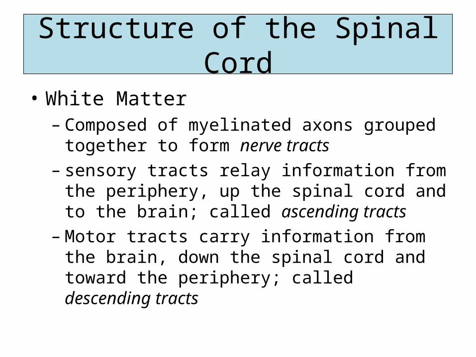
• White Matter– Composed of myelinated axons grouped together
to form nerve tracts – sensory tracts relay information from the
periphery, up the spinal cord and to the brain; called ascending tracts
– Motor tracts carry information from the brain, down the spinal cord and toward the periphery; called descending tracts
Structure of the Spinal Cord

Figure 11-2 Cross section of the spinal cord showing the inner gray matter ("butterfly") and the outer white matter.
Elsevier items and derived items © 2007, 2003, 2000 by Saunders, an imprint of Elsevier Inc.

Function of the Spinal Cord
• Sensory pathway– Provides a pathway for sensory information
traveling from the periphery to the brain• Motor pathway
– Provides a pathway for motor information coming from the brain and going to the periphery
• Reflex center– Acts a major reflex center; many automatic
reflexes occur in the spinal cord, not the brain

Reflexes
• An involuntary response to a stimulus• Occurs repeatedly without conscious thought
or voluntary input• Relexes that occur through the spinal cord
bypass the complicated circuitry of the brain-responses happen quickly– Touch a hot pan and your hand will be withdrawn
quickly, before the brain has time to interpret pain

The Reflex Arc
• The nerve pathway involved in a reflex• Involves 4 parts
– Receptor – Sensory (afferent) neuron– Motor (efferent) neuron– Effector organ
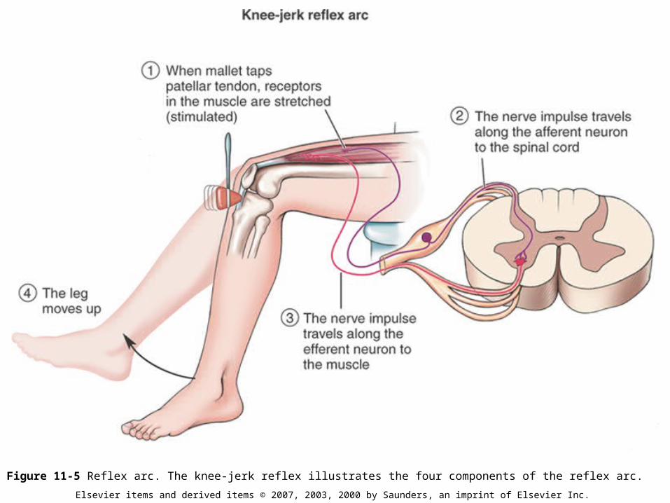
Figure 11-5 Reflex arc. The knee-jerk reflex illustrates the four components of the reflex arc.
Elsevier items and derived items © 2007, 2003, 2000 by Saunders, an imprint of Elsevier Inc.

Figure 11-6 Many reflexes. A, Withdrawal reflex. B, Pupillary reflex. C, Blood pressure, or baroreceptor, reflex. D, Babinski reflex. E, Knee-jerk reflex.
Elsevier items and derived items © 2007, 2003, 2000 by Saunders, an imprint of Elsevier Inc.
Normal
Abnormal- damage to CNS

Peripheral Nervous System
• Neuron vs. Nerve• Neuron- a single nerve cell• Nerve- many neurons bundled together with
blood vessels and wrapped in connective tissue; located outside of the CNS (within the CNS, bundles of neurons are called tracts)

Figure 11-7 Difference between a neuron and nerve.
Elsevier items and derived items © 2007, 2003, 2000 by Saunders, an imprint of Elsevier Inc.
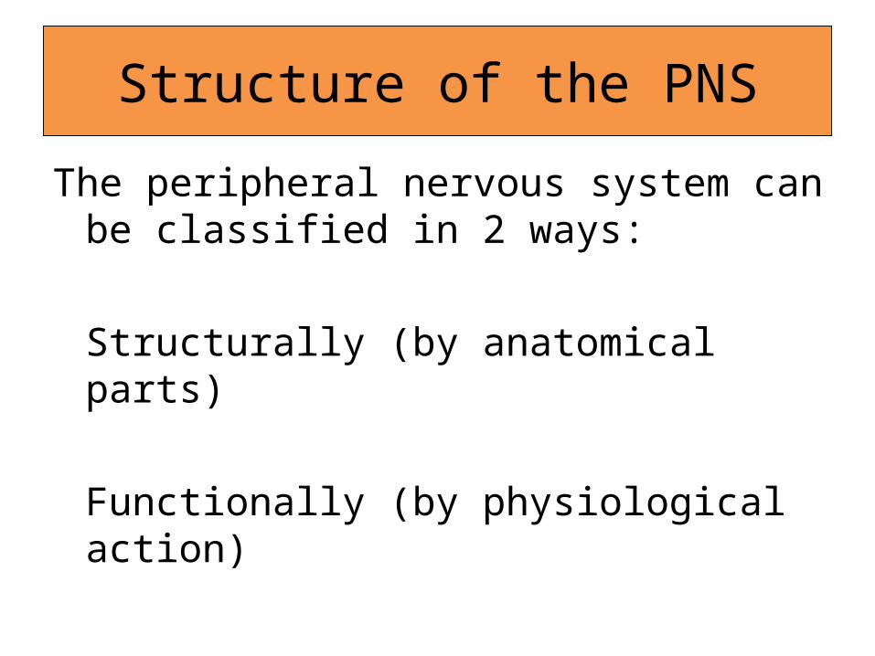
Structure of the PNS
The peripheral nervous system can be classified in 2 ways:
Structurally (by anatomical parts)
Functionally (by physiological action)

Cranial Nerves
• 12 pairs of cranial nerves• Always described with a specific number
(Roman numeral) and a name• Numbers indicate the order in which they exit
the brain (from front to back)• Generally, the name indicates the area
innervated by the nerve (mostly head and neck)

• Four main functions:– Carry sensory information for the special senses
(smell, taste, vision and hearig)– Carry sensory information for the general senses
(touch, pressure, pain, temp)– Carry motor information that results in
contraction of skeletal muscle– Carry motor information that results in secretion
from glands and contraction of smooth and cardiac muscle
Cranial Nerves

Cranial Nerves
• CN I olfactory (smell)
• CN II optic (sight)
• CN III oculomotor (mvmt of eyeball, pupil)
• CN IV trochlear (mvmt of eyeball)
• CN V trigeminal (sensation face)
• CN VI abducens (mvmt of eyeball)
• CN VII facial (expression, tears, taste, blink)
• CN VIII vestibulocochlear (hearing & balance)
• CN IX glossopharyngeal (swallowing, taste, gag)
• CN X vagus (visceral muscle, BP)
• CN XI accessory (swallowing, speaking)
• CN XII hypoglossal (speech & swallowing)


Testing Cranial Nerves
• CN II Optic nerve• How would you test the patient’s optic nerve?• Snellen chart or other test of visual acuity

• CN I Olfactory Nerve• How would you test the patient’s olfactory
nerve?• Have the patient identify various smells
(alcohol pad, coffee, etc.)
Testing Cranial Nerves
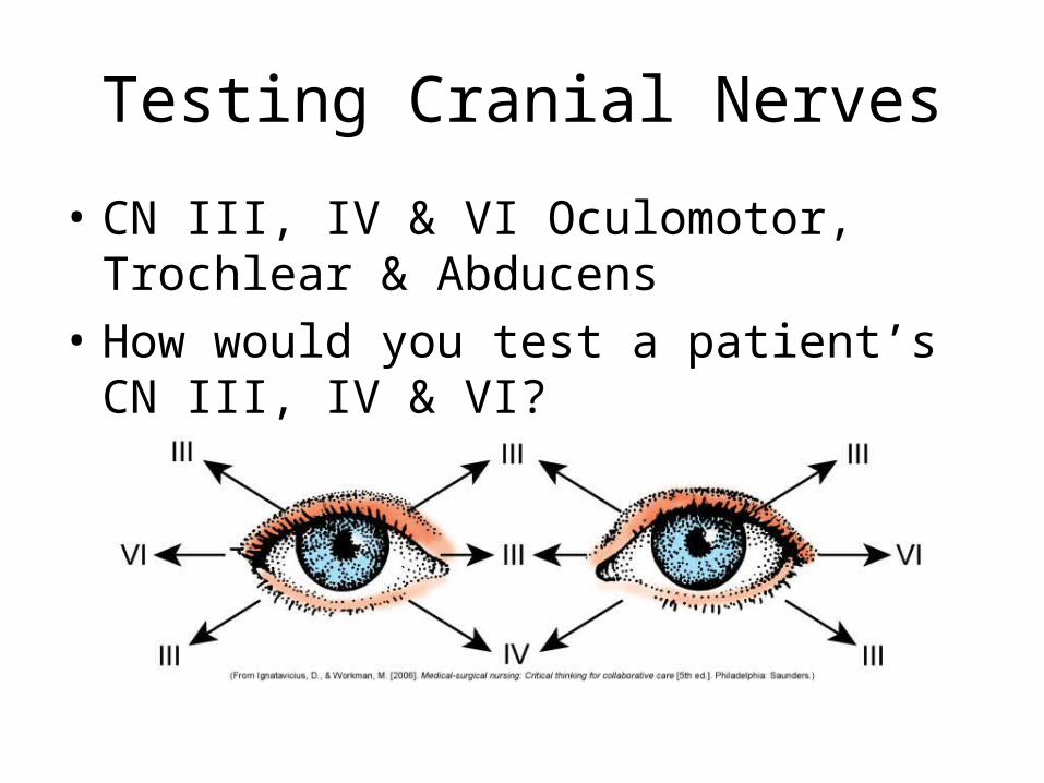
• CN III, IV & VI Oculomotor, Trochlear & Abducens
• How would you test a patient’s CN III, IV & VI?
Testing Cranial Nerves

• CN IX Glossopharyngeal• How would you test a patient’s
glossopharyngeal nerve?
Testing Cranial Nerves

• Spinal Nerves• Thirty one pairs of spinal nerves emerge from
the spinal cord• Each pair is numbered according to the level
of the cord• Cervical (8), thoracic (12), lumbar (5), sacral
(5), coccygeal (1)
Structure of the PNS


• Lumbar and spinal nerves extend the length of the spinal canal before exiting the spinal column (remember the cord ends at L1); the nerves below are called the cauda equina
Structure of the PNS


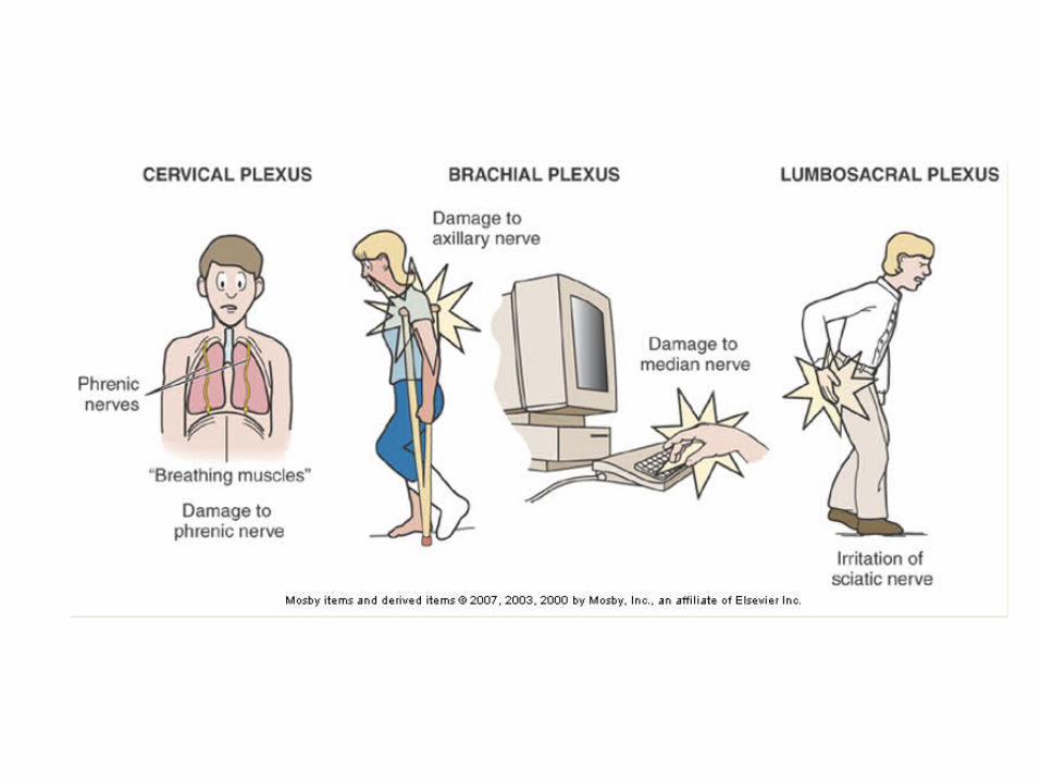
• Nerve Plexuses– Networks of nerve fibers– Three major plexuses: cervical, brachial and
lumbosacral– Each has major nerves that emerge from them
• Cervicle- phrenic nerve• Brachial- axillary, radial, median• Lumbosacral- femoral, sciatic
Structure of the PNS

Examples of nerve plexuses in the body

• Dermatome– The area of skin innervated by each spinal nerve
Structure of the PNS

• Explains where the nerves go and what they do– Somatic afferent nerves (sensory information from
the periphery to the CNS)
– Somatic efferent nerves (bring motor information from the CNS to the skeletal muscles)
– Autonomic nervous system (made up of nerves that supply organs and glands)
Functional Classification of PNS

Cervical spine dislocation C4-C5With cord compression

Cervical Spine fracture and dislocation (C6)Posterior dislocation with cord contusion and edema

NCLEX Question
• A client has an impairment of the cranial nerve II. Specific to this impairment, the nurse would plan to do which of the following the ensure client safety?
1. Speak loudly to the client2. Test the temperature of the shower water3. Check the temperature of the food on the client’s
meal tray4. Provide a clear path for ambulation without
obstacles

Rationale
• 4. Cranial nerve II is the optic nerve. This patient would be expected to have visual disturbances, and therefore need to have trip hazards removed from their room.

NCLEX Question
• When assessing the pupil’s ability to constrict, which cranial nerve (CN) is being tested?
1.II2. III3. IX4.X

Rationale
• Cranial Nerve III (Oculomotor) is one of three cranial nerves that affect pupillary constriction

NCLEX Question
• A patient sustained an injury to his spinal cord at the C2 level. Based on a spinal cord injury at this level, the nurse would most likely expect the patient to be treated with:
1. Diuretics2. Oxygen via a ventilator3. Oxygen via a nasal cannula4. Vasopressors

Rationale
• A spinal cord injury at the level of C2 would cause paralysis of the diaphragm (phrenic nerve exits at the level of C4). The patient will need to be on a ventilator in order to breathe