Multiple adaptive routes of Salmonella enterica Typhimurium to biocide … adaptive... · RESEARCH...
16
RESEARCH ARTICLE Open Access Multiple adaptive routes of Salmonella enterica Typhimurium to biocide and antibiotic exposure Tânia Curiao 1,2* , Emmanuela Marchi 4 , Denis Grandgirard 5 , Ricardo León-Sampedro 1,2 , Carlo Viti 4 , Stephen L. Leib 5 , Fernando Baquero 1,2,3 , Marco R. Oggioni 6 , José Luis Martinez 3,7 and Teresa M. Coque 1,2,3* Abstract Background: Biocides and antibiotics are used to eradicate or prevent the growth of microbial species on surfaces (occasionally on catheters), or infected sites, either in combination or sequentially, raising concerns about the development of co-resistance to both antimicrobial types. The effect of such compounds on Salmonella enterica,a major food-borne and zoonotic pathogen, has been analysed in different studies, but only few works evaluated its biological cost, and the overall effects at the genomic and transcriptomic levels associated with diverse phenotypes resulting from biocide exposure, which was the aim of this work. Results: Exposure to triclosan, clorhexidine, benzalkonium, (but not to hypochlorite) resulted in mutants with different phenotypes to a wide range of antimicrobials even unrelated to the selective agent. Most biocide-resistant mutants showed increased susceptibility to compounds acting on the cell wall (β-lactams) or the cell membranes (poly-L-lysine, polymyxin B, colistin or toxic anions). Mutations (SNPs) were found in three intergenic regions and nine genes, which have a role in energy production, amino acids, carbohydrates or lipids metabolism, some of them involved in membrane transport and pathogenicity. Comparative transcriptomics of biocide-resistant mutants showed over-expression of genes encoding efflux pumps (sugE), ribosomal and transcription-related proteins, cold-shock response (cpeE) and enzymes of microaerobic metabolism including those of the phosphotransferase system. Mainly ribosomal, metabolic and pathogenicity-related genes had affected expression in both in vitro-selected biocide mutants and field Salmonella isolates with reduced biocide susceptibility. Conclusions: Multiple pathways can be involved in the adaptation of Salmonella to biocides, mainly related with global stress, or involving metabolic and membrane alterations, and eventually causing “collateral sensitivity” to other antimicrobials. These changes might impact the bacterial-environment interaction, imposing significant bacterial fitness costs which may reduce the chances of fixation and spread of biocide resistant mutants. Keywords: Biocide resistance, Antimicrobial susceptibility, WGS, Transcriptomics, Collateral sensitivity Background Salmonella enterica is a major food-borne pathogen able to cause diarrhoea or thyphoid/paratyphoid fever [1]. The systemic infection is often preceded by an asymp- tomatic chronic colonization or by a local infection process. One of the major problems associated with per- sistent colonization or infection is the steady rise of antibiotic resistance among strains, which can lead to treatment failures [2]. The association between the over- use of antibiotics and/or biocides in farms, hospitals, in- dustry and homes and the emergence of both co- resistance and cross-resistance to different compounds in Salmonella populations is of concern [3–6]. Unlike antibiotics, most biocides do not act on specific cell targets. In fact, only a few mechanisms by which mi- croorganisms became tolerant to these antimicrobials have been fully characterized. Over-expression of multi- drug efflux pumps such as AcrAB or AcrEF which are * Correspondence: [email protected]; [email protected] 1 Servicio de Microbiología, Instituto Ramón y Cajal de Investigación Sanitaria (IRYCIS), Madrid, Spain Full list of author information is available at the end of the article © 2016 The Author(s). Open Access This article is distributed under the terms of the Creative Commons Attribution 4.0 International License (http://creativecommons.org/licenses/by/4.0/), which permits unrestricted use, distribution, and reproduction in any medium, provided you give appropriate credit to the original author(s) and the source, provide a link to the Creative Commons license, and indicate if changes were made. The Creative Commons Public Domain Dedication waiver (http://creativecommons.org/publicdomain/zero/1.0/) applies to the data made available in this article, unless otherwise stated. Curiao et al. BMC Genomics (2016) 17:491 DOI 10.1186/s12864-016-2778-z
Transcript of Multiple adaptive routes of Salmonella enterica Typhimurium to biocide … adaptive... · RESEARCH...
RESEARCH ARTICLE Open Access
Multiple adaptive routes of Salmonellaenterica Typhimurium to biocide andantibiotic exposureTânia Curiao1,2*, Emmanuela Marchi4, Denis Grandgirard5, Ricardo León-Sampedro1,2, Carlo Viti4, Stephen L. Leib5,Fernando Baquero1,2,3, Marco R. Oggioni6, José Luis Martinez3,7 and Teresa M. Coque1,2,3*
Abstract
Background: Biocides and antibiotics are used to eradicate or prevent the growth of microbial species on surfaces(occasionally on catheters), or infected sites, either in combination or sequentially, raising concerns about thedevelopment of co-resistance to both antimicrobial types. The effect of such compounds on Salmonella enterica, amajor food-borne and zoonotic pathogen, has been analysed in different studies, but only few works evaluated itsbiological cost, and the overall effects at the genomic and transcriptomic levels associated with diverse phenotypesresulting from biocide exposure, which was the aim of this work.
Results: Exposure to triclosan, clorhexidine, benzalkonium, (but not to hypochlorite) resulted in mutants withdifferent phenotypes to a wide range of antimicrobials even unrelated to the selective agent. Most biocide-resistantmutants showed increased susceptibility to compounds acting on the cell wall (β-lactams) or the cell membranes(poly-L-lysine, polymyxin B, colistin or toxic anions). Mutations (SNPs) were found in three intergenic regions andnine genes, which have a role in energy production, amino acids, carbohydrates or lipids metabolism, some ofthem involved in membrane transport and pathogenicity. Comparative transcriptomics of biocide-resistant mutantsshowed over-expression of genes encoding efflux pumps (sugE), ribosomal and transcription-related proteins,cold-shock response (cpeE) and enzymes of microaerobic metabolism including those of the phosphotransferasesystem. Mainly ribosomal, metabolic and pathogenicity-related genes had affected expression in both invitro-selected biocide mutants and field Salmonella isolates with reduced biocide susceptibility.
Conclusions: Multiple pathways can be involved in the adaptation of Salmonella to biocides, mainly related withglobal stress, or involving metabolic and membrane alterations, and eventually causing “collateral sensitivity” toother antimicrobials. These changes might impact the bacterial-environment interaction, imposing significantbacterial fitness costs which may reduce the chances of fixation and spread of biocide resistant mutants.
Keywords: Biocide resistance, Antimicrobial susceptibility, WGS, Transcriptomics, Collateral sensitivity
BackgroundSalmonella enterica is a major food-borne pathogen ableto cause diarrhoea or thyphoid/paratyphoid fever [1].The systemic infection is often preceded by an asymp-tomatic chronic colonization or by a local infectionprocess. One of the major problems associated with per-sistent colonization or infection is the steady rise of
antibiotic resistance among strains, which can lead totreatment failures [2]. The association between the over-use of antibiotics and/or biocides in farms, hospitals, in-dustry and homes and the emergence of both co-resistance and cross-resistance to different compoundsin Salmonella populations is of concern [3–6].Unlike antibiotics, most biocides do not act on specific
cell targets. In fact, only a few mechanisms by which mi-croorganisms became tolerant to these antimicrobialshave been fully characterized. Over-expression of multi-drug efflux pumps such as AcrAB or AcrEF which are
* Correspondence: [email protected]; [email protected] de Microbiología, Instituto Ramón y Cajal de Investigación Sanitaria(IRYCIS), Madrid, SpainFull list of author information is available at the end of the article
© 2016 The Author(s). Open Access This article is distributed under the terms of the Creative Commons Attribution 4.0International License (http://creativecommons.org/licenses/by/4.0/), which permits unrestricted use, distribution, andreproduction in any medium, provided you give appropriate credit to the original author(s) and the source, provide a link tothe Creative Commons license, and indicate if changes were made. The Creative Commons Public Domain Dedication waiver(http://creativecommons.org/publicdomain/zero/1.0/) applies to the data made available in this article, unless otherwise stated.
Curiao et al. BMC Genomics (2016) 17:491 DOI 10.1186/s12864-016-2778-z
controlled by global transcriptional regulators such asMarAB, RamA and SoxRS can lead to diverse levels ofresistance to biocides and/or antibiotics [7–13]. Often,tolerance to triclosan is due to over-expression and/ormutations in FabI, the enoyl-acyl-reductase protein re-quired for fatty acid synthesis [14]. Moreover, exposureand further adaptation to biocides may also impair cellu-lar homeostasis, and/or changes the level of expressionof genes regulating synthesis and modification of cell en-velope, virulence, motility, or stress response [15–20].Whether or not such physiological changes are neededfor adaptation to the presence of biocides, or they justreflect secondary changes associated with restoring fit-ness after adaptation remains to be established. Previousstudies in Salmonella enterica prototype strain SL1344have described the modification of antibiotic susceptibil-ity, growth and regulation of different genes after expos-ure to biocides [5, 6, 21]. However, few studies providedcomprehensive information about the genomic and tran-scriptomic changes of mutants selected after exposure todifferent biocides and antibiotics, which can be used ei-ther coincidentally or sequentially in the clinical practiceand in the food industry [9, 22–24].The aim of this study was to determine the effect of
exposure to some biocides (triclosan, TRI; benzalkoniumchloride, BKC; chlorhexidine, CHX and sodium hypo-chlorite, SHC), or antibiotics (ampicillin, AMP; cipro-floxacin, CIP), widely used in farms, hospitals, industryand homes on the selection of antibiotic/biocide-
resistant Salmonella mutants and to characterize the as-sociated genomic and transcriptomic profiles, as well asthe extended phenotypes (susceptibility to 240 inhibitorycompounds). To address whether these adaptive changesfound in laboratory-selected mutants also occurred innatural populations of Salmonella, the transcriptomes ofa set of field isolates exhibiting reduced susceptibility tobiocides were comparatively studied.
MethodsBacterial strainsThe prototype S. enterica serovar Typhimurium SL1344[25] strain was exposed to biocides (TRI, CHX, BKC andSHC), and antibiotics (the β-lactam ampicillin, AMP;and the fluoroquinolone ciprofloxacin, CIP). The quanti-tative phenotype of this strain against diverse antimicro-bials is summarized in Table 1.Sixteen Salmonella spp. isolates from food-borne ani-
mals with reduced susceptibility to TRI (3 TRIR; MIC 1-2 mg/L), BKC (7 BKCR; MIC = 128 mg/L), CHX (1 beingCHXR/BKCR, MIC = 16 mg/L (Additional file 1: FigureS1) used in a previous work [26], were investigated fortheir transcriptomic profiles. Such isolates, collected in aveterinary surveillance project in Europe, showed 13 dif-ferent PFGE-types and belonged to Salmonella entericasubspecies enterica [serovars Anatum (n = 8), Hadar (n =5), Dublin (n = 2)] and subspecies Typhimurium (n = 1).Most of these strains were susceptible to antibiotics. A
few number of isolates harbored plasmids that contained
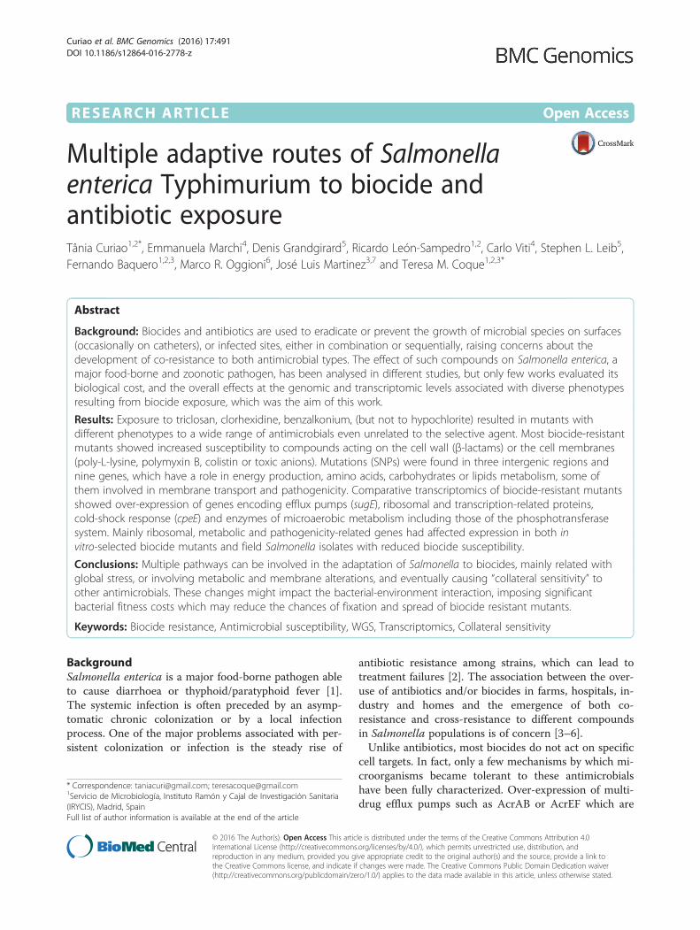
Table 1 Susceptibility profiles of Salmonella mutants respect to SL1344 parental strain
N.° Pre-conditioningagent
Biocide Phenotype Biocide MIC (mg/L) Antibiotic MIC (mg/L) Mutantdesignation
FitnessCost (%)
Frequencyof mutationTRI BKC CHX AMP CAZ CIP ERY GEN CLO TET
- Parental strain SL1344 0.06 16 16 1.5 0.38 0.032 32 1.0 3 3
1 NE TRIR/BKCR/CHXR 2 32 32 1.5 0.38 0.047 32-48 1.5 3 2 NE/TRI1 11 >1.25E-07
2 NE 0.12 32 32 1 0.25 0.032 32 1.0 4 4 NE/CHX2 34 2.92E-09
3 BKC 0.12 32 32 2 0.38 0.023 32 1.5 3 4 BKC/AMP - 2.50E-09
4 CHX 0.12 32 32 1 0.5 0.032 48 1.5 2 2 CHX/BKC3 - 1.67E-09
5 BKC TRIR/BKCR 0.12 32 16 2 0.38 0.032 48 0.5 4 4 BKC/BKC3 - 2.50E-09
6 CIP BKCR 0.06 32 16 1.5 0.5 0.032 24 1.5 2-3 1.5 CIP/TRI1 More fit <1.50E-05
7 TRI 0.06 32 16 2 0.5 0.032 32 1.5 3 2-3 TRI/BKC3 31 1.50E-09
8 NE BKCR/CHXR/TRIHS 0.03 32 32 2 0.38 0.032 32-48 1 4 4 NE/BKC2 - 1.25E-07
9 NE 0.03 32 32 1.5 0.75 0.047 24 1.5 3 1.5 NE/BKC3 more fit 2.08E-11
10 TRI 0.015 32 32 1.5 0.5 0.032 24 1.5 2 1.5 TRI/AMP - 3.06E-09
11 BKC 0.015 64 32 2 0.38 0.023 32 1 3 4 BKC/CHX2 - 1.67E-08
12 CIP TRIHS/CHXHS 0.03 16 8 2 0.38 0.032 32 2 4 3 CIP/CHX1 - <2.50E-06
13 BKC TRIHS 0.015 16 16 1 0.38 0.023 64 1.5 3 1.5 BKC/CIP - 1.50E-09
14 CHX 0.015 16 16 2 0.38 0.023 32-48 0.75 3 4 CHX/AMP 17 4.17E-09
NE: non-exposed, - Not doneIn the designation of mutants, numbers 1-3 refer to the concentration of compounds in plates as follows: 1- 32 mg/L, 2- 64 mg/L and 3-128 mg/LMutants CIP/TRI1 and CIP/CHX1 classified as more fit than SL1344 exhibited -17 % and -21 %, respectivelyWGS was performed in the underlined mutants
Curiao et al. BMC Genomics (2016) 17:491 Page 2 of 16

acquired genes coding for resistance to β-lactams (blaTEM-
1), aminoglycosides (strA, strB), tetracycline (tetA, tetR)and quinolone (qepA). Plasmids from 6 isolates also car-ried genes encoding resistance to metals (As, Co).
Selection of mutantsA colony of S. enterica serovar Typhimurium SL1344grown overnight in Luria Bertani (LB) plates was inocu-lated into LB-broth and LB supplemented with sub-inhibitory concentrations (1/2 ×MIC) of biocides (TRI,CHX, BKC and SHC; Sigma-Aldrich, Inc., St. Louis,MO) or antibiotics (AMP and CIP) and further incu-bated overnight at 37 °C with shaking at 150 rpm. Sub-sequently, aliquots of 100 μl were plated onto LB platescontaining a single biocide or a single antibiotic com-pound at concentrations ranging 2.5-33 ×MIC and incu-bated at 30 °C. These primary selective plates wereexamined for growth during 7 days. A variable numberof viable mutants (one per colony morphotype per plate)were tested for growth on secondary selective platescontaining other biocides or antibiotics. The stability ofmutants was evaluated after serial passages in non-selective LB broth (up to 50 generations). Mutants werenamed by the acronym name of the antimicrobial com-pound added to the broth cultures before plating,followed by the name and concentration of the com-pound added to the selective agar plates from where themutant was retrieved. The mutants obtained from brothcultures not supplemented with any antimicrobial weredesignated as non-exposed (NE). Phenotypes of de-creased and increased susceptibility to biocides and anti-biotics appear represented by the super indexes “R” or“HS”, respectively. The colonial morphology in LB andblood agar plates was compared between mutants andthe parental strain. The variability of XbaI-digested gen-omic DNA profiles of mutants and parental strain wasassessed by pulsed field gel electrophoresis (PFGE) usingstandard protocols for DNA preparation, digestion andPFGE running conditions for Enterobacteriaceae [27].
Antimicrobial susceptibility testingThe minimal inhibitory concentrations (MICs) of bio-cides (TRI, CHX, BKC) and antibiotics (AMP; CAZ;CIP; erythromycin, ERY; gentamicin, GEN; chloram-phenicol, CLO; tetracycline, TET) (BioMérieux, Marcyl’Etoile, France) were determined by both broth micro-dilution using E- test strips following CLSI guidelines.Escherichia coli ATCC10536 and Staphylococcus aureusATCC6538 were used as control strains (3). Minimumbactericidal concentrations (MBCs) were determined bysubculturing 10 μl from each well without visible bacter-ial growth when MIC was determined on Mueller-Hinton broth (Difco, Becton Dickinson, Maryland,USA). The minimal concentration yielding no-growth
after overnight incubation at 37 °C was scored as theMBC. The susceptibility of wild type strains to AMP,streptomycin, sulphonamides, trimethoprim, nalidixicacid, CIP, CLO, TET, GEN and kanamycin was deter-mined by disk diffusion.Further, the susceptibility of mutants and the parental
strain to 240 cell growth-inhibiting chemical compoundswas screened using the Phenotype MicroArray PM11-PM20 in two independent experiments (Biolog,Hayward, CA, USA) as previously described [28]. Thestrains were grown overnight at 30 ° C on BUG agar(Biolog Universal Agar, Biolog Hayward California) andthen, colonies were picked up with a sterile cotton swaband suspended in 15 ml of 1X inoculation fluid (IF-0aGN/GP Base, Biolog 74268). Cell density was adjusted to85 % transmittance (T) on a Biolog turbidimeter. Inocu-lation fluid for PM11-20 was prepared mixing 100 ml ofIF-10a GN Base (1.2X) (Biolog 74264), 1.2 ml of BiologRedox Dye A (100X) (Biolog 74221), 0.6 ml of cell sus-pension at 85 % T, bringing to a final volume of 120 mlwith sterile water. The mixture was inoculated in thePM plates (100 μl per well) and monitored automaticallyfor color development every 15 min for 72 h at 30 °C inan Omnilog reader (Biolog). To identify phenotypes, thekinetic curves of both parental strain and mutants werecompared using Omnilog-PM software (releaseOM_PM_109M). Such comparison was based on thehalf maximal inhibitory concentration (IC50) values for4 concentrations of each antimicrobial, which is definedas the well at which a particular per-well parameter isthe half of its maximal value over the concentrationseries; the reference parameter being the area under thecurve. Raw data were filtered using differences of aver-age area of mutant compared to control taking a differ-ence of 1:3 (33 %) as significant.
Growth kineticsThe growth kinetics of both the parental strain SL1344and biocide-tolerant mutants exhibiting various pheno-types was determined by measuring the optical densityat 600 nm every 5–10 min for 24 h at 37 °C in BioscreenC (ThermoLabsystems, Helsinki, Finland), adapting themethod described by Foucault et al. [29]. Inocula in aconcentration of 104 to 105 CFU/ml were obtained froma 1/1000 dilution of an overnight culture in fresh LBbroth and aliquots of 400 μl were seeded in triplicate ina microtitre plate. Growth rates were determined in theinterval estimated to be exponential using the Growth-Rates 2.1 program [30]. The fitness cost (FC) reflects therelative growth rates, which were based on the individualgrowth rates of mutants relative to the parental strain.For each strain, data from growth rates were averagedand standard deviations calculated.
Curiao et al. BMC Genomics (2016) 17:491 Page 3 of 16
Whole genome sequencing (WGS)Six mutants with different phenotypes were selected forwhole genome sequencing. Genomic DNA was extractedfrom 1 ml of overnight cultures in plain LB broth usinga Promega Wizard Genomic DNA Purification kit ac-cording manufacturer instructions. Genome sequencingwas performed on Illumina MiSeq platform to obtain100–200 bp paired-end reads. Reads were revised andcorrected using Lighter software and further mappedagainst the genome of the SL1344 strain (GenBank acc.Number FQ312003) using Breseq v0.26.1 pipeline(http://barricklab.org/twiki/bin/view/Lab/ToolsBacterial-GenomeResequencing). Single nucleotide polymor-phisms (SNPs) detected in all mutants were not deemedconfident and were excluded, because we cannot dismissdifferences between the laboratory SL1344 strain used aswild-type in this work, and that corresponding to the ca-nonical sequence in the GenBank database. Reads notfound in all mutants were treated as deletions.
Transcriptome analysisArray Design and ProductionAn array was designed to cover the complete genome ofSalmonella enterica subsp. enterica serovar Typhimur-ium, as well as plasmids isolated from various Gram-negative microorganisms. Probe design was performedby the CustomArray Design Service (CustomArray Inc.,Bothell, WA, USA) and included 12,005 capture probes(35–40 bp length), 326 quality control probes and 65non-specific probes derived from plants, phages and un-related bacterial sequences, and also 148 empty spotswith no oligonucleotides. Arrays were synthesized on aCustomArray Synthesizer (CombiMatrix, Mukilteo, WA)and quality tested using the standard protocols providedby the manufacturer.
RNA extractionStrains were grown overnight in 10 ml LB broth at 37 °C, 150 rpm. The cultures were diluted 1:100 in pre-warmed LB and grown to logarithmic phase (OD570 =0.5). 2 ml of the culture (5 x 10⁸ – 1 x 10⁹ colony form-ing units (UFC)/ml) were harvested in 4 ml of RNA pro-tect reagent® (Qiagen GmbH, Hilden, Germany),incubated for 5 min at room temperature and centri-fuged for 10 min at 5000 x g. Bacterial pellets were sus-pended in 200 μl of TE buffer (10 mM Tris/HCl, 1 mMEDTA, pH 8) containing 1 mg/ml lysozyme (Sigma) andincubated for 5 min at room temperature, 600 rpm.Total RNA was then extracted using RNeasy Mini Kit(Qiagen), according to the manufacturer’s instructions.Contaminating DNA was removed using DNA-free™ Kit(Applied Biosystems). The RNA isolation procedure wasvalidated for RNA quality by testing RNA samples on anAgilent 2100 Bioanalyzer (Agilent Technologies). RNA
concentration and purity were determined by Nanodrop®ND-1000 spectrophotometer (Thermo Scientific). Foreach strain, at least 4 RNA samples were prepared fromindependent cultures.
RNA labelling and fragmentationIsolated, unamplified RNA was labelled with Cy5, usingULS™ Labeling Kit for CombiMatrix arrays (KreatechBiotechnology), according to the manufacturer’s instruc-tions. RNA was fragmented with the RNA Fragmenta-tion Reagents (Ambion®).
Array hybridization12 K Custom arrays were hybridized with 2 μg of la-belled, fragmented RNA, according to information pro-vided by the manufacturer (Customarray/CombimatrixIncorporated). In brief, after pre-hybridization of the ar-rays, hybridization was performed at 45 °C for 16 hoursin a hybridization buffer containing 25 % formamide.After washing steps, microarrays were scanned usingPackard ScanArray4000 array scanner and software (Sca-nArray, version 3.1, Packard BioChip Technologies) withincremental laser power from 15 to 100 %. Data wereextracted with Microarray Imager software (version5.8.0, Combi Matrix) and spot intensity expressed as me-dian intensity. After scanning, microarrays were stripedusing 12 K CustomArray™ Stripping kit, according to themanufacturer’s instructions. Quality of the stripping wasverified by scanning the microarray at maximal laser in-tensity and repeated when necessary. Microarrays wereused up to four times.
Data analysisTo adjust for difference in the amount and labelling effi-ciency of hybridized RNAs, the median fluorescence in-tensity values of all spots was determined for all laserintensities used during scanning. Scanning data withsimilar median fluorescence intensity were chosen forfurther analysis. Fluorescence values of spots with max-imal intensity (signal saturation) at a given laser intensitywere extrapolated by linear regression, using valuesgathered with lower laser intensity. For each set of arraysfor a given strain, non-specific binding was determinedfrom fluorescence values of the non-specific probes. Thecut-off for specific binding was set as the upper 95 %confidence interval of the mean signal intensity of thenon-specific probes. Probes were excluded when themean values for the strains compared were under thecut-off value.The fluorescence values were log2 transformed and
stage-wise quantitative normalization was performed foreach set of comparison, using a script written in the statis-tical computing environment of R (R Development CoreTeam, 2011, version 3.3). To identify genes differentially
Curiao et al. BMC Genomics (2016) 17:491 Page 4 of 16

regulated, we analyzed the transformed and normalizedintensities determined by two methods, the SignificanceAnalysis of Microarrays method (SAM, version 5.0, run-ning under Shiny, a web-based interactive applicationframework for R environment, https://github.com/MikeJ-Seo/SAM) and “R”, comprising base package statistics andthe attached LIMMA package (version 3.26.5). The pres-ence of genes identified by both methods in the mutantsanalyzed was searched in the wild type strains included inthe study and transcriptomic profiles were compared. Theexpression profiles of these genes were visualized in aheatmap built with the ‘pheatmap’ package in “R”.
Statistical analysisIn the SAM method, the delta value was set to obtain anaverage. False Discovery Rate (FDR) of 5 % and the foldchange cut-off value was established as 1.5. In LIMMAanalysis, genes with a fold change >1.5 and p < 0.05 wereconsidered as differentially expressed. Only the genesidentified as differentially expressed by both SAM andLIMMA were considered.
Availability of supporting dataThe data sets supporting the results of this article areavailable in the ArrayExpress repository, (http://www.e-bi.ac.uk/arrayexpress/) under accession numbers A-MEXP-2366 (S. Typhimurium combimatrix 12 K custo-marray design) and E-MTAB-2554 (microarray rawresults).
ResultsExposure to biocides or antibiotics yield mutants withdifferent susceptibility to biocides and antimicrobialsTable 1 shows the diversity of mutants exhibiting pheno-types obtained (4 TRIR/BKCR/CHXR, 1 TRIR/BKCR, 4BKCR/CHXR/TRIHS, 2 BKCR, 2 TRIHS and 1 TRIHS/CHXHS). Some mutants with increased MICs to biocideswere obtained without previous exposure to any anti-microbial but using selective plates supplemented withTRI, BKC or CHX. Others were retrieved after exposureto antibiotics such as CIP and AMP. Previous expos-ure to SHC did not yield resistant mutants. A num-ber of biocide tolerant mutants mentioned aboveshowed lower MIC values for AMP, CAZ, CIP, ERY,GEN, CLO and TET than those for the correspondingparental strain (<2-fold).Preexposure to sub-inhibitory concentrations of BKC
resulted in mutants with either decreased susceptibilityto BKC, TRI and/or CHX (TRIR/BKCR/CHXR, TRIR/BKCR) or increased susceptibility to TRI (TRIHS, BKCR/CHXR/TRIHS). Two of these four mutants selected inplates supplemented with BKC also showed a slight in-crease in MICCAZ (1 BKCR and 1 BKCR/CHXR/TRIHS).The TRIR/BKCR/CHXR and the TRIHS phenotypes were
also selected after pre-exposure to CHX, CIP or withoutpre-exposure to any antimicrobial. A hyper-susceptibleTRIHS mutant (BKC/CIP) showed a 2-fold increasedMICERY.Preexposure to sub-inhibitory concentrations of TRI
resulted in mutants that only showed the phenotypesBKCR and BKCR/CHXR, which could even exhibit in-creased susceptibility to TRI. Such BKCR mutantsshowed a minor increase in MIC values to CAZ. Twodifferent mutants were selected on plates supplementedwith TRI, one obtained without previous exposure to an-timicrobials showed an increased MICTRI (33-fold) andsmall increases in MICCIP, MICERY and MICGEN. Theother, obtained after pre-conditioning with CIP did notshow an increase in MICTRI. Pre-exposure to CIP re-sulted on mutants showing BKCR or TRIHS phenotypes,which were closely related with the above ones.A more comprehensive analysis of the effects that the
exposure to biocides and antimicrobials had on Salmon-ella strain SL1344 strain was performed by characteriz-ing the genome and transcriptome of six mutantsrepresenting the phenotypes TRIR/BKCR/CHXR (CHX/BKC3, NE/TRI1, NE/CHX2), BKCR/CHXR/TRIHS (NE/BKC3), BKCR (TRI/BKC3), and TRIHS (CHX/AMP)(Table 2).
Antimicrobial susceptibilityWe identified mutants with a given biocide phenotypeand variable antibiotic susceptibility patterns but alsomutants exhibiting different biocide susceptibility phe-notypes and similar antimicrobial susceptibility profiles(Table 3). The activity of different compounds againstseven mutants and the parental strain was evaluatedconsidering their IC50 values. High IC50 values were ob-served for antibiotics that inhibit protein synthesis (e.g.neomycin and thiamphenicol), specific metabolic routesas the reduction of dihydrofolic acid to tetrahydrofolicacid, which is an essential precursor in the thymidinesynthesis pathway (trimethoprim), or membrane acting
Table 2 Techniques carried out for a representative subset ofmutants
Mutant n° Mutantname
Biolog(Table 3)
WGS(Table 4)
Gene expression(Fig. 1 and Fig. 2)
1 NE/TRI1 Yes Yes Yes
2 NE/CHX2 No Yes Yes
4 CHX/BKC3 Yes Yes Yes
7 TRI/BKC3 Yes Yes Yes
9 NE/BKC3 Yes Yes Yes
10 TRI/AMP Yes No No
11 BKC/CHX2 Yes No No
14 CHX/AMP Yes Yes Yes
Yes and No denotes whether the technique was performed or not
Curiao et al. BMC Genomics (2016) 17:491 Page 5 of 16

Table 3 Antimicrobial susceptibility determined by BIOLOG for Salmonella enterica mutants in comparison to the parental strain
TRIR/BKCR/CHXR BKCR BKCR/CHXR/TRIHS TRIHS
Chemicals Inhibitor Family NE/TRI1 CHX/BKC3 TRI/BKC3 NE/BKC3 TRI/AMP BKC/CHX2 CHX/AMP
ANTIBIOTICS
Neomycin aminoglycosides R R R R R R S
Paromomycin S S S S
Sisomicin S S R R S R R
Chloramphenicol amphenicols S S S
Thiamphenicol R R R R R
Cefazolin cephalosporins R
Ceftriaxone S
Amoxicillin lactams S R S S S R R
Aztreonam S S S
Carbenicillin S S S
Carbenicillin (II) R
Phleomycin DNA oxidants R
Cinoxacin DNA topoisomerasesinhibitors
S S S S S R
Ciprofloxacin R R R R
Enoxacin R S S S S S R
Nalidixic acid R S S R S S R
Novobiocin S
Ofloxacin S S S S S
Pipemidic acid R
Hydroxyurea folate antagonists R S R S R R S
Trimethoprim R R R R R
Troleandomycin macrolides R R R
Rifampicin RNA polymerase inhibitors S
Penimepicycline tetracyclines S S S R S
Tetracycline S
NON-ANTIBIOTICS
1-Hydroxypyridine-2-thione chelators S S S
5,7-Dichloro-8-OHquinoline S S S
5-Chloro-7-iodo-8-OHquinoline S R S R S S
8-Hydroxyquinoline R R R R
Fusaric acid S S S S
2-Phenylphenol DNA intercalators R
Chloroxylenol Fungicides R
Patulin R R R R
Colistin Membrane active agents S S S S S S R
Polymyxin B S S S S
Polymyxin B (II) R
Poly-L-lysine S S S S S S S
Alexidine S S
Ornidazole oxidizing agents S S S
1-Chloro-2,4-dinitrobenzene S S R S R R
Atropine other drugs S S
Curiao et al. BMC Genomics (2016) 17:491 Page 6 of 16
compounds (toxic cations, such as antimony (III) chlor-ide). Conversely, compounds that act on the bacterialcell envelopes (such as colistin, β-lactams, poly-L-lysine,polymyxin B), toxic anions (e.g. potassium chromate),quaternary salts (e.g. sodium metaborate) and proteaseinhibitors showed low IC50 values.Within this common antimicrobial susceptibility profile,
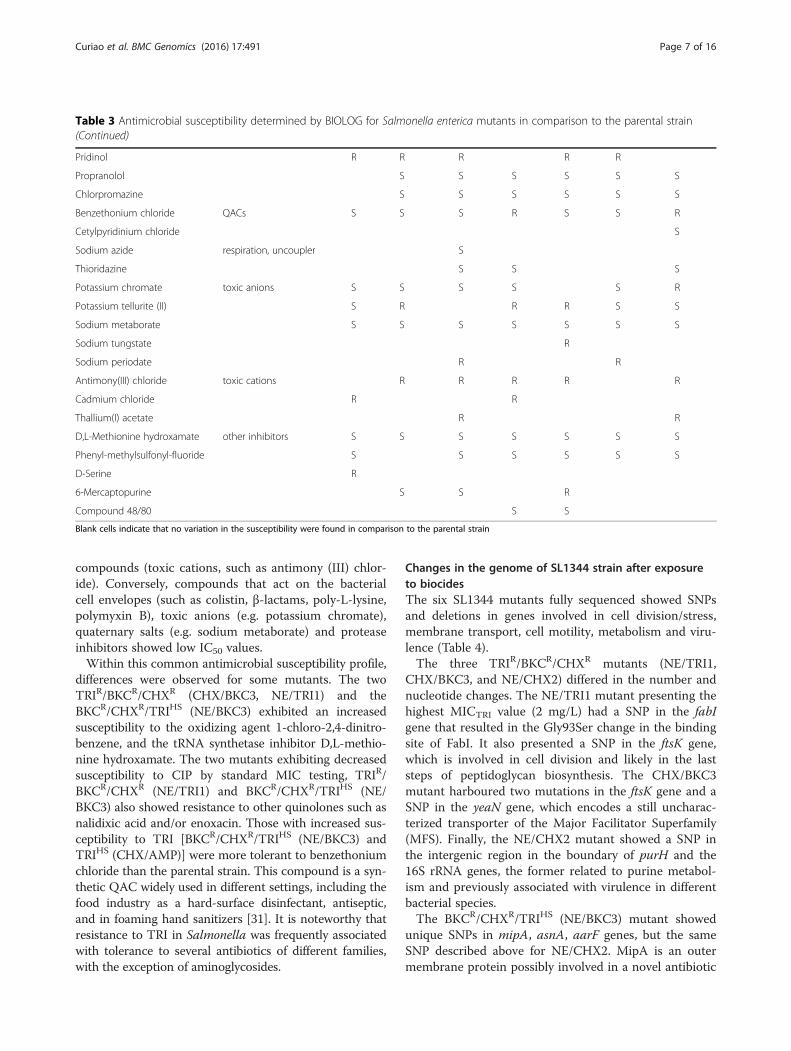
differences were observed for some mutants. The twoTRIR/BKCR/CHXR (CHX/BKC3, NE/TRI1) and theBKCR/CHXR/TRIHS (NE/BKC3) exhibited an increasedsusceptibility to the oxidizing agent 1-chloro-2,4-dinitro-benzene, and the tRNA synthetase inhibitor D,L-methio-nine hydroxamate. The two mutants exhibiting decreasedsusceptibility to CIP by standard MIC testing, TRIR/BKCR/CHXR (NE/TRI1) and BKCR/CHXR/TRIHS (NE/BKC3) also showed resistance to other quinolones such asnalidixic acid and/or enoxacin. Those with increased sus-ceptibility to TRI [BKCR/CHXR/TRIHS (NE/BKC3) andTRIHS (CHX/AMP)] were more tolerant to benzethoniumchloride than the parental strain. This compound is a syn-thetic QAC widely used in different settings, including thefood industry as a hard-surface disinfectant, antiseptic,and in foaming hand sanitizers [31]. It is noteworthy thatresistance to TRI in Salmonella was frequently associatedwith tolerance to several antibiotics of different families,with the exception of aminoglycosides.
Changes in the genome of SL1344 strain after exposureto biocidesThe six SL1344 mutants fully sequenced showed SNPsand deletions in genes involved in cell division/stress,membrane transport, cell motility, metabolism and viru-lence (Table 4).The three TRIR/BKCR/CHXR mutants (NE/TRI1,
CHX/BKC3, and NE/CHX2) differed in the number andnucleotide changes. The NE/TRI1 mutant presenting thehighest MICTRI value (2 mg/L) had a SNP in the fabIgene that resulted in the Gly93Ser change in the bindingsite of FabI. It also presented a SNP in the ftsK gene,which is involved in cell division and likely in the laststeps of peptidoglycan biosynthesis. The CHX/BKC3mutant harboured two mutations in the ftsK gene and aSNP in the yeaN gene, which encodes a still uncharac-terized transporter of the Major Facilitator Superfamily(MFS). Finally, the NE/CHX2 mutant showed a SNP inthe intergenic region in the boundary of purH and the16S rRNA genes, the former related to purine metabol-ism and previously associated with virulence in differentbacterial species.The BKCR/CHXR/TRIHS (NE/BKC3) mutant showed
unique SNPs in mipA, asnA, aarF genes, but the sameSNP described above for NE/CHX2. MipA is an outermembrane protein possibly involved in a novel antibiotic
Table 3 Antimicrobial susceptibility determined by BIOLOG for Salmonella enterica mutants in comparison to the parental strain(Continued)
Pridinol R R R R R
Propranolol S S S S S S
Chlorpromazine S S S S S S
Benzethonium chloride QACs S S S R S S R
Cetylpyridinium chloride S
Sodium azide respiration, uncoupler S
Thioridazine S S S
Potassium chromate toxic anions S S S S S R
Potassium tellurite (II) S R R R S S
Sodium metaborate S S S S S S S
Sodium tungstate R
Sodium periodate R R
Antimony(III) chloride toxic cations R R R R R
Cadmium chloride R R
Thallium(I) acetate R R
D,L-Methionine hydroxamate other inhibitors S S S S S S S
Phenyl-methylsulfonyl-fluoride S S S S S S
D-Serine R
6-Mercaptopurine S S R
Compound 48/80 S S
Blank cells indicate that no variation in the susceptibility were found in comparison to the parental strain
Curiao et al. BMC Genomics (2016) 17:491 Page 7 of 16
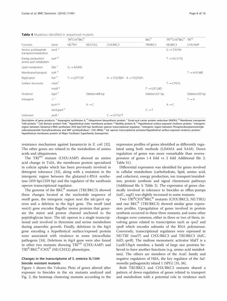
resistance mechanism against kanamycin in E. coli [32].The other genes are related to the metabolism of aminoacids and ubiquinones.The TRIHS mutant (CHX/AMP) showed an amino
acid change in TolA, the membrane protein specializedin colicin uptake which has been previously involved indetergent tolerance [33], along with a mutation in theintergenic region between the glutamyl-t-RNA synthe-tase (459 bp)/(359 bp) and the regulator of the xanthosinoperon transcriptional regulator.The genome of the BKCR mutant (TRI/BKC3) showed
three changes located at the nucleotide sequence ofmotB gene, the intergenic region next the tdc/garA op-eron and a deletion in the bigA gene. The motB (andmotA) gene encodes flagellar motor proteins that gener-ate the stator and proton channel anchored to thepeptidoglycan layer. The tdc operon is a single transcrip-tional unit involved in threonine and serine metabolismduring anaerobic growth. Finally, deletions in the bigAgene encoding a hypothetical surface/exposed proteinwere associated with virulence in some intracellularpathogens [34]. Deletions in bigA gene were also foundin other two mutants showing TRIHS (CHX/AMP) andTRIR/BKCR/CHXR (NE/CHX2) phenotypes.
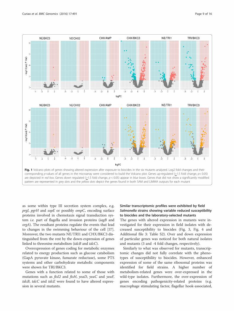
Changes in the transcriptome of S. enterica SL1344biocide resistant mutantsFigure 1 shows the Volcano Plots of genes altered afterexposure to biocides in the six mutants analyzed andFig. 2, the heatmap clustering mutants according to the
expression profiles of genes identified as differently regu-lated using both methods (LIMMA and SAM). Downregulation of genes was more remarkable than overex-pression of genes (-4 fold vs 2 fold Additional file 2:Table S1).Differential expression was identified for genes involved
in cellular metabolism (carbohydrate, lipid, amino acid,and cofactors), energy production, ion transport/metabol-ism, protein synthesis and signal chemotaxis pathways(Additional file 3: Table 2). The expression of genes clas-sically involved in tolerance to biocides as efflux-pumps(tolC, sugE) was slightly increased in some mutants.Two TRIRCHXRBKCR mutants (CHX/BKC3, NE/TRI1)
and one BKCR (TRI/BKC3) showed similar gene expres-sion profiles. Upregulation of genes involved in proteinsynthesis occurred in these three mutants, and some otherchanges were common, either in three or two of them, in-volving genes related to transcription such as rpoZ andrpoB which encodes subunits of the RNA polymerase.Conversely, transcriptional regulators were repressed inNE/TRI (malT) and CHX/BKC3 and TRI/BKC3 (hilC,hilD, sprB). The maltose monomeric activator MalT is aLuxR/UhpA member, a family of large size proteins be-lieved to have another functions (e.g. amino acid metabol-ism). The others are members of the AraC family andnegative regulators of HilA, the key regulator of the Sal-monella pathogenicity island 1 (SPI1) [35, 36].Both TRI/BKC3 and CHX/BKC3 mutants shared a
pattern of down-regulation of genes related to transportand metabolism with a potential role in virulence such
Table 4 Mutations identified in sequenced mutants
TRIRCHXRBKCR BKCR TRIHSCHXRBKCR TRIHS
Function Gene NE/TRI1 NE/CHX2 CHX/BKC3 TRI/BKC3 NE/BKC3 CHX/AMP
Amino acid/peptidetransport/metabolism
asnA a G→ T/A74A
Energy production/amino acid metabolism
aarF b T→ A/L317Q
Lipid metabolism fabI c G→A/G93S
Membrane/transport tolA d T→ A/V168E
Replication ftsK e T→ G/P772P A→ T/Q780H A→ T/Q793H
Surface structures mipA f T→ C/F61L
motB g T→ G/E128D
Virulence bigA h Deletion:408 bp Deletion:331 bp Deletion:293 bp
Intergenic -/- i T→ A
purH j/- A→ C A→ C
tdcA/garA k C→ T
Unknown yeaN l C→ G/T321T
Description of gene products: a Asparagine synthetase A, b Ubiquinone biosynthesis protein; c Enoyl-acyl carrier protein reductase (NADH); d Membrane transporterTolA protein; e Cell division protein FtsK; f Hypothetical outer membrane protein; g Motility protein B; h Hypothetical surface-exposed virulence protein; i Intergenicregion between Glutamyl-t-RNA synthetase (459 bp)/(359 bp) Xanthosin operon transcriptional regulator; j Intergenic region between Phosphoribosylaminoimide-zolecarboxamide fosmyltransferase and IMP cyclohydrolase/- (16S rRNA); k tdc operon transcriptional activator/Hypothetical surface-exposed virulence protein; l
Hypothetical membrane protein of Major Facilitator Superfamily (transporter)
Curiao et al. BMC Genomics (2016) 17:491 Page 8 of 16
as some within type III secretion system complex, e.g.prgI, pgrH and sopE or possibly ompC, encoding surfaceproteins involved in chemotaxis signal transduction sys-tem i.e. part of flagella and invasion proteins (iagB andorgA). The resultant proteins regulate the events that leadto changes in the swimming behaviour of the cell [37].Moreover, the two mutants NE/TRI1 and CHX/BKC3 dis-tinguished from the rest by the down-expression of geneslinked to threonine metabolism (tdcB and tdcC).Overexpression of genes coding for metabolic enzymes
related to energy production such as glucose catabolism(GapA pyruvate kinase, fumarate reductase), some PTSsystems and other carbohydrate metabolic componentswere shown for TRI/BKC3.Genes with a function related to some of those with
mutations such as ftsl2 and ftsH, yeaD, yeaC and yeaF,tdcB, tdcC and tdcE were found to have altered expres-sion in several mutants.
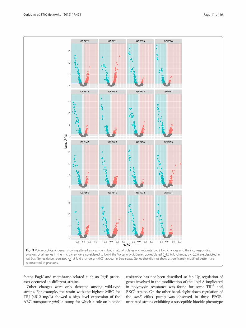
Similar transcriptomic profiles were exhibited by fieldSalmonella strains showing variable reduced susceptibilityto biocides and the laboratory-selected mutantsThe genes with altered expression in mutants were in-vestigated for their expression in field isolates with de-creased susceptibility to biocides (Fig. 3, Fig. 4 andAdditional file 3: Table S2). Over and down expressionof particular genes was noticed for both natural isolatesand mutants (3 and -4 fold changes, respectively).Similarly to what was observed for mutants, transcrip-
tomic changes did not fully correlate with the pheno-types of susceptibility to biocides. However, enhancedexpression of some of the same ribosomal proteins wasidentified for field strains. A higher number ofmetabolism-related genes were over-expressed in thewild-type isolates. Furthermore, the over-expression ofgenes encoding pathogenicity-related proteins (e.g.macrophage stimulating factor, flagellar hook-associated,
Fig. 1 Volcano plots of genes showing altered expression after exposure to biocides in the six mutants analyzed. Log2 fold changes and theircorresponding p-values of all genes in the microarray were considered to build the Volcano plot. Genes up-regulated (>1.5 fold change, p< 0.05)are depicted in red box. Genes down regulated (>1.5 fold change, p < 0.05) appear in blue boxes. Genes that did not show a significantly modifiedpattern are represented in grey dots and the yellow dots depict the genes found in both SAM and LIMMA outputs for each mutant
Curiao et al. BMC Genomics (2016) 17:491 Page 9 of 16
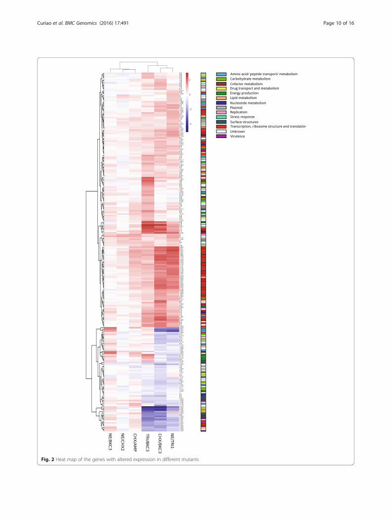
Fig. 2 Heat map of the genes with altered expression in different mutants
Curiao et al. BMC Genomics (2016) 17:491 Page 10 of 16
factor PagK and membrane-related such as PgtE prote-ase) occurred in different strains.Other changes were only detected among wild-type
strains. For example, the strain with the highest MBC forTRI (>512 mg/L) showed a high level expression of theABC transporter ydeY, a pump for which a role on biocide
resistance has not been described so far. Up-regulation ofgenes involved in the modification of the lipid A implicatedin polymyxin resistance was found for some TRIR andBKCR strains. On the other hand, slight down-regulation ofthe acrE efflux pump was observed in three PFGE-unrelated strains exhibiting a susceptible biocide phenotype
Fig. 3 Volcano plots of genes showing altered expression in both natural isolates and mutants. Log2 fold changes and their correspondingp-values of all genes in the microarray were considered to build the Volcano plot. Genes up-regulated (>1.5 fold change, p < 0.05) are depicted inred box. Genes down regulated (>1.5 fold change, p < 0.05) appear in blue boxes. Genes that did not show a significantly modified pattern arerepresented in grey dots.
Curiao et al. BMC Genomics (2016) 17:491 Page 11 of 16
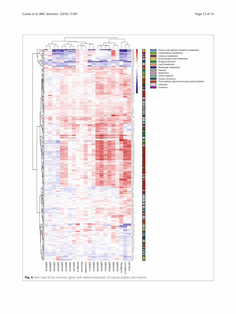
Fig. 4 Heat map of the common genes with altered expression of natural isolates and mutants
Curiao et al. BMC Genomics (2016) 17:491 Page 12 of 16
(TRIS/CHXS/BKCS) while one of them had the sugE geneover-expressed. A number of antibiotic resistance geneswere up- or down-regulated in natural isolates.
Resistance to biocides reduces the fitness of SalmonellaSL1344Variable values of fitness cost (FC) were observed forsome mutants with disparate biocide phenotype (Table 1and Additional file 1: Figure S2). Both NE/CHX2 (TRIR/BKCR/CHXR), which did not show any considerable sus-ceptibility change, and TRI/BKC3 (TRIHS/BKCR/CHXR),showed the highest FC values (34 % and 31 %, respect-ively). They had deleted bigA gene. Mutants TRIR/BKCR/CHXR (NE/TRI1), and TRIHS (CHX/AMP) showed simi-lar FC values (11 % and 16 %, respectively) (Additionalfile 1: Figure S2). Mutants fitter than parental strainshowed increased MICCAZ.Besides changes in growth rates, altered colony
morphology was also observed. Although several mu-tants had smoother and smaller colonies than the paren-tal strain, we did not observe notable differences in therdar (red, dry and rough) morphotype, an aggregativeand resistant physiological state which has been linkedto survival in nutrient-limited environments [38], or inthe genomic XbaI-genomic digested DNA profiles(Additional file 1: Figure S3).
DiscussionThis paper documents a versatile adaptive response ofthe Salmonella enterica strain SL1344 after exposure toinhibitory concentrations of biocides or antibioticswhich resulted on a diversity of phenotypes and genomicand proteomic changes. The emergence of mutants withphenotypes of antimicrobial resistance (antibiotics and/or biocides) which had not been previously exposed toantimicrobial agents but were recovered on selectionplates, suggests that a variety of mutants with alteredsusceptibility to biocides easily arises. Biocides, acting onmultiple cellular targets, would drive random selectionof mutants, eventually causing pleiotropic changes; andtherefore a high diversity of phenotypes were associatedwith biocide-tolerance. The fact that SHC did not selectfor mutants might provide a chance for efficient and safesanitization.Most mutants showed slight variations in their MIC
values to the antimicrobials tested. Although cross-resistance between biocides and antibiotics is frequentlydescribed for biocide resistant mutants, we also observedincreased susceptibility for some antimicrobials, a phe-nomenum that can be attributed to frequent collateraleffects in the emergence of resistance as previously doc-umented for antibiotic resistant bacteria [39, 40]. Indeed,antimicrobial resistant mutants of Salmonella presentedincreased susceptibility to envelope active inhibitory
compounds. Importantly, this sort of compounds turnsthe cytoplasmic membrane more permeable, which oftenresults on reduced viability. Therefore the ability to be-come resistant to these antimicrobials is lower. A partfrom the membrane vulnerability, most mutants poten-tially had alterations in the oxidative metabolism andprotein synthesis. Our data showed that TRI resistancewas often accompanied by higher tolerance to com-pounds of different antibiotic families, with the excep-tion of aminoglycosides. This finding is in line withother studies in Salmonella enterica strains that reportedincreases in the susceptibility to aminoglycosides andCHX accompanying TRIR [41, 42]. While most studieson the field focused on the analysis of cross-resistance ofbiocides and antibiotics, negative epistasis phenomenainferred from the simultaneous emergence of suscepti-bility to aminoglycosides or biocides and resistance toother antimicrobial agents are not uncommon [20, 39].This antagonistic pleiotropy epistatic effect known as“collateral sensitivity” must be taken into considerationwhen evaluating the risks for the acquisition of resist-ance or to envisage methods for reducing or even elim-inating resistant microorganisms in the field [40, 43].Comparative genomic analysis revealed changes in a
variety of genes, some of them previously linked to toler-ance to antibiotics or metals (fabI, yea and fts) [44] andothers newly identified here. Different amino acidchanges at position 93 of the FabI protein resulted inTRIR phenotypes [24, 32, 41, this study]. However, differ-ences in the polarity of amino acids at position 93 mightbe associated with distinct structural conformations ofFabI protein that would affect MICTRI values; highervalues (MICTRI ≥ 2 mg/L for the mutant of this study)occurring when an uncharged amino acid (Gly or Val) issubstituted by a polar amino acid (Ser).Non-functional ftsK gene mutants have previously
shown increased susceptibility to β-lactams and CIP andtolerance to chromate in Pseudomonas aeruginosa [45].In this study mutants with SNPs in ftsK (NE/TRI1 andCHX/BKC3) showed either increased susceptibility toseveral β-lactams or to most CIP-related antibiotics, re-spectively. The tolA gene showed a mutation and differ-ential expression in different mutants. Other SNPs mayalso modulate virulence in the mutants as for instance,mutants in motB (and motA) genes may paralyse the fla-gellar phenotype influencing adhesion and invasion ofcells [46]. Flagellar assembly and/or mobility mayantagonize the T3SS that delivers effectors into the hostcell of some pathogens, revealing the potential impact ofcross talk between some virulence factors depending onthe bacterial colonization phase and infection type [47].While particular changes at genetic level in the mu-
tants were detected, a remarkable alteration of the ex-pression profiles was noted in both mutants and field
Curiao et al. BMC Genomics (2016) 17:491 Page 13 of 16

isolates, with overexpression of ribosomal protein syn-thesis as well the down-regulation of genes involved inglobal stress and regulatory mechanisms, metabolism ofamino acids (lysine, asparagine, threonine), secondarymetabolism, transport, virulence, chemotaxis, invasionpathways and of unknown function. This might indicatea higher cellular activity with lower virulence. Effluxpumps previously involved in biocide tolerance were up-regulated in some mutants and field isolates. They in-cluded SugE, classically implicated in QACs resistanceand frequently found in Salmonella isolates of clinicaland animal origin [48, 49] or AcrAB [41, 50], whoseover-expression is known to contribute to antimicrobialresistance yet at low-level.This is the first study characterizing both the genomic
and expression profiles after antimicrobials challenge, al-though the stress response after exposure to a high di-versity of environmental stressors including biocides orantibiotics has been tackled before. Cold-shock response,allowing the survival of Listeria monocutogenes in thepresence of biocides was previously reported [10]. Simi-larly to the mutants selected in BKC in this study,antibiotic-resistant mutants obtained under differentmetabolic conditions were related to attenuatedvirulence due to low expression of the T3SS [51–53], ormutation in transcriptional regulators. The RNA poly-merase regulates the transcription of genes encodingtransport proteins and enzymes involved in the biosyn-thesis of the metabolic intermediates of exopolysacchar-ides, lipids, lipopolysaccharides, lipoproteins, flagella andpeptidoglycan. This protein is stress-induced and plays acentral role in the control of processes that involve phys-ical interaction of an organism with the environment, ascolonization of host surfaces (virulence) or biofilm for-mation [54]. Polymorphisms in genes coding for RNApolymerase subunit α (rpoA, described in mutants se-lected in QACs) [24, 50], and also σ factors (rpoS andrpoD genes, related to high-level resistance towards TRI)[55]) were previously reported. Despite other genes cod-ing for RNA polymerase activity (rpoZ and rpoB) wereshown here to be up-regulated, it corroborates the im-portance of this protein as a target implicated in intrin-sic resistance to biocides [23, 55]. Conversely, down-regulation of transcriptional and ribosomal genes werepreviously detected after exposure to CHX [50]. Inaddition, we report less transcripts of members of theLuxR/UhpA and AraC family, the last one beingnegative regulator of HilA, the key regulator of the SPI1[35, 36]. Down-regulation of genes encoding virulence-related proteins for several mutants might suggest alower pathogenicity.The finding of similar transcriptomic changes found in
both biocide resistant mutants and field isolates with re-duced susceptibility to biocides, suggests the involvement
of common general responses that include diverse alter-ations in metabolic and chemotaxis pathways, protein syn-thesis, cell envelope or regulation of pathogenicity-islands,which has been reported in other studies analysingbiocide-induced mutants of Salmonella and other species[9, 19, 56].
ConclusionsIn summary, this study shows that growth of Salmonellain the presence of selective concentrations of biocides orantibiotics leads to the selection of mutants with variablesusceptibility to antimicrobials (“cross-resistance” or “col-lateral sensitivity”-like phenotypes) which is consistentwith the “multiples target sites” hypothesis of most bio-cidal agents [57, 58]. The results highlight the wide rangeof pathways employed by Salmonella to counteract bio-cides and achieve stasis/stress survival. Unlike to what hasbeen commonly reported, overexpression of AcrAB-likepumps did not seem to be the main mechanism involvedin biocide tolerance. Detection of SNPs was not associatedwith altered expression of related genes, making data fromgenomic and transcriptomic analysis necessary for a com-prehensive analysis of biocide-challenged strains. Finally,most selected biocide-resistant mutants presented fitnesscosts, an issue that might reduce their chances to spreadunder non-selective conditions.
Additional files
Additional file 1: Figure S1. MIC distributions to triclosan,chlorhexidine and benzalkonium chloride for 62 natural Salmonellaisolates. The number of Salmonella isolates with reduced susceptibility tobiocides analysed for gene expression are indicated above the arrowsand MIC susceptibility values. Colors are according biocide distributions.(*) an isolate showed simultaneously reduced susceptibility to CHX andBKC. Other 6 isolates more susceptible for biocides were analysed forcontrol (TRIS/CHXS/BKCS: 0.06-0.12/2-8/32-64 mg/L).Figure S2. XbaIdigested-chromosomal DNA PFGE of several Salmonella mutants and itsparental strain (5-35 s for 21 h).Figure S3. Growth curves of Salmonellamutants and the parental strain in plain LB at 37 °C with shaking.(DOCX 446 kb)
Additional file 2: Table S1. Differential gene expression of commongenes from LIMMA and SAM methods for mutants. (XLSX 48 kb)
Additional file 3: Table S2. Differential gene expression of commongenes from LIMMA and SAM methods for natural isolates. (XLSX 280 kb)
AcknowledgementsDuring the execution of this study, T.C. was recipient of a predoctoralfellowship from Instituto Carlos III (PFIS-FI09/00901) and R.L.S. was financedby a contract-associated project (PI12-01581). We acknowledge the EuropeanDevelopment Regional Fund “A way to achieve Europe” (ERDF), and forcofounding the Plan Nacional de I + D+ I 2012-2015 (PI12-01581) and CIBERactions (CIBER in Epidemiology and Public Health, CIBERESP; CB06/02/0053).We are also grateful to the anonymous reviewers for their critical input thatcontributed to significantly improve the manuscript, and also to Val F. Lanzafor his helpful advice in using “R” and some tools for bioinformatic analysisof genome sequencing data.
Curiao et al. BMC Genomics (2016) 17:491 Page 14 of 16
FundingThis study was supported by funds from the European Commission (KBBE-2008-2B-227258-BIOHYPO to D.G., C.V., M.R.O., and J.L.M.); the Ministry ofEconomy and Competitiveness of Spain [grant BIO2014-54507-R to J.L.M.,grant PI12-01581 to T.M.C., the CIBERESP Network for Biomedical Research inEpidemiology and Public Health (CB06/02/0053) to F.B. and T.M.C., theSpanish Network for Research on Infectious Diseases (REIPI RD12/0015) fromthe Instituto de Salud Carlos III to J.L.M.] and the Regional Government ofMadrid-CAM (S2010/BMD2414-PROMPT) to J.L.M. and F.B.
Availability of data and materialsAll relevant data are available within the manuscript and its additional files.
Authors’ contributionsT.C., D.G. C.V., M.R.O., J.L.M. and T.M.C. contributed to the study design. T.C.performed the experimental work related to the selection andcharacterization of biocide mutants, participated in the analysis ofphenotypic data, genomic and transcriptomics, and wrote the manuscript.E.M. and C.V. performed the analysis with Phenotypic Microarrays (Biolog).D.G. developed the expression microarrays, and participated in the dataanalysis. R.L.S. performed the bioinformatics analysis of data from genomesequencing and microarrays and participated in the revision of themanuscript. S.L.L. participated in the development of the expressionmicroarrays and in related data analysis. F.B. and M.R.O. provided expertise,participated in the analysis of data, and the revision of the manuscript. J.L.M.and T.M.C. participated in the analysis of data, and wrote the manuscript. Allauthors read and approved the final version of this manuscript.
Competing interestsThe authors declare that they have no competing interests.
Ethics approval and consent to participateNot applicable.
Author details1Servicio de Microbiología, Instituto Ramón y Cajal de Investigación Sanitaria(IRYCIS), Madrid, Spain. 2CIBER Epidemiología y Salud Pública (CIBERESP),Madrid, Spain. 3Unidad de Resistencia a Antibióticos y Virulencia bacterianaasociada al Consejo Superior de Investigaciones Científicas (CSIC), Madrid,Spain. 4Department of Agrifood Production and Environmental Sciences,University of Florence, Firenze, Italy. 5Neuroinfection Laboratory, Institute forInfectious Diseases, Bern, Switzerland. 6University of Leicester, Leicester, UK.7Departamento de Biotecnología Microbiana, Centro Nacional deBiotecnología (CSIC), Darwin 3, Cantoblanco, Madrid 28049, Spain.
Received: 16 October 2015 Accepted: 26 May 2016
References1. Parry CM, Threlfall EJ. Antimicrobial resistance in typhoidal and
nontyphoidal salmonellae. Curr Opin Infect Dis. 2008;21:531–8.2. Barat S, Steeb B, Mazé A, Bumann D. Extensive in vivo resilience of persistent
Salmonella. PLoS One. 2012;7:e42007.3. Condell O, Iversen C, Cooney S, Power KA, Walsh C, Burgess C, Fanning S.
Efficacy of biocides used in the modern food industry to control Salmonellaenterica, and links between biocide tolerance and resistance to clinicallyrelevant antimicrobial compounds. Appl Environ Microbiol. 2012;78:3087–97.
4. Beier RC, Anderson PN, Hume ME, Poole TL, Duke SE, Crippen TL, SheffieldCL, Caldwell DJ, Byrd JA, Anderson RC, Nisbet DJ. Characterization ofSalmonella enterica Isolates from Turkeys in Commercial Processing Plantsfor Resistance to Antibiotics, Disinfectants, and a Growth Promoter.Foodborne Pathog Dis. 2011;8:593–600.
5. Whitehead RN, Overton TW, Kemp CL, Webber MA. Exposure of Salmonellaenterica Serovar Typhimurium to High Level Biocide Challenge Can SelectMultidrug Resistant Mutants in a Single Step. PLoS One. 2011;6:e22833.
6. Randall LP, Cooles SW, Coldham NG, Penuela EG, Mott AC, Woodward MJ,Piddock LJV, Webber MA. Commonly used farm disinfectants can select formutant Salmonella enterica serovar Typhimurium with decreasedsusceptibility to biocides and antibiotics without compromising virulence. JAntimicrob Chemother. 2007;60(September):1273–80.
7. Piddock LJV. Clinically Relevant Chromosomally Encoded Multidrug ResistanceEfflux Pumps in Bacteria Clinically Relevant Chromosomally Encoded MultidrugResistance Efflux Pumps in Bacteria. Clin Microbiol Rev. 2006;19:382–402.
8. Bialek-davenet S, Marcon E, Lavigne J, Moreau R: In Vitro Selection of ramRand soxR Mutants Overexpressing Efflux Systems by Fluoroquinolones asWell as Cefoxitin in Klebsiella pneumoniae. Antimicrob Agents Chemother.2011;55:2795–2802
9. Bailey AM, Constantinidou C, Ivens A, Garvey MI, Webber MA, Coldham N,Hobman JL, Wain J, Woodward MJ, Piddock LJV. Exposure of Escherichia coliand Salmonella enterica serovar Typhimurium to triclosan induces a species-specific response, including drug detoxification. J Antimicrob Chemother.2009;64(September):973–85.
10. Fox EM, Leonard N, Jordan K. Physiological and transcriptionalcharacterization of persistent and nonpersistent Listeria monocytogenesisolates. Appl Environ Microbiol. 2011;77:6559–69.
11. Bore E, Hébraud M, Chafsey I, Chambon C, Skjaeret C, Moen B, Møretrø T,Langsrud Ø, Rudi K, Langsrud S. Adapted tolerance to benzalkonium chloridein Escherichia coli K-12 studied by transcriptome and proteome analyses.Microbiology. 2007;153(Pt 4):935–46.
12. Condell O, Sheridan Á, Power KA, Bonilla-Santiago R, Sergeant K, Renaut J,Burgess C, Fanning S, Nally JE. Comparative proteomic analysis ofSalmonella tolerance to the biocide active agent triclosan. J Proteomics.2012;75:4505–19.
13. Sheridan Á, Lenahan M, Condell O, Bonilla-Santiago R, Sergeant K, Renaut J,Duffy G, Fanning S, Nally JE, Burgess CM. Proteomic and phenotypic analysisof triclosan tolerant verocytotoxigenic Escherichia coli O157:H19. JProteomics. 2013;80:78–90.
14. McMurry LM, Oethinger M, Levy SB. Triclosan targets lipid synthesis. Nature.1998;394:531–2.
15. Karatzas KAG, Webber MA, Jorgensen F, Woodward MJ, Piddock LJV,Humphrey TJ. Prolonged treatment of Salmonella enterica serovarTyphimurium with commercial disinfectants selects for multiple antibioticresistance, increased efflux and reduced invasiveness. J AntimicrobChemother. 2007;60(September):947–55.
16. Cheung H, Wong MM, Cheung S, Liang LY, Lam Y, Chiu S. DifferentialActions of Chlorhexidine on the Cell Wall of Bacillus subtilis and Escherichiacoli. PLoS One. 2012;7:e36659.
17. Bailey AM, Ivens A, Kingsley R, Cottell JL, Wain J, Piddock LJV. RamA, amember of the AraC/XylS family, influences both virulence and efflux inSalmonella enterica serovar Typhimurium. J Bacteriol. 2010;192:1607–16.
18. Mark A. Webber, Andrew M. Bailey, Jessica M. A. Blair, Eirwen Morgan, MarkP. Stevens, Jay C. D. Hinton, Al Ivens, John Wain, Piddock LJ. The GlobalConsequence of Disruption of the AcrAB-TolC Efflux Pump in Salmonellaenterica Includes Reduced Expression of SPI-1 and Other Attributes RequiredTo Infect the Host. J Bacteriol. 2009;191:4276–85.
19. Kastbjerg VG, Larsen MH, Gram L, Ingmer H. Influence of sublethalconcentrations of common disinfectants on expression of virulence genesin Listeria monocytogenes. Appl Environ Microbiol. 2010;76:303–9.
20. Curiao T, Marchi E, Viti C, Oggioni MR, Baquero F, Martinez JL, Coque TM.Polymorphic Variation in Susceptibility and Metabolism of Triclosan-Resistant Mutants of Escherichia coli and Klebsiella pneumoniae ClinicalStrains Obtained after Exposure to Biocides and Antibiotics. AntimicrobAgents Chemother. 2015;59:3413–23.
21. Webber MA, Randall LP, Cooles S, Woodward MJ, Piddock LJV. Triclosanresistance in Salmonella enterica serovar Typhimurium. J AntimicrobChemother. 2008;62:83–91.
22. Karatzas KAG, Randall LP, Webber M, Piddock LJV, Humphrey TJ, Woodward MJ,Coldham NG. Phenotypic and Proteomic Characterization of Multiply Antibiotic-Resistant Variants of Salmonella enterica Serovar Typhimurium Selected FollowingExposure to Disinfectants †. Appl Environ Microbiol. 2008;74:1508–16.
23. Mangalappalli-Illathu AK, Korber DR. Adaptive resistance and differentialprotein expression of Salmonella enterica serovar Enteritidis biofilmsexposed to benzalkonium chloride. Antimicrob Agents Chemother. 2006;50:3588–96.
24. Webber MA, Whitehead RN, Mount M, Loman NJ, Pallen MJ, Piddock LJV.Parallel evolutionary pathways to antibiotic resistance selected by biocideexposure. J Antimicrob Chemother. 2015;70(8):2241-8.
25. Wray CSW. Experimental Salmonella typhimurium infection in calves. Res VetSci. 1978;25:139–43.
26. Morrissey I, Oggioni MR, Knight D, Curiao T, Coque T, Kalkanci A, MartinezJL. Evaluation of epidemiological cut-off values indicates that biocide
Curiao et al. BMC Genomics (2016) 17:491 Page 15 of 16

resistant subpopulations are uncommon in natural isolates of clinically-relevant microorganisms. PLoS One. 2014;9:e86669.
27. Curiao T, Cantón R, Garcillán-Barcia MP, de la Cruz F, Baquero F, Coque TM.Association of composite IS26-sul3 elements with highly transmissible IncI1plasmids in extended-spectrum-beta-lactamase-producing Escherichia coliclones from humans. Antimicrob Agents Chemother. 2011;55:2451–7.
28. Bochner BR, Gadzinski P, Panomitros E. Phenotype microarrays for high-throughput phenotypic testing and assay of gene function. Genome Res.2001;11:1246–55.
29. Foucault ML, Depardieu F, Courvalin P, Grillot-Courvalin C. Inducibleexpression eliminates the fitness cost of vancomycin resistance inenterococci. Proc Natl Acad Sci U S A. 2010;107:16964–9.
30. Hall BG, Acar H, Nandipati A, Barlow M. Growth rates made easy. Mol BiolEvol. 2014;31:232–8.
31. Shintre MS, Gaonkar TA, Modak SM. Efficacy of an alcohol-based healthcarehand rub containing synergistic combination of farnesol and benzethoniumchloride. Int J Hyg Environ Health. 2006;209:477–87.
32. Li H, Zhang D-F, Lin X-M, Peng X-X. Outer membrane proteomics ofkanamycin-resistant Escherichia coli identified MipA as a novel antibioticresistance-related protein. FEMS Microbiol Lett. 2015;362(11) fnv074; doi: 10.1093/femsle/fnv074
33. Levengood-Freyermuth SK, Click EM, Webster RE. Role of the carboxyl-terminal domain of TolA in protein import and integrity of the outermembrane. J Bacteriol. 1993;175:222–8.
34. Czibener C, Merwaiss F, Guaimas F, Del Giudice MG, Serantes DAR, SperaJM, Ugalde JE. BigA is a novel adhesin of Brucella that mediates adhesion toepithelial cells. Cell Microbiol. 2015;18:500–13.
35. Takaya A, Kubota Y, Isogai E, Yamamoto T. Degradation of the HilC and HilDregulator proteins by ATP-dependent Lon protease leads to downregulationof Salmonella pathogenicity island 1 gene expression. Mol Microbiol. 2005;55:839–52.
36. Saini S, Rao CV. SprB is the molecular link between Salmonellapathogenicity island 1 (SPI1) and SPI4. J Bacteriol. 2010;192:2459–62.
37. McEvoy MM, Bren A, Eisenbach M, Dahlquist FW. Identification of thebinding interfaces on CheY for two of its targets, the phosphatase CheZand the flagellar switch protein fliM. J Mol Biol. 1999;289:1423–33.
38. White AP, Gibson DL, Grassl GA, Kay WW, Finlay BB, Vallance BA, Surette MG.Aggregation via the red, dry, and rough morphotype is not a virulenceadaptation in Salmonella enterica serovar Typhimurium. Infect Immun. 2008;76:1048–58.
39. Lázár V, Pal Singh G, Spohn R, Nagy I, Horváth B, Hrtyan M, Busa-Fekete R,Bogos B, Méhi O, Csörgő B, Pósfai G, Fekete G, Szappanos B, Kégl B, Papp B,Pál C. Bacterial evolution of antibiotic hypersensitivity. Mol Syst Biol. 2013;9:700.
40. Pal C, Papp B, Lazar V. Collateral sensitivity of antibiotic-resistant microbes.Trends Microbiol. 2015;23:401–7.
41. Rensch U, Klein G, Kehrenberg C. Analysis of Triclosan-Selected Salmonellaenterica Mutants of Eight Serovars Revealed Increased AminoglycosideSusceptibility and Reduced Growth Rates. PLoS One. 2013;8:e78310.
42. Cottell A, Denyer SP, Hanlon GW, Ochs D, Maillard J-Y. Triclosan-tolerantbacteria: changes in susceptibility to antibiotics. J Hosp Infect. 2009;72:71–6.
43. Baquero F, Lanza VF, Cantón R, Coque TM. Public health evolutionarybiology of antimicrobial resistance: priorities for intervention. Evol Appl.2015;8:223–39.
44. Yu BJ, Kim JA, Ju HM, Choi S-K, Hwang SJ, Park S, Kim E, Pan J-G. Genome-wide enrichment screening reveals multiple targets and resistance genesfor triclosan in Escherichia coli. J Microbiol. 2012;50:785–91.
45. Alvarez-Ortega C, Wiegand I, Olivares J, Hancock REW, Martínez JL. Geneticdeterminants involved in the susceptibility of Pseudomonas aeruginosa tobeta-lactam antibiotics. Antimicrob Agents Chemother. 2010;54:4159–67.
46. Achouri S, Wright JA, Evans L, Macleod C, Fraser G, Cicuta P, Bryant CE. Thefrequency and duration of Salmonella – macrophage adhesion eventsdetermines infection efficiency. Philos Transl B. 2015;370:20140033.
47. Soscia C, Hachani A, Bernadac A, Filloux A, Bleves S. Cross talk between typeIII secretion and flagellar assembly systems in Pseudomonas aeruginosa.J Bacteriol. 2007;189:3124–32.
48. Zou L, Meng J, McDermott PF, Wang F, Yang Q, Cao G, Hoffmann M, ZhaoS. Presence of disinfectant resistance genes in Escherichia coli isolated fromretail meats in the USA. J Antimicrob Chemother. 2014;69:2644–9.
49. Su LH, Chen HL, Chia JH, Liu SY, Chu C, Wu TL, Chiu CH. Distribution of atransposon-like element carrying blaCMY-2 among Salmonella and otherEnterobacteriaceae. J Antimicrob Chemother. 2006;57:424–9.
50. Condell O, Power KA, Händler K, Finn S, Sheridan A, Sergeant K, Renaut J,Burgess CM, Hinton JCD, Nally JE, Fanning S. Comparative analysis ofSalmonella susceptibility and tolerance to the biocide chlorhexidineidentifies a complex cellular defense network. Front Microbiol. 2014;5:373.
51. Giraud E, Baucheron S, Virlogeux-Payant I, Nishino K, Cloeckaert A. Effects ofnatural mutations in the ramRA locus on invasiveness of epidemicfluoroquinolone-resistant Salmonella enterica serovar Typhimurium isolates.J Infect Dis. 2013;207:794–802.
52. Golubeva YA, Sadik AY, Ellermeier JR, Slauch JM. Integrating globalregulatory input into the Salmonella pathogenicity island 1 type III secretionsystem. Genetics. 2012;190:79–90.
53. Linares JF, López JA, Camafeita E, Albar JP, Rojo F, Martínez JL.Overexpression of the multidrug efflux pumps MexCD-OprJ and MexEF-OprN is associated with a reduction of type III secretion in Pseudomonasaeruginosa. J Bacteriol. 2005;187:1384–91.
54. Francke C, Groot Kormelink T, Hagemeijer Y, Overmars L, Sluijter V,Moezelaar R, Siezen RJ. Comparative analyses imply that the enigmaticSigma factor 54 is a central controller of the bacterial exterior. BMCGenomics. 2011;12:385.
55. Gantzhorn MR, Olsen JE, Thomsen LE. Importance of sigma factor mutationsin increased triclosan resistance in Salmonella Typhimurium. BMC Microbiol.2015;15:105.
56. Casey A, Fox EM, Schmitz-Esser S, Coffey A, McAuliffe O, Jordan K.Transcriptome analysis of Listeria monocytogenes exposed to biocide stressreveals a multi-system response involving cell wall synthesis, sugar uptake,and motility. Front Microbiol. 2014;5:1–10.
57. Russell AD. Mechanisms of antimicrobial action of antiseptics anddisinfectants: an increasingly important area of investigation. J AntimicrobChemother. 2002;49:597–9.
58. Maillard J-Y. Bacterial target sites for biocide action. J Appl Microbiol. 2002;92(Suppl):16S–27S.
• We accept pre-submission inquiries
• Our selector tool helps you to find the most relevant journal
• We provide round the clock customer support
• Convenient online submission
• Thorough peer review
• Inclusion in PubMed and all major indexing services
• Maximum visibility for your research
Submit your manuscript atwww.biomedcentral.com/submit
Submit your next manuscript to BioMed Central and we will help you at every step:
Curiao et al. BMC Genomics (2016) 17:491 Page 16 of 16


![SALMONELLA ENTERICA SUBSP. ENTERICA 1,4,[5],12:i:-](https://static.fdocuments.in/doc/165x107/6297d8bb7423086b1b094e2e/salmonella-enterica-subsp-enterica-14512i.jpg)