MRI characteristics of cysts and cyst-like lesions in and around … · 2017-08-28 · ruptured...
16
PICTORIAL REVIEW MRI characteristics of cysts and “cyst-like” lesions in and around the knee: what the radiologist needs to know Evangelos Perdikakis & Vasilios Skiadas Received: 7 November 2012 / Revised: 14 February 2013 / Accepted: 19 February 2013 / Published online: 12 March 2013 # The Author(s) 2013. This article is published with open access at Springerlink.com Abstract Objectives and Methods A variety of benign cystic or “cyst- like” lesions may be encountered during a routine magnetic resonance imaging (MRI) of the knee. These lesions com- prise a diverse group of entities from benign cysts to com- plications of underlying diseases. In addition, normal anatomic bursae and recesses may be misdiagnosed as an intra-articular cystic lesion when they are distended. How- ever, the majority of the aforementioned lesions have char- acteristic MR appearances that allow a confident diagnosis, thus obviating the need for additional imaging or interven- tional procedures. Results This article includes a comprehensive pictorial es- say of the characteristic MRI features of common and un- common benign cysts and “cyst-like” lesions in and around the knee joint. Discussion For accurate assessment of the “cystic struc- ture”, a radiologist should be able to identify typical MRI patterns that contribute in establishing the correct diagnosis and thus guiding specific therapy and avoiding unwarranted interventional procedures such as biopsy or arthroscopy. Teaching points • Cystic lesions are common in knee MRI and the commonest, the Baker’ s cyst, has an incidence of 38 %. • Synovial cysts, meniscal cysts, normal knee bursae and recesses have characteristic MR appearances. • Miscellaneous “cyst-like” lesions may require a more dedicated MR protocol for a correct diagnosis. Keywords Knee . MRI diagnosis . Cyst . Bursae . Recesses Introduction Given the fact that magnetic resonance imaging (MRI) is being performed more frequently for assessment of the knee joint (e.g. post-traumatic, in sport injuries, in rheumatolog- ical disorders, in oncological imaging), the number of inci- dental cystic and “cyst-like” lesions in and around the knee joint found on routine knee MRI scans has also increased [1–4]. The vast majority of these lesions are benign, ranging from benign cysts to complications of underlying diseases and many of them demonstrate characteristic features on MRI, thus allowing a confident diagnosis to be made [1–6]. Knowledge of the common anatomical locations and appearances of bursae, recesses, cysts and ganglia is necessary so that radiologists do not misinterpret these be- nign entities as soft-tissue tumours [1–8]. It is of paramount importance for the radiologist to be aware of the MRI features because understanding the spectrum of appearances of the various benign cystic lesions is vital for optimal patient management. This article is intended to be a com- prehensive pictorial review of the most common and un- common benign cystic and “cyst-like” lesions in and around the knee joint. For easier classification purposes, benign cysts were subdivided into categories as following: (1) synovial cysts, (2) ganglion cysts, (3) meniscal cysts and (4) intraosseous cysts. Similarly, “cyst-like” lesions were subclassified into the following: (1) normal knee bursae, (2) normal knee recesses and (3) miscellaneous cyst-like lesions. E. Perdikakis (*) : V. Skiadas Department of Radiology, 412 General Military Hospital-212 Mobile Army Surgical Hospital, Terma Lefkou Pyrgou, Xanthi 67100, Greece e-mail: [email protected] Insights Imaging (2013) 4:257–272 DOI 10.1007/s13244-013-0240-1
Transcript of MRI characteristics of cysts and cyst-like lesions in and around … · 2017-08-28 · ruptured...

PICTORIAL REVIEW
MRI characteristics of cysts and “cyst-like” lesionsin and around the knee: what the radiologistneeds to know
Evangelos Perdikakis & Vasilios Skiadas
Received: 7 November 2012 /Revised: 14 February 2013 /Accepted: 19 February 2013 /Published online: 12 March 2013# The Author(s) 2013. This article is published with open access at Springerlink.com
AbstractObjectives and Methods Avariety of benign cystic or “cyst-like” lesions may be encountered during a routine magneticresonance imaging (MRI) of the knee. These lesions com-prise a diverse group of entities from benign cysts to com-plications of underlying diseases. In addition, normalanatomic bursae and recesses may be misdiagnosed as anintra-articular cystic lesion when they are distended. How-ever, the majority of the aforementioned lesions have char-acteristic MR appearances that allow a confident diagnosis,thus obviating the need for additional imaging or interven-tional procedures.Results This article includes a comprehensive pictorial es-say of the characteristic MRI features of common and un-common benign cysts and “cyst-like” lesions in and aroundthe knee joint.Discussion For accurate assessment of the “cystic struc-ture”, a radiologist should be able to identify typical MRIpatterns that contribute in establishing the correct diagnosisand thus guiding specific therapy and avoiding unwarrantedinterventional procedures such as biopsy or arthroscopy.Teaching points• Cystic lesions are common in knee MRI and thecommonest, the Baker’s cyst, has an incidence of 38 %.
• Synovial cysts, meniscal cysts, normal knee bursae andrecesses have characteristic MR appearances.
• Miscellaneous “cyst-like” lesions may require a morededicated MR protocol for a correct diagnosis.
Keywords Knee . MRI diagnosis . Cyst . Bursae . Recesses
Introduction
Given the fact that magnetic resonance imaging (MRI) isbeing performed more frequently for assessment of the kneejoint (e.g. post-traumatic, in sport injuries, in rheumatolog-ical disorders, in oncological imaging), the number of inci-dental cystic and “cyst-like” lesions in and around the kneejoint found on routine knee MRI scans has also increased[1–4]. The vast majority of these lesions are benign, rangingfrom benign cysts to complications of underlying diseasesand many of them demonstrate characteristic features onMRI, thus allowing a confident diagnosis to be made[1–6]. Knowledge of the common anatomical locationsand appearances of bursae, recesses, cysts and ganglia isnecessary so that radiologists do not misinterpret these be-nign entities as soft-tissue tumours [1–8]. It is of paramountimportance for the radiologist to be aware of the MRIfeatures because understanding the spectrum of appearancesof the various benign cystic lesions is vital for optimalpatient management. This article is intended to be a com-prehensive pictorial review of the most common and un-common benign cystic and “cyst-like” lesions in and aroundthe knee joint. For easier classification purposes, benigncysts were subdivided into categories as following: (1)synovial cysts, (2) ganglion cysts, (3) meniscal cysts and(4) intraosseous cysts. Similarly, “cyst-like” lesions weresubclassified into the following: (1) normal knee bursae,(2) normal knee recesses and (3) miscellaneous cyst-likelesions.
E. Perdikakis (*) :V. SkiadasDepartment of Radiology, 412 General Military Hospital-212Mobile Army Surgical Hospital, Terma Lefkou Pyrgou,Xanthi 67100, Greecee-mail: [email protected]
Insights Imaging (2013) 4:257–272DOI 10.1007/s13244-013-0240-1

Benign cysts
Synovial cysts
Synovial cysts are defined as juxta-articular fluid collectionsthat are lined by synovial cells [1, 2]. The synovial lining isthe characteristic histological feature that distinguishes themfrom other juxta-articular fluid collections [1, 2, 6, 8]. Froma pathophysiological point of view a synovial cyst repre-sents a focal extension of joint fluid that may extend in anydirection and may, or may not, communicate with the joint.They can be encountered as incidental findings in MRexaminations, but regarding their aetiology, they have beenassociated with other underlying knee disorders such asosteoarthritis, trauma, rheumatoid arthritis, gout, systemiclupus erythematosus and juvenile rheumatoid arthritis [1, 2,4–6]. Although usually asymptomatic, they can manifestwith pain and swelling. The most common examples of asynovial cyst in the knee are the popliteal cyst (Baker’s cyst)and the proximal tibiofibular joint (PTFJ) synovial cyst.
The term popliteal cyst is a “misnomer” since it does notrepresent a true cyst, but actually corresponds to a fluiddistension of the gastrocnemius-semimembranosus bursa,which occasionally communicates with the knee joint [6,8]. Regarding its pathogenesis, a valvular mechanism be-tween the knee joint and the bursa has been implicated,which allows joint fluid to communicate in a unidirectionalmode. On MRI, they manifest as well defined unilocular or
multilocular cystic masses, located posteromedially, arisingbetween the tendon of the semimembranosus and the medialhead of gastrocnemius [1, 2, 4–8]. Baker’s cyst can beeffectively diagnosed with MRI since the fluid-distentedgastrocnemius-semimembranosus bursa is easily depictedon T2-weighted MR images, especially in the axial plane.Fluid signal intensity is seen in all sequences in cases ofpopliteal cysts [8, 9]. Nevertheless, complications such ashaemorrhage, rupture, the presence of intra-articular loose
Fig. 1 Popliteal cysts. Theaxial (a) and sagittal (b) fatsaturated proton densityweighted images show a largemultiseptated popliteal cyst(asterisks) emerging betweenthe medial gastrocnemiustendon (black arrow) and thesemimembranosus tendon(white arrow) and abutting themedial gastrocnemial musclebelly (GN). The axial (c) fatsaturated proton densityweighted image shows aruptured popliteal cyst(arrowheads). The axial (d) fatsaturated proton densityweighted image demonstrates aBaker’s cyst (white arrow) witha single looseosteocartilaginous body insidethe cyst (black arrow)
Fig. 2 Ganglion cyst of the proximal tibiofibular joint. The coronal fatsaturated proton density weighted image shows a ganglion cyst (whitearrow) emerging with a short neck (black arrow) from the proximaltibiofibular joint (T tibia, F fibula)
258 Insights Imaging (2013) 4:257–272

bodies and synovial proliferative reaction-diseases may pro-duce a more heterogeneous appearance. Differential diagno-sis from other cyst-like lesions and soft tissue tumours isthus imperative and requires a dedicated contrast-enhancedMRI examination protocol in the aforementioned cases[8–13]. Increased signal intensity within the cyst on T1-weighted images may appear in an infected or haemorrhagiccyst. The cyst may extend to any direction, but most com-monly inferomedially. Lateral or intramuscular extension isobserved relatively rare. A ruptured popliteal cyst is effi-ciently demonstrated as a high signal intensity oedemadispersing into the adjacent soft tissues and fascial planeson fat-suppressed T2-weighted sequences (Fig. 1).
The proximal tibiofibular joint (PTFJ) synovial cyst isbelieved to represent a joint capsule herniation, due toincreased intra-articular pressure [14–16]. On T1-weighted
and T2-weighted MR sequences the lesion appears as ahomogeneous often fusiform fluid collection in contact withthe proximal tibiofibular joint, while a communicating neckleading from the cyst into the PTFJ is sometimes alsoillustrated [14–16]. Increased signal intensity on T2-weighted images of the peroneally innervated anterior com-partment musculature may be present in cases of commonperoneal nerve impingement. Erosion of adjacent bone mayalso be seen in cases of large cysts. Differential diagnosisfrom soft tissue and nerve sheath tumours with myxomatousor cystic degeneration may be required (Fig. 2).
Ganglion cysts
A ganglion cyst is defined as a benign cystic mass that issurrounded by dense connective tissue, without a synovial
Fig. 3 ACL ganglion cysts.Two sequential sagittal fatsaturated proton densityweighted images in twodifferent patients (upper andlower row) demonstrating acystic lesion (black arrow) atthe upper segment of the ACLwith part of the lesion (whitearrow) dispersing into the ACLfibres
Fig. 4 PCL ganglion cyst. Thesagittal (a) and axial (b) fatsaturated proton densityweighted images show amultiloculated septated cyst(arrowheads) located in contactwith the PCL (white arrows)and along its dorsal surface.Note a small insertional tibialcyst (black arrow)
Insights Imaging (2013) 4:257–272 259

lining and is filled with a gelatinous fluid rich in hyaluronicacid and other mucopolysaccharides. Ganglia are
traditionally divided into the following categories: intra-articular, extra-articular, intraosseous and (rare) periosteal.
Fig. 5 Hoffa’s fat pad ganglioncysts: the sagittal (a) and axial(b) fat saturated proton densityweighted images in twodifferent patients demonstrate amultiloculated septated cysticlesion (asterisks) within theHoffa’s fat pad. Ganglion cystof the suprapatellar bursa: thesagittal (a) and axial (b) fatsaturated T2-weighted imagesshow a lobulated cystic lesion(arrows) in the suprapatellarbursa
Fig. 6 Extra-articularganglion cysts. The axial (a)and sagittal (b) fat saturatedproton density weighted imagesdemonstrate a unilocular cysticfluid collection consistent withan extra-articular ganglion cyst(arrows). The coronal (c) andsagittal (d) fat saturated protondensity weighted images showa multilocular extra-articularganglion cyst (arrows)
260 Insights Imaging (2013) 4:257–272

Intra-articular ganglia They are quite uncommon cystic le-sions and typically arise from the cruciate ligaments, mostcommonly from the anterior cruciate ligament (ACL) [9, 11,17, 18] In MRI part of the lesion is interspersed within theACL fibres and it may extend anteriorly towards the Hoffa’sfat pad or posteriorly towards the femoral intercondylar fossa[1, 2, 19]. Rarely it may be demonstrated between the ACLand posterior cruciate ligament (PCL). PCL ganglia have amore typical appearance and present as well-definedmultilocular cysts adjacent to and along the dorsal PCL sur-face [1, 2, 8, 9]. Differential diagnosis should be made from aposteromedial meniscal cyst extending centrally within thejoint predominantly posterior to the PCL. Ganglia withinHoffa’s fat pad or in the suprapatellar recess-bursa usuallyappear as well-defined multilocular cysts and are most com-monly encountered anterior to the anterior horn of the lateralmeniscus [6, 8, 19]. An intrahoffatic lesion or a synoviallesion such as haemangioma or synovial sarcoma may bemisinterpreted as a ganglion cyst within the fat pad andcontrast-enhanced MRI is warranted in such cases for differ-ential diagnosis (Figs. 3, 4 and 5).
Extra-articular ganglia Ganglion cysts may be encounteredin any of the extra-articular soft tissues around the knee,including the joint capsule, tendons, ligaments, bursae, mus-cles and nerves. In rare cases, extra-articular ganglia maycommunicate with the joint cavity [1, 2, 6–8, 11–13]. OnMR images they present as well-defined rounded or lobulatedfluid collections, often with associated peripheral fluid-filledpseudopodia and sharply defined internal septations (“bunchof grapes appearance”). On T2-weighted images, gangliaappear with high signal intensity. On T1-weighted images,the signal intensities of ganglia range depending on theirprotein concentration. Associated bone erosion and pericysticoedema have been reported. The detection on MRI of apossible communication between the ganglion and the jointcapsule is of great importance in preoperative planning be-cause of the possibility of recurrence. MRI is also the imagingmodality of choice for depicting muscle denervation changesin cases of nerve sheath ganglion cysts [20, 21]. These cystsare most frequently associated with the common peronealnerve and its branches and nerve compression may result infoot drop and paraesthesia over the dorsum of the foot [20,
Fig. 7 Common peronealnerve sheath ganglion cyst. Theaxial (a) T1-weighted, (b)contrast-enhanced fat saturatedT1-weighted, (c) fat saturatedproton density weighted imagesand two sequential coronal (d,e) fat saturated proton densityweighted images show a cysticlesion (arrows) at the lateralaspect of the fibula headextending caudally
Fig. 8 Intraosseous ganglioncyst. The axial (a) and coronal(b) fat saturated proton densityweighted images demonstrate asolitary, unilocular cystic lesion(arrows). Note the sclerotic rim(arrowheads) and mild reactivebone oedema (asterisk)
Insights Imaging (2013) 4:257–272 261

21]. MRI can contribute to treatment planning because itallows visualisation of both the cyst and the presence ofoedema in the innervated muscles [20, 21]. Detection of MRsigns of muscle denervation should be regarded as indicativeof surgical nerve decompression. (Figs. 6 and 7).
Intraosseous ganglia These lesions are located in theepiphyseal-metaphyseal region of long bones, most com-monly the tibia, often within subchondral bone, adjacentto a joint or in proximity to ligament insertion sites [7,17, 18, 22]. Their pathogenesis is unclear and unknownand there is still debate as to whether they are distinctfrom degenerative, insertional or post-traumatic cysts.Regarding MRI appearance, they manifest as solitary,unilocular or multilocular, and a sclerotic rim is usuallypresent. Differential diagnosis may be necessary fromprimary epiphyseal bone tumours, such as giant celltumour, clear cell chondrosarcoma and chondroblastoma
and it can also be extremely difficult to differentiate froma simple bone cyst (Fig. 8).
Periosteal ganglia They are extremely rare and arethought to be produced by mucoid degeneration andcyst formation of the periosteum [1, 2, 6–9]. They aremost commonly located in the proximal tibial shaft, inproximity to pes anserinus. On MR images they typi-cally appear as periosteally based, well-defined homo-geneous lesions with fluid signal intensity. Superficialcortical erosion and scalloping, as well as reactive newbone formation may be present. Other lesions ofperiosteal origin, such as periosteal chondroma,subperiosteal hematoma, chronic subperiosteal abscessor malignant soft tissue tumours, mainly when theyerode the adjacent bone and cause periosteal reaction,may need differential diagnosis from a periostealganglion.
Fig. 9 Intrameniscal cysts. Thecoronal (a) fat saturated protondensity weighted image showsa small cystic fluid collectioninside the anterior horn of thelateral meniscus (white arrow)communicating with ahorizontal meniscal tear (blackarrow). The sagittal (b), (c) fatsaturated proton densityweighted images in twodifferent patients showintrameniscal cysts of theanterior horn of the lateralmeniscus (in b) and of theposterior horn of the medialmeniscus (in c)
Fig. 10 Parameniscal cyst.Three sequential sagittal (a–c)fat saturated proton densityweighted images demonstratinga lobulated cystic fluidcollection (white arrow) incontact with the medialmeniscus and arising with ashort neck (black arrows) froma horizontal meniscal tear
262 Insights Imaging (2013) 4:257–272

Meniscal cysts
Meniscal cysts are divided into intrameniscal andparameniscal cysts [23–27]. An intrameniscal cyst is afocal collection of fluid located within the meniscus. Aparameniscal cyst is a focal joint fluid collection locatedadjacent to a meniscus. Both types of cysts are associat-ed with a meniscal tear [23–28]. Direct communicationwith the meniscal tear may or may not be demonstratedon MR images. Meniscal cysts are typically demonstrat-ed in all pulse sequences as well-defined cystic masseswith fluid signal intensity [23–28]. Occasionally theymay demonstrate isointensity to skeletal muscles onT1-weighted images, due to haemorrhage or high pro-tein content. In addition, low signal intensity on T2-weighted images may be secondary to water resorptionby parameniscal tissues with residual desiccated cystcontents or due to haemosiderin deposition. Mostparameniscal cysts are lobulated and internally septatedand may rarely cause adjacent bone erosion [23–28]. Thecommonest medial parameniscal cyst is that adjacent to theposterior horn of the meniscus, since tears there are far morecommon. The commonest location for a lateral paremeniscalcyst is adjacent to the anterior horn or the body of the lateralmeniscus. Since the lateral meniscus is not tightly bound to thejoint capsule, parameniscal cysts originating from the anteriorhorn and body of lateral meniscus may penetrate the lateral
supporting structures and extend even deep to the iliotibialtract (Figs. 9, 10 and 11).
Intraosseous cysts
Intraosseous cysts can be classified into intraosseous gan-glion cysts, subarticular degenerative cysts (geodes) andinsertional (avulsion-traction) cysts.
Fig. 11 Medial parameniscal cyst: axial (a) and coronal (b) fat satu-rated proton density weighted image demonstrating a parameniscalcyst (asterisk) anterior to the MCL (white arrow) arising from acomplex tear of the body of medial meniscus (black arrow).Anterolateral parameniscal cyst: the sagittal (c) fat saturated protondensity weighted image shows a parameniscal cyst (small white arrow)
extending anteriorly within the Hoffa’s fat pad and continuity with ahorizontal tear of the body of the lateral meniscus is noted (blackarrow). Lateral parameniscal cyst: axial (d) and two sequential coronal(e and f) fat saturated proton density weighted images demonstrate aparameniscal cyst (asterisk) deep to iliotibial tract (thick white arrows)communicating with a lateral meniscal horizontal tear (black arrows)
Fig. 12 Subarticular cyst (geode). The sagittal fat saturated protondensity weighted image shows a cystic lesion (asterisk) in thesubarticular surface of the lateral tibial condyle at the proximaltibiofibular joint. Note chondral defects of the tibial-femoral articularsurfaces (black arrows)
Insights Imaging (2013) 4:257–272 263

Intraosseous ganglion cysts These are covered above in the“Intraosseous ganglia” section.
Degenerative or subarticular cysts (geodes) Geodes are nottrue cysts, since they do not have an epithelial lining[29–34]. They are usually associated with osteoarthritisand most of the times they are multiple, small cystic lesionslocated in both opposing sides of weight-bearing regions ofthe knee joint [29–34]. Common osteoarthritic changes suchas osteophyte formation, absence of overlying cartilage,joint space narrowing and presence of marked surroundingbone oedema are common accompanying diagnostic cluesin MR diagnosis [29–34]. These features are usually ade-quate to differentiate geodes from other cystic lesions thatcan be encountered in the same anatomic area of the bone,such as insertional and ganglion cyst, intraosseous abscess,giant cell tumour, chondroblastoma and chondrosarcoma(Fig. 12).
Insertional cysts As their name implies, they most likelyoriginate from focal bone resorption due to chronic avulsiveand traction stress at the insertional site of the ligament [1, 2,6–9]. They are most commonly observed at the cruciate or atthe meniscotibial ligamentous attachments. Regarding MRI,they are manifested as small, sharply and well defined,usually homogeneous fluid-filled lesions, surrounded byan outer low-signal margin due to fibrous tissue. Most of
the times there is no perilesional bone marrow oedema,although it has been described in large insertional cysts,possibly due to bone weakening and microfractures(Fig. 13).
Cyst-like lesions
Normal knee bursae
Numerous bursae can be encountered around the knee jointand their primary action is to reduce friction between adja-cent moving structures, such as tendons, ligaments and bonesurfaces [6–9, 35, 36]. From a histological point of viewthey are synovium-lined structures and are usually collapsedbut may often contain a small amount of synovial fluid.Typically are not visible on MRI, unless they are inflamedfrom various causes (hence the term bursitis) [6–9, 35, 36].In the following classification, an anatomical location-basedscheme (anterior-medial-lateral-posterior) is used fordescriptional purposes:
Anterior bursitises
Suprapatellar bursitis The suprapatellar bursa lies betweenthe quadriceps tendon and the femur. It commonly commu-nicates with the knee joint cavity, unless the suprapatellar
Fig. 13 Semimembranosusinsertional cyst. The coronal(a), sagittal (b) and axial (c) fatsaturated proton densityweighted images show anintraosseous cyst (whitearrows) located at thesemimembranosus insertion.Semimembranosus tendon isshown with a black arrow.Findings of ACL reconstructionare also visible
Fig. 14 Suprapatellar bursa.The sagittal (a) and coronal (b)fat saturated proton densityweighted images shows thesuprapatellar bursa (whitearrow) with a partiallyperforated suprapatellar plica(black arrow)
264 Insights Imaging (2013) 4:257–272

plica, a normal embryonic remnant, fails to perforate andinvolute. In such cases, MRI reveals a focal fluid accumu-lation anterior to the distal part of femur, separated from theknee joint by a thin intact suprapatellar plica [1, 2, 6–9]. MRsignal may be heterogeneous in chronic post-traumatic bur-sitis and differential diagnosis from pigmented villonodularsynovitis (PVNS), haemangioma or synovial sarcomashould be made. Loose bodies and free osteochondral frag-ments may be present within this bursa, if it communicateswith the knee joint (Fig. 14).
Prepatellar bursitis The prepatellar bursa is located an-teriorly, between the patella and the subcutaneous tis-sues, adjacent to the proximal patellar tendon.Prepatellar bursitis is an inflammation of the prepatellarbursa either due to acute trauma (direct fall on to theknee) or to chronic repetitive microtrauma (housemaid’sknee, carpet-layer’s knee) [6, 8–10]. On MRI prepatellarbursitis presents as a focal fluid collection in all pulsesequences anterior to the patella and the superior part ofthe patellar tendon. However, inflammatory orhaemorrhagic bursitis may present as a more complex,poorly defined, septated collection with heterogeneoussignal intensity and internal debris (Fig. 15).
Superficial infrapatellar bursitis The superficial infrapatellaror pretibial bursa is located between the tibial tubercle andthe overlying skin. It is an uncommon site for bursitis, butdirect trauma or occupational overuse (clergyman’s knee)may result in inflammation and micro-haemorrhage) [1, 2,6]. The characteristic MRI finding is a focally poorly de-fined fluid collection anterior to the tibial tubercle.
Deep infrapatellar bursitis The deep infrapatellar bursa islocated between the posterior margin of the distal part of thepatellar tendon and the anterior tibia, beneath Hoffa’s fat pad[1, 2, 6, 8, 35]. There is no communication with the kneejoint and is usually inflamed in overuse sports injuries, mostcommonly in runners and jumpers. On MRI a fluid collec-tion is seen between the distal patellar tendon and the tibia.However, a small amount of fluid in the deep infrapatellar
bursa may be present in asymptomatic individuals, and forthat reason clinical correlation is warranted (Fig. 16).
Medial bursitises
Anserine bursitis (Pes anserinus bursitis) The anserine bur-sa lies deep to the pes anserinus, superficial to the tibialinsertion of the medial collateral ligament and the medialtibial condyle, and slightly distal to the insertion of thesemimembranosus tendon [35–37]. Clinically, anserine bur-sitis may mimic a medial meniscus tear or injury of theMCL and is more commonly seen as a sports injury inrunners [1, 2, 6–9, 35]. Its MR appearance is a homoge-neous, ovoid fluid collection in the aforementioned location.The differential diagnosis includes an atypical synovial cystand a parameniscal cyst, lesions that may also be found in
Fig. 15 Prepatellar bursitis.The axial (a), sagittal (b) fatsaturated proton densityweighted and the sagittal (c)T1-weighted imagesdemonstrate a distendedprepatellar bursa (asterisk)
Fig. 16 Deep infrapatellar bursitis. The sagittal fat saturated protondensity weighted image demonstrates a small fluid collection in thedeep infrapatellar bursa (arrow), between the distal patellar tendon andthe tibia
Insights Imaging (2013) 4:257–272 265

this position. Chronic pes anserinus bursitis is reported morefrequently and found in overweight middle-aged to elderlywomen and in patients with underlying degenerative jointdisease or rheumatoid arthritis. Its MRI appearance is lessspecific. Thickened synovial lining and heterogeneous sig-nal fluid intensity have been reported. The differential diag-nosis of chronic pes anserinus bursitis includes PVNS andsynovial haemangioma (Fig. 17).
Medial collateral ligament bursitis (MCL bursitis) The me-dial collateral ligament bursa lies vertically between thesuperficial and deep layer of MCL [38–42]. MCL bur-sitis as an isolated finding is extremely rare, with mostcases associated with arthritides and medial intra-articular pathology. MCL bursitis on MR images isdemonstrated as a vertically elongated, well-defined flu-id collection between the superficial and deep layer ofthe MCL [38–42]. Separate femoral and tibial compo-nents may be observed, and this has also been provedin cadaveric studies. Meniscal cyst and ganglion cystshould also be considered in the differential diagnosis ofMCL bursitis (Fig. 18).
Semimembranosus-tibial collateral ligament bursitis Thesemimembranosus-tibial collateral ligament (SMTCL) bursais located between the semimembranosus tendon and theMCL, having a deeper portion extending between thesemimembranosus tendon and medial tibial condyle [6,8]. On MRI, SMTCL bursitis is demonstrated as alongitudinal fluid collection along the semimembranosustendon, in a pattern surrounding the tendon [6, 8]. Onaxial images the SMTCL bursitis has the shape of aninverted U and on coronal images it has a semilunarconfiguration [6, 8]. The deep part is located proximallybetween the semimembranosus tendon and the medialtibial condyle, adjacent to the posterior horn of themedial meniscus, though the superficial part lies distallybetween the semimembranosus tendon and the MCL.These two parts are joined along the anterosuperioraspect of the semimembranosus tendon [6, 8]. Differen-tial diagnosis of SMTCL bursitis should be done from a
parameniscal cyst, since its proximal end abuts theposterior horn of the medial meniscus (Fig. 19).
Lateral bursitises
Iliotibial bursitis The iliotibial bursa is located between thedistal part of the iliotibial band, near its insertion on Gerdy’stubercle, and the adjacent tibial surface. It may mimiciliotibial tendinitis and lateral meniscal or lateral collateralligamentous pathology [1, 2, 6, 8]. On MR images iliotibialbursitis is demonstrated as a well-defined fluid collectionbetween the insertion of the distal iliotibial band and theadjacent bony surface.
Lateral/fibular collateral ligament-biceps femoris bursitis(LCL bursitis) The fibular collateral ligament (FCL)-biceps
Fig. 17 Pes anserinus bursitis.The axial (a) and coronal (b, c)fat saturated proton densityweighted images show a cysticfluid collection (noted withasterisk in a and arrows in b, c)located between the medialaspect of the tibia and the pesanserinus tendons: sartoriusmuscle (grey arrow), gracilistendon (white arrow) andsemitendinosus tendon (blackarrow). T tibia
Fig. 18 MCL bursitis. The coronal fat saturated proton density weight-ed image demonstrates a small fluid collection between the deep MCL(meniscofemoral ligament: grey arrow, meniscotibial ligament: blackarrow) and the superficial portion (white arrow) of the MCL
266 Insights Imaging (2013) 4:257–272

femoris bursa is located superficial to the distal FCL anddeep to the anterior arm of the long head of the bicepsfemoris muscle [1, 2, 6, 8]. On axial MR images it isdemonstrated as a fluid collection around the FCL, formingan inverted J-shape, whose long arm extends along thelateral aspect of the FCL and the hook is curved aroundthe anterior edge of the FCL. The proximal portion is at thesuperior edge of the anterior arm of the long head of thebiceps femoris muscle and the distal one is at the insertion ofthe FCL on the fibula head (Fig. 20).
Posterior bursitises
Gastrocnemius-semimembranosus bursitis (Posteriorbursitis) The poster ior ly located gastrocnemius-semimembranosus bursa (popliteal or Baker’s cyst) togetherwith its symptomatology is covered above in the synovialcyst section.
In summary, the MR characteristics of the various bursaethat can be encountered around the knee joint have been
Fig. 19 Semimembranosus-medial collateral ligamentbursitis. The axial (a–d) fatsaturated proton densityweighted images show a fluiddistended semimembranosus–MCL bursa (white arrow). Incontact with thesemimembranosus-MCL bursaa small Baker cyst isdemonstrated (black arrow). Inimage (b) the lesion takes thetypical configuration of adistended semimembranosus-MCL bursa, that of an invertedU
Fig. 20 LCL bursitis. The axial(a) and coronal (b) fat saturatedproton density weighted imagesshow a fluid collection in thedilated LCL bursa (asterisk).The white arrow points to LCLand the black arrow points tobiceps femoris tendon
Insights Imaging (2013) 4:257–272 267

presented in this section. Above and beyond correctdiagnosis the radiologist can also be implicated in theclinical management of these conditions. Treatment withultrasound-guided aspiration and local injection of long-acting analgesic and steroid may relieve symptoms and
represent the optimal therapy in cases of bursitis[43–46]. Percutaneous-guided treatments have been usedsuccessfully for pain management in bursitis and havebeen proven effective, thus obviating the need for sur-gical therapy [43–46].
Fig. 21 Knee recesses. The sagittal (a) fat saturated proton densityweighted image shows fluid in the subpopliteal-subgastrocnemius(white arrow) and posterior femoral recess (black arrow). Findings ofACL reconstruction are also visible (in dotted line). The axial (b) fatsaturated proton density weighted image shows a fluid distendedposterior capsular recess which is located posteriorly to the PCL. The
axial (c) fat saturated proton density weighted image shows fluid in thesubpopliteal recess (white arrow). A Baker cyst (with asterisk) andgeodes (black arrows) at both tibial condyles are also shown. The axial(d) fat saturated proton density weighted image shows the centralrecesses (white arrows) with fluid extending medially and laterallydeep to the patellar retinacula
Fig. 22 Knee recesses. Thesagittal (a) fat saturated protondensity weighted image showsfluid in the suprahoffatic recess(white arrow), in theinfrahoffatic recess (blackarrow) and in the suprapatellarpouch (grey arrow). A smallfluid intensity lesion in thedistal femoral metaphysis (indotted line) represents a smallenchondroma. The coronal (b)fat saturated proton densityweighted image shows theparameniscal recesses (whitearrows) with fluid above andbelow the lateral meniscalmargins at the level of the body
268 Insights Imaging (2013) 4:257–272

Normal knee recesses
There are numerous anatomical knee recesses that can bedemonstrated in cases of knee effusion and may bemisinterpreted as cyst-like lesions [47–50]. Good knowl-edge of those spaces is essential in order to avoid pitfallsin MRI.
The posterior femoral recesses (subgastrocnemius re-cesses) are found posteriorly to both femoral condyles andthe deep surface of the lateral and medial heads ofgastrocnemius.
The posterior capsular recesses (in the midline) behindthe PCL, may be identified as an extension of the medialfemorotibial compartment.
Fig. 23 Popliteal arteryaneurysm: the axial (a) T1-weighted and the sagittal (b)T2-weighted GRE images showa popliteal aneurysm withthrombus and rim-likecalcifications. The blood flowcreates an artefact (grey arrows)that helps in the diagnosis.Popliteal vein’s varices: theaxial (c) and coronal (d, e) fatsaturated proton densityweighted images demonstratelobulated cystic lesions (blackarrows) in continuity with thepopliteal vein consistent withpopliteal vein varices. Whitesmall arrow in (c) points to thedilated great saphenous vein
Fig. 24 Haematoma. Thesagittal (a) T1-weighted and thesagittal (b) fat saturated protondensity weighted images showa fluid collection in front of thepatellar tendon with high signalintensity in T1-weighted imageconsistent with a haematoma
Insights Imaging (2013) 4:257–272 269
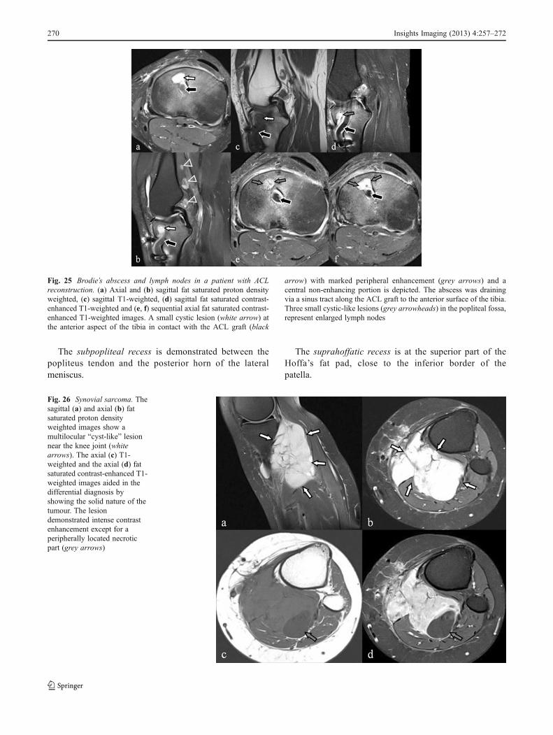
The subpopliteal recess is demonstrated between thepopliteus tendon and the posterior horn of the lateralmeniscus.
The suprahoffatic recess is at the superior part of theHoffa’s fat pad, close to the inferior border of thepatella.
Fig. 25 Brodie’s abscess and lymph nodes in a patient with ACLreconstruction. (a) Axial and (b) sagittal fat saturated proton densityweighted, (c) sagittal T1-weighted, (d) sagittal fat saturated contrast-enhanced T1-weighted and (e, f) sequential axial fat saturated contrast-enhanced T1-weighted images. A small cystic lesion (white arrow) atthe anterior aspect of the tibia in contact with the ACL graft (black
arrow) with marked peripheral enhancement (grey arrows) and acentral non-enhancing portion is depicted. The abscess was drainingvia a sinus tract along the ACL graft to the anterior surface of the tibia.Three small cystic-like lesions (grey arrowheads) in the popliteal fossa,represent enlarged lymph nodes
Fig. 26 Synovial sarcoma. Thesagittal (a) and axial (b) fatsaturated proton densityweighted images show amultilocular “cyst-like” lesionnear the knee joint (whitearrows). The axial (c) T1-weighted and the axial (d) fatsaturated contrast-enhanced T1-weighted images aided in thedifferential diagnosis byshowing the solid nature of thetumour. The lesiondemonstrated intense contrastenhancement except for aperipherally located necroticpart (grey arrows)
270 Insights Imaging (2013) 4:257–272

The infrahoffatic recess lies anterior to the inferior por-tion of the infrapatellar plica (also called ligamentummucosum).
The anterior tibial recess, is a normal capsular recessimmediately anterior to the proximal tibia.
The central synovial recess lies between thepatella/patellar ligaments and the anterior aspect of thefemur.
The parameniscal recess lies just superior and infe-rior to the level of the lateral meniscus in contact withthe lateral femoral and tibial condyle (Figs. 21 and 22).
Other miscellaneous cyst-like lesions
A variety of “cystic” lesions can be encountered in andaround the knee joint that may complicate the differentialdiagnosis even more [1, 2, 6–11]. The most common benignnon-tumoral are the following:
Popliteal artery aneurysms present on MR imageswith variable signal intensity depending on flow charac-teristics and pulse sequences [51, 52]. They are typicallysituated within the popliteal fossa. A laminated MRappearance consistent with multilayered thrombus andoccasionally rim-like calcification may also be demon-strated. The lesion shows continuity with the poplitealartery, which is a hallmark in diagnosis. Popliteal veinvarices are focal dilatations of the popliteal vein [1, 2,6–11]. They present on MR images as lobulated massesin continuity with the popliteal vein. Lymph nodes locat-ed in and around the popliteal fossa may also manifest ascyst-like structures [1, 2, 6–11]. Knowledge of theirlocation as well as of the normal MR appearance ofthe lymph node fatty hillum aids in the differential diag-nosis. Haematomas may simulate a cyst but can bedifferentiated by its signal intensity which depends onblood products’ age (haemoglobin degradation products)[53–55]. Abscesses that can also mimic cysts are associ-ated with infection and inflammation in the surroundingsoft tissues and occasionally underlying osteomyelitis[55–57]. Contrast enhancement is necessary for the cor-rect diagnosis and for unmasking a possible sinus tract(Figs. 23, 24 and 25).
In addition, administration of intravenous contrast isprimarily helpful in the evaluation of soft tissue massesand particularly in differentiating cysts from malignantpseudocystic conditions [9–13]. Solid tumours with cen-tral necrosis, cystic degeneration or myxoid stroma suchas synovial sarcoma, dedifferentiated sarcoma, myxoidliposarcoma, metastases and the benign synovialhaemangioma may have homogeneously high signal onT2-weighted images mimicking a cyst, but they enhanceafter contrast administration contrary to the cysts(Fig. 26).
Conclusion
Benign cystic and “cyst-like” lesions are a common findinginside and around the knee joint. MRI is an excellent meth-od for demonstrating and differentiating these lesions. Aradiologist should be able to identify typical MRI patternsthat contribute in establishing the correct diagnosis and thusguiding specific therapy and avoiding unwarranted interven-tional procedures such as biopsy or arthroscopy.
Conflict of interest None.
Open Access This article is distributed under the terms of the CreativeCommons Attribution License which permits any use, distribution, andreproduction in any medium, provided the original author(s) and thesource are credited.
References
1. Beaman FD, Peterson JJ (2007) MR imaging of cysts, ganglia, andbursae about the knee. Radiol Clin North Am 45:969–982
2. Beaman FD, Peterson JJ (2007) MR imaging of cysts, ganglia, andbursae about the knee. Magn Reson Imaging Clin N Am 15:39–52
3. Ghazikhanian V, Beltran J, Nikac V, Feldman M, Bencardino JT(2012) Tibial tunnel and pretibial cysts following ACL graft re-construction: MR imaging diagnosis. Skeletal Radiol 41:1375–1379
4. Guermazi A, Hayashi D, Roemer FWet al (2010) Cyst-like lesionsof the knee joint and their relation to incident knee pain anddevelopment of radiographic osteoarthritis: the MOST study.Osteoarthr Cartil 18:1386–1392
5. Janzen DL, Peterfy CG, Forbes JR, Tirman PF, Genant HK (1994)Cystic lesions around the knee joint: MR imaging findings. AJRAm J Roentgenol 163:155–161
6. McCarthy CL, McNally EG (2004) The MRI appearance of cysticlesions around the knee. Skeletal Radiol 33:187–209
7. Williams HJ, Davies AM, Allen G, Evans N, Mangham DC (2004)Imaging features of intraosseous ganglia: a report of 45 cases. EurRadiol 14:1761–1769
8. Marra MD, Crema MD, Chung M et al (2008) MRI features ofcystic lesions around the knee. Knee 15:423–438
9. Stacy GS, Kapur A (2011) Mimics of bone and soft tissue neo-plasms. Radiol Clin North Am 49:1261–1286
10. Stein-Wexler R (2009) MR imaging of soft tissue masses in chil-dren. Magn Reson Imaging Clin N Am 17:489–507
11. Subhas N, Bui KL, Sundaram M, Ilaslan H, Recht MP (2009)Incidental tumor and tumor-like lesions around the knee. SeminMusculoskelet Radiol 13:353–370
12. Chung CB, Boucher R, Resnick D (2009) MR imaging of synovialdisorders of the knee. Semin Musculoskelet Radiol 13:303–325
13. Papp DF, Khanna AJ, McCarthy EF, Carrino JA, Farber AJ,Frassica FJ (2007) Magnetic resonance imaging of soft-tissuetumors: determinate and indeterminate lesions. J Bone Joint SurgAm 89(Suppl 3):103–115
14. Jerome D, McKendry R (2000) Synovial cyst of the proximaltibiofibular joint. J Rheumatol 27:1096–1098
15. Mortazavi SM, Farzan M, Asadollahi S (2006) Proximaltibiofibular joint synovial cyst–one pathology with three differentpresentations. Knee Surg Sports Traumatol Arthrosc 14:875–879
16. Pećina HI, Borić I, Pećina TC, Smoljanović T, Pećina M (2008)Double synovial cyst of the proximal tibiofibular joint confirmed
Insights Imaging (2013) 4:257–272 271

by MRI as a cause of the peroneal tunnel syndrome. Acta ChirOrthop Traumatol Cech 75:301–305
17. Battaglia TC, Freilich AM, Diduch DR (2007) An intra-articularknee cyst in a 2-year-old associated with an aberrant anteriorcruciate ligament. Knee Surg Sports Traumatol Arthrosc 15:36–38
18. Krudwig WK, Schulte KK, Heinemann C (2004) Intra-articularganglion cysts of the knee joint: a report of 85 cases and review ofthe literature. Knee Surg Sports Traumatol Arthrosc 12:123–129
19. Ozkur A, Adaletli I, Sirikci A, Kervancioglu R, Bayram M (2005)Hoffa’s recess in the infrapatellar fat pad of the knee on MRimaging. Surg Radiol Anat 27:61–63
20. Yazid Bajuri M, Tan BC, Das S, Hassan S, Subanesh S (2011)Compression neuropathy of the common peroneal nerve secondaryto a ganglion cyst. Clin Ter 162:549–552
21. Rawal A, Ratnam KR, Yin Q, Sinopidis C, Frostick SP (2004)Compression neuropathy of common peroneal nerve caused by anextraneural ganglion: a report of two cases. Microsurgery 24:63–66
22. Başbozkurt M, Hapa O, Demiralp B (2011) Distal femoralintraosseous ganglia: cause or result of a degenerative process:17-year follow-up of a case. Musculoskelet Surg 95:147–150
23. Anderson JJ, Connor GF, Helms CA (2010) New observations onmeniscal cysts. Skeletal Radiol 39:1187–1191
24. De Smet AA, Graf BK, del Rio AM (2011) Association ofparameniscal cysts with underlying meniscal tears as identifiedon MRI and arthroscopy. AJR Am J Roentgenol 196:W180–W186
25. McKnight A, Southgate J, Price A, Ostlere S (2010) Meniscal tearswith displaced fragments: common patterns on magnetic resonanceimaging. Skeletal Radiol 39:279–283
26. Quatman CE, Hettrich CM, Schmitt LC, Spindler KP (2011) Theclinical utility and diagnostic performance of magnetic resonanceimaging for identification of early and advanced knee osteoarthri-tis: a systematic review. Am J Sports Med 39:1557–1568
27. Vande Berg BC, Malghem J, Poilvache P, Maldague B, LecouvetFE (2005) Meniscal tears with fragments displaced in notch andrecesses of knee: MR imaging with arthroscopic comparison.Radiology 234:842–850
28. Wyss JF, Foye PM, Stitik TP (2010) An infected, extruded lateralmeniscal cyst as a cause of knee symptoms. Am J Phys MedRehabil 89:175–176
29. Carrino JA, Blum J, Parellada JA, Schweitzer ME, Morrison WB(2006) MRI of bone marrow edema-like signal in the pathogenesisof subchondral cysts. Osteoarthr Cartil 14:1081–1085
30. Choi JA, Gold GE (2011) MR imaging of articular cartilagephysiology. Magn Reson Imaging Clin N Am 19:249–282
31. Guermazi A, Zaim S, Taouli B, Miaux Y, Peterfy CG, Genant HG(2003) MR findings in knee osteoarthritis. Eur Radiol 13:1370–1386
32. Peterfy CG, Guermazi A, Zaim S et al (2004) Whole-OrganMagnetic Resonance Imaging Score (WORMS) of the knee inosteoarthritis. Osteoarthr Cartil 12:177–190
33. Huétink K, Nelissen RG, Watt I, van Erkel AR, Bloem JL (2010)Localized development of knee osteoarthritis can be predicted fromMR imaging findings a decade earlier. Radiology 256:536–546
34. Hayashi D, Xu L, Roemer FW et al (2012) Detection ofosteophytes and subchondral cysts in the knee with use oftomosynthesis. Radiology 263:206–215
35. Chatra PS (2012) Bursae around the knee joints. Indian J RadiolImaging 22:27–30
36. Nouri H, Ben Hmida F, Ouertatani M et al (2010) Tumour-likelesions of the infrapatellar fat pad. Knee Surg Sports TraumatolArthrosc 18:1391–1394
37. Maheshwari AV, Muro-Cacho CA, Pitcher JD Jr (2007) Pigmentedvillonodular bursitis/diffuse giant cell tumor of the pes anserinebursa: a report of two cases and review of literature. Knee 14:402–407
38. De Maeseneer M, Shahabpour M, Pouders C (2010) MRI spectrumof medial collateral ligament injuries and pitfalls in diagnosis.JBR-BTR 93:97–103
39. De Maeseneer M, Shahabpour M, Vanderdood K, Van Roy F,Osteaux M (2002) Medial meniscocapsular separation: MR imag-ing criteria and diagnostic pitfalls. Eur J Radiol 41:242–252
40. Kramer J, White LM, Recht MP (2009) MR imaging of theextensor mechanism. Semin Musculoskelet Radiol 13:384–401
41. Nandi S, Parker R (2012) Deep medial collateral ligament tearduring knee arthroscopy. J Knee Surg 25:79–81
42. Schein A, Matcuk G, Patel D et al (2012) Structure and function,injury, pathology, and treatment of the medial collateral ligamentof the knee. Emerg Radiol 19:489–498
43. Miller JC, Palmer WE, Goroll AH, Thrall JH, Uppot RN (2009)Anesthetic and steroid injections for musculoskeletal pain. J AmColl Radiol 6:806–808
44. Di Sante L, Paoloni M, Ioppolo F, Dimaggio M, Di Renzo S,Santilli V (2010) Ultrasound-guided aspiration and corticosteroidinjection of Baker’s cysts in knee osteoarthritis: a prospectiveobservational study. Am J Phys Med Rehabil 89:970–975
45. Jose J, Schallert E, Lesniak B (2011) Sonographically guidedtherapeutic injection for primary medial (tibial) collateral bursitis.J Ultrasound Med 30:257–261
46. del Cura JL (2008) Ultrasound-guided therapeutic procedures inthe musculoskeletal system. Curr Probl Diagn Radiol 37:203–218
47. Aydingöz U, Oguz B, Aydingöz O et al (2005) Recesses along theposterior margin of the infrapatellar (Hoffa’s) fat pad: prevalenceand morphology on routine MR imaging of the knee. Eur Radiol15:988–994
48. Fenn S, Datir A, Saifuddin A (2009) Synovial recesses of the knee:MR imaging review of anatomical and pathological features.Skeletal Radiol 38:317–328
49. García-Valtuille R, Abascal F, Cerezal L et al (2002) Anatomy andMR imaging appearances of synovial plicae of the knee.Radiographics 22:775–784
50. Maurel B, Le Corroller T, Cohen M et al (2010) Infrapatellar fatpad: anterior crossroads of the knee. J Radiol 91:841–855
51. Holden A, Merrilees S, Mitchell N, Hill A (2008) Magnetic reso-nance imaging of popliteal artery pathologies. Eur J Radiol67:159–168
52. Wright LB, Matchett WJ, Cruz CP et al (2004) Popliteal arterydisease: diagnosis and treatment. Radiographics 24:467–479
53. Bush CH (2000) The magnetic resonance imaging of musculoskel-etal hemorrhage. Skeletal Radiol 29:1–9
54. Oka K, Yakushiji T, Sato H et al (2008) Ability of diffusion-weighted imaging for the differential diagnosis between chronicexpanding hematomas and malignant soft tissue tumors. J MagnReson Imaging 28:1195–1200
55. Recht MP, Kramer J (2002) MR imaging of the postoperative knee:a pictorial essay. Radiographics 22:765–774
56. Martí-Bonmatí L, Aparisi F, Poyatos C, Vilar J (1993) Brodieabscess: MR imaging appearance in 10 patients. J Magn ResonImaging 3:543–546
57. Soldatos T, Durand DJ, Subhawong TK, Carrino JA, Chhabra A(2012) Magnetic resonance imaging of musculoskeletal infections:systematic diagnostic assessment and key points. Acad Radiol19:1434–1443
272 Insights Imaging (2013) 4:257–272