
Mental Rotation of Objects Retrieved From Memory: A ...
12
Journal of Experimental Psychology: General 2001, Vol. 130, No. 3, 493-504 Copyright 2001 by the American Psychological Association, Inc. 0096-3445/01/$5.00 DOI: 10.1037//0096-3445.130.3.493 Mental Rotation of Objects Retrieved From Memory: A Functional MRI Study of Spatial Processing Marcel Adam Just, Patricia A. Carpenter, Mandy Maguire, Vaibhav Diwadkar, and Stephanie McMains Carnegie Mellon University This functional MRI study examined how people mentally rotate a 3-dimensional object (an alarm clock) that is retrieved from memory and rotated according to a sequence of auditory instructions. We manipulated the geometric properties of the rotation, such as having successive rotation steps around a single axis versus alternating between 2 axes. The latter condition produced much more activation in several areas. Also, the activation in several areas increased with the number of rotation steps. During successive rotations around a single axis, the activation was similar for rotations in the picture plane and rotations in depth. The parietal (but not extrastriate) activation was similar to mental rotation of a visually presented object. The findings indicate that a large-scale cortical network computes different types of spatial information by dynamically drawing on each of its components to a differential, situation-specific degree. Functional neuroimaging provides a new opportunity to exam- ine some of the fundamental characteristics of spatial thinking. Spatial thinking ability has long been studied as a major compo- nent of human intelligence, ranging from Binet's early measure- ments of intelligence to contemporary brain imaging studies. One of the fascinations of spatial thinking is its potential correspon- dence with objects and transformations in the physical world. For example, in mental rotation tasks, which require that an object be represented at a new orientation, questions arise about the three- dimensionality of the object representations and their transforma- tions. This study examined how various types of computational demands affect the activation in a large-scale network of cortical regions that are involved in mental rotation. Research on spatial thinking has attempted to specify the uniquely spatial representations and processes involved, focusing on their geometric properties. For example, one question about the mental rotation process has concerned the effect of the axis of rotation (Shepard & Metzler, 1971), specifically whether rotation in the picture plane is similar to rotation in depth, as it is in the physical world. If the mental representation of the object were only two-dimensional, in the plane of depiction, then the two types of rotation should be different, with the rotation in depth requiring much more processing. The Shepard and Metzler study compared rotation around the z-axis (the axis parallel to the imagined line of sight, such that the object was imagined to rotate in the picture plane) with rotation around the y-axis (a vertical axis around which Marcel Adam Just, Patricia A. Carpenter, Mandy Maguire, Vaibhav Diwadkar, and Stephanie McMains, Center for Cognitive Brain Imaging, Carnegie Mellon University. This work was supported in part by Office of Naval Research Grant N00014-96-1-0322 and National Institute of Mental Health Senior Scien- tist Awards MH-00661 and MH-00662. Correspondence concerning this article should be addressed to Marcel Adam Just, Center for Cognitive Brain Imaging, Psychology Department, Carnegie Mellon University, Pittsburgh, Pennsylvania 15213. Electronic mail may be sent to [email protected]. the object was imagined to be rotating in depth). The behavioral measures generally showed comparable results for the two types of rotation, suggesting that the object representations maintained a three-dimensional structure rather than just a two-dimensional representation that corresponds to the picture plane. The current study also compared various types of rotations, differing in the rotation axis, and sometimes requiring that the object be rotated first around one axis and then around another. The general objec- tive was to discover how various types of geometric properties, such as the uniformity of the rotation axis, affect brain activation in a spatial thinking task such as mental rotation. A second question about mental rotation has concerned the effect of the input modality (e.g., visual vs. haptic). The modality issues have been investigated in behavioral studies that showed that any of several input modalities could engender mental rota- tion, such that response times increased monotonically with the size of the rotation angle (Carpenter & Eisenberg, 1978); however, the input modalities greatly affected the parameters of the behav- ioral response. If the underlying spatial processes are fundamen- tally geometric, then they might engender the same brain activa- tion regardless of input modality. A major innovation of the current study is that all of the image generation and transformation processes occurred without visual input. The participants recalled from memory the object to be rotated, an alarm clock with which they had become familiar. The experimental task was adapted from a venerable test of spatial ability, the Guilford-Zimmerman Spatial Visualization Test (Guil- ford & Zimmerman, 1947-56). In the original test, participants are shown a drawing of an old-fashioned alarm clock and then given visual directions for how to rotate it through a sequence of steps. In the current study, we first familiarized the participants with an old-fashioned alarm clock, with a clock face on the front, various knobs and buttons on the back, and a base at the bottom, as shown in its canonical position in Figure 1. During our task, participants were expected to retrieve their representation of the clock from memory, without visual input, in its canonical position, namely upright and with the clock face facing the viewer. This canonical 493
Transcript of Mental Rotation of Objects Retrieved From Memory: A ...

Journal of Experimental Psychology: General2001, Vol. 130, No. 3, 493-504
Copyright 2001 by the American Psychological Association, Inc.0096-3445/01/$5.00 DOI: 10.1037//0096-3445.130.3.493
Mental Rotation of Objects Retrieved From Memory:A Functional MRI Study of Spatial Processing
Marcel Adam Just, Patricia A. Carpenter, Mandy Maguire, Vaibhav Diwadkar, and Stephanie McMainsCarnegie Mellon University
This functional MRI study examined how people mentally rotate a 3-dimensional object (an alarm clock)that is retrieved from memory and rotated according to a sequence of auditory instructions. Wemanipulated the geometric properties of the rotation, such as having successive rotation steps around asingle axis versus alternating between 2 axes. The latter condition produced much more activation inseveral areas. Also, the activation in several areas increased with the number of rotation steps. Duringsuccessive rotations around a single axis, the activation was similar for rotations in the picture plane androtations in depth. The parietal (but not extrastriate) activation was similar to mental rotation of a visuallypresented object. The findings indicate that a large-scale cortical network computes different types ofspatial information by dynamically drawing on each of its components to a differential, situation-specificdegree.
Functional neuroimaging provides a new opportunity to exam-ine some of the fundamental characteristics of spatial thinking.Spatial thinking ability has long been studied as a major compo-nent of human intelligence, ranging from Binet's early measure-ments of intelligence to contemporary brain imaging studies. Oneof the fascinations of spatial thinking is its potential correspon-dence with objects and transformations in the physical world. Forexample, in mental rotation tasks, which require that an object berepresented at a new orientation, questions arise about the three-dimensionality of the object representations and their transforma-tions. This study examined how various types of computationaldemands affect the activation in a large-scale network of corticalregions that are involved in mental rotation.
Research on spatial thinking has attempted to specify theuniquely spatial representations and processes involved, focusingon their geometric properties. For example, one question about themental rotation process has concerned the effect of the axis ofrotation (Shepard & Metzler, 1971), specifically whether rotationin the picture plane is similar to rotation in depth, as it is in thephysical world. If the mental representation of the object were onlytwo-dimensional, in the plane of depiction, then the two types ofrotation should be different, with the rotation in depth requiringmuch more processing. The Shepard and Metzler study comparedrotation around the z-axis (the axis parallel to the imagined line ofsight, such that the object was imagined to rotate in the pictureplane) with rotation around the y-axis (a vertical axis around which
Marcel Adam Just, Patricia A. Carpenter, Mandy Maguire, VaibhavDiwadkar, and Stephanie McMains, Center for Cognitive Brain Imaging,Carnegie Mellon University.
This work was supported in part by Office of Naval Research GrantN00014-96-1-0322 and National Institute of Mental Health Senior Scien-tist Awards MH-00661 and MH-00662.
Correspondence concerning this article should be addressed to MarcelAdam Just, Center for Cognitive Brain Imaging, Psychology Department,Carnegie Mellon University, Pittsburgh, Pennsylvania 15213. Electronicmail may be sent to [email protected].
the object was imagined to be rotating in depth). The behavioralmeasures generally showed comparable results for the two types ofrotation, suggesting that the object representations maintained athree-dimensional structure rather than just a two-dimensionalrepresentation that corresponds to the picture plane. The currentstudy also compared various types of rotations, differing in therotation axis, and sometimes requiring that the object be rotatedfirst around one axis and then around another. The general objec-tive was to discover how various types of geometric properties,such as the uniformity of the rotation axis, affect brain activationin a spatial thinking task such as mental rotation.
A second question about mental rotation has concerned theeffect of the input modality (e.g., visual vs. haptic). The modalityissues have been investigated in behavioral studies that showedthat any of several input modalities could engender mental rota-tion, such that response times increased monotonically with thesize of the rotation angle (Carpenter & Eisenberg, 1978); however,the input modalities greatly affected the parameters of the behav-ioral response. If the underlying spatial processes are fundamen-tally geometric, then they might engender the same brain activa-tion regardless of input modality.
A major innovation of the current study is that all of the imagegeneration and transformation processes occurred without visualinput. The participants recalled from memory the object to berotated, an alarm clock with which they had become familiar. Theexperimental task was adapted from a venerable test of spatialability, the Guilford-Zimmerman Spatial Visualization Test (Guil-ford & Zimmerman, 1947-56). In the original test, participants areshown a drawing of an old-fashioned alarm clock and then givenvisual directions for how to rotate it through a sequence of steps.In the current study, we first familiarized the participants with anold-fashioned alarm clock, with a clock face on the front, variousknobs and buttons on the back, and a base at the bottom, as shownin its canonical position in Figure 1. During our task, participantswere expected to retrieve their representation of the clock frommemory, without visual input, in its canonical position, namelyupright and with the clock face facing the viewer. This canonical
493

494 JUST, CARPENTER, MAGUIRE, DIWADKAR, AND McMAINS
Figure 1. View of the clock in its canonical, initial position (on left) andhow its orientation should look after mental rotation following the instruc-tion "right."
position was the initial position to which the first step of anysequence of orientation transformations was to be applied. Anotheradaptation was that the rotations to be performed were communi-cated auditorily. For example, participants might be given thespoken instruction "counter," conveying that the most recentlyrepresented orientation of the clock was to be rotated counter-clockwise (i.e., in the picture plane). All transformations were tobe 90°, by convention. This paradigm made it possible to comparethe brain activation with other studies in which the object to bementally rotated is visually presented.
Functional Neuroimaging of Mental Rotation
Spatial thinking not only has distinctive processing characteris-tics in terms of the underlying geometry but is also related toseveral particular parts of the brain. Functional MRI (fMRI) makesit possible to address questions about spatial thinking by measur-ing the activation in various cortical areas while participants men-tally rotate an object. The relative amount of activation in eacharea of a large-scale cortical network indicates not only the com-ponents of the underlying neural system but also the modulation oftheir relative contributions as the computational demands of thetask are varied from condition to condition. For example, one canassess the effect of alternating the rotation axis on the brainactivation in several areas.
The two cortical regions that might be expected to play centralroles in mental rotation are the parietal and inferior temporal areas(i.e., the dorsal and ventral systems, respectively). Several neuro-imaging studies indicate that the parietal areas are implicated inmental rotation, playing a large role in the geometric transforma-tion of the represented object orientation. In a study involving theShepard-Metzler rotation task, fMRI-measured activation wasfound in the left and right parietal regions (Brodmann Areas[BA] 7a and 7b and sometimes BA 40) when a rotation conditionwas compared with a 0° rotation condition (Cohen et al., 1996). APET study found activation in the left posterior-superior parietalcortex during the mental rotation of letters (Alivisatos & Petrides,1997). In two separate parametric studies involving the mentalrotation of Shepard-Metzler figures, the amount of activation inthe left and right intraparietal sulcal region increased linearly withrotation angle (measured in terms of the number of voxels acti-vated above a high fixed threshold), suggesting the amount ofwork that this area did increased with the rotation angle (Carpen-ter, Just, Keller, Eddy, & Thulborn, 1999a). These data suggestthat the amount of activation in the parietal areas that supportvisuospatial processing is related to the amount of computationaldemand.
Parietal involvement has also been found in several event-related potential (ERP) studies of a simpler rotation task in whichparticipants judged whether a letter was normal or mirror imaged(Desrocher, Smith, & Taylor, 1995; Peronnet & Farah, 1989;Wijers, Otten, Feenstra, Mulder, & Mulder, 1989). All three ERPstudies found that as the angular disparity increased, there wasincreasing negativity in the ERP waveforms in the latency range of350-800 ms, particularly in the parietal and occipital leads, whichwas interpreted as reflecting rotation, and lesser but still systematiceffects in central leads (Desrocher et al., 1995; Rosier, Heil, Bajric,Pauls, & Henninghausen, 1995). These studies all implicate theparietal region in some aspect of rotation of an object that isvisually presented.
However, there is more to a mental rotation task than justrotation. In detailed computational models of eye-fixation studiesof mental rotation, Just and Carpenter (1985) proposed that amental rotation of a complex object that is visually presentedentails figural segmentation and rotation of only one segment of acomplex object at a time. Classically, the inferior temporal area isassociated with visual object identification or object-part identifi-cation, so one might expect that area to play a large role inidentifying salient segments of objects to be rotated and evaluated.In their fMRI study, Carpenter et al. (1999a) found that not onlythe parietal but also the inferior temporal areas became activatedduring mental rotation, regions that are primarily associated withthe processes of object and object-part identification. However, inthe absence of visual input in the clock task, one might expect theinferior temporal contribution to a mental rotation task to decrease.More generally, the comparison between the two paradigms per-mits an evaluation of the roles of the dorsal and ventral systems invarious types of mental rotation.
Finally, in a task as complex as clock rotation, the executive andverbal components of the task should engage appropriate corticalareas. The executive components would likely be involved in goalmanagement, namely developing goals and subgoals and keepingtrack of which ones have been satisfied, resulting in activation indorsolateral prefrontal cortex. The verbal components might en-code and rehearse representations of either the state of a clockrepresentation or an action or goal to be pursued, resulting inactivation in inferior frontal areas, posterior superior temporalareas, and inferior parietal areas, particularly in the lefthemisphere.
The main goal of the study was to determine how variousaspects of mental rotation affected the pattern of activation inseveral cortical regions, but there were three specific hypotheses.One hypothesis concerned alternation between two axes of rota-tion. The hypothesis was that a change in rotation axis from onerotation step to the next should be more computationally demand-ing than making successive rotations around the same axis; hence,there should be more brain activation when the rotations alternatedaxes. This hypothesis is based on a proposed model of the process,described further in the Discussion section, which assumes that themental rotation of the clock is performed by reducing an initialthree-dimensional representation to a two-dimensional representa-tion and then applying the orientation transformation. In the caseof alternating axes, a different two-dimensional representationwould have to be generated after each rotation step, namely therepresentation in the plane that is perpendicular to the designated

fMRI OF MENTAL ROTATION 495
rotation axis, requiring extra computational and representationalwork relative to the same-axis rotations.
A second hypothesis was that as the number of successiverotation steps to be applied to a clock increased, the amount ofbrain activation should correspondingly increase. This hypothesisis based on a rotation workload hypothesis, one that has previouslybeen supported in the fMRI study of Carpenter et al. (1999a), inwhich the rotation angle was systematically varied.
A third main interest was the effect of input modality, compar-ing the pattern of cortical activation in mental rotation of retrievedimages with previous studies in which the objects to be rotatedwere visually presented. In addition to the comparison with pre-vious studies, we did a within-subject comparison for three par-ticipants who also performed a mental-rotation task involvingvisually presented three-dimensional objects in the same testingsession. The third hypothesis was that similar areas of the parietallobule would activate in the two types of studies, but there shouldbe dissimilarities in more sensory and perceptual areas.
Method
Participants
The participants contributing fMRI data were 12 right-handed volunteercollege students (7 men and 5 women). Each participant gave signed,informed consent (approved by the University of Pittsburgh and the Car-negie Mellon Institutional Review Boards). Participants were first famil-iarized with an abbreviated behavioral version of the mental rotation task,and a larger group was screened for acceptable accuracy scores. Onlythe 12 participants who made less than 40% error responses in the mostdifficult condition and had fMRI data sufficiently free of head motionartifacts were entered into the analyses.
fMRI Parameters
Images were acquired on a GE Medical Systems 3.0 Tesla scanner usingthe following parameters: TR (repetition time) = 3,000 ms, TE (echotime) = 25 ms, flip angle = 90°, and voxel size = 3.125 x 3.125 X 5 mmwith a 1-mm gap. For 10 of the 12 participants, 14 functional slice imageswere acquired in an oblique-axial plane and were positioned to cover asmuch of the parietal and temporal lobules as possible; the pitch angle of theimages ranged from 9" to 21°. For 2 of the 12 participants, the 14 sliceimages were acquired in a coronal plane and were positioned to cover asmuch of the parietal and temporal lobes as possible. The latter sliceprescription is limiting in that it does not cover the frontal areas. These twosubjects are included in analyses concerning the parietal and temporalregions of interest (ROIs) but not in dorsolateral prefrontal cortex, frontaleye fields, or the inferior frontal gyrus.
Design
The experiment had two independent variables: the axis about which theclock was to be rotated (z-axis only, y-axis only, or z- and y-axes) and thenumber of successive 90° rotation steps that were to be applied to the clock.The latter variable, the number of rotation steps, is referred to as the pathlength variable, and it was either a short path (either two or three rotationsteps) or a long path (four rotation steps). The three levels of the rotationaxis variable and the two levels of the path length variable combined toproduce six conditions or epoch types.
Procedure
To determine how much time to allow for performing various rotations,we ran a pilot study in which the time intervals were under the participants'
control. Rotation commands were presented, and the participants pressed abutton when they had completed the instructed rotation. The durationschosen for the main experiment corresponded to a point that included about75% or more of the self-paced response tunes in the pilot study. Thesedurations were 3.2 s for the first rotation in a problem, 2.2 s for anysubsequent rotations in a single-axis problem, and 5.7 s for any subsequentrotations in a z and y axis problem.
The presentation of each of the various types of problems always startedwith a visual "ready" text lasting 1.5 s, providing the participant an intervalto mentally retrieve or construct an image of the clock hi its canonicalposition, face-forward and upright. The first rotation command was thenauditorally presented, followed by silence, consuming a total of 3.2 s,during which the participant was to perform the first mental rotation.One-word commands indicated the rotation axis and the rotation direction.The command "clock" indicated a 90° clockwise rotation around the z-axis(i.e., the axis parallel to the line of sight, such that the clock was to berotated in the picture plane). "Counter" indicated the corresponding coun-terclockwise rotation. The command "right" indicated a 90° rightwardrotation around the y-axis (the vertical axis), and "left" indicated thecorresponding leftward rotation. In problems with rotations about two axes,the rotations alternated axes from one rotation command to the next. Afterall of the commands had been presented, a picture of the clock in someorientation was presented for 3.5 s, during which time the participantindicated whether the picture correctly depicted the final position of theclock. Approximately 20% of the probes were "different," constructed byapplying the final rotation around the specified axis but in the wrongdirection. Participants were instructed to respond "same" or "different" bypressing one of two hand-held response buttons. Any participant with over40% errors in any condition was excluded from the analysis.
The presentation duration of a problem ranged from 12.6 to 25.3 s,depending on the path length and rotation axis. The problems were pre-sented in groups of the same problem type, composing epochs. To keep thedurations of the six epoch types (conditions) similar at 25 to 30 s, thenumber of problems per epoch type was varied. The short path, one-axis(either z-axis only or y-axis only) epoch consisted of two 3-move problemstotaling 25.2 s. The long path, one-axis (either z-axis only or y-axis only)epoch consisted of two 4-move problems totaling 29.6 s. The short path,two-axis epoch consisted of two 2-move problems totaling 27.8 s. Thelong, two-axis epoch consists of one 4-move problem totaling 25.3 s. Thus,the assessment of activation comparing fewer or more rotation steps peritem compares such duration-equated epochs.
There were four tokens of each of the six types of epochs in a session,presented in four successive unique permutations of the six epoch types.Successive epochs were separated by 6-s rest periods during which indi-viduals fixated an asterisk in the middle of the screen. There were also fourpresentations of 24-s fixation epochs distributed across the session, toprovide a baseline measure of the brain activation.
Data Analysis
The image preprocessing corrected for head motion and signal driftusing procedures and software developed by William Eddy and his col-leagues in the Carnegie Mellon Statistics Department (Eddy, Fitzgerald,Genovese, Mockus, & Noll, 19%). The data analysis focuses on quanti-fying BOLD (blood oxygen level dependenQ-contrast-related changes inthe fMRI-measured signal using a dependent measure that takes intoaccount both the volume of activation and the percentage change in signal,relative to a baseline level (Xiong, Rao, Gao, Woldorff, & Fox, 1998). Datafrom the first 6 s of each epoch were discarded to accommodate the rise ofthe hemodynamic response. For each voxel in the a priori defined ROIs(described below), the distribution of signal in an experimental conditionwas compared with that for the rest condition using a t test with a thresholdof t > 4.5, corresponding to a high Bonferroni-corrected alpha level. Wethen calculated the integral of the percentage change in signal intensity for
496 JUST, CARPENTER, MAGUIRE, DIWADKAR, AND McMAINS
each region in each condition by summing the percentage change in signalintensity relative to the baseline estimate across voxels that reached thecritical threshold in that condition. These data from each of the ROIs werethen submitted to a 3 (z-axis, y-axis, both axes) X 2 (short or long pathlength) X 10 or 12 (participants; 10 only for regions uncovered by coronalslices) repeated measures analysis of variance.
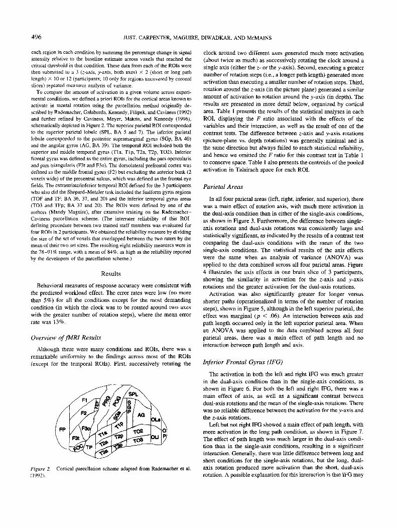
To compare the amount of activation in a given volume across experi-mental conditions, we defined a priori ROIs for the cortical areas known toactivate in mental rotation using the parcellation method originally de-scribed by Rademacher, Galaburda, Kennedy, Filipek, and Caviness (1992)and further refined by Caviness, Meyer, Makris, and Kennedy (1996),schematically depicted in Figure 2. The superior parietal ROI correspondedto the superior parietal lobule (SPL, BA 5 and 7). The inferior parietallobule corresponded to the posterior supramarginal gyrus (SGp, BA 40)and the angular gyrus (AG, BA 39). The temporal ROI included both thesuperior and middle temporal gyrus (Tla, Tip, T2a, T2p, TO2). Inferiorfrontal gyrus was defined as the entire gyrus, including the pars opercularisand pars triangularis (F3t and F3o). The dorsolateral prefrontal cortex wasdefined as the middle frontal gyrus (F2) but excluding the anterior bank (2voxels wide) of the precentral sulcus, which was defined as the frontal eyefields. The extrastriate/inferior temporal ROI defined for the 3 participantswho also did the Shepard-Metzler task included the fusiform gyrus regions(TOP and TF; BA 36, 37, and 20) and the inferior temporal gyrus areas(TO3 and TFp; BA 37 and 20). The ROIs were defined by one of theauthors (Mandy Maguire), after extensive training on the Rademacher-Caviness parcellation scheme. (The interrater reliability of this ROI-defining procedure between two trained staff members was evaluated forfour ROIs in 2 participants. We obtained the reliability measure by dividingthe size of the set of voxels that overlapped between the two raters by themean of their two set sizes. The resulting eight reliability measures were inthe 78-91% range, with a mean of 84%, as high as the reliability reportedby the developers of the parcellation scheme.)
Results
Behavioral measures of response accuracy were consistent withthe predicted workload effect. The error rates were low (no morethan 5%) for all the conditions except for the most demandingcondition (in which the clock was to be rotated around two axeswith the greater number of rotation steps), where the mean errorrate was 13%.
Overview offMRI Results
Although there were many conditions and ROIs, there was aremarkable uniformity to the findings across most of the ROIs(except for the temporal ROIs). First, successively rotating the
Figure 2. Cortical parcellation scheme adapted from Rademacher et al.(1992).
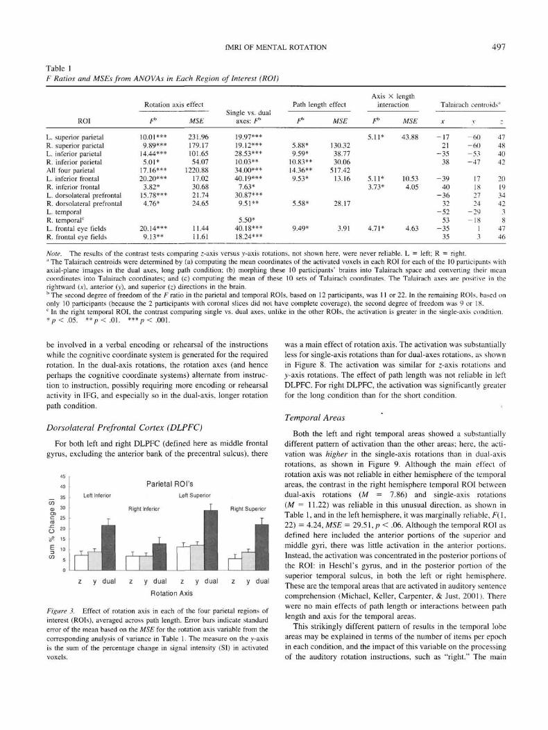
clock around two different axes generated much more activation(about twice as much) as successively rotating the clock around asingle axis (either the z- or the y-axis). Second, executing a greaternumber of rotation steps (i.e., a longer path length) generated moreactivation than executing a smaller number of rotation steps. Third,rotation around the z-axis (in the picture plane) generated a similaramount of activation to rotation around the y-axis (in depth). Theresults are presented in more detail below, organized by corticalarea. Table 1 presents the results of the statistical analyses in eachROI, displaying the F ratio associated with the effects of thevariables and their interaction, as well as the result of one of thecontrast tests. The difference between z-axis and y-axis rotations(picture-plane vs. depth rotations) was generally minimal and inthe same direction but always failed to reach statistical reliability,and hence we omitted the F ratio for this contrast test in Table 1to conserve space. Table 1 also presents the centroids of the pooledactivation in Talairach space for each ROI.
Parietal Areas
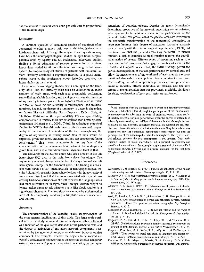
In all four parietal areas (left, right, inferior, and superior), therewas a main effect of rotation axis, with much more activation inthe dual-axis condition than in either of the single-axis conditions,as shown in Figure 3. Furthermore, the difference between single-axis rotations and dual-axis rotations was consistently large andstatistically significant, as indicated by the results of a contrast testcomparing the dual-axis conditions with the mean of the twosingle-axis conditions. The statistical results of the axis effectswere the same when an analysis of variance (ANOVA) wasapplied to the data combined across all four parietal areas. Figure4 illustrates the axis effects in one brain slice of 3 participants,showing the similarity in activation for the z-axis and y-axisrotations and the greater activation for the dual-axis rotations.
Activation was also significantly greater for longer versusshorter paths (operationalized in terms of the number of rotationsteps), shown in Figure 5, although in the left superior parietal, theeffect was marginal (p < .06). An interaction between axis andpath length occurred only in the left superior parietal area. Whenan ANOVA was applied to the data combined across all fourparietal areas, there was a main effect of path length and nointeraction between path length and axis.
Inferior Frontal Gyrus (IFG)
The activation in both the left and right IFG was much greaterin the dual-axis condition than in the single-axis conditions, asshown in Figure 6. For both the left and right IFG, there was amain effect of axis, as well as a significant contrast betweendual-axis rotations and the mean of the single-axis rotations. Therewas no reliable difference between the activation for the y-axis andthe z-axis rotations.
Left but not right IFG showed a main effect of path length, withmore activation in the long path condition, as shown in Figure 7.The effect of path length was much larger in the dual-axis condi-tion than in the single-axis conditions, resulting in a significantinteraction. Generally, there was little difference between long andshort conditions for the single-axis rotations, but the long, dual-axis rotation produced more activation than the short, dual-axisrotation. A possible explanation for this interaction is that IFG may
fMRI OF MENTAL ROTATION 497
Table 1F Ratios and MSEs from ANOVAs in Each Region of Interest (ROI)
ROI
L. superior parietalR. superior parietalL. inferior parietalR. inferior parietalAll four parietalL. inferior frontalR. inferior frontalL. dorsolateral prefrontalR. dorsolateral prefrontalL. temporalR. temporal0
L. frontal eye fieldsR. frontal eye fields
Rotation
F*
10.01***9.89***
14.44***5.01*
17.16***20.20***
3.82*15.78***4.76*
20.14***9.13**
axis effect
MSE
231.96179.17101.6554.07
1220.8817.0230.6821.7424.65
11.4411.61
Single vs. dualaxes: f*
19.97***19.12***28.53***10.03**34.00***40.19***
7.63*30.87***9.51**
5.50*40.18***18.24***
Path length
F»
5.88*9.59*
10.83**14.36**9.53*
5.58*
9.49*
Axis X lengtheffect interaction
MSE F" MSE
5.11* 43.88130.3238.7730.06
517.4213.16 5.11* 10.53
3.73* 4.05
28.17
3.91 4.71* 4.63
Talairach centroids'1
X
-1721
-3538
-3940
-3632
-5253
-3535
V
-60-60-53-47
17182724
-29-18
13
-
47484042
2019344238
4746
Note. The results of the contrast tests comparing z-axis versus y-axis rotations, not shown here, were never reliable. L = left; R = right." The Talairach centroids were determined by (a) computing the mean coordinates of the activated voxels in each ROI for each of the 10 participants withaxial-plane images in the dual axes, long path condition; (b) morphing these 10 participants' brains into Talairach space and converting their meancoordinates into Talairach coordinates; and (c) computing the mean of these 10 sets of Talairach coordinates. The Talairach axes are positive in therightward (x), anterior (y), and superior (z) directions in the brain.b The second degree of freedom of the F ratio in the parietal and temporal ROIs, based on 12 participants, was 11 or 22. In the remaining ROIs, based ononly 10 participants (because the 2 participants with coronal slices did not have complete coverage), the second degree of freedom was 9 or 18.c In the right temporal ROI, the contrast comparing single vs. dual axes, unlike in the other ROIs, the activation is greater in the single-axis condition.*p<.05. ** p < . 01. ***/><.001.
be involved in a verbal encoding or rehearsal of the instructionswhile the cognitive coordinate system is generated for the requiredrotation. In the dual-axis rotations, the rotation axes (and henceperhaps the cognitive coordinate systems) alternate from instruc-tion to instruction, possibly requiring more encoding or rehearsalactivity in IFG, and especially so in the dual-axis, longer rotationpath condition.
Dorsolateral Prefrontal Cortex (DLPFC)
For both left and right DLPFC (defined here as middle frontalgyrus, excluding the anterior bank of the precentral sulcus), there
Parietal ROI'sLeft Inferior Left Superior
Right Inferior tnlm Right Superior
45
40
_ 35C/30) 30en<= 25m
0 20
S? 15
1 10OT 5
0
z y dual z y dual z y dual z y dual
Rotation Axis
Figure 3. Effect of rotation axis in each of the four parietal regions ofinterest (ROIs), averaged across path length. Error bars indicate standarderror of the mean based on the MSE for the rotation axis variable from thecorresponding analysis of variance in Table 1. The measure on the y-axisis the sum of the percentage change in signal intensity (SI) in activatedvoxels.
was a main effect of rotation axis. The activation was substantiallyless for single-axis rotations than for dual-axes rotations, as shownin Figure 8. The activation was similar for z-axis rotations and>'-axis rotations. The effect of path length was not reliable in leftDLPFC. For right DLPFC, the activation was significantly greaterfor the long condition than for the short condition.
Temporal Areas
Both the left and right temporal areas showed a substantiallydifferent pattern of activation than the other areas; here, the acti-vation was higher in the single-axis rotations than in dual-axisrotations, as shown in Figure 9. Although the main effect ofrotation axis was not reliable in either hemisphere of the temporalareas, the contrast in the right hemisphere temporal ROI betweendual-axis rotations (M = 7.86) and single-axis rotations(M = 11.22) was reliable in this unusual direction, as shown inTable 1, and in the left hemisphere, it was marginally reliable, F(\,22) = 4.24, MSE = 29.51, p < .06. Although the temporal ROI asdefined here included the anterior portions of the superior andmiddle gyri, there was little activation in the anterior portions.Instead, the activation was concentrated in the posterior portions ofthe ROI: in Heschl's gyrus, and in the posterior portion of thesuperior temporal sulcus, in both the left or right hemisphere.These are the temporal areas that are activated in auditory sentencecomprehension (Michael, Keller, Carpenter, & Just, 2001). Therewere no main effects of path length or interactions between pathlength and axis for the temporal areas.
This strikingly different pattern of results in the temporal lobeareas may be explained in terms of the number of items per epochin each condition, and the impact of this variable on the processingof the auditory rotation instructions, such as "right." The main

498 JUST, CARPENTER, MAGUIRE, DIWADKAR, AND McMAINS
z-axis y-axis dual-axes
Figure 4. Activation in the most active parietal slice of 3 representative participants (one row per participant),shown for rotation around the z-axis, y-axis, and alternating between the y- and z-axes. The activated voxels areshown in red, superimposed on the structural image of each participant's own brain. The green outline shows theboundary of the superior parietal region of interest as it intersects this slice. The amount of activation is similarfor z-axis and y-axis rotations but substantially greater for dual-axis rotations.
sense in which the dual-axis conditions could be construed as lessdemanding is that they contained only four rotation steps (hence,rotation instructions) per epoch, whereas the single-axis conditionsentailed six or eight steps per epoch. It is likely that the greateractivation in the temporal lobe areas in the single-axis rotation
conditions reflects the perceptual and linguistic processing of theauditory rotation instructions. However, this explanation must befurther elaborated to account for the greater activation in the rightthan in the left temporal area. During the auditory processing oflanguage, there is typically more activation in the left than in the

fMRI OF MENTAL ROTATION 499
Parietal ROI's
Left Superior
Right Superior
Short Long Short Long Short Long
Rotation Path Length
Short Long
Figure 5. Path length effect in each of the four parietal regions of interest(ROIs), averaged across rotation axes. Error bars indicate standard error ofthe mean for this variable based on the MSE. The measure on the y-axis isthe sum of the percentage change in signal intensity (SI) in activatedvoxels.
right temporal area (Schlosser, Aoyagi, Fulbright, Gore, & Mc-Carthy, 1998). The right dominance of the obtained temporalactivation probably entails other levels of processing besides thesensory, perceptual, and linguistic. The right dominance mightreflect the spatial referent of the linguistic content. The righttemporal area may be more involved when there is a mapping to bemade from the language to some spatial representation. When thereferent is spatial, then the relative activation in right temporalareas has been observed to be greater than otherwise, even in thecase of visually presented sentences (Carpenter, Just, Keller, Eddy,& Thulborn, 1999b).
This distinction between the response of the temporal areas andthe other areas reflects the relatively greater involvement of thetemporal areas in the language-related processing versus the otherareas' relatively greater involvement in the spatial processing.More generally, the results show the differential sensitivity ofdifferent parts of a large-scale cortical network to the demandsimposed by different types of computations.
Inferior Frontal Gyrus
y dual dualRotation Axis
Figure 6. Effect of rotation axis in left and right inferior frontal gyrus,averaged across path length. Error bars indicate standard error of the meanfor this variable based on the MSE. The measure on the y-axis is the sumof the percentage change in signal intensity (SI) in activated voxels.
Inferior Frontal Gyrus
RightHemisphere
Short Long Short Long
Rotation Path Length
Figure 7. Path length effect in left and right inferior frontal gyrus,averaged across rotation axes. Error bars indicate standard error of themean for this variable based on the MSE. The measure on the y-axis is thesum of the percentage change in signal intensity (SI) in activated voxels.
Frontal Eye Fields
Recall that during the mental rotation, nothing was being dis-played on the screen, so the frontal eye fields are unlikely to becontrolling eye movements, although they could well be involvedin controlling spatial attention within an internal image space.Earlier research has shown that the control of attention within theexternal visual environment, even in the absence of eye move-ments, produces activation in the frontal eye fields (Corbetta et al.,1998). Both left and right frontal eye fields showed a main effectof rotation axis. Furthermore, the average activation for rotationsin a single axis was significantly lower than dual-axis rotations.There was no reliable difference between z-axis and y-axis rota-tions in these areas.
In the left but not the right frontal eye fields there was a maineffect of path length (with the long conditions producing moreactivation than the short conditions), as well as an interactionbetween path length and axis,
Laterality
The laterality effects in the clock task were rather systematic. Ingeneral, the amount of activation for most areas (except temporal)
Dorsolateral Prefrontal Cortex
Right
Figure 8. Effect of rotation axis in left and right dorsolateral prefrontalcortex, averaged across path length. Error bars indicate standard error ofthe mean for this variable based on the MSE. The measure on the y-axis isthe sum of the percentage change in signal intensity (SI) in activatedvoxels.

500 JUST, CARPENTER, MAGUIRE, DIWADKAR, AND McMAINS
21
18
W 15tocn
§«-C
" 9
E 6
CO3
0
Temporal ROILeft Right
Hemisphere Hemisphere
z y dual z y dual
Rotation Axis
Figure 9. Effect of rotation axis in left and right superior and middletemporal area regions of interest (ROIs), averaged across path length. Errorbars indicate standard error of the mean for this variable based on the MSB.The measure on the y-axis is the sum of the percentage change in signalintensity (SI) in activated voxels.
was greater on the left than on the right, particularly in some of themore demanding conditions. For example, in the inferior parietalROI, the activation was particularly large in the left hemisphere inthe dual-axis condition; there was a significant interaction betweenrotation axis and hemisphere for this ROI, F(2, 22) = 3.46,MSE = 70.10, p < .05. This effect is shown in Figure 3. A similarform of interaction occurred in the superior parietal ROI but wasnot reliable. The left dominance in the areas associated with spatialprocessing is similar to previous neuroimaging findings reportedfor an imagery task (D'Esposito et al., 1997). However, a previousstudy of mental rotation in the Shepard-Metzler task found that theright parietal area had slightly more fMRI-measured activation(Carpenter et al., 1999b). It may be that the difference in inputmodality between the Shepard-Metzler rotation and clock rotationtasks accounts for the small but reliable hemispheric differencesbeing in opposite directions.
Another example of a hemisphere by complexity interactionoccurred in the inferior frontal gyri. This area showed an interac-tion between path length and laterality, F(l, 9) = 11.57,MSE = 2.36, p < .01. The activation in left IFG (Broca's area)was particularly large for the long path problems, as shown inFigure 7.
The temporal ROI was unlike the others in that it showed a maineffect of hemisphere with the right temporal ROI having greateractivation, F(l, 11) = 7.65, MSE = 105.42, p < .05, as shown inFigure 9. There was also an interaction between length, axis, andhemisphere, F(2, 22) = 5.63, MSE = 15.24, p < .05.
Individual Differences
Although the sample size is small for examining individualdifferences, it nevertheless yielded results that are consistent withrecent fMRI individual-difference findings (Reichle, Carpenter, &Just, 2000). In that study, participants with poorer verbal skills hadlarger increments in activation volume in left inferior frontal gyrus(L. IFG) when using a verbal strategy as compared with a visualstrategy. Verbal ability was measured in that study and in thecurrent one with the Daneman and Carpenter (1980) Reading SpanTest. In the current clock rotation task, the scores on the Reading
Span Test were also negatively related to the brain activation in L.IFG and the left inferior parietal area, namely the areas implicatedin verbal rehearsal (Awh et al., 1996). The amount of brainactivation was measured not as the absolute activation volume butas the number of additional voxels activated in the most demand-ing condition (dual-axis, long path) compared with the least de-manding condition (single-axis, short path), that is, the slope of theactivation as a function of the computational demand. The corre-lation between Reading Span Test scores and the activation slopesin L. IFG and left inferior parietal was —.70 and —.62, respec-tively (p < .05 for both correlations; n = 10 and 12, respectively).This pattern of correlations implies, quite plausibly, that people oflower verbal ability rehearse more. The correlations betweenvisual-spatial ability (measured here with the Paper Folding Test;French, Ekstrom, & Price, 1963) and the activation slopes in L.IFG and left inferior parietal were negative, as expected, but theywere smaller and not reliable. The individual difference findingssuggest that the role of verbal rehearsal in this mental rotation taskincreases with decreasing verbal ability.
The Effect of Input Modality on Activationin Mental Rotation
The location of the brain activation in the clock rotation taskwas compared with the activation in the Shepard-Metzler task forthe three participants with data of appropriate technical quality.The main goal was to determine whether the very same areas of theparietal lobule were activated for both tasks, despite the differencein the source or modality of the object to be rotated. The dataanalytic procedures for the two tasks were very similar, and thethreshold used to define a voxel as "activated" was the same. Thecomparisons were between the more demanding conditions of eachexperiment: the dual-axis, longer-rotation conditions for clockrotation and the 80°-rotation condition in Shepard-Metzler, con-ditions that produced approximately similar amounts of activationin the parietal areas.
To determine whether the two tasks activated precisely thesame 3.125 X 3.125 X 5 mm voxels, we compared the number ofsuch overlapping voxels to the total number of voxels activated(within each participant). The mean overlap in activated voxels inthe left parietal ROI between the two tasks was .18 (i.e., thenumber of voxels that activated in both experiments divided by themean of the number activated in the two tasks). In the right parietalROI, this measure was .21. Figure 10 illustrates this overlap indetail for one slice of one participant. In addition to countingoverlap in precisely the same voxels, we also counted whether atleast one of the 18 voxels adjacent to an activated voxel in theclock task was also activated in the Shepard-Metzler task (adja-cent was defined in terms of a voxel's center being less than 6 mmfrom the center of the activated voxel). The adjacency measureallowed for a possible slight change in the locus of activation or inthe participant's head position between the two tasks. When theoverlap measure additionally counted whether at least one adjacentvoxel was activated, then the mean overlap measure in the left andright parietal ROIs was .62 and .58, respectively. These findingsindicate a fairly close topographical similarity in the activation inthe parietal areas between mental rotation of visually presented

fMRI OF MENTAL ROTATION 501
Figure 10. Comparison of activation in the clock rotation task andShepard-Metzler rotation in one slice of 1 participant. Overlap (voxelsactivated to threshold in both studies) is shown in red. Voxels activatedonly in the clock study are shown in yellow. Voxels activated only in theShepard-Metzler rotation are shown in blue. The green outline shows theboundary of the superior parietal region of interest as it intersects that slice.
objects and mental rotation of mentally retrieved objects, down tothe level of individual voxels.1
Another cortical area worthy of comparison across the two tasksis the extrastriate cortex, the area between the primary visualregion and the inferior temporal regions. The defined ROI includesthe posterior portions of the inferior temporal lobule and thefusiform gyrus. Mellet, Petit, Mazoyer, Denis, and Tzourio (1998)cited a number of studies that reported activation in such areaswhen participants were performing mental imagery tasks in theabsence of visual input. (It is important to note in the context ofthis particular comparison that the clock task did entail some visualstimulation, not as input to the rotation, but in the form of theprobe stimulus.) There was some reliable activation in the extra-striate area in the clock task (a mean total of 4.7 voxels activatedfor left and right extrastriate combined in the hardest condition),but the activation was small in absolute volume and small relativeto the volume in Shepard-Metzler (a mean total of 27.3 voxelsactivated in both hemispheres in the hardest condition). A largerstudy of the Shepard-Metzler task (Carpenter et al., 1999a) ob-tained a similar amount of activation in this region and found theactivation volume to be modulated (but not monotonically) byrotation angle. So the retrieval and manipulation of a visual imagewithout visual input may produce some activation in extrastriatecortex, but it is a very small amount.
Discussion
The main conclusions from this study are that the mental rota-tion of three-dimensional objects does not occur in a true three-dimensional space; that the rotation processing is substantiallysubserved by the parietal lobules bilaterally; and that the parietalcontribution is similar regardless of input modality, whereas theextrastriate contribution is very small in the absence of visualinput, but is substantial when the rotation operates on visual input.DLPFC, left prefrontal, and left inferior parietal may contribute tothe goal management and rehearsal aspects of the task. Besidesproviding these broad conclusions, the results also indicated somefiner grain characteristics of mental rotation that were previouslyunknown.
Cognitive Processing of Three-Dimensionality
Given that the world is three-dimensional, cognitive represen-tations and transformations of physical objects might be expectedto routinely incorporate their three-dimensional characteristics(Attneave, 1972; Shepard, 1981, 1984). However, several otherstudies indicate that there is an extra cost associated with repre-senting three- as opposed to two-dimensional space. Imagining themovement of an object in a mentally generated array as signaledby visual or auditory cues (e.g., "up," "left") is easier in animagined two-dimensional than three-dimensional array (Diwad-kar, Carpenter, & Just, 2000; Kerr, 1987, 1993). Studies of theinterpretation of visual form also indicate that the three-dimensional interpretation of drawings can be effortful, and itsease is affected by cues such as shading (Hemenway & Palmer,1978) and the number of line segments or angles in the two-dimensional rendition of the space (Attneave & Frost, 1969; Hoch-berg & Brooks, 1960). Imagining the rotation of the three-dimensional object around an arbitrary axis is extremely errorful(Hinton, 1979; Pani, 1993). Such studies suggest that the cognitiveinterpretation of three-dimensional space requires more computa-tion and maintenance than two-dimensional interpretations (Rock.1983; Roth & Kosslyn, 1988). In a task requiring the mentalrotation of a cube containing a single letter or numeral on eachface, the verbal reports and eye fixations of participants who werelow in spatial ability indicated that they operated on only one cubeface at a time when the item required dual-axis rotation (Just &
1 To help to interpret these values of overlap measures, we also com-puted the degree of activation overlap between the first and second half ofthe clock experiment for these 3 participants. We made the number ofobservations in each half comparable with the number used in the intertaskcomparisons by computing the overlap between the two halves of a new"condition" that consisted of the two hardest conditions in the clock study.The proportion of identical voxels activated in the left and right parietalROIs. respectively, was .33 and .38. This degree of identical topographicoverlap of activation between the two halves of the clock rotation exper-iment is almost twice as high as that between the clock rotation and theShepard-Metzler task. When the overlap measure between the two halvesof the clock task additionally counted whether at least one adjacent voxelwas activated, overlap measures in the left and right parietal ROIs were .71and .55, respectively. This degree of identical or adjacent topographicoverlap of activation between the two halves of the clock rotation exper-iment is similar to that between the clock rotation and the Shepard-Metzlertask.

502 JUST, CARPENTER, MAGUIRE, DIWADKAR, AND McMAINS
Carpenter, 1985). The general conclusion is that the mind can dothe three-dimensional work, but it requires extra computation. Themental three-dimensionality in rotation appears to be provided asit is needed and not as a default condition.
This general conclusion can be illustrated in terms of the effectof alternating rotation axes in the clock task. We briefly describehere a theoretical account of the effect. We propose that the mentalrotation is performed by starting with a three-dimensional repre-sentation of the object and then reducing the representation to atwo-dimensional projection in the relevant plane before rotating it.This may occur because of limits on the complexity of a repre-sentation to which a rotation transformation can be applied. Asimilar limitation appeared in a study of mental rotation of three-dimensional blocks with an alphabetic character on each face,where most participants reported rotating only one cube face at atime (Just & Carpenter, 1985). The limitation was manifested inthe verbal protocols of the participants and was implemented in thecomputational model. In the case of single-axis clock rotations,this reduction to two dimensions needs to be performed only once,and then all the subsequent rotations can be applied to the two-dimensional representation. This account is consistent with thefinding that in the self-paced norming study, participants tooklonger on the first rotation step than on subsequent rotation steps.In the condition that requires alternation of rotation axes, eachsubsequent rotation after the first requires that the three-dimensional representation be regenerated from the two-dimensional projection that was the outcome of the previousrotation. Then, the alternate two-dimensional projection must begenerated before the next rotation is applied. The proposed algo-rithm indicates both a cognitive limitation on spatial processingcapacity and a method for circumventing the limitation.
The fMRI results showing much greater activation (greater by atleast a factor of 2) in the dual-axis condition indicate how the extramental work engendered by alternating the rotation axes affects anetwork of cortical areas, including the parietal areas, but alsoextending to Broca's area and DLPFC. The added "spatial" workrequires the added participation of language and executive areas,reflecting a systemwide demand on extra resources. The extrawork performed by the nonparietal areas in the dual-axis condi-tions may include DLPFC's keeping track of whether variousgoals and subgoals are achieved, and Broca's area generating andrehearsing verballike expressions of intermediate states and goals.Any rehearsal of verbal representations could well involve phono-logical storage in the area of the angular gyrus, which is includedin the inferior parietal ROI definition. Thus, when mental rotationis made difficult in this way, the difficulty is propagated wellbeyond the areas and processes that transform representedorientations.
Relation of Perception and Mental Processing
A classic issue in visual thinking concerns the degree of simi-larity (in terms of common neural substrate and algorithms) be-tween perceptual processes and their purely conceptual counter-parts (Finke, 1980; Kosslyn & Koenig, 1992). As the underlyingprocessing becomes better understood, the similarities and differ-ences become clearer. The results of this and other neuroimagingstudies of mental rotation indicate that there are multiple brain
areas involved, so evaluating the similarity in neural .substrateentails a multidimensional comparison.
The results comparing the clock comparison task with theShepard-Metzler task show remarkable similarity in the largeamount of parietal activation, as well as in the precise overlap ofactivation locations within the superior parietal areas. It seemslikely that the parietal area underlies the geometric processing ofobject orientation regardless of input modality. At the same time,the comparison between the two tasks illustrates that the percep-tual processing beyond the primary sensory cortex, namely in theextrastriate cortex, is very different between the two tasks. Therewas enormously less activation in this area in the clock than in theShepard-Metzler task, indicating that the activation there is notpart of the geometric transformation itself. Nevertheless, the largeamount of extrastriate activation in Shepard-Metzler varies withrotation angle (Carpenter et al., 1999a), so the processing there issensitive to the two objects' relative orientations. The comparisonof the two tasks contributes to an understanding of what the dorsaland ventral streams do, as well as their interplay.
The specific hypothesis that mental imagery activates primaryvisual cortex in the absence of visual input could not be evaluatedin this study because the probe was presented visually.
Similarity of z- and y-Axis Rotations
The finding of similar amounts of activation throughout theactivated cortical network for z- and y-axis rotations suggests thatparticipants are initially representing the clock as a three-dimensional object. The alternative possibility, which is ruled outby these data, is that the clock was initially represented as atwo-dimensional object as projected in the picture plane. Had thisalternative been correct, then there would have been extra com-putations needed (and hence, more activation) for rotations indepth (around the y-axis) than in the picture plane (around thez-axis). The findings thus suggest that the clock was initially beingrepresented as a three-dimensional object.
Consistent with this view, the rate for rotating Shepard-Metzlerfigures in depth in behavioral studies is similar to the rotation ratein the picture plane (Shepard & Metzler, 1971), and higher orderapparent motion of complex figures is as easily realized in three-dimensional as in two-dimensional space (Shepard & Judd, 1976).
Path Length
The amount of activation in many areas increased with thenumber of 90° rotation steps made. The results confirm that thecomplexity of the mental work done during an epoch is a largedeterminant of the amount of activation, even when the epochdurations are equated. Having to rotate a clock through a longerseries of steps imposes extra burdens of storing additional inter-mediate products and discriminating among them. The generalfinding of more activation for a greater number of rotation steps ofa fixed magnitude extends the previous finding of an approxi-mately linear increase in parietal activation obtained for singlerotation steps of increasing magnitude from 0° to 120°, in 40° steps(Carpenter et al., 1999a). Mental rotation takes time proportionalto the rotation angle (as behavioral studies have long indicated);the newer results indicate that not just the duration of the process
fMRI OF MENTAL ROTATION 503
but the amount of mental work done per unit time is proportionalto the rotation angle.
Laterality
A common question in behavioral studies of cognition oftenconcerned whether a given task was a right-hemisphere or aleft-hemisphere task. Although the origin of such questions mayhave been the neuropsychological studies on split-brain surgicalpatients done by Sperry and his colleagues, behavioral studiesfinding a 40-ms advantage of sensory presentation to a givenhemisphere tended to attribute the task processing to that hemi-sphere. Neuropsychological studies of patients with unilateral le-sions similarly attributed a cognitive function to a given hemi-sphere (namely, the hemisphere whose lesioning produced thelarger deficit in the function).
Functional neuroimaging studies cast a new light on the later-ality issue. First, the laterality issue must be assessed in an entirenetwork of brain areas, with each area presumably performingsome distinguishable function, and the degree or even the directionof asymmetry between pairs of homologous areas is often differentin different areas. So the laterality is multiregional and multide-termined. Second, the degree of asymmetry in a task is dependenton factors like task difficulty (Just, Carpenter, Keller, Eddy, &Thulborn, 1996) and on the input modality. For example, readingcomprehension is reliably more left-lateralized than listening com-prehension (Michael et al., 2001). Third, the ubiquitous empiricalfinding in fMRI is that although there is often a systematic asym-metry in the amount of activation of the two hemispheres, thedegree of asymmetry is usually much smaller than would beexpected, given that focal, unilateral lesions can produce profoundimpairments.2 Thus, lateral asymmetry is just one facet of thecharacterization of the large-scale brain network that underpins agiven task, and it is a multidimensional, dynamic facet at that.
The activation in the current study was often greater in the lefthemisphere ROI than in the right hemisphere homologue. Theasymmetry was not always reliable, but it always favored the lefthemisphere, except for the temporal areas. The finding is consis-tent with Farah's (1984) meta-analysis of neuropsychological re-sults linking left posterior hemisphere lesions with image retrievalimpairment. We found that the areas associated with spatial pro-cessing had more activation on the left, whereas the language areashad more activation on the right. Such findings illustrate why it nolonger makes sense to ask whether a task like clock rotation is aright-hemisphere task. The true situation can now be understood inmost of its complexity, rendering a simplistic answer inaccurateand unuseful.
Summary
The characterization of the laterality results are prototypical ofthe more general implications of this study. The large-scale corti-cal network underlying mental rotation is dynamically configuredas a function of the qualitative demands of the particular task, andthe degree of activation of any given network component is de-termined by the amount of computational demand imposed on thatcomponent. For example, whether the objects to be rotated arevisually presented or not determines whether the inferior temporal/extrastriate areas will play a major role in operating on the repre-
sentations of complex objects. Despite the many dynamicallydetermined properties of the network underlying mental rotation,what appears to be relatively stable is the participation of theparietal lobules. We presume that the parietal areas are involved inthe geometric transformation of the represented orientation, inlarge part because their degree of activation increases approxi-mately linearly with the rotation angle (Carpenter et al., 1999a). Atthe same time that the parietal areas may be central to mentalrotation, a task as complex as clock rotation requires the coordi-nated action of several different types of processes, such as stra-tegic and verbal processes that engage a number of areas in thefrontal and temporal lobes. Thus, the fMRI findings permit thepartial decomposition of the task performance by cortical area andallow the measurement of the workload of each area as the com-putational demands are manipulated from condition to condition.The resulting partial decomposition provides a more precise ac-count of modality effects, individual differences, and lateralityeffects in mental rotation than was previously available, yielding afar richer explanation of how such tasks are performed.
2 One inference from the combination of fMRI and neuropsychologicalfindings on laterality is that although the participation of the "subordinate"hemisphere can be substantial in terms of brain activation, it may not beabsolutely essential for task performance when the degree of difficulty isrelatively undemanding. An additional inference is that although the twohemispheres may normally coperform the task, one hemisphere can exer-cise control over the other, and a lesion to the controlling hemisphere maydisable not only the controlling hemisphere's participation but also theparticipation of the undamaged, controlled hemisphere. This type of con-trol relation between the two hemispheres is not apparent from brainimaging studies of intact individuals, but a surgical intervention canprovide relevant evidence. For example, surgical removal of a lesioned lefthemisphere allowed a 9-year-old to acquire language for the first time(Vargha-Khadem et al., 1997).
References
Alivisatos, B., & Petrides, M. (1997). Functional activation of the humanbrain during mental rotation. Neuropsychologia, 35, 111-118.
Attneave, F. (1972). Representation of physical space. In A. W. Mellon &E. Martin (Eds.), Coding processes in human memory (pp. 283-306).Washington, DC: Winston.
Attneave, F., & Frost, R. (1969). The determination of perceived tridimen-sional orientation by minimum criteria. Perception & Psychophysics, 6,391-396.
Awh, E., Jonides, J., Smith, E. E., Schumacher, E. H., Koeppe, R. A., &Katz, S. (1996). Dissociation of storage and rehearsal in verbal workingmemory: Evidence from positron emission tomography. PsychologicalScience, 7, 25-31.
Carpenter, P. A., & Eisenberg, P. (1978). Mental rotation and the frame ofreference in blind and sighted individuals. Perception & Psychophys-ics, 23, 117-124.
Carpenter, P. A., Just, M. A., Keller, T., Eddy, W. F., & Thulborn, K. R.(1999a). Graded functional activation in the visuospatial system with theamount of task demand. Journal of Cognitive Neuroscience, 11, 9-24.
Carpenter, P. A., Just, M. A., Keller, T. A., Eddy, W. F., & Thulborn, K. R.(1999b). Time course of fMRI-activation in language and spatial net-works during sentence comprehension. Neurolmage, 10, 216-224.
Caviness, V. S., Jr., Meyer, J., Makris, N., & Kennedy, D. N. (1996).MRI-based topographic parcellation of human neocortex: An anatomi-

504 JUST, CARPENTER, MAGUIRE, DIWADKAR, AND McMAINS
cally specified method with estimate of reliability. Journal of CognitiveNeuroscience, 8, 566-587.
Cohen, M. S., Kosslyn, S. M, Breiter, H. C., DiGirolamo, G. J., Thomp-son, W. L., Anderson, A. K., Bookheimer, S. Y., Rosen, B. R., &Belliveau, J. W. (1996). Changes in cortical activity during mentalrotation: A mapping study using functional MRI. Brain, 119, 89-100.
Corbetta, M., Akbudak, E., Conturo, T. E., Snyder, A. Z., Ollinger, J. M.,Drury, H. A., Linenweber, M. R., Petersen, S. E., Raichle, M. E., VanEssen, D. C., & Shulman, G. L. (1998). A common network of func-tional areas for attention and eye movements. Neuron, 21, 761-773.
Daneman, M., & Carpenter, P. A. (1980). Individual differences in workingmemory and reading. Journal of Verbal learning and Verbal Behav-ior, 19, 450-466.
D'Esposito, M., Detre, J. A., Aguirre, G. K., Stallcup, M., Alsop, D. C.,Tippet, L. J., & Farah, M. J. (1997). A functional MRI study of mentalimage generation. Neuropsychologia, 35, 725-730.
Desrocher, M. E., Smith, M. L., & Taylor, M. J. (1995). Stimulus and sexdifferences in performance of mental rotation: Evidence from event-related potentials. Brain and Cognition, 28, 14—38.
Diwadkar, V. A., Carpenter, P. A., & Just, M. A. (2000). Collaborativeactivity between parietal and dorso-lateral prefrontal cortex in dynamicspatial working memory revealed by fMRI. Neurolmage, 12, 85-99.
Eddy, W. F., Fitzgerald, M., Genovese, C. R., Mockus, A., & Noll, D. C.(1996). Functional imaging analysis software—computational olio. InA. Prat (Ed.), Proceedings in computational statistics (pp. 39-49).Heidelberg, Germany: Physica-Verlag.
Farah, M. J. (1984). The neurological basis of mental imagery: A compo-nential analysis. Cognition, 18, 1-27.
Finke, R. A. (1980). Levels of equivalence in imagery and perception.Psychological Review, 87, 113-132.
French, J. W., Ekstrom, R. B., & Price, L. A. (1963). Manual for a kit ofreference tests of cognitive factors. Princeton, NJ: Educational TestingService.
Guilford, J. P., & Zimmerman, W. S. (1947). The Guilford-ZimmermanAptitude Survey: Manual of instructions and interpretations. Palo Alto,CA: Sheridan Psychological Services.
Hemenway, K., & Palmer, S. E. (1978). Organizational factors in perceiveddimensionality. Journal of Experimental Psychology: Human Percep-tion and Performance, 4, 388-396.
Hinton, G. (1979). Some demonstrations of the effects of structural de-scriptions in mental imagery. Cognitive Science, 3, 231-250.
Hochberg, J., & Brooks, V. (1960). The psychophysics of form:Reversible-perspective drawings of spatial objects. American Journal ofPsychology, 73, 337-354.
Just, M. A., & Carpenter, P. A. (1985). Cognitive coordinate systems:Accounts of mental rotation and individual differences in spatial ability.Psychological Review, 92, 137-172.
Just, M. A., Carpenter, P. A., Keller, T. A., Eddy, W. F., & Thulborn, K. R.(1996). Brain activation modulated by sentence comprehension. Science,274, 114-116.
Kerr, N. H. (1987). Locational representation in imagery: The third dimen-sion. Memory & Cognition, 15, 521-530.
Kerr, N. H. (1993). Rate of imagery processing in two versus threedimensions. Memory & Cognition, 21, 467-476.
Kosslyn, S. M., & Koenig, O. (1992). Wet mind: The new cognitiveneuroscience. New York: Free Press.
Mellet, E., Petit, L., Mazoyer, B., Denis, M., & Tzourio, N. (1998).Reopening the mental imagery debate: Lessons from functional anat-omy. Neurolmage, 8, 129-139.
Michael, E. B., Keller, T. A., Carpenter, P. A., & Just, M. A. (2001). AnfMRI investigation of sentence comprehension by eye and by ear:Modality fingerprints on cognitive processes. Human Brain Mapping,13, 239-252.
Pani, J. R. (1993). Limits on the comprehension of rotational motion:Mental imagery of rotations with oblique components. Perception, 22,785-808.
Peronnet, F., & Farah, M. J. (1989). Mental rotation: An event-relatedpotential study with a validated mental rotation task. Brain and Cogni-tion, 9, 279-288.
Rademacher, J., Galaburda, A. M., Kennedy, D. N., Filipek, P. A., &Caviness, V. S., Jr. (1992). Human cerebral cortex: Localization, par-cellation, and morphometry with magnetic resonance imaging. Journalof Cognitive Neuroscience, 4, 352-374.
Reichle, E. D., Carpenter, P. A., & Just, M. A. (2000). The neural basis ofstrategy and skill in sentence-picture skill in sentence verification. Cog-nitive Psychology, 40, 261-295.
Rock, I. (1983). The logic of perception. Cambridge, MA: MIT Press.Rosier, P., Heil, M., Bajric, J., Pauls, A. C., & Henninghausen, E. (1995).
Patterns of cerebral activation while mental images are rotated andchanged in size. Psychophysiology, 32, 135-149.
Roth, J. D., & Kosslyn, S. M. (1988). Construction of the third dimensionin mental imagery. Cognitive Psychology, 20, 344-361.
Schlosser, M. J., Aoyagi, N., Fulbright, R. K., Gore, J. C., & McCarthy, G.(1998). Functional MRI studies of auditory comprehension. HumanBrain Mapping, 6, 1-13.
Shepard, R. N. (1981). Psychophysical complementarity. In M. Kubovy &J. R. Pomerantz (Eds.), Perceptual organization (pp. 279-341). Hills-dale, NJ: Erlbaum.
Shepard, R. N. (1984). Ecological constraints on internal representation:Resonant kinematics of perceiving, imagining, thinking, and dreaming.Psychological Review, 91, 417-447.
Shepard, R. N., & Judd, S. A. (1976). Perceptual illusion of rotation ofthree-dimensional objects. Science, 191, 952-954.
Shepard, R. N., & Metzler, J. (1971). Mental rotation of three-dimensionalobjects. Science, 171, 701-703.
Vargha-Khadem, F., Carr, L. J., Isaacs, E., Brett, E., Adams, C., &Mishkin, M. (1997). Onset of speech after left hemispherectomy in anine-year-old boy. Brain, 120, 159-182.
Wijers, A. A., Otten, L. J., Feenstra, S., Mulder, G., & Mulder, L. J. M.(1989). Brain potentials during selective attention, memory search, andmental rotation. Psychophysiology, 26, 452—467.
Xiong, J., Rao, S., Gao, J.-H., Woldorff, M., & Fox, P. T. (1998). Evalu-ation of hemispheric dominance for language using functional MRI: Acomparison with positron emission tomography. Human Brain Map-ping, 6, 42-58.
Received March 6, 2000Revision received August 28, 2000
Accepted October 2, 2000 •


