Mechanisms of Oxidative Stress and Therapeutic Targets ...
12
Review Article Mechanisms of Oxidative Stress and Therapeutic Targets following Intracerebral Hemorrhage Zhenjia Yao, 1 Qinqin Bai, 1 and Gaiqing Wang 1,2 1 Neurology, Shanxi Medical University, 030001 Taiyuan, Shanxi, China 2 Neurology, Sanya Central Hospital (Hainan Third People’s Hospital), 572000 Sanya, Hainan, China Correspondence should be addressed to Gaiqing Wang; [email protected] Received 29 September 2020; Revised 17 January 2021; Accepted 10 February 2021; Published 22 February 2021 Academic Editor: John H. Zhang Copyright © 2021 Zhenjia Yao et al. This is an open access article distributed under the Creative Commons Attribution License, which permits unrestricted use, distribution, and reproduction in any medium, provided the original work is properly cited. Oxidative stress (OS) is induced by the accumulation of reactive oxygen species (ROS) following intracerebral hemorrhage (ICH) and plays an important role in secondary brain injury caused by the inflammatory response, apoptosis, autophagy, and blood-brain barrier (BBB) disruption. This review summarizes the current state of knowledge regarding the pathogenic mechanisms of brain injury after ICH, markers for detecting OS, and therapeutic strategies that target OS to mitigate brain injury. 1. Introduction ICH is a type of stroke characterized by spontaneous and nontraumatic bleeding in the brain that is associated with high morbidity and mortality rates [1]. ICH can be classified as primary and secondary. While treatment options for the former are limited, various strategies have been proposed for managing the latter [1]. Hematomal and perihematomal regions are biochemi- cally active environments that sustain oxidative damage fol- lowing ICH [2]. OS is defined as an imbalance between the formation of strong oxidants and physiologic antioxidant capacity [3]. ROS such as oxygen free radicals (e.g., superox- ide (O 2 − ) and hydroxyl radicals (OH − )) and nonradical com- pounds (e.g., hydrogen peroxide (H 2 O 2 ) and hypochlorous acid), as well as reactive nitrogen species (RNS; e.g., nitric oxide (NO)) and a variety of nitrogenous compounds pro- duced as metabolic byproducts, are the major drivers of oxi- dative damage [4] to proteins, lipids, and nucleic acids, which can induce inflammation, autophagy, apoptosis, and destruc- tion of the BBB. OS is associated with dysregulation of cellu- lar oxidation and reduction (redox) mechanisms; redox- sensitive thiols that are easily oxidized by nonradical oxi- dants such as H 2 O 2 after ICH are essential for transcription factor regulation (e.g., nuclear factor erythroid 2-related fac- tor (Nrf) 2 and nuclear factor- (NF-) κB) [5]. The Kelch-like ECH-associated protein (Keap) 1/Nrf2/antioxidant response element (ARE) signaling path- way is the main regulatory system protecting cells against oxidative damage. Nrf2 is a master regulator of the cellular response to oxidative stress, which is associated with the expression of antioxidant and detoxification enzymes and factors such as NAD(P)H: quinone oxidoreductase (NQO) 1, catalase (CAT), superoxide dismutase (SOD), heme oxy- genase- (HO-) 1, glutathione peroxidase (GPX), and glutathione-S-transferase (GST) [6]. Nrf2 was shown to mit- igate early brain injury after ICH by translocating to the nucleus following activation and binding to AREs to activate the transcription of genes encoding antioxidant enzymes [7]. 2. ROS Production after ICH 2.1. Production of ROS by Activated Phagocytes and Nonphagocytic Cells following ICH. Activated neutrophils, microglia, and macrophages are the main sources of ROS fol- lowing ICH. Nicotinamide adenine dinucleotide phosphate (NADPH) oxidase (NOX) is expressed on the surface of neu- trophils and macrophages and stimulates the production of Hindawi Oxidative Medicine and Cellular Longevity Volume 2021, Article ID 8815441, 12 pages https://doi.org/10.1155/2021/8815441
Transcript of Mechanisms of Oxidative Stress and Therapeutic Targets ...

Review ArticleMechanisms of Oxidative Stress and Therapeutic Targetsfollowing Intracerebral Hemorrhage
Zhenjia Yao,1 Qinqin Bai,1 and Gaiqing Wang 1,2
1Neurology, Shanxi Medical University, 030001 Taiyuan, Shanxi, China2Neurology, Sanya Central Hospital (Hainan Third People’s Hospital), 572000 Sanya, Hainan, China
Correspondence should be addressed to Gaiqing Wang; [email protected]
Received 29 September 2020; Revised 17 January 2021; Accepted 10 February 2021; Published 22 February 2021
Academic Editor: John H. Zhang
Copyright © 2021 Zhenjia Yao et al. This is an open access article distributed under the Creative Commons Attribution License,which permits unrestricted use, distribution, and reproduction in any medium, provided the original work is properly cited.
Oxidative stress (OS) is induced by the accumulation of reactive oxygen species (ROS) following intracerebral hemorrhage (ICH)and plays an important role in secondary brain injury caused by the inflammatory response, apoptosis, autophagy, and blood-brainbarrier (BBB) disruption. This review summarizes the current state of knowledge regarding the pathogenic mechanisms of braininjury after ICH, markers for detecting OS, and therapeutic strategies that target OS to mitigate brain injury.
1. Introduction
ICH is a type of stroke characterized by spontaneous andnontraumatic bleeding in the brain that is associated withhigh morbidity and mortality rates [1]. ICH can be classifiedas primary and secondary. While treatment options for theformer are limited, various strategies have been proposedfor managing the latter [1].
Hematomal and perihematomal regions are biochemi-cally active environments that sustain oxidative damage fol-lowing ICH [2]. OS is defined as an imbalance between theformation of strong oxidants and physiologic antioxidantcapacity [3]. ROS such as oxygen free radicals (e.g., superox-ide (O2
−) and hydroxyl radicals (OH−)) and nonradical com-pounds (e.g., hydrogen peroxide (H2O2) and hypochlorousacid), as well as reactive nitrogen species (RNS; e.g., nitricoxide (NO)) and a variety of nitrogenous compounds pro-duced as metabolic byproducts, are the major drivers of oxi-dative damage [4] to proteins, lipids, and nucleic acids, whichcan induce inflammation, autophagy, apoptosis, and destruc-tion of the BBB. OS is associated with dysregulation of cellu-lar oxidation and reduction (redox) mechanisms; redox-sensitive thiols that are easily oxidized by nonradical oxi-dants such as H2O2 after ICH are essential for transcription
factor regulation (e.g., nuclear factor erythroid 2-related fac-tor (Nrf) 2 and nuclear factor- (NF-) κB) [5].
The Kelch-like ECH-associated protein (Keap)1/Nrf2/antioxidant response element (ARE) signaling path-way is the main regulatory system protecting cells againstoxidative damage. Nrf2 is a master regulator of the cellularresponse to oxidative stress, which is associated with theexpression of antioxidant and detoxification enzymes andfactors such as NAD(P)H: quinone oxidoreductase (NQO)1, catalase (CAT), superoxide dismutase (SOD), heme oxy-genase- (HO-) 1, glutathione peroxidase (GPX), andglutathione-S-transferase (GST) [6]. Nrf2 was shown to mit-igate early brain injury after ICH by translocating to thenucleus following activation and binding to AREs to activatethe transcription of genes encoding antioxidant enzymes [7].
2. ROS Production after ICH
2.1. Production of ROS by Activated Phagocytes andNonphagocytic Cells following ICH. Activated neutrophils,microglia, and macrophages are the main sources of ROS fol-lowing ICH. Nicotinamide adenine dinucleotide phosphate(NADPH) oxidase (NOX) is expressed on the surface of neu-trophils and macrophages and stimulates the production of
HindawiOxidative Medicine and Cellular LongevityVolume 2021, Article ID 8815441, 12 pageshttps://doi.org/10.1155/2021/8815441

ROS in response to extracellular signals such as hormonesand cytokines [8]. Nonphagocytic cells such as neurons,microglia, astrocytes, and cerebrovascular endothelial cellsalso express NOX [8–10]. To date, 7 NOX isozymes havebeen identified in nonphagocytic cells that use NADH orNADPH as an electron donor for ROS production [11].While NOX activity is generally low in these cells, they con-tinuously produce O2
− even in the absence of external stimu-lation [11]. Hypoxia after ICH induces conformationalchanges in gp91phox, the heme-binding subunit of NOX,which activates the protein and leads to the formation ofNOX complexes and increased ROS production [12] Theactivation of NOX was also reported to be the main mecha-nism underlying ROS generation in a rabbit model of intra-ventricular hemorrhage (IVH) [13], and OS resulting fromNOX activation was shown to contribute to collagenase-induced ICH and brain injury [14]. NOX2 protein level wasupregulated in the striatum of mice 12 h after ICH, whichpeaked at 24 h [15], and another study found that gp91phoxwas primarily expressed in activated microglia and coloca-lized with peroxynitrite (ONOO−) 24 h after ICH in theinjured hemisphere [16]. However, following ICH, activatedleukocytes release myeloperoxidase (MPO), which catalyzeslipid peroxidation and causes OS at the site of injury [17].Additionally, increased expression of inducible NO synthase(iNOS) in M1 microglia in conjunction with the release ofproinflammatory mediators and cytotoxic substances causedsignificant tissue damage after ICH [18].
2.2. Increased ROS Production in Mitochondria followingICH. Another important ROS is O2
− produced by mitochon-dria, which is generated as a byproduct of biological oxida-tion during mitochondrial respiration under physiologicconditions [19]. In most cells, the electron transport chainconsumes 90% of cellular oxygen; 2% of this is transformedinto oxygen free radicals in the mitochondrial inner mem-brane and matrix [20, 21]. Electrons that leak from the respi-ratory chain react with oxygen to form O2
−. MitochondrialO2
− is detoxified to H2O2 by SOD, then to O2 and H2O byantioxidant enzymes such as CAT and GPX. However, O2
−
that elude antioxidant mechanisms can damage proteins,lipids, and DNA [21]. There are 7 known sources of O2
− inmammalian mitochondria: the ubiquinone-binding sites incomplex I (site IQ) and complex III (site IIIQo), glycerol 3-phosphate dehydrogenase, complex I flavin (site IF),electron-transferring flavoprotein: Q oxidoreductase in fattyacid beta oxidation, pyruvate, and 2-oxoglutarate dehydroge-nase, with site IQ and site IIIQo having the highest produc-tion capacities [21].
Mitochondria are storage sites for calcium ions (Ca2+).Under ischemia/reperfusion (I/R), excessive glutamate levelscan cause an influx of Ca2+ into neurons via N-methyl-d-aspartic acid receptor (NMDAR), a ligand-gated ion channel[22]. Activation of the NMDAR leads to further Ca2+ influx,with increased levels in the cytosol and mitochondrial Ca2+
loading. Thrombin produced after ICH leads to Src kinaseactivation by activating protease-activated receptor 1(PAR1), which phosphorylates and activates NMDAR. PARsare a subfamily of G protein-coupled receptors (GPCRs) with
four members, namely, PAR1, PAR2, PAR3, and PAR4.PAR1 is highly expressed in many different cell types.PAR1 plays an important role in astrocyte proliferation,stimulus-induced long-term potentiation (LTP), and nervegrowth factor (NGF) secretion. PAR1 enhances Src-mediated tyrosine phosphorylation of NMDA receptor inICH [23]. Activation of α-amino-3-hydroxy-5-methyl-4-iso-xazolepropionic acid (AMPA) receptor by glutamate afterICH in motor neurons also increased Ca2+ and Na+ influxand mitochondrial Ca2+ loading [22]. Following ICH, Ca2+
stored in the endoplasmic reticulum (ER) is thought to besequestered by mitochondria. Mitochondrial Ca2+ loadingreduces mitochondrial membrane potential (MMP) andopens the mitochondrial permeability transition pore(MPTP), resulting in mitochondrial damage and disruptionof the mitochondrial respiratory chain; together, these pro-cesses result in the release of excess ROS [22].
2.3. Increased ROS Production by the ER following ICH. TheER is the site of protein synthesis, posttranslational modifica-tion, folding, and trafficking. ICH can cause ER stress (ERS),which is characterized by protein misfolding, accumulationof abnormal proteins, and Ca2+ imbalance, all of which trig-ger the unfolded protein response (UPR) [24]. Glutamateexcitotoxicity and the inflammatory response can result inERS. When ICH causes ERS, ROS are generated by NOX4in the internal membrane. ROS then acts as a signaling inter-mediate that subsequently mitigates ERS via the UPR. If ERSis not alleviated, the delayed expression of proteins such asC/EBP homologous protein (CHOP) causes a secondaryincrease in ROS levels [25]. Additionally, disulfide bonds inproteins translated in the ER are highly sensitive to changesin redox balance; thus, both reducing and oxidizing condi-tions can disrupt protein folding and cause ERS. On the otherhand, oxidative protein folding is a major source of intracel-lular ROS production [26]; during this process, thiol groupson the cysteines of peptides are oxidized and form disulfidebonds [26]. After accepting electrons from protein disulfideisomerase (PDI), ER oxidoreductin (ERO) 1 transfers elec-trons to molecular oxygen to generate H2O2, the major typeof ROS formed in the ER lumen [26]. In the ERS followingICH, disruption of disulfide bond formation leads to ROSaccumulation and OS [26]. Inositol 1,4,5-trisphosphatereceptor and voltage-dependent anion channel—which arelocated in the ER and mitochondria, respectively—form acomplex with the chaperone protein glucose-regulated pro-tein (GRP) 75, thus physically connecting the 2 organelles[22]. Upon ERS, Ca2+ transfer at the contact points betweenthe ER and mitochondria leads to mitochondrial dysfunc-tion, thereby increasing mitochondrial ROS production,resulting in cellular stress or neuronal death. Although therehave been few studies investigating the relationship betweenERS and ICH, given the interaction between ERS and micro-glial activation [27], neuroinflammation, and autophagy afterICH, clarification of this point can inspire new avenues forICH treatment.
2.4. Hemoglobin (Hb) Toxicity after ICH. Hb toxicity isinduced by free radicals generated via Fenton-type reactions
2 Oxidative Medicine and Cellular Longevity

and by oxidative damage to proteins, nucleic acids, and lipids[28]. During its conversion to methemoglobin, oxyhemoglo-bin releases O2
−, which in turn forms OH− and contributesto ROS production [29]. Hb, a major component of erythro-cytes, is a heterotetramer composed of α and β globin sub-units that each bind a heme molecule. Hb induces theexpression of iNOS by M1 microglia and neutrophils afterICH. NOS is expressed by endothelial cells, macrophages,neurophagocytes, and nerve cells; there are 2 isoenzymesbesides iNOS—namely, neural and endothelial NOS [30].Overexpression of iNOS or endothelial NOS and the conse-quent overproduction of NO lead to changes in tight junctionproteins and can potentially disrupt the BBB [31]. FollowingICH, heme is released by Hb and decomposed into bilirubin,free iron, and carbon monoxide.
2.5. Increased ROS Production by Heme following ICH.Heme(ferrous protoporphyrin IX) is a reactive, low molecularweight form of iron that participates in Fenton-type oxygenradical reactions in neurons, microglia, and neutrophils[32]. Hemin, the oxidized form of heme, accumulates inintracranial hematomas and is a potent oxidant [33]. Heminis bound by hemopexin in serum, and the complex is translo-cated into the cell via lipoprotein receptor-related protein(LRP) 1. Intracellular hemin is degraded into bilirubin,Fe2+, and carbon monoxide. Fe2+ derived from hemin cangenerate OH−—the most reactive oxygen radical—via theFenton reaction, leading to an increase in ROS levels [22].
2.6. Increased ROS Production from Ferrous Iron and Ferritinfollowing ICH. Ferrous iron is one of the main contributors toOS following ICH. Free iron catalyzes the conversion of O2
−
and H2O2 into OH− via the Fenton reaction while oxidizingiron from a divalent to a trivalent form [34]. Ferrous iron istransported into the cell through divalent metal transporter(DMT) 1 and into mitochondria by ATP-binding cassette-(ABC-) 7 [22], resulting in OS. Ferritin functions as a sourceof iron in lipid peroxidation; the release of iron from ferritinis mediated by O2
− [34] (Figure 1). Knowledge of the mech-anisms and dynamics of ROS generation following ICH canguide the development of drugs for the treatment of ICH thatact by mitigating OS.
3. OS-Induced Brain Damage following ICH
3.1. Organelle Damage by OS. Oxidative damage in DNAincludes base modifications and hydrogen bond breakage[35]; in proteins, it can include amino acid modifications,peptide chain fractures, and protein cross-linking [36]. Freeradicals cause cellular membrane damage by promoting lipidperoxidation (especially of polyunsaturated fatty acids); thebinding of free radicals to membrane receptors also leads tothe destruction of membrane integrity [37].
3.2. OS-Induced Autophagy in ICH. Autophagy is a cellularprocess for the clearance of damaged organelles and proteinsthat are misfolded or no longer required. Autophagy is trig-gered by ICH-induced OS [38]. Studies have indicated thatsuperoxide is the major form of ROS regulating autophagy[39]. A hallmark of the process is the reversible conjugation
of autophagy-related protein (Atg) 8 to the autophagosomemembrane. Under conditions of serum starvation, ROS(especially H2O2) induces the inactivation of Atg4 duringautophagosome formation, which promotes the lipidationof Atg8. As the autophagosome matures and fuses to a lyso-some, the reduction in H2O2 levels promotes the activation ofAtg4 along with the delipidation and recycling of Atg8 [39].The regulation of autophagy is closely related top62/Keap1/Nrf2 redox signaling. The autophagy-related fac-tor p62 binds to ubiquitinated protein aggregates, and itsaffinity for Keap1 is increased upon phosphorylation atSer351 [40]. This induces the degradation of Keap1 viaautophagy and releases Nrf2, which accumulates and translo-cates to the nucleus where it binds to AREs to activate thetranscription of genes encoding antioxidant enzymes andp62 as well as other autophagy-related factors [40]. ROS pro-duced as a result of ICH act on the mammalian target ofrapamycin complex (mTORC) 1/UNC51-like kinase (ULK)1 and AMP-activated protein kinase (AMPK)/ULK1 signal-ing pathways that regulate autophagy. AMPK activationand mTORC1 inhibition in response to ROS lead to ULK1activation and induction of autophagy [40]. ActivatedULK1 phosphorylates its interaction partners Atg13 andFAK family-interacting protein of 200 kDa (FIP200), result-ing in the activation of the class III phosphoinositide 3-kinase (PI3K) complex via activating molecule in BECN1-regulated autophagy (AMBRA) 1, thus initiating the nucle-ation of autophagosomes from the ER or mitochondria[40]. Some research data suggest that oxidative stress inducesautophagy activation, which may make ICH-induced braininjury disappear [41]. This may also reduce early brain dam-age in SAH [42]. However, others believe that under- or over-activation of autophagy may lead to cell damage and death.The potential role of selective autophagy in the clinical treat-ment of hemorrhagic stroke has been recognized. The mech-anism of autophagy activation mediated by mitochondrialand ERS induced by OS after ICH remains to be studied.
3.3. Apoptosis Induced by OS following ICH. Severe OS and ahigh intracellular concentration of Ca2+ following ICHinduces MPTP opening and a reduction inMMP, with subse-quent release of cytochrome C and other proapoptotic pro-teins from the mitochondrial membrane into the cytosol,which activates the intrinsic neuronal apoptosis pathway inmitochondria [43]. After cerebral hemorrhage (ICH), oxida-tive stress leads to DNA and protein damage, which leads toneuronal apoptosis.
3.4. Matrix Metalloproteinase (MMP) Activation by OS afterICH. Following ICH, O2
− and NO levels are increased as aresult of endothelial NOS and iNOS activities, respectively.In the context of cerebral I/R, NO reacts with O2
− producedby gp91phox to form ONOO−. The activation of MMP-9and MMP-2 by ONOO− results in the degradation of thetight junction proteins claudin-5 and occludin as well as theextracellular matrix, leading to disruption of the BBB [44].ONOO− induces other forms of cellular damage includingprotein oxidation, DNA damage, lipid peroxidation, tyrosinenitration, and mitochondrial dysfunction. Notably, tyrosine
3Oxidative Medicine and Cellular Longevity
nitration leads to the modification of functional proteins andvascular endothelial cell injury [45]. Following ICH, Hb-induced OS resulting from MMP activation disrupted theBBB and induced cell apoptosis, which was reversed by over-expression of SOD1 [29].
3.5. OS Mediates the Inflammatory Response following ICH.ROS-induced activation of the NACHT, LRR, and PYDdomain-containing protein (NLRP) 3 inflammasome follow-ing ICH results in the release of interleukin- (IL-) 1β, whichpromotes neutrophil infiltration, inflammation, and brainedema [46]. ROS and RNS produced by neutrophils regulatethe inflammatory response after ICH by modulating phago-cytosis, cellular function, gene expression, and apoptosis[47]. In the context of ICH, MPO from activated white bloodcells catalyzes the oxidation of chloride ions by H2O2, pro-ducing the strong oxidant hypochlorous acid (HOCl), whichcan cause further tissue damage and promote inflammation[48]. Thiol redox circuits are a normal part of cell signalingand physiologic regulation; their destruction in vascular dis-ease causes OS, leading to the activation of a proinflamma-
tory signaling cascade [5]. The net effect of NOX activationmay be proinflammatory, as evidenced by its activation inphagocytes after ICH and the resultant generation of reactiveoxygen intermediates that enhance inflammation and tissuedamage [8] and further increase OS. The infiltration ofinflammatory cells induces an influx of Ca2+, increasing freeradical production, and lipid peroxidation. After ICH, M1phenotypic microglia can be activated by thrombin to releaseproinflammatory cytokines and chemokines such as interleu-kin-1β (IL-1β), tumor necrosis factor-α (TNF-α), and ROS,thereby attracting surrounding inflammatory mediators. Inaddition, microglia strongly express HO-1, which convertsheme into iron, carbon monoxide (CO), and biliverdin [45](Figure 2).
4. Markers for Detecting OS after ICH
4.1. Oxidative DNA Damage
4.1.1. 8-Oxo-7,8-Dihydro-2′Deoxyguanosine (8-OHdG) and8-Oxo-7,8-Dihydro-Guanine (8-oxoGua). 8-OHdG and 8-
Glutamate GlutamateNMDA
Mitochondrial uniporter
AMPA
MPTPROS
GRP75
VDAC
ABC-7CO
HO-1Fe2+
Fe2+
DMT1
Bilirubin
LRP1
Hemin
Hemin
Hemopexin
Hemopexin Hemopexin
IP(3)R
ROS
UPR
NOX4 ERS CHOP
PDI ERO1-SHGSSG GSH
S-S + ROS(+)
(–)(+)
(+) (+)+
Mitochondria
Endoplasmic reticulum
Ca2+ Ca2+
Ca2+
Fe2+ Fenton
Fe(III)
ROSH2O2
Site IQSite IIIQo
Ca2+
�rombin
Src(+)
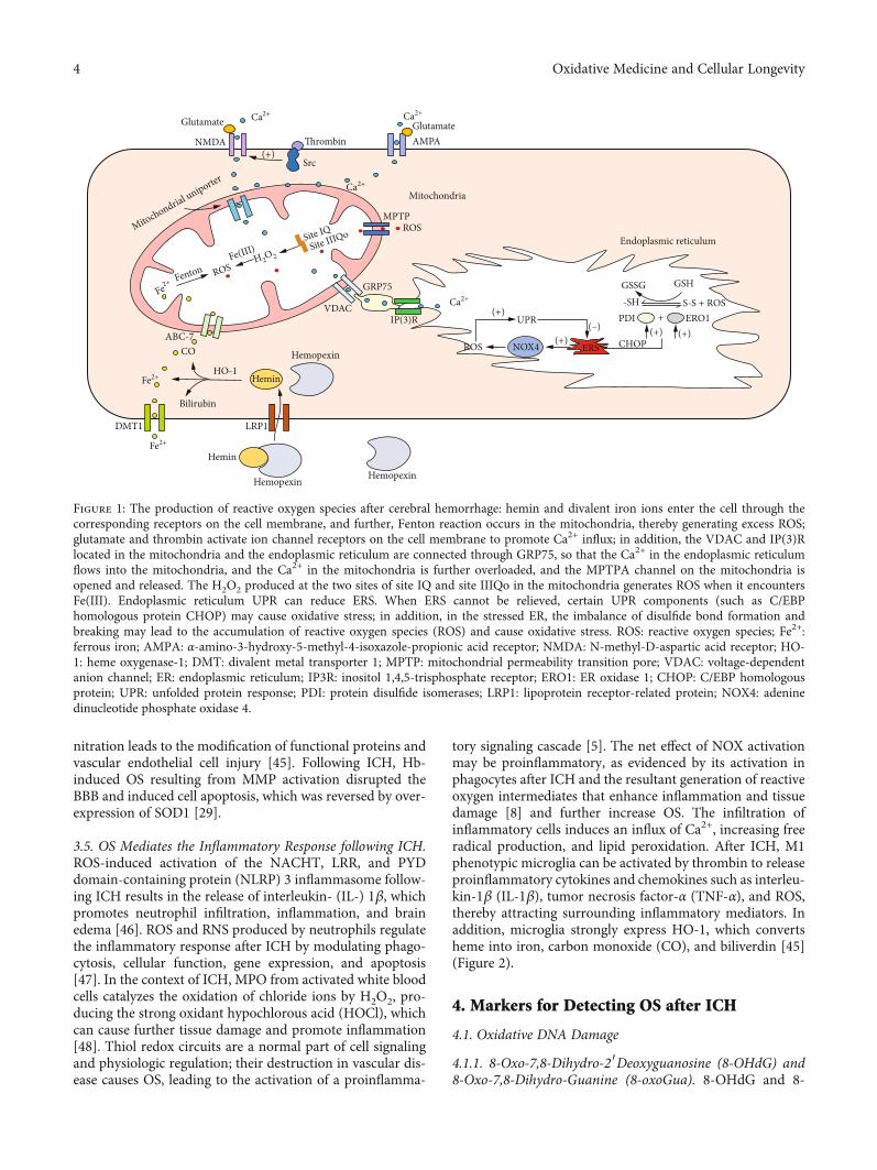
Figure 1: The production of reactive oxygen species after cerebral hemorrhage: hemin and divalent iron ions enter the cell through thecorresponding receptors on the cell membrane, and further, Fenton reaction occurs in the mitochondria, thereby generating excess ROS;glutamate and thrombin activate ion channel receptors on the cell membrane to promote Ca2+ influx; in addition, the VDAC and IP(3)Rlocated in the mitochondria and the endoplasmic reticulum are connected through GRP75, so that the Ca2+ in the endoplasmic reticulumflows into the mitochondria, and the Ca2+ in the mitochondria is further overloaded, and the MPTPA channel on the mitochondria isopened and released. The H2O2 produced at the two sites of site IQ and site IIIQo in the mitochondria generates ROS when it encountersFe(III). Endoplasmic reticulum UPR can reduce ERS. When ERS cannot be relieved, certain UPR components (such as C/EBPhomologous protein CHOP) may cause oxidative stress; in addition, in the stressed ER, the imbalance of disulfide bond formation andbreaking may lead to the accumulation of reactive oxygen species (ROS) and cause oxidative stress. ROS: reactive oxygen species; Fe2+:ferrous iron; AMPA: α-amino-3-hydroxy-5-methyl-4-isoxazole-propionic acid receptor; NMDA: N-methyl-D-aspartic acid receptor; HO-1: heme oxygenase-1; DMT: divalent metal transporter 1; MPTP: mitochondrial permeability transition pore; VDAC: voltage-dependentanion channel; ER: endoplasmic reticulum; IP3R: inositol 1,4,5-trisphosphate receptor; ERO1: ER oxidase 1; CHOP: C/EBP homologousprotein; UPR: unfolded protein response; PDI: protein disulfide isomerases; LRP1: lipoprotein receptor-related protein; NOX4: adeninedinucleotide phosphate oxidase 4.
4 Oxidative Medicine and Cellular Longevity

oxoGua are sensitive markers for DNA oxidative damagethat are produced through hydroxylation at the C-8 positionof guanine. Various methods are used to measure 8-oxoGuaand 8-oxodG concentrations in blood and urine samples,including gas chromatography-mass spectrometry (GC-MS), high-performance liquid chromatography-electrochemical detection (HPLC-ECD), immunohisto-chemistry, and enzyme-linked immunosorbent assay(ELISA). GC-MS and HPLC-ECD have higher specificityand are more accurate than ELISA [49]. At 24h after ICH,8-OHdG was detected at high levels at the borders of dam-aged and normal tissues, coinciding with an increase in thenumber of apurinic/apyrimidinic sites; expression peaked inthe first 3 days post-ICH and returned to baseline startingfrom day 7 [50]. The OS marker leukocyte 8-hydroxy-2′-deoxyguanosine was shown to be an independent predictorof 30-day outcome following ICH [51], while serum 8-OHdGand 8-oxoGua levels were related to 30-day mortality in ICHpatients [52]. According to reports, the optimal cutoff ofserum OGS levels in ICH patients is 4.94 ng/mL (accordingto Youden’s J index). When the OGS level of patients ishigher than the cutoff value, the mortality rate is higher [52].
4.2. Lipid Peroxidation
4.2.1. Malondialdehyde (MDA). ICH leads to progressivelyhigher levels of lipid peroxidation, as evidenced by increaseddiene conjugation and MDA levels [52]. MDA is the mostcommonly used biomarker of lipid peroxidation in clinicalstudies. Serum MDA levels were shown to increase rapidlyat the early stage of ICH and were closely related to the sever-ity of clinical symptoms [53]. MDA can be accurately
detected by HPLC [53], whereas the detection of thiobarbitu-ric acid-reactive substances by ultraviolet light (UV) spectro-photometry overestimated MDA levels in human plasma[43]. Serum MDA level at diagnosis of severe spontaneousICH was shown to be associated with early mortality [54].The study found that patients with spontaneous cerebralhemorrhage have a higher risk of death when the serumMDA is higher than 2.48 nmol/mL [54]. It is common touse the thiobarbituric acid reactive substance (TBARS)method to determine the serum MDA of cerebral hemor-rhage mouse models and patients with ICH [2, 52, 54–58].
4.2.2. 4-Hydroxynonenal (4-HNE). 4-HNE is a product oflipid peroxidation in the cell membrane that can be quanti-fied by HPLC, GC-MS, LC-MS, and aldehyde-reactive probes[59]. 4-HNE levels in cerebrospinal fluid (CSF) and plasmasamples of patients with Parkinson’s disease have also beendetermined by GC-negative-ion chemical ionization massspectrometry-based detection of O-pentafluorobenzyl oxime[60]. Vasospasm in patients with SAH has been evaluatedbased on measurement of lipid peroxidation; the levels ofpolyunsaturated fatty acid cyclization products, F2-isoprostanes (F2-IsoPs), and neuroprostanes were higheston day 1 post-SAH and decreased over time, with similartrends observed in the levels of 4-HNE, 4-oxononenal, andMDA [61].
4.2.3. F2-IsoPs. Another hallmark of lipid peroxidation is theproduction of F2-IsoPs, which can be detected by LC-MS andthe gold standard method GC-MS, while ELISA is less effec-tive [62]. 8-Iso-prostaglandin (8-iso-PG) F2α is an isomerderivative of F2-IsoP that serves as a biomarker for
Activated leukocytes, microglia,and phagocytes Mitochondria and ER Glutamate, thrombin, Hb, heme, and Fe2+
ICH
ROS
OS
Inflammatory response BBB destruction Autophagy Apoptosis
SBI
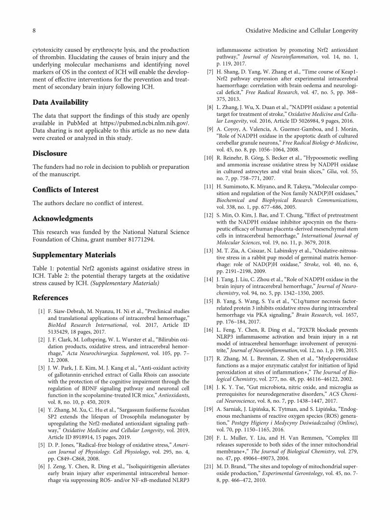
Figure 2: Summary of mechanisms by which OS aggravates SBI after ICH: after ICH, activated phagocytes, mitochondria, ER, and RBClysates all cause excess release of ROS. This increase in OS exacerbates the inflammatory response, apoptosis, autophagy, and BBBdisruption, with further SBI aggravation. BBB: blood-brain barrier; ICH: intracerebral hemorrhage; OS: oxidative stress; ROS: reactiveoxygen species; SBI: secondary brain injury.
5Oxidative Medicine and Cellular Longevity

evaluating OS and lipid peroxidation. 8-Iso-PGF2α is presentin a free form in tissue or in esterified form in lipids; levels inthe blood were shown to be increased after ICH, which waspositively correlated with the National Institute of HealthStroke Scale score and hematoma volume [60], and elevatedplasma concentrations of 8-Iso-PGF2α were reported to beassociated with the clinical severity and outcome of ICH.The plasma level of 8-iso-PGF2α is an independent prognos-tic factor in ICH [63] and indicator of oxidation in analysesof F2-IsoP, isofuran, and F(4)-neuroprostane concentrationsfollowing aneurysmal SAH and traumatic brain injury. Ahigher level of F(4)-neuroprostanes in CSF more accuratelyreflects neural dysfunction than the elevated F2-IsoP level[64].
4.3. Enzyme Activity
4.3.1. SOD. Studies have determined that the level of SOD inpatients with cerebral hemorrhage (113:62 ± 9:14U/mL) issignificantly lower than healthy (161:20 ± 21:12U/mL) [65].It is a common indicator for evaluating oxidative stress inmouse models of ICH [2, 6, 56, 58, 66, 67].
4.3.2. MPO. The activity of MPO, a potent oxidizing enzyme,can be determined by quantifying the level of 3-Cl-Tyr andthe conversion of hydroethidine to 2-chloroethidium. TheMPO/H2O2/chloride system of leukocyte activation isresponsible for the generation of 3-Cl-Tyr, which is a bio-marker of neuroinflammation [68]. Moreover, serum MPOconcentration was found to be increased in ICH patients,which was correlated with ICH severity and prognosis [69].In ICH animal experiments, MPO can be measured withimmunofluorescence assay [55].
4.4. Evaluation of Antioxidant Levels
4.4.1. Glutathione (GSH) and Oxidized Glutathione (GSSG).The molar ratio of GSH to GSSG is a useful index of OS inICH. Total GSH in cells is quantified by measuring the con-centration of the glutathione-N-ethylmaleimide conjugateby UV/visible HPLC. However, under pathologic conditions,total GSH content is lower than that of GSSG. Spectropho-tometry can be used to determine GSSG concentration byeither the GSH recycling method or HPLC [70], while com-mercial GSH/GSSG detection kits are commonly used instudies involving ICH models [6, 57, 66].
4.4.2. Allantoin. Allantoin is a physiologic antioxidant thatcan be detected in human plasma and serum samples byGC-MS [71]. It was also demonstrated that determinationof urinary allantoin concentrations by GC-MS was usefulfor evaluating the efficacy of clinical interventions in pretermneonates diagnosed with germinal matrix IVH [64].
4.4.3. Thioredoxin (TRX). TRX is an antioxidant that elimi-nates oxygen as well as OH− radicals. Serum concentrationsof TRX can be determined by ELISA, with commercial kitswidely available. Increased serum concentrations of TRXwere shown to be related to hemorrhage severity and long-term mortality in patients with ICH [72].
Besides, the mouse ICHmodel can also use the ROS anal-ysis kit to detect the level of oxidative stress. This kit uses theprinciple of the fluorescent probe 2′,7′-dichlorofluoresceindiacetate to determine [6, 57, 67, 73].
5. Therapeutic Strategies Targeting OSfollowing ICH (Supplementary Table 1–2)
5.1. Regulation of Oxidant Signaling Pathways
5.1.1. Keap1/Nrf2/ARE Signaling (Supplementary Table 1).The Keap1/Nrf2/ARE signaling pathway is one of the mostimportant defense mechanisms against OS in ICH [74] as itpromotes the expression of endogenous antioxidant enzymesincluding NQO1, CAT, SOD, HO-1, and GPX [75]. Nrf2 wasupregulated following ICH, with peak level occurring at 24 h;the time course of expression was shown to be correlatedwith the severity of brain edema and neurologic deficits.Heme oxygenase-1 is resistant to OS in the early stages ofICH but is thought to promote oxidation in subsequentstages of the disease process. Drugs targeting the Nrf2/AREsignaling pathway have therapeutic potential for reducingbrain damage caused by OS and inflammation followingICH (Figure 3). The drugs used to treat ICH animal modelsby regulating the Nrf2-ARE signaling pathway include gly-cyrrhizin [6], simvastatin [76], methyl hydrogen fumarate[77], nicotinamide mononucleotide [56], astaxanthin [55],mangiferin [71], RS9 [78], silymarin [79], sulforaphane[80], Hb pretreatment [81], melatonin [82], and recombinanthuman erythropoietin [83], calycosin [84], (-)-epicatechin[67], luteolin [85], and ghrelin [86].
5.1.2. Peroxisome Proliferator-Activated Receptor (PPAR) γSignaling. PPARγ regulates CAT expression and is anotherantioxidant signaling pathway. CAT is ubiquitouslyexpressed in all cell types including glia and neurons and ispredominantly localized in peroxisomes. A PPAR responseelement is present in the CAT gene promoter, indicating adirect regulatory interaction. 15-Deoxy-Δ12,14-prostaglan-din J2 (15d-PGJ2) is a nonenzymatic breakdown product ofprostaglandin D2; unlike synthetic thiazolidinediones, 15d-PGJ2 acts as an endogenous ligand for PPARγ to promotethe expression of CAT, which was shown to be associatedwith decreased inflammation, oxidative damage, and neuro-nal loss in a rat model of ICH [87]. It was noted in the cere-bral hemorrhage model that telmisartan can induce theexpression of receptor γ activated by endothelial nitric oxidesynthase and peroxisome proliferators and reduce oxidativestress, apoptosis signals, and tumor necrosis factor-α andcyclooxygenase-2 expression [88].
5.2. Decreased ROS Production following ICH
5.2.1. NOX. The inhibition of NOX reduces the generation ofendogenous ROS. A small ubiquitin-related modifier wasshown to negatively regulate NOX5 in human neutrophilsand vascular smooth muscle cells, thus limiting the produc-tion of ROS. However, there is little known about theinvolvement of small ubiquitin-related modifiers in ICH[89]. Melatonin was previously found to inhibit ROS
6 Oxidative Medicine and Cellular Longevity

generation and OS after ICH [73]. Meanwhile, overexpres-sion of the ubiquitin ligase ring finger (RNF) 34 exacerbatedbrain injury after ICH by promoting peroxisomeproliferator-activated receptor coactivator- (PGC-) 1α degra-dation while stimulating the generation of mitochondrialROS. Thus, genetic ablation of RNF34 is a potential strategyfor the treatment of ICH [90]. The NADPH oxidase inhibitorapocynin improved the therapeutic efficacy of mesenchymalstem cells in the acute stage of ICH by exerting neuroprotec-tive effects and enhancing the integrity of cerebral vascula-ture [12].
5.2.2. Mitochondria. Sodium benzoate was reported to miti-gate OS-induced secondary brain injury, inhibit neuronalapoptosis, and suppress ROS production in mitochondriaafter ICH via DJ-1/protein kinase B (AKT)/IκB kinase(IKK)/NF-κB signaling [91]. The alleviation of neurologicdeficits by deferoxamine via inhibition of PGC-1α signalingwas shown to reduce OS caused by mitochondrial dysfunc-tion [92]. Besides, drugs that can reduce the oxidative stress
induced by mitochondrial function damage in ICH animalmodels include Dexmedetomidine (Dex) [92], Pyrroloquino-line Quinone (PQQ) [93], and melatonin [73].
5.3. Elimination of ROS following ICH. Intracerebroventricu-lar injection of recombinant α1-microglobulin (A1M)resulted in its coexistence with extracellular Hb, while injec-tion of human A1M mitigated the inflammatory responseand mitochondrial damage in a rabbit model of IVH [94].As a radical scavenger, A1M eliminates both heme and radi-cals, thus providing early protection to the immature brain inpreterm IVH [94]. The novel free radical scavenger NSP-116was found to alter cerebral blood flow and alleviate neuro-logic deficits in a model of I/R injury caused by middle cere-bral artery occlusion; it also suppressed the expansion ofhematomas and reduced neurologic deficits [95]. Edaravone,a free radical scavenger, reduced cerebral edema, and lipidperoxidation following IVH in rats and repeated administra-tion improved learning and memory performance [96].Hydrogen gas was found to reduce OS damage by eliminat-ing OH− in a rat model of cerebral I/R [97], and inhaledhydrogen diminishes OS and brain edema 24h after ICH,although it did not improve clinical outcome [98]. In addi-tion, Glibenclamide (GLI) [99] and tempol [100] are also freeradical scavengers for oxidative stress after ICH.
5.4. Effects of Antioxidants following ICH.Melatonin (N-ace-tyl-5-methoxytryptamine) is an indolamine that is primarilysynthesized by the pineal gland and can easily pass throughthe BBB. Melatonin improved severe ICH-induced braininjury by mitigating OS, apoptosis, inflammation, DNAdamage, brain edema, and BBB damage and by inhibitingMPTP opening [73], and in a SAH model, it mitigated cere-bral OS by increasing the expression of HO-1, NQO1,NADPH, and GST-α1, possibly via activation of theNrf2/ARE signaling pathway [82]. Baicalein treatmentreduced OS in rats by increasing the activity of SOD andGPX while downregulating the expression of MDA in thebrain. Thus, baicalein can potentially be used to treat ICHand related brain injuries [101]. Danhong—a traditional Chi-nese medicine extracted from 2 herbs (Salviae miltiorrhizaBunge (Danshen, China) and Carthamus tinctorius L (Hon-ghua, China))—contains flavonoids and phenolic com-pounds and was reported to increase the expression ofperoxiredoxin (Prx) 1 in astrocytes, thereby preventingsevere brain injury following ICH in aged rats [102]. In addi-tion, carnosine [103], COA-Cl (a novel synthesized nucleo-side analog) [104], AE1-259-01 EP2 receptor agonists [105],green tea and red tea [57], protocatechuic acid (PCA)[106], nebivolol [107], 14Adiponectin (APN) [108], metfor-min [109], C1q/tumor necrosis factor-related protein 3(CTRPs) [15], gastrodin [110], Naringin (NGN) [111], andParthenolide (PN) [112] are all effective antioxidants inICH animal models.
6. Summary and Outlook
The pathophysiologic processes that occur after ICH arecomplex, involving the neuroinflammatory response, OS,
Physiologicalcytoplasm
Ubiquitin
Ubiquitin
Cul3
Cul3
Keap1
Keap1
Nrf2
Nrf2
Nrf2
Nrf2 Maf
Action
Action
OS statecytoplasm
Nucleus
Accessory protein
ARE
HO-l, CAT, SOD,GPX, NQ01, P62
Degradation
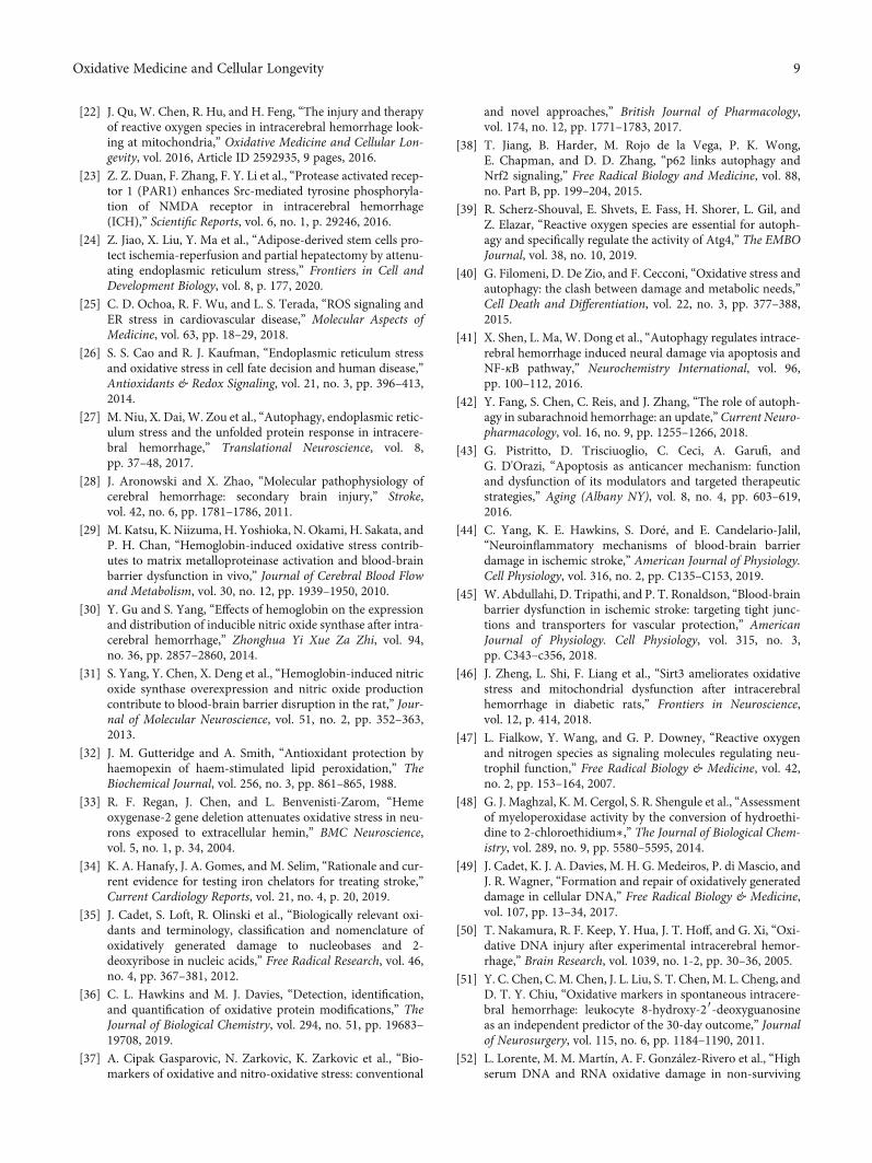
Figure 3: Description of antioxidant enzyme system regulation viathe Keap1-Nrf2-ARE pathway. (a) Under normal basal conditions,Keap1 binds Nrf2 and keeps its level low by ubiquitination andproteasomal degradation; (b) under OS conditions, Keap1 isoxidized by OS, and dissociation of Nrf2 from Keap1 enables Nrf2to translocate to the nucleus. Nrf2 combines with the small Mafprotein to form a Nrf2-Maf heterodimer, and Nrf2 binds toaccessory protein and then ARE activates gene expression of HO-1, NQO1, GPX, SOD, CAT, and the autophagy protein p62. ARE:antioxidant response element; CAT: catalase; Cul3: Cullin3; GPX:glutathione peroxidase; HO-1: heme oxygenase-1; ICH:intracerebral hemorrhage; Keap1: Kelch-like ECH-associatedprotein 1; Maf: musculoaponeurotic fibrosarcoma; NQO1:NAPDH quinone oxidoreductase 1; Nrf2: nuclear factor erythroid2-related factor 2; OS: oxidative stress; SOD: superoxide dismutase.
7Oxidative Medicine and Cellular Longevity
cytotoxicity caused by erythrocyte lysis, and the productionof thrombin. Elucidating the causes of brain injury and theunderlying molecular mechanisms and identifying novelmarkers of OS in the context of ICH will enable the develop-ment of effective interventions for the prevention and treat-ment of secondary brain injury following ICH.
Data Availability
The data that support the findings of this study are openlyavailable in PubMed at https://pubmed.ncbi.nlm.nih.gov/.Data sharing is not applicable to this article as no new datawere created or analyzed in this study.
Disclosure
The funders had no role in decision to publish or preparationof the manuscript.
Conflicts of Interest
The authors declare no conflict of interest.
Acknowledgments
This research was funded by the National Natural ScienceFoundation of China, grant number 81771294.
Supplementary Materials
Table 1: potential Nrf2 agonists against oxidative stress inICH. Table 2: the potential therapy targets at the oxidativestress caused by ICH. (Supplementary Materials)
References
[1] F. Siaw-Debrah, M. Nyanzu, H. Ni et al., “Preclinical studiesand translational applications of intracerebral hemorrhage,”BioMed Research International, vol. 2017, Article ID5135429, 18 pages, 2017.
[2] J. F. Clark, M. Loftspring, W. L. Wurster et al., “Bilirubin oxi-dation products, oxidative stress, and intracerebral hemor-rhage,” Acta Neurochirurgica. Supplement, vol. 105, pp. 7–12, 2008.
[3] J. W. Park, J. E. Kim, M. J. Kang et al., “Anti-oxidant activityof gallotannin-enriched extract of Galla Rhois can associatewith the protection of the cognitive impairment through theregulation of BDNF signaling pathway and neuronal cellfunction in the scopolamine-treated ICRmice,”Antioxidants,vol. 8, no. 10, p. 450, 2019.
[4] Y. Zhang, M. Xu, C. Hu et al., “Sargassum fusiforme fucoidanSP2 extends the lifespan of Drosophila melanogaster byupregulating the Nrf2-mediated antioxidant signaling path-way,” Oxidative Medicine and Cellular Longevity, vol. 2019,Article ID 8918914, 15 pages, 2019.
[5] D. P. Jones, “Radical-free biology of oxidative stress,” Ameri-can Journal of Physiology. Cell Physiology, vol. 295, no. 4,pp. C849–C868, 2008.
[6] J. Zeng, Y. Chen, R. Ding et al., “Isoliquiritigenin alleviatesearly brain injury after experimental intracerebral hemor-rhage via suppressing ROS- and/or NF-κB-mediated NLRP3
inflammasome activation by promoting Nrf2 antioxidantpathway,” Journal of Neuroinflammation, vol. 14, no. 1,p. 119, 2017.
[7] H. Shang, D. Yang, W. Zhang et al., “Time course of Keap1-Nrf2 pathway expression after experimental intracerebralhaemorrhage: correlation with brain oedema and neurologi-cal deficit,” Free Radical Research, vol. 47, no. 5, pp. 368–375, 2013.
[8] L. Zhang, J. Wu, X. Duan et al., “NADPH oxidase: a potentialtarget for treatment of stroke,” Oxidative Medicine and Cellu-lar Longevity, vol. 2016, Article ID 5026984, 9 pages, 2016.
[9] A. Coyoy, A. Valencia, A. Guemez-Gamboa, and J. Morán,“Role of NADPH oxidase in the apoptotic death of culturedcerebellar granule neurons,” Free Radical Biology &Medicine,vol. 45, no. 8, pp. 1056–1064, 2008.
[10] R. Reinehr, B. Görg, S. Becker et al., “Hypoosmotic swellingand ammonia increase oxidative stress by NADPH oxidasein cultured astrocytes and vital brain slices,” Glia, vol. 55,no. 7, pp. 758–771, 2007.
[11] H. Sumimoto, K. Miyano, and R. Takeya, “Molecular compo-sition and regulation of the Nox family NAD(P)H oxidases,”Biochemical and Biophysical Research Communications,vol. 338, no. 1, pp. 677–686, 2005.
[12] S. Min, O. Kim, J. Bae, and T. Chung, “Effect of pretreatmentwith the NADPH oxidase inhibitor apocynin on the thera-peutic efficacy of human placenta-derived mesenchymal stemcells in intracerebral hemorrhage,” International Journal ofMolecular Sciences, vol. 19, no. 11, p. 3679, 2018.
[13] M. T. Zia, A. Csiszar, N. Labinskyy et al., “Oxidative-nitrosa-tive stress in a rabbit pup model of germinal matrix hemor-rhage: role of NAD(P)H oxidase,” Stroke, vol. 40, no. 6,pp. 2191–2198, 2009.
[14] J. Tang, J. Liu, C. Zhou et al., “Role of NADPH oxidase in thebrain injury of intracerebral hemorrhage,” Journal of Neuro-chemistry, vol. 94, no. 5, pp. 1342–1350, 2005.
[15] B. Yang, S. Wang, S. Yu et al., “C1q/tumor necrosis factor-related protein 3 inhibits oxidative stress during intracerebralhemorrhage via PKA signaling,” Brain Research, vol. 1657,pp. 176–184, 2017.
[16] L. Feng, Y. Chen, R. Ding et al., “P2X7R blockade preventsNLRP3 inflammasome activation and brain injury in a ratmodel of intracerebral hemorrhage: involvement of peroxyni-trite,” Journal of Neuroinflammation, vol. 12, no. 1, p. 190, 2015.
[17] R. Zhang, M. L. Brennan, Z. Shen et al., “Myeloperoxidasefunctions as a major enzymatic catalyst for initiation of lipidperoxidation at sites of inflammation∗,” The Journal of Bio-logical Chemistry, vol. 277, no. 48, pp. 46116–46122, 2002.
[18] J. K. Y. Tse, “Gut microbiota, nitric oxide, and microglia asprerequisites for neurodegenerative disorders,” ACS Chemi-cal Neuroscience, vol. 8, no. 7, pp. 1438–1447, 2017.
[19] A. Sarniak, J. Lipińska, K. Tytman, and S. Lipińska, “Endog-enous mechanisms of reactive oxygen species (ROS) genera-tion,” Postȩpy Higieny i Medycyny Doświadczalnej (Online),vol. 70, pp. 1150–1165, 2016.
[20] F. L. Muller, Y. Liu, and H. Van Remmen, “Complex IIIreleases superoxide to both sides of the inner mitochondrialmembrane∗,” The Journal of Biological Chemistry, vol. 279,no. 47, pp. 49064–49073, 2004.
[21] M. D. Brand, “The sites and topology of mitochondrial super-oxide production,” Experimental Gerontology, vol. 45, no. 7-8, pp. 466–472, 2010.
8 Oxidative Medicine and Cellular Longevity
[22] J. Qu, W. Chen, R. Hu, and H. Feng, “The injury and therapyof reactive oxygen species in intracerebral hemorrhage look-ing at mitochondria,” Oxidative Medicine and Cellular Lon-gevity, vol. 2016, Article ID 2592935, 9 pages, 2016.
[23] Z. Z. Duan, F. Zhang, F. Y. Li et al., “Protease activated recep-tor 1 (PAR1) enhances Src-mediated tyrosine phosphoryla-tion of NMDA receptor in intracerebral hemorrhage(ICH),” Scientific Reports, vol. 6, no. 1, p. 29246, 2016.
[24] Z. Jiao, X. Liu, Y. Ma et al., “Adipose-derived stem cells pro-tect ischemia-reperfusion and partial hepatectomy by attenu-ating endoplasmic reticulum stress,” Frontiers in Cell andDevelopment Biology, vol. 8, p. 177, 2020.
[25] C. D. Ochoa, R. F. Wu, and L. S. Terada, “ROS signaling andER stress in cardiovascular disease,” Molecular Aspects ofMedicine, vol. 63, pp. 18–29, 2018.
[26] S. S. Cao and R. J. Kaufman, “Endoplasmic reticulum stressand oxidative stress in cell fate decision and human disease,”Antioxidants & Redox Signaling, vol. 21, no. 3, pp. 396–413,2014.
[27] M. Niu, X. Dai, W. Zou et al., “Autophagy, endoplasmic retic-ulum stress and the unfolded protein response in intracere-bral hemorrhage,” Translational Neuroscience, vol. 8,pp. 37–48, 2017.
[28] J. Aronowski and X. Zhao, “Molecular pathophysiology ofcerebral hemorrhage: secondary brain injury,” Stroke,vol. 42, no. 6, pp. 1781–1786, 2011.
[29] M. Katsu, K. Niizuma, H. Yoshioka, N. Okami, H. Sakata, andP. H. Chan, “Hemoglobin-induced oxidative stress contrib-utes to matrix metalloproteinase activation and blood-brainbarrier dysfunction in vivo,” Journal of Cerebral Blood Flowand Metabolism, vol. 30, no. 12, pp. 1939–1950, 2010.
[30] Y. Gu and S. Yang, “Effects of hemoglobin on the expressionand distribution of inducible nitric oxide synthase after intra-cerebral hemorrhage,” Zhonghua Yi Xue Za Zhi, vol. 94,no. 36, pp. 2857–2860, 2014.
[31] S. Yang, Y. Chen, X. Deng et al., “Hemoglobin-induced nitricoxide synthase overexpression and nitric oxide productioncontribute to blood-brain barrier disruption in the rat,” Jour-nal of Molecular Neuroscience, vol. 51, no. 2, pp. 352–363,2013.
[32] J. M. Gutteridge and A. Smith, “Antioxidant protection byhaemopexin of haem-stimulated lipid peroxidation,” TheBiochemical Journal, vol. 256, no. 3, pp. 861–865, 1988.
[33] R. F. Regan, J. Chen, and L. Benvenisti-Zarom, “Hemeoxygenase-2 gene deletion attenuates oxidative stress in neu-rons exposed to extracellular hemin,” BMC Neuroscience,vol. 5, no. 1, p. 34, 2004.
[34] K. A. Hanafy, J. A. Gomes, and M. Selim, “Rationale and cur-rent evidence for testing iron chelators for treating stroke,”Current Cardiology Reports, vol. 21, no. 4, p. 20, 2019.
[35] J. Cadet, S. Loft, R. Olinski et al., “Biologically relevant oxi-dants and terminology, classification and nomenclature ofoxidatively generated damage to nucleobases and 2-deoxyribose in nucleic acids,” Free Radical Research, vol. 46,no. 4, pp. 367–381, 2012.
[36] C. L. Hawkins and M. J. Davies, “Detection, identification,and quantification of oxidative protein modifications,” TheJournal of Biological Chemistry, vol. 294, no. 51, pp. 19683–19708, 2019.
[37] A. Cipak Gasparovic, N. Zarkovic, K. Zarkovic et al., “Bio-markers of oxidative and nitro-oxidative stress: conventional
and novel approaches,” British Journal of Pharmacology,vol. 174, no. 12, pp. 1771–1783, 2017.
[38] T. Jiang, B. Harder, M. Rojo de la Vega, P. K. Wong,E. Chapman, and D. D. Zhang, “p62 links autophagy andNrf2 signaling,” Free Radical Biology and Medicine, vol. 88,no. Part B, pp. 199–204, 2015.
[39] R. Scherz-Shouval, E. Shvets, E. Fass, H. Shorer, L. Gil, andZ. Elazar, “Reactive oxygen species are essential for autoph-agy and specifically regulate the activity of Atg4,” The EMBOJournal, vol. 38, no. 10, 2019.
[40] G. Filomeni, D. De Zio, and F. Cecconi, “Oxidative stress andautophagy: the clash between damage and metabolic needs,”Cell Death and Differentiation, vol. 22, no. 3, pp. 377–388,2015.
[41] X. Shen, L. Ma, W. Dong et al., “Autophagy regulates intrace-rebral hemorrhage induced neural damage via apoptosis andNF-κB pathway,” Neurochemistry International, vol. 96,pp. 100–112, 2016.
[42] Y. Fang, S. Chen, C. Reis, and J. Zhang, “The role of autoph-agy in subarachnoid hemorrhage: an update,” Current Neuro-pharmacology, vol. 16, no. 9, pp. 1255–1266, 2018.
[43] G. Pistritto, D. Trisciuoglio, C. Ceci, A. Garufi, andG. D'Orazi, “Apoptosis as anticancer mechanism: functionand dysfunction of its modulators and targeted therapeuticstrategies,” Aging (Albany NY), vol. 8, no. 4, pp. 603–619,2016.
[44] C. Yang, K. E. Hawkins, S. Doré, and E. Candelario-Jalil,“Neuroinflammatory mechanisms of blood-brain barrierdamage in ischemic stroke,” American Journal of Physiology.Cell Physiology, vol. 316, no. 2, pp. C135–C153, 2019.
[45] W. Abdullahi, D. Tripathi, and P. T. Ronaldson, “Blood-brainbarrier dysfunction in ischemic stroke: targeting tight junc-tions and transporters for vascular protection,” AmericanJournal of Physiology. Cell Physiology, vol. 315, no. 3,pp. C343–c356, 2018.
[46] J. Zheng, L. Shi, F. Liang et al., “Sirt3 ameliorates oxidativestress and mitochondrial dysfunction after intracerebralhemorrhage in diabetic rats,” Frontiers in Neuroscience,vol. 12, p. 414, 2018.
[47] L. Fialkow, Y. Wang, and G. P. Downey, “Reactive oxygenand nitrogen species as signaling molecules regulating neu-trophil function,” Free Radical Biology & Medicine, vol. 42,no. 2, pp. 153–164, 2007.
[48] G. J. Maghzal, K. M. Cergol, S. R. Shengule et al., “Assessmentof myeloperoxidase activity by the conversion of hydroethi-dine to 2-chloroethidium∗,” The Journal of Biological Chem-istry, vol. 289, no. 9, pp. 5580–5595, 2014.
[49] J. Cadet, K. J. A. Davies, M. H. G. Medeiros, P. di Mascio, andJ. R. Wagner, “Formation and repair of oxidatively generateddamage in cellular DNA,” Free Radical Biology & Medicine,vol. 107, pp. 13–34, 2017.
[50] T. Nakamura, R. F. Keep, Y. Hua, J. T. Hoff, and G. Xi, “Oxi-dative DNA injury after experimental intracerebral hemor-rhage,” Brain Research, vol. 1039, no. 1-2, pp. 30–36, 2005.
[51] Y. C. Chen, C. M. Chen, J. L. Liu, S. T. Chen, M. L. Cheng, andD. T. Y. Chiu, “Oxidative markers in spontaneous intracere-bral hemorrhage: leukocyte 8-hydroxy-2′-deoxyguanosineas an independent predictor of the 30-day outcome,” Journalof Neurosurgery, vol. 115, no. 6, pp. 1184–1190, 2011.
[52] L. Lorente, M. M. Martín, A. F. González-Rivero et al., “Highserum DNA and RNA oxidative damage in non-surviving
9Oxidative Medicine and Cellular Longevity

patients with spontaneous intracerebral hemorrhage,” Neu-rocritical Care, vol. 33, no. 1, pp. 90–96, 2020.
[53] H. F. Moselhy, R. G. Reid, S. Yousef, and S. P. Boyle, “A spe-cific, accurate, and sensitive measure of total plasma malon-dialdehyde by HPLC,” Journal of Lipid Research, vol. 54,no. 3, pp. 852–858, 2013.
[54] L. Lorente, M. M. Martín, P. Abreu-González et al., “Serummalondialdehyde levels and mortality in patients with spon-taneous intracerebral hemorrhage,” World Neurosurgery,vol. 113, pp. e542–e547, 2018.
[55] Q. Wu, X. S. Zhang, H. D. Wang et al., “Astaxanthin activatesnuclear factor erythroid-related factor 2 and the antioxidantresponsive element (Nrf2-ARE) pathway in the brain aftersubarachnoid hemorrhage in rats and attenuates early braininjury,” Marine Drugs, vol. 12, no. 12, pp. 6125–6141, 2014.
[56] C. C. Wei, Y. Y. Kong, G. Q. Li, Y. F. Guan, P. Wang, andC. Y. Miao, “Nicotinamide mononucleotide attenuates braininjury after intracerebral hemorrhage by activatingNrf2/HO-1 signaling pathway,” Scientific Reports, vol. 7,no. 1, p. 717, 2017.
[57] P. M. Sosa, M. A. de Souza, and P. B. Mello-Carpes, “Greentea and red tea from Camellia sinensis partially preventedthe motor deficits and striatal oxidative damage induced byhemorrhagic stroke in rats,” Neural Plasticity, vol. 2018, Arti-cle ID 5158724, 8 pages, 2018.
[58] S. Duan, F. Wang, J. Cao, and C. Wang, “Exosomes derivedfrom microRNA-146a-5p-enriched bone marrow mesenchy-mal stem cells alleviate intracerebral hemorrhage by inhibit-ing neuronal apoptosis and microglial M1 polarization,”Drug Design, Development and Therapy, vol. Volume 14,pp. 3143–3158, 2020.
[59] C. M. Spickett, “The lipid peroxidation product 4-hydroxy-2-nonenal: advances in chemistry and analysis,” Redox Biology,vol. 1, no. 1, pp. 145–152, 2013.
[60] M. L. Selley, “Determination of the lipid peroxidation prod-uct (E)-4-hydroxy-2-nonenal in clinical samples by gas chro-matography–negative-ion chemical ionisation massspectrometry of the O-pentafluorobenzyl oxime,” Journal ofChromatography. B, Biomedical Sciences and Applications,vol. 691, no. 2, pp. 263–268, 1997.
[61] I. Jarocka-Karpowicz, A. Syta-Krzyżanowska,J. Kochanowicz, and Z. D. Mariak, “Clinical prognosis forSAH consistent with redox imbalance and lipid peroxida-tion,” Molecules, vol. 25, no. 8, p. 1921, 2020.
[62] D. Il'yasova, J. D. Morrow, A. Ivanova, and L. E. Wagen-knecht, “Epidemiological marker for oxidant status: compar-ison of the ELISA and the gas chromatography/massspectrometry assay for urine 2,3-dinor-5,6-dihydro-15-F2t-isoprostane,” Annals of Epidemiology, vol. 14, no. 10,pp. 793–797, 2004.
[63] Q. du, W. H. Yu, X. Q. Dong et al., “Plasma 8-iso-prostaglandin F2α concentrations and outcomes after acuteintracerebral hemorrhage,” Clinica Chimica Acta, vol. 437,pp. 141–146, 2014.
[64] T. B. Corcoran, E. Mas, A. E. Barden et al., “Are isofurans andneuroprostanes increased after subarachnoid hemorrhageand traumatic brain injury?,” Antioxidants & Redox Signal-ing, vol. 15, no. 10, pp. 2663–2667, 2011.
[65] M. Zheng, X. Wang, J. Yang, S. Ma, Y. Wei, and S. Liu,“Changes of complement and oxidative stress parameters inpatients with acute cerebral infarction or cerebral hemor-
rhage and the clinical significance,” Experimental and Thera-peutic Medicine, vol. 19, no. 1, pp. 703–709, 2020.
[66] Z. Wang, S. Guo, J. Wang, Y. Shen, J. Zhang, and Q. Wu,“Nrf2/HO-1 mediates the neuroprotective effect of mangi-ferin on early brain injury after subarachnoid hemorrhageby attenuating mitochondria-related apoptosis and neuroin-flammation,” Scientific Reports, vol. 7, no. 1, p. 11883, 2017.
[67] X. Lan, X. Han, Q. Li, and J. Wang, “(-)-Epicatechin, a naturalflavonoid compound, protects astrocytes against hemoglobintoxicity via Nrf2 and AP-1 signaling pathways,” MolecularNeurobiology, vol. 54, no. 10, pp. 7898–7907, 2017.
[68] T. Nybo, M. J. Davies, and A. Rogowska-Wrzesinska, “Anal-ysis of protein chlorination by mass spectrometry,” RedoxBiology, vol. 26, p. 101236, 2019.
[69] G. R. Zheng, B. Chen, J. Shen et al., “Serum myeloperoxidaseconcentrations for outcome prediction in acute intracerebralhemorrhage,” Clinica Chimica Acta, vol. 487, pp. 330–336,2018.
[70] D. Giustarini, D. Tsikas, G. Colombo et al., “Pitfalls in theanalysis of the physiological antioxidant glutathione (GSH)and its disulfide (GSSG) in biological samples: an elephantin the room,” Journal of Chromatography. B, Analytical Tech-nologies in the Biomedical and Life Sciences, vol. 1019, pp. 21–28, 2016.
[71] J. Gruber, S. Y. Tang, A. M. Jenner et al., “Allantoin in humanplasma, serum, and nasal-lining fluids as a biomarker of oxi-dative stress: avoiding artifacts and establishing real in vivoconcentrations,” Antioxidants & Redox Signaling, vol. 11,no. 8, pp. 1767–1776, 2009.
[72] S. Q. Qian, X. C. Hu, S. R. He, B. B. Li, X. D. Zheng, and G. H.Pan, “Prognostic value of serum thioredoxin concentrationsafter intracerebral hemorrhage,” Clinica Chimica Acta,vol. 455, pp. 15–19, 2016.
[73] Z.Wang, F. Zhou, Y. Dou et al., “Melatonin alleviates intrace-rebral hemorrhage-induced secondary brain injury in rats viasuppressing apoptosis, inflammation, oxidative stress, DNAdamage, and mitochondria injury,” Translational StrokeResearch, vol. 9, no. 1, pp. 74–91, 2018.
[74] X. Zhao, G. Sun, J. Zhang et al., “Transcription factor Nrf2protects the brain from damage produced by intracerebralhemorrhage,” Stroke, vol. 38, no. 12, pp. 3280–3286, 2007.
[75] S. Petri, S. Körner, and M. Kiaei, “Nrf2/ARE signaling path-way: key mediator in oxidative stress and potential therapeu-tic target in ALS,” Neurology Research International,vol. 2012, Article ID 878030, 7 pages, 2012.
[76] C. Y. Zhang, X. M. Ren, H. B. Li et al., “Simvastatin alleviatesinflammation and oxidative stress in rats with cerebral hem-orrhage through Nrf2-ARE signaling pathway,” EuropeanReview for Medical and Pharmacological Sciences, vol. 23,no. 14, pp. 6321–6329, 2019.
[77] Y. Y. Shi, H. F. Cui, and B. J. Qin, “Monomethyl fumarateprotects cerebral hemorrhage injury in rats via activatingmicroRNA-139/Nrf2 axis,” European Review for Medicaland Pharmacological Sciences, vol. 23, no. 11, pp. 5012–5019, 2019.
[78] T. Sugiyama, T. Imai, S. Nakamura et al., “A novel Nrf2 acti-vator, RS9, attenuates secondary brain injury after intracere-bral hemorrhage in sub-acute phase,” Brain Research,vol. 1701, pp. 137–145, 2018.
[79] R. Yuan, H. Fan, S. Cheng et al., “Silymarin prevents NLRP3inflammasome activation and protects against intracerebral
10 Oxidative Medicine and Cellular Longevity

hemorrhage,” Biomedicine & Pharmacotherapy, vol. 93,pp. 308–315, 2017.
[80] G. Chen, Q. Fang, J. Zhang, D. Zhou, and Z. Wang, “Role ofthe Nrf2-ARE pathway in early brain injury after experimen-tal subarachnoid hemorrhage,” Journal of NeuroscienceResearch, vol. 89, no. 4, pp. 515–523, 2011.
[81] Y. Yang, Z. Xi, Y. Xue et al., “Hemoglobin pretreatmentendows rat cortical astrocytes resistance to hemin- inducedtoxicity via Nrf2/HO-1 pathway,” Experimental CellResearch, vol. 361, no. 2, pp. 217–224, 2017.
[82] Z. Wang, C. Ma, C. J. Meng et al., “Melatonin activates theNrf2-ARE pathway when it protects against early brain injuryin a subarachnoid hemorrhage model,” Journal of PinealResearch, vol. 53, no. 2, pp. 129–137, 2012.
[83] J. Zhang, Y. Zhu, D. Zhou, Z. Wang, and G. Chen, “Recombi-nant human erythropoietin (rhEPO) alleviates early braininjury following subarachnoid hemorrhage in rats: possibleinvolvement of Nrf2-ARE pathway,” Cytokine, vol. 52,no. 3, pp. 252–257, 2010.
[84] C. Chen, J. Cui, X. Ji, and L. Yao, “Neuroprotective functionsof calycosin against intracerebral hemorrhage-induced oxi-dative stress and neuroinflammation,” Future MedicinalChemistry, vol. 12, no. 7, pp. 583–592, 2020.
[85] X. Tan, Y. Yang, J. Xu et al., “Luteolin exerts neuroprotectionvia modulation of the p62/Keap1/Nrf2 pathway in intracere-bral hemorrhage,” Frontiers in Pharmacology, vol. 10, p. 1551,2020.
[86] Y. Cheng, B. Chen, W. Xie et al., “Ghrelin attenuates second-ary brain injury following intracerebral hemorrhage by inhi-biting NLRP3 inflammasome activation and promotingNrf2/ARE signaling pathway in mice,” International Immu-nopharmacology, vol. 79, p. 106180, 2020.
[87] X. Zhao, Y. Zhang, R. Strong, J. C. Grotta, and J. Aronowski,“15d-Prostaglandin J2Activates peroxisome proliferator-activated Receptor-γ, promotes expression of catalase, andreduces inflammation, behavioral dysfunction, and neuronalloss after intracerebral hemorrhage in rats,” Journal of Cere-bral Blood Flow and Metabolism, vol. 26, no. 6, pp. 811–820, 2005.
[88] K. H. Jung, K. Chu, S. T. Lee et al., “Blockade of AT1 receptorreduces apoptosis, inflammation, and oxidative stress in nor-motensive rats with intracerebral hemorrhage,” The Journalof Pharmacology and Experimental Therapeutics, vol. 322,no. 3, pp. 1051–1058, 2007.
[89] D. Pandey, F. Chen, A. Patel et al., “SUMO1 negatively regu-lates reactive oxygen species production from NADPH oxi-dases,” Arteriosclerosis, Thrombosis, and Vascular Biology,vol. 31, no. 7, pp. 1634–1642, 2011.
[90] X. Qu, N. Wang, W. Chen, M. Qi, Y. Xue, and W. Cheng,“RNF34 overexpression exacerbates neurological deficitsand brain injury in a mouse model of intracerebral hemor-rhage by potentiating mitochondrial dysfunction-mediatedoxidative stress,” Scientific Reports, vol. 9, no. 1, p. 16296,2019.
[91] W. Xu, T. Li, L. Gao et al., “Sodium benzoate attenuates sec-ondary brain injury by inhibiting neuronal apoptosis andreducing mitochondria-mediated oxidative stress in a ratmodel of intracerebral hemorrhage: possible involvement ofDJ-1/Akt/IKK/NFκB pathway,” Frontiers in Molecular Neu-roscience, vol. 12, p. 105, 2019.
[92] J. Huang and Q. Jiang, “Dexmedetomidine protects againstneurological dysfunction in a mouse intracerebral hemor-
rhage model by inhibiting mitochondrial dysfunction-derived oxidative stress,” Journal of Stroke and Cerebrovascu-lar Diseases, vol. 28, no. 5, pp. 1281–1289, 2019.
[93] H. Lu, J. Shen, X. Song et al., “Protective effect of pyrroloqui-noline quinone (PQQ) in rat model of intracerebral hemor-rhage,” Cellular and Molecular Neurobiology, vol. 35, no. 7,pp. 921–930, 2015.
[94] O. Romantsik, A. A. Agyemang, S. Sveinsdóttir et al., “Theheme and radical scavenger α1-microglobulin (A1M) confersearly protection of the immature brain following pretermintraventricular hemorrhage,” Journal of Neuroinflamma-tion, vol. 16, no. 1, p. 122, 2019.
[95] T. Imai, S. Iwata, D. Miyo, S. Nakamura, M. Shimazawa, andH. Hara, “A novel free radical scavenger, NSP-116, amelio-rated the brain injury in both ischemic and hemorrhagicstroke models,” Journal of Pharmacological Sciences,vol. 141, no. 3, pp. 119–126, 2019.
[96] Z. Chen, J. Zhang, Q. Chen, J. Guo, G. Zhu, and H. Feng,“Neuroprotective effects of edaravone after intraventricularhemorrhage in rats,” Neuroreport, vol. 25, no. 9, pp. 635–640, 2014.
[97] I. Ohsawa, M. Ishikawa, K. Takahashi et al., “Hydrogen actsas a therapeutic antioxidant by selectively reducing cytotoxicoxygen radicals,” Nature Medicine, vol. 13, no. 6, pp. 688–694, 2007.
[98] A. Manaenko, T. Lekic, Q. Ma, R. P. Ostrowski, J. H. Zhang,and J. Tang, “Hydrogen inhalation is neuroprotective andimproves functional outcomes in mice after intracerebralhemorrhage,” Acta Neurochirurgica. Supplement, vol. 111,pp. 179–183, 2011.
[99] F. Zhou, Y. Liu, B. Yang, and Z. Hu, “Neuroprotective poten-tial of glibenclamide is mediated by antioxidant and anti-apoptotic pathways in intracerebral hemorrhage,” BrainResearch Bulletin, vol. 142, pp. 18–24, 2018.
[100] Y. Wanyong, T. Zefeng, X. Xiufeng et al., “Tempol alleviatesintracerebral hemorrhage-induced brain injury possibly byattenuating nitrative stress,” Neuroreport, vol. 26, no. 14,pp. 842–849, 2015.
[101] N. Wei, Y. Wei, B. Li, and L. Pang, “Baicalein promotes neu-ronal and behavioral recovery after Intracerebral hemorrhagevia suppressing apoptosis, oxidative stress and neuroinflam-mation,” Neurochemical Research, vol. 42, no. 5, pp. 1345–1353, 2017.
[102] S. Wang, L. Yu, G. Sun et al., “Danhong injection protectshemorrhagic brain by increasing peroxiredoxin 1 in agedrats,” Frontiers in Pharmacology, vol. 11, p. 346, 2020.
[103] R. X. Xie, D. W. Li, X. C. Liu et al., “Carnosine attenuatesbrain oxidative stress and apoptosis after intracerebral hem-orrhage in rats,” Neurochemical Research, vol. 42, no. 2,pp. 541–551, 2017.
[104] F. Lu, T. Nakamura, N. Okabe et al., “COA-Cl, a novel syn-thesized nucleoside analog, exerts neuroprotective effects inthe acute phase of intracerebral hemorrhage,” Journal ofStroke and Cerebrovascular Diseases, vol. 25, no. 11,pp. 2637–2643, 2016.
[105] H. Wu, T. Wu, X. Han et al., “Cerebroprotection by the neu-ronal PGE2 receptor EP2 after intracerebral hemorrhage inmiddle-aged mice,” Journal of Cerebral Blood Flow andMetabolism, vol. 37, no. 1, pp. 39–51, 2017.
[106] Z. Xi, X. Hu, X. Chen et al., “Protocatechuic acid exerts pro-tective effects via suppression of the P38/JNK- NF-κB
11Oxidative Medicine and Cellular Longevity

signalling pathway in an experimental mouse model of intra-cerebral haemorrhage,” European Journal of Pharmacology,vol. 854, pp. 128–138, 2019.
[107] M. A. Aladag, Y. Turkoz, H. Parlakpinar, and M. Gul, “Nebi-volol attenuates cerebral vasospasm both by increasing endo-thelial nitric oxide and by decreasing oxidative stress in anexperimental subarachnoid haemorrhage,” British Journal ofNeurosurgery, vol. 31, no. 4, pp. 439–445, 2016.
[108] S. Wang, D. Li, C. Huang et al., “Overexpression of adiponec-tin alleviates intracerebral hemorrhage-induced brain injuryin rats via suppression of oxidative stress,” Neuroscience Let-ters, vol. 681, pp. 110–116, 2018.
[109] B. Qi, L. Hu, L. Zhu et al., “Metformin attenuates neurologicaldeficit after intracerebral hemorrhage by inhibiting apoptosis,oxidative stress and neuroinflammation in rats,” Neurochem-ical Research, vol. 42, no. 10, pp. 2912–2920, 2017.
[110] X. C. Liu, C. Z.Wu, X. F. Hu et al., “Gastrodin attenuates neu-ronal apoptosis and neurological deficits after experimentalintracerebral hemorrhage,” Journal of Stroke and Cerebrovas-cular Diseases, vol. 29, no. 1, p. 104483, 2020.
[111] N. Singh, Y. Bansal, R. Bhandari et al., “Naringin reversesneurobehavioral and biochemical alterations in intracerebro-ventricular collagenase-induced intracerebral hemorrhage inrats,” Pharmacology, vol. 100, no. 3-4, pp. 172–187, 2017.
[112] J. A. Wang, M. L. Tong, B. Zhao, G. Zhu, D. H. Xi, and J. P.Yang, “Parthenolide ameliorates intracerebral hemorrhage‐induced brain injury in rats,” Phytotherapy Research,vol. 34, no. 1, pp. 153–160, 2019.
12 Oxidative Medicine and Cellular Longevity