Mechanism of CO3- substitution in carbonate … of CO3- substitution in carbonate-fluorapatite:...
9
American Mineralogist, Volume 79, pages 809-818, 1994 Mechanism of CO3- substitution in carbonate-fluorapatite: Evidence from FTIR spectroscopV, t3C NM& and quantum mechanical calculations P. Rrcxrrn * A. C. L.l,sl,c.l, R. A. Bpnrvsn** Department of Geology and Geophysics, Yale University, New Haven, Connecticut 06511, U.S.A. O. H. Hrx,f K. W. Znu Department of Chemistry, Yale University, New Haven, Connecticut 0651I, U.S.A. Ansrucr The substitution of COI in carbonate-fluorapatite("francolite") has a destabilizing effect on the apatite structure that results in increasedsolubility. A commonly proposed substitution mechanism involves the replacementof a POI ion by CO3F3-, in which C is at the center ofa tetrahedron and is surrounded by three O atoms and one F atom at the corners. However, no spectroscopic evidenceexists for this hypothetical anion. We have investigated the microenvironment of CO! in '3C-enriched type B synthetic and natural sedimentarycarbonate-fluorapatite samples by '3C static and MAS NMR and FTIR spectroscopies. (True COI- substitution in the apatite structure was ascertained by X-ray diffraction and FTIR.) The COI- ion itself consists of an sp'?-hybridized planar arrangement of three O atoms about the central C atom. If the CO3- geometry were changedto an sp3 hybrid with F at one comer, the NMR chemical isotropic shift would changeconsiderably, owing to the altered environment about the central C atom. Also, there would be an internuclear dipolar interaction between t3C and teF. Simulated NMR spectra for r3c-reF distances of 1.38 and 2.00 A based on the sp3 model are very different from those obtained on our apatite samples, which are instead characteristicof C in a planar sp2arrangementand no r3c-reF dipolar coupling. No evi- dence for direct bonding betweenCOI- and F was found. This result is confirmed by ab- initio quantum mechanical calculations, which show that the hypothetical COtF3- ion is unstable in an apatitic environment. Ab-initio results, as well as FTIR data, favor the existenceof an interstitial position for F- if nonstoichiometric amounts of this element are present rn apatrte. Ixrnooucrrox The most abundant authigenic phosphate phasein ma- rine sediments is a highly substitutedform of fluorapatite, carbonate-fluorapatite, also known as "francolite" (McConnell, 1973; Jahnke, 1984).It is well established that carbonate substitution has a destabilizing effect on the apatite structure resulting in increasedsolubility (Chien and Black, 1976; Vieillard, 1978; Jahnke, 1984). The concentration variations of structure-bound COI in car- bonate-fluorapatite have also been interpreted as diagenetic or paleoenvironmental indicators (for an extensive review, see Schuffert et al., 1990). However, the true crystal chem- ical nature of this substitution is still controversial. The association of the COr2- ions with apatiteshasbeen investigated by many methods(Beshah et al., 1990): X-ray diffraction, chemical analyses, infrared and Raman spec- troscopy, electron paramagnetic resonance,and proton * Present address: Department of Chemical Oceanography, University of Brussels, CP208, 1050 Brussels, Belgium. ** To whom reprint requests should be sent. f Present address: Korea Basic Science Center,Yoosung-goo, Yeueun-dong224 - l, T aejon 305 - 333, South Korea. and '3C NMR. However, most of thesestudieshave been carried out for medical purposes on pure synthetic hy- droxylapatite, bone tissues, or dental enamel, i.e., in F-poor environments. From chemical studies and mineralogical data of sed- imentary apatites, Borneman-Starinkevich and Belov (1953),Elliott (1964),Smith and khr (1966),Trueman (1966),Guldbrandsen (1966),McClellan and khr (1969), and Binder and Troll (1989)have suggested that when a planar CO3 ion substitutes for a tetrahedral POI- group, the vacant O site is occupied by a F ion. The mechanism is also supported by Bacquet et al. (1980) from EPR ex- periments. This COrF3 arrangement would preserve electroneutrality and complete the coordination of the cations in the structure. However, these results seem to be ambiguous. Okazaki (1983) and Jahnke (1984) found no clear positive relationship between the fluoride con- tent and the amount of COI- incorporated during apatite precipitation. Furthermore, natural carbonate-fluorapa- tite formation takes place in a complex ionic environ- ment, and the electroneutrality of the crystal structure could be maintained by severalother chemically simpler 0003-004x/94109 l 0-o809$02.00 809
Transcript of Mechanism of CO3- substitution in carbonate … of CO3- substitution in carbonate-fluorapatite:...
American Mineralogist, Volume 79, pages 809-818, 1994
Mechanism of CO3- substitution in carbonate-fluorapatite: Evidence from FTIRspectroscopV, t3C NM& and quantum mechanical calculations
P. Rrcxrrn * A. C. L.l,sl,c.l, R. A. Bpnrvsn**Department of Geology and Geophysics, Yale University, New Haven, Connecticut 06511, U.S.A.
O. H. Hrx,f K. W. ZnuDepartment of Chemistry, Yale University, New Haven, Connecticut 0651I, U.S.A.
Ansrucr
The substitution of COI in carbonate-fluorapatite ("francolite") has a destabilizingeffect on the apatite structure that results in increased solubility. A commonly proposedsubstitution mechanism involves the replacement of a POI ion by CO3F3-, in which Cis at the center ofa tetrahedron and is surrounded by three O atoms and one F atom atthe corners. However, no spectroscopic evidence exists for this hypothetical anion.
We have investigated the microenvironment of CO! in '3C-enriched type B syntheticand natural sedimentary carbonate-fluorapatite samples by '3C static and MAS NMR andFTIR spectroscopies. (True COI- substitution in the apatite structure was ascertained byX-ray diffraction and FTIR.) The COI- ion itself consists of an sp'?-hybridized planararrangement of three O atoms about the central C atom. If the CO3- geometry werechanged to an sp3 hybrid with F at one comer, the NMR chemical isotropic shift wouldchange considerably, owing to the altered environment about the central C atom. Also,there would be an internuclear dipolar interaction between t3C and teF.
Simulated NMR spectra for r3c-reF distances of 1.38 and 2.00 A based on the sp3 modelare very different from those obtained on our apatite samples, which are insteadcharacteristic of C in a planar sp2 arrangement and no r3c-reF dipolar coupling. No evi-dence for direct bonding between COI- and F was found. This result is confirmed by ab-initio quantum mechanical calculations, which show that the hypothetical COtF3- ion isunstable in an apatitic environment. Ab-initio results, as well as FTIR data, favor theexistence of an interstitial position for F- if nonstoichiometric amounts of this elementare present rn apatrte.
IxrnooucrroxThe most abundant authigenic phosphate phase in ma-
rine sediments is a highly substituted form of fluorapatite,carbonate-fluorapatite, also known as "francolite"(McConnell, 1973; Jahnke, 1984). It is well establishedthat carbonate substitution has a destabilizing effect onthe apatite structure resulting in increased solubility (Chienand Black, 1976; Vieillard, 1978; Jahnke, 1984). Theconcentration variations of structure-bound COI in car-bonate-fluorapatite have also been interpreted as diageneticor paleoenvironmental indicators (for an extensive review,see Schuffert et al., 1990). However, the true crystal chem-ical nature of this substitution is still controversial.
The association of the COr2- ions with apatites has beeninvestigated by many methods (Beshah et al., 1990): X-raydiffraction, chemical analyses, infrared and Raman spec-troscopy, electron paramagnetic resonance, and proton
* Present address: Department of Chemical Oceanography,University of Brussels, CP208, 1050 Brussels, Belgium.
** To whom reprint requests should be sent.f Present address: Korea Basic Science Center, Yoosung-goo,
Yeueun-dong 224 - l, T aejon 305 - 3 3 3, South Korea.
and '3C NMR. However, most of these studies have beencarried out for medical purposes on pure synthetic hy-droxylapatite, bone tissues, or dental enamel, i.e., inF-poor environments.
From chemical studies and mineralogical data of sed-imentary apatites, Borneman-Starinkevich and Belov(1953), Elliott (1964), Smith and khr (1966), Trueman(1966), Guldbrandsen (1966), McClellan and khr (1969),and Binder and Troll (1989) have suggested that when aplanar CO3 ion substitutes for a tetrahedral POI- group,the vacant O site is occupied by a F ion. The mechanismis also supported by Bacquet et al. (1980) from EPR ex-periments. This COrF3 arrangement would preserveelectroneutrality and complete the coordination of thecations in the structure. However, these results seem tobe ambiguous. Okazaki (1983) and Jahnke (1984) foundno clear positive relationship between the fluoride con-tent and the amount of COI- incorporated during apatiteprecipitation. Furthermore, natural carbonate-fluorapa-tite formation takes place in a complex ionic environ-ment, and the electroneutrality of the crystal structurecould be maintained by several other chemically simpler
0003-004x/94109 l 0-o809$02.00 809
8 1 0 REGNIER ET AL.: CO? SUBSTITUTION IN CARBONATE-FLUORAPATITE
Mdf id r lmJmOn
at6.95 to avoid precipitation of other minerals, especiallycalcite.
The phosphoite analyzed in this study was a phos-phate sample from the Babcock Deep core, located insouthwestern Florida, furnished by John Compton of theUniversity of South Florida. The chemical compositionof the carbonate-fluorapatite from this sample, given byCompton (personal communication), is (Cao roMgouN?o,o-Sro o,)(POo), r?(CO3)0,5r(SOo)o ,rFo ,r.
ExpnnrprnNrAr. METHoDS
Chemical analysis
Ca and P from the synthetic sample were analyzed byinductively coupled plasma-emission spectroscopy (ICPVarian Liberty-200) after the dissolution ofthe syntheticsample in dilute acid. Fluoride content was determinedby ion-selective electrode. K incorporation in the crystalswas checked by electron microprobe analysis using crys-tal dispersion (PET crystal, 15 kV, l0-8 A). Under suchconditions, the minimum concentration of certain detec-tion for K is approximately 0. l0l0.
X-ray diffraction and infrared spectroscopy
The samples were checked by X-ray diffraction to con-f,rm the mineral identity and the crystallinity of the ma-terial. The apatites were scanned on a Scintag Pad-V dif-fractometer (CuKa, 45 kV, 40 mA). The COI- content ofcarbonate-fluorapatite was determined from measure-ments of the A20 for either the 004-410 or the 300-002pair of reflections. This method relies upon an empiricalequation to convert the A20 measurement to a COr2- con-centration (Guldbrandsen, 19701, Schuffert et al., 1990).The experimental conditions and the relationship givenby Schuffert et al. (1990) were used for our calculations:y : 10.643* - 52.512x + 56.986, where y : COI- wto/sand x: L20 (004-410).
In addition Io the A20 measurements, XRD was alsoused to check each sample for the presence ofcalcite. Thepeak of highest intensity for this mineral occurs at ap-proximately 29.4 20. Under the conditions employed(Schuffert et al., 1990), the detection limit is approxi-mately 1.0 wto/o.
The infrared spectra were obtained using KBr pellets;5 and 15 mg of sample were mixed in an agate mortarwith I g of KBr. Transparent pellets were obtained undervacuum at a pressure of 200 kg/cm2. The FTIR spectrawere recorded using a Brucker IFS25 apparatus.
Nuclear magnetic resonance
All the experiments were carried out on a homemadespectrometer with a magnetic field strength of 7.05 T(Larmor frequency: trc : 75.648 MHz). The nuclearmagnetic resonance (NMR) instrument is equipped witha Tecmag data system and an Oxford Instruments 89-mm bore superconducting solenoid. A magic angle spin-ning (MAS) multinuclear resonance probe (Doty Scien-tific, Inc.) was used for,3C MAS NMR experiments (l.lolo
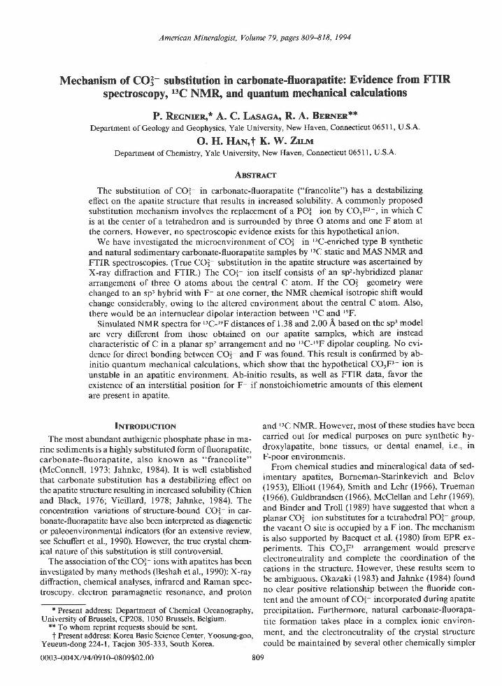
Fig. 1. Experimental system used in the pH-stat precipita-tion experiments. See text for details.
substitution mechanisms. For example, replacement ofCa2* by Na+ (LeGeros et al., 1968; McClellan and Lehr,1969; Bonel et al., 1973; Nelson and Featherstone, 1982:Driessens et al., 1983) and lattice defect formation (Bonelet al.,1973;, Tochon-Danguy et al., 1980) have been ex-tensively studied.
Direct spectroscopic evidence for the existence of thishypothetical COrFr- ion is essentially missing. The aimof this paper is to identifu the microenvironment ofCO3 existing in synthetic and natural sedimentary car-bonate-fluorapatite, using FTIR spectroscopy, '3C NMRspectroscopy, and quantum mechanical calculations.
Sarvrpr.rcs
Synthetic samples were prepared using the pH-stat ap-paratus of Van Cappellen and Berner (1991). The systemwas modified to follow the experimental method of Jahnke( l 984).
A plastic vessel containing I L ofreaction solution wasstirred by a suspended magnetic stir bar to avoid thegrinding of crystals against the bottom of the vessel (Fig.l). Temperature was maintained at 70 "C. A Ross com-bination pH electrode with an internal reference was cou-pled with an automatic titrator that delivers either a con-centrated KOH solution or a concentrated HNO. solutionto maintain a constant solution pH. During the experi-ments, fluctuations in pH were kept within 0.05 units(Van Cappellen and Berner, l99l).
Reagents in the form of 0.2 M Ca(NOr)r.4H20,0.12M KTIJPOA wirh 0.048 M I<F solution, and 0.0027 MK2I3CO3 were added to the reaction vessel by means ofperistaltic pump at a rate of 8 mllh. Dramatic variationsin ionic strength were avoided by using 0. I ,l1 KNO3solution as the reaction medium (Jahnke, 1984). KNO,was used because K+ substitutes into apatite to a muchlesser extent than Na+ (Bonel et al., 1973; Jahnke, 1984).AfIer 24 h, the addition of reagents by means of the peri-staltic pump was stopped, and the precipitate was al-lowed to equilibrate for an additional 5 h; it then wasfiltered, washed with distilled HrO, and dried at roomtemperature. The pH of the experiment was maintained
REGNIER ET AL.: CO?_ SUBSTITUTION IN CARBONATE-FLUORAPATITE 8 l l
abundance, I : Vz). Sapphire rotors, of 7-mm outer di-ameter, were spun at 4-4.8 kHz. The MAS spin rateswere measured before and after each experiment elec-tronically. A homemade static probe was employed toacquire static-echo spectra.
In all experiments, spin-echo sequences and phase cy-cling of the pulses were used to suppress pulse break-through and oth€r artifacts (Stejskal and Schaefer, 1974).For MAS, a spin echo with the ?r pulse timed to occurexactly one rotor period after the initial r/2 pulse wasemployed. If the echo sequence is not synchronous withthe sample rotation, the spin echo and the MAS can in-terfere, resulting in the destruction of the signal (Maricqand Waugh, 1979; Olejniczak et al., 1984). All echo sig-nals were acquired in quadrature, with the carrier placedclose to the center ofgravity ofthe resonance. A typicalil2 pulse length was 6 ps.
Chemical shifts reported here are all relative to externaltetramethylsilane (TMS), with downfield shifts taken aspositive, and have a precision of about I ppm.
Ab-initio quantum rnechanical calculations
Investigation of the bonding and atomic dynamics ofminerals can answer significant questions concerning thestability of a crystal structure (e.9., Sauer, 1989; Lasagaand Gibbs, 1989). The bulk of the ab-initio work haseffectively emphasized the use of suitable molecular clus-ters to replace the infinite periodic structure of a mineral.
In our simple cluster model, geometric optimization ofa central ion (either POI- or CO3F3 ) through energyminimization was performed in a point-charge array thatmimics the spatial atomic distribution in the apatitestructure. These calculations were carried out with theGaussian-90 (Frish et al., 1990) computer program usingseveral basis sets to expand the molecular orbitals. A ba-sis set is defined by the linear combination of atomicorbitals, which are usually themselves expanded in termsof Gaussian functions. The geometric optimization washandled by the Fletcher-Powell method (Fletcher andPowell, 1963). In this case, the gradient of the SCF en-ergy, which is the first derivative of the energy with re-spect to displacement in the nuclear coordinates, is eval-uated numerically (Hehre et al., 1986).
Rssulrs AND DrscussroN
Chemical analysis
The stoichiometric formula of the synthetic sample,given in ions per unit cell and normalized to ten Ca atomsis: Ca,o(POo), 64(CO3)o 24^(F,OH), 58?(HrO)0.,r. OH ions weredetermined from charge balance conditions and accountfor 27o/o occupancy of the monovalent positions. (HrOcontent was calculated from the mass balance require-ment.) Trace amounts of K (<0.10/o) in the sample wereneglected in these calculations. CO32- content was calcu-lated from the L20 ofthe 004-410 diffraction peaks.
The Ca:PO.:F,OH molar ratio of the synthetic carbon-ate-fluorapatite indicates an excess ofmonovalent anionsfrom the stoichiometry of pure apatite (10:6:2).
20 30 40 50 60
DIFFRACTION ANGLE (DEGREES)
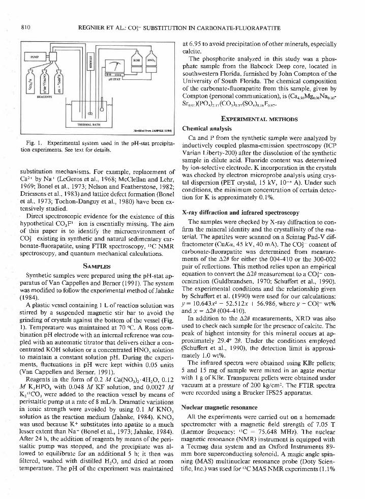
Fig. 2. X-ray diffraction scm of the synthetic carbonate-fluorapatite at 120/min using a Scintag Pad-V diffractometer.The angular distance between the 410-004 and 002-300 diffrac-tion peaks is used to determine the carbonate content of theapatite sample.
X-ray diffraction
The X-ray diffractogram of the synthetic carbonate-fluorapatite is shown in Figure 2. The presence of othermineral phases is unlikely, since every peak is accountedfor by the apatite structure. Most important, the sampledid not contain any crystalline calcium carbonate. TheCq content of the synthetic sample determined fromthe relationship given by Schuffert et al. (1990) is 1.36 +0.61 wto/0. The relatively small amount of CO3- incorpo-rated in the synthetic sample can be correlated with thealmost neutral pH at which the experiment was conduct-ed (Jahnke, 1984).
A fast scan of the natural sample has also been per-formed. The XRD measurement indicates that this sed-iment is a mixture of quartz and well-crystallized phos-phorite grains.
FTIR spectroscopy
Figure 34' shows the infrared absorption spectrum ofsynthetic carbonate-fluorapatite in the region 1600-400cm '. The same domain was also covered at a lower sen-sitivity (Fig. 3B). The z, mode of the POl- ion is repre-sented in apatites by a very narrow band (965 cm-'), andthe z, mode produces an absorption peak at 470 cm-l(Fig. 3A). As shown by Rey et al. (1991), the main ab-sorbance signal of POI appears in the triply degeneratez, domain (Fig. 3B). Finally, the zo mode gives two mainbands at 600 and 560 cm-' and a shoulder at 575 cm-'(Fig. 3A', 3B). The anisotropic electric field of crystallineapatite splits the degeneracy ofthis absorption band intoa well-defined doublet, and a high crystallinity index (CI: 5.1) was extracted from the amount of splitting (Ter-mine and Posner, 1966; Rey et al., 1990; Shemesh, 1990).No additional bands are present in either the v, or vo
Fazti2
.l
812 REGNIER ET AL.: CO3- SUBSTITUTION IN CARBONATE-FLUORAPATITE
Fl(Jz&o
1600 1200 800 ,t00
WAVEI{I,JMBER (cm-l)
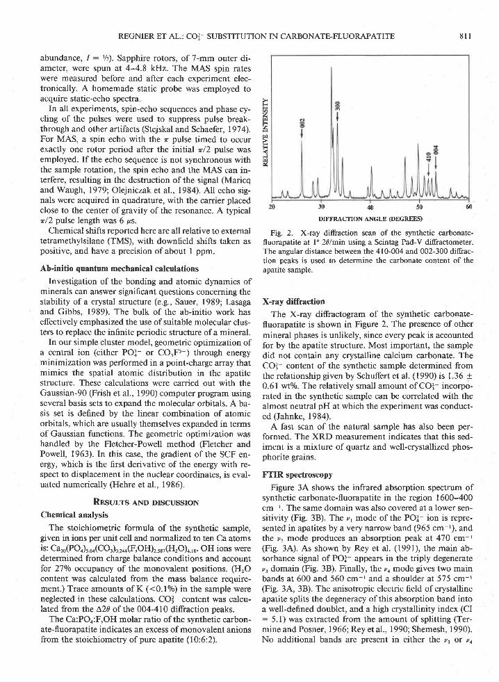
Fig. 3. Fourier transform infrared (FTIR) spectra of(A) syn-thetic carbonate-fluorapatite (-15 mg sample per g KBr); (B)synthetic carbonate-fluorapatite (-5 mg sample per g KBr); (C)natural carbonate-fluorapatite. Qz are the SiOi vibrationalmodes attributed to quartz. The absence of the C-O absorptionbands at about 710 cm-' (thick arrows) in the spectra of bothsamples indicates that no detectable CaCO, is associated withthe marine apatites. The detailed structure of the COI- vibra-tional bands are discussed in Fig. 4.
domains, and, therefore, no distinguishable nonapatiticenvironments or ions (e.g., HPOI ions) are present inthe precipitated mineral phase.
The IR spectrum of the natural sample is shown inFigure 3C. The broad peak at around 1000 cm ' is dueto both the vr-v. POI- modes and the most intenseSiOX- absorption band. The z, POI signal is also maskedby another quartz peak, and the only well-defined POl-band is thus the doubly degenerate ,,4 mode. The well-defined doublet at 798-779 cm-r and the band at 695cm-r are all attributed to the presenca of quartz in thissample.
Finally, a very weak band at 740 cm-t is observed inFigure 3,A, which can be attributed to the substitution ofF by OH ions in synthetic fluorapatite (Freund and Kno-bel, 1977; Klee, 1974; Baumer et al., 1985, 1990). The
presence of some OH- groups is confirmed by a faintabsorption band at 3570 cm ' (not shown in Fig. 3).
Among the four internal vibrational modes of the freeCO3- ion, only two are of importance for IR investiga-tions in calcium phosphates: u, and v, (Rey et al., 1989).The absence ofthe C-O absorption bands at about 710cm-' in the spectrum (identified by heavy arrows in Fig.3) indicates that no detectable CaCO, is associated withour synthetic carbonate-fluorapatite (I-eGeros et al., I 980).This confirms our XRD results.
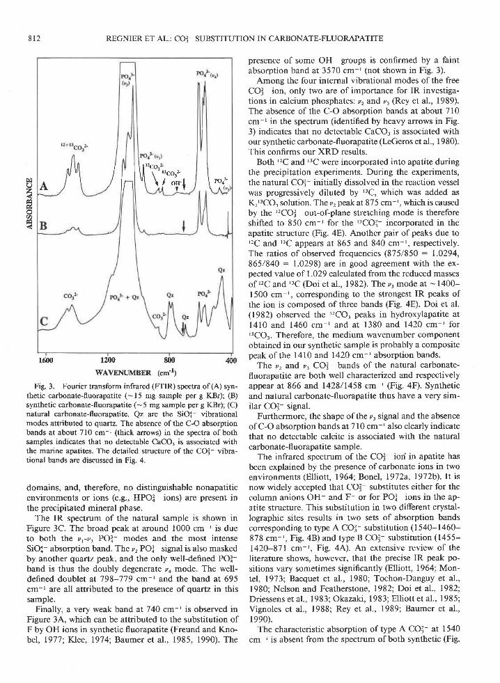
Both '2C and '3C were incorporated into apatite duringthe precipitation experiments. During the experiments,the natural CO3- initially dissolved in the reaction vesselwas progressively diluted by ''C, which was added asKr'3CO3 solution. The z, peak at 875 cm-', which is causedby the ''?CO3- out-of-plane stretching mode is thereforeshifted to 850 cm ' for the '3CO3- incorporated in theapatite structure (Fig. aE). Another pair of peaks due to'2C and '3C appears at 865 and 840 cm r, respectively.The ratios of observed frequencies (875/850 : 1.0294,865/840 : 1.0298) are in good agreement with the ex-pected value of 1.029 calculated from the reduced massesof '2C and "C (Doi et al., 1982). The z. mode at - 1400-1500 cm-', corresponding to the strongest IR peaks ofthe ion is composed of three bands (Fig. 4E). Doi et al.(1982) observed the '2CO, peaks in hydroxylapatite atl4 l0 and 1460 cm- 'and at 1380 and 1420 cm- ' forr3CO.. Therefore, the medium wavenumber componentobtained in our synthetic sample is probably a compositepeak of the l4l0 and 1420 cm-t absorption bands.
Ttre v, and z, COI bands of the natural carbonate-fluorapatite are both well characterized and respectivelyappear at 866 and,1428/1458 cm ' (Fig. aD. Syntheticand natural carbonate-fluorapatite thus have a very sim-ilar COI- signal.
Furthermore, the shape of the z, signal and the absenceof C-O absorption bands at 710 cm-' also clearly indicatethat no detectable calcite is associated with the naturalcarbonate-fl uorapatite sample.
The infrared spectrum of the CO3- ion in apatite hasbeen explained by the presence ofcarbonate ions in twoenvironments (Elliott, 1964; Bonel, 1972a, 1972b). It isnow widely accepted that CO3 substitutes either for thecolumn anions OH- and F or for POI ions in the ap-atite structure. This substitution in two different crystal-lographic sites results in two sets of absorption bandscorresponding to type A CO3- substitution (1540-1460-878 cm-', Fig. 4B) and type B CO3- substitution (1455-1420-87 1cm-', Fig. 4A). An extensive review of theliterature shows, however, that the precise IR peak po-sitions vary sometimes significantly (Elliott, 1964; Mon-tel, 1973; Bacquet et al., 1980; Tochon-Danguy et al.,1980; Nelson and Featherstone, 1982; Doi et aI., 19821,Driessens et al., 1983; Okazaki, 1983; Ell iott et al., 1985;Vignoles et al., 1988; Rey et al., 1989; Baumer et al.,le90).
The characteristic absorption of type A CO3- at 1540cm I is absent from the spectrum of both synthetic (Fig.
/q ll l\1"\1i..,". l\A i \ / lnl ' \ \ lst i \ l . l

REGNIER ET AL.: CO3 SUBSTITUTION IN CARBONATE-FLUORAPATITE 8 1 3
4E) and natural (Fig. aD samples, which clearly indicatesthat no COI- is incorporated in the column anion sites.By contrast, the z. mode of type B apatite (Fig. aA) is clearlyexhibited by both the synthetic and natural samples.
The interpretation of the v, COI- band is more com-plex. Rey et al. (1989) found a band ar 871 (Fig. 4,{) and878 cm-' (Fig. aB) for type B and type A hydroxylapatitesamples, respectively. However, the origin of the 878-cm-' band is still the center of several controversies. Nel-son and Featherstone (1982) effectively showed that italso appeared in apatites without the z, spectral charac-teristics of type A CO]-. They consequently attributedthis band to a second kind of type B COI-. The samespectra (a band at 878 cm-r without the presence of aband at 1540 cm-': Fig. 4C) were later obtained by Reyet al. (l 989) for synthetic hydroxylapatite and are in goodagreement with the results obtained for the synthetic ap-atite (Fig. 4E). These z, spectral characteristics are lackingfor the natural sample, and, in this case, the only ob-served component is found at 866 cm ' (Fig. 4F).
Another interesting feature of Figure 4 is the slight dis-placement of the z, peak positions toward lower wave-numbers (878 - -874 cm-t1,871 - 866 cm-') relatedto an increase in the degree of sample fluoridation (com-pare Fig. 4C with Fig. 4D). It is thus normal that thevalues obtained in our study (875, and 865 cm-' for thesynthetic sample, 866 cm ' for the natural carbonate-fluorapatite) are displaced with respect to the positionsgenerally accepted for hydroxylapatite (Fig. 4A-4C). Sinceit is well known that absorption bands can be broadened,split, or shifted by mutual interactions of adjacent ionsor combination with low wavenumber lattice vibrations,Okazaki (1983) speculated that F- greatly affects theneighboring ions. Vignoles (1984), on the basis of a semi-quantitative study ofthe IR spectra, identified these twotype B COI- peaks as COI--tr (O vacancy) and COI -F-associations. However, kGeros et al. (1968) and Rey etal. (1989) found these data to be ambiguous and simplyattributed the shifting of the 878- and 871-cm-r bands ofnonfluoridated apatites to a change of composition.
Nuclear magnetic resonance spectroscopy
The static-echo spectra of both samples are shown inFigure 5. The spectrum of l0o/o '3C enriched BaCO, isshown in Figure 5C for comparison. The spectrum has atypical line shape due to an axially symmetric chemical-shift anisotropy (Abragam, 196l). The center of gravityand singularities ofthe pattern can be regarded as a fin-gerprint for CO3 ions (Duncan, 1990). (The humps near30 ppm, marked by stars in Figure 5A and 58, are fromthe parafilm used to cap the sample tubes.) The line widthsbecome broader in the following order: BaCOr, syntheticapatite, and natural apatite. However, the MAS spectraof the two apatite samples have an identical isotropicchemical shift of 170 + I ppm, as shown in Figure 6.(The peaks near ll5 ppm are background signals fromthe probe and kel-F rotor caps.) One of the two spinningside-band peaks ofthe apatites happens to appear at the
1530 1470 1410 880 860 840
WAVTNLMBER (cm-l)
Fig. 4. FTIR spectra ofCOl vibrational bands in carbonate-fluorapatite samples. (A) type B carbonate hydroxylapatite; (B)type A carbonate hydroxylapatite; (C) hydroxylapatite withoutthe usual type A location band at 1540 cm ' but with an 878-cm ' band; (D) a fluoridation experiment showing the z, COI-wibrational band shift; @) synthetic apatite from the present study;(F) marine carbonate-fluorapatite from the present study. Seetext for a complete description. Data for curves A-D are fromRey et al. (1989).
Oz
.t)
8 1 4 REGNIER ET AL.: CO3- SUBSTITUTION IN CARBONATE-FLUORAPATITE
600 400 200 -200 -400ppm
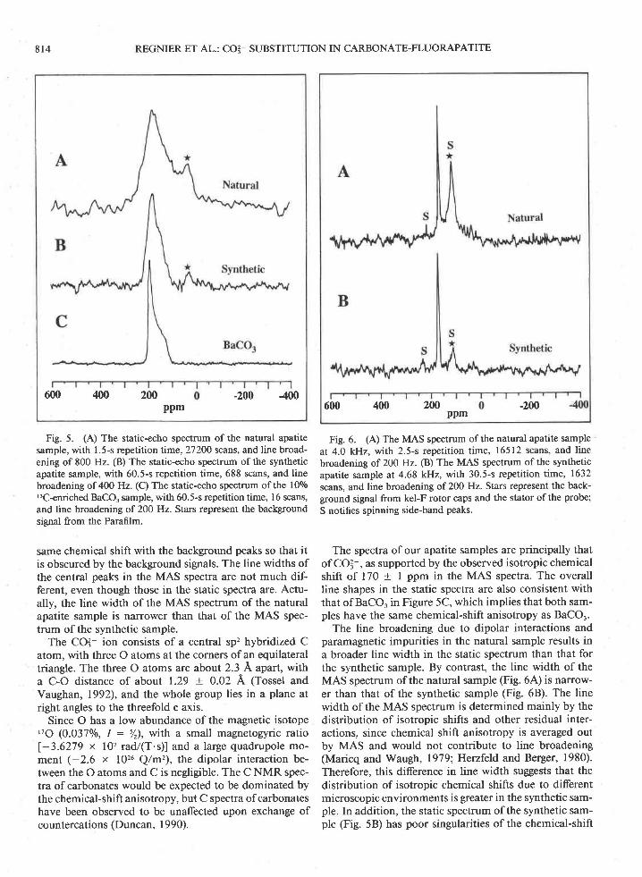
Fig. 5. (A) The static-echo spectrum of the natural apatitesample, with 1.5-s repetition time,27200 scans, and line broad-ening of 800 Hz. (B) The static-echo spectrum ofthe syntheticapatite sample, with 60.5-s repetition time, 688 scans, and linebroadening of 400 Hz. (C) The static-echo spectrum of the 100/o'3C-enriched BaCO, sample, with 60.5-s repetition time, l6 scans,and line broadening of 200 Hz. Stars represent the backgroundsignal from the Parafilm.
same chemical shift with the background peaks so that itis obscured by the background signals. The line widths ofthe central peaks in the MAS spectra are not much dif-ferent, even though those in the static spectra are. Actu-ally, the line width of the MAS spectrum of the naturalapatite sample is narrower than that of the MAS spec-trum of the synthetic sample.
The CO? ion consists of a central sp'z hybridized Catom, with three o atoms at the corners of an equilateraltriangle. The three O atoms are about 2.3 A apart, witha C-O distance of about 1.29 + 0.02 A (Tossel andVaughan, 1992), and the whole group lies in a plane atright angles to the threefold c axis.
Since O has a low abundance of the magnetic isotopetlO (0.037o/o, I : %), with a small magnetogyric ratio
l-3.6279 x l0? radl(T.s)l and alarge quadrupole mo-ment (-2.6 x 1026 Q/m'), the dipolar interaction be-tween the O atoms and C is negligible. The C NMR spec-tra of carbonates would be expected to be dominated bythe chemical-shift anisotropy, but C spectra of carbonateshave been observed to be unaffected upon exchange ofcountercations (Duncan. I 990).
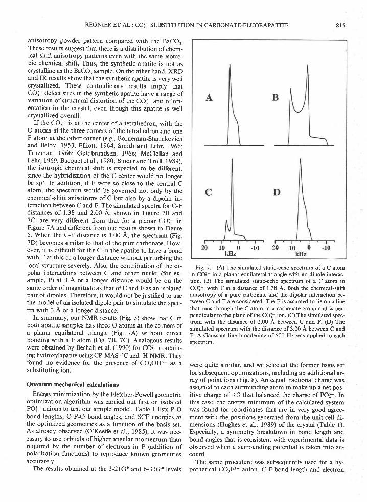
Fig. 6. (A) The MAS spectrum of the natural apatite sampleat 4.0 kHz, with 2.5-s repetition time, 16512 scans, and linebroadening of 200 Hz. (B) The MAS spectrum of the syntheticapatite sample at 4.68 kH4 with 30.5-s repetition time, 1632scans, and line broadening of 200 Hz Stars represent the back-ground signal from kel-F rotor Glps and the stator of the probe;S notifies spinning side-band peaks.
The spectra of our apatite samples are principally thatof COI-, as supported by the observed isotropic chemicalshift of 170 + I ppm in the MAS spectra. The overallline shapes in the static spectra are also consistent withthat of BaCO. in Figure 5C, which implies that both sam-ples have the same chemical-shift anisotropy as BaCOr.
The line broadening due to dipolar interactions andparamagnetic impurities in the natural sample results ina broader line width in the static spectrum than that forthe synthetic sample. By contrast, the line width of theMAS spectrum of the natural sample (Fig. 6,4') is narrow-er than that of the synthetic sample (Fig. 6B). The linewidth of the MAS spectrum is determined mainly by thedistribution of isotropic shifts and other residual inter-actions, since chemical shift anisotropy is averaged outby MAS and would not contribute to line broadening(Maricq and Waugh, 1979;Henfeld and Berger, 1980).Therefore, this difference in line width suggests that thedistribution of isotropic chemical shifts due to differentmicroscopic environments is greater in the synthetic sam-ple. In addition, the static spectrum of the synthetic sam-ple (Fig. 58) has poor singularities of the chemical-shift
2W600 400 -200ppm
REGNIER ET AL.: CO3_ SUBSTITUTION IN CARBONATE-FLUORAPATITE 8 1 5
anisotropy powder pattern compared with the BaCOr.These results suggest that there is a distribution of chem-ical-shift anisotropy patterns even with the same isotro-pic chemical shift. Thus, the synthetic apatite is not ascrystalline as the BaCO, sample. On the other hand, XRDand IR results show that the synthetic apatite is very wellcrystallized. These contradictory results imply thatCOI- defect sites in the synthetic apatite have a range ofvariation of structural distortion of the CO3 and of ori-entation in the crystal, even though this apatite is wellcrystallized overall.
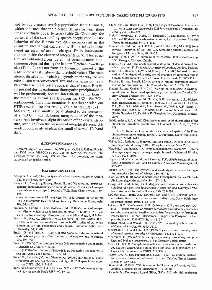
If the COI is at the center of a tetrahedron, with theO atoms at the three corners ofthe tetrahedron and oneF atom at the other corner (e.g., Borneman-Starinkevichand Belov, 1953; Elliott, 1964; Smith and Lehr, 1966;Trueman, 1966; Guldbrandsen, 1966: McClellan andLehr, I 969; Bacquet et al., I 980; Binder and Troll, I 989),the isotropic chemical shift is expected to be different,since the hybridization of the C center would no longerbe sp'?. In addition, if F were so close to the central Catom, the spectrum would be governed not only by thechemical-shift anisotropy of C but also by a dipolar in-teraction between C and F. The simulated spectra for C-Fdistances of 1.38 and 2.00 A, shown in Figure 78 and7C, are very different from that for a planar CO!- inFigure 7A and different from our results shown in Figure5. When the C-F distance is 3.00 A, the spectrum (Fig.7D) becomes similar to that of the pure carbonate. How-ever, it is difficult for the C in the apatite to have a bondwith F at this or a longer distance without perturbing thelocal structure severely. Also, the contribution of the di-polar interactions between C and other nuclei (for ex-ample, P) at 3 A or a longer distance would be on thesame order of magnitude as that of C and F as an isolatedpair of dipoles. Therefore, it would not be justified to usethe model of an isolated dipole pair to simulate the spec-tra with 3 A or a longer distance.
In summary, our NMR results (Fig. 5) show that C inboth apatite samples has three O atoms at the corners ofa planar equilateral triangle (Fig. 7A) without directbonding with a F atom (Fig. 7B, 7C). Analogous resultswere obtained by Beshah et al. (1990) for CO3 contain-rng hydroxylapatite using CP-MAS ,3C and 'H NMR. Theyfound no evidence for the presence of COTOH3- as asubstituting ion.
Quantum mechanical calculations
Energy minimization by the Fletcher-Powell geometricoptimization algorithm was carried out first on isolatedPOI- anions to test our simple model. Table I lists P-Obond lengths, O-P-O bond angles, and SCF energies atthe optimized geometries as a function of the basis set.As already observed (O'Keeffe et al., 1985), it was nec-essary to use orbitals of higher angular momentum thanrequired by the number of electrons in P (addition ofpolarization functions) to reproduce known geometriesaccurately.
The results obtained at the 3-2lG* and 6-3lG* levels
Fig. 7. (A) The simulated static-echo spectrum of a C atomin COI- in a planar equilateral triangle with no dipole interac-tion. (B) The simulated static-echo spectrum of a C atom inCO3-, with F at a distance of 1.38 A. notl the chemical-shiftanisotropy of a pure carbonate and the dipolar interaction be-tween C and F are considered. The F is assumed to lie on a linethat runs through the C atom in a carbonate group and is per-pendicular to the plane of the CO32 ion. (C) The simulated spec-trum with the distance of 2.00 A between C and F. (D) Thesimulated spectrum with the distance of 3.00 A between C andF. A Gaussian line broadening of 500 Hz was applied to eachspectrum.
were quite similar, and we selected the former basis setfor subsequent optimizations, including an additional ar-ray of point ions (Fig. 8). An equal fractional charge wasassigned to each surrounding atom to make up a net pos-itive charge of +3 that balanced the charge of POI-. Inthis case, the energy minimum of the calculated systemwas found for coordinates that are in very good agree-ment with the positions generated from the unit-cell di-mensions (Hughes et al., 1989) of the crystal (Table l).Especially, a symmetry breakdown in bond length andbond angles that is consistent with experimental data isobserved when a surrounding potential is taken into ac-count.
The same procedure was subsequently used for a hy-pothetical CO3F3- anion. C-F bond length and electron
20 100 -10kHz

8 1 6 REGNIER ET AL.: CO3 SUBSTITUTION IN CARBONATE-FLUORAPATITE
Fig. 8. Apatite molecular cluster centered around a fully op-timized POI- ion. The nonphosphate atoms are generalized pointsof fractional charge, making up a net positive charge of + 3 thatbalances the negative charge ofthe POI ion. The vertical linesare parallel to the screw axis of the mineral and perpendicularto the (001) plane. O : open spheres, Ca : shaded spheres, F isrepresented by the letter F, and the P atom : the small sphere.
TABLE 1. P-O bond lengths, O-P-O angles, and SCF energiesat the optimized geometries for PO! as a function ofthe basis set
SCFBond angle energy
f) (Hartrees)
Without lattice charges
Fig. 9. Apatite molecular cluster after the replacement ofPO? by CO'F3 (crystal orientation as indicated in Fig. 8). O: open spheres, Ca : shaded spheres, F is represented by theletter F, and the C atom : the small sphere. Note the interstitialposition for F (thick arrow).
overlap population calculated at the 3-2lG and 6-3lG*levels, respectively, are compared from the structure ob-tained after three Fletcher-Powell optimization cycles(Table 2). The results are qualitatively the same to justify
the use of the computationally simpler 3-2lG basis set.The same three-dimensional array of equal lattice
charges selected for POI- calculations was subsequentlyincluded in the quantum mechanical calculations. Asshown in Table 2, no optimal structure is found, anddirect bonding between CO3- and F is lacking. This isdemonstrated clearly by the large C-F distance (3.61 A)
Tlau 2. C-F distances and electron overlap population ob-tained at nonoptimized geometries for a hypotheticalCO.F3 ion
CyclesC-F distance C-F electron
(A) overlap population
Bond lengthBasis (A)
STO-3G
3-21G'
631G.
1 .7131 . 7 1 21 .7131 .7131.5621.5611.5621.5621.5681.5671.5681 568
1.5601 .5741.5581.559
1.5381.5391.5311532
109.49109.52109.43
109.s1109.45109.45
109.50109.45109.45
With lattice charges109 .18108.03110 .04
Observed108.00'1o7.45
111. ' t2
-630.83528
-636.39530
-639.70781
-637.248403-21G
6€1G'3-21G
Without lattice charges5 38327 9-360
With lattice charges3 2.5753 2.4945 2.808
10 3.61 015 3.61 120 3.611
0.0004200
0.0880.0900.0360.000 6530.0006590.000647
/Vote.' the observed values have been obtained from the unit-cell dimen-sion of the apatite crystal (data from Hughes et al., 1989). one Hartree:627 Kcal/mol.
Note.' cycles are Fletcher-Powell cycles. Overlap populations are fromMulliken population analysis. See text for the complete description.
REGNIER ET AL.: CO3- SUBSTITUTION IN CARBONATE-FLUORAPATITE 8 1 7
and by the electron overlap population from C and F,which indicates that the electron density between theseions is virtually equal to zero (Table 2). Obviously, thepotential of the surrounding species deeply modifies thebehavior of the F atom explicitly incorporated in thequantum mechanical calculations. If one takes into ac-count an array of atomic charges, F- is interstitiallytrapped inside the cluster of interest (Fig. 9). This struc-ture was obtained from the almost constant atomic po-sitioning observed during the last ten Fletcher-powell cy-cles (Table 2) and not from an optimized geometry (theRMS force was still above the threshold value). The exactatomic distribution probably depends on the way the ap-atite cluster was constructed (size and charge assignment).Nevertheless, these results suggest that if excess F is in-corporated during carbonate-fluorapatite precipitation, itwill be preferentially located interstitially rather than inthe remaining vacant site left from the COI- for pOi-replacement. This interpretation is consistent with ourFTIR results. The observedv,COl- band shift (871 -866 cm ') is too small to be attributed to the formationsf 3 tnrQQrpr ion. A better interpretation of the mea-surements involves a slight distortion of the crystal struc-ture, resulting from the presence ofinterstitial F ions: thismodel could easily explain the small observed IR bandshift.
AcxNowr,nocunrvrs
Research support was provided by NSF grant OCE-9115569 (to R.A.B.)and DOE grant DE-FG22-91-PC91285 (to K.W.Z.). We thank JohnCompton of the University of South Florida for providing the naturalcarbonate-fl uorapatite sample.
RerrnpNcns crrEDAbragam, A. (1961) The principles of nuclear magnetism, 599 p. Oxford
University Press. New York.Bacquet, G., Vo Quang Truong, Bonel, G., and Vignoles, M. (1980) R6-
sonance paramagn6tique 6l6ctronique du centre F* dans les fluorapa-tites carbonatees de type B. Journal ofSolid State Chemisrry, 33, 189-I 95 .
Baumer, A., Ganteaume, M., and Klee, W. (1985) Determination of OHions in fluorapatire by infrared spectroscopy. Bulletin de Mineralogie,1 0 8 , 1 4 5 - 1 5 2 .
Baumer, A., Caruba, R., and Ganteaume, M. (1990) Carbonate-fluorapa-tite: Mise en 6vidence de la substitution 2POl - SiOX + SO.2 parspectrom6trie infrarouge. European Journal of Mineralo gy, 2, 297 -304.
Beshah, K., Rey, C., Glimcher, M.J., Schimizu, M., and Griffin, R.G.(1990) Solid state carbon-I3 and proton NMR studies ofcarbonatecontaining calcium phosphates and enamel. Joumal of Solid StareChemistry, 84, 7l-81.
Binder, G., and Troll, G. (1989) Coupled anion subsritution in naturalcarbon-bearing apatites. Contributions to Mineralogy and petrology,r0r,394-40r.
Bonel, G. (1972a) Contribution a l'6tude de la carbonatation des apatites.L Annales de Chimie. 7. 65-87 .
- (1972b) Contribution a l'6rude de la carbonatation des apatites. IIand III. Annales de Chimie, 7,127-144.
Bonel, G., Iabarthe, J.C., and Vignoles, C. (1973) Contribution a l'6tudestructurale des apatites carbonatees de type B. Colloques Internation-aux du CNRS, 23O,117-125.
Borneman-Starinkevich, I.D., and Belov, N.V. ( I 9 5 3) Carbonate-apatites.Doklady Akademie Nauk SSSR, 90, 89-92.
Chien, S.H., and Black, C.A. (l 976) Free energy offormation ofcarbonateapatites in some phosphate rocks. Soil Science Society ofAmerica Pro-ceedings, 40,234-239.
Doi, Y., Moriwaki, Y., Aoba, T., Takahashi, J., and Joshin, IC (1982)ESR and IR studies of carbonate-containing hydroxyapatites. CalcifiedTissue International. 34. 178-18 l.
Driessens, F.C.M., Verbeeck, R.M-H., and Heiiligers, H.J.M. (1983) Somephysical properties of Na- and COr-containing apatites synthesized.Inorganica Chimica Acta, 80, 19-23.
Duncan, T.M. (1990) A compilation of chemical shift anisotropies, p.36C. Farragut, Chicago, Illinois.
Elliott, J.C. (1964) The crystallographic structure of dental enamel andrelated apatites. Ph.D. thesis, University ofl-ondon, London, England.
Elliott, J.C., Holcomb, D.W., and Young, R.A. (1985) Infrared determi-nation of the degree of substitution of hydroxyl by carbonate ions inhuman dental enamel. Calcified Tissue International, 37, 372-375.
Fletcher, R., and Powell, M.J.D. (1963) A rapidly convergent desc€ntmethod for minimization. The Computer Journal, 6, 163-168.
Freund, F., and Knobel, R. (1977) Dstribution of fluorine in hydroxy-apatite studied by infrared spectroscopy. Joumal of the Chemical So-ciety, Dalton Transactions, I 136-1 140.
Frish, M.J., Head-Gordon, M., Trucks, G.W., Foresman, J.B., Schlegel,H.B., Raghavachari, K., Robb, M., Binkley, J.S., Gonzalez, C., Defrees,D.J., Fox, D.J., Whiteside, R.A., Seeger, R., Melius, C.F., Baker, J.,Martin, R.L., Kahn, L.R., Stewart, J.J.P., Topiol, S., and Pople, J. A.(1990) Gaussian 90, Revision F. Gaussian, Inc., Pittsburgh, Pennsyl-vanla,
Guldbrandsen, R.A. ( I 966) Chemical composition of phosphorites of thephosphoria formation. Geochimica et Cosmochimica Acta, 30,769-778.
-(1970) Relation of carbon dioxide content of apatite of the Phos-phoria formation to regional facies. U.S. Geological Survey Profession-al Paper, 700-8,9-13.
Hehre, W.J., Radom, L., Schleyer, P.R., and Pople, J.A. (1986) Ab-initiomolecular orbital theory, 548 p. Wiley Interscience, New York.
Herzfeld, J., and Berger, E.A. (l980) Sideband intensities in NMR spectraof samples spinning at the magic angle. Journal of Chemical Physics,73 (t2),602r-6030.
Hughes, J.M., Cameron, M., and Crowley, K.D. (1989) Structural varia-tions in natural F, OH, and Cl apatites. American Mineralogist, 74,870-876.
Jahnke, R.A. (1984) The synthesis and solubility ofcarbonate fluorapa-tite. American Joumal ofScience, 284, 58-78.
Klee, W. (1974) OH-Ionen in naturlichen Fluorapatiten. Neues JahrbuchfiiLr Mineralogie Monatshefte, 3/4, 127-143.
Lasaga, A.C., and Gibbs, G.V. ( I 989) Ab-initio quantum mechanical cal-culations of water-rock interactions: Adsorption and hydrolysis reac-tions. American Journal of Scien cn, 29O, 263-29 5.
lrGeros, R.Z.,Ttatul'z, O.R., I-eGeros, J.P., and Klein, E. (1968) Carbon-ate substitution in the apatite structure. Bulletin de la Soci6t6 Chimiquede France, special issue, 1712-1718.
kGeros, R.2., Vandemaele, K.H., Quirolgico, G.B., and l-eGeros, D.J.(1980) Transformation ofcalcium carbonates and calcium phosphatesto carbonate apatites: Possible mechanism for phosphorite formation.Proceedings of the 2nd International Congress on Phosphorus Com-pounds, Boston, IMPHOS (Paris), 4l-57.
Maricq, M.M., and Waugh, J.S. (1979) NMR in rotating solids. Journalof Chemical Physics, 70, 3300-3316.
McClellan, G.H., and Lehr, J.R. (1969) Crystal chemical investigationsof natural apatites. American Mineralogist, 54, | 37 4- | 39 1.
McConnell, D. (1973) Apatite, its crystal chemistry, mineralogy, and geo-logic and biologic occurrences, I I I p. Springer-Yerlag, Berlin.
Montel, G. (1973) Conceptions actuelles sur la structure et la constitutiondes apatites synth6riques comparables aux apatites biologiques. Col-loques Internationaux du CNRS, 230, l3-18.
Nelson, D.G.A., and Featherstone, J.D.B. (1982) Preparation, analysis,and characterization of carbonated apatites. Calcified Tissue lnterna-tional. 34. 569-581.
Okazaki, M. (1983) F COI interaction in IR spectra of fluoridated CO.-apatites. Calcified Tissue Intemational, 35, 78-81.
O'Keeffe, M., Domenges, B., and Gibbs, c.V. (1985) Ab-initio molecutar