
Mechanics of the Cardiovascular System COPYRIGHTED...
7
Overview The cardiovascular system consists of two pri- mary components: the heart and blood vessels. The lymphatic system also has a cardiovascular exchange function but does not contain blood. This chapter will highlight the mechanics of the cardiovascular system and present an overview of the essential elements and structures involved in the flow of blood through the venous and arterial systems. It will also highlight how abnormalities in the mechanics of the cardiovascular system can result in degrees of cardiac disease states. Basic heart anatomy The human heart is essentially a muscular pump which delivers blood containing oxygen, nutri- ents and other vital elements to the body tissues and major organs. The structure and location of the heart was described by Henry Gray in 1918. It is conical in shape, about the size of a human fist and weighs between 230 and 340 g in an adult. The heart is located in the mediastinum, with one-third lying to the right of the sternum and two-thirds to the left. The top of the heart is known as the base, and this is located behind the sternum; the bot- tom of the heart, known as the apex, is located 1 Mechanics of the Cardiovascular System B. Greaney & A.M. Kucia Learning objectives After reading this chapter, you should be able to: ● Identify the anatomical location of the heart and its basic function. ● Identify the key structures within the heart, which are involved in the flow of blood through the heart and identify their specific function. ● Define the term ‘cardiac cycle’ and explain the key physiological changes that occur in the heart during this process. ● Define the terms ‘cardiac output’ (CO) and ‘stroke volume’ (SV), and explain their physiolog- ical significance in relation to the cardiac cycle. ● Define the terms ‘preload’, ‘afterload’ and ‘contractility’, and explain their physiological impact upon myocardial contraction. Key concepts Cardiac cycle; cardiac output; cardiac chambers; cardiac valves; layers of the heart COPYRIGHTED MATERIAL
Transcript of Mechanics of the Cardiovascular System COPYRIGHTED...

Overview
The cardiovascular system consists of two pri-mary components: the heart and blood vessels. The lymphatic system also has a cardiovascular exchange function but does not contain blood. This chapter will highlight the mechanics of the cardiovascular system and present an overview of the essential elements and structures involved in the fl ow of blood through the venous and arterial systems. It will also highlight how abnormalities in the mechanics of the cardiovascular system can result in degrees of cardiac disease states.
Basic heart anatomy
The human heart is essentially a muscular pump which delivers blood containing oxygen, nutri-ents and other vital elements to the body tissues and major organs. The structure and location of the heart was described by Henry Gray in 1918. It is conical in shape, about the size of a human fi st and weighs between 230 and 340 g in an adult. The heart is located in the mediastinum, with one-third lying to the right of the sternum and two-thirds to the left. The top of the heart is known as the base, and this is located behind the sternum; the bot-tom of the heart, known as the apex, is located
1 Mechanics of the Cardiovascular System
B. Greaney & A.M. Kucia
Learning objectives
After reading this chapter, you should be able to:
● Identify the anatomical location of the heart and its basic function.
● Identify the key structures within the heart, which are involved in the fl ow of blood through the heart and identify their specifi c function.
● Defi ne the term ‘cardiac cycle’ and explain the key physiological changes that occur in the heart during this process.
● Defi ne the terms ‘cardiac output’ (CO) and ‘stroke volume’ (SV), and explain their physiolog-ical signifi cance in relation to the cardiac cycle.
● Defi ne the terms ‘preload’, ‘afterload’ and ‘contractility’, and explain their physiological impact upon myocardial contraction.
Key concepts
Cardiac cycle; cardiac output; cardiac chambers; cardiac valves; layers of the heart
COPYRIG
HTED M
ATERIAL

2 Acute Cardiac Care: A Practical Guide for Nurses
in the fi fth intercostal space in the mid-clavicular line. The heart is a four-chambered structure – the upper chambers known as the right and left atria, the lower two chambers known as the right and left ventricles, with right and left-sided chambers divided by the septum.
The bulk of the heart’s wall is the myocardium, which is a thick contractile mass of cardiac mus-cle cells. It is the myocardium that provides the force of contraction to move blood out of the ven-tricles at the end of each cardiac cycle. The heart is surrounded by the pericardium, which is com-prised of two principal layers that surround and protect the heart. The outer layer is known as the fi brous pericardium, which is made up of tough and fi brous connective tissue. This layer provides both protection and anchorage for the heart. The second layer, the serous pericardium, is a thin-ner, more delicate layer and forms two distinct layers around the heart. The outer parietal layer is adhered to the inner side of the fi brous pericar-dium, whilst the inner visceral layer, also known as the epicardium, is adhered tightly to the myo-cardium. Between these two layers there exists a potential space termed the pericardial cavity. Within this cavity is a very thin fi lm of serous fl uid known as pericardial fl uid, which is nor-mally between 15 and 35 mL in volume (Spodick 1997). The key function of this fl uid is to reduce friction between the pericardial layers as the heart contracts. The inner layer lining the heart is a con-tinuous sheet of squamous epithelium, continu-ing into the tunica intima of blood vessels, and is known as the endocardium.
The heart is divided into four chambers: two upper atria and two lower ventricles. These chambers are separated by a set of heart valves termed the atrioventricular (AV) valves; the tri-cuspid valve separates the right atrium (RA) and right ventricle (RV) and the bicuspid valve or mitral valve separates the left atrium (LA) and left ventricle (LV) (Figure 1.1a). Attached to each AV valve are two structures: the chordae tendinae and the papillary muscles. These two structures are adhered to the walls of each ventricle (Figure 1.1a). Their function is to prevent the valve cusps inverting or swinging upward into the atria dur-ing ventricular systole. The key function of the heart valves is to permit the fl ow of blood in one direction only as it fl ows through the heart.
The heart can be viewed functionally as two pumps serving the pulmonary and systemic cir-culations. The pulmonary circulation refers to the fl ow of blood within the lungs that is involved in the exchange of gases between the blood and the alveoli. Deoxygenated blood returns to the RA via the inferior and superior vena cavae. It then passes through the tricuspid valve to the RV before entering the pulmonary circulation via the pulmonary artery, where gases are exchanged. The pulmonary artery has a pulmonary valve or semi-lunar valve which opens and closes during con-traction and relaxation of the heart, again having a similar function to the AV valves, allowing the fl ow of blood in one direction only (Figure 1.1). The systemic circulation consists of all the blood vessels within and outside of all organs excluding the lungs. Once oxygenated, the blood returns to the LA via the pulmonary veins and then passes through the mitral valve into the thicker-walled left ventricle, which ejects the oxygenated blood through the aortic valve into the aorta and into the systemic circulation. The aorta also has a valve, the aortic valve, which prevents the back-fl ow of blood during myocardial contraction (Figure 1.1a).
The cardiac cycle
In simple terms, the heart is a pump that receives blood from the venous system at low pressure and generates pressure through contraction to eject the blood into the arterial system. The mechani-cal action of the heart is created by a synchronised contraction and relaxation of the cardiac mus-cle, referred to as systole and diastole. The actual mechanical function of the heart is infl uenced by pressure, volume and fl ow changes that occur within the heart during one single cardiac cycle.
When the heart muscle contracts (systole) and relaxes (diastole), sequential changes in pressure are produced in the heart chambers and blood vessels, which result in blood fl owing from areas of high pressure to areas of lower pressure. The valves prevent backfl ow of blood. Under normal conditions, this cycle will take place in the human heart between 60 and 100 times per minute.
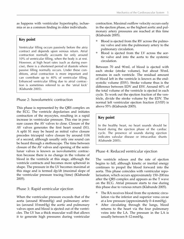
Figure 1.2a demonstrates the seven phases of the cardiac cycle.

Mechanics of the Cardiovascular System 3
Phase 1: Atrial systole
Atrial systole begins after a wave of depolarisation passes over the atrial muscle. Atrial depolarisation is represented by the P wave on the electrocardio-graph (ECG). As the atria contract, pressure builds up inside the atria forcing blood through the tricuspid and mitral valves into the ventricles. Atrial contraction causes a small increase in proxi-mal venous pressure (in the pulmonary veins and vena cavae). This is represented by the ‘a’ wave of the jugular venous pulse, which is used to meas-ure jugular venous pressure (JVP) (Klabunde 2005).
● Blood fl ows from the RA across the tricuspid valve into the RV.
● Blood fl ows from the LA through the mitral valve into the LV.
Pressure in the atria falls and the AV valves fl oat upward. Ventricular volumes are now at their maximum (around 120 mL) and this is known as end diastolic volume (EDV). Left ventricular end diastolic pressure (LVEDP) is approximately 8–12 mmHg; right ventricular end diastolic pres-sure (RVEDP) is usually around 3–6 mmHg. A fourth heart sound (S4) may be heard in this phase if ventricular compliance is reduced, such
CarotidsAortic arch I
ZZ M
IA A
H
Pulmonaryartery
Pulmonaryvalve
Superiorvena cava
Rightatrium
Sarcomere
MyosinActin
Inferiorvena cava
Tricuspid valve
Right ventricle
Sarcolemma
T tubule
[Inside cell]
Corbular(tubular) SR
Terminalcisternal SR
Diad
Leftatrium
Septum
Leftventricle
Mitral valve
Papillary muscles
Myocardium
Intercalated disc
50 µm
Capillary
Nucleus
Desmosome
Connexons
Gapjunction(nexus)
(a)(c)
(b)
(e)
(f) Coronary circulation
(d) 2.2 µm
Sarcolemma
Sarcoplasmicreticulum
T tubuleMitochondria
Right coronaryartery
Left maincoronary artery
Marginalbranch of
right coronaryartery
Posteriordescending
artery Left anteriordescendingartery
Diagonal branchof left anteriordescending artery
Pulmonary artery
Left circumflexmarginal artery
Aorta
Left circumflexartery
Figure 1.1 Gross anatomy of the heart. Source: From Aaronson and Ward (2007).

4 Acute Cardiac Care: A Practical Guide for Nurses
(b)
(a)
Dicrotic notch
Aorta
G A B C D E F G
Left atriumpressure
a
a
c
c
x
x
v
v
y
y
S1 S2 S3
Left ventriclepressure
Aortic valveopens
Mitral valvecloses
Aortic valvecloses
120
Heart sounds
130
6520
0
100
Pre
ssur
e (m
mH
g)Ju
gula
rve
nous
pul
se
Ven
tric
ular
volu
me
(ml)
Aor
ticflo
w (
l/min
)
80
60
40
20
0
Mitral valveopens
G A B C D E F G
P T
S
200 ms
Q
R
EC
G (
mV
)
Ventricular pressure–volume loop
Pressures, volumes and key events during the cardiac cycle
50 150100Left ventricular volume (ml)
40
80
120
160
A
B
C
E
F G
Pre
ssur
e (m
mH
g)
Contractility
Normal
EDV
0
A Atrial systole B Isovolumetriccontraction
C Rapid ventricular ejectionD Reduced ventricular ejection
E Isovolumetricrelaxation
F Rapid ventricular fillingG Reduced ventricular filling
RA
RV
LA
LV
Figure 1.2 Cardiac cycle. Source: From Aaronson and Ward (2007).
Mechanics of the Cardiovascular System 5
as happens with ventricular hypertrophy, ischae-mia or as a common fi nding in older individuals.
contraction. Maximal outfl ow velocity occurs early in the ejection phase, so the highest aortic and pul-monary artery pressures are reached at this time (Klabunde 2005).
● Blood is ejected from the RV across the pulmo-nic valve and into the pulmonary artery to the pulmonary circulation.
● Blood is ejected from the LV across the aor-tic valve and into the aorta to the systemic circulation.
Between 70 and 90 mL of blood is ejected with each stroke (stroke volume), but about 50 mL remains in each ventricle. The residual amount of blood left in the ventricle is known as the end-systolic volume (ESV). Stroke volume thus is the difference between EDV and ESV. Around 60% of the total volume of the ventricle is ejected in each cycle. To work out the ejection fraction of the ven-tricle, divide the stroke volume by the EDV. The normal left ventricular ejection fraction (LVEF) is above 55% (Klabunde 2005).
Key point
Ventricular fi lling occurs passively before the atria contract and depends upon venous return. Atrial contraction normally accounts for only around 10% of ventricular fi lling, when the body is at rest. However, at high heart rates (such as during exer-cise), there is a shortened period of diastole where passive fi lling normally occurs. Under these con-ditions, atrial contraction is more important and can contribute up to 40% of ventricular fi lling. Enhanced ventricular fi lling due to atrial contrac-tion is sometimes referred to as the ‘atrial kick’ (Klabunde 2005).
Phase 2: Isovolumetric contraction
This phase is represented by the QRS complex on the ECG. The ventricle depolarises and initiates contraction of the myocytes, resulting in a rapid increase in ventricular pressure. This rise in pres-sure causes the AV valves to close. Closure of the AV valves generates the fi rst heart sound (S1). A split S1 may be heard as mitral valve closure precedes tricuspid valve closure by around 0.04 of a second, although usually only one sound can be heard through a stethoscope. The time between closure of the AV valves and opening of the semi-lunar valves is known as isovolumetric contrac-tion because there is no change in the volume of blood in the ventricle at this stage, although the ventricle contracts and becomes more spheroid in shape. The pressure in the LV becomes maximal at this stage and is termed dp/dt (maximal slope of the ventricular pressure tracing/time) (Klabunde 2005).
Phase 3: Rapid ventricular ejection
When the ventricular pressure exceeds that of the aorta (around 80 mmHg) and pulmonary arter-ies (around 10 mmHg) the aortic and pulmonary valves open and blood is ejected out of the ventri-cles. The LV has a thick muscular wall that allows it to generate high pressures during ventricular
Key point
In the healthy heart, no heart sounds should be heard during the ejection phase of the cardiac cycle. The presence of sounds during ejection indicates valvular disease or intracardiac shunts (Klabunde 2005).
Phase 4: Reduced ventricular ejection
The ventricle relaxes and the rate of ejection begins to fall, although kinetic or inertial energy continues to propel the blood forward into the aorta. This phase coincides with ventricular repo-larisation, which occurs approximately 150–200 ms after the QRS complex and appears as the T wave on the ECG. Atrial pressure starts to rise during this phase due to venous return (Klabunde 2005).
● The RA receives blood from the systemic circu-lation via the inferior and superior vena cavae at a low pressure (approximately 0–4 mmHg).
● After circulating through the lungs, blood returns to the heart via the four pulmonary veins into the LA. The pressure in the LA is usually between 8–12 mmHg.

6 Acute Cardiac Care: A Practical Guide for Nurses
Phase 5: Isovolumetric relaxation
In this phase, the pressure in the ventricles continues to fall and when the point is reached where the pressure is less in the ventricles than that in the outfl ow tracts (aorta and pulmonary veins), the aortic and pulmonary valves close abruptly, causing a second heart sound (S2). Aortic and pulmonary artery pressures fall slowly due to a combination of stored energy in the elastic walls of these vessels which controls pressure and fl ow, and because forward fl ow is impeded by systemic and pulmonic vascular resistance as blood is dis-tributed through the systemic and pulmonary cir-culations (Klabunde 2005).
Phase 7: Reduced ventricular fi lling
There is no clear demarcation as to when this phase begins, but this is a stage during diastole when passive ventricular fi lling is near comple-tion. As the ventricles fi ll, they become less com-pliant, causing intraventricular pressure to rise and the rate of ventricular fi lling starts to fall. Immediately following this phase, atrial systole occurs following fi ring of the sino-atrial node.
Key point
As the aortic valve closes before the pulmonic valve, there is a physiological splitting of the S2 sound and this may be heard with a stethoscope. Closure of the aortic and pulmonary valves result in a characteristic notch in aortic and pulmonary artery pressure tracings (Figure 1.2a). The aortic notch is important in setting timing for intra-aortic balloon counterpulsation.
Phase 6: Rapid ventricular fi lling
Low pressures in the heart allow blood to pas-sively return to the atria. When the ventricular pressure falls below the atrial pressure, the AV valves open and the ventricles fi ll quickly. Blood fl ows into the atria and ventricles throughout diastole with the rate of fi lling decreasing as the amount of blood in the chambers distends the walls. About 70% of ventricular fi lling occurs pas-sively at this time.
Key point
No prominent heart sounds should be heard at this time. If a third heart sound (S3) is heard during ven-tricular fi lling in adults, it may indicate tensing of the chordae tendinae and AV ring, often associated with ventricular dilation. It is a normal fi nding in children.
Key point
At slow heart rates, diastole is lengthened, resulting in increased fi lling time. In rapid heart rates, there is less fi lling time. This would compromise CO, if not for compensatory mechanisms.
Cardiac output
CO is an important index of cardiac function, and refers to the amount of blood that is ejected with each contraction (stroke volume) multiplied by heart rate (HR):
CO � SV � HR
At typical resting values, if the heart rate is 75 beats/min and the stroke volume is 70 mL/beat, the CO should equal 5.25 L/min. Therefore the body’s total volume of blood (4–6 L/min) passes through the body each minute (Saladin 2001).
CO never remains at a constant rate: any factor that alters stroke volume or heart rate will alter CO and it can vary signifi cantly according to normal physical exercise as well as impaired cardiac func-tion. Other factors such as preload, afterload and contractility (inotropy) will indirectly affect CO.
Preload is defi ned as the actual stretch or ten-sion on the ventricular myocardium prior to con-traction (Totora & Gabowski 2002). The greater the preload on the myocardium (the larger the amount of blood that has fi lled the heart dur-ing diastole), the greater the contraction will be. A simple analogy to explain this concept is that the further you stretch an elastic band prior to releas-ing it, the further it will recoil. The same principle applies here: the greater the stretch or tension on the myocardium, the greater the force of contrac-tion. When venous return to the heart increases,

Mechanics of the Cardiovascular System 7
ventricular fi lling and preload also increase. The Frank Starling Law of the Heart (Starling’s Law) asserts that the more the ventricle is fi lled with blood during diastole (EDV), the greater the vol-ume of blood that will be ejected (stroke volume) during the ensuing systolic contraction. Thus, altered preload is a mechanism by which the force of contractility can be affected (Klabunde 2005).
Contractility, also known as inotropy, is the ability of a cardiac myocyte to alter its tension development independently of preload changes (Klabunde 2005). Contractility is affected by autonomic innervation and circulating catecho-lamines (adrenaline, noradrenaline), and addi-tionally changes in afterload and heart rate can augment contractility. A number of pharmaco-logical agents positively or negatively affect con-tractility. Agents that affect contractility are called positive or negative inotropes, depending upon whether they increase or decrease contractility. Loss of myocardial contractility results in heart failure.
Afterload is defi ned as the force or pressure against which the ventricular myocardium must push prior to contraction (Totora & Grabowski 2003). This force or pressure is constantly present in the arteries as arterial blood pressure. Therefore, any increase in systemic blood pressure will result in the left ventricular myocardium having to contract more forcefully to eject its volume of blood. Any increase in the pressure of the pulmonary circula-tion, such as pulmonary oedema, or the presence of any physical obstruction to the pulmonary circula-tion, such as lung scar tissue, will result in the right ventricular myocardium having to contract more forcefully. In the long term, this increased workload for the myocardium will eventually result in the abnormal enlargement of the myocardium (hyper-trophy), which may in turn lead to heart failure.
Conclusion
This chapter has provided you with an overview of anatomical and physiological underpinnings underlying much of the assessment and nurs-ing care of the patient with a cardiovascular dis-order. When next you check a patient’s heart rate or blood pressure, or listen to their heart sounds, consider in detail the anatomical and physiologi-cal determinants of those measures.
Key point
The myocardium requires oxygen to regenerate ade-nosine triphosphate (ATP) that is hydrolysed to pro-duce energy during contraction and relaxation. Any change to the force or frequency of contraction will have an effect on myocardial oxygen consumption (MVO2). Imbalances in the supply and demand of oxygen to the myocardium may result in myocardial ischaemia or infarction.
Learning activities
There are a number of interactive online websites where you can test your knowledge of cardiac anat-omy and physiology. The Columbia University Medical Center Department of Surgery in New York has some great heart animations and information at http://www.columbiasurgery.org/pat/cardiac/anatomy.html
The Texas Heart Institute at St Luke’s Episcopal Hospital Heart Information Center likewise has some good cardiovascular information and animations at http://texasheart.org/HIC/Anatomy/index.cfm
References
Aaronson, P.I. & Ward, J.P.T. (2007). The Cardiovascular System at a Glance3E. Wiley Blackwell, Oxford.
Gray, H. (1918). Anatomy of the Human Body. Lea & Febiger, Philadelphia.
Klabunde, R. (2005). Cardiovascular Physiology Concepts. Lippincott Williams & Wilkins, Philadelphia.
Saladin, K.S. (2001). Anatomy & physiology: The Unity of Form & Function. McGraw Hill, New York.
Spodick, D.H. (1997). Pericardial macro- and micro-anatomy: A synopsis. In: D.H. Spodick, (ed.), The Pericardium: A Comprehensive Textbook. Marcel Dekker, New York, pp. 7–14.
Totora, G.J. & Grabowski, S.R. (2003). Principles of Anatomy and Physiology, 10th edn. John Wiley & Sons, New Jersey.
Useful Websites and Further Reading
Klabunde, R.E. (2007). Cardiovascular physiology con-cepts. Retrieved online 4th October 2007 from http://www.cvphysiology.com/
Rogers, J. (1999). Cardiovascular physiology. Retrieved online 4th October 2007 from http://www.nda.ox.ac.uk/wfsa/html/u10/u1002_01.htm


