Marta Julia Bionanocompositos de Fe3O4/SiO2 e alginato ... · Este trabalho incluiu a preparação...
68
Universidade de Aveiro 2014 Departamento de Química Marta Julia Busse Bionanocompositos de Fe 3 O 4 /SiO 2 e alginato para remoção magnética de espécies de Cr(III) em água Bionanocomposites of Fe 3 O 4 /SiO 2 and alginate for magnetic removal of Cr(III) species from water
Transcript of Marta Julia Bionanocompositos de Fe3O4/SiO2 e alginato ... · Este trabalho incluiu a preparação...

Universidade de Aveiro
2014
Departamento de Química
Marta Julia Busse
Bionanocompositos de Fe3O4/SiO2 e alginato para remoção magnética de espécies de Cr(III) em água Bionanocomposites of Fe3O4/SiO2 and alginate for magnetic removal of Cr(III) species from water

Universidade de Aveiro
2014
Departamento de Química
Marta Julia Busse
Bionanocompositos de Fe3O4/SiO2 e alginato para remoção magnética de espécies de Cr(III) em água Bionanocomposites of Fe3O4/SiO2 and alginate for magnetic removal of Cr(III) species from water
dissertação apresentada à Universidade de Aveiro para cumprimento dos requisitos necessários à obtenção do grau de Mestre em Química,realizada sob a orientação científica do Proffesor Doutor Tito da Silva Trindade, Professor Associado do Departamento de Química da Universidade de Aveiro e Doutora Ana Luísa Daniel da Silva, Investigadora Auxiliar do Departamento de Química da Universidade de Aveiro


o júri
presidente Prof. Doutor Artur Manuel Soares da Silva Professor Catedrático do Departamento de Qumíca da Universidade de Aveiro
Prof. Doutora Olinda Coelho Monteiro Professora Auxiliar Convidada da Faculdade de Ciências da Universidade de Lisboa
Doutora Ana Luísa Daniel da Silva Investigadora Auxiliar do Departamento de Química da Universidade de Aveiro

agradecimentos
I would like to thank Professor Tito da Silva Trindade, Doctor Ana Luísa Daniel da Silva and Professor Wojciech Kinart for help me to prepare this thesis and I would like to thank to C (undação para a Ciência e a Tecnologia) for funding this work through the project PTDC/CTM-NAN/120668/2010, FEDER through COMPETE and by national funding through FCT in the frame of project CICECO-FCOMP-01-0124-FEDER-037271 (Ref. FCT Pest-C/CTM/LA0011/2013). Also I would like to thank to Dra. Teresa Caldeira (University of Aveiro) for AAS measurements.

Palavras-chave
Nanopartículas magnéticas, alginato, remoção de poluentes, Cr (III).
resumo
O objectivo do presente trabalho foi o de estudar a eliminação de Cr (III), a partir de soluções aquosas, por processo de adsorção, utilizando nanopartículas magnéticas funcionalizadas com grupos amina e com o polissacarídeo alginato. A poluição causada por metais pesados está na base de graves problemas de saúde pública e ambientais em todo o mundo. O crómio é um dos metais mais tóxicos, pondo em perigo a vida humana. O Cr (III) é frequentemente usado na indústria, causando a poluição da água. Por esta razão, aumentou o interesse dos investigadores na utilização de nanomateriais para a remoção de poluentes e no estudo de adsorventes para substituir materiais caros, nomeadamente matérias-primas de baixo custo, tais como, materiais provenientes de polímeros residuais ou subprodutos agrícolas. Este trabalho incluiu a preparação de nanopartículas magnéticas, o seu revestimento com uma capa de sílica amorfa funcionalizada com grupos de amina e a ligação covalente do alginato à nanopartícula magnética. As propriedades das nanopartículas foram avaliadas utilizando várias técnicas experimentais nomeadamente difracção de raios-X, espectroscopia de infravermelho, análise elementar, termogravimetria e medições do potencial zeta. As nanopartículas magnéticas revestidas com sílica e funcionalizadas com grupos de amina foram investigadas para a adsorção de Cr (III) / complexos de EDTA de águas, enquanto que as nanopartículas ligadas às moléculas de alginato foram testadas para a remoção de iões Cr (III) em meio aquoso. Os resultados de cinética foram ajustados aos modelos de pseudo-primeira e pseudo-segunda ordem, e os resultados de equilíbrio de adsorção foram ajustados aos modelos adequados, tais como a isotérmica de Langmuir e a de Freundlich. As nanopartículas compósitas revelaram ter afinidade para as espécies de Cr (III). Devido às suas propriedades, estas nova nanopartículas compósitas podem encontrar aplicações interessantes na purificação de águas.

keywords
Magnetic nanoparticles, alginate, pollutant removal, Cr (III).
abstract
The aim of the present work was to study the removal of Cr (III) species from aqueous solutions by adsorption process, using magnetic nanoparticles functionalized with amine groups and with the polysaccharide alginate. Heavy metal pollution is serious environmental and public health problem worldwide. Chromium is one of the most toxic metal endangering human life. Cr(III) is often used in industry which caused water pollution. Therefore, increased interest by the researchers to employ nanomaterials for the removal contamination and explore adsorbents to replace expensive materials, particularly low-cost raw materials such as, residual polymers materials or agricultural by-products. The work included the preparation of magnetic nanoparticles, coating them with amorphous silica shell functionalized with amine groups and the covalent attachment of alginate to the magnetic nanoparticle. The properties of the nanoparticles were assessed by a number of experimental techniques namely x-ray diffraction, infrared spectroscopy, elemental analysis, thermogravimetry and zeta potential measurements. Silica coated magnetic nanoparticles functionalized with amine groups were investigated for the uptake of Cr(III)/EDTA complexes from water, while the nanoparticles attached to the alginate molecules were tested for the removal of aqueous Cr(III) ions. The kinetic results were fitted to models of pseudo first and pseudo second order and the equilibrium adsorption results were adjusted to suitable isotherm models such as Langmuir and Freundlich. The composite nanoparticles revealed affinity to the Cr(III) species. Due to its properties, this new composite nanoparticles may find interesting applications in purification of water.

Contents
Abbreviations........................................................................................................................1
List of figures and tables......................................................................................................2
1.Introduction.......................................................................................................................4
1.1 Magnetic nanoparticles: fundamental aspects on synthesis and properties ........6
1.2 Bionanocomposites of magnetite .....................................................................15
1.3 Sorbents for metal complexes in water .............................................................17
1.4 Objectives and outline .......................................................................................25
2. Experimental ..................................................................................................................26
2.1 Chemicals ..........................................................................................................26
2.2 Synthesis of magnetite (Fe3O4) nanoparticles ...................................................26
2.3 Coating of magnetite nanoparticles with an amorphous silica shell
functionalized with amine groups (Fe3O4@SiO2-NH2)............................................27
2.4 Covalent attachment of alginate to the magnetite (Fe3O4@SiO2-NH2-ALG) ...27
2.5 Synthesis of [Cr(HY)(H2O)] ..............................................................................27
2.6 Instrumentation ..................................................................................................28
2.7 Adsorption experiments .....................................................................................29
2.7.1 The effect of pH [Cr(HY)(H2O)] complex .........................................29
2.7.2 Kinetic experiments ............................................................................30
2.7.3 Equilibrium isotherms experiments ....................................................30
2.8 Kinetics modelling .............................................................................................31
2.9 Isotherm modelling.............................................................................................31
3. Results and discussion ...................................................................................................33

3.1 Characterization of magnetic nanoparticles ......................................................33
3.2 Adsorption experiments ....................................................................................40
3.2.1 Uptake of [Cr(HY)(H2O)] complexes using Fe3O4@SiO2-
NH2particles.................................................................................................40
3.2.1.1 The effect of pH on [Cr(HY)(H2O)] complex .....................40
3.2.1.2 Kinetic studies .....................................................................43
3.2.1.3 Isotherm studies ...................................................................44
3.2.2 Uptake of Cr(III) species using Fe3O4@SiO2-NH2-ALG particles ....46
3.2.2.1 Kinetic studies .....................................................................46
3.2.2.2 Isotherms studies .................................................................47
4. Conclusion and perspectives of future work ...............................................................49
Reference ............................................................................................................................50

1
Abbreviations
AAS atomic absorption spectrometry
ALD alginate
APTES 3-aminopropyltriethoxysilane
APTMS 3-aminopropyltrimethoxysilane
DLS Dynamic Light Scattering
DMSA dimercaptosuccinic
EDC N-(3-dimethylaminopropyl)-N’-ethylcarbodiimide hydrochloride
EDTA-Na2 ethylenediaminetetraacetic acid
FTIR Fourier Transform Infrared spectroscopy
HA humic acid
NHS N-hydroxysuccinimide
ODS octadecylsilane
SA Salicylic acid
SEM Scanning Electron Microscopy
TEM Transmission Electron Microscopy
TEOS tetraethyl orthosilicate
TGA Thermogravimetric Analysis
TGA thermogravimetric analysis
UV-VIS Ultraviolet-Visible
XRD Powder X-ray Diffraction
Cr(HY)(H2O) chromium(III) complex with EDTA

2
List of figures and tables
Figures
Figure 1. Comparative sizes of various entities to perceive nanoscale measurements
Figure 2. TEM images of (a) quantum dots; (b) mesoporous carbon nanotube; (c)
AuNPs; (d) Fe3O4 (16,0 ± 1,4 nm)
Figure 3. Various arrangements of individual atomic magnetic moments (a)
paramagnetic, (b) ferromagnetic, (c) ferrimagnetic
Figure 4. Dependence of magnetic behavior on the particle size
Figure 5. Chemical functionalization of the silica shells at the magnetite surfaces
Figure 6. Surface modification of SiO2 nanoparticles using APTES
Figure 7. A possible mechanism for adsorption of metal ions by amine functionalized
magnetic nanoparticles
Figure 8. Structure of sodium alginate polymer
Figure 9. Molecular structure of (a) chitosan and (b) chitin
Figure 10. pH dependent removal efficiency of some metal ions by amine
functionalized nanoparticles
Figure 11. Schematic of the preparation of adsorbent (a) and solid phase extraction of
the analytes (b)
Figure 12. Recovery of metal ions in function of pH, using salicylic acid
Figure 13. X-ray diffraction patterns of Fe3O4
Figure 14. SEM micrograph of Fe3O4 nanoparticles (courtesy: Dr. Ana Estrada,
University of Aveiro)
Figure 15. Reactions of chemical modified of magnetic nanoparticles (R= -(CH2)3NH2)
Figure 16. Structure of the new nanoparticle covalent attachment of alginate
Fe3O4@SiO2-NH2-ALG
Figure 17. FT-IR spectra of (a) Fe3O4 (b) Fe3O4@SiO2-NH2 (c) Fe3O4@SiO2-NH2ALG
Figure 18. TGA graphs of Fe3O4; Fe3O4@SiO2-NH2 and Fe3O4@SiO2-NH2-ALG
Figure 19. Chemical structure of Y4-
anion
Figure 20. FTIR spectrum of the complex [Cr(HY)(H2O)]
Figure 21. Potentiometric titration curve of [Cr(HY)(H2O)] with NaOH

3
Figure 22. Color of the solution at different pHs (a) 2,38 (b) 7,66 (c) 8,06 (d) 11,24 (e)
11,55
Figure 23. The time profile of the [Cr(HY)(H2O)] uptake by Fe3O4@SiO2-NH2 (a)
concentration of complex in aqueous solution (b)
Figure 24. Isotherm of the adsorption of [Cr(HY)(H2O)] using Fe3O4@SiO2-NH2
nanoparticles (a) fit of Langmuir isotherm model for adsorption of above
complex
Figure 25. The time profile of adsorption capacity of Cr(III) species onto Fe3O4@SiO2-
NH2-ALG (a) Change in the concentration of Cr(III) vs time (b)
Figure 26. Isotherm of the adsorption of Cr(III) using Fe3O4@SiO2-NH2-ALG
nanoparticles and fit of Freundlich isotherm model for adsorption of above
complex
Tables
Table 1. Comparise of the synthetic methods for magnetite nanoparticles
Table 2. Nanoparticle characterization techniques
Table 3. Key advantages and shortcomings of biopolymers
Table 4. Examples of surface modified magnetite nanoparticles for the removal of
heavy metal ions from water
Table 5. Results of elemental analysis of the nanoparticles.
Table 6. Values of Zeta potential obtained in ultra pure water at variable pH
Table 7. Kinetic parameters estimated from pseudo 1st and 2
nd order equations and
evaluations of its fittings
Table 8. Langumir and Freundlich isotherm model parameters for adsorption
[Cr(HY)(H2O)] complex
Table 9. Kinetic parameters estimated from pseudo 1st and 2
nd order equations and
evaluations of its fittings
Table 10. Langmuir and Freundlich isotherm model parameters for adsorption Cr(III)
species

4
1. Introduction
In the past decade nanomaterials have gained increasing attention for scientists
and engineers, because of their special properties. Size effects and increased fraction of
“surface” atoms result in the change of chemical and physical properties of there small
particles [1]. Typically, nanomaterials have at least one dimension between 1 and 100 nm
[2].
Figure 1. Comparative sizes of various entities to perceive nanoscale measurements [2].
The unique physical and chemical properties of nanomaterials lead to an increase
interest for the nanotechnology sector [1, 2] and make their potential use in different areas
such as optics, ceramics, electronics and magnetic data storage. For example, one of the
specific properties of nanomaterials is that a high percentage of the atoms of the
nanoparticle is of the surface. This, highly chemical activity is expected, because the
surface atoms are unsaturated so can bind with other atoms very easily [3]. Other
important surface related property is the adsorption capacity, because nanomaterials can
strongly adsorb many substances including trace metals and polar organic compounds [1]
thes providing opportunities to treat contaminants in water [4]. The synthesis of many
nanoadsorbents development of new water treatments procedures [5].
As illustrated above, nanomaterials show properties obtendent on surface effects.
Other effects are due to influence of size on the intrinsic properties of the materials [6].
Example is magnetic properties of thenanomaterials. Nanoparticles can be classified
depending on different parameters, such as their origin (natural or anthropogenic),

5
formation (biogenic, geogenic, anthropogenic and atmospheric), chemical composition
(organic and inorganic), their size, characteristics and shape or applications in research and
industry. Examples of different nanostructures are shown in figure 2.
Figure 2. TEM images of (a) quantum dots; (b) mesoporous carbon nanotube; (c) AuNPs;
(d) Fe3O4 (16,0 ± 1,4 nm) (figure removed from [1,2]
A brief describe of important classes of nanoparticles follows.
1) Carbon-based nanostructures are made up of pure carbon. Carbon-based
nanostructures are divided into units groups. Fullerenes, carbon nanotubes (CNTs) and
graphene [2]. The simplest form of fullerene is C60, a ball made up from 60 carbon atoms
[7] CNTs have different properties and can be synthesized by different methods. These
nanotubes have good electrical, mechanical, chemical properties [2] and sorbent properties
[1]. Due to their high adsorption capacities, CNTs can be used in many applications such
as elimination of pathogens, cyanobacterial toxins and natural organic matter from water
systems [2].
2) Metal oxide nanoparticles includes single faced metallic oxide nanoparticles
(aluminium trioxide or titanium dioxide), nanostructured mixed oxides (nanostructured
binary iron- titanium mixed oxide particles) and also magnetic nanoparticles such as iron

6
oxides [1]. Metal oxide nanoparticles are employed in different kind of consumer products,
for example sunscreens, cosmetics, as catalysts and in biomedicine.
3) Quantum dots. There are fluorescent semiconductor nanocrystals that have been
employed for biomedical imaging. Quantum dots have unique optical properties, which
depend on their size. When the size is reduced, the surface area to volume ratio increases
and surface structure strongly affects on optical and electronic properties [8]. Another
important properties of quantum dots are high quantum yield, special chemical properties
and high photo- stability. The binary metal dichalcogenides such as CdSe, CdS and ZnS
are the most common constituents of quantum dots.
4) Elemental metallic nanoparticles involves inorganic nanoparticles composed, for
example of Au or Ag elements but also others metals, like Fe or Zn. Gold nanoparticles are
colloids with dimensions ranging from 0,8 – 250 nm and they can be conjugated with
different biomolecules through their functional groups and used as elemental markers and
chemical anchors [3, 2]. Silver nanoparticles has been employed in consumer products and
paints and also as an antimicrobial agent in textiles [9] due to the close contact between the
Ag nucleus and the cell walls causing their disruption [2].
5) Magnetic nanoparticles have magnetic properties determined by many factors. One of
them are the chemical composition, the type and the degree of defectiveness of the crystal
lattice, the particle shape and size, the morphology, the interaction of the particle with the
surrounding matrix and the neighboring particles. Changing the nanoparticle size,
composition, shape and structure, one can control to an extent the magnetic characteristics
of the material based on them. The properties of nanomaterials of the same chemical
composition can be different, due to size and surface effects [10].
1.1 Magnetic nanoparticles: fundamental aspects on synthesis and properties
When crystals such as bulk iron are formed from atoms having a net magnetic
moment a number of the individual atoms are aligned with respect to each other. In case
when the magnetic moments are arranged in the same direction, even when no magnetic
field is applied, these materials are called ferromagnetic. These crystals have a magnetic
moment and behaves like a bar magnet producing a magnetic field outside of it. While,

7
ferrimagnetic is made of two types of atoms, each having a magnetic moment of a different
strength [11].
Also different types of magnetic materials can be classified according to their
magnetic susceptibility. In the first group are materials, which have susceptibility small
and negative. These materials are called diamagnetic and their magnetic response opposes
the applied external magnetic field. Examples belonging to this group are copper, silver,
gold and beryllium. A second group are materials called paramagnets for which
susceptibility is small and positive. The magnetization is weak but aligned parallel with the
direction of the magnetic field. Examples can be aluminium, platinium and manganese. In
many paramagnets the susceptibility is inversely proportional to temperature but in other is
independent of temperature. Exist two theories explaining these two types of
paramagnetism. First, the localized moment model which leads to the Curie law. Whereas
second, the conduction band electron model due to Pauli which leads to temperature-
independent and rather weaker susceptibility [12]. Figure 3 illustrates possible
arrangements that can occur in two dimensions for magnetic materials.
Figure 3. Various arrangements of individual atomic magnetic moments (a) paramagnetic,
(b) ferromagnetic, (c) ferrimagnetic [12].
Superparamagnetism is a result of magnetic anisotropy. This is due to the existence
of preferred crystallographic directions along which the electron spins are most readily
aligned and the substance most easily magnetized. The preferred direction for easy
magnetization is along the [111] directions for magnetite. If sufficient energy is supplied,
magnetism can be reversed along these axes. Superparamagnetic relaxation occurs when
the thermal energy of the particles exceeds the activation energy barrier between the spin

8
states. The effect of these spin causes that the observed magnetic field is reduced or even
absent. Superparamagnetic effect depends on the particle size and on the anisotropy
constant. For this reason, this effect is displayed at room temperature by iron oxide
nanoparticles less than 10 nm size [13]. Figure 4 shows Weiss domains – phenomena,
which is formed due to capabilities of magnetic fields and spins to couple together in very
sophisticated ways, generating spin waves and other sorts of weird phenomena. It is visible
that superparamagnetism depends on the particle size. Iron oxide crystals are formed by a
multitude of Weiss domains within which the magnetic dipoles are parallel to each other.
Point 1 on the figure shows nonmagnetic particle, because each Weiss domain has a dipole
which is randomly oriented with respect to others. Increase of the applied field causes
increasingly alignment of each Weiss domain, till saturation (point 2). Upon a decrease of
the applied field to zero, the Weiss domain will be unable to revert to a completely random
orientation leading to a remanent magnetization (point 3). Apply a magnetic field of
opposite direction and intensity HC causes cancel magnetization as is shown in point 4 on
the graph. This behavior is strongly affected by the size of the particles. Thus, if the crystal
is smaller than typical size of a Weiss domain, the magnetic dipole of the particles will
return to a zero-average magnetization upon turning off the applied field [14].
Figure 4. Dependence of magnetic behavior on the particle size [14]
ferromagnetic
superparamagnetic

9
Magnetic nanoparticles are used in a wide range of disciplines like, magnetic fluids,
catalysis, biotechnology, biomedicine, magnetic resonance imaging, data storage. Among
the magnetic nanoparticles, iron oxides are widely used in chemistry, materials science and
physics. Among the several types of iron oxides that have been invesigated in the field of
nanosized magnetic particles, maghemite- γ-Fe2O3 or magnetite- Fe3O4 are the most
significant [15] both have superparamagnetic properties at very small sizes. These
superparamagnetic nanoparticles have large magnetic moments that make them behave as
a giant paramagnetic atoms with a fast response to the external magnetic field and with
insignificant remanence and coercivity. Magnetic nanoparticles can be easily attracted to a
magnetic field, but after their removal no magnetic property remains thus this
characteristic and the resulting low risk of agglomeration, these nanoparticles useful
biomedical applications [1] and environmental applications [16]. Magnetic iron oxide is
often used because of its low toxicity and price. In addition, iron oxide nanoparticles have
high surface to volume ratios, which depend on the particle size. The high surface to
volume ratio associated to their ability for surface chemical modification can show
enhanced capacity for metal uptake in water treatment procedures [17].
Magnetic nanoparticles have strong magnetic dipole-dipole and Van der Waals
attraction. Due to these properties, magnetic nanoparticles tend to aggregate, which can
change their magnetic properties, and consequently researchers had the necessity to
overcome these aggregation phenomena. This can be done by using desired functional
polymers or surfacants coated in the nanoparticles’ surface. Surface modification also
stabilizes the nanoparticles and precludes their oxidation. The modifying strategies are
divided into two groups: (1) coating with inorganic components such as silica and (2) use
organic molecules for example octadecylsilane (ODS), polymer or surfactant. Silica gel is
useful, because is stable under acidic conditions, has high mass exchange, does not swell
and shows high thermal resistance. Silica-functionalized magnetic materials are stable
under aqueous conditions, and it is easy to control the interparticle interactions and surface
modification [13].
Magnetic nanoparticles, such as iron oxides have been synthesized with a number
of different compositions and phases. There are many methods for the synthesis of shape-
controlled, highly stable and monodispersed magnetic nanoparticles, but techniques like
co-precipitation, thermal decomposition, reduction, micelle synthesis, hydrothermal

10
synthesis and laser pyrolysis can all be directed at the synthesis of high-quality magnetic
nanoparticles [18]. Important examples of methods of synthesis for magnetite
nanoparticles are describe below.
A facile and convenient method to synthesize magnetite is co-precipitation of Fe(II)
and Fe(III) ions, which was described by Massart more than 30 years ago [19]. The
reaction takes place according to equation:
OHOFeOHFeFe 243
32 482 (1)
The reaction is not direct and other iron species are formed as intermediated:
OHHFeOOHOHFe 2
3
62
3 4 3 (2)
(3)
OHOFeOHFeFeOOH 2432 2 2 (4)
This method consist of synthesis of iron oxide from aqueous Fe2+
/Fe3+
salt solutions by the
addition of a base under inert atmosphere at room temperature or at slightly temperature.
Factors such as the type of salts used (usually chlorides, sulfates and nitrates), the Fe2+
/Fe3+
ratio, the pH value, ionic strength of the media and the reaction temperature affect on the
size, shape and composition of the magnetic nanoparticles [18]. The temperature, the effect
of heating the precipitate in the alkaline medium and the effect of addition of surfactant to
the reaction mixture affect on particle size. When uncoated particles are heated in the
alkaline medium then particle size increases [20].
Another method is thermal decomposition, which is inspired by the synthesis of
high-quality semiconductor nanocrystals and oxides in non-aqueous media. This method of
metal-containing compounds has been studied in detail in relation to the development of
the scientific grounds of the metal organic chemical vapour depisition technique. Through
this method one can obtain high quality nanoparticles [21]. Similar methods for the
synthesis of magnetic particles, with control over size and shape have been developed [18].
For example Yang et al. [22] synthesized iron oxide nanorods by thermal decomposition of
[Fe(CO)5] in ionic liquid [23]. Also Butter et al. [24] used this compound in the presence
of polyisobutene in decalin under nitrogen atmosphere at 170ºC.
22 2 OHFeOHFe

11
A method which includes amphoteric surfactants to create water-swollen reversed
micellar structures in nonpolar solvents, is reverse micelles and micro-emulsion
technology [25]. This method is a thermodynamically stable isotropic dispersion of two
immiscible liquids. Between which is microdomain, which is stabilized by an interfacial
film of surfactant molecules [26]. Microemulsions can be prepared by addition of for
example pentanol, hexanol or butanol to milky emulsions to make transparent solutions
comprising dispersions of either water-in-oil or oil-in-water in nanometer or colloidal
dispersions [27]. In water-in-oil microemulsions, the aqueous phase forms microdroplets,
which size is typically 1-50 nm in diameter, surrounded by a monolayer of surfactant
molecules in the continuous hydrocarbon phase [18]. When the soluble metal salt is
incorporated in the aqueous phase of the microemulsion, it will reside in the aqueous
microdroplets surrounded by oil. These microdroplets will continuously collide, coalesce
and break again [28]. Addition of solvents, such as acetone or ethanol, to the
microemulsions can facilitate the extract of precipitate by filtering or centrifuging the
mixture. In this sense, a microemulsion can be used as a nanoreactor for the formation of
nanoparticles [18]. The main advantage of the reverse micelle or emulsion technology is
the diversity of nanoparticles that can be formed by varying the nature and amount of
surfactant and cosurfacant, the oil phase, or the reacting conditions. In addition, by
changing temperature and the surfactant concentration, the size of the magnetite can be
controlled [25]. This method has many advantages, but also has disadvantages. One of
them is low yield of nanoparticles compared to other methods, such as thermal
decomposition and co-precipitation. Large amounts of solvent are necessary to synthesize
significant amounts of material. Though many advantages, this method is not very efficient
process and also rather difficult to scale-up [18].
Under hydrothermal conditions a broad range of nanostructured materials can be
formed [18]. Hydrothermal synthesis is performed in aqueous media in reactors or
autoclaves where the pressure is higher than 2000 psi and the temperature can be above
200ºC [25]. There are two ways for the formation of magnetite via hydrothermal
conditions. First is hydrolysis and oxidation; second- neutralization of mixed metal
hydroxides. Both are very similar, except that only ferrous slats are used in the first
method. The reaction conditions, such as solvent, time and temperature have very
important effects on the products. It was observed that particle size of Fe3O4 powders

12
increased with long reaction time and that higher water content resulted in the precipitation
of larger magnetite particles [29]. During hydrothermal process, the particle size is
controlled through the rate processes of nucleation and grain growth [25].
Table 1 shows advantages and disadvantages of the four above-mentioned methods.
Considering the simplicity of the synthesis, co-precipitation is the preferred route. In terms
of morphology and size control of the nanoparticles, thermal decomposition seems the best
method developed to date. Also microemulsions can be used to synthesize monodispersed
nanoparticles with various morphologies, but this method has disadvantages, mainly that
requires a large amount of solvent. High-quality nanoparticles can be synthesized by
hydrothermal synthesis but is a relatively little explored method. To sum up, co-
precipitation and thermal decomposition are the best studied methods which allow
prepared nanoparticles on a large scale.
Table 1. Comparise of the synthetic methods for magnetite nanoparticles [18]
Method Synthesis Reaction
temp. [ºC] Solvent
Size
distribution
Shape
control Yield
Co-
precipitation
Very simple,
ambient
conditions
20-90 Water Relatively
narrow
Not
good High/scalable
Thermal
decomposition
inert
atmosphere 100-320
Organic
compound Very narrow
Very
good High/scalable
Microemulsion ambient
conditions 20-50
Organic
compound
Relatively
narrow Good Low
Hydrothermal
synthesis
Simple, high
pressure 220
Water-
ethanol Very narrow
Very
good Medium

13
Chemical composition, size, shape and structure play a major role in the particular
and special properties that characterize nanoparticles and their environmental and health
impact [2]. Transmission Electron Microscopy (TEM), Scanning Electron Microscopy
(SEM), Fourier Transform Infrared spectroscopy (FTIR), Powder X-ray Diffraction
(XRD) Technique, Thermogravimetric Analysis (TGA) are methods widely used in the
literature for Fe3O4 nanoparticle characterization and surface determination [25].
There are several investigated methods to determine the sizes characterizing
nanoparticles. “Size” can be define in two different ways. First, include different parts of
the nanoparticles, such as the whole iron core, the crystalline part of the core, the core, the
shell and the hydrated layer. In this case, size can also be interpreted as a no geometrical
meaning on the particle but just a physical meaning. Second, nanoparticles are
polydisperse. This double meaning of size gives rise to different values depending if the
technique gives access to a volume, intensity or number [29].
Among the methods used for the determination of the size and size distribution of
nanoparticles is TEM [27] and DLS [25]. This technique is used for measuring the size of
particles typically in the submicron region. In dynamic light scattering, the speed at which
the particles are diffusing due to Brownian motion is measured. This is possible by
measuring the rate at which the intensity of the scattered light fluctuates when detected
using a suitable optical arrangement. Concentration of particle suspension, composition of
solvents, dust and other additivies, such as ions or free radicals may affect on the particle
size distribution measurement [30]. DLS has many advantages for sizing magnetic
nanoparticles, first of all, the measuring time for DLS is short and it is almost all
automated. This technique does not destroy the sample, so its can be employed for other
purposes after the measurement and is extremely sensitive towards the presence of small
aggregates [31]. Only one disadvantage is impossibility use to analyze as powders or
solids, because DLS is limited to dilute colloids [25].
Also TEM have been employed to measure the size or size distribution of magnetic
nanoparticles. This technique can give the total particle size of the core and gives access to
a number-weighted mean value [29]. The use of the short wavelengths achievable with
highly accelerated electrons is possible to investigate the structure of magnetic
nanoparticles down to the atomic level of detail, whereas by performing image analysis on
the TEM micrograph obtained, it is possible to give quantitative results on the size

14
distribution of the magnetic nanoparticles. A typical nanoparticles suspension composed of
1010
to 1015
particles/mL, give a relatively small sample pool to draw statistically
conclusive remarks [31].
The SEM technique provides information about the sample’s morphology, surface,
elemental composition, shape, size and other properties such as electrical conductivity [2].
The sample preparation is simple, because some nanomaterials can be observed by SEM
directly through loading them on carbon tape but size can limit observation [32]. Useful
technique to determine the crystal structure and characterization of Fe3O4 nanoparticles, is
Powder XRD [25]. This technique is highly surface-specific and gives information on
coatings and surface properties or elemental composition.
Other techniques, such as thermogravimetric analysis (TGA) and FTIR, have been
used to examine the surface properties of coated iron oxide nanoparticles [29]. FTIR have
been used to characterize the phase of the magnetic core of magnetite nanoparticles [25].
FTIR is useful, because without any specific preparation, allows characterization of
molecular or chemical composition of many types of samples [33]. The use of FTIR can be
useful to check the chemical achievement between the Fe3O4 core and the organic surface
coverage [34]. For the determination of the weight of surfactant, which covered the particle
surface per mass of magnetite and for the determination of desorption binding heat of the
covering layers used thermogravimetric analysis [25].
Table 2. Nanoparticle characterization techniques [2]
Characterization technique Information provided
Microscopic techniques SEM
Surface, size, shape morphology, crysrallographic
composition
TEM Elemental composition, electrical conductivity
X-ray based techniques XRD Surface, crystallographic and elemental
composition
Light scattering DLS Particle size

15
1.2 Bionanocomposites of magnetite
Research on bionanocomposites brings together biology, material science and
nanotechnology. The main task in the design and fabrication of all composites has been to
combine two or more different materials to produce a single material, which must behave
as a homogenous entity and has predictable and reproducible properties. Proper design,
composite combines the best qualities of each component and allows to produce a material
with some properties [35].
Biopolymer nanocomposites are the result of the combination of biopolymers and
inorganic solids at the nanometer scale. They could be formed from different biopolymers,
such as polypeptides, proteins, nucleic acids and polysacharides, but also from inorganic
solid particles, for example layered silicates, hydroxyapatite, metal oxides and silica [36].
They can act as molecular bridges in the polymer matrix, because enables interaction
between the filler components and the matrix. This is the basis for enhanced mechanical,
thermal, biological, magnetic, electronic, optical and optoelectronic properties as compared
with the corresponding inorganic or polymer components [35]. Possibility of applications
are related to their thermal, mechanical and barrier properties, making bionanocomposites
useful for uses in controlled drug and pesticides delivery, membranes for food processing,
drinking water purification, oxygen barrier films and food package. In addition,
bionanocomposites can be used as biomedical materials, because they are biocompatible
and biodegradable compounds [36]. In table 3 are presented some advantages and
shortcomings of biopolymers.
Table 3. Key advantages and shortcomings of biopolymers [37]
Biopolymers
Advantages Shortcomings
Limited environmental impact, low
cost Low mechanical strength
Favorable interactions with living
systems High degradation rate
Chemical and structural diversity Preformed polymers
Hierarchically organized structures
- Dynamic properties
Molecular recognition and
bioresponsiveness

16
Silica is a material, which can be used to coat magnetic nanoparticles. These
coatings prevents their aggregation in liquid, improves their chemical stability, provides
better protection against toxicity and can also help in binding various biological ligands to
the nanoparticle surface. There are different ways to generate magnetic silica nanospheres.
One of them is the well-known Stöber process, in which silica was formed in situ through
the hydrolysis and condensation of a sol-gel precursor, such as tetraethyl orthosilicate
(TEOS). The surfaces of silica-coated magnetic nanoparticles are hydrophilic and are
easily modified with other functional groups [18]. This method was successfully used by
Im et al. [38], which showed that the final size of silica the coated colloids depends upon
the concentration of iron oxide nanoparticles and the type of solvent. This is due to the fact
that the size of the coated silica particles is closely related to the number of iron oxide
seeds. Larger particles were obtained at lower concentrations of iron oxide nanoparticles
and in alcohols with higher molecular weights [29]. The attachment of silane-coupling
materials, such as, to a nano-object is made using one or several covalent bonds to link the
matrix to a precursor [1, 29]. These materials are able to form covalent bonds with
bioaffinity adsorbents through organofunctionalities [29]. A scheme of the silane-coupling
process is shown in figure 5, for cubic magnetite particles coated with SiO2.
Figure 5. Chemical functionalization of the silica shells at the magnetite surfaces [11]
Silica nanoporous materials have been often used due to their properties, such as
high surface area. Modified silica can be used for preconcentration of organic pollutants
and many metal ions [1]. Another advantage is the presence of surface silanol groups that
can easily react with various coupling agents to attach specific ligands to magnetic
particles. A good example can be amine groups, which have been introduced on the surface
of silica-coated magnetite nanoparticles by condensation and hydrolysis of an
organosilane, such as 3-aminopropyltriethoxysilane (APTES), on the surface of magnetite
nanoparticles [29].

17
A methods for the introduction of amine groups onto the surface of magnetite and
silica coated magnetite nanoparticles have been reported. Amine modified particles can be
used to covalently link specific biomolecules [39], drugs and other organic compounds
[40], generaly “bioactive” nanoparticles [37]. Figure 6 presents a scheme of the surface
modification of nanoparticles by APTS. In solution phase, the hydrolysis of the
alkoxysilane molecules results into highly reactive silanol species while on the solid phase
their condensation with surface free –OH groups render strong Si-O-Si bonds, forming a
SiO2 shell at the surface of the particles [39].
Figure 6. Surface modification of SiO2 nanoparticles using APTES [39]
1.3 Sorbents for metal complexes in water
Among the several applications of bionanocomposites has been also investigated
water purification. Contamination of water with toxic metal ions such as Cr3+
, Ni2+
, Co2+
,
Hg2+
or Pb2+
is a severe environmental and public health problem. Various techniques such
as adsorption, precipitation, ion exchange, reverse osmosis, electrochemical treatment,
membrane filtration, evaporation, flotation, oxidation and biosorption have been
extensively used. Among these, adsorption is an selective technique to remove toxic metal
ions from water. For water purification numerous adsorbents have been developed, but
magnetic nanoparticles functionalized with biocompatible organic/inorganic molecules are

18
more effective, because of the surface there are functional groups, which provide a large
number of active sites, for the successful adsorption of metal ions [40].
A schematic representation of a possible mechanism for the adsorption of metal
ions by amine functionalized magnetic nanoparticles is shown in figure 7. In this
representation, the effect of pH has been highlighted.
Figure 7. A possible mechanism for adsorption of metal ions by amine functionalized
magnetic nanoparticles [40]
At pH< pHpzc (pH of zero point charge) the protonation of amine groups (-NH3+)
and surface complexation of metal ions (-NH2Mn+
) may occur of on the surface of amine
magnetic nanoparticles. Conversion of –NH2 groups to –NH3+ causes fewer –NH2 sites
available of the Fe3O4 surfaces for the adsorption of metal ions through complexation. The
large number of –NH3+ causes increase of the electrostatic repulsion between M
n+ and
surfaces of magnetite. All these effects can affect on the reduction of Mn+
adsorption on
amine magnetic nanoparticles at low pH. At higher pH the surface of amine magnetic
nanoparticles is negatively charged due to the formation of –NH2OH-. Thus, the adsorption

19
of metal ions increases with increasing pH of the solution, which can give almost 100%
removal for certain metal ions [41].
Heavy metal pollution is a serious environmental and public health problem in
many places worldwide. Chromium is one of the most toxic metal endangering human life.
Cr(III) ion compounds are used in leather making, wood preservation, textile dyeing, metal
finishing and petroleum refining. The wide use of chromium ions increases the risks of
water pollution. This is extremely harmful to human beings because Cr(III) ions can
change the valence from III to VI causes formation of Cr(VI) ions. There are non-
biodegradable in living tissues and would induce toxic and carcinogenic health effects on
humans [42]. Therefore, it has become increasingly urgent to develop new technologies for
heavy metals removal from industrial effluents. As mentioned previously, for this purpose
several methods have been used such as, adsorption and biosorption processes. Among
these techniques, some of these, can be very expensive and sometimes not very effective
[43]. All these facts resulted in an increased interest to explore other adsorbents to replace
expensive materials, particularly low-cost raw materials such as agricultural by-products,
residual polymer materials, carbonized coals or wood materials.
One material that has been studied as adsorbent is rice husk. This material is a
renewable agricultural residue and is a by-product from the rice milling industry. Rice
husks have granular structure, chemical stability, mechanical strength and water
insolubility. This type of adsorbent has already been successfully applied for the removal
of lead [44], mercury [45], gold [46] and chromium [47]. Other low-cost materials for
metal removal from water include wastes from industrial processes such as cork and
yohimbe bark, grape stalks and olive pits [48]. Other examples of biosorbents are sawdust
[49], oil palm [45] and tea leaves [51].
Other type of adsorbents that may be very useful for removing inorganic
contaminants from water, are modified biopolymers and hydrogels. This is due to their
particular structure, pshychico-chemical characteristics, chemical stability, high reactivity
and selectivity towards heavy metal ions, resulting from the presence of reactive chemical
groups in polymer chains. Among the group of biopolymers, polysaccharides appear as
renewable and biodegradable resources, having a capacity to associate with a wide variety
of molecules by physical and chemical interactions. As important and useful
polysaccharide is alginate (figure 8), which has the ability to complex various heavy metal
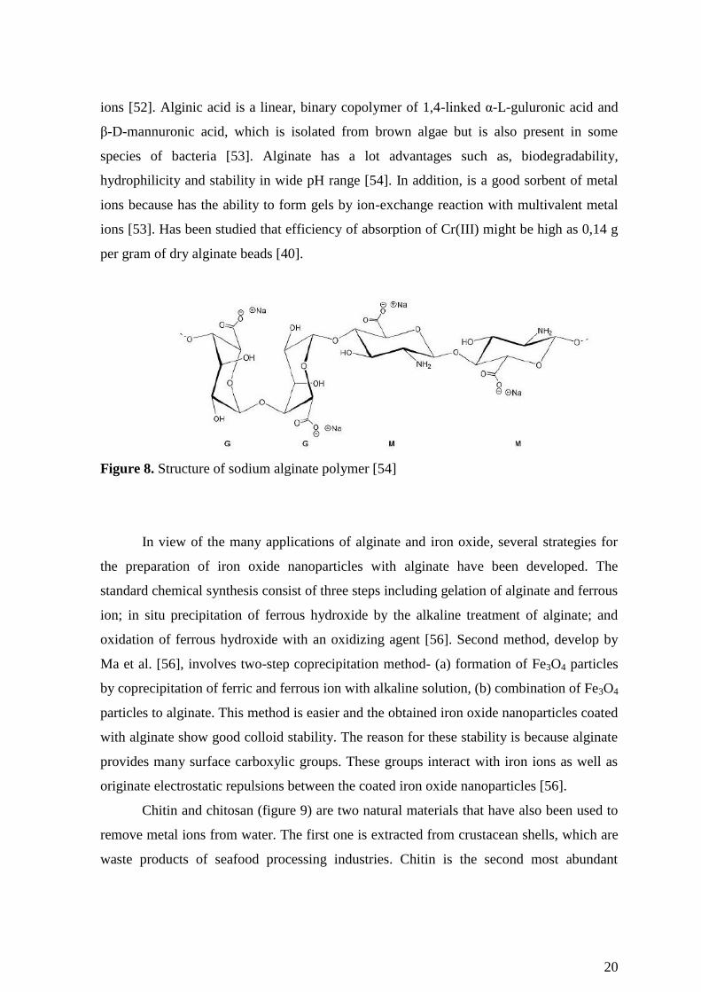
20
ions [52]. Alginic acid is a linear, binary copolymer of 1,4-linked α-L-guluronic acid and
β-D-mannuronic acid, which is isolated from brown algae but is also present in some
species of bacteria [53]. Alginate has a lot advantages such as, biodegradability,
hydrophilicity and stability in wide pH range [54]. In addition, is a good sorbent of metal
ions because has the ability to form gels by ion-exchange reaction with multivalent metal
ions [53]. Has been studied that efficiency of absorption of Cr(III) might be high as 0,14 g
per gram of dry alginate beads [40].
Figure 8. Structure of sodium alginate polymer [54]
In view of the many applications of alginate and iron oxide, several strategies for
the preparation of iron oxide nanoparticles with alginate have been developed. The
standard chemical synthesis consist of three steps including gelation of alginate and ferrous
ion; in situ precipitation of ferrous hydroxide by the alkaline treatment of alginate; and
oxidation of ferrous hydroxide with an oxidizing agent [56]. Second method, develop by
Ma et al. [56], involves two-step coprecipitation method- (a) formation of Fe3O4 particles
by coprecipitation of ferric and ferrous ion with alkaline solution, (b) combination of Fe3O4
particles to alginate. This method is easier and the obtained iron oxide nanoparticles coated
with alginate show good colloid stability. The reason for these stability is because alginate
provides many surface carboxylic groups. These groups interact with iron ions as well as
originate electrostatic repulsions between the coated iron oxide nanoparticles [56].
Chitin and chitosan (figure 9) are two natural materials that have also been used to
remove metal ions from water. The first one is extracted from crustacean shells, which are
waste products of seafood processing industries. Chitin is the second most abundant

21
polymer in nature after cellulose, whereas, chitosan can be formed by deacetylation of
chitin. The chitosan has drawn particular attention for its feasible application in a variety of
forms, from flake-types to gels, beads and fibers [57]. Because of the high content of
aminic and hydroxyl groups in chitosan, this by-product material has been found to be able
to adsorb several metal ions including those of chromium [58] and lead [57]. According to
Keng et al. [57], the extension of metal adsorption by this material depends strongly on the
chitosan source, deacetylation degree, nature of metal ion, variations in crystallinity, amino
content and solution pH.
Figure 9. Molecular structure of (a) chitosan [58] and (b) chitin
Yantasee et al. [59] prepared iron oxide nanoparticles, which were functionalized
with dimercaptosuccinic acid (DMSA). This way allowed to prepare a novel dispersible
sorbent, which can be magnetically collected and has a high capacity and selectivity for the
softer heavy metals. Hg(II), Ag(I), Pb(II), Cd(II) and Tl(I) ions effectively bind to the
DMSA ligands, while As(III) binds to the iron oxide lattices. In the article is described also
the ease with which nanoparticles can be separated from solution- within a minute using a
magnet. The authors compared the chemical affinity, capacity, kinetics and stability of the
magnetic nanoparticles to those of conventional resin based sorbents, activated carbon and
nanoporous silica of similar surface chemistries in river water, ground water and seawater.
These studies have shown an advantage of iron oxide functionalized with DMSA.

22
DMSAFe3O4 showed a capacity of 227 mg of Hg/g, a 30-fold larger value than that of
resin based sorbents.
Liu et al. [55] have studied a novel low-cost magnetic sorbent material prepared by
coating Fe3O4 magnetic nanoparticles with humic acid (HA). These nanoparticles were
developed for the removal of heavy metal ions, such as Hg(II), Pb(II), Cd(II) and Cu(II)
from water. HA has high affinity to magnetite and in the use its functionalization have a
number of advantages. One of them is that sorption of HA on the Fe3O4 nanoparticles
enhanced the stability of nanodispersions by preventing their aggregation. This acid has a
skeleton of alkyl and aromatic units that comprises carboxylic acid, phenolic hydroxyl and
quinone functional groups. Due to these functional groups, which have high coordination
capacity for heavy metal ions, HA was applied to remove heavy metal ions from water.
These studies have shown that sorption of the heavy metals to Fe3O4/HA reached
equilibrium in less than 15 min and agreed to the Langmuir adsorption model with
maximum adsorption capacites ranging from 45 to 100 mg/g. 99% of Hg(II) and Pb(II),
and over 95% of Cu(II) and Cd(II) were removed from tap water at optimized pH.
Also amino groups were used to functionalization of magnetic nanoparticles. Wang
et al. [61] developed amino-functionalized magnetic nanomaterial by covalently grafting
amino groups onto the surfaces of Fe3O4@SiO2. This material was used to remove heavy
metal ions from aqueous media. Cu(II), Co(II), Ni(II), Zn(II), Pb(II) and Cd(II) ions can be
removed from aqueous solution owing to the strong metal complexing capability of amino
groups. In fact, amino-functionalized nanoadsorbent exhibited high affinity for aqueous
Cu(II), Pb(II) and Cd(II) ions. In this study the results of adsorption affinity were not much
satisfactory. The reason for this was the presence of a cosolute of HA or alkali/earth metal
ions (Na+, K
+, etc.). Fe3O4@SiO2-NH2 nanoparticles were easily recovered from aqueous
solution by magnetic separation and regenerated easily by acid treatment.
Singh et al. [41] have used Fe3O4 magnetic nanoparticles functionalized with amine
groups for removal of Cr3+
ions from water. Experiment showed that removal efficiency of
metal ions is strongly dependent on pH of the solution, which is seen in figure 10. The
removal of ions increase with increasing pH of the solution till extraction rates approach a
plateau. About pH 6 obtained almost 100% removal of Cr3+
ions from waste-water. The
experiment carried out with different concentration of nanoadsorbents. This confirmed that
the removal efficiency of metal ions is also dependent on the concentration of

23
nanoadsorbents- adsorption of metal ions increases with increasing the concentration of
nanoadsorbents.
Figure 10. pH dependent removal efficiency of some metal ions by amine functionalized
nanoparticles [41]
Salicylic acid (SA) is a commercial ligand with a carboxylic and a phenolic
function site which can act as electron donor reacting with most of hard and intermediate
cations. For these reasons Shishehbore et al. [62] developed a method for the pre-
concentration of trace heavy metal ions in environmental samples. It consist in the sorption
of Cu(II), Cd(II), Ni(II) and Cr(III) ions with SA as chelate on silica-coated magnetite
nanoparticles. Figure 11 shows steps of synthesis of magnetic nanoparticles with SA and
also solid phase extraction of the analytes.
Figure 11. Schematic of the preparation of adsorbent (a) and solid phase extraction of the
analytes (b) [62]

24
The solution pH is an important parameter to obtain quantitative recovery for heavy
metal ions which may confirm figure 12. Could be seen that quantitative recovery was
obtained for metal ions within the pH range 5-7 and the decrease in recovery at pH values
lower than 5. May be this due to the competition of proton with cations in binding to donor
atoms. In fact, the method was successfully applied to the evaluation of these trace and
toxic metals in various waters.
Figure 12. Recovery of metal ions in function of pH, using salicylic acid [62]
All the methods described above are summarized in table 4.
Table 4. Examples of surface modified magnetite nanoparticles for the removal of heavy
metal ions from water
Synthesis method Coating agents Surface functionalized
group
Metal
removed Ref
Thermolysis of
precursors
Dimercaptosuccinic
acid Thiol group
Hg(II), Ag(I),
Pb(II), Cd(II) [59]
Co-precipitation Humic acid Carboxylic and phenolic
group
Hg(II), Pb(II),
Cd(II), Cu(II) [60]
Co-precipitation
followed by a sol-gel
process using sodium
silicate
(3-
aminopropyl)trimethox
ysilane
Amine group Pb(II), Cd(II),
Cu(II) [61]
Thermolysis of
precursors ethylenediamine Amine group
Cr(III), Co(II),
Ni(II), Cu(II),
Cd(II), Pb(II),
As(III)
[41]
Co-precipitation
followed by
hydrolysis of TEOS
Salicylic acid
Functionalized silica Carboxylic group
Cr(III), Cu(II),
Cd(II), Ni(II) [62]

25
1.4 Objectives and outline
Over the past decade, there has been great interest for nanoparticles as new sorbents
due to their unique size/surface dependent properties and possibility for removal of several
pollutants from wastewaters using adequate surface chemical functionalization.
In spite of the many advantages, colloidal nanoparticles might have also limitations
related to their colloidal instability over long periods of time. Small particles tend to form
agglomerates in order to reduce the energy associated with the high surface area per
volume ratio. Therefore it is necessary to develop coating strategies to chemically improve
the stability of colloidal magnetic nanoparticles because this is a crucial requirement for
their application as nanosorbents. Concerning magnetite nanoparticles, there are no reports
on the use of alginate as a coating material aiming sorption applications. Therefore the
main objective of this thesis was to investigate the use of this polysaccharide as a coating
material over magnetite nanoparticles aiming sorption applications. In particular, the
covalent attachment of alginate to silica modified magnetite surfaces was investigated and
the final materials were evaluated as sorbents for Cr(III) species dissolved in water.
Hence, in a first part of the thesis, magnetite nanoparticles coated with silica shells
have been functionalized with a coupling agent containing amine groups and then these
materials have been functionalized with alginate. These materials have been characterized
using several techniques such as FTIR spectroscopy, powder X-ray diffraction,
thermogravimetric analysis, zeta potential measurements, elemental analysis and TEM. In
a second stage, the magnetic nanoparticles functionalized with alginate were evaluated as
magnetic sorbents for the removal of [Cr(HY)(H2O)] complex and Cr(III) ions dissolved in
water.
The time profile for the adsorption behavior of those species onto magnetic
nanoparticles was assessed in order to investigate the kinetics involved. This model
describes the uptake rate of adsorbate, which in turn controls the resident time in the
adsorbent-solution interface. Several theoretical models have been used in order to explain
the sorption kinetics of the new materials in various experimental conditions. The results of
this research show that the covalent attachment of alginate to the magnetic nanoparticles
was successful and these new hybrid materials have potential as colloidal nanosorbents.

26
2. Experimental
2.1 Chemicals
Ferrous sulfate heptahydrated (FeSO4·7H2O) (>99%) and ethanol (C2H5OH)
(>99%) were obtained from Panreac (Spain). Potassium nitrate (KNO3) (>99%), tetraethyl
ortosilicate (Si(OC2H5)4; TEOS, >99%) and 3-aminopropyltrimethoxysilane
(H2N(CH2)3Si(OCH3)3, APTMS, >99%), N-hydroxysuccinimide (C4H5NO3, NHS, >98%),
disodium salt of ethylenediaminetetraacetic acid ([-CH2N(CH2CO2Na)CH2CO2H]2,
EDTA-Na2 >99%), N-(3-dimethylaminopropyl)-N’-ethylcarbodiimide hydrochloride
(C8H17N3·HCl, EDC >99%), alginic acid sodium salt from brown algae and potassium
chloride (KCl) (>99%) were purchased from Sigma-Aldrich. Potassium hydroxide (KOH)
(>86%) and sodium hydroxide (NaOH) (>98%) were purchased from Pronolab (Portugal).
Ammonia (NH4OH) (25% NH3) was purchased from Riedel-de-Häen (Germany) and
hydrochloric acid (HCl) (37%) from Fluka Chemie (Denmark). All chemicals were used as
received.
2.2 Synthesis of magnetite (Fe3O4) nanoparticles
Magnetic iron oxide nanoparticles were synthesized by oxidative hydrolysis of
iron (II) sulphate in alkaline conditions. 25 mL of deoxygenated water, which was first
deoxygenated with N2 under vigorous stirring during 2 hours, was added to a 250 mL
round flask. Then, 1,98g and 1,5327 g of KOH and KNO3 respectively were added. The
mixture was heated at 60ºC with bubbling N2 and mechanically stirred at 500 rpm. When
salt was dissolved, 25 mL of an aqueous solution containing 4,775 g of FeSO4·7H2O was
added drop-by-drop and the stirring was increased to 700 rpm. The resulting solution
presented a dark-green color after complete addition of Fe2+
salt. After the addition of all
above mentioned compounds, the solution was left to react for 30 min at 60ºC. After
reaction, the temperature was increased to 90ºC and the mixture was left under N2 with no
stirring during 4 hours.
The resulting black powder was washed several times with deoxygenated water and
ethanol and collected using a NdFeB magnet. After washing, the particles were dried by
evaporating the solvent at 60ºC.

27
2.3 Coating of magnetite nanoparticles with an amorphous silica shell functionalized
with amine groups (Fe3O4@SiO2-NH2)
Silica coating of the magnetite and amino-functionalized magnetite@SiO2 was
performed in one unique step in the following way: 50 mg of Fe3O4 nanoparticles were
added to 80 mL ethanol and then the suspension was left to sonicate for 10 min, allowing
for the particles to be highly dispersed in the solution. After, 100 µL of TEOS, 3 mL of
ammonia (25%) and 35 µL of APTMS were added to the solution which was left for 2
hours at room temperature under sonification. The resulting particles were washed with
distilled water and ethanol. The powder was then collected magnetically from the
suspension. After washing, particles were left to dry by solvent evaporation at 60ºC.
2.4 Covalent attachment of alginate to the magnetic particles (Fe3O4@SiO2-NH2-
ALG)
For the covalent attachment of alginate to the magnetite two solutions were
prepared. The first one comprised 50 mg of alginate dissolved in 5 mL distilled water to
which 20 mg and 25 mg of EDC and NHS, respectively, were added. This is necessary
because the reaction between the –NH2 groups of the surface of the particles and the –
COOH groups of the alginate is achieved by carbodiimide activation, using EDC and NHS.
The second solution comprised 30 mg of Fe3O4@SiO2-NH2 nanoparticles dispersed in
distilled water (5 mL). Then, the second solution was added to the first solution and the pH
was adjusted to 4,5 by adding 865 µL of 0,01 M HCl and 40 µL of 0,1 M HCl. The
resulting suspension was stirred for 24 h, at room temperature (23ºC) using a mini rotator.
After 24 h the nanoparticles were magnetically recovered using a NdFeB magnet, rinsed
with distilled water and freeze-dried.
2.5 Synthesis of [Cr(HY)(H2O)]
0,93 g of EDTA-Na2, was added to a solution of 1 g of Cr(NO3)3·9H2O, previously
dissolved in 13 mL of hot water, under vigorous stirring. Then, the resulting purple

28
solution was heated, with constant stirring, at a temperature slightly lower than boiling
temperature for 30 minutes. At the end the formation of a purple solid has been observed.
After reaction the mixture was left to cool at room temperature. The formed crystals were
filtered and washed with 10 mL of distilled water and then with a little ethanol. After this,
the crystals were dried at room temperature.
2.6 Instrumentation
Fourier transform infrared (FTIR) spectra of the Fe3O4, Fe3O4@SiO2-NH2,
Fe3O4@SiO2-NH2-ALG and [Cr(HY)(H2O)] were collected using a spectrometer Bruker
optics tensor 27 coupled to a horizontal attenuated total reflectance (ATR) cell, using 128
scans at a resolution of 2 cm-1
. The crystallite phase of the magnetic nanoparticles was
identified by powder X-ray diffraction (XRD). The X-ray powder diffraction patterns of
the samples were recorded using X-ray diffractometer Philips X’Pert equipped with a Cu
Kα monochromatic radiation source. The alginate content in the functionalzed magnetic
nanoparticles was calculated by thermogravimetric analysis (TGA). These experiments
were performed using a TGA 50 from a Shimadzu instrument. Samples were heated from
25ºC to 900ºC at 10ºC min-1
, under nitrogen atmosphere. The surface charge of the Fe3O4,
Fe3O4@SiO2-NH2 and Fe3O4@SiO2-NH2-ALG were assessed by zeta potential
measurements, using a Zeta-sizer Nanoseries instrument from Malvern Instruments. The
zeta potential measurements were performed in aqueous solutions prepared with distilled
water. Elemental analysis (C, H and N) was performed by Truspec 630-200-200 at
combustion furnance temperature 1075ºC. Carbon and hydrogen were detected using
infrared absorption and nitrogen using thermal conductivity. The morphology and size of
bare and coated nanoparticles was investigated by transmission electron microscopy
(TEM). For this purpose, used a transmission electron microscope Hitachi 9000 operating
at an accelerating voltage of 300 kV. Samples were prepared by evaporating dilute
suspensions of the nanoparticles on a copper grid coated with an amorphous carbon film.

29
2.7 Adsorption experiments
The nanomaterials described above were investigated for the uptake of
[Cr(HY)(H2O)] complex and Cr(III) ions from water, using Fe3O4@SiO2-NH2 and
Fe3O4@SiO2-NH2-ALG nanoparticles respectively. As a source of Cr(III) used
Cr(NO3)3·9H2O. Experimental solutions of the desired concentrations were obtained by
succesive dilution. The pH of the solution was adjusted by adding 0,01 M HCl or NaOH
solution before adsorption experiment. The general procedure for both compounds was
performed as follows. Accurately weighted amounts of magnetic nanoparticles were added
to a [Cr(HY)(H2O)] and Cr(III) aqueous solution of known concentration prepared with
ultra pure water. This time was considered the starting point of the experiment. The
samples were placed on a mini rotator at room temperature (24ºC). Supernatants were
collected for analysis at known times, and the magnetic nanoparticles were separated using
NdFeB magnet. The chromium equilibrium concentration was spectrophotometrically
measured. Cr(III) ions were determined using atomic absorption spectrometry (AAS) and
[Cr(HY)(H2O)] complex equilibrium concentration was measured using a standard
procedure based on the measurement of the absorbance at λ=544 nm.
The amount of Cr(III) ions and [Cr(HY)(H2O)] complex adsorbed into the
nanoparticles at time t (qt, mg/g) was calculated by using the following expression:
m
VCCq tt 0 (5)
where C0 (mg/L) is the initial concentration of Cr(III) or [Cr(HY)(H2O)], Ct is the
concentration at time t (mg/L), V is the total volume (L) and m is the mass of the dried
nanoparticles (g) used as sorbent.
2.7.1 The effect of the pH on [Cr(HY)(H2O)] complex
In order to determine the effect of the pH on the complex, a potentiometric titration
of a solution of the [Cr(HY)(H2O)] complex with NaOH was carried out. For this purpose
prepared 50 mL of a solution of the complex with a concentration 0,005 M was prepared
usng as a solvent a solution of 0,2 M KCl. During titration 50 mL of 0,02 M NaOH were

30
added. Value of pH solution of the complex before and after addition of NaOH was 2,38
and 11,65, respectively.
2.7.2 Kinetic experiments
The time profile of above compounds adsorption onto magnetic nanoparticles was
assessed in order to investigate the kinetics of adsorption. For this purpose, 10 mg of
Fe3O4@SiO2-NH2 and Fe3O4@SiO2-NH2-ALG (accurately weighted) were added to the
aqueous solution of [Cr(HY)(H2O)] complex (20 mL, 300 mg/L) and Cr(III) (20 mL,
30 mg/L), respectively. The pH of solutions was previously adjusted to 6. The solutions
were shaken at room temperature (24ºC) and after 15 min, 30 min, 45 min, 1 h, 2 h, 3 h,
4 h, 8 h, 24 h and 48 h 1 mL of aliquots were collected. The amount of complex and Cr(III)
ions adsorbed onto the magnetic nanoparticles at each time interval (qt, mg/g) was
determined and plotted against time (t, min) for kinetic modelling.
2.7.3 Equilibrium isotherms experiments
To obtain equilibrium isotherms ca. 1,5 mg (accurately weighted) of coated
magnetic nanoparticles were dispersed in 3 mL solution. Initial concentration for
[Cr(HY)(H2O)] complex was 50, 100, 200, 300, 400 and 500 mg/L but for Cr(III) ions the
initial concentration was 5, 10, 30, 50, 75 and 100 mg/L, both at pH=6.. The experiments
were conducted at 24ºC for a total period of 24 h and for 8 h for [Cr(HY)(H2O)] and Cr3+
,
respectively. The amount of complex and Cr(III) ions adsorbed at equilibrium (qe, mg/g)
was assessed by UV-Vis spectroscopy for [Cr(HY)(H2O)] and AAS for Cr(III). The
calculations were performed using equation (5) for C=Ce.

31
2.8 Kinetics modelling
An analysis of the kinetic data is important because the kinetics describe the uptake
rate of adsorbate, which in turn controls the resident time in the adsorbent-solution
interface. To describe adsorption data two kinetic models, the pseudo-first-order (equation
6) and the pseudo-second-order equations (equation 7) [58] were used
tk
et eqq 11
(6)
tqk
tqkq
e
e
t
2
2
2
1 (7)
In these equations k1 (h-1
) is the pseudo-first-order rate constant and k2 (g·mg-1
h-1
)
is the of pseudo-second-order rate constant. The pseudo-first-order equation has been used
to calculate the adsorption of solutes from liquid solutions in systems near equilibrium and
in systems with a time independent solute concentration or linear behavior in equilibrium
adsorption isotherms [64]. The pseudo-second-order kinetic equation is the opposite of
previous model, because the second one usually predicts the behavior over the whole range
of adsorption [65].
2.9 Isotherm modelling
Equilibrium data are the basic requirements for the design of adsorption systems
and provide valuable information to assess the adsorption capacity of an adsorbent. Also
give and understanding on the interactions between the adsorbate and the adsorbent, at
equilibrium under isothermal conditions [70]. Many isotherm equations are commonly
used, including Langmuir, Freundlich, Redlich-Peterson, Temkin and Elovich. In this
work, the equilibrium adsorption data of Cr(III) ions and [Cr(HY)(H2O)] has been
investigated using the Langmuir and Freundlich isotherm models.

32
The first one is based on the assumption that all sites possess equal affinity for the
adsorbate and a monolayer is formed when the solid surface reaches saturation [66]. The
mathematical expression shows equation (8) and the linear expression (9)
eL
eLe
CK
CKqq
1
0
(8)
00
1
q
C
qKq
C e
Le
e (9)
where q0
is the Langmuir monolayer adsorption capacity (mg/g), KL is the Langmuir
isotherm constant (L/mg) and qe (mg/g) and Ce (mg/L) are the amount of adsorbate
adsorbed and the final adsorbate concentration in solution at equilibrium, respectively.
The Freundlich isotherm model is an empirical equation based on heterogenous
surfaces suggesting that binding sites are not equivalent and/or independent which is given
as:
neFe CKq
1
(10)
eFe Cn
Kq ln1
lnln (11)
Where KF Freundlich isotherm constant [(mg/g)(L/mg)(1-n/n)] and n Freundlich exponent
(dimensionsless) related to adsorption capacity and adsorption intensity, respectively

33
3. Results and discussion
3.1 Characterization of magnetic nanoparticles
Magnetic iron oxide nanoparticles were synthesized by oxidative hydrolysis of
iron(II) sulphate in alkaline conditions and under an atmosphere of N2. KOH and KNO3
were first heated and then an aqueous solution of FeSO4·7H2O was added to the mixture.
Reaction of this synthesis is shown in equation 12.
OHSOOFeOHSOFeN
2
2
343
,2
4
2 92186 2 (12)
To confirm the crystal structure of the magnetite, XRD experiment was performed
and the resulting difractogram is shown in figure 16. According to the literature [68, 69,
70], the lattice spacing calculated from the diffraction peaks observed at 30º, 35º, 37º, 43º,
53º, 57º and 62º matched the [220], [311], [222], [400], [422], [511] and [440] planes of
Fe3O4 crystals, respectively. These results suggest that the main crystalline phase obtained
is magnetite (Fe3O4).
Figure 13. X-ray diffraction patterns of Fe3O4
20 30 40 50 60 70
Inte
nsi
ty (
a. u
.)
2θ (º)
(222)
(400)
(422)
(511)
(440)
(311)
(220)

34
The Fe3O4 nanoparticles are spheroidal with an average particle diameter of
55 nm, as seen by scanning electron microscopy (SEM) in figure 14.
Figure 14. SEM micrograph of Fe3O4 nanoparticles (courtesy: Dr. Ana Estrada, University
of Aveiro)
Prepared magnetic nanoparticles, as described above, were coated with amorphous
silica shell functionalized with amine groups. This reaction was performed by hydrolysis
of TEOS in alkaline conditions with ammonia solution acting as a catalyst. The surface
amino modification of the silica coated magnetite was performed via hydrolysis and
condensation of APTMS. Equations 13 and 14 show chemical reactions described above.
OHHCSiOOHHOCSiNH
5222452 42 3 (13)
243 SiOOFe (14)

35
Figure 15. Reactions of chemical modified of magnetic nanoparticles (R= -(CH2)3-NH2)
Covalent attachment of alginate to the particles coated with amorphous silica shell
functionalized with amine groups was carried out using EDC and NHS. The reaction
between –NH2 groups of the surface of the particles with silica shell and the –COOH
groups of the alginate is achieved by carbodiimide activation, using above compounds.
Figure 16 shown the structure of the new nanoparticle obtained above method.
Figure 16. Structure of the new nanoparticle covalent attachment of alginate Fe3O4@SiO2-
NH2-ALG

36
Figure 17 shows Fourier transform infrared (FT-IR) spectra of the iron oxide
nanoparticles (Fe3O4), iron oxide nanoparticles coated with silica shell functionalized with
amine groups (Fe3O4@SiO2-NH2) and after covalent linkage with alginate (Fe3O4@SiO2-
NH2-ALG). The spectra of magnetite show strong bands in the low-frequency region
(1000-400 cm-1
) due to the iron oxide skeleton [66]. The characteristic band of ν(Fe-O) is
at 579cm-1
in the FT-IR spectrum. The formation of a silica-coating on the magnetite
surface was confirmed by bands at 1124 cm-1
, 1052 cm-1
, 779 cm-1
and
455cm-1
assigned to the Si-OH vibration, Si-O-Si and Fe-O-Si groups [67]. The peak at
3421 cm-1
is attributed to the O-H stretching vibrations of the hydroxyl group on the
surface of silica [68]. Successful amino-functionalization of the silica layer on magnetite
surface was confirmed by the absorption bands at 1635 cm-1
, 1558 cm-1
,
1496 cm-1
. These bands ascribed to the N-H stretching and bending vibrations of amino
groups. The FT-IR spectrum of magnetite with alginate show bands at 1632 cm-1
attributed
to the antisymmetric stretch of νas (COO-) and 1413 cm
-1 attributed to the symmetric
stretch νs(COO-) which corroborate the presence of the polysaccharide alginate.
Figure 17. FT-IR spectra of (a) Fe3O4 (b) Fe3O4@SiO2-NH2 (c) Fe3O4@SiO2-NH2-ALG

37
Table 5 presents the experimental values of C, H and N percentage of Fe3O4,
Fe3O4@SiO2-NH2 and Fe3O4@SiO2-NH2-ALG measured by elemental analysis. The
highest percentage of N appears in sample Fe3O4@SiO2-NH2, which confirms the presence
of amine groups. After the covalent linkage with the alginate the C percentage increases
significantly which may be a proof of the presence of alginate in the last sample. Thus this
technique allowed to confirm the successful coating of magnetite nanoparticles with
amorphous silica shell functionalized with amine groups and the attachment of alginate to
the magnetic nanoparticles.
Table 5. Results of elemental analysis of the nanoparticles.
Sample % C % H % N
Fe3O4 0,1247 0,1263 -
Fe3O4@SiO2-NH2 1,0948 0,4464 0,3998
Fe3O4@SiO2-NH2-ALG 2,2449 0,6579 0,3768
The modification of the surface of Fe3O4 nanoparticles was further confirmed by
thermogravimetric analysis (TGA). Figure 18 compares the TGA curves of the several
samples. It can be seen that the weight loss of the Fe3O4 is much higher than the
Fe3O4@SiO2-NH2 and the Fe3O4@SiO2-NH2-ALG. This may be caused by the presence of
contamination of magnetite.
Figure 18. TGA graphs of Fe3O4; Fe3O4@SiO2-NH2 and Fe3O4@SiO2-NH2-ALG
70
75
80
85
90
95
100
0 500 1000
We
igh
t [%
]
Temperature [ºC]
Fe3O4
Fe3O4@SiO2-NH2
Fe3O4@SiO2-NH2-ALG

38
The nanoparticles prepared in this work were magnetic as they could be collected
by using an NdFeB magnet. A detailed characterization of the magnetic properties of the
nanoparticles here investigated was not carried out. However previous studies performed in
the same laboratory in similar magnetic samples have confirmed the magnetic properties of
magnetic nanoparticles. For instance Girginowa et al. [15] performed magnetic
measurements for Fe3O4 nanoparticles with an average size of 80 nm and Fe3O4@SiO2
samples at T=293 K. In these magnetometric measurements the sample is exposed to an
external magnetic field and the magnetization is recorded with a magnetometer. Uncoated
particles shown high values of magnetization saturation (92 emu·g-1
, 90 emu·g-1
) that
decreases after silica coating. Both exhibited coercivity and remanent magnetization. The
saturation magnetization has decreased with the modification of the pure magnetite while
the coercivity has increased. Similar research has been studied by Goya et al. [69].
Magnetic properties were confirmed also by Salgueiro et al. [70], but in this case magnetic
measurements were performed for bare and carrageenan coated magnetic nanoparticles
with an average diameter of 20 nm at T=300 K. Values of magnetization saturation show
that after polymer coating the magnetization saturation decreases to approximately half of
the initial value.
The surface charge of the nanoparticles Fe3O4@SiO2-NH2 and Fe3O4@SiO2-NH2-
ALG was assessed by zeta potential measurements. This is important to confirm the
potential application of these nanoparticles for the removal of chromium(III) complex with
EDTA and Cr(III) ions, used zeta potential measurements. These measurements were
performed at variable pH and the results are presented in table 6.
Table 6. Values of Zeta potential obtained in ultra pure water at variable pH
Sample pH Zeta Potential [mv]
Fe3O4@SiO2-NH2
4,66 5,71 ± 1,80
5,14 2,30 ± 0,35
6,24 5,69 ± 0,87

39
7,31 -26,8 ± 0,30
8,23 -21,23 ± 1,0
Fe3O4@SiO2-NH2-
ALG
4,03 -35,53 ± 1,78
5,42 -41,83 ± 2,08
6,27 -40,50 ± 0,50
7,86 -50,47 ± 1,20
8,06 -53,27 ± 2,75
In aqueous media the pH of the samples is one of the most important factors that
affect its zeta potential. Is visible that if more alkali is added to the solution then the
particles tend to aquire more negative charge, and is opposite way is when acid is added to
the solution. Therefore, a zeta potential versus pH curve is positive at low pH and negative
at high pH. In the case of Fe3O4@SiO2-NH2 was expected positive charge, due to
–NH2 group, but for Fe3O4@SiO2-NH2-ALG, negative charge, due to COO- groups of
alginate. Zeta potential measurements revealed a positive and negative surface charge for

40
the above nanomaterials. In pH 6 these values were 5,69 and -40,50 mV for nanoparticles
with amine groups and with alginate, respectively.
3.2 Adsorption experiments
3.2.1 Uptake of [Cr(HY)(H2O)] complexes using Fe3O4@SiO2-NH2 particles
3.2.1.1 The effect of pH on [Cr(HY)(H2O)] complex
[Cr(HY)(H2O)] complex is composed of Cr(III) ion, an water molecule and EDTA
anion, which in this case is present in the HY3-
form, where Y4-
represents the EDTA
molecule totally deprotonated (Figure 19).
Figure 19. Chemical structure of Y4-
anion
Figure 20. FTIR spectrum of the complex [Cr(HY)(H2O)]
1743
1648

41
Figure 20 shows the FTIR spectrum of the complex [Cr(HY)(H2O)] presenting
intense peaks. A strong band appears at 1743 cm-1
and can be assigned to the non-ionized
and uncoordinated COO stretching band. The vibration band of the ionized and
coordinated COO- appears at 1650-1590 cm-1
, but for coordination to metal ions such as
Cr(III) it occurs at 1648 cm-1
.
In aqueous solution the complex [Cr(HY)(H2O)] behaves like a diprotic acid, due to
the arrangement of ligands, which is shown in equations 15 and 16.
OHOHYCrOHOHHYCr 3222 (15)
OHOHYCrOHOHYCr 3
2
22 (16)
The first form of the complex ([Cr(HY)(H2O)] ) is purple, but the second ([Cr(Y)(OH)]2-
)
is blue and thus it is relatively easy to recognize which form exists at a specific pH value.
With this purpose, the potentiometric titration of an acid solution of the complex using
NaOH(aq) as a titrant was carried out The graphical representation of the pH value versus
the volume of base added is shown in figure 21. Two equivalence points are visible. Before
the first point solution is still acidic, so the equilibrium concentration of [Cr(Y)(OH)]2-
is
negligible. The first equivalence point in the graph is attributed to the formation of
[Cr(Y)(H2O)]-, while the second point is assigned to the formation of the blue compound
[Cr(Y)(OH)]2-
[72].

42
Figure 21. Potentiometric titration curve of [Cr(HY)(H2O)] with NaOH
Figure 22. Color of the solution at different pHs (a) 2,38 (b) 7,66 (c) 8,06 (d) 11,24
(e) 11,55
0
2
4
6
8
10
12
14
0 10 20 30 40 50 60
pH
Na OH [mL]

43
3.2.1.2 Kinetic studies
The time profile of the [Cr(HY)(H2O)] uptake by the nanoparticles coated with
amorphous silica shell functionalized with amine groups, for initial concentration of
complex (300 mg/L) at 24ºC is present in figure 23. Is visible that the adsorption curve
displays an usual shape. A rapid adsorption takes place at the beginning and after 24 h
decreases. Figure 23 represents the concentration of the complex that remains in solution
with time. As is shown in figure, on the beginning concentration reached variable values,
but after 4 hours is visible decrease of the concentration. These different values may be due
to errors during experiment or during characterization of samples. As was written previous,
the magnetic nanoparticles were separate magnetically and then the supernatant were
analyzed by UV-VIS spectroscopy. Due to the dark color of the nanoparticles, they absorb
the radiation so, even residual amount of nanoparticles, may affect to final calculations.
Therefore, the concentration of [Cr(HY)(H2O)] complex has unusual values.
Figure 23. The time profile of the [Cr(HY)(H2O)] uptake by Fe3O4@SiO2-NH2 (a)
concentration of complex in aqueous solution (b)

44
The adsorption kinetics was investigated and the experimental data was fitted with
well-known kinetic models such as the pseudo-first-order and the pseudo-second-order
kinetic equation, given by equation (6) and (7), respectively. Table 7 shows kinetic
parameters obtained using both models. The results show that both models describe not
well the kinetics of [Cr(HY)(H2O)] complex removal. To compare both models, the
pseudo-second-order model describe better the kinetic of complex removal as confirmed
by the higher value of R2 (table 7).
Table 7. Kinetic parameters estimated from pseudo 1st and 2
nd order equations and
evaluations of its fittings
Model Parameters R2
Pseudo-1st order
qe (mg/g)
31,7852 0,0055
k1 (h-1
)
1·10-4
Pseudo-2nd
order
qe (mg/g)
64,5161 0,954
k2 (g·mg-1
· h-1
)
0,4820·10-4
3.2.1.3 Isotherm studies
A plot of the solute concentration in the adsorbent phase qe (mg/g) as function of
the solute concentration in the solution Ce (mg/L) at equilibrium gives an adsorption
isotherm (figure 24). As shown the figure 24 adsorption capacity decrease with increasing
of concentration of [Cr(HY)(H2O)] complex. This indicates that the [Cr(HY)(H2O)]
complex are adsorbed onto the surface of the Fe3O4@SiO2-NH2 particles.

45
Figure 24. Isotherm of the adsorption of [Cr(HY)(H2O)] using Fe3O4@SiO2-NH2
nanoparticles (a) fit of Langmuir isotherm model for adsorption of above complex
In order to understand and clarify the adsorption process, Langumir adsorption
isotherm and Freundlich adsorption isotherm models were applied in this study. The
isotherm constants were calculated from the experimental data and presented in table 8.
Taking into consideration the values of the correlation coefficient as a criterion for
goodness of fit for the system, the Langumir isotherm model (figure 24) shows better
correlation (R2=0,9191) than the Freundlich adsorption isotherm model. The adsorption
data confirm that the Langumir isotherm can be interpreted as indicating a homogeny
adsorption process, leading to monolayer binding.
Table 8. Langumir and Freundlich isotherm model parameters for adsorption
[Cr(HY)(H2O)] complex
Langmuir isotherm model Freundlich isotherm model
q0 (mg·g
-1) KL (L·mg
-1) R
2
KF
((mg/g)·(mg/L)n)
n R2
62,9 0,042 0,9191 12,1 3,51 0,6062
0
10
20
30
40
50
60
70
80
90
0 200 400 600
qe
[mg/
g]
C final [mg/L]
Experimental
langmuir model

46
3.2.2 Uptake of Cr(III) species using Fe3O4@SiO2-NH2-ALG particles
3.2.2.1 Kinetic studies
The adsorption of Cr(III) species, with initial concentration 30 mg/L, by
Fe3O4@SiO2-NH2-ALG particles as a function of the contact time is shown in figure 25 a.
It shows that the plots are not linear over the whole time range which confirm that Cr(III)
species were adsorbed. Also is shown that the adsorption capacity increases with
increasing contact time. The figure 25 b presents changing of concentration with time. Is
shown that concentration of Cr(III) decreases with increasing contact time, so this may
indicate that magnetic nanoparticles with covalent attachment of alginate are able to
remove of Cr(III) species from aqueous solution.
Figure 25. The time profile of adsorption capacity of Cr(III) species onto
Fe3O4@SiO2-NH2-ALG (a) Change in the concentration of Cr(III) vs time (b)
Table 9 lists the kinetics constants evaluated from the adsorption kinetics, along
with the correlation coefficients and the experimental adsorption capacities.The pseudo-
first-order model and the pseudo-second-order model fit equally because R2 is similar in
both cases.

47
Table 9. Kinetic parameters estimated from pseudo 1st and 2
nd order equations and
evaluations of its fittings
Model Parameters R2
Pseudo-1st order
qe (mg/g)
9,7796 0,965
k1 (h-1
)
0,0033
Pseudo-2nd
order
qe (mg/g)
12,9199 0,966
k2 (g·mg-1
· h-1
)
6,01·10-4
3.2.2.2 Isotherms
Figure 26 shows the adsorption isotherm curves present isotherms of Cr(III) on
Fe3O4@SiO2-NH2-ALG particles in aqueous solution. The isotherm curves present the
Cr(III) uptake as a function of the equilibrium Cr(III) solution concentration at room
temperature. It can be seen that in the beginning the adsorption capacity decreases, which
may be the result of errors during characterization of solution after the experiment. After
this point the adsorption capacity increases and the graph resume normal shape. This
behavior may prove that magnetic nanoparticles can be employed to removal Cr(III)
species from water.
Figure 26. Isotherm of the adsorption of Cr(III) using Fe3O4@SiO2-NH2-ALG
nanoparticles and fit of Freundlich isotherm model for adsorption of above complex
0
2
4
6
8
10
12
14
0 50 100
qe [
mg/
g]
C final [mg/L]
Freundlich model
Experimental

48
The Langmuir and Freundlich isotherm models parameters for Cr(III) ions obtained
from the fitting of experimental data are listed in table 10. The results in table 10 suggested
that Freundlich isotherm model (figure 26) describes the adsorption of metal ions better
than of Langmuir model, evidenced by higher R2. Values of n implies stronger interaction
between adsorbent and metal and also is in the range of 1<n<10, which also demonstrates
that these particle act as a favorable adsorbent for the removal of Cr(III) from aqueous
solution. Nevertheless, the R2 obtained for the Freundlich model fitting was still low,
which indicates that both models do not describe the adsorption capacity satisfactory.
Table 10. Langmuir and Freundlich isotherm model parameters for adsorption Cr(III)
species
Langmuir isotherm model Freundlich isotherm model
q0 (mg·g
-1) KL (L·mg
-1) R
2
KF
((mg/g)·(mg/L)n)
n R2
19,8019 0,01266 0,3863 0,4934 1,4337 0,781

49
4. Conclusion and perspectives of future work
Magnetite nanoparticles coated with an amorphous silica shell functionalized with
amine groups were prepared and evaluated as magnetic sorbents for the removal of Cr(III)
EDTA complexes, here designated as [Cr(HY)(H2O)], from aqueous solutions, using
simple NdFeB magnet. Under the experimental conditions used (pH=6, room temperature),
the nanoparticles showed affinity for the complex and allowed its removal . The adsorption
followed a pseudo-second order kinetics, while the equilibrium adsorption was better
described by the Langmuir isotherm model, that estimated a maximum adsorption capacity
of 62 mg/g.
The biopolymer alginate was successfully covalently attached to the amine surface
functionalized nanoparticles, to our knowledge for the first time. The reaction between the
–NH2 groups of the surface of the particles Fe3O4@SiO2-NH2 and the –COOH groups of
the alginate was achieved by carbodiimide activation, using EDC and NHS. The resulting
nanoparticles were employed to uptake Cr(III) species from aqueous solutions. The
adsorption kinetics was well described by both pseudo-first and pseudo-second order
equations. For the isotherm studies, the Freundlich model gave better results than the
Langmuir model, although still unsatisfactory due to low R2.Results described in this work
indicate that these particles can be used for Cr(III) species removal from aqueous solution.
To assess the full potential of the materials here developed for the uptake of Cr(III)
species from water, additional studies are necessary for the evaluation of optimal uptake
conditions, such as initial concentration, pH and temperature, as well as the study of the
kinetics and equilibrium isotherms for the removal of different metal cations, and the
evaluation of their performance in the purification of real effluents of different chemical
composition.

50
Reference
[1] Khajeh M., Laurent S., Dastafkan K., Nanosorbents: classification, preparation, and
applications (with emphasis on aqueous media), Chemical Reviews (2012)
[2] Lopez-Serrano A., Munoz Olivas R., Sanz Landaluze J., Camara C., Nanoparticles: a
global vision. Characterization, separation, and quantification methods. Potential
environmental and health impact, Analytical Methods 6 (2014) 38-56
[3] Liu Y., Liang P., Guo L., Nanometer titanium dioxide immobilized on silica gel as
sorbent for preconcentration of metal ions prior to their determination by inductively
coupled plasma atomic emission spectrometry, Talanta 68 (2005) 25-30
[4] Mohmood I., Lopes C., Lopes I., Ahmad I., Duarte A., Pereira E., Nanoscale materials
and their use in water contaminants removal- a review, Environmental Science and
Pollution Research 20 (2013) 1239-1260
[5] Zhang L., Fang M., Nanomaterials in pollution trace detection and environmental
improvement, Nano Today 5 (2010) 128-142
[6] Wigginton N., Haus K., Hochella M., Aquatic environmental nanoparticles, Jurnal of
Environmental Monitoring 9 (2007) 1285-1432
[7] Kroto H. W., Heath J.R., O’Brien S. C., Curl R. F., Smalley R. E., C60:
Buckminsterfullerene, Nature 318 (1985) 162-163
[8] Elyasi P., SalmanOgli A., Investigation of potential profile effects in quantum dot and
onion- like quantum dot-quantum well on optical properties, Optics Communications 318
(2014) 26-30
[9] Ferreira da Silva B., Perez S., Gardinalli P., Singhal R., Mozeto A., Barcelo D.,
Analytical chemistry of metallic nanoparticles in natural environments, Trends in
Analytical Chemistry 30 (2011) 528-540
[10] Gubin S., Koksharov Y., Khomutov G., Yurkov G., Magnetic nanoparticles:
preparation, structure and properties, Russian Chemical Reviews 74 (2005) 489-520

51
[11] Poole Ch., Owens F., Introduction to nanotechnology, Wiley Interscience, (2003),
Chapter 7
[12] Jiles D., Introduction to magnetism and magnetic materials, Taylor & Francis Group,
Chapter 4
[13] Cornell R., Schwertmann U., The iron oxides structure, properties, reactions,
occurrences and uses, Wiley-Vch (2000)
[14] Cademartiri L., Ozin G., Concepts of nanochemistry, Wiley-Vch (2009) Chapter 6
[15] Girginova P., Daniel-da-Silva A., Lopes C., Figueira P., Otero M., Amaral V., Pereira
E., Trindade T., Silica coated magnetite particles for magnetic removal of Hg2+
from
water, Journal of Colloid and Interface Science 345 (2010) 234-240
[16] Suleiman J., Hu B., Peng H., Huang Ch., Separation/preconcentration of trace
amounts of Cr, Cu and Pb in environmental samles by magnetic solid-phase extraction
with Bismuthiol-II-immobilized magnetic nanoparticles and their determination by ICP-
OES, Talanta 77 (2009) 1579-1583
[17] Chen L., Wang T., Tong J., Application of derivatized magnetic materials to the
separation and the preconcentration of pollutants in water samples, Trends in Analytical
Chemistry 30 (2011) 1095-1108
[18] Lu A., Salabas E., Schuth F., Magnetic nanoparticles: synthesis, protection,
functionalization and application, Angewandte Chemie 46 (2007) 1222-1244
[19] Massart R., Preparation of aqueous magnetic liquids in alkaline and acidic media, 17
(1981) 1247
[20] Charles S., The preparation of magnetic fluids, Lecture Notes in Physics 594 (2002)
3-18
[21] Gubin S., Koksharov Yu., Khomutov G., Yurkov G., Magnetic nanoparticles:
preparation, structure and properties, Russian Chemical Reviews 74 (2005) 489-520
[22] Wang Y., Yang H., Synthesis of iron oxide nanorods and nanocubes in an
imidazolium ionic liquid, Chemical Engineering Journal 147 (2009) 71-78

52
[23] Jiao H., Wang J., Thermal decomposition fabrication, growth mechanism and
characterization of Rod-Like γ-Fe2O3, Metal-Organic and Nano-Metal Chemistry 44
(2014) 1354-1357
[24] Butter K., Philipse A., Vroege G., Synthesis and properties of iron ferrofluids, Journal
of Magbetism and Magnetic Materials 252 (2002) 1-3
[25] Carlos L., Garcia Einschlag F., Gonzalez M., Martire D., Applications of megnetite
nanoparticles for heavy metal removal from wastewater, ISBN 978-953-51-0882-5 (2013)
Chapter 3
[26] Langevin D., Micelles and microemulsions, Annual Review of Physical Chemistry 43
(1992) 327-349
[27] Paul B., Moulik S., Uses and applications of microemulsions, Current Science 80
(2001) 990-1001
[28] Gupta A., Gupta M., Synthesis and surface engineering of iron oxide nanoparticles
for biomedical applications, Biomaterials 26 (2005) 3995-4021
[29] Laurent S., Forge D., Port M., Roch A., Robic C., Elst L., Muller R., Magnetic Iron
Oxide Nanoparticles: Synthesis, stabilization, vectorization, physicochemical
characterizations, and biomedical applications, Chemical Review 108 (2008) 2064-2110
[30] Yang S., Paik S., Ryu J., Choi K., Kang T., Lee J., Song Ch., Ko S., Dynamic light
scattering- based method to determine primary particle size of iron oxide nanoparticles in
simulated gastrointestinal fluid, Food Chemistry 161 (2014) 185-191
[31] Lim J., Yeap S., Che H., Low S., Characterization of magnetic nanoparticle by
dynamic light scattering, Nanoscale research letters 8 (2013) 1-14
[32] Zhou W., Apkarian R., Wang Z., Joy D., Fundamentals of Scanning Electron
Microscopy, Scanning Microscopy for Nanotechnology (2007) 1-40
[33] Chorupa I., Douziech-Eyrolles L., Nagaboini-Okassa L., Fouquenet J., Cohen-
Jonathan S., Souce M., Marchais H., Dubois P., Molecular composition of iron oxide

53
nanoparticles, precursors for magnetic drug targeting, as characterized by confocal
Raman microspectroscopy, Analyst 130 (2005) 1395-1403
[34] Russo P., Acierno D., Palomba M., Carotenuto G., Rosa R., Rizzuti A., Leonelli C.,
Ultrafine magnetite nanopowder: Synthesis, characterization and preliminary use as filler
of polymethylmethacrylate nanocomposites, Jurnal of Nanotechnology 2012 (2012) 1-8
[35] Bajpai A., Gupta R., Evaluation of water sorption behavior and In Vitro blood
compatibility of polyvinyl alcohol based magnetic bionanocomposites, Journal of Applied
Polymer Science 114 (2009) 3548-3560
[36] Ruiz-Hitzky E., Darder M., Aranda P., Functional biopolymer nanocomposites based
on layered solids, Journal of Materials Chemistry 15 (2005) 3650-3662
[37] Aime C., Coradin T., Nanocomposites from biopolymer hydrogels: blueprints for
white biotechnology and green materials chemistry, Journal of polymer science 50 (2012)
669-680
[38] Im S., Herricks T., Lee Y., Xia Y., Synthesis and characterization of monodisperse
silica colloids loaded with superparamagnetic iron oxide nanoparticles, Cgemical physics
letters 401 (2005) 19-23
[39] del Campo A., Sen T., Lellouche J., Bruce I., Multifunctional magnetite and silica-
magnetite nanoparticles: Synthesis, surface activation and applications in life sciences,
Journal of Magnetism and Magnetic Materials 293 (2005) 33-40
[40] Yamaura M., Camilo R., Sampaio L., Macȇdo M., Nakamura M., Toma H.,
Preparation and characterization of (3-aminopropyl)triethoxysilane- coated magnetite
nanoparticles, Journal of Magnetism and Magnetic Materials 279 (2004) 210-217
[41] Singh S., Barick K., Bahadur D., Surface engineered magnetic nanoparticles for
removal of toxic metal ions and bacterial pathogens, Journal of hazardous materials 192
(2011) 1539-1547
[42] Chen J., Xing H., Guo H., Li G., Weng W., Hu S., Preparation, characterization and
adsorption properties of a novel 3-aminopropyltriethoxysilane functionalized sodium

54
alginate porous membrane adsorbent for Cr(III) ions, Journal of Hazardous Materials 248-
249 (2013) 285-294
[43] Araujo M., Teixeira J., Trivalent chromium sorption on alginate beads, International
Biodeterioration & Biodegradation 40 (1997) 63-74
[44] Vassileva P., Detcheva A., Uzunov I., Uzunova S., Removal of metal ions from
aqueous solutions using pyrolyzed rice husks: adsorption kinetics and equilibria, Chemical
Engineering Communications 200 (2013) 1578-1599
[45] Rocha L., Lopes C., Borges J., Duarte A., Pereira E., Valuation of unmodified rice
husk waste as an eco-friendly sorbent to remove mercury: a study using environmental
realistic concentrations, Water air and soil pollution 224 (2013) 1-18
[46] Nakbanpote W., Thiravavetyan P., Kalambaheti C., Comparison of gold adsorption by
Chlorella vulgaris, rice husk and activated carbon, minerals Engineering 15 (2002) 549-
552
[47] Marshall W., Champagne E., Evans W., Use of rice milling byproducts (hulls and
bran) to remove metal ions from aqueous solution, Journal of Environmental Science and
Health 28 (1993) 1977-1992
[48] Fiol N., Serarols J., Poch J., Martinez M., Miralles N., Villaescusa I., Low cost
materials for metal uptake from aqueous solutions, Environmental Chemistry, Chapter 24
(2005) 251-258
[49] Sidiras D., Politi D., batzias F., Boukos N., Efficient removal of hexavalent chromium
from aqueous solutions using autohydrolyzed Scots Pine (Pinus Sylvestris) sawdust as
adsorbent, International Journal of environmetal science and technology 6 (2013) 1337-
1348
[50] Khosravihaftkhany S., Morad N., Teng T., Abdullah A., Norli I., Biosorption of Pb(II)
and Fe(III) from aqueous solutions using oil palm biomasses as adsorbents, Water air and
soil pollution 3 (2013) 1455

55
[51] Lavecchia R., Pugliese A., Zuorro A., Removal of lead from aqueous solutions by
spent tea leaves, CISAP4: 4th international conference on safety & environment in process
industry (2010) 73-78
[52] Abdel-Halim E., Al-Deyab, Removal of heavy metals from their aqueous solutions
through adsorption onto natural polymers, Carbohydrate Polymers 84 (2011) 454-458
[53] Choong J., Nah I., Hwang K., Adsorption of heavy metals using magnetically modified
alginic acid, Hydrometallurgy 86 (2007) 140-146
[54] Nayak D., Banerjee A., Roy S., Lahiri S., Speciation dependent studies on chromium
absorption using calcium alginate and iron doped calcium alginate biopolymer, Journal of
Radioanalytical and Nuclear Chemistry 274 (2007) 219-224
[55] Covaliu C., Matei C., Ianculescu A., Jitaru I., Berger D., Fe3O4 and CoFe2O4
nanoparticles stabilized in sodium alginate polymer, U.P.B. Scientific Bulletin, Series B
71 (2009) 1454-2331
[56] Ma H., Qi X., Maitani Y., Nagai T., Preparation and characterization of
superparamagnetic iron oxide nanoparticles stabilized by alginate, International Journal of
Pharmaceutics 333 (2007) 177-186
[57] Keng P., Lee S., Ha S., Hung Y., Ong S., Removal of hazardous heavy metals from
aqueous environment by low-cost adsorption materials, Environmental Chemistry Letters
12 (2013) 15-25
[58] Vieira R., Meneghetti E., Baroni P., Guibal E., Cruz V., Caballero A., Castellon E.,
Beppu M., Chromium removal on chitosan-based sorbents- An EXAFS/XANES
investigation of mechanism, Materials Chemistry and Physic 146 (2014) 412-417
[59] Yantasee W., Warner C., Sangvanich T., Addleman R., Carter T., Wiacek R., Fryxell
G., Timchalk Ch. Warner M., Removal of heavy metals from aqueous systems with thiol
functionalized superparamagnetic nanoparticles, Environmental Science and Technology
41 (2007) 5114-5119

56
[60] Liu J., zhao Z., Jiang G., Coating Fe3O4 magnetic nanoparticles with humic acid for
high efficient removal of heavy metals in water, Environmental Science and Technology 42
(2008) 6949-6954
[61] Wang J., Zheng S., Shao Y., Liu J., Xu Z., Zhu D., Amino-functionalized Fe3O4@SiO2
core-shell magnetic nanomaterial as a novel adsorbent for aqueous heavy metals removal,
Journal of Colloid and Interface Science 349 (2010) 293-299
[62] Shishehbore M., Afkhami A., Bagheri H., Salicylic acid functionalized silica-coated
magnetite nanoparticles for solid phase extraction and preconcentration of some heavy
metal ions from various real samples, Chemistry Central Journal 5 (2011)
[63] Lin J., Wang L., Comparison between linear and non-linear forms of pseudo-first-
order and pseudo-second-order adsorption kinetic models for the removal of methylene
blue by activated carbon, Frontiers of environmental science & engineering in China 3
(2009) 320-324
[64] Soto M., Moure A., Dominguez H., Parajo J., Recovery, concentration and
purification of phenolic compounds by adsorption: A review, Journal of food engineering
105 (2011) 1-27
[65] Ho Y., McKay G., Pseudo-second order model for sorption processes, Process
Biochemistry 34 (1999) 451-465
[66] Mochamed Ch., Applicability of some statistical tools to predict optimum adsorption
isotherm after linear and non-linear regression analysis, Journal of Hazardous Materials
153 (2008) 207-212
[67] Cao J., Wang Y., Yu J., Xia J., Zhang Ch., Yin D., Urs O., Preparation and
radiolabeling of surface-modified magnetic nanoparticles with rhenium-188 for magnetic
targeted radiotherapy, Journal of Magnetism and Magnetic Materials 277 (2004) 165-174
[66] Lu M., Liu Y., Hu X., Ben Y., Zeng X., Li T., Wang H., Competitive adsorption of
Cu(II) and Pb(II) ions from aqueous solutions by Ca-alginate immobilized activated
carbon and Saccharomyces cerevisiae, Journal of Central South University 20 (2013)
2478-2488 (od tego wszystkie w górę)

57
[67] Mürbe J., Rechtenbach A., Töpfer J., Synthesis and physical characterization of
magnetite nanoparticles for biomedical applications, Materials Chemistry and Physics 110
(2008) 426-433
[68] Mahdavi M., Ahmad M., Haron M., Gharayebi Y., Shameli K., Nadi B., Fabrication
and characterization of SiO2/(3-aminopropyl)triethoxysilane- coated magnetite
nanoparticles for Lead(II) removal from aqueous solution, Journal of inorganic and
organometallic polymers and materials 23 (2013) 599-607
[69] Shen M., Cai H., Wang X., Cao X., Li K., Wang S., Guo R., Zheng L., Zhang G., Shi
X., Facile one-pot preparation, surface functionalization, and toxicity assay of APTS-
coated iron oxide nanoparticles, Nanotechnology 23 (2012) 105601
[70] Goya G., Berquo T., Fonseca F., Morales M., Static and dynamic magnetic properties
of spherical magnetite nanoparticles, Journal of Applied Physics 94 (2003) 3520-3528
[71] Salgueiro A., Daniel-da-Silva A., Girao A., Pinheiro P., Trindade T., Unusual dye
adsorption behavior of κ-carrageenan coated superparamagnetic nanopartices, Chemical
Engineering Journal 229 (2013) 276-284
[72] Hoppe J., Howell P., Education in Chemistry, 12 (1975) 12-15
Web sides:
[1] http://www.ph.utexas.edu/~laser/projects/shelled.html (16.07.2014)
[2] http://www.glantreo.com/otherparticles.php (16.07.2014)

58
ANNEX I
Figure 1. Calibration curve for [Cr(HY)(H2O)]