Management of uterine adenomyosis: current trends and ... · Management of uterine adenomyosis:...
9
EDUCATIONAL REVIEW Open Access Management of uterine adenomyosis: current trends and uterine artery embolization as a potential alternative to hysterectomy Riham Dessouky 1* , Sherif A. Gamil 2 , Mohamad Gamal Nada 1 , Rola Mousa 1 and Yasmine Libda 1 Abstract Adenomyosis is a challenging clinical condition that is commonly being diagnosed in women of reproductive age. To date, many aspects of the disease have not been fully understood, making management increasingly difficult. Over time, minimally invasive diagnostic and treatment methods have developed as more women desire uterine preservation for future fertility or to avoid major surgery. Several uterine-sparing treatment options are now available, including medication, hysteroscopic resection or ablation, conservative surgical methods, and high- intensity focused ultrasound each with its own risks and benefits. Uterine artery embolization is an established treatment option for uterine fibroids and has recently gained ground as a safe and cost-effective method for treatment of uterine adenomyosis with promising results. In this review, we discuss current trends in the management of uterine adenomyosis with a special focus on uterine artery embolization as an alternative to hysterectomy. Keywords: Adenomyosis, Uterine artery embolization, Hysterectomy Key points Uterine artery embolization (UAE) seems to be the most promising uterine-sparing, minimally invasive treatment option for adenomyosis. Results of ongoing randomized controlled trial (QUESTA) will soon show whether UAE can be validated as a treatment option for adenomyosis. Ability to preserve fertility will be one of the main factors determining whether UAE can replace hysterectomy in treatment of adenomyosis, but further randomized controlled trials are needed. Introduction Adenomyosis is defined by the abnormal location of endometrial tissue within the myometrium associated with hypertrophy or hyperplasia of the myometrial stroma [1, 2]. Although pathogenesis and etiology of adenomyosis remain unknown, two main theories have been proposed: invagination of the endometrial basal layer and metaplasia of embryonic stem cells [3]. Preva- lence of adenomyosis varies widely from 5 to 70% [4–7] with recent studies showing about 20% prevalence [8– 10] among which the majority were premenopausal. Despite the absence of specific (pathognomonic) diag- nostic features for uterine adenomyosis, typical symp- toms include menorrhagia, chronic pelvic pain, and dysmenorrhea [11]. These symptoms are commonly en- countered in other gynecological disorders including leiomyomas and endometriosis, often confounding the clinical diagnosis [12]. For more than a century, diagnosis was dependent on histopathologic examination of post-hysterectomy speci- mens till the introduction of noninvasive ultrasound and MR techniques [13]. Since then, several studies have il- lustrated high sensitivities and specificities for both two-dimensional transvaginal sonography (TVS) and magnetic resonance imaging (MRI) [13–17]. Current © The Author(s). 2019 Open Access This article is distributed under the terms of the Creative Commons Attribution 4.0 International License (http://creativecommons.org/licenses/by/4.0/), which permits unrestricted use, distribution, and reproduction in any medium, provided you give appropriate credit to the original author(s) and the source, provide a link to the Creative Commons license, and indicate if changes were made. * Correspondence: [email protected] 1 Radiology Department, Faculty of Medicine, Zagazig University, Koliat Al Tob Street, Zagazig 44519, Egypt Full list of author information is available at the end of the article Insights into Imaging Dessouky et al. Insights into Imaging (2019) 10:48 https://doi.org/10.1186/s13244-019-0732-8
Transcript of Management of uterine adenomyosis: current trends and ... · Management of uterine adenomyosis:...

EDUCATIONAL REVIEW Open Access
Management of uterine adenomyosis:current trends and uterine arteryembolization as a potential alternative tohysterectomyRiham Dessouky1*, Sherif A. Gamil2, Mohamad Gamal Nada1, Rola Mousa1 and Yasmine Libda1
Abstract
Adenomyosis is a challenging clinical condition that is commonly being diagnosed in women of reproductive age.To date, many aspects of the disease have not been fully understood, making management increasingly difficult.Over time, minimally invasive diagnostic and treatment methods have developed as more women desire uterinepreservation for future fertility or to avoid major surgery. Several uterine-sparing treatment options are nowavailable, including medication, hysteroscopic resection or ablation, conservative surgical methods, and high-intensity focused ultrasound each with its own risks and benefits. Uterine artery embolization is an establishedtreatment option for uterine fibroids and has recently gained ground as a safe and cost-effective method fortreatment of uterine adenomyosis with promising results. In this review, we discuss current trends in themanagement of uterine adenomyosis with a special focus on uterine artery embolization as an alternative tohysterectomy.
Keywords: Adenomyosis, Uterine artery embolization, Hysterectomy
Key points
� Uterine artery embolization (UAE) seems to be themost promising uterine-sparing, minimally invasivetreatment option for adenomyosis.
� Results of ongoing randomized controlled trial(QUESTA) will soon show whether UAE can bevalidated as a treatment option for adenomyosis.
� Ability to preserve fertility will be one of the mainfactors determining whether UAE can replacehysterectomy in treatment of adenomyosis, butfurther randomized controlled trials are needed.
IntroductionAdenomyosis is defined by the abnormal location ofendometrial tissue within the myometrium associatedwith hypertrophy or hyperplasia of the myometrial
stroma [1, 2]. Although pathogenesis and etiology ofadenomyosis remain unknown, two main theories havebeen proposed: invagination of the endometrial basallayer and metaplasia of embryonic stem cells [3]. Preva-lence of adenomyosis varies widely from 5 to 70% [4–7]with recent studies showing about 20% prevalence [8–10] among which the majority were premenopausal.Despite the absence of specific (pathognomonic) diag-nostic features for uterine adenomyosis, typical symp-toms include menorrhagia, chronic pelvic pain, anddysmenorrhea [11]. These symptoms are commonly en-countered in other gynecological disorders includingleiomyomas and endometriosis, often confounding theclinical diagnosis [12].For more than a century, diagnosis was dependent on
histopathologic examination of post-hysterectomy speci-mens till the introduction of noninvasive ultrasound andMR techniques [13]. Since then, several studies have il-lustrated high sensitivities and specificities for bothtwo-dimensional transvaginal sonography (TVS) andmagnetic resonance imaging (MRI) [13–17]. Current
© The Author(s). 2019 Open Access This article is distributed under the terms of the Creative Commons Attribution 4.0International License (http://creativecommons.org/licenses/by/4.0/), which permits unrestricted use, distribution, andreproduction in any medium, provided you give appropriate credit to the original author(s) and the source, provide a link tothe Creative Commons license, and indicate if changes were made.
* Correspondence: [email protected] Department, Faculty of Medicine, Zagazig University, Koliat Al TobStreet, Zagazig 44519, EgyptFull list of author information is available at the end of the article
Insights into ImagingDessouky et al. Insights into Imaging (2019) 10:48 https://doi.org/10.1186/s13244-019-0732-8
treatment options for symptomatic adenomyosis includehysterectomy, medication, conservative surgery, or min-imally invasive techniques including uterine arteryembolization [18]. To date, hysterectomy remains thedefinitive treatment. This is mainly due to difficult diag-nosis, the diffuse nature of the disease, and littleevidence-based literature needed to standardize treat-ments [19]. This consequently results in a managementdilemma, particularly in symptomatic patients who wishto preserve their uterus [18].Uterine artery embolization (UAE) was first described
in 1995 by Ravina et al. [20] then later established as aneffective treatment option for patients with symptomaticuterine fibroids [21, 22]. Since then, UAE has been in-vestigated as a noninvasive treatment option for adeno-myosis with initial promising results [23, 24]. Whatremains to be known is whether UAE can be validatedas a safer, noninvasive, uterine-sparing alternative tohysterectomy. This article summarizes current trends inmanagement of uterine adenomyosis with special focuson the emerging role of UAE.
EtiologyThe precise etiology and pathophysiology leading to thedevelopment of adenomyosis remains undetermined.Several theories have been introduced, including trau-matic, immunological, hormonal, metaplastic, and stemcell [25]. Traumatic and immunological theories suggestdisruption of endometrial-myometrial interface with in-vagination of eutopic (normally located) endometrialcells [26, 27], while hormonal, metaplastic, and stem celltheories rely on the altered behavior of atopic (displaced)cells [28–30]. These mechanisms, in addition to variousrisk factors, such as age, parity, previous uterine surgery,smoking, ectopic pregnancy, antidepressant, and tamoxi-fen therapies, are believed to contribute to the develop-ment of adenomyosis [19]. Regardless of etiology,histopathologic features remain the same, and definitivediagnosis is established by the presence of “ectopic,non-neoplastic, endometrial glands and stroma sur-rounded by hypertrophic and hyperplastic myometrium”on hysterectomy specimens [1].
DiagnosisAdenomyosis remains an underdiagnosed condition.This is largely due to lack of pathognomonic symptomsrelated to this condition [31]. Symptomatic patientsvaryingly present with menorrhagia, dysmenorrhea,chronic pelvic pain, dyspareunia, and subfertility [32–34], and up to 30% of patients are asymptomatic [34].Furthermore, confounding coexisting pathologies (usu-ally fibroids and endometriosis) add to the difficulty ofdiagnosis, as both entities present with similar clinicalfeatures [31].
Role of ultrasound and MRI in diagnosisWith the introduction and advancement of ultrasoundand MR techniques, various criteria have been utilizedin the noninvasive narrowing of the clinical differen-tial [15, 35, 36], determining the depth of myometrialinvasion and monitoring treatment response [37].Transvaginal ultrasound (TVS) represents a cost-effective
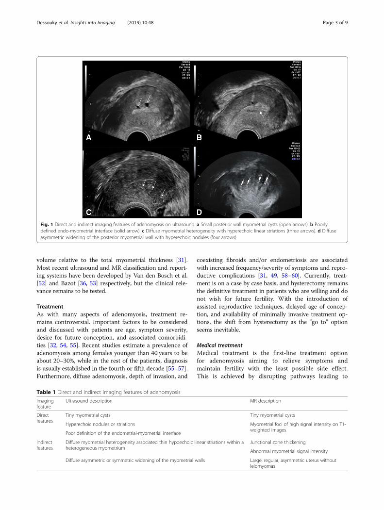
initial screening modality for adenomyosis. Ultrasound fea-tures of adenomyosis can be divided into direct or indirectfeatures (Fig. 1). Direct features are due to the presence ofendometrial tissue within the myometrium, and indirectfeatures are due to a hypertrophied myometrium as de-scribed by Atri et al. [38]. Table 1 describes ultrasound fea-tures of adenomyosis as described in previous literature[14, 16, 38–43]. To report the diagnostic accuracy of TVSin adenomyosis, several meta-analyses have been published[17, 44–46]. Estimated pooled sensitivities of 72 to 82%,pooled specificities of 81 to 85%, and pooled positive likeli-hood ratios 3.7 to 4.67 have been reported [17, 44]; how-ever, one meta-analysis suggested that variability betweenstudies does not allow for accurate statistical pooling [45].With the introduction of color and power Doppler ultra-sound, three-dimensional TVS and elastography techniquesto the work-up of adenomyosis, there is promise for furtherimprovement in diagnostic accuracy [46].Magnetic resonance imaging (MRI) represents a
second line, detailed imaging modality for the detec-tion of adenomyosis (Fig. 2). Similar to ultrasound,various direct and indirect features can be used todescribe adenomyosis, but need more knowledge ofuterine anatomy and its cyclic variations [36]. Table 1describes MRI features of adenomyosis as describedin previous literature [14–16, 35, 47]. Few prospectivestudies have evaluated the diagnostic accuracy of MRIin the diagnosis of adenomyosis [15, 16, 48]. Thesestudies have reported sensitivity between 70 and 93%and specificities between 86 and 93%. Despite beingless operator dependent, MRI needs more reader ex-perience and optimization of imaging technique toachieve higher diagnostic accuracy [36].
Classification of adenomyosisThe use of complex imaging techniques has revealedvarious subtypes of adenomyosis, often associated withhistopathologic variation in glandular and muscularcomponents [31]. Furthermore, initial studies havelinked various imaging criteria to symptoms of adeno-myosis [49–51]. Therefore, the need for a more holisticapproach to identify various disease characteristics incorp-orating symptomatology, morphology, and pathologic fea-tures is rising in order to improve the diagnostic accuracyand adequately guide treatment decisions. Important fac-tors to be included in classification systems would be thesite and location of pathology, configuration, and size/
Dessouky et al. Insights into Imaging (2019) 10:48 Page 2 of 9
volume relative to the total myometrial thickness [31].Most recent ultrasound and MR classification and report-ing systems have been developed by Van den Bosch et al.[52] and Bazot [36, 53] respectively, but the clinical rele-vance remains to be tested.
TreatmentAs with many aspects of adenomyosis, treatment re-mains controversial. Important factors to be consideredand discussed with patients are age, symptom severity,desire for future conception, and associated comorbidi-ties [32, 54, 55]. Recent studies estimate a prevalence ofadenomyosis among females younger than 40 years to beabout 20–30%, while in the rest of the patients, diagnosisis usually established in the fourth or fifth decade [55–57].Furthermore, diffuse adenomyosis, depth of invasion, and
coexisting fibroids and/or endometriosis are associatedwith increased frequency/severity of symptoms and repro-ductive complications [31, 49, 58–60]. Currently, treat-ment is on a case by case basis, and hysterectomy remainsthe definitive treatment in patients who are willing and donot wish for future fertility. With the introduction ofassisted reproductive techniques, delayed age of concep-tion, and availability of minimally invasive treatment op-tions, the shift from hysterectomy as the “go to” optionseems inevitable.
Medical treatmentMedical treatment is the first-line treatment optionfor adenomyosis aiming to relieve symptoms andmaintain fertility with the least possible side effect.This is achieved by disrupting pathways leading to
Fig. 1 Direct and indirect imaging features of adenomyosis on ultrasound. a Small posterior wall myometrial cysts (open arrows). b Poorlydefined endo-myometrial interface (solid arrow). c Diffuse myometrial heterogeneity with hyperechoic linear striations (three arrows). d Diffuseasymmetric widening of the posterior myometrial wall with hyperechoic nodules (four arrows)
Table 1 Direct and indirect imaging features of adenomyosis
Imagingfeature
Ultrasound description MR description
Directfeatures
Tiny myometrial cysts Tiny myometrial cysts
Hyperechoic nodules or striations Myometrial foci of high signal intensity on T1-weighted images
Poor definition of the endometrial-myometrial interface
Indirectfeatures
Diffuse myometrial heterogeneity associated thin hypoechoic linear striations within aheterogeneous myometrium
Junctional zone thickening
Abnormal myometrial signal intensity
Diffuse asymmetric or symmetric widening of the myometrial walls Large, regular, asymmetric uterus withoutleiomyomas
Dessouky et al. Insights into Imaging (2019) 10:48 Page 3 of 9

inflammation, neuroangiogenesis, and impaired apoptosis[61]. Currently, several hormonal and non-hormonal op-tions, namely gonadotropin-releasing hormone (GnRH)analogues, progestins, combined oral contraceptives, andnon-steroidal anti-inflammatory drugs are being used inan “off label” manner for the symptomatic treatment ofadenomyosis [57, 62]. Also, newer drugs, such as aroma-tase inhibitors, have been investigated by Badawy et al.and Tosti et al. [63, 64], while other therapies such as se-lective progesterone receptor modulators, GnRH antago-nists, valproic acid, and anti-platelet therapies are stillunder investigation [55].The main advantage of medication is symptomatic re-
lief without the need for surgical treatment. Neverthe-less, many drawbacks still need to be addressed. Thisincludes the temporary relieve of symptoms, and thecommon (i.e., menopausal symptoms, irregular bleeding,amenorrhea) and occasionally severe (i.e., thrombo-embolic) side effects of some drugs. Lack of evidenceneeded to base choice of drugs also raises the need toperform research into the comparative efficacy of cur-rently used drugs and develop a more standardized ap-proach for patients wanting to conceive while usingmedication. With a better understanding of pathogeneticmechanisms of adenomyosis, advances in drug develop-ment will soon be possible [55].
Minimally invasive techniquesThese are second-line treatment options aiming to curesymptoms and preserve the uterus in patients with failedmedical therapy. Conservative surgical treatments aim toremove adenomyosis and preserve the remaining normaluterine muscles through laparotomy, laparoscopy, hys-teroscopy, or combined approach. Excisional adenomyo-mectomy involves the complete removal of focal lesions(adenomyomas), while myometrectomy is the surgicaldebulking of diffuse adenomyosis. Non-excisional treat-ments aim to induce necrosis of focal or diffuse
adenomyosis through selective vascular occlusion or fo-cused ultrasound/thermal energy without direct tissuedissection. In some cases, a combination of surgical andnon-excisional methods, i.e., hysteroscopic resection/ab-lation, are used to achieve maximum cytoreduction andreduce myometrial tissue damage.
Conservative surgical treatmentDebulking/cytoreductive surgeries aim to remove visiblydiseased tissue with repair of the remaining myometrialtissue [65]. Several laparotomic techniques have beendescribed, including wedge resection and its modifica-tions, transverse H-shaped incision [66], wedge-shapeduterine wall removal [67], double and triple flap [68, 69],and asymmetric dissection methods [70]. Laparoscopictechniques have also been described in more focal path-ology, where longitudinal or transverse incisions [71, 72]are used to access adenomyotic lesions followed by re-section using monopolar needle or laser knife [73, 74],bag removal, and repair in layers or using double flaps[72, 75]. To date, there is no consensus on the best sur-gical method, but initial results are promising. In a sys-tematic review by Grigoris et al., dysmenorrheareduction, menorrhagia control, and pregnancy successrates ranged from 81 to 82%, 50 to 69%, and 47 to 61%among partial versus complete adenomyosis excisionsrespectively [76], and a recent review by Younes et al.showed 75% symptom relief on short-term follow-ups[77]. The main issue with conservative surgical methodsis the high risk for complications, i.e., uterine ruptureand complicated pregnancy [54, 65] (especially in diffuselesions and on long-term follow-up), making this optionsafer in focal adenomyomas.
Hysteroscopic resection/ablationHysteroscopic resection/ablation is a combined treatmentmethod involving the dissection and or coagulation of cys-tic adenomyotic lesions and crypts [78–82]. Hysteroscopic
Fig. 2 Coronal (a) and sagittal (b) T2W 1.5-T pelvic MRI images of a 42-year-old female with persistent pelvic pain following cesarean sectionshow focal thickening of the posterior uterine wall transitional zone (asterisk) with tiny myometrial cyst (solid arrow head), suggestingfocal adenomyosis
Dessouky et al. Insights into Imaging (2019) 10:48 Page 4 of 9

resections can be performed using yttrium aluminum gar-net (YAG) laser, rollerball resection, thermal balloon abla-tion, cryoablation, circulated hot fluid ablation, microwaveablation, bipolar radiofrequency ablation, and electro-coagulation [19].
High-intensity focused ultrasound (HIFU)High-intensity focused ultrasound (HIFU) is the use ofintense ultrasound energy directly targeting abnormaltissues and their vascularity through heating and cavita-tion, sparing the normal surrounding tissues. Thisprocess can be guided and monitored through MRI orultrasound [83]. High-intensity focused ultrasound hasbeen used since 2008 for the treatment of adenomyosis[84]. Since then, literature has shown promising resultsregarding symptom relief and uterine preservation withfew reported complications (namely pain, numbness, va-ginal or urinary discharge, fever, skin burn, or contactdermatitis) [83]. Recent studies have also investigatedthe use of ultrasound contrast agents (microbubbles)and hormonal (GnRH) and non-hormonal (metformin)treatments to enhance the efficacy of HIFU. Microbub-bles are believed to improve the ablative effects of HIFUby changing the acoustic characteristics, thus increasingenergy deposition in target tissues, while GnRH andmetformin inhibit cellular proliferation and induce apop-tosis [85–87]. Limited literature on treatment outcomesfor HIFU in adenomyosis has shown highly variable re-sults regarding symptom and uterine volume reduction[88–97]. Rates of menorrhagia, dysmenorrhea, and uter-ine volume reduction varied widely from 12.4 to 44.8%,25 to 100%, and 12.7 to 54% respectively, increasinggradually overtime (from 1 to 24 months). Nevertheless,paucity of literature comparing HIFU to other minimallyinvasive treatment options, limited availability, overallcost, unknown fertility outcomes, and strict indications,including lesions no more than 10 cm in diameter [88,90], no pelvic adhesions [84, 89, 90, 93], body weight lessthan 100 kg [98], and abdominal wall thickness less than5 cm [93] may limit its widespread use.
Uterine artery embolization (UAE)Uterine artery embolization is the use of transarterialcatheters aiming to induce more than 34% necrosiswithin adenomyotic tissues [99, 100]. The techniquefor UAE in adenomyosis is similar to that used in fi-broids. In many parts of the world, UAE is performedunder conscious sedation. Vascular access is gainedthrough a femoral or radial artery puncture using 4–6-French (F) arterial sheath for femoral [99, 101] and4-F sheath for radial access [102]. Under fluoroscopicguidance, aortography is followed by selective andsuper selective arteriography using 4–5-F catheters forthe internal iliac and 2–3-F microcatheters for the
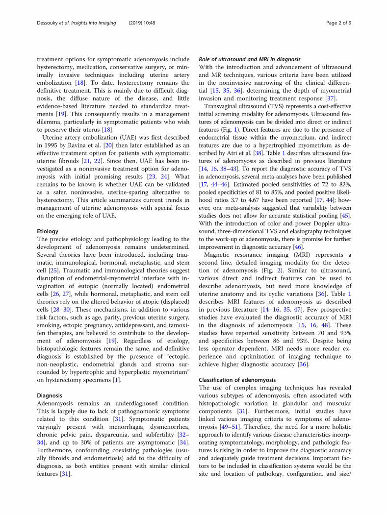
uterine artery and its branches respectively.Embolization is usually performed using variable-sizedpermanent particulate agents [103, 104]. Special atten-tion is paid to visualization of the cervicovaginal andovarian artery branches (Fig. 3). Distal embolizationavoids vaginal necrosis and unwanted reflux of micro-spheres into the ovarian artery [105].Despite being established in fibroids as a cost-effective,
short recovery alternative to surgery with minimal com-plications [19, 23, 100], it was believed to have lower ef-ficacy in adenomyosis [106]. In the past 15 years, UAEhas been considerably studied for the treatment ofsymptomatic adenomyosis [107]. Earlier studies byPopovic, Keung, and Zhou et al. demonstrate long-termimprovement in patient symptoms (in over 60% of pa-tients) and a short-term decrease in uterine volumes (inover 20% of patients), especially in vascular lesions [23,107, 108]. Current literature by Dueholm and Bruijn etal. show up to 67% long-term (40 month) treatment suc-cess and up to and 72% patient satisfaction rates respect-ively [24, 100]. In the latest systematic review andmeta-analysis by de Bruijn et al., patients were dividedinto four groups to report short- and long-term out-comes. Short-term improvement was achieved in 89.6%of patients with pure adenomyosis and 94.3% of patientswith adenomyosis with fibroids, while long-term im-provement was achieved in 74.0% of patients with pureadenomyosis and 84.5% of patients with adenomyosiswith fibroids [109].Overall, UAE shows favorable clinical outcomes, but ran-
domized controlled trials are still lacking [110]. In an at-tempt to fill this gap in knowledge, the “Quality of Life afterEmbolization vs Hysterectomy in Adenomyosis” (QUESTA)trial was set up. This multicenter non-blinded randomizedcontrolled trial is currently ongoing in the Netherlands. Ithas started since November 2015, and its primary outcomesare expected by May 2020 [101]. The calculated sample sizefor this trial was 96 patients (divided into 52 embolizationand 34 hysterectomy, including a 10% expected drop-out)made on assumptions from the embolization versus hyster-ectomy (EMMY) trial outcomes [111].Inclusion criteria were premenopausal women with
symptomatic pure adenomyosis or dominant adeno-myosis when both adenomyosis and fibroids coexistand women with an indication for hysterectomy (ei-ther failed or refused medical treatment). Exclusioncriteria were patients under 18 years of age, pelvic in-fection, suspected or confirmed malignancy, currentor future desire to conceive, any absolute contraindi-cation to angiography, deep infiltrating endometriosisrequiring surgery or obstructing the bowel, or coexist-ing hysteroscopically removable submucous fibroids.Following selection, TVUS and MRIs were performedto confirm the adenomyosis and eligible patients are
Dessouky et al. Insights into Imaging (2019) 10:48 Page 5 of 9

informed of the trial. Patients with written informedconsents were randomly allocated (in a 2:1 ratio) be-tween both experimental intervention (UAE) andstandard care control groups (hysterectomy), whilepatients refusing randomization are given the stand-ard of care (hysterectomy) [101].Following the procedure (UAE or hysterectomy), pa-
tients are followed up immediately, then at 6 weeks, 3months, 6 months, 12 months, and 24months using anonline questionnaire system. Three outcome parameterswere measured. Primary outcomes (quality of life) weremeasured at 6, 12, and 24months using a combinationof World Health Organization Quality of Life Scale andShort Form-12 Questionnaires. Secondary outcomes(clinical, symptom and quality of life, recovery related,cost utility analysis, laboratory, and pathology outcomes)were measured at 6 weeks and 3, 6, 12, and 24months.Imaging outcomes were also determined to identify po-tential predictive parameters for therapy effect usingspecific TVUS criteria (uterine size/fibroid volume re-duction in case of associated fibroids, vascular index by3D power Doppler) at baseline, 6 weeks, and 6monthsand MRI criteria (uterine size/fibroid volume reductionin case of associated fibroids, junctional zone reduction,
infarction rate, and presence of endometriosis) at base-line and at 6 months postprocedure [101].
UAE as an alternative to hysterectomyTo date, UAE seems to be the most investigated andhighest potential minimally invasive treatment optionfor adenomyosis. Results of ongoing randomized con-trolled (QUESTA) trial will soon show whether UAE canbe validated as a treatment option for adenomyosis. Al-though comparative information regarding quality of life,patient satisfaction, side effects, and complications postUAE versus hysterectomy will soon be available, ques-tions regarding fertility post UAE remain to be an-swered. Current American College of Obstetrics andGynecology and Society of Interventional Radiologyguidelines still consider desire for future fertility a rela-tive contraindication to UAE, but conflicting reports re-garding effects of UAE on fertility [112] still give roomfor debate. Nevertheless, further randomized studies arestill needed to give a clear answer for physicians and pa-tients alike.In conclusion, lack of information is the main hur-
dle to overcome the complexity in management ofadenomyosis. With randomized controlled trials and
Fig. 3 Digital subtraction angiography (DSA) images (of the same patient in Fig. 2) with selective injections of the left (a) uterine arterydemonstrate with multiple tortuous uterine artery branches and (b) lesion blush (most prominent at the anatomic site of the posterioruterine wall). Right uterine artery injection (not shown) was unremarkable for pathology. Post-embolization DSA images show occlusion oftoursous feeding vessels (c) with absence of lesion blush (d)
Dessouky et al. Insights into Imaging (2019) 10:48 Page 6 of 9

more evidence-based research, optimal treatment pro-tocols can be developed according to patient needs.Whether or not UAE can replace hysterectomy willlargely depend on the results of ongoing QUESTAtrial and other randomized trials comparing fertilityoutcomes among minimally invasive therapies.
AbbreviationsEMMY: Embolization versus hysterectomy; GnRH: Gonadotropin-releasinghormone; HIFU: High-intensity focused ultrasound; QUESTA: Quality of Lifeafter Embolization vs Hysterectomy in Adenomyosis; UAE: Uterine arteryembolization; YAG: Yttrium aluminum garnet
AcknowledgementsThe authors would like to acknowledge Prof. Adel Gamil for providingultrasound images for this manuscript.
FundingNo funding was received for this work.
Availability of data and materialsNot applicable.
Authors’ contributionsRD contributed to the manuscript preparation and revision. SAG and MGNcontributed to the manuscript editing and revision, image collection, editing,and preparation. RM and YL contributed to the preparation of themanuscript draft and editing and revision of final manuscript. All authorssignificantly contributed to the preparation of this manuscript. All authorsread and approved the final manuscript.
Ethics approval and consent to participateNot applicable.
Consent for publicationNot applicable.
Competing interestsThe authors declare that they have no competing interests.
Publisher’s NoteSpringer Nature remains neutral with regard to jurisdictional claims inpublished maps and institutional affiliations.
Author details1Radiology Department, Faculty of Medicine, Zagazig University, Koliat Al TobStreet, Zagazig 44519, Egypt. 2Radiology Department, Al-Ahrar TeachingHospital, Zagazig, Egypt.
Received: 15 December 2018 Accepted: 14 March 2019
References1. Bird CC, McElin TW, Manalo-Estrella P (1972) The elusive adenomyosis of the
uterus--revisited. Am J Obstet Gynecol 112:583–5932. Benagiano G, Brosens I (2006) History of adenomyosis. Best Pract Res Clin
Obstet Gynaecol 20:449–463 https://doi.org/10.1016/j.bpobgyn.2006.01.0073. García-Solares J, Donnez J, Donnez O, Dolmans MM (2018) Pathogenesis of
uterine adenomyosis: invagination or metaplasia? Fertil Steril 109:371–379https://doi.org/10.1016/j.fertnstert.2017.12.030
4. Azziz R (1989) Adenomyosis: current perspectives. Obstet Gynecol Clin N Am 16:221–235
5. Vercellini P, Oldani S, Parazzini F, Panazza S, Bramante T, Crosignani PG(1995) Adenomyosis at hysterectomy: a study on frequency distribution andpatient characteristics. Hum Reprod 10(5):1160–1162
6. Bergholt T, Berendt N, Eriksen L, Jacobsen M, Hertz JB (2001) Prevalence and riskfactors of adenomyosis at hysterectomy. Hum Reprod Update 16:2418–2421
7. Parazzini F, Mais V, Cipriani S, Busacca M, Venturini P, GISE (2009)Determinants of adenomyosis in women who underwent hysterectomy for
benign gynecological conditions: results from a prospective multicentricstudy in Italy. Eur J Obstet Gynecol Reprod Biol 143:103–106 https://doi.org/10.1016/j.ejogrb.2008.12.010
8. Wallwiener M, Taran F-A, Rothmund R et al (2013) Laparoscopicsupracervical hysterectomy (LSH) versus total laparoscopic hysterectomy(TLH): an implementation study in 1,952 patients with an analysis of riskfactors for conversion to laparotomy and complications, and of procedure-specific re-operations. Arch Gynecol Obs 288:1329–1339 https://doi.org/10.1007/s00404-013-2921-x
9. Naftalin J, Hoo W, Pateman K, Mavrelos D, Holland T, Jurkovic D (2012) Howcommon is adenomyosis? A prospective study of prevalence usingtransvaginal ultrasound in a gynaecology clinic. Hum Reprod 27:3432–3439https://doi.org/10.1093/humrep/des332
10. Di Donato N, Montanari G, Benfenati A et al (2014) Prevalence ofadenomyosis in women undergoing surgery for endometriosis. Eur J ObstetGynecol Reprod Biol 181:289–293 https://doi.org/10.1016/j.ejogrb.2014.08.016
11. Tamai K, Togashi K, Ito T, Morisawa N, Fujiwara T, Koyama T (2005) MRimaging findings of adenomyosis: correlation with histopathologic featuresand diagnostic pitfalls. Radiographics 25:21–40 https://doi.org/10.1148/rg.251045060
12. Ascher SM, Jha RC, Reinhold C (2003) Benign myometrial conditions:leiomyomas and adenomyosis. Top Magn Reson Imaging 14:281–304
13. Habiba M, Benagiano G (2015) Uterine adenomyosis. Springer, Cham14. Reinhold C, Tafazoli F, Mehio A et al (1999) Uterine adenomyosis:
endovaginal US and MR imaging features with histopathologic correlation.Radiographics 19:S147–S160 https://doi.org/10.1148/radiographics.19.suppl_1.g99oc13s147
15. Dueholm M, Lundorf E, Hansen ES, Sørensen JS, Ledertoug S, Olesen F(2001) Magnetic resonance imaging and transvaginal ultrasonography forthe diagnosis of adenomyosis. Fertil Steril 76:588–594
16. Bazot M, Cortez A, Darai E et al (2001) Ultrasonography compared withmagnetic resonance imaging for the diagnosis of adenomyosis: correlationwith histopathology. Hum Reprod 16:2427–2433
17. Champaneria R, Abedin P, Daniels J, Balogun M, Khan KS (2010) Ultrasoundscan and magnetic resonance imaging for the diagnosis of adenomyosis:systematic review comparing test accuracy. Acta Obs Gynecol Scand 89:1374–1384 https://doi.org/10.3109/00016349.2010.512061
18. Radzinsky VE, Khamoshina MB, Nosenko EN et al (2016) Treatment strategiesfor pelvic pain associated with adenomyosis. Gynecol Endocrinol 32:19–22https://doi.org/10.1080/09513590.2016.1232673
19. Taran FA, Stewart EA, Brucker S (2013) Adenomyosis: epidemiology, riskfactors, clinical phenotype and surgical and interventional alternatives tohysterectomy. Geburtshilfe Frauenheilkd 73:924–931 https://doi.org/10.1055/s-0033-1350840
20. Ravina JH, Herbreteau D, Ciraru-Vigneron N et al (1995) Arterial embolisationto treat uterine myomata. Lancet 346:671–672
21. de Bruijn AM, Ankum WM, Reekers JA et al (2016) Uterine arteryembolization vs hysterectomy in the treatment of symptomatic uterinefibroids: 10-year outcomes from the randomized EMMY trial. Am J ObstetGynecol 215:e741–e745
22. Edwards RD, Moss JG, Lumsden MA et al (2007) Uterine-artery embolizationversus surgery for symptomatic uterine fibroids. N Engl J Med 356:360–370https://doi.org/10.1056/NEJMoa062003
23. Popovic M, Puchner S, Berzaczy D, Lammer J, Bucek RA (2011) Uterine arteryembolization for the treatment of adenomyosis: a review. J Vasc IntervRadiol 22:901–909quiz 909 https://doi.org/10.1016/j.jvir.2011.03.013
24. de Bruijn AM, Smink M, Hehenkamp WJK et al (2017) Uterine arteryembolization for symptomatic adenomyosis: 7-year clinical follow-up usingUFS-Qol questionnaire. Cardiovasc Intervent Radiol 40:1344–1350 https://doi.org/10.1007/s00270-017-1686-1
25. Tinelli A, Malvasi A (2015, 2015) Uterine myoma, myomectomy andminimally invasive treatments. Springer International Publishing, pp 1–281https://doi.org/10.1007/978-3-319-10305-1
26. Habiba M, Benagiano G, Brosens I (2016) The pathophysiology ofadenomyosis. In: Habiba M, Benagiano G (eds) Uterine adenomyosis, vol 4.Springer, Cham, pp 45–70 https://doi.org/10.1007/978-3-319-13012-5_3
27. Ota H, Hatazawa J, Igarashi S, Tanaka T (1998) Is adenomyosis an immunedisease? Hum Reprod 4:360–367
28. Bergeron C, Amant F, Ferenczy A (2006) Pathophysiology and physiology ofadenomyosis. Best Pr Res Clin Obs Gynaecol 20:511–521
Dessouky et al. Insights into Imaging (2019) 10:48 Page 7 of 9

29. Matsumoto Y, Iwasaka T, Yamasaki F, Sugimori H (1999) Apoptosis and Ki-67expression in adenomyotic lesions and in the corresponding eutopicendometrium. Obstet Gynecol 94:71–77
30. Du H, Taylor HS (2009) Stem cell and female reproduction. Reprod Sci 16:126–13931. Gordts S, Grimbizis G, Campo R (2018) Symptoms and classification of
uterine adenomyosis, including the place of hysteroscopy in diagnosis. FertilSteril 109:380–388.e1 https://doi.org/10.1016/j.fertnstert.2018.01.006
32. Struble J, Reid S, Bedaiwy MA (2016) Adenomyosis: a clinical review of achallenging gynecologic condition. J Minim Invasive Gynecol 23:164–185https://doi.org/10.1016/j.jmig.2015.09.018
33. Garcia L, Isaacson K (2011) Adenomyosis: review of the literature. J MinimInvasive Gynecol 18:428–437 https://doi.org/10.1016/j.jmig.2011.04.004
34. Peric H, Fraser IS (2006) The symptomatology of adenomyosis. Best PractRes Clin Obstet Gynaecol 20:547–555 https://doi.org/10.1016/j.bpobgyn.2006.01.006
35. Togashi K, Nishimura K, Itoh K, Morisawa N, Fujiwara T, Koyama T (1988)Adenomyosis: diagnosis with MR imaging. Radiology 166:111–114 https://doi.org/10.1148/radiology.166.1.3336669
36. Bazot M, Daraï E (2018) Role of transvaginal sonography and magneticresonance imaging in the diagnosis of uterine adenomyosis. Fertil Steril 109:389–397 https://doi.org/10.1016/j.fertnstert.2018.01.024
37. Sugino N (2018) Uterine fibroids and adenomyosis. Springer NatureSingapore Pte Ltd, Singapore
38. Atri M, Reinhold C, Mehio AR, Chapman WB, Bret PM (2000) Adenomyosis:US features with histologic correlation in an in-vitro study. Radiology 215:783–790 https://doi.org/10.1148/radiology.215.3.r00jn06783
39. Bazot M, Darai E, Rouger J, Detchev R, Cortez A, Uzan S (2002) Limitations oftransvaginal sonography for the diagnosis of adenomyosis, withhistopathological correlation. Ultrasound Obstet Gynecol 20:605–611https://doi.org/10.1046/j.1469-0705.2002.00852.x
40. Fedele L, Bianchi S, Dorta M, Arcaini L, Zanotti F, Carinelli S (1992)Transvaginal ultrasonography in the diagnosis of diffuse adenomyosis. FertilSteril 58:94–97
41. Kepkep K, Tuncay YA, Goynumer G, Tutal E (2007) Transvaginal sonographyin the diagnosis of adenomyosis: which findings are most accurate?Ultrasound Obstet Gynecol 30:341–345 https://doi.org/10.1002/uog.3985
42. Sun YL, Wang CB, Lee CY et al (2010) Transvaginal sonographic criteria forthe diagnosis of adenomyosis based on histopathologic correlation. TaiwanJ Obstet Gyneco 49:40–44 https://doi.org/10.1016/S1028-4559(10)60007-1
43. Exacoustos C, Brienza L, Di Giovanni A et al (2011) Adenomyosis: three-dimensional sonographic findings of the junctional zone and correlationwith histology. Ultrasound Obstet Gynecol 37:471–479 https://doi.org/10.1002/uog.8900
44. Meredith SM, Sanchez-Ramos L, Kaunitz AM (2009) Diagnostic accuracy oftransvaginal sonography for the diagnosis of adenomyosis: systematicreview and metaanalysis. Am J Obstet Gynecol 201:107e1–107e6 https://doi.org/10.1016/j.ajog.2009.03.021
45. Dartmouth K (2014) A systematic review with meta-analysis: the commonsonographic characteristics of adenomyosis. Ultrasound 22:148–157 https://doi.org/10.1177/1742271X14528837
46. Andres MP, Borrelli GM, Ribeiro J, Baracat EC, Abrão MS, Kho RM (2018)Transvaginal ultrasound for the diagnosis of adenomyosis: systematic reviewand meta-analysis. J Minim Invasive Gynecol 25:257–264 https://doi.org/10.1016/j.jmig.2017.08.653
47. Togashi K, Konishi I, Itoh H, Nishimura K, Fujisawa IOH (1989) Enlargeduterus: differentiation between adenomyosis and leiomyoma with MRimaging. Radiology 171:531–534
48. Reinhold C, McCarthy S, Bret PM et al (1996) Diffuse adenomyosis:comparison of endovaginal US and MR imaging with histopathologiccorrelation. Radiology 199:151–158 https://doi.org/10.1148/radiology.199.1.8633139
49. Naftalin J, Hoo W, Pateman K, Mavrelos D, Foo X, Jurkovic D (2014) Isadenomyosis associated with menorrhagia? Hum Reprod 29:473–479https://doi.org/10.1093/humrep/det451
50. Naftalin J, Hoo W, Nunes N, Holland T, Mavrelos D, Jurkovic D (2016) Associationbetween ultrasound features of adenomyosis and severity of menstrual pain.Ultrasound Obstet Gynecol 47:779–783 https://doi.org/10.1002/uog.15798
51. Eisenberg VH, Arbib N, Schiff E, Goldenberg M, Seidman DS, Soriano D(2017) Sonographic signs of adenomyosis are prevalent in womenundergoing surgery for endometriosis and may suggest a higher risk ofinfertility. Biomed Res Int 2017:9 https://doi.org/10.1155/2017/8967803
52. Van den Bosch T, de Bruijn AM, de Leeuw RA et al (2018) A sonographicclassification and reporting system for diagnosing adenomyosis. UltrasoundObstet Gynecol https://doi.org/10.1002/uog.19096
53. Bazot M (2017) Pathologie Myometriale. In: Nahum H (ed) Imagerie de lafemme - Gynecologie. Lavoisier, Paris, p 668
54. Oliveira MAP, Crispi CP Jr, Brollo LC, De Wilde RL (2018) Surgery inadenomyosis. Arch Gynecol Obstet 297:581–589 https://doi.org/10.1007/s00404-017-4603-6
55. Vannuccini S, Luisi S, Tosti C, Sorbi F, Petraglia F (2018) Role of medicaltherapy in the management of uterine adenomyosis. Fertil Steril 109:398–405 https://doi.org/10.1016/j.fertnstert.2018.01.013
56. Tan J, Moriarty S, Taskin O et al (2018) Reproductive outcomes after fertility-sparingsurgery for focal and diffuse adenomyosis: a systematic review. J Minim InvasiveGynecol 25:608–621 https://doi.org/10.1016/j.jmig.2017.12.020
57. Pontis A, D’Alterio MN, Pirarba S, de Angelis C, Tinelli R, Angioni S (2016)Adenomyosis: a systematic review of medical treatment. GynecolEndocrinol 32:696–700 https://doi.org/10.1080/09513590.2016.1197200
58. Pinzauti S, Tosti C, Centini G et al (2015) Transvaginal sonographic featuresof diffuse adenomyosis in 18-30-year-old nulligravid women withoutendometriosis: association with symptoms. Ultrasound Obstet Gynecol 46:730–736 https://doi.org/10.1002/uog.14834
59. Mochimaru A, Aoki S, Oba MS, Kurasawa K, Takahashi T, Hirahara F(2015) Adverse pregnancy outcomes associated with adenomyosis withuterine enlargement. J Obstet Gynaecol Res 41:529–533 https://doi.org/10.1111/jog.12604
60. Taran FA, Weaver AL, Coddington CC, Stewart EA (2010) Understandingadenomyosis: a case control study. Fertil Steril 94:1223–1228 https://doi.org/10.1016/j.fertnstert.2009.06.049
61. Vannuccini S, Tosti C, Carmona F et al (2017) Pathogenesis of adenomyosis:an update on molecular mechanisms. Reprod BioMed Online 35:592–601https://doi.org/10.1016/j.rbmo.2017.06.016
62. Fedele L, Bianchi S, Frontino G (2008) Hormonal treatments foradenomyosis. Best Pr Res Clin Obs Gynaecol 22:333–339 https://doi.org/10.1016/j.bpobgyn.2007.07.006
63. Badawy AM, Elnashar AM, Mosbah AA (2012) Aromatase inhibitors orgonadotropin-releasing hormone agonists for the management of uterineadenomyosis: a randomized controlled trial. Acta Obstet Gynecol Scand 91:489–495 https://doi.org/10.1111/j.1600-0412.2012.01350.x
64. Tosti C, Vannuccini S, Lazzeri L, Luisi S, Petraglia F, Troìa L (2016) Currentand future medical treatment of adenomyosis. J Endometr 8:127–135
65. Osada H (2018) Uterine adenomyosis and adenomyoma: the surgicalapproach. Fertil Steril 109:406–417 https://doi.org/10.1016/j.fertnstert.2018.01.032
66. Fujishita A, Masuzaki H, Khan KN, Kitajima M, Ishimaru T (2004) Modifiedreduction surgery for adenomyosis. A preliminary report of the transverse Hincision technique. Gynecol Obstet Investig 57:132–138 https://doi.org/10.1159/000075830
67. Saremi A, Bahrami H, Salehian P, Hakak N, Pooladi A, Bahrami H (2014)Treatment of adenomyomectomy in women with severe uterineadenomyosis using a novel technique. Reprod BioMed Online 28:753–760
68. Huang X, Huang Q, Chen S et al (2015) Efficacy of laparoscopicadenomyomectomy using double-flap method for diffuse uterineadenomyosis. BMC Womens Health 15:24
69. Kim TH, Lee HH, Chung SH, Lee W (2012) The triple-flap method for hugeuterine adenomyosis with pelvic adhesions. Reprod BioMed Online 25:649author reply 650 https://doi.org/10.1016/j.rbmo.2012.09.011
70. Nishida M, Takano K, Arai Y, Ozone H, Ichikawa R (2010) Conservativesurgical management for diffuse uterine adenomyosis. Fertil Steril 94:715–719 https://doi.org/10.1016/j.fertnstert.2009.03.046
71. Nabeshima H, Murakami T, Terada Y, Noda T, Yaegashi N, Okamura K(2003) Total laparoscopic surgery of cystic adenomyoma underhydroultrasonographic monitoring. J Am Assoc Gynecol Laparosc 10:195–199
72. Takeuchi H, Kitade M, Kikuchi I et al (2006) Laparoscopicadenomyomectomy and hysteroplasty: a novel method. J Minim InvasiveGynecol 13:150–154 https://doi.org/10.1016/j.jmig.2005.12.004
73. Suginami H, Tokushige M, Taniguchi F (2008) Surgical treatment ofadenomyosis. Obstet Gynecol (Tokyo) 75:72–78
74. Kishi Y, Yabuta M, Taniguchi F (2014) Who will benefit from uterus-sparingsurgery in adenomyosis-associated subfertility? Fertil Steril 102:802–807e1https://doi.org/10.1016/j.fertnstert.2014.05.028
Dessouky et al. Insights into Imaging (2019) 10:48 Page 8 of 9

75. Wang PH, Chao HT, Liu WM, Cheng MH, Chao KC, Fuh JL (2009) Is thesurgical approach beneficial to subfertile women with symptomaticextensive adenomyosis? J Obstet Gynaecol Res 35:495–502
76. Grimbizis GF, Mikos T, Tarlatzis B (2014) Uterus-sparing operative treatmentfor adenomyosis. Fertil Steril 101:472–487.e8 https://doi.org/10.1016/j.fertnstert.2013.10.025
77. Younes G, Tulandi T (2018) Conservative surgery for adenomyosis andresults: a systematic review. J Minim Invasive Gynecol 25:265–276 https://doi.org/10.1016/j.jmig.2017.07.014
78. Giana M, Montella F, Surico D, Vigone A, Bozzola C, Ruspa G (2005) Largeintramyometrial cystic adenomyosis: a hysteroscopic approach with bipolarresectoscope: case report. Eur J Gynaecol Oncol 26:462–463
79. Ryo E, Takeshita S, Shiba M, Ayabe T (2006) Radiofrequency ablation forcystic adenomyosis: a case report. J Reprod Med 51:427–430
80. Kamio M, Taguchi S, Oki T et al (2007) Isolated adenomyotic cyst associatedwith severe dysmenorrhea. J Obstet Gynaecol Res 33:388–391 https://doi.org/10.1111/j.1447-0756.2007.00543.x
81. Takeuchi H, Kitade M, Kikuchi I, Kumakiri J, Kuroda K, Jinushi M (2010)Diagnosis, laparoscopic management, and histopathologic findings ofjuvenile cystic adenomyoma: a review of nine cases. Fertil Steril 94:862–868https://doi.org/10.1016/j.fertnstert.2009.05.010
82. Sun W, Guo X, Zhu L, Fei X, Zhang Z, Li D (2018) Hysteroscopictreatment of a uterine cystic adenomyosis. J Minim Invasive Gynecol25:374–375 https://doi.org/10.1016/j.jmig.2017.07.015
83. Cheung VYT (2017) Current status of high-intensity focused ultrasound forthe management of uterine adenomyosis. Ultrasonography 36:95–102https://doi.org/10.14366/usg.16040
84. Fukunishi H, Funaki K, Sawada K, Yamaguchi K, Maeda T, Kaji Y (2008) Earlyresults of magnetic resonance-guided focused ultrasound surgery ofadenomyosis: analysis of 20 cases. J Minim Invasive Gynecol 15:571–579https://doi.org/10.1016/j.jmig.2008.06.010
85. Jingqi W, Lu Z, Jun Z et al (2018) Clinical usefulness of the microbubblecontrast agent SonoVue in enhancing the effects of high-intensity focusedultrasound for the treatment of adenomyosis. J Ultrasound Med https://doi.org/10.1002/jum.14638
86. Xiao-Ying Z, Ying-Shu G, Jiu-Mei C et al (2018) Effect of pre-treatment withgonadotropin-releasing hormone analogue GnRH-α on high-intensityfocussed ultrasound ablation for diffuse adenomyosis: a preliminary study.Int J Hyperth:1–9 https://doi.org/10.1080/02656736.2018.1440014
87. Hou Y, Qin Z, Fan K, Xu Y, Huang X (2018) Combination therapeuticeffects of high intensity focused ultrasound and metformin for thetreatment of adenomyosis. Exp Ther Med 15:2104–2108 https://doi.org/10.3892/etm.2017.5601
88. Fan TY, Zhang L, Chen W et al (2012) Feasibility of MRI-guided highintensity focused ultrasound treatment for adenomyosis. Eur J Radiol 81:3624–3630 https://doi.org/10.1016/j.ejrad.2011.05.036
89. Liu X, Wang W, Wang Y, Wang Y, Li Q, Tang J (2016) Clinical predictors oflong-term success in ultrasound-guided high-intensity focused ultrasoundablation treatment for adenomyosis: a retrospective study. Medicine(Baltimore) 95:e2443 https://doi.org/10.1097/MD.0000000000002443
90. Long L, Chen J, Xiong Y et al (2015) Efficacy of high-intensity focusedultrasound ablation for adenomyosis therapy and sexual life quality. Int JClin Exp Med 8:11701–11707
91. Zhou M, Chen JY, Tang LD, Chen WZ, Wang ZB (2011) Ultrasound-guidedhigh-intensity focused ultrasound ablation for adenomyosis: the clinicalexperience of a single center. Fertil Steril 95:900–905 https://doi.org/10.1016/j.fertnstert.2010.10.020
92. Wang W, Wang Y, Tang J (2009) Safety and efficacy of high intensityfocused ultrasound ablation therapy for adenomyosis. Acad Radiol 16:1416–1423 https://doi.org/10.1016/j.acra.2009.06.005
93. Lee JS, Hong GY, Park BJ, Kim TE (2015) Ultrasound-guided high-intensityfocused ultrasound treatment for uterine fibroid & adenomyosis: a singlecenter experience from the Republic of Korea. Ultrason Sonochem 27:682–687 https://doi.org/10.1016/j.ultsonch.2015.05.033
94. Ferrari F, Arrigoni F, Miccoli A et al (2016) Effectiveness of magneticresonance-guided focused ultrasound surgery (MRgFUS) in the uterineadenomyosis treatment: technical approach and MRI evaluation. Radiol Med121:153–161 https://doi.org/10.1007/s11547-015-0580-7
95. Polina L, Nyapathy V, Mishra A, Yellamanthili H, Vallabhaneni MP (2012)Noninvasive treatment of focal adenomyosis with MR-guided focused
ultrasound in two patients. Indian J Radiol Imaging 22:93 https://doi.org/10.4103/0971-3026.101078
96. Kim KA, Yoon SW, Lee C et al (2011) Short-term results of magneticresonance imaging-guided focused ultrasound surgery for patients withadenomyosis: symptomatic relief and pain reduction. Fertil Steril 95:1152–1155 https://doi.org/10.1016/j.fertnstert.2010.09.024
97. Shui L, Mao S, Wu Q et al (2015) High-intensity focused ultrasound (HIFU)for adenomyosis: two-year follow-up results. Ultrason Sonochem 27:677–681https://doi.org/10.1016/j.ultsonch.2015.05.024
98. Wang S, Meng X, Dong Y (2016) The evaluation of uterine arteryembolization as a nonsurgical treatment option for adenomyosis. Int JGynaecol Obstet 133:202–205 https://doi.org/10.1016/j.ijgo.2015.09.016
99. Kim MD, Kim YM, Kim HC et al (2011) Uterine artery embolization forsymptomatic adenomyosis: a new technical development of the 1-2-3protocol and predictive factors of MR imaging affecting outcomes. J VascInterv Radiol 22:497–502 https://doi.org/10.1016/j.jvir.2011.01.426
100. Dueholm M (2018) Minimally invasive treatment of adenomyosis. Best PractRes Clin Obstet Gynaecol https://doi.org/10.1016/j.bpobgyn.2018.01.016
101. de Bruijn AM, Lohle PN, Huirne JA et al (2018) Uterine artery embolizationversus hysterectomy in the treatment of symptomatic adenomyosis:protocol for the randomized questa trial. JMIR Res Protoc 20:e47 https://doi.org/10.2196/resprot.8512
102. Resnick NJ, Kim E, Patel RS, Lookstein RA, Nowakowski FS, Fischman AM(2014) Uterine artery embolization using a transradial approach: initialexperience and technique. J Vasc Interv Radiol 25:443–447 https://doi.org/10.1016/j.jvir.2013.11.010
103. Bilhim T, Pisco JM, Duarte M, Oliveira AG (2011) Polyvinyl alcohol particlesize for uterine artery embolization: a prospective randomized study ofinitial use of 350-500μm particles versus initial use of 500-700μm particles. JVasc Interv Radiol 22:21–27 https://doi.org/10.1016/j.jvir.2010.09.018
104. Firouznia K, Ghanaati H, Sanaati M, Jalali AH, Shakiba M (2009) Pregnancyafter uterine artery embolization for symptomatic fibroids: a series of 15pregnancies. AJR Am J Roentgenol 192:1588–1592 https://doi.org/10.2214/AJR.07.3904
105. Das C, Rathinam D, Manchanda S, Srivastava D (2017) Endovascular uterineartery interventions. Indian J Radiol Imaging 27:488 https://doi.org/10.4103/ijri.IJRI_204_16
106. Rabinovici J, Stewart EA (2006) New interventional techniques foradenomyosis. Best Pr Res Clin Obs Gynaecol 20:617–636 https://doi.org/10.1016/j.bpobgyn.2006.02.002
107. Keung JJ, Spies JB, Caridi TM (2017) Uterine artery embolization: a review ofcurrent concepts. Best Pract Res Clin Obstet Gynaecol 46:66–73 https://doi.org/10.1016/j.bpobgyn.2017.09.003
108. Zhou J, He L, Liu P et al (2016) Outcomes in adenomyosis treated withuterine artery embolization are associated with lesion vascularity: a long-term follow-up study of 252 cases. PLoS One 11:e0165610 https://doi.org/10.1371/journal.pone.0165610
109. de Bruijn AM, Smink M, Lohle PNM et al (2017) Uterine artery embolizationfor the treatment of adenomyosis: a systematic review and meta-analysis. JVasc Interv Radiol 28:1629–1642.e1 https://doi.org/10.1016/j.jvir.2017.07.034
110. Caridi TM, Spies JB (2018) Management of adenomyosis. A review ofcharacteristic imaging findings and treatment options, with an emphasis onthe use of uterine artery embolization. Endovasc Today 17:57–61
111. Hehenkamp WJK, Volkers NA, Birnie E, Reekers JA, Ankum WM (2008)Symptomatic uterine fibroids: treatment with uterine artery embolization orhysterectomy—results from the randomized clinical embolisation versushysterectomy (EMMY) trial. Radiology 246:823–832 https://doi.org/10.1148/radiol.2463070260
112. Mohan PP, Hamblin MH, Vogelzang RL (2013) Uterine artery embolizationand its effect on fertility. J Vasc Interv Radiol 24:925–930 https://doi.org/10.1016/j.jvir.2013.03.014
Dessouky et al. Insights into Imaging (2019) 10:48 Page 9 of 9