Management of Patients with ST-Elevation Myocardial...
60
University of Debrecen Universitas Debreceniensis Management of Patients with ST-Elevation Myocardial Infarction (STEMI) István Lőrincz MD Division of Emergency Medicine, Institute of Internal Medicine, University of Debrecen
Transcript of Management of Patients with ST-Elevation Myocardial...

University of Debrecen Universitas Debreceniensis
Management of Patients with
ST-Elevation Myocardial Infarction
(STEMI)
István Lőrincz MD Division of Emergency Medicine, Institute of Internal Medicine, University of Debrecen

In-hospital mortality (STEMI)
30
15
10
70
5
10
15
20
25
30
35
60-s 70-s 80-s 90-s
CCU + defibrillator
thrombolysis
primary PCI
In-hospital mortality dramatically decreased under the last 30 years:

Figure 13.1 Terminology used in acute coronary syndromes. Reproduced with permission from
Hamm et al. Lancet 2001; 358: 1533–1538 [151

Prizmetal's or 'variant' angina
Angina associated with ST elevation may be due to
coronary artery vasospasm.
This may occur with or without a fixed coronary
abnormality and may be indistinguishable from an acute
MI until changes resolve rapidly with GTN as pain is
relieved.

Options for Transport of Patients With STEMI and
Initial Reperfusion Treatment
EMS Transport
Onset of
symptoms of
STEMI
9-1-1
EMS
Dispatch
EMS on-scene • Encourage 12-lead ECGs.
• Consider prehos. fibrinolytic if capable
and EMS-to-needle within 30 min. GOALS
PCI
capable
Not PCI
capable
Hospital fibrinolysis:
Door-to-Needle
within 30 min.
Inter-
Hospital
Transfer
Golden Hour = first 60 min. Total ischemic time: within 120 min.
Patient
EMS Prehospital fibrinolysis
EMS-to-needle
within 30 min.
EMS transport EMS-to-balloon within 90 min.
Patient self-transport Hospital door-to-balloon
within 90 min.
Dispatch
1 min.
5 min.
8 min.
Antman EM, et al. J Am Coll Cardiol 2008. Published ahead of print on December 10, 2007. Available at
http://content.onlinejacc.org/cgi/content/full/j.jacc.2007.10.001. Figure 1.

Transmission of Patient Data
EMS ED Cath Lab
Cellular Site

Ischemia
Injury
Necrosis
Time after onset Onset <20-40 min. 30 min. 1 hour 2 hours 6 hours 24 hours
Extent of necrosis 0% 0% 10% 30% 50% 90% 100%
Important Evolution of Heart Attack

MI diagnosis The diagnosis of acute MI requires 2 out of the
following 3 features:
• A history of cardiac-type ischaemic chest pain.
• Evolutionary changes on serial ECGs.
• A rise in serum cardiac markers.

MI diagnosis
Note that 50-60% of pts will not have a
diagnostic ECG on arrival and up to 17%
will have an entirely normal initial ECG.
Late presentation does not improve
diagnostic accuracy of the ECG.
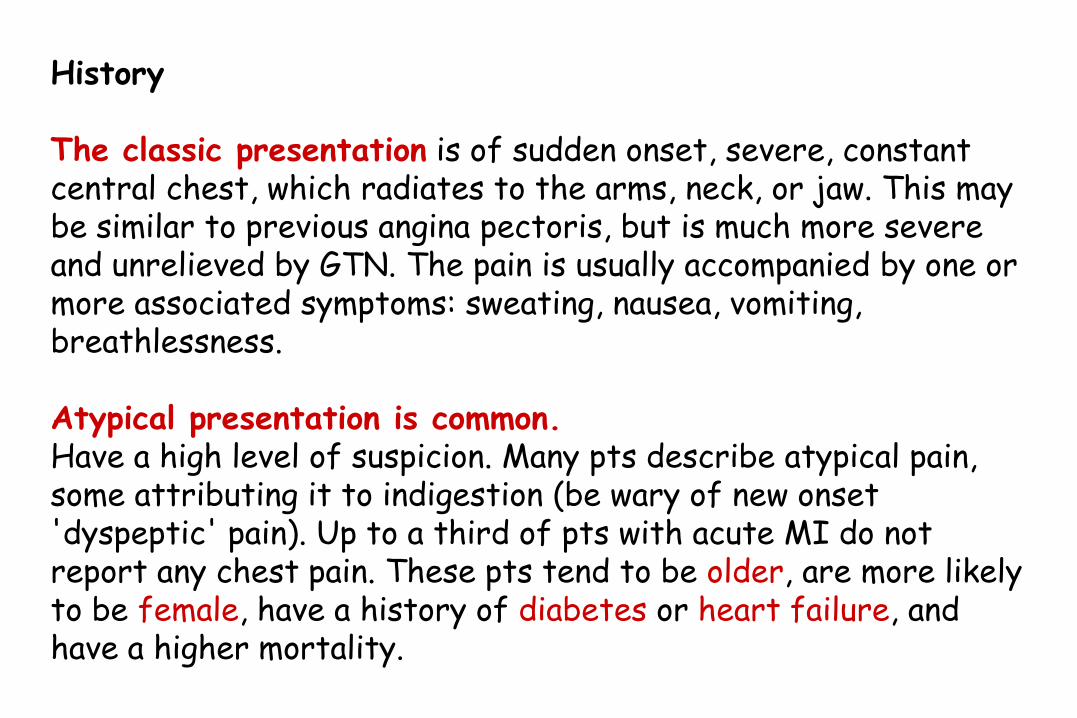
History The classic presentation is of sudden onset, severe, constant central chest, which radiates to the arms, neck, or jaw. This may be similar to previous angina pectoris, but is much more severe and unrelieved by GTN. The pain is usually accompanied by one or more associated symptoms: sweating, nausea, vomiting, breathlessness. Atypical presentation is common. Have a high level of suspicion. Many pts describe atypical pain, some attributing it to indigestion (be wary of new onset 'dyspeptic' pain). Up to a third of pts with acute MI do not report any chest pain. These pts tend to be older, are more likely to be female, have a history of diabetes or heart failure, and have a higher mortality.

These patients may present with:
• LVF.
• Collapse or syncope (often with
associated injuries eg head injury).
• Confusion.
• Stroke.
• An incidental ECG finding at a later date.

Examination
Examination and initial resuscitation (maintain Sp02 in
normal range. IV cannula, analgesia) go hand in hand.
The patient may be pale, sweaty, and distressed.
Examination is usually normal unless complications have
supervened (e.g. arrhythmias, LVF). Direct initial
examination towards searching for these complications
and excluding alternative diagnoses:

Examination
Check pulse. BP and monitor trace (?arrhythmia or
cardiogenic shock).
Listen to the heart (murmurs or 3rd heart sound).
Listen to the lung fields (?LVF, pneumonia, ptx).
Check peripheral pulses (?aortic dissection).
Check legs for evidence of deep vein thrombosis
(?PE).
Palpate for abdominal tenderness or masses
(?cholecystitis, pancreatitis, perforated peptic
ulcer, ruptured aortic aneurysm).

Investigations If the diagnosis of ST segment elevation MI within the
first few hours is based upon history and ECG
changes (serum cardiac markers may take several hours
to rise-see below).
Record an ECG as soon as possible, ideally within a few
minutes of arrival at hospital. Sometimes pts arrive at
hospital with ECGs of diagnostic quality already recorded
by paramedics. If the initial ECG is normal, but symptoms
are suspicious, repeat the ECG every 15min and re-
evaluate,

Investigations
Request old notes (these may contain previous ECGs for
comparison).
Ensure continuous cardiac monitoring and pulse oximetry.
Monitor BP and respiratory rate.
Obtain venous access and send blood for cardiac markers.
U&E. glucose. FBC, lipids.
Obtain a CXR if there is suspicion of LVF or aortic
dissection.

Cardiac markers
Troponins are now universally used. Troponin T (cTnT) and
Troponin I (cTnl) are proteins virtually exclusive to cardiac
myocytes. They are highly specific and sensitive, but are only
maximally accurate after 12hr.
Troponin T and I cannot be used to rule out MI in the first few
hours. In addition, cardiac cells may release troponin into the
blood when cardiac muscle is damaged by pericarditis,
pulmonary embolism with a large clot burden, or sepsis. Renal
failure reduces excretion of troponin.
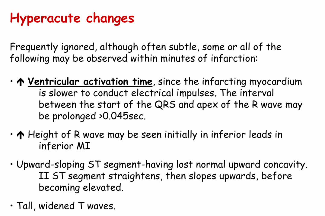
Hyperacute changes Frequently ignored, although often subtle, some or all of the following may be observed within minutes of infarction: • Ventricular activation time, since the infarcting myocardium is slower to conduct electrical impulses. The interval between the start of the QRS and apex of the R wave may be prolonged >0.045sec.
• Height of R wave may be seen initially in inferior leads in inferior MI
• Upward-sloping ST segment-having lost normal upward concavity. II ST segment straightens, then slopes upwards, before becoming elevated.
• Tall, widened T waves.

Evolving acute changes In isolation, none of these changes are specific to MI. In combination, with an appropriate history, they can diagnose MI: • ST elevation: the most important ECG change. ST segments become concave down and are significant if elevated >1mm in 2 limb leads, or >2mm in 2 adjacent chest leads. • Reciprocal ST depression may occur on the 'opposite side' of the heart. • Pathological Q waves reflect electrically inert necrotic myocardium. ECG leads over a large transmural infarct, the deep QS waves. Leads directed towards the periphery of a large infarct or over a smaller infarct may show a QR complex or a loss of R amplitude. • T wave inversion: typically deeply inverted, symmetrical and pointed.

Evolving acute changes Conduction problems may develop
LBBB in a patient with acute cardiac chest pain makes
interpretation of the ECG very difficult. LBBB does not have to be
new to be significant. Do not delay intervention in pall with a good
clinical history of MI in order to obtain old ECGs.
Sgarbossa criteria for diagnosing ACS in the presence of LBBB
• ST segment elevation >1 mm in leads with positive QRS
complexes.
• ST segment depression in leads V1, V2, or V3.
• ST segment elevation >5mm in lead with negative QRS complexes.
If all 3 are present, MI is likely.

Chronic changes
In the months following an MI, ECG changes resolve to a
variable extent. ST segments become isoelectric, unless
a ventricular aneurysm develops, T waves gradually
become positive again. Q waves usually remain, indicating
MI at some time in the past.

Evolution of AMI
• Hyperacute
– Early change suggestive of AMI
– Tall & Peaked
– May precede clinical symptoms
– Only seen in leads looking at
infarcting area
– Not used as a diagnostic finding

Evolution of AMI
• Acute
– ST segment elevation
– Implies myocardial injury
occurring
– Elevated ST segment
presumed acute rather than
old

Evolution of AMI
• Acute
– ST segment Elevated
– Q wave at least 40 ms wide =
pathologic
– Q wave associated with some
cellular necrosis

Evolution of AMI
• Age Undetermined
– Wide (pathologic) Q wave
– No ST segment elevation
– Old or “age undetermined”
MI

MI usually affects the left ventricle (LV), occasionally the right ventricle (RV), but virtually never the atria. The part of myocardium affected is implied by which
leads show changes Table
ECG leads Location of MI
V 1-3 Anteroseptal
V 5-6, AVL Anterolateral
V2-4 Anterior
V1-6 Extensive anterior
I.II. aVL, V6 Lateral
II. III. aVF Inferior
V1, V4R Rigth ventricle

Localization
Inferior: II, III, AVF
Septal: V1, V2
Anterior: V3, V4
Lateral: I, AVL, V5, V6
I
II
III
aVR
aVL
aVF
V1
V2
V3
V4
V5
V6

Localization
I Lateral
II Inferior
III Inferior
aVR
aVL Lateral
V1 Septal
aVF Inferior
V2 Septal
V3 Anterior
V4 Anterior
V5 Lateral
V6 Lateral
Which coronary arteries are most likely
associated with each group of contiguous
leads?

Localization: Left Coronary Artery
Left Main
Left Circumflex
Lateral Wall
Anterior Wall of Left Ventricle
Septal Wall
Right Ventricle
Right Coronary Artery
Anterior Descending Artery

Localization Practice ECG

Localization Practice ECG

Localization Practice ECG

Localization: Right Coronary Artery
Right Coronary Artery
Posterior Descending Artery
Inferior Wall of left ventricle
Posterior Wall
Lateral Wall
Left Ventricle
Left Coronary Artery
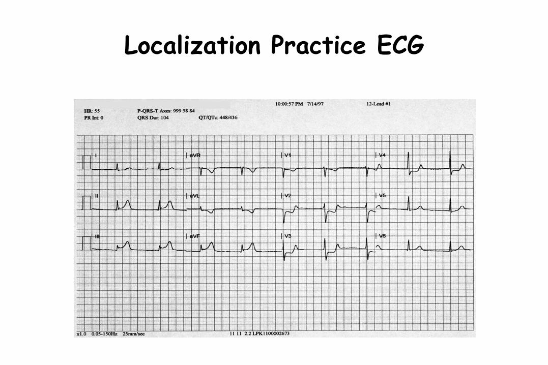
Localization Practice ECG

Localization Practice ECG
Note: “R” designation manually placed on this ECG for teaching purposes

Localization Practice ECG

Localization Practice ECG

Localization Summary
• Left Coronary Artery – Septal
– Anterior
– Lateral
– Possibly Inferior
• Right Coronary Artery – Inferior
– Right Ventricular Infarct
– Posterior

Does your institution achieve these goals?
Primary PCI 90 minutes Thrombolysis 30 minutes
? Time is the major predictor in patient outcomes
Type of reperfusion method (PCI vs. fibrinolysis) less imperative
Proper Early Management

Reperfusion Strategies
Several factors to consider:
• Time from the onset of symptoms
• Risk from the STEMI
• Risk of bleeding
• Time required for transport to
cath lab
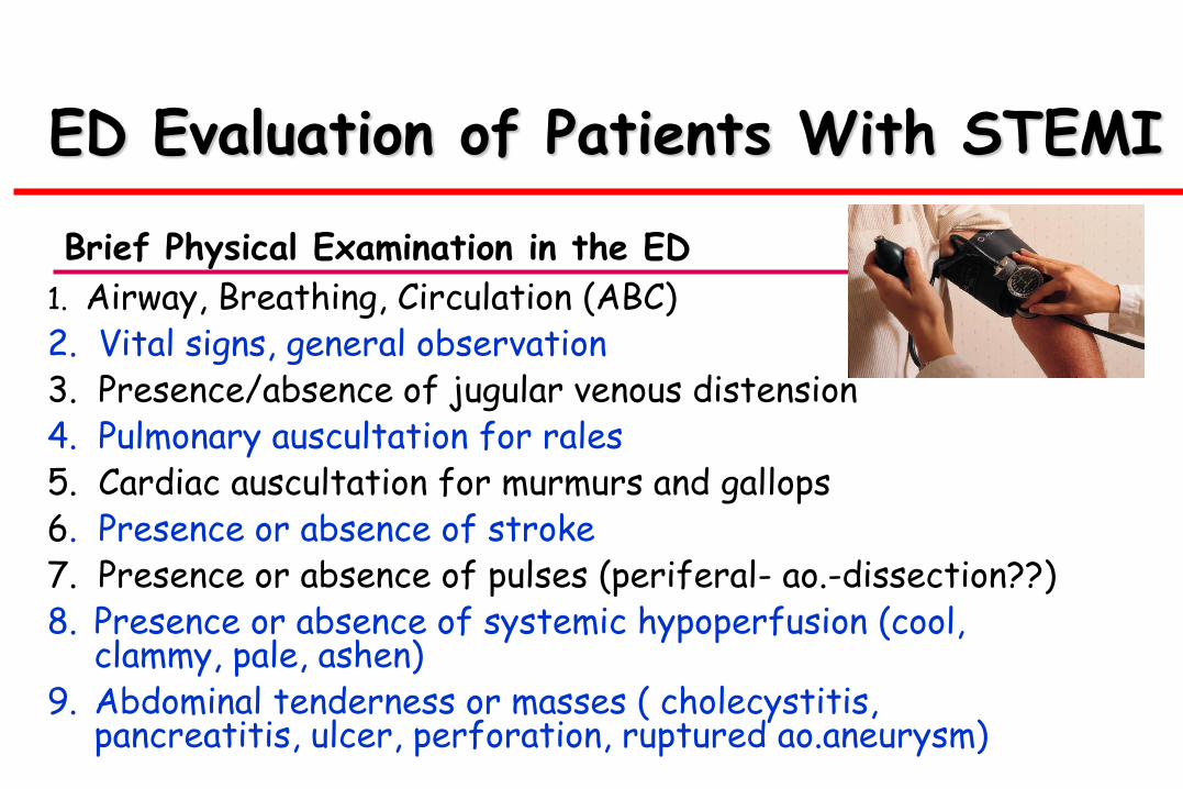
ED Evaluation of Patients With STEMI
1. Airway, Breathing, Circulation (ABC) 2. Vital signs, general observation 3. Presence/absence of jugular venous distension 4. Pulmonary auscultation for rales 5. Cardiac auscultation for murmurs and gallops 6. Presence or absence of stroke 7. Presence or absence of pulses (periferal- ao.-dissection??) 8. Presence or absence of systemic hypoperfusion (cool,
clammy, pale, ashen) 9. Abdominal tenderness or masses ( cholecystitis,
pancreatitis, ulcer, perforation, ruptured ao.aneurysm)
Brief Physical Examination in the ED

Laboratory Examinations
Lab. examinations should be performed as part of the management of STEMI pts, but should not delay the implementation of reperfusion therapy.
Serum biomarkers for cardiac damage Complete blood count (CBC) with platelets International normalized ratio (INR) Activated partial thromboplastin time (aPTT) Electrolytes and magnesium Blood urea nitrogen (BUN) Creatinine Glucose Complete lipid profile
III IIaIIaIIa IIbIIbIIb IIIIIIIIIIII IIaIIaIIa IIbIIbIIb IIIIIIIIIIII IIaIIaIIa IIbIIbIIb IIIIIIIIIIIaIIaIIa IIbIIbIIb IIIIIIIII

Pts with STEMI should have a portable chest
X-ray, but this should not delay implementation of
reperfusion th. (unless a potential contraindication is
suspected, such as aortic dissection). – Class I
Imaging studies such as a high quality portable chest X-
ray, transthoracic and/or transesophageal
echocardiography, and a contrast chest CT scan or an
MRI scan should be used for differentiating STEMI
from aortic dissection in pts for whom this distinction is
initially unclear. - Class I
Imaging
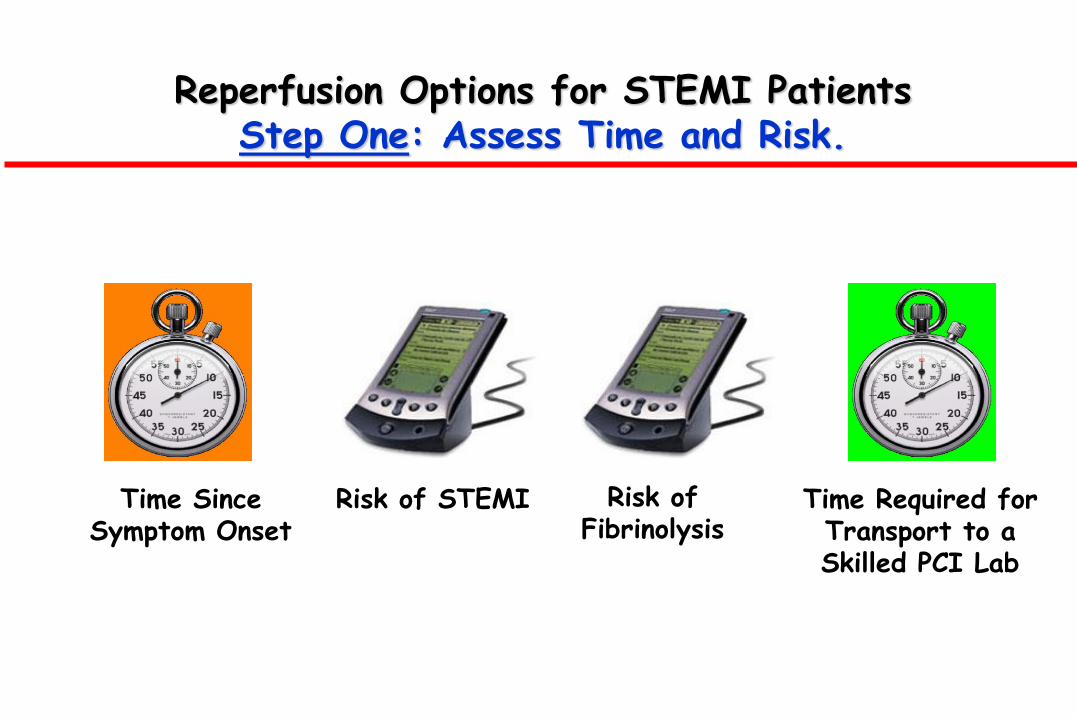
Reperfusion Options for STEMI Patients Step One: Assess Time and Risk.
Time Since Symptom Onset
Time Required for Transport to a Skilled PCI Lab
Risk of STEMI Risk of Fibrinolysis

Reperfusion Options for STEMI Patients Step 2: Select Reperfusion Treatment.
If presentation is < 3 hours and there is no delay to an invasive strategy, there is no preference for either strategy.
Fibrinolysis generally preferred Early presentation ( ≤ 3 hours from symptom onset and delay to invasive strategy) Invasive strategy not an option Cath lab occupied or not available Vascular access difficulties
No access to skilled PCI lab Delay to invasive strategy Prolonged transport
Door-to-balloon more than 90 minutes > 1 hour vs fibrinolysis (fibrin-specific agent) now

Reperfusion Options for STEMI Patients Step 2: Select Reperfusion Treatment.
If presentation is < 3 hours and there is no delay to an invasive strategy, there is no preference for either strategy.
Invasive strategy generally preferred Skilled PCI lab available with surgical backup
Door-to-balloon < 90 minutes
• High Risk from STEMI Cardiogenic shock, Killip class ≥ 3
Contraindications to fibrinolysis, including increased risk of bleeding and ICH
Late presentation
> 3 hours from symptom onset
Diagnosis of STEMI is in doubt

Algorithm
for
management
of pts with
suspected
AMI in the
ED


Extension / Ischemia
Complications of Acute MI
Acute MI
Arrhythmia
Heart Failure
Expansion / Aneurysm RV Infarct
Pericarditis
Mechanical Mural Thrombus

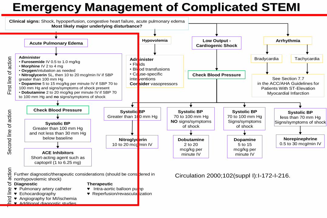
Emergency Management of Complicated STEMIEmergency Management of Complicated STEMI
Administer
• Fluids
• Blood transfusions
• Cause-specific
interventions
Consider vasopressors
Arrhythmia
Bradycardia Tachycardia
Systolic BP
Greater than 100 mm Hg
Systolic BP
70 to 100 mm Hg
NO signs/symptoms
of shock
Systolic BP
70 to 100 mm Hg
Signs/symptoms
of shock
Systolic BP
less than 70 mm Hg
Signs/symptoms of shock
Dobutamine
2 to 20
mcg/kg per
minute IV
Low Output -
Cardiogenic Shock
Nitroglycerin
10 to 20 mcg/min IV
Dopamine
5 to 15
mcg/kg per
minute IV
Norepinephrine
0.5 to 30 mcg/min IV
Hypovolemia
Administer
• Furosemide IV 0.5 to 1.0 mg/kg
• Morphine IV 2 to 4 mg
• Oxygen/intubation as needed
• Nitroglycerin SL, then 10 to 20 mcg/min IV if SBP greater than 100 mm Hg
• Dopamine 5 to 15 mcg/kg per minute IV if SBP 70 to
100 mm Hg and signs/symptoms of shock present
• Dobutamine 2 to 20 mcg/kg per minute IV if SBP 70
to 100 mm Hg and no signs/symptoms of shock
Fir
st li
ne
of
act
ion
Se
con
d li
ne
of a
ctio
nT
hir
d li
ne
of
act
ion
See Section 7.7
in the ACC/AHA Guidelines for
Patients With ST-Elevation
Myocardial Infarction
Check Blood Pressure
Clinical signs: Shock, hypoperfusion, congestive heart failure, acute pulmonary edema
Most likely major underlying disturbance?
Further diagnostic/therapeutic considerations (should be considered in
nonhypovolemic shock)
Diagnostic Therapeutic
♥ Pulmonary artery catheter ♥ Intra-aortic balloon pump
♥ Echocardiography ♥ Reperfusion/revascularization
♥ Angiography for MI/ischemia
♥ Additional diagnostic studies
Acute Pulmonary Edema
Check Blood Pressure
Systolic BP
Greater than 100 mm Hg
and not less than 30 mm Hg
below baseline
ACE Inhibitors
Short-acting agent such as
captopril (1 to 6.25 mg)
Circulation 2000;102(suppl I):I-172-I-216.

Emergency Management of Complicated STEMIEmergency Management of Complicated STEMI
Administer
• Fluids
• Blood transfusions
• Cause-specific
interventions
Consider vasopressors
Arrhythmia
Bradycardia Tachycardia
Systolic BP
Greater than 100 mm Hg
Systolic BP
70 to 100 mm Hg
NO signs/symptoms
of shock
Systolic BP
70 to 100 mm Hg
Signs/symptoms
of shock
Systolic BP
less than 70 mm Hg
Signs/symptoms of shock
Dobutamine
2 to 20
mcg/kg per
minute IV
Low Output -
Cardiogenic Shock
Nitroglycerin
10 to 20 mcg/min IV
Dopamine
5 to 15
mcg/kg per
minute IV
Norepinephrine
0.5 to 30 mcg/min IV
Hypovolemia
Administer
• Furosemide IV 0.5 to 1.0 mg/kg
• Morphine IV 2 to 4 mg
• Oxygen/intubation as needed
• Nitroglycerin SL, then 10 to 20 mcg/min IV if SBP greater than 100 mm Hg
• Dopamine 5 to 15 mcg/kg per minute IV if SBP 70 to
100 mm Hg and signs/symptoms of shock present
• Dobutamine 2 to 20 mcg/kg per minute IV if SBP 70
to 100 mm Hg and no signs/symptoms of shock
Fir
st lin
e o
f a
ction
Se
co
nd
lin
e o
f a
ctio
nT
hir
d lin
e o
f a
ction
See Section 7.7
in the ACC/AHA Guidelines for
Patients With ST-Elevation
Myocardial Infarction
Check Blood Pressure
Clinical signs: Shock, hypoperfusion, congestive heart failure, acute pulmonary edema
Most likely major underlying disturbance?
Further diagnostic/therapeutic considerations (should be considered in
nonhypovolemic shock)
Diagnostic Therapeutic
♥ Pulmonary artery catheter ♥ Intra-aortic balloon pump
♥ Echocardiography ♥ Reperfusion/revascularization
♥ Angiography for MI/ischemia
♥ Additional diagnostic studies
Acute Pulmonary Edema
Check Blood Pressure
Systolic BP
Greater than 100 mm Hg
and not less than 30 mm Hg
below baseline
ACE Inhibitors
Short-acting agent such as
captopril (1 to 6.25 mg)
Circulation 2000;102(suppl I):I-172-I-216.

Emergency Management of Complicated STEMIEmergency Management of Complicated STEMI
Administer
• Fluids
• Blood transfusions
• Cause-specific
interventions
Consider vasopressors
Arrhythmia
Bradycardia Tachycardia
Systolic BP
Greater than 100 mm Hg
Systolic BP
70 to 100 mm Hg
NO signs/symptoms
of shock
Systolic BP
70 to 100 mm Hg
Signs/symptoms
of shock
Systolic BP
less than 70 mm Hg
Signs/symptoms of shock
Dobutamine
2 to 20
mcg/kg per
minute IV
Low Output -
Cardiogenic Shock
Nitroglycerin
10 to 20 mcg/min IV
Dopamine
5 to 15
mcg/kg per
minute IV
Norepinephrine
0.5 to 30 mcg/min IV
Hypovolemia
Administer
• Furosemide IV 0.5 to 1.0 mg/kg
• Morphine IV 2 to 4 mg
• Oxygen/intubation as needed
• Nitroglycerin SL, then 10 to 20 mcg/min IV if SBP greater than 100 mm Hg
• Dopamine 5 to 15 mcg/kg per minute IV if SBP 70 to
100 mm Hg and signs/symptoms of shock present
• Dobutamine 2 to 20 mcg/kg per minute IV if SBP 70
to 100 mm Hg and no signs/symptoms of shock
Fir
st li
ne
of
act
ion
Se
con
d li
ne
of a
ctio
nT
hir
d li
ne
of
act
ion
See Section 7.7
in the ACC/AHA Guidelines for
Patients With ST-Elevation
Myocardial Infarction
Check Blood Pressure
Clinical signs: Shock, hypoperfusion, congestive heart failure, acute pulmonary edema
Most likely major underlying disturbance?
Further diagnostic/therapeutic considerations (should be considered in
nonhypovolemic shock)
Diagnostic Therapeutic
♥ Pulmonary artery catheter ♥ Intra-aortic balloon pump
♥ Echocardiography ♥ Reperfusion/revascularization
♥ Angiography for MI/ischemia
♥ Additional diagnostic studies
Acute Pulmonary Edema
Check Blood Pressure
Systolic BP
Greater than 100 mm Hg
and not less than 30 mm Hg
below baseline
ACE Inhibitors
Short-acting agent such as
captopril (1 to 6.25 mg)
Circulation 2000;102(suppl I):I-172-I-216.

Thank
you !