
Acute Respiratory Insufficiency Acute Respiratory Failure...
40
University of Debrecen Universitas Debreceniensis István Lőrincz MD,PhD Division of Emergency Medicine, Institute of Internal Medicine, University of Debrecen Acute Respiratory Failure
Transcript of Acute Respiratory Insufficiency Acute Respiratory Failure...
University of Debrecen Universitas Debreceniensis
István Lőrincz MD,PhD Division of Emergency Medicine, Institute of Internal Medicine, University of Debrecen
Acute Respiratory Failure

Acute respiratory failure
The goal of breathing is to fill the blood with the sufficient amount of oxygen necessary for the tissues and clear the blood of carbon dioxide
ARF= the insufficiency of the breathing to fulfill the above task- that is insufficient respiratory performance of the lungs

Acute respiratory failure
- Not an independent entity – it is always a
consequence of various pathologic processes
- The cause can be mechanical insufficiency of
the breathing or alveolo-capillary dysfunction
(hypercapnic and hypoxic types of respiratory
insufficiency)

Classification
Acute/ chronic respiratory failure + acute exacerbation of a chronic process
Partial or total (global) ARI (hypoxia alone or +hypercapnia)
Ventilation/ Diffusion/ Perfusion abnormalities
Obstructive or restrictive RI
Acute respiratory failure

Alveolar phase of breathing
erythrocyte
Membrane
Intracellular fluid
Hgb molecule

Causes of ventilation problems
Central: CNS –spinal cord Injuries Drug action - e.g. opioids!
Neurologic, neuromuscular, muscular failures E.g. myasthenia gravis, Gillain Barré sy., muscle
relaxants
Mechanical causes Thoracic cage – rib fractures, burns, scars… Compression of the lungs – hydrothorax,
hemothx, pneumothx
Airway obstruction Upper airway obstruction – foreign body, stenosis… Lower airways – bronchospasm, asthma…..
Problems in the lung-parenchyma itself

Acute Lung Injury, Acute Respiratory Distress Syndrome (ALI/ARDS)
Acute bronchospasm – severe asthma
Acute on chronic airflow limitation acute exacerbation of COPD
Severe pneumonia
Pulmonary embolism
Pulmonary edema
Aspiration, inhalation
Acute respiratory failure
Clinical signs of respiratory insufficiency
dyspnoea
use of ventilatory auxiliary muscles
cyanosis
progressive elevation of the resp. rate
tachycardia
agitation, confusion, somnolentia, coma

Diagnosis
Inspection– dyspnoea, thoracic movements, etc.
Respiratory rate
Pulsoximetry
Blood gases (arterial, venous) – irepeated! – Reaction to oxygen inhalation?
Asthma: peak flow
Further investigations: Thorax X ray? CT, MRI
Sputum - bacteriology, serology
Laboratory testing
ECG, US (TEE?)

Acute lung injury, Acute respiratory distress syndrome (ALI/ARDS)
Diffuse lung disease with severe hypoxia- characterized by loss of ventilated alveoli (loss of surfactant activityedema of the lung tissue) reduced ventilated lung-capacity reduced compliance severe hypoxemia (intrapulmonary shunts)

ALI/ARDS
Diagnosis: •Thorax x-ray /CT •Severe hypoxia – not reacting on oxygen inhalation •PaO2/FiO22 < 300 (ALI) or 200 (ARDS) •Lung compliance↓
Diffuse bilateral infiltration caused not by LV insufficiency (Paop 18 Hgmm)
American/European Consensus Comittee 1994

Causes of ALI/ARDS
Pulmonary: Extrapulmonary: - infektion/pneumonia - sepsis - aspiration/inhalation - trauma - near drowning - TRALI - contusion - CPB
TRALI = transfusion related acute lung injury CPB = Cardiopulmonary bypass

ALI/ARDS
A complex interaction between the cells and
the inflammatory mediators - lesion of
epithelial cells, alveolar macrophags and
endothelial cells
exsudation - edema, inflammation,
coagulation disorders
proliferative phase – regeneration
fibrotic phase -

Acute bronchospasm, severe asthmatic attack
Components of the insufficiency Bronchospasmus Edema of the bronchiolar mucous membranes Secretion – sticky secretions Obscruction of small bronchioli Air trapping – Exhalation incomplete
The pressure never returns to zero! "dynamic hyperinflation" (TLC↑, RV↑, FRC↑)
Lung inflation - intrinsic or autoPEEP
Respiratory work elevated - Exhaustion!

Acute asthma: assessment Pts with severe asthma and one or more adverse psychosocial factors (psychiatric illness. alcohol or drug abuse, unemployment) have t mortality. Measure the peak expiratory flow rate and compare it against that expected. The peak flow acts as an immediate triage tool: remember that pts with life-threatening asthma may be too dyspnoeic to do this. Make an initial assessment of the severity of acute asthma based upon a combination of clinical features, peak flow measurement and pulseoximetry as outlined below.
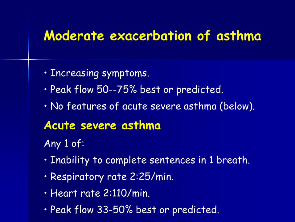
Moderate exacerbation of asthma
• Increasing symptoms.
• Peak flow 50--75% best or predicted.
• No features of acute severe asthma (below).
Acute severe asthma
Any 1 of:
• Inability to complete sentences in 1 breath.
• Respiratory rate 2:25/min.
• Heart rate 2:110/min.
• Peak flow 33-50% best or predicted.

Life-threatening asthma
A patient with severe asthma with any 1 of:
• Cyanosis.
• Exhaustion. Confusion, coma.
• Feeble respiratory effort.
• Sp02<92%.
• Silent chest.
• Bradycardia, arrhythmia, hypotension.
• p02<8kPa.
• Normal pC02 (4.6-6.0kPa).
• Peak flow <33% best or predicted.

Near fatal asthma • pC02 and/or requiring mechanical ventilation with inflation pressures.

Other investigations
Obtain ABG if Sp02<92% or if there are other features
of life-threateninr. asthma.
Obtain a CXR (Without delaying treatment) if there is:
• Suspected pneumomediastinum or pneumothorax.
• Suspected consolidation.
• Life-threatening asthma.
. Failure to respond to treatment satisfactorily.
• Requirement for ventilation.

Acute asthma: management Initial treatment 1
Follow BTS/SIGN guidelines summarized as follows:
• Provide high flow 02.
• Put the trolley back and side rails up so the pt is
sitting up and holding on to the side rails (to use
pectoral muscles as accessory m. of respiration).
• If the patient cannot talk, start treatment, but get
senior ED and ICU help in case intubation and
ventilation are required.
• Check trachea and chest signs for pneumothorax.
• Ask about previous admissions to ICU.

Acute asthma: management Initial treatment 2
• Administer high dose nebulized β2 agonist (eg salbutamol 5mg or terbutaline 10mg), or 10 puffs of salbutamol into spacer device and face mask. For severe asthma or asthma that reponds poorly to the initial nebulizer, consider continuous nebulization. • Give a corticosteroid: either prednisolone 4O-50mg PO or hydrocortysone (as sodium succinate) 100mg IV. • Add nebulized ipratropium bromide (500mcg) to beta2 agonist treatment for patients with acute severe or life-threatening asthma or those with a poor initial response to beta2 agonist therapy.

Acute asthma: management Initial treatment 4
• Consider a Single dose of IV Mg-sulphate (1.2-2g IVI over 20min) for pts with acute severe asthma without a good initial response to inhaled bronchodilator th. or for those with near-fatal asthma. • Use IV aminophylline. Some individual pts with near-fatal or life-threatening asthma with a poor response to initial th. may gain additional benefit. The loading dose of IVI aminophylline is 5mg/kg over 20min unless on maintenance th, in which case check blood theophylline level and start IV of aminophylline at 0.5-0.7mg/kg/hr. ?????????????

Acute asthma: management Initial treatment 5
• IV salbutamol is an alternative in severe asthma,
after consultation with senior staff. Draw up
5mg salbutamol into 500mL 5% dextrose and
run at a rate of 30-60mUhr.
• A patient who cannot talk will be unable to drink
fluids and may be dehydrated,
• Avoid 'routine' antibiotics.
• Repeat ABG within an hour.
• Hypokalaemia may be caused or exacerbated by
beta2 agonist and/or steroid therapy.

Criteria for admission
Admit patients with any features of
A life-threatening or near-fatal
attack.
Severe attack persisting after initial
treatment.

Refferral to intensive care unit
Refer any patient requiring ventilatory support or
with acute severe or life-threatening asthma failing
to respond to therapy, evidenced by:
Drowsiness, confusion.
Exhaustion, feeble respiration.
Coma or respiratory arrest.
Persisting or worsening hypoxia.
Hypercapnoea.
ABG showing pH.
Deteriorating peak flow.

Cardiac arrest in acute asthma
The underlying rhythm is usually PEA. This may reflect
one or more of the following: prolonged severe hypoxia
(secondary to severe bronchospasm and mucous
plugging), hypoxia-related arrhythmias or tension
pneumothorax (may be bilateral). Give advanced life
support according to the guidellines in Cardiac arrest,
and treat tension pneumothorax if present. Aim to
achieve tracheal intubation early in view of the higher
than normal required lung inflation pressures and the
attendant risk of gastric inflation in the absence of a
tracheal tube.

PEAK FLOW METER 12

Arterial blood gases
PaO 2 Severity
Mild
Medium
Severe Normal
Life danger!
PaCO 2
16

Pneumonia – infective infiltration of the lungs
Epidemiology: Home aquired
Community aquired (CAP)
Hospital aquired (HAP)
Ventilator aquired (VAP)
Infective agent Bakterial - pneumocc., haemophylus, stacc., mycoplasma
Viral pneumonia (influenza, adenovirus, etc.)
Clinical appearance Typic pneumonia (sudden beginning, high fever, productive cough…)
Atypic pneumonia (less characteristic symptoms)

Pulmonary edema
Dynamic balance state:
intravascular – interstitial – alveolar
compartments
Starling equation
(fluid movement through semipermeable membranes):
Qf = K /(Pc – Pi) – σ(Pc – Pi)/ K: filtration coefficient,
σ: protein permeability Pc, Pi: capillary + interstitial oncotic pressure
Factors: Alveolocapillary membrane permeability
Hydrostatic pressure in the capillaries
Onkotic pressure in the interstitium
Capacity of the lymph-system

Common causes of pulmonary edema:
Cardial edema: main cause is the elevated hydrostatic
pressure in the pulmonary vessels (AMI, IHF, CMP, MS, MI, hypertensive crisis…)
Nono cardiac causes: Chemical irritation (gases,fumes, aspiration of acidic gastric content, etc.)
Fluid overload
Followinf upper airway obstruction, near drowing
Pneumothx (interstitial neg.pressure↓), re-expansion
High altitude
Infection, sepsis
Pharmacons, toxins (sedato-hypnotica, salicylate overdose, paraquate…)
…..

Therapy
Elimination of the cause
Half-sitting position
Oxygen therapy
Positive pressure ventilation (IPPV,
PEEP) – in case of hypercapnia, severe hypoxia Gyógyszeres
th:
Morphine 5-10 mg IV
Furosemide 20-40 mg IV
TNG sublinqual 0,3-0,6 mg, or spray or IV infusion

Oxygen Supplementation low flow systems 1-10 LPM
100% O2 mixes with room air to determine FIO2 - definition
FIO2 varies with patient’s breathing pattern – Rapid inspiration entrains more room air
– Deep breaths entrain more room air
– Rapid respiratory rate entrains more room air
– Patients in more distress get lower FIO2
FIO2 is unknown since amount of entrainment is unknown
Any humidity in gas comes from entrained air- wall O2 has 0% relative humidity
Low flow devices Simple Nasal Cannulas
Simple masks 33

High Flow O2 Devices > 20 - 60 lpm
34
Device provides 100% of gas to patient - definition
No entrainment of room air if mask fits
FIO2 is known and exact
Relative humidity depends on the device
High flow devices:
– High flow nasal cannula
– Venturi mask
– Aerosol mask – heated or cool
– Nonrebreather mask – some characteristics of both high and low

O2 Devices
35

Aerosol O2 devices
36

BiPAP or NPPV
37
Bilevel positive airway pressure (BiPAP) is a proprietary name of Respironics, Inc. For continouous positive airway pressure (CPAP) with pressure support breaths. It is used during noninvasive positive pressure ventilation. It delivers a preset inspiratory positive airway pressure (IPAP) during inspiration and expiratory positive airway pressure (EPAP). BiPAP can be described as a continuous positive airway pressure system with a time-cycled or flow-cycled change of the applied pressure level. CPAP, BPAP and other non-invasive ventilation modes have been shown to be effective management tools for COPD, acute and chronic respiratory failure. Another term for bilevel positive airway pressure, and the term becoming increasingly adopted by the medical community, is non-invasive positive pressure ventilation (NIPPV) or non-invasive ventilation (NIV).

The setup for BIPAP using a mechanical ventilator
38

BiPAP or NPPV
Contraindications
– Cardiac or respiratory arrest
– Inability to cooperate, protect the airway, or clear secretions
– Nonrespiratory organ failure, esp shock
– Facial surgery, trauma, or deformity
– Prolonged duration of mechanical ventilation anticipated
– Recent esophageal anastomosis
A need for emergent intubation is an absolute contraindication to NPPV
Set inspiratory pressure (IP) and exp pressure (PEEP)
Mean pressure determines oxygenation
IP – PEEP determines ventilatory assist
39

Thank you !


