Lipids - JU Med: Class of 2019jumed2019.weebly.com/uploads/2/6/8/5/26855942/biom_lipids_2013... ·...
82
-
Upload
nguyenkien -
Category
Documents
-
view
217 -
download
4
Transcript of Lipids - JU Med: Class of 2019jumed2019.weebly.com/uploads/2/6/8/5/26855942/biom_lipids_2013... ·...

Lipids Dr. Mamoun Ahram Lecture 7 Summer 2013-2014

Resources
This lecture Campbell and Farrell’s Biochemistry, Chapter 8

Lipids
Lipids are a heterogeneous class of naturally occurring organic compounds that share some properties based on structural similarities, mainly a dominance of nonpolar groups. They are Amphipathic in nature. They are insoluble in water, but soluble in fat or organic solvents (ether, chloroform, benzene, acetone). They are widely distributed in plants & animals.

Classes Simple lipids (fats, oils, and waxes) Complex lipids (glycerides , glycerophospholipids, sphingolipids, glycolipids, lipoproteins) Derived lipids (fatty acids, alcohols, eicosanoids) Cyclic lipids (steroids)

Lipid Functions
Lipids include: Storage Lipids Structural Lipids in Membranes Lipids as Signals, Cofactors & Pigments
Source of energy They are storable to unlimited amount (vs. carbohydrates) They provide considerable amount of energy to the body (25% of body needs) & provide a high-energy value (more energy per gram vs. carbohydrates & proteins)
Structural components (cell membranes) Precursors of hormone and vitamins Shock absorbers thermal insulator

Fatty acids Aliphatic mono-carboxylic acids Formula: R-(CH2)n-COOH Lengths
Physiological (12-24) Abundant (16 and 18)
Degree of unsaturation Amphipathic molecules
Function: • Building blocks of other lipids • Modification of many proteins
(lipoproteins) • Important fuel molecules • Derivatives of important cellular
molecules

Types of fatty acids
Saturated fatty acids Short chain F.A. (1-6 carbons) Medium-chain F.A. (7-10 carbons) Long chain F.A.(more the 10 carbon)
Unsaturated fatty acids Monounsaturated Polyunsaturated

Cis vs. trans bonds
cis isomer predominates; trans is rare
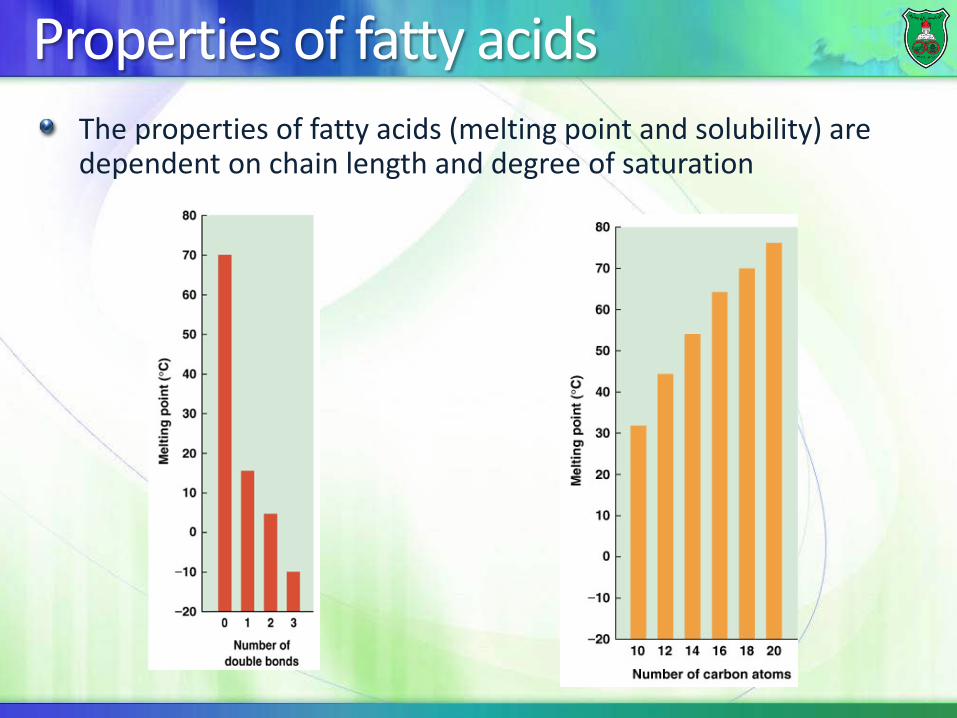
Properties of fatty acids The properties of fatty acids (melting point and solubility) are dependent on chain length and degree of saturation

Properties of fatty acids
Short chain F.A. Medium-chain F.A. Long chain F.A. They are liquid in nature
Solids at room temperature
Solids at room temperature
Water-soluble Water-soluble Water-insoluble Volatile ar room temperature
Non-volatile at room temperature
Non-volatile &
acetic, butyric, & caproic acids
Examples: caprylic & capric F.A.
Examples: palmitic, stearic, & lignoceric F.A Occur in hydrogenated oils, animal fats, butter & coconut & palm oils
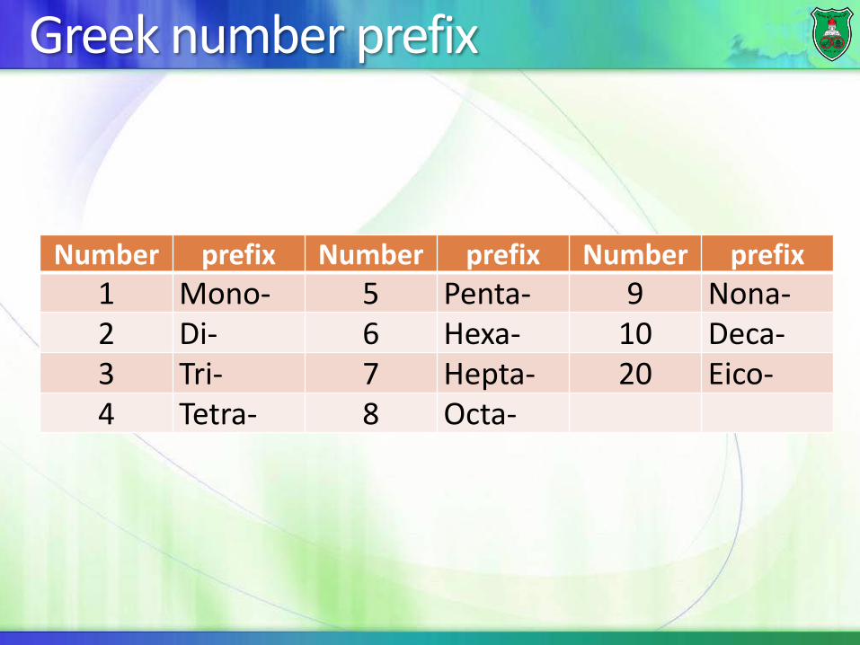
Greek number prefix
Number prefix Number prefix Number prefix 1 Mono- 5 Penta- 9 Nona- 2 Di- 6 Hexa- 10 Deca- 3 Tri- 7 Hepta- 20 Eico- 4 Tetra- 8 Octa-

Naming of a fatty acid Alkane to oic
Octadecane (octa and deca) is octadecanoic acid One double bond = octadecenoic acid Two double bonds = octadecadienoic acid Three double bonds = octadecatrienoic acid
Designation of carbons and bonds 18:0 = a C18 fatty acid with no double bonds
stearic acid (18:0); palmitic acid (16:0) 18:2 = two double bonds (linoleic acid)
Designation of location of bonds Δn: The position of a double bond
cis-Δ9 :a cis double bond between C 9 and 10 trans-Δ2:a trans double bond between C 2 and 3


Number of carbons
Number of double
bonds
Common name
Systematic name Formula
14 0 Myristate n-Tetradecanoate CH3(CH2)12COO- 16 0 Palmitate n-Hexadecanoate CH3 (CH2) 14COO- 18 0 Stearate n-Octadecanoate CH3(CH2) 16COO- 18 1 Oleate cis-Δ9-Octadecenoate CH3(CH2) 7CH=CH(CH2) 7COO- 18 2 Linoleate cis,cis-Δ9,Δ12-
Octadecadienoate CH3(CH2) 2(CH=CHCH2) 2(CH2)
6COO- 18 3 Linolenate all-cis-Δ9,Δ12,Δ15-
Octadecatrienoate CH3CH2(CH=CHCH2) 3(CH2) 6COO-
20 4 Arachidonate all-cis-Δ5,Δ8,Δ11,Δ14-Eicosatetraenoate
CH3 (CH2) 4(CH=CHCH2) 4(CH2)
2COO-

Another way of naming (ω)-C: distal methyl C as #1
• Linoleic acid: precursor of arachidonates • Linolenic acid: precursor of EPA and DHA

Numerical Symbol Common Name and Structure Comments
18:1Δ9 Oleic acid
Omega-9 monounsaturated
18:2Δ9,12 Linoleic acid Omega-6 polyunsaturated
18:3Δ9,12,15 α-Linolenic acid (ALA) Omega-3 polyunsaturated
20:4Δ5,8,11,14 Arachidonic acid Omega-6 polyunsaturated
20:5Δ5,8,11,14,17 Eicosapentaenoic acid (EPA)
Omega-3 polyunsaturated (fish oils)
22:6Δ4,7,10,13,16,19 Docosahexaenoic acid (DHA)
Omega-3 polyunsaturated (fish oils)


Arachidonate all cis-Δ5,Δ8,Δ11,Δ14-eicosatetraenoate, CH3(CH2)4(CH=CHCH2)4(CH2)2COO-
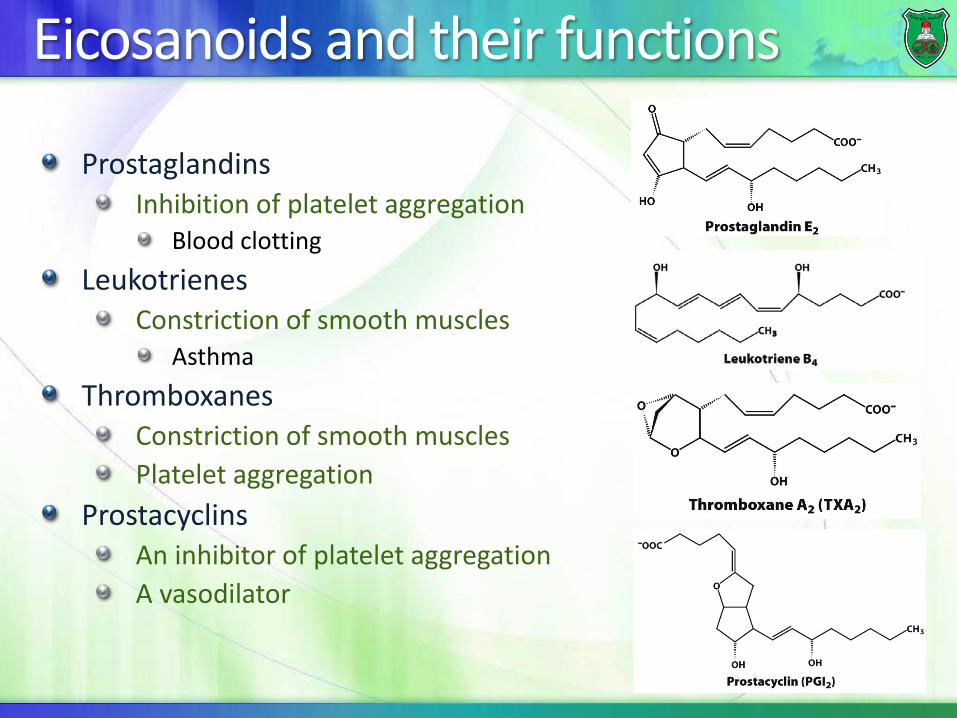
Eicosanoids and their functions
Prostaglandins Inhibition of platelet aggregation
Blood clotting Leukotrienes
Constriction of smooth muscles Asthma
Thromboxanes Constriction of smooth muscles Platelet aggregation
Prostacyclins An inhibitor of platelet aggregation A vasodilator

Aspirin and the heart
Thromboxane A2 leads to platelet activation and aggregation. Aspirin acts as a potent antiplatelet agent by inhibiting cyclooxygenase preventing thromboxane A2 (TXA2) generation.

Aspirin
Aspirin is anti-inflammatory and fever-reducing (antipyretic). It irreversibly inhibits cyclooxygenase (COX), the enzyme that catalyzes conversion of arachidonic acid to prostaglandins.
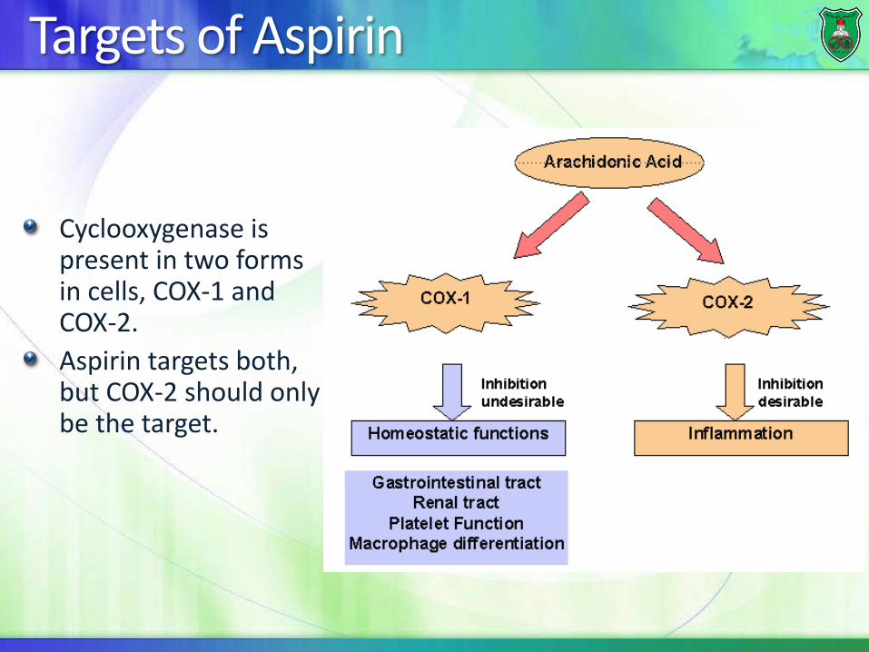
Targets of Aspirin
Cyclooxygenase is present in two forms in cells, COX-1 and COX-2. Aspirin targets both, but COX-2 should only be the target.

Celebrex
A new generation drug, Celebrex, targets COX2, but is prescribed with a strong warning of side effects on the label.

Omega fatty acids Omega-3 fatty acids
α-linolenic acid → eicosapentaenoic acid (EPA) → docosahexaenoic acid (DHA)
They reduce inflammatory reactions by: Reducing conversion of arachidonic acid into eicosanoids Promoting synthesis of anti-inflammatory molecules
Omega-6 fatty acids: Arachidonic acid
stimulates platelet and leukocyte activation, signals pain, Induces bronchoconstriction, regulates gastric secretion
Omega-9 fatty acids Oleic acid
Reduces cholesterol in the circulation


Triglycerides
Ester linkage
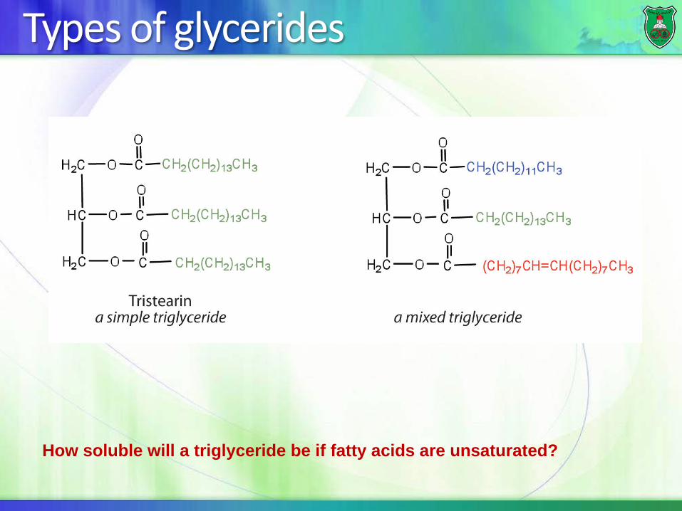
Types of glycerides
How soluble will a triglyceride be if fatty acids are unsaturated?

Solid vs. liquid fats
Vegetable oils consist almost entirely of unsaturated fatty acids, whereas animal fats contain a much larger percentage of saturated fatty acids.
This is the primary reason for the different melting points of fats and oils

Saponification
Hydrolysis : steam, acid, enzyme (e.g., lipase of pancreas) Saponification: Alkaline hydrolysis produces salts of fatty acids (soaps). Soaps cause emulsification of oily material

How does soap work?
When mixed with water, the hydrophobic hydrocarbon tails cluster together to create a nonpolar microenvironment and the hydrophilic ionic heads interact with water. The resulting spherical clusters are called micelles. Grease and dirt are trapped inside micelles and the complex can be rinsed away.
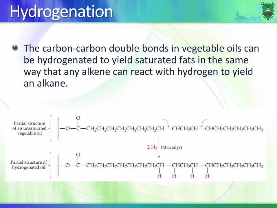
Hydrogenation
The carbon-carbon double bonds in vegetable oils can be hydrogenated to yield saturated fats in the same way that any alkene can react with hydrogen to yield an alkane.
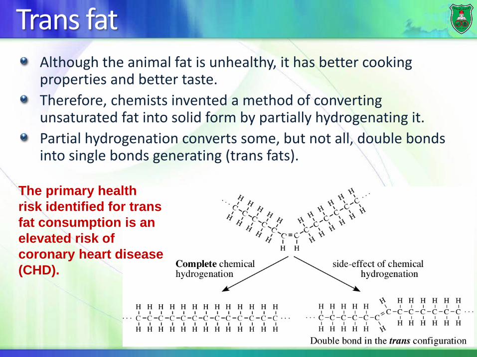
Trans fat Although the animal fat is unhealthy, it has better cooking properties and better taste. Therefore, chemists invented a method of converting unsaturated fat into solid form by partially hydrogenating it. Partial hydrogenation converts some, but not all, double bonds into single bonds generating (trans fats).
The primary health risk identified for trans fat consumption is an elevated risk of coronary heart disease (CHD).

Example: margarine
In margarine, only about two-thirds of the double bonds present in the starting vegetable oil are hydrogenated, so that the margarine remains soft in the refrigerator and melt on warm toast.

Chemical Properties of fats & oils Halogenation: added to unsaturated F.A (e.g., iodination)
Used to determine the degree of unsaturation of the fat or oil that determines its biological value
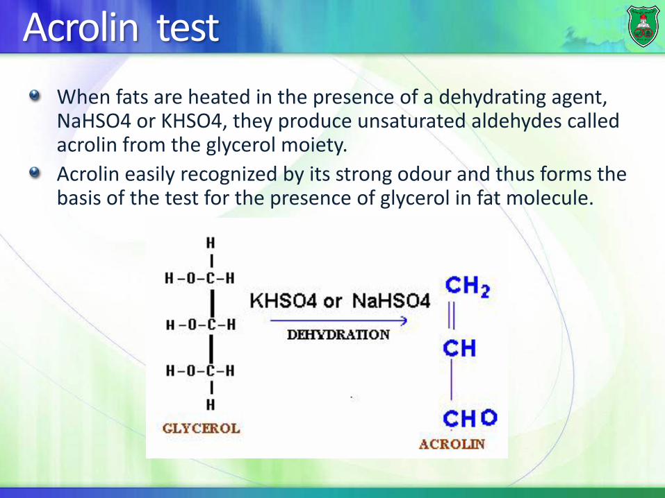
Acrolin test When fats are heated in the presence of a dehydrating agent, NaHSO4 or KHSO4, they produce unsaturated aldehydes called acrolin from the glycerol moiety. Acrolin easily recognized by its strong odour and thus forms the basis of the test for the presence of glycerol in fat molecule.

Waxes
Solid simple lipids containing a monohydric alcohol (C16 ~ C30, higher molecular weight than glycerol) esterified to long-chain fatty acids (C14 ~ C36). Examples: palmitoyl alcohol Insoluble in water & Negative to acrolein test Are not easily hydrolyzed (fats) & are indigestible by lipases Are very resistant to rancidity Are of no nutritional value Coatings that prevent loss of water by leaves of plants

Membrane lipids
The most prevalent class of lipids in membranes is the glycerophospholipids

Phospholipids (phosphoacylglycerols)

Classification of Glycerophospholipids
Phosphatidic acids Phosphatidylcholine (lecithins)
Most abundant membrane lipid Cephalins
Phosphatidylethanolamine Phosphatidylserine
abundant in brain Phosphatidylinositol
sends messages across cell membranes Cardiolipin Plasmalogens

Glycerophospholipids - Lecithins
Snake venom contain lecithinase, which hydrolyzes polyunsaturated fatty acids and converting lecithin into lysolecithin
hemolysis of RBCs

Emulsification
Because of their amphipathic nature, they act as emulsifying agents, that is substances that can surround nonpolar molecules and keep them in suspension in water

Glycerophospholipids - Cardiolipins
Diphosphatidyl-glycerol Found in the inner membrane of mitochondria Initially isolated from heart muscle (cardio) Structure: 3 molecules of glycerol, 4 fatty acids & 2 phosphate groups

Plasmalogens They are found in the cell membrane phospholipids fraction of brain & muscle, liver, and semen. They have a protective role against reactive oxygen species Structure:
Precursor: Dihydroxyacetone phosphate Unsaturated fatty alcohol at C1 connected by ether bond In mammals: at C3; phosphate + ethanolamine or choline

Major classes of plasmalogens
Ethanolamine plasmalogen (myelin-nervous tissues) Choline plasmalogen (cardiac tissue)
Platelet activating factor Serine plasmalogens

Glycerophospholipids - Inositides Phosphatidyl inositol Nitrogenous base: cyclic sugar alcohol (inositol) Structure: glycerol, saturated FA, unsaturated FA, phosphoric acid, & inositol Source: Brain tissues
Functions: Major component of cell membrane Second messenger during signal transduction On hydrolysis by phospholipase C, phosphatidyl-inositol-4,5-
diphosphate produces diacyl-glycerol (DAG) & inositol-triphosphate (IP3); which liberates calcium
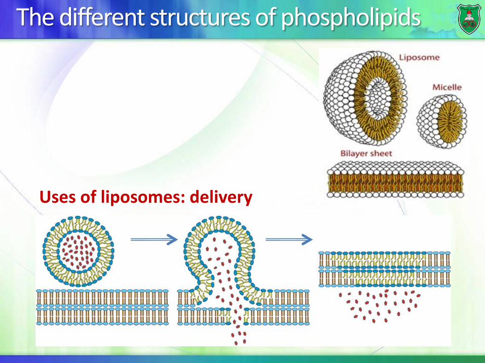
The different structures of phospholipids
Uses of liposomes: delivery
Sphingolipids
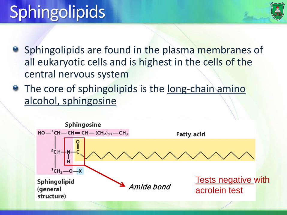
Sphingolipids are found in the plasma membranes of all eukaryotic cells and is highest in the cells of the central nervous system The core of sphingolipids is the long-chain amino alcohol, sphingosine
Amide bond Tests negative with acrolein test

Ceramide
Types of sphingolipids
The sphingolipids are divided into the two subcategories:
Sphingomyelins Glycosphingolipid (or glycolipids)

Sphoingomyelin
Sphoingomyelin is a sphinglolipid that is a major component of the coating around nerve fibers The group attached to C1 is a phosphocholine

Glycolipids
Sphingolipids can also contain carbohydrates attached at C-1 and these are known as glycolipids Glycolipids are present on cell membranes and act as cell surface receptors that can function in cell recognition (e.g., pathogens) and chemical messengers There are three types of glycolipids
Cerebrosides Globosides Gangliosides

Glycolipids • Cerebrosides: the simplest
glycolipids, contain a single hexose (galactose or glucose).
• Globosides and gangliosides are more complex glycolipids.
• Both contain glucose, galactose, and N-acetylgalactosamine, but gangliosides must also contain sialic acid.
Gangliosides are targeted by cholera toxin in the human intestine.

Sulfatides Synthesized from galactocerebroside Abundant in brain myelin

Sphingolipids and blood groups

Lipoproteins Function: transport of different types of lipids (cholesterol, cholesterol esters, phospholipids & triacylglycerols) in blood plasma.
As lipid content increases, the density decreases
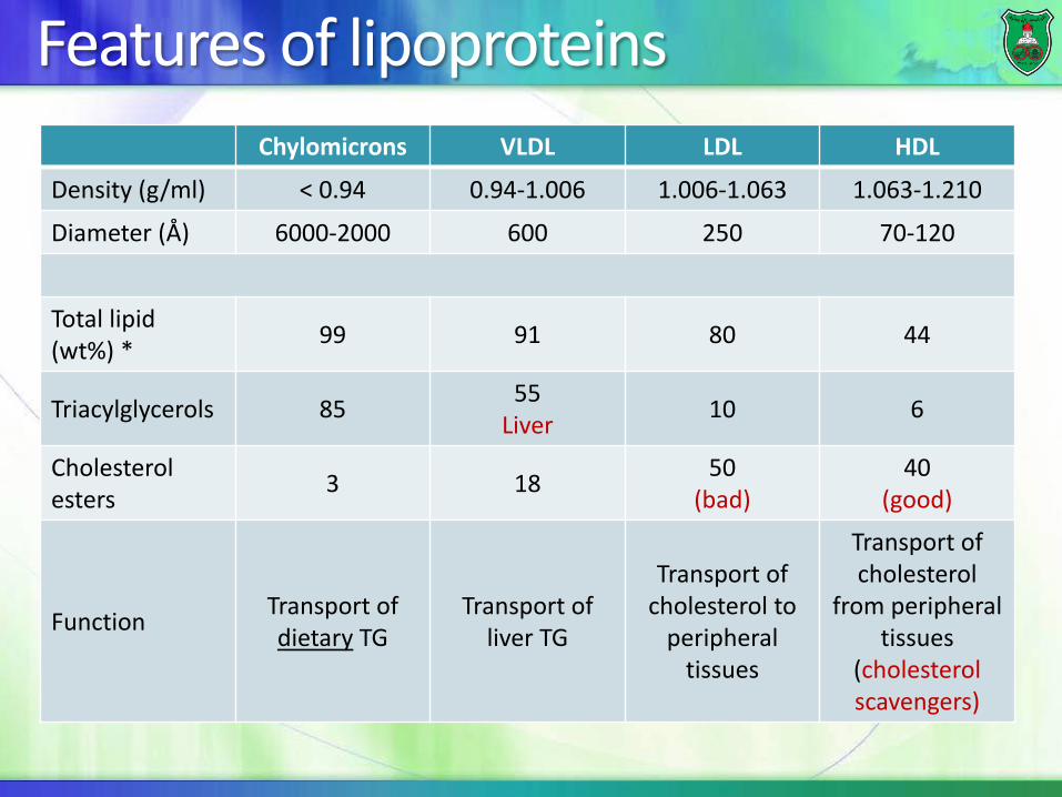
Features of lipoproteins Chylomicrons VLDL LDL HDL
Density (g/ml) < 0.94 0.94-1.006 1.006-1.063 1.063-1.210
Diameter (Å) 6000-2000 600 250 70-120
Total lipid (wt%) * 99 91 80 44
Triacylglycerols 85 55 Liver 10 6
Cholesterol esters 3 18 50
(bad) 40
(good)
Function Transport of dietary TG
Transport of liver TG
Transport of cholesterol to
peripheral tissues
Transport of cholesterol
from peripheral tissues
(cholesterol scavengers)


Steroids
The precursor The nucleus
The most common steroid

Products of cholesterol
Hormones sex hormones (androgens,. estrogens, progestins)
Some vitamins such as vitamin D Vitamins A, D, E, and K are made from isoprenoids
Bile acids (intestinal absorption of fat)

Cholesterol esters
A cholesterol with a fatty acid attached at (-OH) of C3
Name the molecules?

Atherosclerosis

Cell membranes The membrane is hypothesized in a model known as the fluid mosaic model. Components: 45% lipid, 45% protein and 10% carbohydrate They exist side by side without forming some other substance of intermediate nature.

Phospholipids The outer: phosphatidylcholine, sphingomyelin, and glycolipids(cell recognition) The inner: phosphatidylethanolamine, phosphatidylserine, and phosphatidylinositol (signaling)
Cholesterol is distributed in both leaflets
Animal cells vs. plant cells vs.
prokaryotic cells
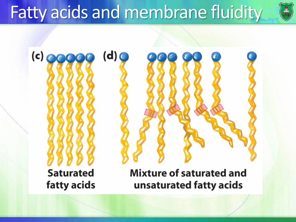
Fatty acids and membrane fluidity

Membrane fluidity and temperature
Melting temperature (transition temperature
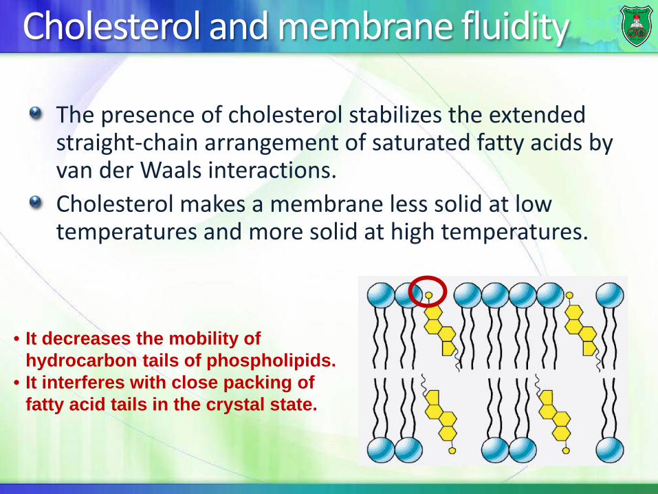
Cholesterol and membrane fluidity
The presence of cholesterol stabilizes the extended straight-chain arrangement of saturated fatty acids by van der Waals interactions. Cholesterol makes a membrane less solid at low temperatures and more solid at high temperatures.
• It decreases the mobility of hydrocarbon tails of phospholipids.
• It interferes with close packing of fatty acid tails in the crystal state.

Membrane proteins

Types of membrane proteins
Peripheral proteins: are associated with the exterior of membranes via noncovalent interactions
Integral membrane proteins: anchored into membrane via hydrophobic regions
Lipid-anchored: associated via a lipid group

Peripheral membrane proteins They are associated with membranes but do not penetrate the hydrophobic core of the membrane
often associated with integral membrane proteins They are not strongly bound to the membrane and can be removed without disrupting the membrane structure
treatment with mild detergent

Integral membrane proteins
The membrane integral domains are: 1. Single or multiple 2. α-helix or β-sheet
The integral proteins can be associated with the lipid bilayer in several ways.
Some can form channels
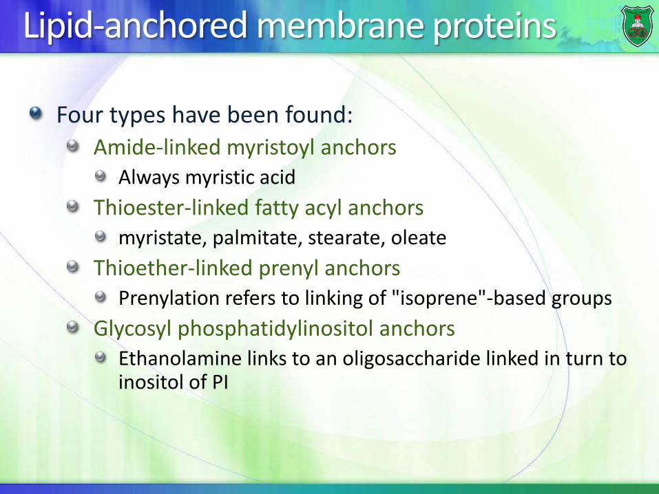
Lipid-anchored membrane proteins
Four types have been found: Amide-linked myristoyl anchors
Always myristic acid Thioester-linked fatty acyl anchors
myristate, palmitate, stearate, oleate Thioether-linked prenyl anchors
Prenylation refers to linking of "isoprene"-based groups Glycosyl phosphatidylinositol anchors
Ethanolamine links to an oligosaccharide linked in turn to inositol of PI


Structure-Function of Membranes
Transport: Membranes are impermeable barrier Proteins can be carriers or channels
Signaling Protein receptors and small molecules (some can be lipids themselves)
Catalysis Enzyme-linked receptors

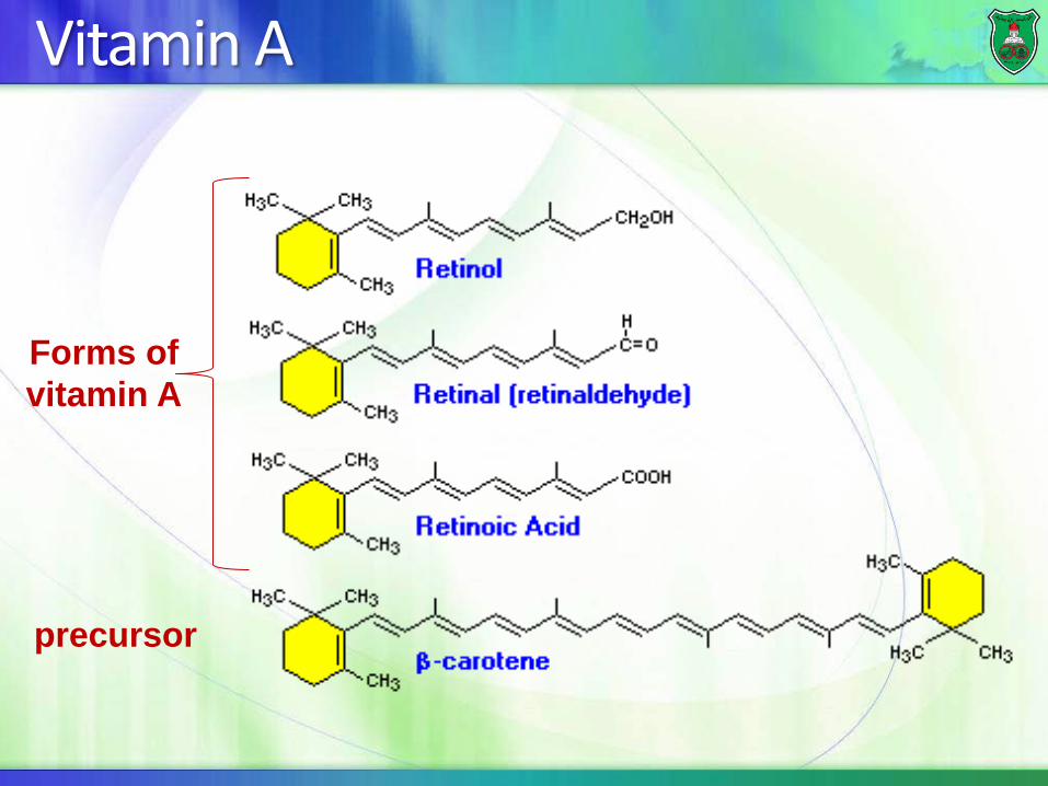
Vitamin A
precursor
Forms of vitamin A
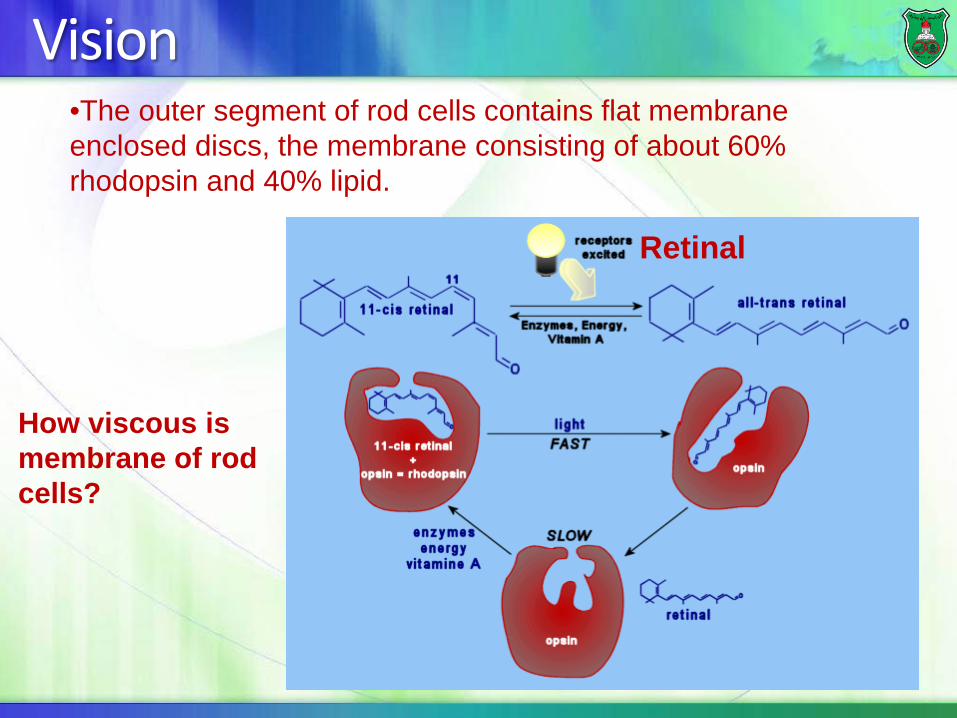
Vision
Retinal
•The outer segment of rod cells contains flat membrane enclosed discs, the membrane consisting of about 60% rhodopsin and 40% lipid.
How viscous is membrane of rod cells?
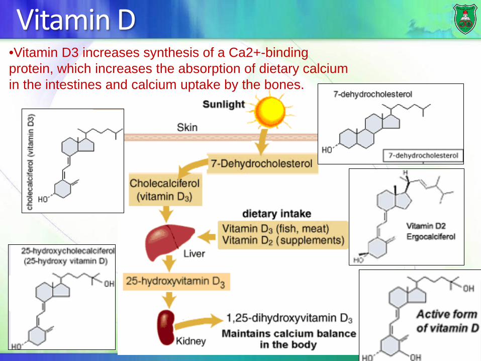
Vitamin D •Vitamin D3 increases synthesis of a Ca2+-binding protein, which increases the absorption of dietary calcium in the intestines and calcium uptake by the bones.

Vitamin E
• Vitamin E is a good reducing agent and an antioxidant (it reacts with oxidizing agents before they can attack other biomolecules).

Vitamin K The bicyclic ring system contains two carbonyl groups and a long unsaturated hydrocarbon side chain that consists of repeating isoprene Units.
(synthetic vitamin K)

Biological function of vitamin K Vitamin K is important in the carboxylation of glutamate producing γ-carboxyglutamate residues in the prothrombin protein. The two carboxyl groups bind Ca2+ ion form a bidentate (“two teeth”) ligand, which is required for blood clotting. Two well-known anticoagulants, dicumarol and warfarin (a rat poison), are vitamin K antagonists.