Limits on the bathymetric distribution of keratose sponges...
17
Vol. 174: 123-139.1998 MARINE ECOLOGY PROGRESS SERIES Mar Ecol Prog Ser Published November 26 Limits on the bathymetric distribution of keratose sponges: a field test in deep water Manuel ~aldonado'**, Craig M. young2 'Department of Aquatic Biology, Centre de Estudios Avanzados de Blanes (CSIC), Camino de Santa Barbara sin, E-17300 Blanes, Girona, Spain Department of Larval Ecology, Harbor Branch Oceanographic Institution, 5600 U.S. 1 Hwy North, Fort Pierce, Florida 34946, USA ABSTRACT The keratose sponges (1.e. those in which the mineral skeleton is replaced by a collage- nous skeleton) are generally restricted to shallow-water habitats, but the causes of this distinct bathy- metric pattern remain unclear. Sharp pycnoclines at the depth of the upper slope may hinder coloniza- tion of deep waters because of thermal stress or reduced light and particulate food below the pycnoclines. It is also possible that oligotrophy and loss of symbiotic cyanobacteria below the pycno- cline may lead to a nutritional stress. Using manned submersibles in Exuma Sound, Bahamas, we deter- mined that the pycnocline lies between 70 and 100 m. We transplanted individuals of 2 keratose sponges (Aplysina fistulans and Ircinia felix) from their natural habitat on a shallow reef (4 m deep) to 3 depths (100, 200, 300 rn) within or below the pycnocline to investigate mortality and changes in body size, shape and histology as a function of depth. We also recorded changes in populations of photosyn- thetic and heterotrophic symbiotic bacteria, as well as the parasitic polychaete Haplosyllis spongicola. By transplanting individuals of A. fistularis bearing buds for asexual propagation (fistules)and individ- uals of I. felix brooding embryos, we also tested the viability of reproductive propagules in deep-water environments. We found that, although these 2 sponges do not naturally occur at depths below 40 rn, 62.5% of A. fistularis and 42.8% of I. felix survived at 100 m for 12 nlo. No A. fistularis survived at 200 m, whereas 28.5 % of I. felix did. All sponges transplanted to 300 m died within 2 mo. Water tem- perature was the most likely cause of sudden mortality at this depth. There were no significant differ- ences in growth between individuals at the slope and controls on the shallow reef. Cyanobacteria were lost in individuals of I. felix that survived at 100 and 200 rn, and these sponges repositioned oscules and formed chimney-like processes, probably to enhance water flow through the sponge and compensate for nutritional stress. By contrast, cyanobacteria were still abundant in individuals of A. fistularis sur- viving at a depth of 100 rn, and these sponges did not change shape significantly, apart from the loss of fistules. It appears, therefore, that the loss of cyanobacteria and the increasing oligotrophy with depth do not set the lower bathymetric limits of species. Removal of sponge tissues by the parasitic polychaete H. spongicola also appears not to aggravate significantly the nutritional stress experienced by sponges transplanted to deep water, at least to the extent that it may restrict the bathymetric distribution of the host. Despite the facts that only the species I. felix was heavily parasitized and that parasites survived within hosts at all depths, there was no significant difference in survival with depths between sponge species. A TEM (transmission electron microscope) examination of the mesohyl did not reveal signifi- cant cytological differences among sponges transplanted to various depths. At all depths, surviving individuals of both species showed archeocytes engaged in phagocytosis and digestion of cyanobacte- ria and/or heterotrophic bacteria. Similarly, collencytes and spongocytes were apparently secreting collagen, indicating that temperatures at 100 and 200 rn do not inhibit the formation of the skeleton. Sponge recruitment derived from either asexual or sexual propagules was never observed at slope depths. Since adult sponges survived when they were artificially transported to deep waters, the inhi- bition of larval dispersal or settlement success (perhaps caused by the sharp decrease in temperature With increasing depth) emerges as the most plausible explanation for the shallow-water confinement of these keratose sponges. KEY WORDS: Keratose sponges . Bathymetric distributions Sponge ecology . Sponge symbionts . Sponge infauna . Slope megafauna 0 Inter-Research 1998 Resale of full article not permitted 'E-mail: [email protected]
-
Upload
nguyenngoc -
Category
Documents
-
view
221 -
download
0
Transcript of Limits on the bathymetric distribution of keratose sponges...
Vol. 174: 123-139.1998 MARINE ECOLOGY PROGRESS SERIES
Mar Ecol Prog Ser Published November 26
Limits on the bathymetric distribution of keratose sponges: a field test in deep water
Manuel ~ a l d o n a d o ' * * , Craig M. young2
'Department of Aquatic Biology, Centre de Estudios Avanzados de Blanes (CSIC), Camino de Santa Barbara sin, E-17300 Blanes, Girona, Spain
Department of Larval Ecology, Harbor Branch Oceanographic Institution, 5600 U.S. 1 Hwy North, Fort Pierce, Florida 34946, USA
ABSTRACT The keratose sponges (1.e. those in which the mineral skeleton is replaced by a collage- nous skeleton) are generally restricted to shallow-water habitats, but the causes of this distinct bathy- metric pattern remain unclear. Sharp pycnoclines at the depth of the upper slope may hinder coloniza- tion of deep waters because of thermal stress or reduced light and particulate food below the pycnoclines. It is also possible that oligotrophy and loss of symbiotic cyanobacteria below the pycno- cline may lead to a nutritional stress. Using manned submersibles in Exuma Sound, Bahamas, we deter- mined that the pycnocline lies between 70 and 100 m. We transplanted individuals of 2 keratose sponges (Aplysina fistulans and Ircinia felix) from their natural habitat on a shallow reef (4 m deep) to 3 depths (100, 200, 300 rn) within or below the pycnocline to investigate mortality and changes in body size, shape and histology as a function of depth. We also recorded changes in populations of photosyn- thetic and heterotrophic symbiotic bacteria, as well as the parasitic polychaete Haplosyllis spongicola. By transplanting individuals of A. fistularis bearing buds for asexual propagation (fistules) and individ- uals of I. felix brooding embryos, we also tested the viability of reproductive propagules in deep-water environments. We found that, although these 2 sponges do not naturally occur at depths below 40 rn, 62.5% of A. fistularis and 42.8% of I. felix survived at 100 m for 12 nlo. No A. fistularis survived at 200 m, whereas 28.5 % of I. felix did. All sponges transplanted to 300 m died within 2 mo. Water tem- perature was the most likely cause of sudden mortality at this depth. There were no significant differ- ences in growth between individuals at the slope and controls on the shallow reef. Cyanobacteria were lost in individuals of I. felix that survived at 100 and 200 rn, and these sponges repositioned oscules and formed chimney-like processes, probably to enhance water flow through the sponge and compensate for nutritional stress. By contrast, cyanobacteria were still abundant in individuals of A. fistularis sur- viving at a depth of 100 rn, and these sponges did not change shape significantly, apart from the loss of fistules. It appears, therefore, that the loss of cyanobacteria and the increasing oligotrophy with depth do not set the lower bathymetric limits of species. Removal of sponge tissues by the parasitic polychaete H. spongicola also appears not to aggravate significantly the nutritional stress experienced by sponges transplanted to deep water, at least to the extent that it may restrict the bathymetric distribution of the host. Despite the facts that only the species I. felix was heavily parasitized and that parasites survived within hosts at all depths, there was no significant difference in survival with depths between sponge species. A TEM (transmission electron microscope) examination of the mesohyl did not reveal signifi- cant cytological differences among sponges transplanted to various depths. At all depths, surviving individuals of both species showed archeocytes engaged in phagocytosis and digestion of cyanobacte- ria and/or heterotrophic bacteria. Similarly, collencytes and spongocytes were apparently secreting collagen, indicating that temperatures at 100 and 200 rn do not inhibit the formation of the skeleton. Sponge recruitment derived from either asexual or sexual propagules was never observed at slope depths. Since adult sponges survived when they were artificially transported to deep waters, the inhi- bition of larval dispersal or settlement success (perhaps caused by the sharp decrease in temperature With increasing depth) emerges as the most plausible explanation for the shallow-water confinement of these keratose sponges.
KEY WORDS: Keratose sponges . Bathymetric distributions Sponge ecology . Sponge symbionts . Sponge infauna . Slope megafauna
0 Inter-Research 1998 Resale of full article not permitted
'E-mail: [email protected]
Mar Ecol Prog Ser 174: 123-139, 1998
INTRODUCTION
Sponges are well adapted to a great variety of eco- logical conditions despite their simple body organiza- tion (Bergquist 1978, Vacelet 1979, 1988). There are 3 major groups of sponges, namely Hexactinellida (with triaxonic silica spicules and sincytial organiza- tion), Calcarea (with calcareous spicules), and Demo- spongiae (with either monaxonic or tetraxonic silica spicules, or with a collagenous skeleton replacing silica spicules). Most Hexactinellida live at bathyal depths (Tabachnick 1994), whereas most Calcarea are found in relatively shallow waters, between 0 and 200 m (Reid 1968). Nevertheless, a few shallow-water hexactinellids and deep-sea Calcarea are also known (e.g. Mackie & Singla 1983, Vacelet et al. 1989). To out- line the bathymetric distribution of Demospongiae is somewhat more complicated, since this class contains about 95% of the living sponges. Indeed, demo- sponges are present from freshwater springs high in the mountains to hadal depths in the ocean. Neverthe- less, distinct patterns of vertical zonation can be found for a few taxonomic subgroups (reviewed by Vacelet 1988). One of the most striking examples is provided by the so-called keratose demosponges (orders Den- droceratida, Dictyoceratida and Verongida). In these sponges, the silica skeleton has been evolutionarily replaced by collagenous elements, which may be spongin fibers, spongin filaments, or spongin spicules (reviewed by Bergquist 1980). It has long been known that most keratose sponges are only present in shallow waters (Burton 1928), with very few species inhabiting bathyal depths (e.g. Levi & Levi 1983, Ilan et al. 1994, Maldonado & Young 1998). Although the causes of this vertical distribution remain unclear, 2 processes are commonly invoked to account for it: (1) the nutritional advantages of a symbiosis between sponges and cyanobacteria within the euphotic zone (Sara & Vace- let 1973, Cheshire & Wilkinson 1991), and (2) the dele- terious effects of cold water on the physiology of keratose sponges (Vacelet 1988).
Many keratose sponges contain photosynthetic sym- b i o n t ~ (mostly cyanobacteria) from which the sponge may obtain a substantial part of its nutrition (e.g. Sara 197 1, Vacelet 1971, Wilkinson 1983, Wilkinson & Cheshire 1990). This association is obligate for some species (phototrophic sponges) and facultative for others (mixotrophic sponges). Cheshire & Wilkinson (1991) developed a model describing how the vertical distribution of phototrophic sponges is limited by photosynthetically active radiation, They concluded that these sponges should not survive at depths greater than 30 m. At greater depths, respiration should ex- ceed photosynthetic production over a 24 h period, This argument does not explain, however, why the
lower depth limit in many rnixotrophic keratose sponges is similar to that predicted for phototrophic species.
Some keratose sponges are heavily parasitized by sponge-eating polychaetes (e.g. Pawlik 1983, Tsurumi & Reiswig 1997). This host-parasite relationship appears to be benign in shallow-water habitats, where sponges benefit from symbiosis with cyanobacteria and particulate food is not limiting. However, sponges recruiting at deeper sites on the slope may be nutri- tionally disadvantaged by the loss of cyanobacteria and lower densities of particulate foods. Under these conditions, we hypothesize that the continuous re- moval of biomass by the parasite may have fatal conse- quences for the host sponge. The idea that a counter- balance between parasitic and symbiotic relationships may play some role in determining the vertical distrib- ution has not been investigated.
Many observations suggest that water temperature may limit vertical distributions of keratose sponges. Although a few species are known from cold waters (Burton 1928, Bergquist 1961, Dayton et al. 1974), the importance of the keratose fauna declines dramatically in both latitudes and depths where temperature drops below 18 to 20° (Vacelet 1988). In a pioneer study on Antarctic sponges, Hentschel (1923) reported that the ratio between spongin and siliceous spicules is lower in sponges living at low temperatures than in con- generic or conspecific sponges living in warmer water. As a result of this work, it is widely believed that low temperatures enhance secretion of siliceous spicules. The fact that these sponges are much better repre- sented in tropical and subtropical areas than in high latitudes is commonly viewed as indirect evidence sup- porting this idea (Burton 1928, Sara & Vacelet 1973, Vacelet 1988). The alternative possibility that low tem- peratures affect negatively the synthesis of collagen- spongin has never been investigated.
If a decrease in irradiance, temperature and particu- late food availability with increasing depth actually has deleterious effects on the physiology of keratose sponges, one might expect pycnoclines to be important environmental barriers. Sharp pycnoclines usually in- volve significant reductions in light availability, tem- perature and particulate food (Kinne 1971). Moreover, pycnoclines may also hinder larval dispersal. In pycno- clines, numerous physical parameters that are relevant to the physiology of invertebrate larvae, such as dis- solved gases, seawater density, viscosity, osmolality, etc., change rapidly over a short vertical distance (reviewed by Kinne 1971). Several studies have sug- gested that larvae of benthic invertebrates are physi- cally unable to pass through these environmental bar- riers (e.g. Angel 1968, Vaquez & Young 1996, Metaxas & Young 19981, thereby preventing recruitment of benthic organisms on one side of the pycnocline or
Maldonado & Young: Bathymetnc distribution of keratose sponges 125
the other. Laboratory experiments with shallow-water tropical sponges indicated that a 5OC decrease in water temperature, from 20 to 15OC, reduces significantly both larval dispersal and settlement success (Maldo- nado & Young 1996a). There is thus a body of indirect evidence suggesting that sharp pycnoclines may be barriers that hinder colonization of the slope by kera- tose sponges.
In this study, we investigated the potential causes of the shallow-water confinement of 2 mixotrophic kera- tose sponges, Aplysina fistularis (Pallas 1766) and Ircinia felix (Duchassaing & Michelotti 1864). These 2 species are common in Caribbean lagoons, sand flats and reefs between 1 and 5 m deep (e.g. Wiedenmayer 1977, Van Soest 1978, Zea 1987), but their abundances decrease dramatically with depth. By using manned submersibles, we investigated the location of the pycnocline on a steep Caribbean slope, and confirmed that A. fistularis and I. felix were not present at or below the pycnocline depth. Individuals of both spe- cies were transplanted from their natural habitat in a shallow reef to greater depths along the slope, at and below the pycnocline. We recorded the incidence of mortality over time, and investigated changes in body size, external morphology and histology as a function of depth. Changes in the intra-sponge populations of photosynthetic and heterotrophic symbiotic bacteria, as well as in the population of the parasitic polychaete Haplosyllis spongicola, were also examined. By trans- planting to the slope individuals of A. fistularis bearing buds for asexual propagation and individuals of I. felix brooding high numbers of developing larvae, we also tested the hypothesis that a limited dispersal of repro- ductive propagules may cause the shallow-water con- finement of these species. We reasoned that if limited larval dispersal rather than physiological tolerance to deep-water conditions limits colonization of the slope by shallow-water keratose sponges, some short-term recruitment should be expected from these trans- plants.
MATERIALS AND METHODS
Sponge features. Both Aplysina fistularis and Ircinia felix are known to be mixotrophic sponges, associated with the cyanobacteria Aphanocapsa feldmani (Reis- wig 1981, Rutzler 1990, Vicente 1990).
Bahamian individuals of Ircinia felix (order Dic- tyoceratida, family Irciniidae) are massive, lobate sponges, reddish brown in color, with purplish or greenish tinges (Wiedenmayer 1977). Oscules are 3 to 6 mm in diameter, often at the top of the lobes, and dis- play a characteristic black rim. The sponge surface is finely conulose, with conules 0.5 to 2 mm high and 1 to
3 mm apart. The skeleton consists of an irregular, coarse network of spongin fibers made of ascending primary fibers connected transversally by secondary fibers. The primaries are grouped in fascicles and heavily filled with debris (exogenous spicules and sand grains), whereas the secondaries are non-fasciculate and contain a moderate amount of debris. Apart from the fiber skeleton, there are numerous spongin fila- ments permeating the flesh at the internal portion of the sponge (endosome). Spongin filaments are 2 to 5 pm thick and end with an oval knot (8 to 10 pm in diameter). In virtually all individual sponges examined from the natural population, the endosoine served as host for numerous parasitic syllid polychaetes of the species Haplosyllis spongicola. As in other Dictyo- ceratida, I. felix broods embryos and releases free- swimming parenchymella larvae. Larval release in the studied population takes place in May and June (authors' pers. obs.).
Individuals of Aplysina fistularis fistularis (order Verongida, family Aplysinidae) are bright-yellow, vase-shaped sponges with a single oscule at the top provided with a sphincter-like membrane (Wieden- mayer 1977). The apex of the sponge commonly bears numerous thin, digitate processes (fistules). Fistules are 1 to 2 mm in diameter and up to 2 cm long, The sponge skeleton is a regular network of nearly hexa- gonal meshes made of a single type of fiber. Fibers are amber-colored and pithed, with a laminated bark and lacking exogenous inclusions, Parasitic poly- chaetes were scarce or absent in most sponges ex- amined. Thus, the effects of parasitism on the host biology were considered to be negligible at the popu- lation level. As is typical for Verongida, A. fistularis is oviparous, with an unknown larval form. Grafting studies have shown that many local populations are single genets, suggesting that asexual propagation through fistule detachment is the main reproductive pattern (Neigel & Schmahl 1984).
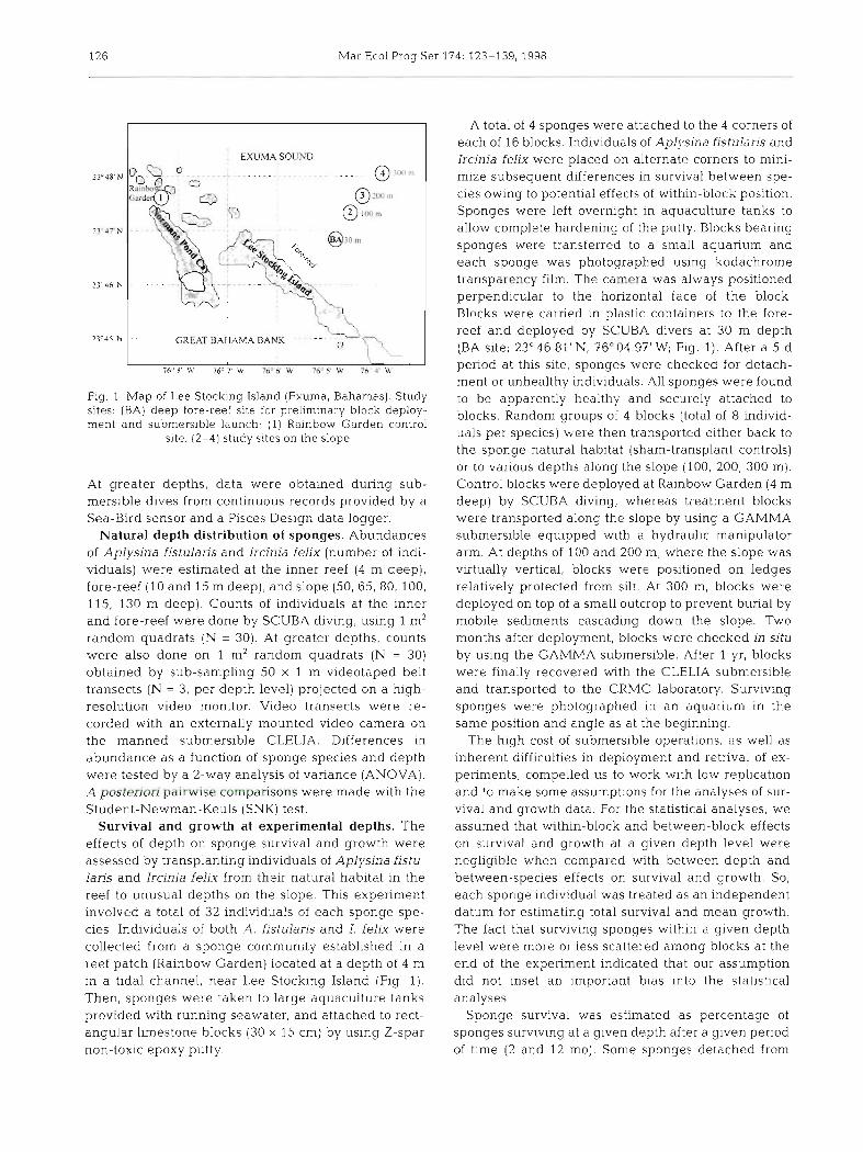
Physical setting and environmental data. Field experiments were conducted near the Caribbean Marine Research Center (CMRC) at Lee Stocking Is- land, in the southern Exuma Cays, Bahamas (Fig. 1). The Exumas are a group of subtropical islands and cays lining the eastern margin of the Great Bahama Bank. Shallow patch reefs are scattered on the lee sides of the islands and in some of the tidal channels connecting the Bank with Exuma Sound. On the Exuma Sound (windward) side, there is a well-devel- oped fore-reef. The fore-reef at Lee Stocking Island consists of a shallow terrace at 18 m and a deep terrace at 45 m, then cascades to a virtually vertical cliff that angles to a more gentle slope below 200 m.
Water temperature was recorded at shallow sites by using underwater thern~on~eters during SCUBA dives.
126 Mar Ecol Prog Ser 174: 123-139, 1998
EXUMA SOUND
GREAT BAHAMA BANK -
Fig 1 Map of Lee Stocking Island (Exuma, Bahamas) Study sites (BA) deep fore-reef site for preliminary block deploy- ment and submersible launch, (1) Rainbow Garden control
site, (2-4) study sites on the slope
At greater depths, data were obtained during sub- mersible dives from continuous records provided by a Sea-Bird sensor and a Pisces Design data logger
Natural depth distribution of sponges. Abundances of Aplysina fistulans and Ircinia felix (number of indi- viduals) were estimated at the inner reef (4 m deep) , fore-reef (10 and 15 m deep) , and slope (50 65, 80, 100, 115, 130 m deep) Counts of individuals at the inner and fore-reef were done by SCUBA diving, using 1 m2 random quadrats ( N = 30) At greater depths, counts were also done on 1 m2 random quadrats (N = 30) obtained by sub-sampling 50 x 1 m videotaped belt transects (N = 3, per depth level) projected on a high- resolution video monitor Video transects were re- corded with an externally mounted video camera on the manned submersible CLELJA Differences in abundance as a function of sponge species and depth were tested by a 2-way analysis of vanance (ANOVA) A p ^ k 3 ~ 1 ~ i i p ~ i ~ ~ i h v cbmpdrisons were made with the Student-Newman-Keuls (SNK) test
Survival and growth at experimental depths. The effects of depth on sponge survival and growth were assessed by transplanting individuals of Aplysina fistu- lans and Ircinia felix from their natural habitat in the reef to unusual depths on the slope This expenment involved a total of 32 individuals of each sponge spe- cies Individuals of both A fistulans and I felix were collected from a sponge community established in a reef patch (Rainbow Garden) located at a depth of 4 m in a tidal channel, near Lee Stocking Island (Fig 1) Then, sponges were taken to large aquaculture tanks provided with running seawater and attached to rect- angular limestone blocks (30 x 15 cm) by using Z-spar non-toxic epoxy putty
A total of 4 sponges were attached to the 4 corners of each of 16 blocks Individuals of Aph'sina fistulans and Ircma fehx were placed on alternate corners to mmi- mize subsequent differences in survival between spe- cies owing to potential effects of within-block position Sponges were left overnight in aquaculture tanks to allow complete hardening of the putty Blocks beanng sponges were transferred to a small aquanum and each sponge was photographed using kodachrome transpdrency film The camera was always positioned perpendicular to the horizontal face of the block Blocks were earned in plastic containers to the fore- reef and deployed by SCUBA divers at 30 m depth (BA site 23'46 81 'N , 76"04 97 W, Fig 1) After a 5 d period at this site, sponges were checked for detach- ment or unhealthy individuals All sponges were found to be apparently healthy and securely attached to blocks Random groups of 4 blocks (total of 8 individ- uals per species) were then transported either back to the sponge natural habitat (sham-transplant controls) or to various depths along the slope (100, 200, 300 m) Control blocks were deployed at Rainbow Garden (4 m deep) by SCUBA diving, whereas treatment blocks weie transported along the slope by using a GAMMA submersible equipped with a hydraulic manipulator arm At depths of 100 and 200 m, where the slope was virtually vertical, blocks were positioned on ledges relatively protected from silt At 300 m, blocks were deployed on top of a small outcrop to prevent burial by mobile sediments cascading down the slope Two months after deployment, blocks were checked in situ by using the GAMMA submersible After 1 yr, blocks were finally recovered with the CLELIA submersible and transported to the CRMC laboratory Surviving sponges were photographed in an aquarium in the same position and angle as at the beginning
The high cost of submersible operations as well as inherent difficulties in deployment and retrival of ex- penments compelled us to work with low replication and to make some assumptions for the analyses of sur- vival and growth data For the statistical analyses, we assumed that within-block and between-block effects on survival and growth at a given depth level were negligible when compared with between-depth and between-species effects on survival and growth So, each sponge individual was treated as an independent datum for estimating total survival and mean growth The fact that surviving sponges within a given depth level were more or less scattered among blocks at the end of the experiment indicated that our assumption did not inset an important bias into the statistical analyses
Sponge survival was estimated as percentage of sponges surviving at a given depth after a given period of time (2 and 12 mo) Some sponges detached from

Maldonado & Young; Bathyrnetnc distribution of keratose sponges 127
blocks while being transported by submersible, so that only 7 individuals of Aplysina fistularis were deployed at 200 m, 7 individuals of Ircinia felix were trans- planted to 100 and 200 m depths, and 5 I. felix were used as controls.
We constructed a 3-factor frequency table by record- ing the number of sponges that died and survived after 12 mo (survival factor: live vs dead) as a function of transplantation depth (depth factor: 4, 100, 200, 300 m) and sponge species (species factor: Aplysina fistularis vs Ircinia felix). Then, we tested the independence of survival (response variable or dependent variable), sponge species and depth factors using log-linear models (Bishop et al. 1975). We approached the final model step by step, adding and eliminating terms hier- archically (main effects and interactions) to examine the partial and marginal association of each term in the model. The partial association between, for example, survival and depth factors was computed by compar- ing the fit (i.e. evaluating the chi-square difference) of the model that includes all 2-way interactions with the model that excludes the interaction under considera- tion. Alternatively, the marginal association between, for example, survival and depth was computed by comparing the fit of the model that excludes all main effects (i.e. all effects of lower order than the one of interest) with the model including the interaction under consideration. A term was retained or excluded from the model on the basis of the significance of its partial and marginal association. The statistical signifi- cance of the goodness of fit of a particular model, that is, whether or not the expected cell frequencies under the respective model were significantly different from the observed cell frequencies, was evaluated by using the Pearson chi-squared (x2) statistic. When significant differences were found (p < 0.05), the model was rejected.
As the log-linear analysis revealed that sponge sur- vival was not dependent on the species factor; the 3- way table was collapsed across levels of the species factor and became a 4 x 2 table (depth factor: 4, 100, 200, 300 m; survival factor: live vs dead). Subse- quently, we tested the association between sponge survival (both species pooled) and particular pairs of depth levels by decomposing the table in a total of six 2 x 2 sub-tables. Each sub-table was evaluated using the Yates-corrected Pearson chi-squared statistic ( x ~ ~ ) , Analyses of frequency data were done using Statistica software (Statsoft Inc, OK, USA).
Sponge growth after 12 mo was assessed by analyz- ing photographs taken at the beginning and the end of the transplantation experiment. Growth was expressed as the positive or negative increment in the area as viewed from the side. We did not attempt to obtain a accurate quantitative value for sponge growth, but
rather assessed the relative magnitude of positive ver- sus negative growth in 2 dimensions. Others authors have used 1-dimensional measurements of linear hori- zontal or vertical growth to evaluate total growth in similar reef sponges (e.g. Hoppe 1988). Changes in body form during the 12 mo period at the experimental depths were assessed using the circularity index (C). Circularity was calculated by dividing the sponge area by the area of a circle with a perimeter equivalent to that of the sponge (Turon & Becerro 1992). All parame- ters needed to estimate sponge growth and re-shaping were measured on digitized pictures using Sigmascan software. Differences in sponge growth (projected area) and shape (circularity of projected area) in the species Aplysina fistularis as a function of depth were tested by the non-parametric Mann-Whitney rank sum (MWRS) test, as sponges survived at just 2 depths. In Ircinia felix, differences were tested by the Kruskal- Wallis non-parametric 1-way ANOVA, as there were survivors at 3 depth levels. Non-parametric tests were used, because data were either non-normally distrib- uted or heteroscedastic.
Recruitment at experimental depths. To assess the viability of asexual propagation at different experi- mental depths, we used the individuals of Aplysina fis- tularis transplanted to depths of 4, 100, 200 and 300 m, as described in the section above. These sponges bore numerous fistules, a type of bud for asexual propaga- tion. The presence of sponge recruits on blocks and rocks near the deployment sites (within an approxi- mately 5 m2 quadrat around the block), as well as the formation of new fistules by transplanted sponges, was checked after 2 and 12 mo.
To determine the success of larval recruitment at experimental depths, we conducted a second trans- plantation experiment using the method explained above. Nevertheless, we used only brooding indi- viduals of Ircinia felix this time. A total of 4 brooding sponges were attached to the 4 corners of each block, Random pairs of blocks were deployed at a depth of 15 m in the fore-reef (controls), and at 100 and 200 m on the slope (i.e. 8 sponges per depth level). After 3 mo, blocks deployed on the slope were retrieved and the surrounding bottom (an approximately 5 m2 quadrat around the block) was checked for young recruits by using the GAMMA submersible. Unfortu- nately, controls blocks were lost,
In both experiments, we reasoned that any recruit- ment by short-lived larvae or buds would support the hypothesis that limited dispersal hinders colonization rather than physiological tolerance of adults or juve- niles.
Sponge histology, symbionts and parasites. We investigated depth-related changes in the symbiotic populations of cyanobacteria and heterotrophic bac-
128 Mar Ecol Prog Ser 174: 123-139, 1998
teria, as well as in cytological aspects of sponge feeding and collagen-spongin secretion, by using transmission electron microscopy (TEM]. Material for TEM was prepared according to the methodology described by Rutzler (1990). Although Rut- zler recommends that tissue samples should not be held in fixative solution longer than 2 to 4 h, an emergency hurri- cane evacuation forced us to retain mater- ial in the fixative for 4 d , This extended fixation did not seem to compromise the cells or tissues in any way. Sections were viewed with a Philips EM-301 electron microscope. The proportion of cyanobacte- ria and heterotrophic bacteria (mean * SE %) with respect to the peripheral sponge
Salinity (ppt)
20 22 24 26 28 20 22 24 26 28 20 22 24 26 28
Temperature PC)
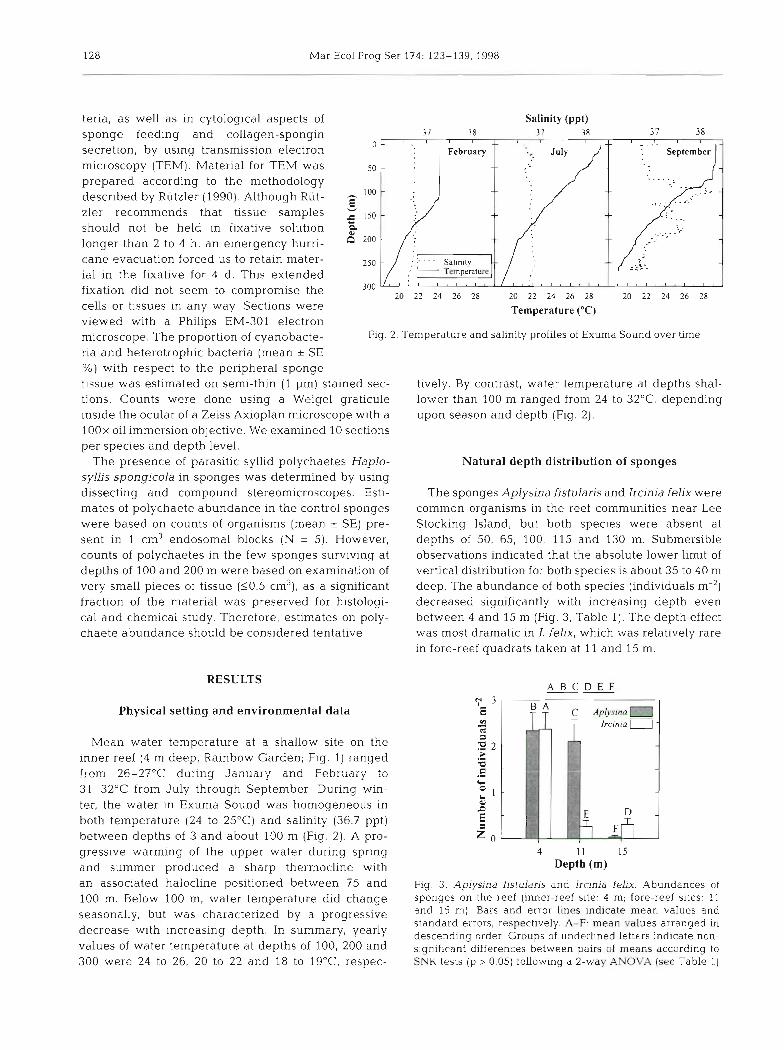
Fig. 2. Temperature and salinity profiles of Exuma Sound over time
tissue was estimated on semi-thin (1 pm) stained sec- tively. By contrast, water temperature at depths shal- tions. Counts were done using a Weigel graticule lower than 100 m ranged from 24 to 32OC, depending inside the ocular of a Zeiss Axioplan microscope with a upon season and depth (Fig. 2). 1OOx oil immersion objective. We examined 10 sections per species and depth level.
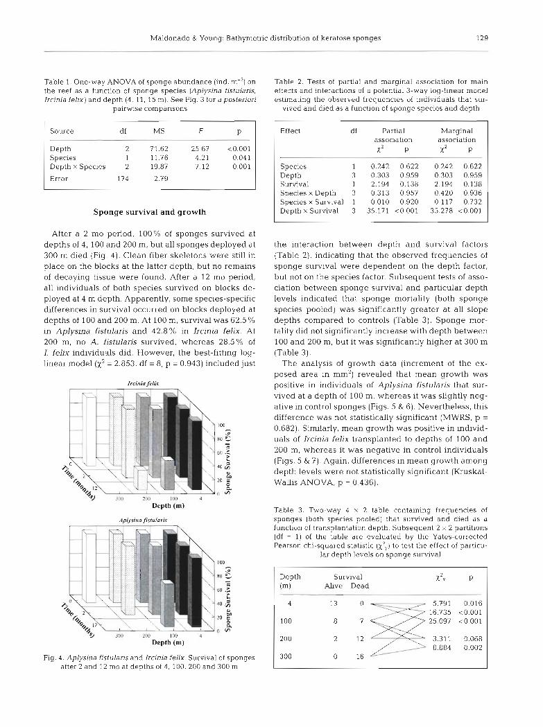
The presence of parasitic syllid polychaetes Haplo- Natural depth distribution of sponges syllis spongicola in sponges was determined by using dissecting and compound stereomicroscopes. Esti- The sponges Aplysina fistularis and Ircinia felix were mates of polychaete abundance in the control sponges common organisms in the reef communities near Lee were based on counts of organisms (mean SE) pre- Stocking Island, but both species were absent at sent in 1 cm3 endosomal blocks ( N = 5) . However, depths of 50, 65, 100, 115 and 130 m. Submersible counts of polychaetes in the few sponges surviving at observations indicated that the absolute lower limit of depths of 100 and 200 m were based on examination of vertical distribution for both species is about 35 to 40 m very small pieces of tissue (50.5 cm3), as a significant deep. The abundance of both species (individuals m-2) fraction of the material was preserved for histologi- decreased significantly with increasing depth even cal and chemical study. Therefore, estimates on poly- between 4 and 15 m (Fig. 3, Table 1). The depth effect chaete abundance should be considered tentative. was most dramatic in 1. felix, which was relatively rare
in fore-reef quadrats taken at 11 and 15 m.
RESULTS
Physical setting and environmental data
Mean water temperature at a shallow site on the inner reef (4 m deep, Rainbow Garden; Fig. 1) ranged from 26-27OC during January and February to 31-32OC from July through September During win- ter, the water in Exuma Sound was homogeneous in both temperature (24 to 25'C) and salinity (36.7 ppt) between depths of 3 and about 100 m (Fig. 2). A pro- gressive warming of the upper water during spring and summer produced a sharp thermocline with an associated halocline positioned between 7.5 and 100 m. Below I00 m, water temperature did change seasonally, but was characterized by a progressive decrease with increasing depth. In summary, yearly values of water temperature at depths of 100, 200 and 300 were 24 to 26, 20 to 22 and 18 to lg°C respec-
A B C D E F --
4 11 I s Depth (m)
Fig. 3. Aplysina fistulans and Ircinja felix. Abundances of sponges on the reef (inner-reef site: 4 m: fore-reef sites: 11 and 15 m) Bars and error 11nes indicate mean values and standard errors, respectively. A-F: mean values arranged in descending order. Groups of underlined letters indicate non- significant differences between pairs of means according to SNK tests [p > 0.05) following a 2-way ANOVA (see Table 1)
Maldonado & Young: Bathymetric distribution of keratose sponges 129
Table 1. One-way ANOVA of sponge abundance (ind. m-2] on the reef as a function of sponge species (Aplysina fjstularjs, Ircinia felm) and depth (4, 11, 15 m). See Fig. 3 for a posteriori
pairwise comparisons
Source df MS F P
Depth 2 71.62 Species 1 11.76 Depth x Species 2 19,87
Error 174 2.79 -
Sponge survival and growth
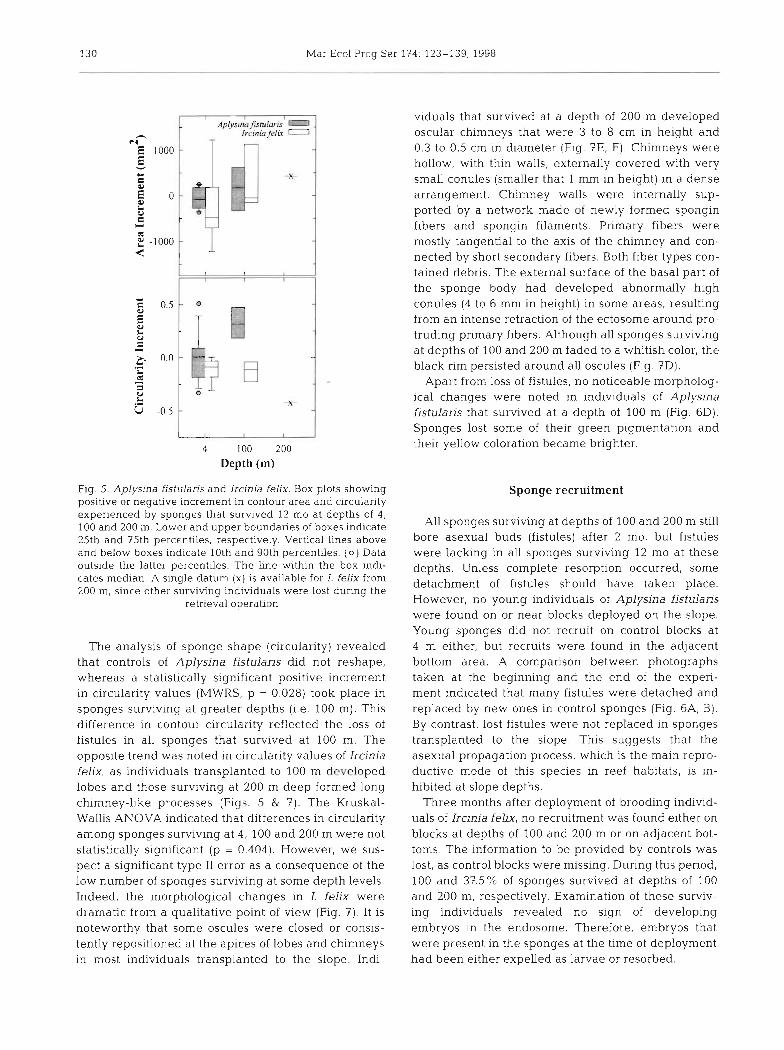
After a 2 mo period, 100% of sponges survived at depths of 4,100 and 200 m, but all sponges deployed at 300 m died (Fig. 4). Clean fiber skeletons were still in place on the blocks at the latter depth, but no remains of decaying tissue were found. After a 12 mo period, all individuals of both species survived on blocks de- ployed at 4 m depth. Apparently, some species-specific differences in survival occurred on blocks deployed at depths of 100 and 200 m. At 100 m, survival was 62.5 % in Aplysina fistulaxis and 42.8% in Ircinia felix. At 200 m, no A. fistularis survived, whereas 28.5% of I. feljx individuals did. However, the best-fitting log- linear model (x2 = 2.853, df = 8, p = 0.943) included just
Depth (m)
4 Depth (m)
Fig. 4. Aplysina fistularis and Ircinia felix. Survival of sponges after 2 and 12 mo at depths of 4 , 100, 200 and 300 m
Table 2. Tests of partial and marginal association for main effects and interactions of a potential 3-way log-linear model estmatinq the observed frequencies of individuals that sur-
vived a i d died as a functioi of sponge species and depth
Effect
Species Depth Survival Species x Depth Species x Survival Depth x Survival
df Partial association x 2 P
1 0.242 0.622 3 0.303 0.959 1 2.194 0.138 3 0.313 0.957 1 0.010 0.920 3 35.171 <O.OOl
Marginal association x2 P
0.242 0.622 0.303 0.959 2.194 0.138 0.420 0.936 0.117 0.732
35.278 <O.OOl
the interaction between depth and survival factors (Table 21, indicating that the observed frequencies of sponge survival were dependent on the depth factor, but not on the species factor, Subsequent tests of asso- ciation between sponge survival and particular depth levels indicated that sponge mortality (both sponge species pooled) was significantly greater at all slope depths compared to controls (Table 3). Sponge mor- tality did not significantly increase with depth between 100 and 200 m, but it was significantly higher at 300 m (Table 3).
The analysis of growth data (incren~ent of the ex- posed area in mm2) revealed that mean growth was positive in individuals of Aplysina fistularis that sur- vived at a depth of 100 m, whereas it was slightly neg- ative in control sponges (Figs. 5 & 6). Nevertheless, this difference was not statistically significant (MWRS, p =
0.682). SinGlarly, mean growth was positive in individ- uals of Ircinia felix transplanted to depths of 100 and 200 m, whereas it was negative in control individuals (Figs. 5 & 7). Again, differences in mean growth among depth levels were not statistically significant (Kruskal- Wallis ANOVA, p = 0.436).
Table 3. Two-way 4 x 2 table containing frequencies of sponges (both species pooled) that survived and died as a function of transplantation depth. Subsequent 2 x 2 partitions (df = 1) of the table are evaluated by the Yates-corrected Pearson chi-squared statistic (x2,,) to test the effect of particu-
lar depth levels on sponge survival
Depth Survival x" Y
(m) Ahve Dead
130 Mar Ecol Prog Ser 174 123-139, 1998
4 100 200 Depth (rn)
Fig 5. Aplysina fist~~laris and Ircinia felix. Box plots showing positlve or negative increment in contour area and circularity expenenced by sponges that survived 1.2 mo at depths of 4 , 100 and 200 m. Lower and upper boundaries of boxes indicate 25th and 75th percentiles, respectively. Vertical lines above and below boxes indicate 10th and 90th percentiles. (0) Data outside the latter percentiles. The line within the box indi- cates median. A single datum (x) is available for I. felix from 200 m, since other surviving individuals were lost during the
retrieval operation
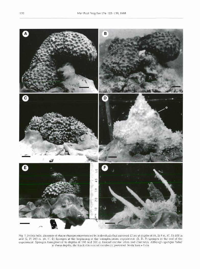
The analysis of sponge shape (circulanty) revealed that controls of Aplysina fistularis did not reshape, whereas a statistically significant positive increment in circularity values (MWRS, p = 0.028) took place in sponges surviving at greater depths (1.e. 100 m). This difference in contour circulanty reflected the loss of fistules in all sponges that survived at 100 m. The opposite trend was noted in circulanty values of Ircinia felix, as individuals transplanted to 100 m developed lobes and those surviving at 200 m deep formed long chlmney-like processes (Figs. 5 & 7). The Kruskal- Wallis ANOVA indicated that differences in circulanty among sponges surviving at 4, 100 and 200 m were not statistically significant (p = 0.404). However, we sus- pect a significant type I1 error as a consequence of the low number of sponges surviving at some depth levels. Indeed, the morphological changes in I. felix were dramatic from a qualitative point of view (Fig. ?) , It is noteworthy that some oscules were closed or consis- tently repositioned at the apices of lobes and chimneys in most individuals transplanted to the slope. Indi-
viduals that survived at a depth of 200 m developed oscular chimneys that were 3 to 8 cm in height and 0.3 to 0.5 cm in diameter (Fig. ?E, F). Chimneys were hollow, with thin walls, externally covered with very small conules (smaller that 1 mm in height) in a dense arrangement. Chimney walls were internally sup- ported by a network made of newly formed spongin fibers and spongin filaments. Primary fibers were mostly tangential to the axis of the chimney and con- nected by short secondary fibers. Both fiber types con- tained debris. The external surface of the basal part of the sponge body had developed abnormally high conules (4 to 6 mm in height) in some areas, resulting from an intense retraction of the ectosome around pro- truding primary fibers. Although all sponges surviving at depths of 100 and 200 m faded to a whitish color, the black rim persisted around all oscules (Fig, ?D).
Apart from loss of fistules, no noticeable morpholog- ical changes were noted in individuals of Aplysina fjstulans that survived at a depth of 100 m (Fig, 6D), Sponges lost some of their green pigmentation and their yellow coloration became brighter.
Sponge recruitment
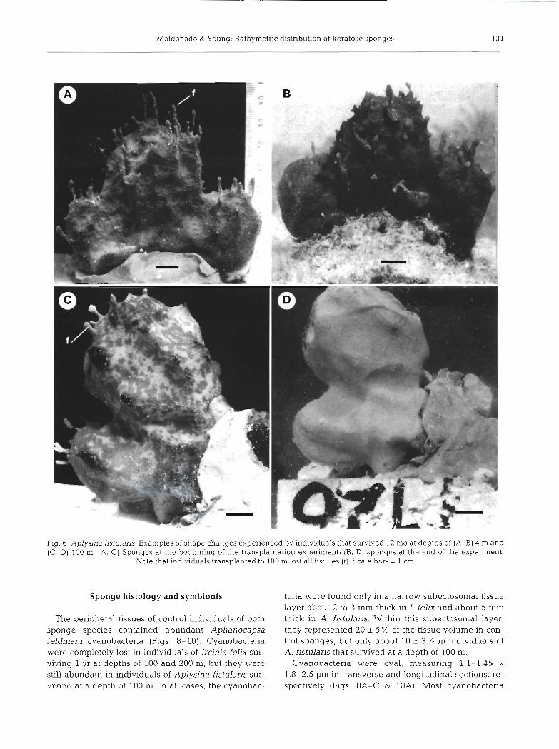
All sponges surviving at depths of 100 and 200 m still bore asexual buds (fistules) after 2 mo, but fistules were lacking in all sponges surviving 12 mo at these depths. Unless complete resorption occurred, some detachment of fistules should have taken place. However, no young individuals of Aplysina fistulans were found on or near blocks deployed on the slope. Young sponges did not recruit on control blocks at 4 m either, but recruits were found in the adjacent bottom area. A comparison between photographs taken at the beginning and the end of the expen- ment indicated that many fistules were detached and replaced by new ones in control sponges (Fig. 6A, B). By contrast, lost fistules were not replaced in sponges transplanted to the slope. This suggests that the asexual propagation process, which is the main repro- ductive mode of this species in reef habitats, is in- hibited at slope depths.
Three months after deployment of brooding individ- uals of Ircinia felix, no recruitment was found either on blocks at depths of 100 and 200 m or on adjacent bot- toms. The information to be provided by controls was lost, as control blocks were missing. During this period, 100 and 37.5 % of sponges survived at depths of 100 and 200 m, respectively. Examination of these surviv- ing individuals revealed no sign of developing embryos in the endosome. Therefore, embryos that were present in the sponges at the time of deployment had been either expelled as lawae or resorbed.
Maldonado & Young: Bathymetric &sknbution of keratme sponges
Fig. 6. Aplysjna fistularis. Examples of shape changes experienced by individuals that survived 12 mo at depths of (A, B) 4 m and (C, D) 100 m. (A, C) Sponges at the beginning of the transplantation experiment; (B, D) sponges at the end of the experiment.
Note that individuals transplanted to 100 m lost all fistules ( f ) . Scale bars = 1 cm
Sponge histology and symbionts
The peripheral tissues of control individuals of both sponge species contained abundant Aphanocapsa feldrnani cyanobacteria (Figs. 8-10). Cyanobactena were completely lost in individuals of Ircjnia felix sur- viving 1 yr at depths of 100 and 200 m, but they were still abundant in individuals of Aplysina flstularis sur- viving at a depth of 100 m. In all cases, the cyanobac-
tena were found only in a narrow subectosomal tissue layer about 2 to 3 mm thick in I. felix and about 5 mm thick in A. fistularis. Within this subectoso~mal layer, they represented 20 & 5 Yo of the tissue volume in con- trol sponges, but only about 10 k 3 Yo in individuals of A. f~stularjs that survived at a depth of 100 m.
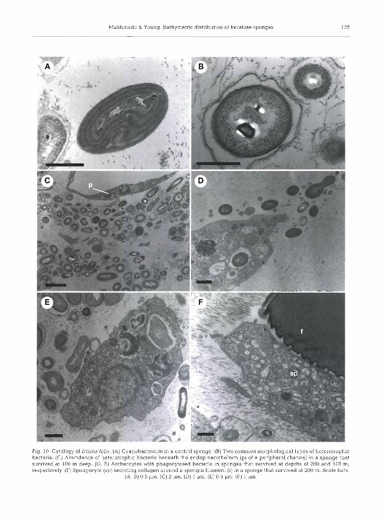
Cyanobactena were oval, measuring 1.1-1,45 x 1,8-2.5 pm in transverse and longitudinal sections, re- spectively (Figs. 8A-C & 10A). Most cyanobactena
Mar Ecol Prog Ser 174: 123-139, 1998
Fig. 7. Ircinia fehx. Example of shape changes experienced by indiv~duals that survived 12 mo at depths of (A, B) 4 rn, (C, D) 100 m and (E, F) 200 m. [A, C, E) Sponges at the beginning of the transplantation expenment; (B, D, F) sponges at the end of the expenment. Sponges transplanted to depths of 100 and 200 m formed oscular lobes and chimneys. Although sponges faded
at these depths, the black rim around oscules (0) persisted. Scale bars = 1 cm

Maldonado & Young: Bathymetric distribution of keratose sponges
Fig. 8. Cytology of Aplysina fistularis. (A) Abundance of cyanobacteria (cy) and heterotrophic bacttIiu ,", in the peiip..eral tissue of a control sponge. (B) Cyanobacteria in a control sponge. (C) Cyanobacteria in a sponge that survived at 100 m. (D) Intracellular cyanobacterium parasitized by a Bdellovlbrio-like bacterium (db) in a sponge that survived at 100 m (E) Cyanobacterium being phagocytosed by a sponge cell in a sponge that survived at 100 m. (F) Archeocyte of control sponge phagocytosing a bacterium
and containing cyanobacteria at different states of digestion. Scale bars: (A) 1 pm, (B, C) 0.5 pm, (D) 2 pm, (E, F) 1 pm
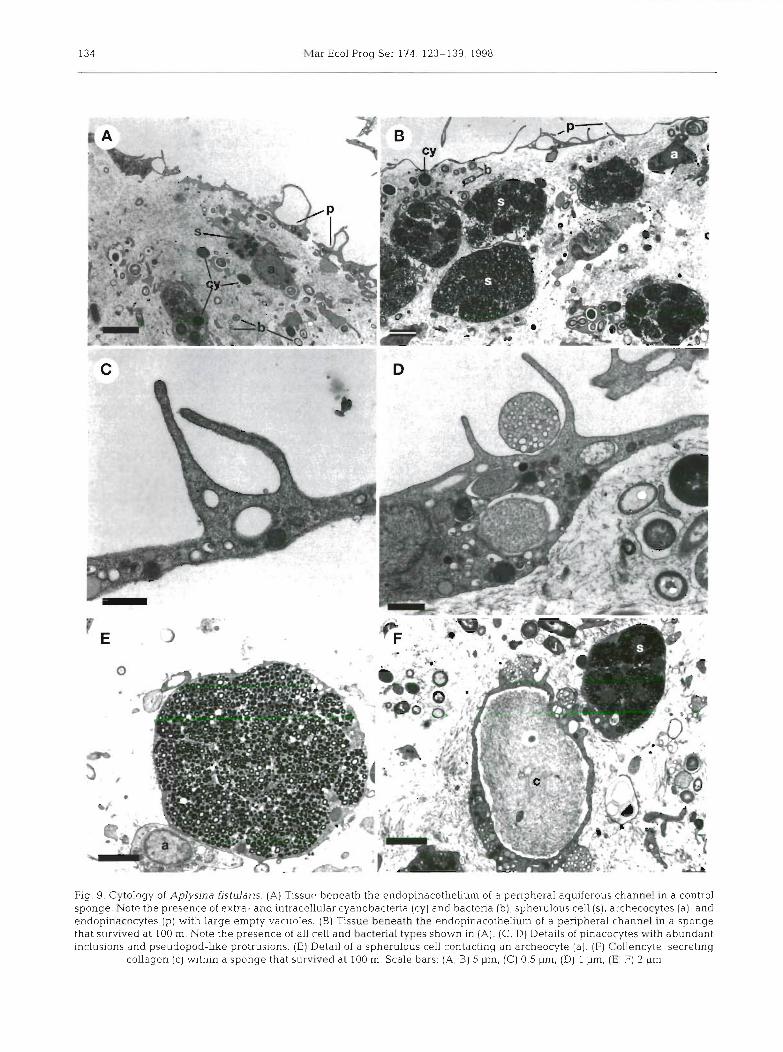
were extracellular in both sponge species, though they were also found in intracellular vacuoles. Intracellular cyanobacteria were found in an apparent living state and also at different states of digestion (Figs. 8D-F & 9A, B). In Aplysina fistularis, cyanobacteria were occa- sionally observed dividing, both in control sponges and in individuals that survived at a depth of 100 m. The thylakoid of most cyanobacteria showed between 4 and 6 spirals. The number of spirals was not found to be cor- related with either depth in the sponge tissue or depth at which sponges lived. Indeed, individuals of A. fistu-
laris surviving at a depth of 100 m showed some cyanobacteria with thylakoid somewhat less developed than those of cyanobacteria in control sponges (Fig. 8B, C). The only noticeable difference between cyano- bacteria populations living in control and 100 m deep A. fistularis sponges was the occurrence of a low per- centage of cyanobacteria parasitized by a Bdellovibrio- like bacterium at the greater depth (Fig. 8D).
Heterotrophic bacteria were well represented in both sponge species. At a depth of 5 mm in the sponge tissue, bacteria accounted for about 20 Â 6 % and 30 :
Mar Ecol Prog Ser 174: 123-139, 1998
Fig. 9. Cytology of Aplysina fistularis. (A) Tissue beneath the endopinacothelium of a peripheral aquiferous channel in a control sponge. Note the presence of extra- and intracellular cyanobacteria (cy) and bacteria (b), spherulous cell (s), archecocytes (a), and endopinacocytes (p) with large empty vacuoles. (B) Tissue beneath the endopinacothelium of a peripheral channel in a sponge that survived at 100 m. Note the presence of all cell and bacterial types shown in (A). (C, D ) Details of pinacocytes with abundant inclusions and pseudopod-like protrusions. (E) Detail of a spherulous cell contacting an archeocyte (a) . (F] Collencyte secreting
collagen (c) within a sponge that survived at 100 m. Scale bars: (A, B) 5 urn, (C) 0,5 urn, (Dl 1 urn, (E; F) 2 urn
Maldonado & Young Bathyn~etnc distribution of keratose sponges
Fig 10 Cytology of Ircinia fe11x (A) Cyanobactenum in a control sponge (B) Two common morphological types of heterotrophic bactena (C) Abundance of heterotrophic bactena beneath the endopinacotheliurn (p ) of a peripheral channel in a sponge that survived at 100 m deep (D, E) Archeocytes with phagocytosed bactena in sponges that survived at depths of 200 and 100 m, ~espectively (F] Spongocyte (sp] secreting collagen around a spongin filament ( f ) in a sponge that survived at 200 m Scale bars
(A, B) 0 5 urn, (C) 2 urn, (D) 1 urn, (El 0 5 urn, (F) 1 urn
136 Mar Ecol Prog Ser 174: 123-139, 1998
8 % of the sponge tissue volume in control individuals of Aplysina fistularis and Ircinia felix, respectively. Similar proportions were found in sponges surviving at 100 m, whereas the proportion was lower (20 Â 8 %) in individuals of I. felix surviving at 200 m. The bacterial population of both sponge species was dominated by 3 major morphological bacterial types (Figs. 8A, C &
10B, C) that were comparable to the B, C and D types described by Vacelet (1975) from 2 Mediterranean species of Aplysina.
A TEM examination of the mesohyl of surviving sponges did not reveal significant differences in sponge cytology as a function of transplantation depth. At depths of both 4 and 100 m, individuals of Aplysina fistularis showed archeocytes engaged in phagocytosis and digestion of cyanobacteria and heterotrophic bac- teria (Figs. 8D-F & 9A, B). Similarly, endopinacocytes showed pseudopod-like protrusions, suggesting active capture of particulate material. These cells were also characterized by the presence of large, empty vacuoles (Fig. 9A-D). The significance of these structures remains unclear. Immediately below the endopina- cothelium, spherulous cells were abundant and a few, scattered chromocyte-like cells were also found (Fig. 9A, B, E, F). Interestingly, we observed collen- cytes apparently secreting collagenous fibrils in both controls and individuals transplanted to 100 m (Fig. 9F). Similarly, we found apparently active spon- gocytes in close contact with spongin filaments in Ircinia felix individuals that survived at depths of 100 and 200 m (Fig. 10F). These observations indicate that spongin synthesis was not inhibited at these depths. Active capture and digestion of bacteria by archeo- cytes was also observed in individuals of I. felix surviv- ing at control, 100 and 200 m depth levels (Fig. 10D, E).
Parasitic polychaetes, Haplosyllis spongicola, were present in the endosomal tissues of all surviving indi- viduals of Ircinia felix, irrespective of sponge trans- plantation depth. Polychaetes measured 1000-4000 x 300-350 pm and appeared at mean (Â SE) densities as high as 35 Â 15 syllids c m 3 Casual inspection sug- gested that neither polychaete size nor abundance was significantly different among populations inhabiting control sponges and sponges that survived at depths of 100 and 200 m.
DISCUSSION
The keratose sponges Aplysina fistularis and Ircinia felix were common in shallow habitats on the reef, but their abundance decreased significantly with increas- ing depth along the outer reef, and both were com- pletely absent from the upper slope. This vertical dis- tribution agrees with data from the abundant literature
available for these 2 species (e.g. Wiedenmayer 1977, Zea 1987 and literature therein). Nevertheless, 3 deep- water specimens collected from Puerto Rico (Pisca- dera, 500 m deep), Barbados (100 m) and Venezuela (72 m) were tentatively assigned to the species I. felix by Van Soest (1978), although the author did not exclude the possibility that they belong to a separate deep-water taxon. Our numerous submersible obser- vations at Lee Stocking Island and throughout other parts of the Bahamas (e.g. Maldonado & Young 1996b) are consistent with the large body of the literature indicating that these species do not occur naturally at depths much greater than 35 to 40 m. However, both species survived at greater depths when artificially transplanted, and some of the transplanted individuals even experienced a positive mean growth after 1 yr. Therefore, these results suggest that limited larval dis- persal or unsuccessful settlement at deep habitats may be responsible for the confinement of this species to shallow water. We found no sign of recruitment from either sexual or asexual origin at experimental depths, but cannot discard the possibility that we failed to ob- served recruitment because detached buds or expelled larvae dispersed and recruited out of the area we sur- veyed. Nevertheless, our field experience with other littoral sponges indicates that at least a few recruits should be found on blocks or the nearby bottom if buds or brooded larvae were actually viable at these depths e . g . Uriz et al. 1998).
It is not surprising that sponges transplanted to bathyal depths showed a tendency to grow better than at control depths. At bathyal depths, some reproduc- tive processes (i.e. formation of asexual buds) were inhibited, and many embryos probably either were resorbed or did not develop at all. Therefore, the energy usually allocated for reproduction may have been reallocated to somatic growth. Wilkinson & Vacelet (1979) found a similar effect on the growth of other sponges. In their study, individuals of the sponges Aplysina cavernicola and Chondrilla nucula transplanted to different light conditions often grew better than control individuals that remained in their natural habitat.
Symbiosis with cyanobacteria does not appear to play an important role in determining the lower limit of depth distribution for either sponge species. Aplysina fistularis does not occur deeper than 40 m, but trans- planted sponges survived to at least a depth of 100 m. At this depth, symbiotic cyanobacteria were still abun- dant in the peripheral sponge tissue. Therefore, the absence of this species in deep fore-reef and upper- slope habitats is not directly caused by loss of cyano- bacteria. hcinja Eelix survived even deeper (200 m), despite the fact that they lost cyanobacteria at a depth even shallower than 100 m . The fact that symbiotic
Maldonado & Young: Bathymetric distribution of keratose sponges 137
cyanobacteria were present in A. fistularis but lost in I. felix under virtually identical environmental con- ditions (same block) suggests that the relationship between Aphanocapsa feldmani and its host varies with sponge species, especially in the case of mixotrophic sponges. On the other hand, the presence of symbiotic cyanobacteria in sponges living as deep as 100 m is not probably an exceptional condition. This observation is consistent with studies reporting that zooxanthellate scleractinians occur to depths of about 100 m in the Caribbean area (Lang et al. 1988).
Sponge survival results do not support the hypothe- sis that parasitism by Haplosyllis spongicola may hinder the colonization of deeper habitats by these species. The log-linear analyses indicated that there was no significant difference in survival between both sponge species with depth, despite the fact that only Ircinia felix was heavily parasitized at all depths. Fur- thermore, some individuals of this latter species sur- vived at greater depths than non-parasitized individ- uals of Aplysina fistularis, Further studies are needed to understand this interesting polychaete-sponge asso- ciation. The large number of polychaetes still present in sponges that survived at 200 m, where oligotrophy should also be a problem for the sponge, suggests that the sponge-polychaete relationship is mutualistic or commensalistic rather than parasitic. Otherwise, one cannot easily understand how sponges under nutri- tional stress in deep-sea conditions can counterbal- ance the effects of grazing by such a high concentra- tion of parasites.
Cytological observations indicate that both feeding and collagen-spongin secretion proceed normally in sponges surviving at depths greater that the lower natural limit of these species. The positive growth ex- perienced by some sponges at the experimental depths was the strongest evidence for this (Fig, 10D-F). The abundant population of symbiotic heterotrophic bac- teria still present in the endosome of Aplysina fistularis and especially Ircinia felix at these depths may con- tribute to palliate the negative effects derived from the loss of cyanobacteria and the impoverishment of avail- able particulate food in deep waters. The reshaping process that took place in individuals of I. felix with increasing depth was probably triggered by the need to enhance filter feeding and compensate for lower food levels. Vogel (1978) showed how sponges may exploit ambient currents by inducing passive flow with chimney-like extensions of the oscules. Indeed, the reshaping of I. felix that survived at depths of 100 and 200 m may be one of the most clear empirical confir- mations of Vogel's theories about the significance of body form in sponges. Oscular chimneys similar to those formed by I. felix were described in specimens of the Mediterranean species Ircinia pipetta (Schmidt)
collected from an undetermined depth between 70 and 120 m (Uriz & Maldonado 1993). The species A. fistu- laris did not reshape, possibly because its body form was already appropriate for exploiting ambient cur- rents (Vogel 1978).
Although lower food with increasing depth ap- peared to have an important impact on the shape of transplanted sponges, food does not emerge as the key factor hindering the colonization of the slope by these sponges. The fact that individuals of both species died relatively quickly (within 2 mo) when transplanted to a depth of 300 m suggests that sponge physiology was dramatically affected at that depth. A process of slow shrinking and delayed mortality should be expected in the case of death by starvation or parasitism. Predation is unlikely to play a significant role in determining the lower limit of depth distribution of these sponges. The fiber skeleton of individuals of both sponge species remained intact even after death in both species trans- planted to bathyal depths. There was no sign of preda- tion in any individual. In our opinion, water tempera- ture was most likely the reason for sudden mortality after transplantation, since it is the environmental factor, apart from light, that changes most dramatically with depth. Temperature at a depth of 300 m (18OC) was about 10° lower, on average, than on the reef (27 to 32OC). Our observations agree with Vacelet's (1988) suggestion that a temperature below 18 to 20° has a deleterious effect on keratose sponges. Never- theless, the negative effects of low temperature may not affect adult sponges as severely as larvae, asexual buds or early recruits. Some information in the sponge literature suggests that adult individuals of some ker- atose species are little affected by temperature. For example, Ilan et al. (1994) have recently described 2 keratose sponges living at a depth of 830 m in the Red Sea, where water temperature is around 1O0C. These individuals were tentatively regarded as conspecifics of 2 sublittoral sponge species, Ircinia cf. retiderma and Sarcotragus cf. muscarurn. Both sponges served as host for an interesting invertebrate infauna that included sublittoral polychaetes. Although the authors did not offer any explanation for these unexpected deep-water records of shallow sublittoral sponges and polychaetes, one cannot exclude the possibility that sponges have been transported from the shelf to the slope by fishing trawling-nets. This fishing practice has been shown to affect significantly both the biogeographical and bathymetric distribution of some bathyal sponge spe- cies (Uriz 1990). This mechanism would also explain some unexpected deep records of typical shallow- water keratose sponges in Mediterranean areas that are subject to intense exploitation by trawling-fishing nets (Pansini & Musso 1991). This hypothesis is also consistent with the results of our transplantation exper-
138 Mar Ecol Prog Ser 174: 123-139, 1998
iments. Thus, if adult keratose sponges artificially transported to great depths can survive in these new environmental deep-sea conditions, the hypothesis that a inhibition of larval dispersal or settlement suc- cess (probably by a decreasing temperature with increasing depth) emerges as the most plausible expla- nation for the shallow-water confinement of most ker- atose sponge.
It is widely thought that the keratose orders are the most modern demosponges (e.g. Levi 1973). Our experimental results appear to be consistent with the ideas that they evolved from ancestors inhabiting shal- low, warm waters, and that they would have to modify some of their physiological processes (e.g. reproduc- tion) in order to colonize successfully slope habitats.
Acknowledgements. Special thanks are due to the staff of The Caribbean Marine Research Center at Lee Stocking Island for their help and encouragement in field tasks, pilots of the GAMMA and CLELIA submersibles for their kind and valu- able help during the cruises, and the staff of the Servicio de Microscopia de la Universidad de Barcelona for assistance with TLM. We also thank Prof. Maria J. Uriz for comments on TEM micrographs. This research was funded by a NOAA grant (CMRC-95-3047) and has also benefited from funds of a Postdoctord1 Fulbright fellowship to M.M. dnd a DCCYT grant (PB-94-0015-C02-01) This is Harbor Branch contri'bu- tion number 1236.
LITERATURE CITED
Angel IMV (1968) The thermocline as an ecological boundary. Sarsia 34:299-312
Bergquist PR (19611 The Keratosa (Porifera) collected by the Chatham Islands 1954 Expedition. Bull NZ Dep Sci 1,n.d Res 139:207-219
Bergquist PR (1978) Sponges. University of California Press, Berkeley
Bergquist PR (1980) A revision of the supraspecific classifica- tion of the orders Dictyoceratida, Dendroceratida and Verongida (class Demospongiae). NZ J Zoo1 7:443-503
Bishop YMM, Fienberg SE, Holland PW (1975) Discrete multivariate analysis. MIT Press, Cambridge, MA
Burton M (1928) A comparative study of the characteristics of shallow-water and deep-sea sponges, with notes on their external form and reproduction. J Queket Micro Club 16(95):48-70
Cheshire AC, Wilkinson CR (1991) Modeling the photosyn- thetic production by sponges on Davies Reef, Great Barner Reef. Mar Biol 109:13-18
Dayton PK, Robilliard GA, Paine RT, Dayton LB (1974) Bio- logical accommodation in the benthic community at the McMurdo Sound, Antarctica. Ecol Monogr 44:105-128
Duchassainq P, Michelotti G (1864) Spongiaires de la mer Carai'br Natk Verh HoU Maatsch Wetensch Haarlem 21. 1-124
Hentschol E (1923) Ponfera. In: Kukenthal W, Krumbach T (eds) Hcindbuch der Zoologic. Walter de Gruyter und Co, Berlin
Hoppe WF (1988) Growth, regeneration and predation in three species of large coral reef sponges. Mar Ecol Prog Ser 50-1 17-125
Ilan M, Ben-Eliahu MM, Galil BS (1994) Three deep water sponges from the eastern Mediterranean and their associ- ated fauna. Ophelia 39:45-54
Kinne 0 (1971) Marine ecology. A comprehensive, integrated treatise on life in oceans and coastal waters. I. Lnviron- mental factors, Part 2. Wiley-Interscience, London
Lang JC, Wicklund RI , Dill RF (1988) Depth- and habitat- related bleaching of zooxanthellate reef organisms ncdr Lee Stocking Island, Exuma Cays, Bahamas Proc 6th Int Symp Coral Reef 3:269-274
Levi C (1973) Systcmatique de la classe des Demospongiaria (Demosponqes). In: Grasse PP (ed) Spongiaires. Anatomie, physiologie, systcmatique, ecologie. Masson et Cie, Paris
Levi C, Levi P (1983) Demosponges bathyales recoltees par Ie N / 0 'Vauban' au sud de la Nouvelle Caledonie. Bull Mus Natl Hist Nat 4eme Serie 5:931-997
Mackie GO, Singla CL (1983) Studies on hexactinellid sponges. I. Histology of Rhabdocalyptus dawsoni (Lambe, 1873). Phil Trans R Soc Lond 301:365-400
Maldonado M, Young CM (1996a) Effects of physical factors on larval behavior, settlement and recruitment of four tropical demosponges. Mar Ecol Prog Ser 138:169-180
Mdldonado M, Young CM (1996b) Bathymetric patterns of sponge distribution on the Bahamian slope Deep-sea Res 143:897-915
Maldonado M, Young CM (1998) Reevaluation of stalked aplysinid sponges, with description of a new species from the upper Bahamian slope. Bull Mar Sci (in press)
Metaxas A, Young CM (1998) Behaviour of echinoid larvae around sharp haloclines: effects of the salinity gradient and dietary conditioning. Mar Biol 131-443-459
Neigel JF, Schmahl GP (1984) Phenotypic variation within histocompatibility-defined clones of marine sponges. Sci- ence 224:413-415
Pallas PS (1766) Elenchus zoophytorum sistens generum adumbrationes generaliores et specierum cognitarum succinctas descriptiones cum selectis auctorum synonyirus. P van Cleef, The Hague
Pansini M, Musso B (1991) Sponges from trawl-exploitable bottoms of Ligunan and Thyrrhenian seas: distribution and ecology. PSZN 1: Mar Ecol 1.2:317-329
Pawlik JR (1983) A sponge-eating worm from Bermuda: Bran- chiosyllis oculata (Polychaeta. Syllidae). PSZN I: Mar Ecol 4:65-79
Reid REH (1968) Bathymetric distributions of Calcarea and Hexactinellida m the present and the past. Geol Mag 105: 546-559
Reiswig HM (1981) Partial carbon and energy budgets of the bacteriosponge Verongia fistuians (Porifera: Demospon- giae) in Barbados. PSZN I: Mar Ecol2:273-293
Rutzler K (1990) Associations between Caribbean sponges and photosynthetic organisms. In: Rutzler K (ed) New per- spectives in sponge biology. Smithsonian Institution Press, Washington, DC
Sara M (1971) Ultrastr~~ctural aspects of the symbiosis be- tween two species of the genus Aphanocapsa (Cyano- phyceae) and Ircima variabilis (Demospongiae]. Mar Biol 11:214-221
Sara M, Vacelet J (1973) Ecologie des Demosponges. In: Grasse PP (ed) Spongiaires. Anatomie, physiologie, syste- matique, ecologie. Masson et Cie, Paris
Tabachnick KR (1994) Distribution of recent Hexactinellida. In: Van Soest RMW, Van Kempen TMG, Braekman JC (eds) Sponges in time and space. AA Balkema, Rotterdam
Tsurumi M, Reiswig HM (1997) Sexual versus asexual repro- duction in a oviparous rope-form sponge, Aplysina c a d -
Maldonado & Young: Bathymetric distribution of keratose sponges
Turon X, Becerro MA (1992) Growth and survival of several ascidian species from the northwestern Mediterranean. Mar Ecol Prog Ser 82:235-247
Uriz MJ (1990) Possible influence of trawl fishery on recent expansion in the range of Suberites tylobtusa in the southeast Atlantic. In: Riitzler K (ed) New perspectives in sponge biology. Smithsonian Institution Press, Washing- ton, DC
Unz MJ, Maldonado M (1993) Redescription of some rare sponge species in the western Mediterranean. In: Uriz MJ, Rutzler K (eds) Recent advances in ecology and system- atics of sponges. Scientia Marina, Barcelona
Uriz MJ, Maldonado M, Turon X, Marti R (1998) How do reproductive output, larval behaviour, and recruitment contribute to adult spatial patterns in Mediterranean encrusting sponges? Mar Ecol Prog Ser 167:137-148
Vacelet J (1971) Etude en microscopie electronique de l'association entre une cyanophycee chroococcale et une eponge du genre Verongia. J Microsc (Oxf) 12:363-380
Vacelet J (1975) Etude en microscopie electronique de l'asso- ciation entre bacteries et spongiaires du genre Verongia (Dictyoceratida). J Microsc Biol Cell 23:271-288
Vacelet J (1979) Le place des spongiaires dans les systemes trophiques marins. In: Lev! C, Boury-Esnault N (eds) Biologie des spongiaires. CNRS, Paris
Vacelet J (1988) Indications de profondeur donnkes par les Spongiaires dans les milieux benthiques actuels. Geol Mediterr 1513-26
Editorial responsibility: Otto Kinne (Editor), OldendorfILuhe, Germany
Vacelet J, Boury-Esnault N, Zibrowius H (1989) Unexpected deep-water records of calcareous sponges (Calcarea). Deep-sea News1 1524-25
Van Soest RWM (1978) Marine sponges from Curacao and other Caribbean localities, Part I. Keratosa. Stud Fauna Curacao 179:l-94
Vazquez E, Young CM (1996) Responses of compound ascid- ian larvae to haloclines. Mar Ecol Prog Ser 133:179-190
Vicente VP (1990) Response of sponges with autotrophic endo- symbionts during the coral-bleaching episode in Puerto Rico. Coral Reefs 8:199-202
Vogel S (1978) Organisms that capture currents. Sci Am 239: 128-135
Wiedenmayer F (1977) Shallow-water sponges of the western Bahamas. Experientia (Suppl) 28:l-287
Wilkinson CR (1983) Phylogeny of bacterial and cyanobac- terial symbionts in marine sponges. In: Schenk HEA, Schwenunler W (eds) Endocytobiology. Intracellular space as oligogenetic ecosystem. Walter de Gruyter, Berlin
Wilkinson CR, Cheshire AC (1990) Comparison of sponge populations across the barrier reefs of Australia and Belize evidence for higher productivity in the Caribbean. Mar Ecol Prog Ser 67:285-294
Wilkinson CR, Vacelet J (1979) Transplantation of marine sponges to different conditions of light and current. J Exp Mar Biol Ecol37:91-104
Zea S (1987) Esponjas del Caribe colombiano. Editorial Catal- ogo Cientifico, Santa Marta
Submitted: March 17, 1998; Accepted: August 5, 1998 Proofs received from a uthor(s): November 9, 1998