LEAF DRUGS - bspublications.net Lab... · II. Ergastic Substances: These are non protoplasmic cell...
21
10 LEAF DRUGS General Account I. Morphology: (a) Type of leaf - simple, compound, pinnately compound, palmately compound. (b) Condition - fresh/dried. (c) Shape - ovate, lanceolate , cordate etc. (d) Petiole - petiolate /sessile. (e) Margin - entire, dentate, wavy etc. (f) Apex - acute, acuminate, mucronate etc. (g) Base - lamina base equal, unequal etc. (h) Surface - upper (ventral) – color, nature, glabrous etc. lower (dorsal) - color, nature, glabrous etc. (i) Venation - reticulate-pinnate, palmate, parallel etc. (j) Size - length, breadth. (k) Odor (l) Taste (m) Any other special characters-dotted glands, marginal veins etc. Draw diagrams to their natural size. II. Surface preparation: A peeling of the lower epidermis is to be mounted in chloral hydrate (CH) soln. Observe and record the type of stomata, covering and glandular trichome if any (draw few stomata long with the surrounding epidermal cells and trichomes if any). III. Microscopy: To study the anatomical details of a leaf, one has to take a thin transverse section passing through midrib along with a portion of lamina on either side. A slab of potato/the mocol may be used as a pith to support the leaf pieces so that sections can be taken easily. Split two third portion of potato piece as shown in the figure and insert the leaf piece in such a manner that the cut surface is horizontal. Hold the potato piece with the leaf piece inside, in your left hand between
Transcript of LEAF DRUGS - bspublications.net Lab... · II. Ergastic Substances: These are non protoplasmic cell...

10
LEAF DRUGS
General Account
I. Morphology:
(a) Type of leaf - simple, compound, pinnately compound, palmately compound.
(b) Condition - fresh/dried.
(c) Shape - ovate, lanceolate , cordate etc.
(d) Petiole - petiolate /sessile.
(e) Margin - entire, dentate, wavy etc.
(f) Apex - acute, acuminate, mucronate etc.
(g) Base - lamina base equal, unequal etc.
(h) Surface - upper (ventral) – color, nature, glabrous etc.
lower (dorsal) - color, nature, glabrous etc.
(i) Venation - reticulate-pinnate, palmate, parallel etc.
(j) Size - length, breadth.
(k) Odor
(l) Taste
(m) Any other special characters-dotted glands, marginal veins etc.
Draw diagrams to their natural size.
II. Surface preparation:
A peeling of the lower epidermis is to be mounted in chloral hydrate (CH) soln. Observe and record the type of stomata, covering and glandular trichome if any (draw few stomata long with the surrounding epidermal cells and trichomes if any).
III. Microscopy:
To study the anatomical details of a leaf, one has to take a thin transverse section passing through midrib along with a portion of lamina on either side. A slab of potato/the mocol may be used as a pith to support the leaf pieces so that sections can be taken easily. Split two third portion of potato piece as shown in the figure and insert the leaf piece in such a manner that the cut surface is horizontal. Hold the potato piece with the leaf piece inside, in your left hand between

11
the thumb and index finger. Hold the sharp blade between the right hand thumb and index· finger. Cut thin and complete sections horizontally all along the lamina and midrib, along with the potato piece. Keep both blade and material wet by frequently dipping in water. Transfer the sections from the blade to watch glass containing water. Check the potato piece after cutting few sections. If you have been getting oblique sections, once again cut the potato slab horizontally and continue cutting the sections. In this way take 20 to 30 sections within 10-15 min.
Pick up 3 to 4 sections on a glass slide, add CH saln. boil thoroughly for few minutes. Plenty of air bubbles appear, if not cleared properly. When CH evaporates, add more CH. Place the section in the middle of the slide (parallel to the slide in case of leaf sections) and put the coverslip slowly with the help of a needle avoiding the appearance of air bubbles. Remove excess of CH with a blotting paper and observe under a microscope (LP objective). Retain one good section. Transfer other good sections carefully to another slide, add 2-3 drops of phloroglucinol followed by equal volume of Con. HCl. Set it aside for 3 to 5 min. The stained sections are now to be mounted either in CH or Glycerin. Put on a coverslip again carefully and observe under a microscope. Leaf sections being very delicate, extra care is to be taken while clearing, staining, putting coverslip etc.

12
EPIDERMAL TISSUE SYSTEM
Epidermal tissue system consists of the outermost covering or skin of the various plant organs called as 'epidermis'. It is this layer which has the contact between the environment and plant body. Primarily this is a protective tissue. Except for the stomatal pores, the epidermis is a continuous layer, normally made up of a single layer of compactly arranged tabular of lenticular cells without any intercellular spaces.
I. Epidermal Tissue System:
A: Type of Epidermal Cells:
(i) Simple Epidermal Cells:
eg. Datura. Mount the upper epidermal layer of the leaf.
(ii) Beaded Epidermal Cells:
eg. Digitalis lanata (Powder).
The anticlinal walls of the epidermal cells have thickening giving the appearance of beads.
Fig. 1
B: Study of Trichomes:
Trichomes are plant hairs and are elongated epidermal outgrowths consisting of a portion embedded in the epidermis called as foot and a free projecting portion called the body. In the pharmacognostical study trichomes are of immense help in the identification of drugs as they are specific to certain drugs.

13
Fig. 2
Trichomes are of two types:
(i) The Covering trichomes:
These are usually protective in function and may be unicellular or multicellular.
(a) unicellular trichomes: eg. Senna (powder or epidermal peeling).
(b) unicellular lignified trichomes: eg. Tea (powder). (c) Multicellular trichomes:
eg. Datura (epidermal peeling). (d) Branched trichomes:
eg. Verbascum (powder). These consist of central uniseriate axis from which side branches arise. Also called as Candelabra trichomes.
Fig. 3

14
(e) Collapsed trichomes: Certain cells of the trichomes are
collapsed or have become placid. eg. Digitalalis purpurea (powder).
(ii) Glandular Trichomes:
These are secretory in function.
(a) Sessile: Glandular trichomes are small, sessile with quadricellular heads.
eg. vasaka (epidermal peeling)
(b) Multicellular and uniseriate: eg. Digitalis purpurea (powder) and Datura (epidermal peeling).
Fig. 4

15
C. Stomata:
Stomata are openings in the epidermal layer. As these are also specific to particular plants, these act as diagnostic characters, in the identification of drugs. Each stoma (sing.) is made of a pair of guard cells enclosing a small pore called as stomal pore. The epidermal cells surrounding the stoma are called subsidiary cells. Depending upon the number and arrangement of subsidiary cells in relation to stoma, there are 4 types of stomata:
(i) Anomocytic (Ranunculaceous) or Irregular Celled Type:
Here the stoma is surrounded by more than three subsidiary cells which are irregularly arranged and cannot be differentiated from other epidermal cells.eg. Eucalyptus (epidermal peeling).
(ii) Anisocytic / Cruciferous / Unequal Celled Type:
This type of stoma consists of 3 subsidiary cells of which one is smaller than the other two. eg. Datura (lower epidermal peeling).
(iii) Paracytic / Rubiaceous / Parallel - Celled Type:
The stoma here has two subsidiary cells, the long axis of which lie parallel to the guard cells . eg. Senna (epidermal peeling).
(iv) Diacytic / Caryophyllaceous/Cross- Celled Type:
Here again two subsidiary cells at right angles to the axis of stoma. eg. Vasaka (lower epidermal peeling).
Fig. 5

16
II. Ergastic Substances:
These are non protoplasmic cell contents and being specific to certain drugs, are of immense help in the identification.
(i) Cystolith: It is the aggregation of calcium carbonate crystals. eg.Vasaka (clearly seen in the longitudinal section and TS of the petiole mounted in water). Both in HCI and CH3 COOH, it dissolves giving effervescence.
Fig. 6
(ii) Calcium oxalate crystals: Occur in dirrerentent forms. Unlike calcium carbonate these are soluble in HCI with no effervescence and insoluble in CH3 COOH.
(a) Prisms: Calcium oxalate crystals in the form of prisms.eg: Quillaia and Senna (powders).
(b) Cluster crystals: These are aggregates of numerous prisms in the form of a spherical mass which have projecting points and angles. eg: Senna (powder), Datura (TS).
(c) Acicular raphides: calcium oxalate crystals in the form of needles. eg: Squill (powder)
(d) Microsphenoidal crystals: These are small solitary crystals occurring in a cell in large numbers, sometimes completely fill the cells. eg: Sandy balls of Belladonna (powder).

17
Fig. 7

18
DIGITALIS
Digitalis purpurea Fam: Scrophulariaceae
1. Morphology : Observe fresh (if available) or dried (herbarium) leaf. Salient features given in SCD are to be observed and recorded in your journal.
2. Powder : Observe the color, odor and taste of the powder. Mount the powder in CH and study the identifying characters as indicated in PCD.
Record your observations and draw the same.
Record; Page I – 1; II – 2
Digitalis lanata
1. Morphology : Here again observe fresh (if available) or dried specimens and note the salient features.
Draw an entire leaf in your journal.
2. Powder : Observe the color, odor and taste of the powder. As usual mount the powder in CH and study the identifying characters as indicated in PCD.
Record your observation and draw the same.
Record : Page I-1; II-2
Note: While D. purpurea powder can be identified on the basis of the typical collapsed trichomes, the adulterants like Verbascum thapsus and symphytum officinale show branched trichome called as candelabra in case Verbascum and unicellular hook shaped trichomes in Symphytum. These are to be noted if the adulterants are available. Powder of D. lanata can be identified based on beaded epidermal cells.
19
EUCALYPTUS
Eucalyptus species Fam: Myrtaceae
1. Morphology : Observe salient external features of a fresh leaf as indicated in SCD. Draw a neat labelled diagram in your journal.
2. TS : Mount in chloral hydrate to observe prisms and cluster crystals. Mount in phloroglucinol + HCI (1:1) to observe the lignified tissues like; Xylem vessels and pericyclic fibres. Also note the disappearance of prisms and cluster crystals in the stained section. Study the different tissues as indicated in ACD and draw a neat labelled diagram in your record.
3. Surface preparation : It is difficult to take a peeling of the epidermal layers of
the leaf. Therefore scrape the upper or lower epidermal layer with the help of a blade and mount the transparent piece of the leaf in chloral hydrate. Observe and draw the characteristic epidermal cells along with the anomocytic stomata (PCD character No.2)
4. Powder : Mount both in chloral hydrate and later in phloroglucinol and HCI. Study the identifying characters as indicated in PCD.
5. Distillation : Observe the distillation of the Eucalyptus oil from the fresh leaves and note the nature of the oil (Take 100g. of leaves and find out the % yield of the oil.)
6. TLC of the oil : Adsorbent: Silica gel plate
Solvent system: Toluene (or Benzene) + Ethyl acetate (93 : 7). Take 30 ml (27.9 + 2.1) in the jar and saturate atleast for 1 h.
Application: Applied in the form of spots.
Eucalyptus oil (diluted in toluene or chloroform in the proportion of 1:10): 10 I Cineole (1: 30 dilution in same solvent): 10 l
Running distance: 10 cm.

20
Drying: Air drying for 15 min. and later in an oven for 3 to 5 minutes.
Detection: Cool the plate, spray thoroughly with vanillin H2SO4 and heat at 110 oC for 5-10 min. under observation till blue colored spots appear.
Caution: Whole plate turns violet if over heated.
Record Rf of Cineole (approx. 0.7) in visual light and draw the chromatographic chart in your record.
SPOT OUEST IONS ON TS:
Describe the TS of Eucalyptus.
On what basis would you identify the TS of Eucalyptus leaf ?
Which are the lignified tissues in the TS?
What is meant by isobilateraI leaf?
What is the active constituent here and where exactly it is located?
Bring out the differences between a stained and an unstained section.
Why do you think this is a TS?
Why do you think this is TS of a leaf?
Name the ergastic substances in the TS.
Diagrams in the record:
Page – I - 1,3 ; II – 2; III – 4; IV – 5,6
a 7.8=
b 10.5
= 0.742

21
MENTHA
Mentha species Fam: Labiatae
For want of fresh specimen, both morphology and TS are usually not done in the practical class.
1. Powder : (A) Observe the color, odor and taste of the powder (B) Mount the powder in CH and study the identifying
characters as indicated in PCD. Record your observations and draw the same.
2. TLC of Mentha oil: Observe the color and odor of the oil.
Adsorbent: Silica gel plate.
Solvent system:
Toluene: Ethyl acetate (93: 7). Chromatographic jar to be saturated for 1 h.
Mentha oil to be diluted in the proportion of 1:10 in toluene or chloroform and 10 l of this applied in band form.
Menthol, also diluted in 1:5 ratio in toluene or chloroform and 10 l of this applied in band form.
Running distance: 10 - 12 cm.
Drying:
Air drying for 15 - 20 min. and then in oven for 3 to minutes.
Detection: Spray thoroughly with vanillin - H2SO4 reagent. Heat at 110 oC - for 5 to 10 min. under observation (do not over heat). Observe in visual light for blue colored spots of menthol and a small orange colored spot of piperitone just above menthol.
Record Rf value of menthol (approximately 0.4 - 0.5)
NOTE: Powder illustrations to be done on one page and TLC chart to be shown on a different page.
Record: Page I - 1; II - 2

22
SENNA
Cassia angustifolia Fam: Leguminosae
1. Morphology : Observe the salient external features of a fresh leaflet (if available) or of a dry leaflet as indicated in SCD. Draw a neat labelled diagram in your record. Also observe the dried pods (fruits) which also contain the active constituents hence used in medicine.
2. TS: Dried leaflets are to be soaked for 20 to 30 min. in water and then sections are to be taken. The sections being very delicate are to be handled carefully while transferring to the glass slide, clearing, staining or while placing the cover - slip.
Observe in: CH preparation: Cluster crystals in mesophyll crystal sheath in the midrib
Stained preparation: Lignified tissues like xylem vessels, sclerenchyma in the midrib region and vascular strands in the lamina region.
Observe all the tissues and draw a neat labelled diagram in your record as given in ACD.
3. Surface Preparation : Observe and draw the characteristic epidermal cells
along with paracytic stomata and unicellular trichomes.
4. Powder: Observe the color, odor and taste. Mount the powder in chloral hydrate and also in phloroglucinol + HCI. Study the identifying characters as indicated in PCD.
5. Chemical Test : Senna being an anthraquinone containing drug it is
customary to perform the so called anthraquinone test or Borntrager's test (refer SCD). However, instead of powder as mentioned therein, one could also perform the test by taking the crushed leaflets.
6. TLC of Senna: Preparation of the extract:
0.5 g. of leaf powder is extracted by warming for 10 min. on water bath with 5 ml of methanol (50%). The filtrate is used for TLC.
Solvent system:
n - propanol: ethyl acetate: water (4:4:3)

23
Application: all in band form Senna extract: 20 l
Sennoside A (1% dilution in MeOH): 10 l
Sennoside B (1% dilution in MeOH): 10 l (or mixture of sennoside A and B)
Running distance: 15 cm.
Drying: Air drying for 20 min In oven for 5 min.
Detection:
Cool and first spray with 25% HNO3 and heat the plate for 10 min. at 120 oC (do not overheat). Cool the plate, again spray with 10% methanolic KOH solution and warm the plate carefully.
6 to 7 reddish brown and yellow spots are seen in visual light.
Record Rf values of
Sennoside A (approx. 0.4) and
Sennoside B (approx. 0.2)
Record: Page I - 1,3; II - 2; III - 4, 5 ; IV - 6

24
SOLANACEOUS DRUGS
Datura metel var. fastuosa Fam: Solanaceae
1. Morphology : Observe the general morphological features of the plant in general and then the salient features of the leaf in particular as given in SCD. Draw a neat labelled diagram in your record.
2. Microscopy of the leaf : A thin TS to be mounted in CH solution and the different
types of calcium oxalate crystals - prisms, microsphenoidal crystals (sandy balls) and cluster crystals are to be noted. There after the section is to be stained in the usual way and the lignified tissue, here namely the xylem vessels only, to be noted. Draw the TS as in ACD and label the parts.
3. Surface preparation : This is done to see the epidermal cells in surface view
and so also the individual trichomes both covering and glandular and the cruciferous (anisocytic) stomata. For this peeling of the leaf to be mounted in chloral hydrate. Record your observation in your journal.
Powder Study: The powders of the following drugs are always to be cleared thoroughly with strong chloral hydrate till the pieces of lamina are clear enough to show the type of calcium oxalate crystals,based on which the Solanaceous drugs can be identified.
4. Datura stramonium : Observe cluster crystals in a particular pattern in the
mesophyll region.
5. Hyoscyamus niger : Observe prisms also in a particular pattern in the
mesophyll region.
6. Atropa belladonna : Observe sandy balls in the mesophyll. The market
samples of dried Belladonna leaves usually contain the leaves of the adulterant - Phytolacca decandra. These however, can be identified on the basis of bundles of acicular raphides in the mesophyll region.
Use full page to draw these features as in PCD

25
7. TLC Studies of Solanaceous drugs :
Preparation of extract:
Take 2 g. of powdered drug and shake for 5 min. with 10 ml of 0.05 NH2SO4 and filter. Add 1 ml concentrated ammonia solution to the filtrate. Dilute the mixture to 10 ml with water and extract by shaking with 10 ml ether. Dry the ethereal layer over Na2SO4
(anhydrous). Filter and evaporate the filtrate to dryness on a water bath. Dissolve the residue in 0.5 ml methanol.
Adsorbent: Silica gel plate.
Solvent system: Toluene: Ethyl
acetate: Diethylamine (7:2:1).
Application: To be applied in band form
Drug extract: 30 l
Atropine standard (1% atropine sulphate in methanol): 10 l
Running distance: 10 cm.
Drying: Air drying for 15-20 min. and then in oven for 3-5 min.
Detection: Cool the plate and first spray with Dragendorff's reagent; as the spots are unstable, plate is sprayed again with 1% Sodium nitrite solution.
Record Rf value of atropine in visual light. (Atropine takes brownish violet color. Approximate Rf value varies from 0.4 to 0.6 depending upon the saturation).
Diagrams in the journal:
Page I - 1, 3; II - 2; III - 4; IV - 5; V – 6
(draw mesophyll tissue of, Phytolacca showing acicular raphides); VI – 7.

26
SQUILL
Urginea indica Fam: Liliaceae
1. Morphology : Observe the entire plant, both the fresh bulbs and dried slices of the bulb as described in SCD and draw neat labelled diagrams in your record.
2. Microscopy : TS of the fresh scale leaf to be taken and mounted.
(a) one in CH solution to observe the characteristic acicular raphides (needle shaped crystals)
(b) one in phloroglucinol + con. HCI so as to see the lignified xylem vessels.
(c) one in ruthenium red to observe the fact that inspite of a mucilaginous drug, this mucilage here does not take any color; however with corallin soda, the mucilage does take the red color.
(d) one in Iodine solution. The mucilage present in the parenchymatous mesophyll takes up reddish purple color while that present in European squill does not take any color. Thus this is a distinguishing test.
Draw a neat labelled diagram in your record using ACD as the model.
3. Powder : Observe the color, odor and taste and record these observations. Also look for bundles of acicular raphides in a chloral hydrate preparation, that being the characteristic feature of Squill. Draw this in your record (PCD).
Record: Page I - 1; II - 2; III – 3.

27
TEA
Camellia sinensis (Syn : Thea sinensis) Fam: Theaceae
1. Powder : a) Observe the color, odor and taste of the powder b) Mount the powder in CH and phloroglucinol + con. HCI
and study the identifying characters as indicated in PCD. Record your observations and draw the same.
2. Chemical test : Normally this test is not done in the laboratory in view of
the use of potassium chlorate. However one is expected to know this test - Murexide Test. An extract of tea or for that matter even coffee is taken on a small watch glass and to this few drops of HCI is added and there after one or two crystals of potassium chlorate. The watch glass is kept on a water bath in a fume cupboard so as to evaporate the contents of the watch glass to dryness. Caffeine, the ophylline and the obromine - purine alkaloids - give a purple color. 3. TLC of Tea / Coffee / Purine alkaloids: Preparation of the extract: 2g.of the drug (tea/coffee) is taken in a flask containing 10 ml mixture of ethanol: chloroform: water (4: 2: 1) and heated on a water bath and filtered. The residue on the filter paper is washed with the same mixture, so as to make 15 ml of the filtrate. This filtrate is to be applied directly.
Adsorbent: Silica gel
Solvent system:
Ethyl acetate : Methanol : Water (100: 16.5 : 13.5). Take 39 ml in the proportion of (30: 5: 4). Chamber to be saturated for 1h.
Application : In band form.
Tea/Coffee extract - 20 l
Caffeine (l% in MeOH/ CHC13): 10 l
Running distance: 10 cm

28
Drying: Air drying for 20 min and later in an oven for 3 to 5 minutes and the plate to be cooled before spraying.
Detection : First spray with alcoholic iodine solution and alter 2 min. with alcoholic HCI. Record Rf values of caffeine (chocolate brown spots with approx. Rf value 0.7) in visual light. If the spots disappear after spraying with alcoholic HCI spray again with alcoholic iodine.
Record: Page I - 1; II - 3.
29
VASAKA
Adhatoda vasica Fam: Acanthaceae
1. Morphology : Observe salient external features of a fresh leaf as indicated in SCD. Draw a neat labelled diagram in your record.
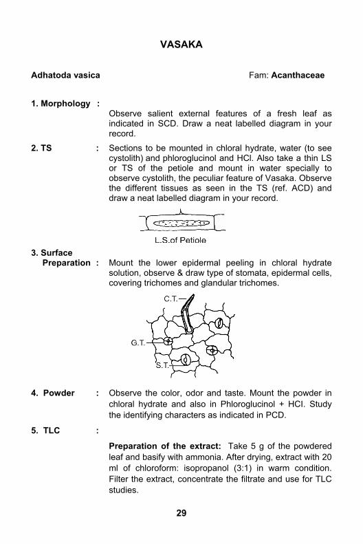
2. TS : Sections to be mounted in chloral hydrate, water (to see cystolith) and phloroglucinol and HCl. Also take a thin LS or TS of the petiole and mount in water specially to observe cystolith, the peculiar feature of Vasaka. Observe the different tissues as seen in the TS (ref. ACD) and draw a neat labelled diagram in your record.
3. Surface Preparation : Mount the lower epidermal peeling in chloral hydrate
solution, observe & draw type of stomata, epidermal cells, covering trichomes and glandular trichomes.
4. Powder : Observe the color, odor and taste. Mount the powder in chloral hydrate and also in Phloroglucinol + HCI. Study the identifying characters as indicated in PCD.
5. TLC :
Preparation of the extract: Take 5 g of the powdered leaf and basify with ammonia. After drying, extract with 20 ml of chloroform: isopropanol (3:1) in warm condition. Filter the extract, concentrate the filtrate and use for TLC studies.

30
Adsorbent: Silica gel pate.
Solvent System:
(a) Toluene/benzene: Ethyl acetate: Die-thylamine (7:2:1) or
(b) Dichloromethane: methanol (3:1) Take 30 ml in a chamber and saturate for atleast an hour.
Application: To be applied in band form
Vasaka extract - 20 l
Vasicine (1% CH3OH soln.) - 10 l
Running distance: 10 to 12 cm.
Drying: Plates are air dried for 15 min and then in an oven for 5 min.
Detection: Cool the plate and spray with Dragendorff 's reagent or keep the plate in Iodine chamber.
Observation:
(a) With Dragendorff's reagent 3 prominent orange brown colored spots appear in the Rf range between 0.4 and 0.95
(b) Treatment with iodine vapour also shows 3-4 spots in the same Rf range.
Record:
The Rf value of Vasicine (approx. Rf value is between 0.45 and 0.7 depending upon the solvent system and saturation of the chamber).
Reproduce the plate in the journal with yellow background and orange colored spots.
Record: Page I - 1,3 ; II - 2 ; III - 4 ; IV - 5