Iontophoretic Drug Delivery - -:: Welcome to BS...
26
1 1 Iontophoretic Drug Delivery Ramesh Gannu 1 and Y. Madhusudan Rao 2 1 AET Laboratories Pvt Ltd, Hyderabad. 2 Vaagdevi College of Pharmacy, Hanamkonda, Warangal. 1.1 Introduction Most preferred route is the oral route among the various routes of drug delivery. Solid oral dosage forms for peroral administration e.g., tablet, capsule, etc., has been the traditional way to systemically administer the drugs for over centuries. Some drugs administered by this route exhibit poor bioavailability either because of the inherent properties of the drug e.g., acid lability or pre-systemic metabolism or limitations associated with the gastrointestinal tract (e.g., chemically hostile environment). In some cases the problems associated with the drug can be solved by modifying the formulation e.g., reduction of gastric irritation by application of enteric coating. On the other hand, poor bioavailability associated with the peroral route can often be improved only, by formulating the drug for delivery via a different route such as parenteral route (intravenous infusion), transdermal route or buccal route. These routes bypass the hepatic “first-pass’’ elimination and also maintain a constant, prolonged, and therapeutically-effective drug level in the body. Transdermal drug delivery (TDD) systems have been developed aiming to achieve the objective of systemic medication through topical application on the intact skin surface. Drug molecule should posses certain properties such as low molecular weight, low dose and sufficient lipophilicity for designing transdermal delivery systems. Conventional TDDS are not suitable for the hydrophilic and ionizable drugs. Hence an
Transcript of Iontophoretic Drug Delivery - -:: Welcome to BS...

1
1 Iontophoretic Drug Delivery
Ramesh Gannu1 and Y. Madhusudan Rao2
1AET Laboratories Pvt Ltd, Hyderabad. 2Vaagdevi College of Pharmacy, Hanamkonda, Warangal.
1.1 Introduction
Most preferred route is the oral route among the various routes of drug delivery. Solid oral dosage forms for peroral administration e.g., tablet, capsule, etc., has been the traditional way to systemically administer the drugs for over centuries. Some drugs administered by this route exhibit poor bioavailability either because of the inherent properties of the drug e.g., acid lability or pre-systemic metabolism or limitations associated with the gastrointestinal tract (e.g., chemically hostile environment). In some cases the problems associated with the drug can be solved by modifying the formulation e.g., reduction of gastric irritation by application of enteric coating. On the other hand, poor bioavailability associated with the peroral route can often be improved only, by formulating the drug for delivery via a different route such as parenteral route (intravenous infusion), transdermal route or buccal route. These routes bypass the hepatic “first-pass’’ elimination and also maintain a constant, prolonged, and therapeutically-effective drug level in the body.
Transdermal drug delivery (TDD) systems have been developed aiming to achieve the objective of systemic medication through topical application on the intact skin surface. Drug molecule should posses certain properties such as low molecular weight, low dose and sufficient lipophilicity for designing transdermal delivery systems. Conventional TDDS are not suitable for the hydrophilic and ionizable drugs. Hence an

2 Advances in Drug Delivery Volume - III
alternative mode of delivery would be desired for the delivery of ionizable drug molecules in to the systemic circulation.
The present chapter describes anatomy of skin, iontophoretic principles, factors influencing the process and drug selection criteria for iontophoresis etc
1.2 Components of Normal Human Skin
The skin of an average adult human body covers a surface of approximately 2 m2 and receives about one third of the blood circulating through the body1. It is one of the most readily accessible organs of the human body with a thickness of only a few millimeters (2.97 0.28 mm). Its major roles are to regulate body temperature, protect tissues from infection, prevent fluid loss, and cushion internal structures. The skin can be divided into three distinct layers: the stratified cellular epidermis and underlying dermis of connective tissue (Fig. 1.1). The dermal-epidermal junction is undulating and consists of ridges of the epidermis, known as rete ridges. The ridges project into the dermis and the junction provides mechanical support to the epidermis and acts as a partial barrier against exchange of cells and large molecules. Fatty layer is present below the dermis and is usually designated as subcutaneous layer. This is separated from the rest of the body by a vestigial layer of striated muscle, the panniculus carnosus.
The corneocytes are suspended in the lipid matrix, in addition to the lipid envelope surrounding the cells. It forms a brick and mortar barrier that permits retention of water within the corneocytes in addition to hampering the penetration of foreign particles2. The major lipid classes within the stratum corneum are ceramides, cholesterol, and fatty acids. The stratum granulosum or granular layer lies below the stratum corneum followed by stratum spinosum. The stratum spinosum has an abundance of desmosomes that give a spiny appearance to the cells (prickly layer). The stratum basale, also known as the stratum germinativum, is a single layer of columnar basal cells that are attached to the basement membrane, or basal lamina, via hemidesmosomes3. The stratum basale is the regenerative layer composed of undifferentiated keratinocytes and stem cells4. Within the epidermis, there are several other cell populations, namely melanocytes, which donate pigment to the keratinocytes, Langerhans’ cells, which have immunological functions and Merkel cells.

Iontophoretic Drug Delivery 3
The dermis, approximately 2-3 mm thick, forms the bulk of the skin and is made up primarily of fibroblasts5. It consists of a network of collagen tissue fibers with interweaving blood and lymph vessels, sweat and sebaceous glands, hair follicles and nerve endings. The dermis consists of two regions, the papillary or adventitial that interfaces the basal lamina and reticular dermis. The reticular dermis make up the bulk of the structural dermis. The lowest layer of the skin is the hypodermis, which is primarily composed of fibroblasts and adipocytes. The hypodermis binds skin to the underlying structures, in addition to serving as a thermo regulator and a cushion to internal organs against trauma6.
There are two kinds of human skin (i) glabrous skin (is also called as non-hairy skin), found on the palms and soles, is grooved on its surface by continuously alternating ridges and sulci, in individually unique configurations known as dermatoglyphics. It is characterized by a thick epidermis divided into several well-marked layers, including a compact stratum corneum, by the presence of encapsulated sense organs within the dermis and by a lack of hair follicles and sebaceous glands. (ii) Hair-bearing skin has both hair follicles and sebaceous glands but lacks encapsulated sense organs. There is also wide variation between different body sites. For example, the scalp with its large hair follicles may be contrasted with the forehead, which has only small vellus-producing follicles, albeit associated with large sebaceous glands. The axilla is notable because it has apocrine glands in addition to the eccrine sweat glands, which are found throughout the body.
Fig. 1.1 The skin and its appendages (Adopted from Reference7).
4 Advances in Drug Delivery Volume - III
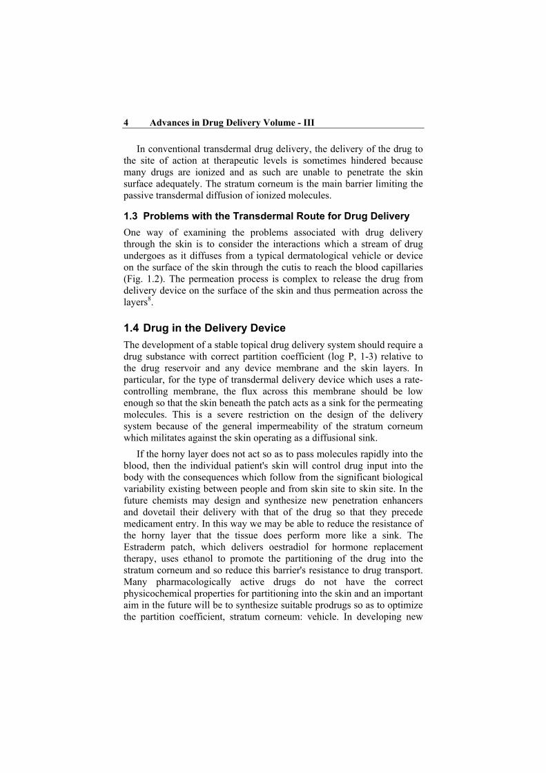
In conventional transdermal drug delivery, the delivery of the drug to the site of action at therapeutic levels is sometimes hindered because many drugs are ionized and as such are unable to penetrate the skin surface adequately. The stratum corneum is the main barrier limiting the passive transdermal diffusion of ionized molecules.
1.3 Problems with the Transdermal Route for Drug Delivery
One way of examining the problems associated with drug delivery through the skin is to consider the interactions which a stream of drug undergoes as it diffuses from a typical dermatological vehicle or device on the surface of the skin through the cutis to reach the blood capillaries (Fig. 1.2). The permeation process is complex to release the drug from delivery device on the surface of the skin and thus permeation across the layers8.
1.4 Drug in the Delivery Device
The development of a stable topical drug delivery system should require a drug substance with correct partition coefficient (log P, 1-3) relative to the drug reservoir and any device membrane and the skin layers. In particular, for the type of transdermal delivery device which uses a rate-controlling membrane, the flux across this membrane should be low enough so that the skin beneath the patch acts as a sink for the permeating molecules. This is a severe restriction on the design of the delivery system because of the general impermeability of the stratum corneum which militates against the skin operating as a diffusional sink.
If the horny layer does not act so as to pass molecules rapidly into the blood, then the individual patient's skin will control drug input into the body with the consequences which follow from the significant biological variability existing between people and from skin site to skin site. In the future chemists may design and synthesize new penetration enhancers and dovetail their delivery with that of the drug so that they precede medicament entry. In this way we may be able to reduce the resistance of the horny layer that the tissue does perform more like a sink. The Estraderm patch, which delivers oestradiol for hormone replacement therapy, uses ethanol to promote the partitioning of the drug into the stratum corneum and so reduce this barrier's resistance to drug transport. Many pharmacologically active drugs do not have the correct physicochemical properties for partitioning into the skin and an important aim in the future will be to synthesize suitable prodrugs so as to optimize the partition coefficient, stratum corneum: vehicle. In developing new

Iontophoretic Drug Delivery 5
drug entities, more attention could profitably be paid to producing chemicals with low melting points (preferably liquids at biological temperatures) and, more speculatively, to including penetration-enhancer groups in the active molecule.
Fig. 1.2 Percutaneous absorption of a drug from a typical transdermal
drug delivery device (adopted from Reference9).
6 Advances in Drug Delivery Volume - III
Iontophoresis has the potential to overcome the limitations of conventional transdermal systems making it feasible to deliver ionic, hydrophilic and some of the high molecular weight compounds10,11.
1.5 Iontophoresis
The highly lipophilic nature of the skin restricts the permeation of hydrophilic, high molecular weight and charged compounds through the stratum corneum into the systemic circulation. However, many therapeutically active drug molecules are hydrophilic and possess high molecular weights for example, peptides.
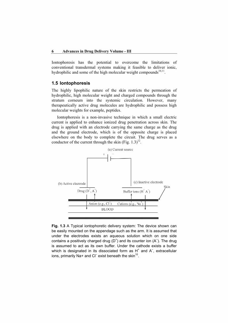
Iontophoresis is a non-invasive technique in which a small electric current is applied to enhance ionized drug penetration across skin. The drug is applied with an electrode carrying the same charge as the drug and the ground electrode, which is of the opposite charge is placed elsewhere on the body to complete the circuit. The drug serves as a conductor of the current through the skin (Fig. 1.3)12.
Fig. 1.3 A Typical iontophoretic delivery system: The device shown can be easily mounted on the appendage such as the arm. It is assumed that under the electrodes exists an aqueous solution which on one side contains a positively charged drug (D+) and its counter ion (A–). The drug is assumed to act as its own buffer. Under the cathode exists a buffer which is designated in its dissociated form as H+ and A–, extracellular ions, primarily Na+ and Cl– exist beneath the skin10.

Iontophoretic Drug Delivery 7
This technique is capable of expanding the range of compounds that can be delivered transdermally.
The advantages summarized as below:
1. Avoidance of hepatic first pass effect: Drugs are administered by topical route bypasses the metabolism by gastrointestinal and liver metabolic enzymes. That enhances the bioavailability.
2. Higher patient compliance: No skilled personals are involved to administer the drugs (as required in parenteral medication) could enhances the patient compliance.
3. Facilitates the delivery of ionized and unionized drugs.
4. Enabling continuous or pulsatile delivery of drug by programmable iontophoretic delivery.
5. Permitting easier termination of drug delivery if needed, by simply by stopping drug input from the iontophoretic delivery system.
6. Offers better control over the amount of drug delivered since the amount of compound delivered depends on applied current, duration of applied current, and area of skin exposed to the current.
7. Restoration of the skin barrier function without producing severe skin irritation.
8. Improving the delivery of polar molecules as well as high molecular weight compounds.
9. Ability to be used for systemic delivery or local (topical) delivery of drugs.
10. Reducing considerably the inter and intra-individual variability since the rate of drug delivery is more dependent on applied current than on stratum corneum characteristics.
11. Provide predictable and extended duration of action.
12. Reduce frequency of dosage.
13. Self-administration is possible.
14. It eliminates problems like toxicity, adverse reaction, formulation problems associated with presence of chemical enhancers in pharmaceuticals.
The disadvantages are summarized as below:
1. Major side effects are very rare. However minor reactions such as itching, erythyma and general irritation of the iontophoretic skin surface are common.
8 Advances in Drug Delivery Volume - III
2. There is an increased risk of minor reactions, if the exposure time and/or current are increased. And with some drugs like histamine, capsaicin and acetylcholine.
3. Drug induced long lasting skin pigmentation after iontophoretic application, where the intensity of skin discoloration is proportional to the exposure time.
4. Current density across the pores in the skin may be higher than the current per unit area applied, depending on the pores in a given area. These spots of high current density increase the possibility of current-induced skin damage.
5. Limited utility of the technique for negatively charged drug substances as the skin’s net negative charge could repel the ions from the skin surface.
1.6 Selection Criteria for Drug Candidates
Drug molecule for iontophoretic delivery should posses the following properties
1. Dose: the therapeutic dose should be low for transdermal iontophoresis
2. Low molecular weight: The molecular weight should be about 500 daltons for better penetrability
3. Charge: molecule should possess ionizability at physiological pH
4. Should be hydrophilic in nature for efficient iontophoretic transport
5. Cationic molecules are favoured over anionic compounds because, in addition to electromigration effect, the iontophoretic transport of the former is supplemented by electroosmosis, while the transport of the latter is against the stream of solvent flow. The stream of solvent flow is created indirectly as a result of the skin’s net negative charge.
1.7 Principle and Basic Device
A basic iontophoretic device consists of a power supply (a battery) connected to an anode and a cathode, which separately contact the skin via an appropriate conducting medium (Fig. 1.3). A preferred material for the electrodes is silver/silver chloride (Ag/AgCl); such electrodes are reversible and do not induce pH changes in the media in contact with the skin. The main disadvantage of Ag/AgCl electrodes is the consumption and accumulation of chloride ions at the anode and cathode, respectively. For example, the flux of anionic drugs may be progressively reduced due to the increasing chloride at the cathode13-15. During process, oxidation

Iontophoretic Drug Delivery 9
and reduction takes place at the anode and cathode respectively. The flux of electrons through the outside circuit is exactly balanced by the flux of ions through the inside circuit (Fig. 1.2). Oxidation at the Ag/AgCl anode results in the loss of a chloride ions from the solution, and this is balanced either by pushing a cation (Na+, Drug+) through the skin or by the arrival of an anion from beneath the skin (Cl-). The use of inert electrodes such as platinum is inadvisable as they result in the hydrolysis of water and progressive and significant pH alterations. This results in risks of skin irritation and changes in skin perme-selectivity and/or drug ionization. To minimize the changes, an alternative way is use of high ionic strength buffers to keep the pH constant; however it decreases the transport efficiency of the ion of interest16.
The reduction at the cathode releases a chloride ion, an extra negative charge, which is neutralized either by the electromigration of an ion (Cl- or Drug- for example) through the skin or by the arrival of cation from beneath. In other words, to deliver n monovalent ions through the skin, n electrons must be generated at the anode (oxidation) and transferred to the cathode, where the reduction consumes these n electrons. The number of electrons flowing through the external circuit is a direct reflection of the amount of ionic charge flowing through the skin.
The application of current across membrane creates an electric potential gradient and ions on either side will migrate in the direction dictated by their charge. The speed of migration of an ion is determined by its physicochemical characteristics and the properties of the media through which the ion is moving. The sum of the individual ion fluxes must equal the current supplied by the power source; in other words, there is a “competition” among all the ions present to carry the charge. Obviously, the chances of being a major carrier, and in consequence being efficiently transported through the skin, increase with the electrical mobility and concentration of the ion concerned.
1.8 Factors Affecting Iontophoretic Delivery
A complex multitude of factors have been recognized to influence iontophoretic process17-19. In order to understand the delivery profiles and to be able to use this technology commercially, it is important to understand the various formulations, electrochemical and biological factors involved in the process. The formulation or electro-chemical factors can be evaluated by conventional techniques such as one factor at a time or using statistical methods such as response surface methodology and factorial design. Statistical methods are advisable as they give
10 Advances in Drug Delivery Volume - III
maximum information with minimum number of experiments20-22. Various factors which influence the iontohporetic delivery are explained below.
1.8.1 Physiochemical Factors Affecting Iontophoresis
1.8.1.1 pH of the Drug Solution
Drug solution pH will have a very significant influence on iontophoretic delivery of drug from the formulation. Changes in the pH of the fluid at the driving electrode can influence the transport. The pH can determine whether or not the drug is charged or it can affect the ratio of the charged and uncharged species23. For the delivery of polypeptides, the type of charge is also controlled by the formulation pH relative to the isoelectric point of the polypeptide. Ideal candidates for iontophoresis should be water-soluble, potent drugs that exist in their salt form with high charge density24, 25. Conductivity experiments can be done to speculate on which drugs may be the best candidates for iontophoresis and to select the optimum pH for maximum delivery. The conductivity of a drug can also be used to estimate the competitive transport between the drug and other extraneous ions during iontophoretic transport. The salt form of the drug can also play an important role in controlling delivery efficiency if a protonated drug is being used and is the primary current-carrying species in the formulation. At neutral pH, cationic drugs often exist as a mixture of protonated and unprotonated species. As the protonated drug migrates towards the skin under the electric field, an imbalance is created with more protonated drug in the boundary layer. This in turn lowers the pH in the boundary layer which results in higher proton transport. The pH may even drop below the pKa of the acid. The problem will not occur with positively charged drugs which are not protonated, such as quaternary ammonium salts. This problem can be avoided by using a weak acid salt of the drug, such as the acetate rather than the hydrochloride. Non-ionized molecules will also be delivered typically by iontophoresis owing to electro-osmosis. Using a series of n-alkanols, it was shown that iontophoresis hindered the lipoidal transport pathways. The iontophoretic enhancement values decreased linearly with increasing alkyl chain length, with the transport being even less than passive diffusion at alkyl chain lengths of greater than six26.
Co-solvents may be added to improve the solubility of drug in the formulation. The presence of high concentrations (> 15 % v/v) of co-solvents in the formulations (such as propylene glycol, poly ethylene glycol) hinders the iontophoretic delivery. This could be due to a

Iontophoretic Drug Delivery 11
decrease in the conductivity of the drug solution as well as a decrease in electro-osmotic flow27. The effect of vehicle pH on the rate and the extent of iontophoretic delivery of lidocaine through human stratum corneum was investigated by Siddiqui et al.28. The rate of penetration was greatest at the pH at which lidocaine existed in the ionic form. The importance of solute transport by iontophoresis was shown pH in enhancing for other drug molecules such as insulin29.
A shift in pH becomes particularly important for protein and peptide drugs, since the pH of the solution determines the charge on these molecules29, 30. Skin membrane ionization induced by pH change has been implicated to explain the anomalous behavior of verapamil iontophoretic flux which increases with increasing the pH of the solution.
1.8.1.2 Size and Salt
The charge, size, structure and lipophilicity of the drug will all influence its potential to be an iontophoresis candidate. The molecular size of the solute is a major factor determining the feasibility of iontophoretic delivery and the amount transported. The efficiency of delivery of carboxylate ions showed the following rank order: acetate> hexanoate > dodecanoate, suggesting that smaller and more hydrophilic ions are transported faster than larger ions31,32. The molecular volume may be more important than the molecular weight as a compact folded molecule such as a globular protein may be able to pass through pores more readily than an open extended fibrous one, such as an unfolded globular or a fibrous protein. Thus, the tertiary and quaternary structure of a protein will play a role in the overall feasibility and efficiency of delivery. The permeability coefficients for a number of positively charged, negatively charged and uncharged solutes across excised human skin were a function of molecular size have been reported33. In general, as the size of the molecule being transported increases, the permeability coefficient for that molecule across a membrane decreases33,34.
1.8.1.3 Ion Competition for Iontophoretic Transport
The condition of electro-neutrality in solution requires that an equal quantity of positively charged and negatively charged molecules exist in a given volume of solution. Therefore, the migrating ion requires an ion of opposite charge in close proximity. This oppositely charge ion is referred to as the counter-ion. An ion of like charge, but of different type is termed a co-ion. Use of divalent cations in drug solution is believed to diminish the selective perm-selectivity of skin towards cations by binding to negatively charged sites on the skin35. The ionic composition of the

12 Advances in Drug Delivery Volume - III
receptor solution can also alter the efficiency of drug ion transport. Replacing the competing ion Cl– with a poly-acrylic acid yielded a transport efficiency of 80%36.
1.8.2 Formulation Factors Affecting Iontophoresis
1.8.2.1 Current Strength and Types
Iontophoretic dosage is controlled by current level (mA) and time (min) and is expressed as milli ampere minutes (mA-min). This follows Coulomb’s law where Q is the electric force, I equals current in milli amperes and T is time in minutes (Q = IT). Current levels are limited by patient comfort and safety. Drug flux increases with increasing current density and a linear relationship has been observed for a wide range of compounds37, the response can plateau at higher current levels. Once this level has been reached, there are no significant increases in drug flux with further increases in current. Current range used is from 0.05 to 0.5 mA20, as the current density increases the upper limit could cause discomfort and pain38. Bellantone39 reported that the increase in the applied current produced a linear increase in benzoate flux. The flux of thyrotropin releasing hormone40, verapamil41, and morphine hydrochloride42 was found to be directly proportional to applied current density.
Continuous use of direct current in skin tissue sometimes resulted in a polarisation effect, which could reduce the efficiency of iontophoretic delivery proportional to the length of time direct current is applied. This polarised current build-up can be overcome with pulsed direct current43. During the off portion of the pulsed cycle, the skin polarisation returns to its near initial electric condition. Pulsed frequency selection is important because enhanced skin polarisation can decrease the efficiency of transport if the frequency is high44.
1.8.2.2 Electrode Types
The choice of electrode is an important parameter for successful iontophoresis. If inert electrodes like platinum, carbon and graphite are used, electrolysis of water takes place at these electrodes, resulting in the formation of hydrogen ions at anode and hydroxyl ions at cathode. Formation of these ions affects the pH of drug solution, and thereby affecting the drug flux45. Moreover, the hydrogen ion being charged and highly mobile competes with drug ion for transport. The use of buffer does little to solve the problem. However, it does prevent the pH change, but provides its own ion for transport. Thus, decreasing the efficiency of drug delivery. These difficulties associated with electrolysis reactions that
Iontophoretic Drug Delivery 13
occur with inert electrodes in direct current iontophoresis can be corrected by using a size exclusion membrane35. This membrane prevents direct drug solution interaction with the electrode.
Use of Ag/AgCl electrodes prevents the electrolysis and the pH change. In addition, if drug molecules present in the system do not undergo redox reactions at Ag/AgCl electrode interface, the use of an Ag/AgCl electrode system provides a more convenient way to apply a voltage difference across the skin (if the total charge transfer across the electrodes during the experiment does not delete the Ag/AgCl deposited on the electrode). The Ag/AgCl electrodes are expensive and need constant replacement. Continuous use of Ag/AgCl electrodes may result in deposition of black silver chloride over the gray Ag/AgCl layer as a result of chlorination. Mechanical removal of the deposit is generally not recommended. In such cases, dilute amonium hydroxide solution may be used46. If a layer of bright silver appears, the electrodes can be re-chlorinated, but the electrode layer is not uniformly composed of a sintered Ag/AgCl. An alternate way is the use of Pt/Pt electrodes and DC reversing current.
Electrochemical Reactions
Inert electrodes (platinum electrode) and direct current
Anode (+) H2O 2H+ + 1/2O2 + 2e–
Cathode (–) 2e– + 2H2O2OH– + H2
Reactive electrodes
Current
Anode (+) Ag+ + Cl–AgCl + e–
Cathode (–) AgC1 + e–Ag+ + Cl–
Inert Electrodes with reversing DC Current
H+(from anode reaction) + OH– (from cathode reaction) no pH change in the drug solution
1.8.3 Biological Factors
Iontophoresis reduces intra and inter-subject variation in drug delivery rate, which is unachievable with passive absorption studies. Iontophoretic fluxes of sodium chloride and mannitol across human thigh skin obtained from the skin regions of low and high density hair follicles are comparable35. Skin blood perfusion also influences drug flux. The
14 Advances in Drug Delivery Volume - III
presence of a vasoconstrictor (epinephrine) or vasodilator (lidocaine) decreases the flux of co administered drug such as a lidocaine due to vasoconstricting and dilating properties respectively47,48.
1.9 Mechanism of Iontophoresis
Skin consists of 15-20 % of lipids (triglycerides, free fatty acids, cholesterol and phospholipids), proteins (keratin) and 40 % of water. The isoelectric point of skin is between 3 and 4, which is another way of saying that pores have a positive charge below pH 3 and a negative charge above 449,50. The negative charge could favor the permeation of drugs in ionized state for basic drugs such as methylene blue51. There are two mechanisms for the transdermal permeation by iontophoresis- (1) electrorepulsion and (2) electroosmosis
1. Electrorepulsion: An electrode with defined charge is used to repel a drug with a similar charge, which attracted by an oppositely charged electrode placed elsewhere on the body. In anodal iontophoresis, positively charged drug is placed in the anodal reservoir system, where as the cathode is placed on a different site on the skin. On application of current all cations including the positively charged drug move away from the anode and into skin. At the same time, negatively charged ions in the body move from the body into the donor reservoir35.
2. Electroosmosis: The transport of water as a whole accorss skin. This causes migration of non ionic molecules across skin. The negative charge of skin will affect movement of water into the body from positive pole electroosmotically toward the outer surface of the skin at the negative pole. This leads to shrinkage of skin pores at the positive pole and causes swelling at the negative pole after intensive iontophoresis. This process helps in cation transfer from the positive pole. Electroosmosis is highest in solutions with a low conductivity; on the other hand iontophoresis is greatest in fluids with a high electrolyte concentration52.
1.10 Applications of Iontophoresis
1.10.1 Topical Delivery
The ability to control the delivery rates of drugs by changes in current makes iontophoresis an attractive technique to use. The charged nature of the prodrug, valacyclovir enabled it to be more efficiently iontophoresed into the skin than the parent molecule acyclovir53.

Iontophoretic Drug Delivery 15
1.10.2 Treatment of Hyperhydrosis
Hyperhydrosis is a condition that most often results in excessive sweating in the hands and feet. Water via iontophoresis is one of the most popular treatments used in this condition. The procedure uses a mild electrical current that is passed through the water to temporarily shut off sweat glands54.
1.10.3 Diagnostic Applications
Iontophoretic application of the drug pilocarpine produces intense sweating, allowing sufficient amounts of sweat to be collected and analyzed55.
Table 1.1 Reverse iontophoresis in diagnostic applications56.
S. No. Drug Indication
1 Phenytoin Ratio of extracted amount correlated well with subdermal concentration.
2 Lithium Excellent correlation between subdermal lithium and iontophoretic extraction flux and iontophoresis tracked sudden changes in lithium concentration.
3 Caffeine, Theophyline
Better extraction through stratum corneum than the stratum corneum stripped skin.
1.10.4 Otorhinolaryngology
Iontophoresis is a preferred method for obtaining anesthesia of the tympanic membrane prior to simple surgical procedures involving that structure. Iontophoresis of zinc has also been used for the treatment of patients with allergic rhinitis55.
1.10.5 Dentistry
Gangarosa described the use of iontophoresis for three basic applications in dentistry:
1. Treatment of hypersensitive dentin (eg, in teeth sensitive to air and cold liquids) using negatively charged fluoride ions;
2. Treatment of oral ulcers and herpes orolabialis lesions using negatively charged corticosteroids and antiviral drugs, respectively; and
3. The application of local anesthetics to produce profound topical anesthesia, as is done in some physical therapy applications57.

16 Advances in Drug Delivery Volume - III
Table 1.2 Summary of conditions treated via iontophoresis58.
S. No. Condition Drug used
1 Ischemic ulcer Zinc oxide
2 Plantar warts Sodium salicylate
3 Aphthous stomatitis Triamcinolone acetonide
4 Ulcers Histamine
5 Herpes simplex Idoxuridine
6 Lichen planus Methylprednisone
7 Hyperhidrosis Poldine methyl sulfate, glycopyrro-nium bromide, atropine, tap water
8 Infected burn wound Penicillin
9 Local skin anesthesia Lidocaine with epinephrine
10 Hyperkeratosis of palms and soles Sodium salicylate
11 Vitiligo Meladine
12 Scleroderma Hyaluronidase
13 Lymphedema Hyaluronidase
14 Patch testing for contact eczema
Pigment for dermabraded tattoos
Iron and titanium oxide
15 Sweat test Pilocarpine
16 Scar tissue Iodine
1.11 Case Study
Ramesh G, Vamshi VY, Chinna RP, Shravan KY and Madhusudan RY, Iontophoretic delivery of Lisinopril: Optimization of process variables by Box-Behnken statistical design
1.11.1 Rational for Drug Selection
Lisinopril (LSP) is an angiotensin converting enzyme inhibitor used for the treatment of hypertension, congestive heart failure and to alleviate strain on hearts damaged as a result of a heart attack. LSP is slowly and incompletely absorbed after oral administration with a bioavailability of 25-30 %59. To improve bioavailability and for effective treatment of chronic hypertension, an alternative long-acting formulations via transdermal route could be beneficial. A classical transdermal administration would not be adequate because of the low permeability of the skin and the prolonged lag time resulting from the excellent barrier

Iontophoretic Drug Delivery 17
properties of the horny layer. Ionic, neutral, and/or polar molecules typically show limited skin penetration ability60-61. LSP is an ideal candidate for iontophoretic delivery, because of its clinical need, low molecular weight (405.5 g/mol), low dose (2.5-20 mg/day) and ionizability. LSP, with apparent dissociation constants (pKa) of 2.5, 4.0, 6.7 and 9.8, exhibits a negative charge (pKa 2.5 because of proline group)62.
1.11.2 Materials and Methods
Lisinopril, Ag (99.9 % purity), AgCl (99 %), Dulbecco’s buffer (pH 7.4), methanol and acetonitrile.
1.11.2.1 Preparation of Rat Abdominal Skin
Albino rats weighing 150-200 gm were selected for permeation studies and the study was conducted with the approval of institutional ethical committee. The animals were sacrificed using anesthetic ether, hair of test animals was carefully trimmed short (<2 mm) with electrical clippers and the full thickness skin was removed from the abdominal region. The epidermis was prepared surgically by heat separation technique63, which involved soaking the entire abdominal skin in water at 60° C for 45 sec, followed by careful removal of the epidermis. The epidermis was washed with water and used for iontophoretic studies.
1.11.2.2 Preparation of Electrodes
Silver/Silver chloride (Ag/AgCl) electrodes were used for the application of the iontophoretic current. Silver wire of 0.5 mm diameter (about 5 cm) was rinsed with concentrated hydrochloric acid, followed by water to remove surface contamination. The wire electrode was then immersed in molten silver chloride for 30 min. A gray silver chloride layer was gradually coated on the silver wire.
1.11.2.3 Iontophoresis of LSP
Iontophoresis experiments were conducted using Franz diffusion cells. The skin was clamped between the two halves of diffusion cell so as to face the stratum corneum towards the donor compartment, cathodal chamber of the cell in which 3 mL (30 mg) of drug solution was placed. A power supply (Sharada Electronics Inc., India) was used to deliver a constant direct current for 4 h via Ag/AgCl electrodes. LSP, with apparent dissociation constants (pKa) of 2.5, 4.0, 6.7 and 9.8, were found to have electrically enhanced drug movement under the influence of cathodal iontophoresis. The receptor solution (anodal chamber) was
18 Advances in Drug Delivery Volume - III
12 mL of phosphate buffer pH 7.4 and was magnetically stirred. The samples were collected from receptor compartment at predetermined intervals and replenished with fresh buffer. The drug content in the samples was determined by HPLC and the concentration was corrected for sampling effects according to the following equation64.
1 1n n T T S n 1 n 1C C (V / V V ) C / C
where 1nC is the corrected concentration of the nth sample, Cn is the
measured concentration of LSP in the nth sample, C n-1 is the measured concentration of the LSP in the (n –1)th sample, VT is the total volume of the receiver fluid and VS is the volume of the sample drawn.
1.11.2.4 HPLC Analysis of LSP
Analysis of samples was performed with a Shimadzu HPLC system equipped with LC-10AT pump, UV-Vis spectrophotometric detector (SPD-10A) and C18 column (Phenomenex 250 × 4.6 mm; 5 µm) at ambient temperature. The mobile phase used was a mixture of phosphate buffer (25 mM potassium dihydrogen ortho phosphate, pH 5.0) and acetonitrile (88:12). A flow rate of 1 mL min−1 was maintained and the detection wavelength was 215 nm. A calibration curve was plotted for LSP in the range of 50-2500 ng mL−1. A good linear relationship was observed between the concentration of LSP and the peak area of LSP with a correlation coefficient (r2 = 0.999). The required studies were carried out to estimate the precision and accuracy of the HPLC method. Sample preparation briefly involved the filtration of iontophoretic sample through 0.45 μ membrane filter, diluted with mobile phase and 20 µL was spiked into column.
1.11.2.5 Experimental Design
Box-Behnken statistical design was used to statistically optimize the iontophoresis process parameters and evaluate main effects, interaction effects and quadratic effects of the process parameters on the amount permeated in 4 h and amount permeated in 24 h. A 3 factor, 3 level Box-Behnken design was used to explore quadratic response surfaces and constructing second order polynomial models with Design Expert (Version 7.1, Stat-Ease Inc., Minneapolis, MN, USA). The Box-Behnken design was specifically selected since it requires fewer runs than a Central Composite Design in cases of three or four variables. This cubic design is characterized by set of points lying at the mid point of each edge and a replicate centre point of the multidimensional cube. A design

Iontophoretic Drug Delivery 19
matrix comprising of 13 experimental runs was constructed. The non-linear computer generated quadratic model is given as
Y = b0 + b1x1 + b2x2 + b3x3 + b12x1x2 + b1
2 x1x3 + b12x2x3 + b11 x1
2 + b22 x22
+ b33 x32
where Y is the measured response associated with each factor level combination; b0 is an intercept; b1 to b33 are regression coefficients computed from the observed experimental values of Y; and X1, X2 and X3 are the coded levels of independent variables. The terms X1X2 and Xi2 (i = 1, 2 or 3) represent the interaction and quadratic terms, respectively. The dependent and independent variables selected are shown in Table 1.4 along with their low, medium and high levels, which were selected based on the results from preliminary experimentation. The current density (X1), ionic strength (X2) and medium/pH (X3) used to prepare the 13 experimental trials and the respective observed responses are given in Table 1.3 and 1.4.
1.11.3 Results and Discussion
Based on the preliminary trials, the current densities were retained at 0.05 and 0.5 mA for the iontophoretic optimization process. Thirteen experi-ments were conducted by varying current density, salt concentration and pH of vehicle (Table 1.3) and corresponding results are in (Table 1.4). The range of response were found to be 1.10 mg in P03 to 4.68 mg in P07; 3.45 mg in P03 to 14.07 mg in P07 and 0.41 h in P07 to 0.82 h in P3 as Y1, Y2 and Y3 respectively.
Table 1.3 Variables in Box-Behnken design.
Factor Levels used, Actual (Coded)
Low (-1) Medium (0) High (+1) Independent variables X1= Current density (mA) 0.05 0.28 0.5 X2=Ionic Strength (Nacl, mM) 0 50 100
X3= Medium/pH Buffer (pH 5.8) Water Buffer
(pH 7.4) Y1= Q4 (Cumulative amount of LSP permeated in 4 h, µg) 1≤Y1≤5 Y2 = Q24 (Cumulative amount of LSP permeated in 24 h, µg) 3≤Y2≤15 Y3= Lag time (h) 0.4≤Y3≤0.9
The cumulative amount of LSP permeated in 4 h (Q4) and 24 h (Q24) were found to be increased from 1.10 and 4.68 mg to 3.45 and 14.07 mg in P03 and P07 respectively using water as medium. The results suggest that increase in the ionic strength decreased the iontophoretic permeation of

20 Advances in Drug Delivery Volume - III
LSP. Chloride ions might be competing with the transport of LSP anions; therefore the permeation of LSP ions was decreased with the increasing ionic strength of sodium chloride.
Table 1.4 Observed responses in the iontophoresis of LSP by Box-Behnken design.
Batch
Independent variables
Dependant variables (mean ± S.D)
X1 X2 X3 Y1 (µg)a Y2 (µg) a Y3 (h) a
P01 -1 -1 0 1.47 ± 0.100 4.90 ± 0.261 0.45 ± 0.012
P02 -1 0 1 1.40 ± 0.159 4.39 ± 0.421 0.50 ± 0.022
P03 -1 1 0 1.10 ± 0.057 3.45 ± 0.655 0.82 ± 0.018
P04 0 -1 1 2.31 ± 0.142 7.35 ± 0.319 0.45 ± 0.016
P05 0 0 0 1.95 ± 0.068 6.58 ± 0.213 0.60 ± 0.018
P06 0 1 -1 1.70 ± 0.042 5.49 ± 0.131 0.61 ± 0.021
P07 1 -1 0 4.68 ± 0.299 14.07 ± 0.775 0.41 ± 0.011
P08 1 0 -1 4.12 ± 0.247 12.88 ± 0.621 0.60 ± 0.023
P09 1 1 0 2.34 ± 0.221 7.41 ± 0.695 0.80 ± 0.027
P10 1 0 1 4.19 ± 0.145 12.11 ± 0.460 0.51 ± 0.018
P11 0 1 1 1.68 ± 0.027 5.16 ± 0.173 0.52 ± 0.021
P12 -1 0 -1 1.38 ± 0.195 4.37 ± 0.760 0.45 ± 0.011
P13 0 -1 -1 2.29 ± 0.156 7.33 ± 0.608 0.48 ± 0.013 aValues represented are mean ± SD (n=3)
1.11.3.1 Data Analysis
The experiments, P07, P08 and P10 had the highest Q4 and Q24. The dependent variables obtained at various levels of the three independent variables (X1, X2 and X3) was subjected to multiple regression to yield a second-order polynomial equation (full model)
Y1 = 1.98 + 1.23X1 – 0.49X2 + 0.025X3 – 0.49X1X2 + 0.040X1X3 – 0.010X2X3 + 0.58X1
2 – 0.17X22 – 0.18X3
2
Y2 = 7.28 + 3.32X1 – 1.52X2 + 0.022X3 – 1.30X1X2 + 0.49X1X3 –0.090X2X3 + 0.79X1
2 – 0.62X22 – 0.33X3
2

Iontophoretic Drug Delivery 21
Fig. 1.4 Iontophoretic delivery of LSP across rat abdominal skin.
1.11.3.2 Optimization
The optimum process parameters were selected based on the criteria of maximum Q4 and Q24 values, by applying constraints on Y1 (1 ≤ Y1 ≤ 5) and Y2 (3 ≤ Y2 ≤ 15). Upon trading of various response variables and extensive grid search, the process parameters with current density of 0.50 mA, sodium chloride of 8 mM and water as medium were found to fulfil the maximum requisite of an optimum iontophoretic process because of maximum Q4 and Q24.
1.12 Conclusion
The penetration was influenced by ionic strength (i.e. sodium chloride concentration) of the donor solution and current density.
References
1. Abla N, Naik A, Guy RH, Kalia YN. Topical iontophoresis of valaciclovir hydrochloride improves cutaneous acyclovir delivery. Pharm Res. 23(8):1842-1849 (2006).
2. Abramson HA and Gorin MH. Skin reactions. IX. The electrophoretic demonstration of the patent pores of the living

22 Advances in Drug Delivery Volume - III
human skin; its relation to the charge of the skin. J Phys Chem 44:1094-1102 (1940).
3. Alvarez-Figueroa MJ, Delgado-Charro MB, Blanco-Méndez J. Passive and iontophoretic transdermal penetration of methotrexate. Int. J. Pharm. 212: 101–107 (2001).
4. Azad Khan, Mohd Yasir, Mohd Asif, Iti Chauhan, Alok P. Singh, Rajat Sharma, Pradeep Singh and Shubham Rai. Iontophoretic drug delivery: History and applications. J App Pharm Sci 1(3): 11-24 (2011).
5. Babiuk S, Maria BE, Lorne AB, Catherine E and Marianna F. Cutaneous vaccination: the skin as an immunologically active tissue and the challenge of antigen delivery. J Control Rel. 66: 199-214 (2000).
6. Baeyens V, Percicot C, Zignani M, et al., Ocular drug delivery in veterinary medicine, Advanced Drug Delivery Reviews 28 (3) (1997) 335– 361.
7. Bagniefski T and Burnette RR. A comparison of pulsed and continuous current iontophoresis. J Controll Rel. 11: 113-122 (1990).
8. Banga A and Chein YW: Iontophoretic delivery of drugs: Fundamentals, developments and biomedical applications. J. Cont. Rel. 7:1-24 (1988).
9. Barry B.W. Modern methods of promoting drug absorption through the skin. Molec. Aspects 12: 195-241(1991).
10. Barry, B. W. Transdermal drug delivery. In: Drug Delivery. Systems (Johnson, P. and J.G. Lloyd-Jones, eds.), pp. 200-223. Ellis Horwood, Chichester (1987c).
11. Boucsein W. Electrodemal activity. Plenum press, New York (1992).
12. Burnette RR and Ongipiattanakul B. Characterization of the permselective properties of excised human skin during iontophoreis. J. Pham Sci. 76(8):765-773 (1987).
13. Burnette RR. Iontophoresis In: Transdermal Drug Delivery: Development Issues and Research Initiatives. Edited by J Kadgraft and RH Guy, NY, Marcel Dekker, 247-291 (1989).
14. Clarke's Analysis of Drugs and Poisons: The Pharmaceutical Press, London, www.medicinecomplete.com.

Iontophoretic Drug Delivery 23
15. Comeau M and Vernon J. Local anesthesia of the ear by iontophoresis. Arch Otolaryngol 98:114-120 (1973).
16. Corish J, Corrigan OI and Foley D. Iontophoretic transport of analgesics, Chemica Oggi, 6: 17-22 (1989).
17. Cullander C and Guy RH. Routes of delivery: Case studies (6) Transdermal delivery of peptides and proteins. Adv. Drug Del. Rev., 8, 291–329 (1992).
18. Dahl JC and Glent-Madsen L. Treatment of hyperhidrosis manuum by tap water iontophoresis. Acta Derm Venereol. 69(4):346-348 (1989).
19. Echolas DF, Norris CH and Tabb HG. Anesthesia of the ear by iontophoresis of lidocaine. Arch Otolaryngol 101:418-421 (1975).
20. Farmer ER, Hood and Antoinette, H. The Pathology of the Skin, 2nd Edition. New York: McGraw-Hill Health Professions Division, 2000.
21. Gangarosa LP, Park NH, Fong BC, Scott DF and Hill JM. Conductivity of drugs used for iontophoresis J. Pharm. Sci., 67: 1439–1443 (1978).
22. Gay CL, Green PG, Guy RH and Francoeur ML. Iontophoretic delivery of piroxicam across the skin. J Control Rel 22:57–68(1992).
23. Gopinathan and Menon K. New insights into skin structure: scratching the surface. Adv Drug Del Rev 54 (1): S3-S17 (2002).
24. Green PG, Hinz RS, Cullander C, Yamane G and Guy RH. Iontophoretic delivery of a series of amino acids and amino acid derivatives across the skin in vitro. Pharm Res 8:1113–1120 (1991).
25. Green PG, Hinz RS, Cullander C, Yamane G, Guy RH. Iontophoretic delivery of amino acids and amino acid derivatives across the skin in-vitro. Pharm. Res. (1991) 8: 1113-1120.
26. Green PG, Hinz RS, Kim A, Szoka FC and Guy RH. Iontophoretic delivery of a series of tripeptides across the skin in vitro. Pharm Res 8:1121–1127 (1991).
27. Harper BN, Rim S, Francoeur ML, Rasadi B. Enhanced percutaneous absorption via iontophoresis I. Evaluation of an in vitro system and transport of model compounds. Int J Pharm 30(1): 63–72 (1986).
28. Hayton WL, Chen T. Correction of perfusate concentration for sample removal. J Pharm Sci 71: 820- 821 (1982).
24 Advances in Drug Delivery Volume - III
29. Huang YY, Wu SM and Wang CY. Response surface method: A novel strategy to optimize iontophoretic transdermal delivery of thyrotropin-releasing hormone. Pharm. Res., 13, 547–552 (1996).
30. Huang YY, Wu SM, Wang CY and Jiang TS A strategy to optimize the operation conditions in iontophoretic transdermal delivery of pilocarpine. Drug Develop. Ind. Pharm 21:1631–1648 (1995).
31. Jadoul A, Mesens J, Caers W, De Beukelaar F, Crabbe R and Preat V. Transdermal permeation of alnitidan by iontophoresis: in vitro optimization and human pharmacokinetic data. Pharm Res 13:1348–1353 (1996).
32. Jadoul A, Regnier V and Preat V. Influence of ethanol and propylene glycol addition on the transdermal delivery by iontophoresis and electroporation, Pharm.Res., 14, S308– S309 (1997).
33. Karakoc Y, Aydemir EH, Kalkan MT and Unal G. Safe control of palmoplantar hyperhidrosis with direct electrical current. Int. J. Dermatol. 41: 602–605 (2002).
34. Lancaster SG and Todd PA. Lisinopril: A preliminary review of its pharmacodynamic and pharmacokinetic properties and therapeutic use in hypertension and congestive heart failure. Drugs. 35: 646-669 (1988).
35. Lattin GA, Padmanabhan RV and Phipps JB. Electronic control of iontophoretic drug delivery, Ann. N.Y. Acad. Sci., 618: 450–464 (1991).
36. Ledger PW. Skin biological issues in electrically enhanced transdermal delivery. Adv Drug Deliv Rev 9:289–307 (1992).
37. McGrath, J. A., Eady, R. A. J. and Pope, F. M. (2008) Anatomy and Organization of Human Skin, in Rook's Textbook of Dermatology, Seventh Edition (eds T. Burns, S. Breathnach, N. Cox and C. Griffiths), Blackwell Publishing, Inc., Malden, Massachusetts, USA.
38. Michael J.P. Transport Mechanisms in Iontophoresis. I. A Theoretical Model for the Effect of Electroosmotic Flow on Flux Enhancement in Transdermal Iontophoresis. Pharm Res. 7: 118-126 (1990).
39. Miller LL and Smith GA. Iontophoretic transport of acetate and carboxylate ions through hairless mouse skin. A cation exchange membrane model, Int. J. Pharm.,49, 15–22 (1989).

Iontophoretic Drug Delivery 25
40. Miller LL, Smith GA, Chang A and Zhou Q. Electrochemically controlled release, J. Control. Release, 6, 293–296 (1987).
41. Moser K, Katrin K, Aarti N, Kalia YN and Guy RH. Passive skin penetration enhancement and its quantification in vitro. Eur J Pharm Biopharm. 52:103-112 (2001).
42. Omalley EP and Oester YT. Influence of some physical chemical factors on iontophoresis using radio isotopes. Arch. Phys. Med.Rrehabi, 36: 310–316 (1955).
43. Phipps JB and Gyory JR.Transdermal ion migration. Adv. Drug. Del. Rev. 9:137-176 (1992).
44. Potts RO, Tamada JA and Tierney MJ. Glucose monitoring by reverse iontophoresis. Diabetes Metab. Res. Rev. 18: S49–S53 (2002).
45. Ramesh G, Vamshi VY, Chinna RP, Shravan KY and Madhusudan RY. Iontophoretic delivery of Lisinopril: Optimization of process variables by Box-Behnken statistical design. Pharm Dev Tech 15(2):169-177 (2010).
46. Rao G, Guy RH, Glikfel P, La Course WR, Reverse iontophoresis: noninvasive glucose monitoring in vivo in human. Pharm. Res. 12 (12): 1869–1873 (1995).
47. Reena R and Srinivas CR. Iontophoresis in dermatology. Indian J Dermatol Venereol Leprol. 71(4): 238-241 (2005).
48. Rivlere JE and Monteiro-Riviere NA. The isolated perfused porcine skin flap as an in vitro model for percutaneous absorption and cutaneous toxicities. Crit Rev. Toxicol, 21: 329-349 (1991).
49. Rivlere JE, Sage B, Williams PI Effects of vasoactive drugs on transdermal lidocaine ionophoresis. J. Pharm. Sei. 80: 615-627 (1991).
50. Sanderson JE, Caldwell RW, Hsiao W and Dixon R. Non invasive delivery of a noval inotropic catelcholamines: Iontophoresis versus intravenous infusions in dogs. J. Pham Sci. 76 (3): 215-218 (1987).
51. Schaefer H. In Schaefer H, Zesch A and Stuttgen G (Eds), Skin Permeability, Springer-Verlag, Boston, 1982, pp 640-641.
52. Siddiqui O and Chein YW. Non parenteral administration of peptides and protein drugs. Crit. Rev Ther Drug Carrier Sys 3:195-221 (1987).

26 Advances in Drug Delivery Volume - III
53. Siddiqui O, Roberts MS and Polack AE. Iontophoretic transport of weak electrolytes through the excised human stratum corneum. J Pham Pharmacol. 41: 430-442 (1985).
54. Siddiqui O, Sun Y, Liu JC and Chein YW. Facilitated transport of insulin. J Phar. Sci. 76(3): 341-345 (1987).
55. Singh P and Maibach HI: Iontophoresis in drug delivery: Basic principles and applications. Critical Reviews in Therapeutic Drug Carrier Systems 11:161-213 (1994).
56. Sitruk-Ware R, De Lignieres B, Basdevant A and Mauvais-Jarvis P. Absorption of percutaneous oestradiol in postmenopausal women. Maturitas. 2(3): 207-211 (1980).
57. Sloan JB and Soltani K. Iontophoresis in dermatology. J. Am. Acad. Dermatol., 15: 671-684 (1986).
58. Stanley J.R, Woodley DT, Katz SI and Martin GR. Structure and function of basement membrane. J. Invest. Dermatol. 79 (1): 695-725 (1982).
59. Terzo SD, Behl CR and Nash RA. Iontophoretic transport of a homologous series of ionized and nonionized model compounds: Influence of hydrophobicity and mechanistic interpretation, Pharm. Res., 6: 85–90 (1989).
60. Wearley L, Jue-Chen L and Chien Y.W. Iontophoresis-facilitated transdermal delivery of verapamil I. In vitro evaluation and mechanistic studies. J Controll Rel. 8, 237-250 (1989).
61. Yoshida NH and Roberts MS. Solute molecular size and transdermal iontophoresis across excised human skin. J. Cont. Rel. 9:239 (1993).
62. Zankel HT and Durham NC. Effect of physical modalities upon Ra I131 iontophoresis Arch. Phys. Med.Rrehabil, 44: 93–97 (1963).
63. Zankel HT, Cress RH and Kamin H. Iiontophoresis studies with a radioactive tracer. Aarch.Pphys.Mmed.Rrehabil, 40: 193–196 (1959).
64. Zhao K, Singh J. Mechanism of percutaneous absorption of tamoxifen by terpenes: euginol, D-limonen and menthone. J. Control Rel 1998: 55: 359-366.