Involvement of the Tubular ClC-Type Exchanger ClC-5 in ... · Involvement of the Tubular ClC-Type...
7
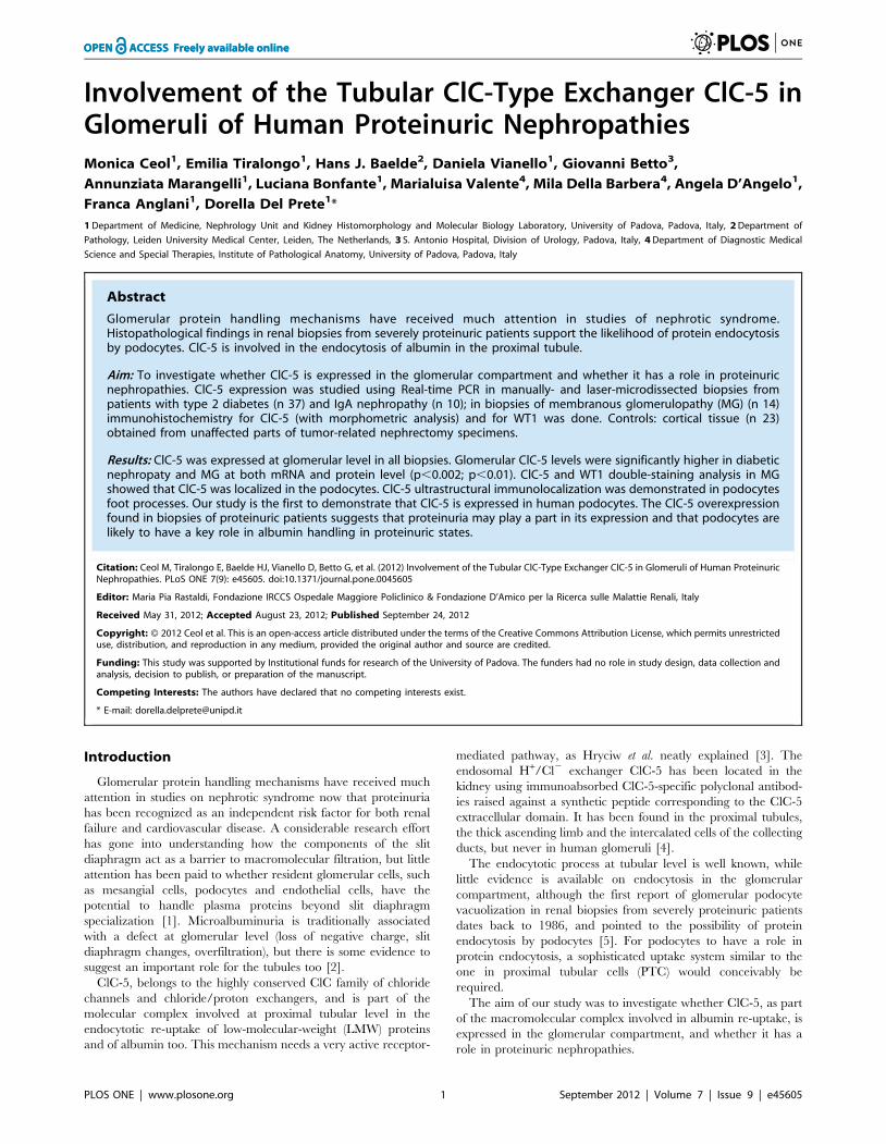
Involvement of the Tubular ClC-Type Exchanger ClC-5 in Glomeruli of Human Proteinuric Nephropathies Monica Ceol 1 , Emilia Tiralongo 1 , Hans J. Baelde 2 , Daniela Vianello 1 , Giovanni Betto 3 , Annunziata Marangelli 1 , Luciana Bonfante 1 , Marialuisa Valente 4 , Mila Della Barbera 4 , Angela D’Angelo 1 , Franca Anglani 1 , Dorella Del Prete 1 * 1 Department of Medicine, Nephrology Unit and Kidney Histomorphology and Molecular Biology Laboratory, University of Padova, Padova, Italy, 2 Department of Pathology, Leiden University Medical Center, Leiden, The Netherlands, 3 S. Antonio Hospital, Division of Urology, Padova, Italy, 4 Department of Diagnostic Medical Science and Special Therapies, Institute of Pathological Anatomy, University of Padova, Padova, Italy Abstract Glomerular protein handling mechanisms have received much attention in studies of nephrotic syndrome. Histopathological findings in renal biopsies from severely proteinuric patients support the likelihood of protein endocytosis by podocytes. ClC-5 is involved in the endocytosis of albumin in the proximal tubule. Aim: To investigate whether ClC-5 is expressed in the glomerular compartment and whether it has a role in proteinuric nephropathies. ClC-5 expression was studied using Real-time PCR in manually- and laser-microdissected biopsies from patients with type 2 diabetes (n 37) and IgA nephropathy (n 10); in biopsies of membranous glomerulopathy (MG) (n 14) immunohistochemistry for ClC-5 (with morphometric analysis) and for WT1 was done. Controls: cortical tissue (n 23) obtained from unaffected parts of tumor-related nephrectomy specimens. Results: ClC-5 was expressed at glomerular level in all biopsies. Glomerular ClC-5 levels were significantly higher in diabetic nephropaty and MG at both mRNA and protein level (p,0.002; p,0.01). ClC-5 and WT1 double-staining analysis in MG showed that ClC-5 was localized in the podocytes. ClC-5 ultrastructural immunolocalization was demonstrated in podocytes foot processes. Our study is the first to demonstrate that ClC-5 is expressed in human podocytes. The ClC-5 overexpression found in biopsies of proteinuric patients suggests that proteinuria may play a part in its expression and that podocytes are likely to have a key role in albumin handling in proteinuric states. Citation: Ceol M, Tiralongo E, Baelde HJ, Vianello D, Betto G, et al. (2012) Involvement of the Tubular ClC-Type Exchanger ClC-5 in Glomeruli of Human Proteinuric Nephropathies. PLoS ONE 7(9): e45605. doi:10.1371/journal.pone.0045605 Editor: Maria Pia Rastaldi, Fondazione IRCCS Ospedale Maggiore Policlinico & Fondazione D’Amico per la Ricerca sulle Malattie Renali, Italy Received May 31, 2012; Accepted August 23, 2012; Published September 24, 2012 Copyright: ß 2012 Ceol et al. This is an open-access article distributed under the terms of the Creative Commons Attribution License, which permits unrestricted use, distribution, and reproduction in any medium, provided the original author and source are credited. Funding: This study was supported by Institutional funds for research of the University of Padova. The funders had no role in study design, data collection and analysis, decision to publish, or preparation of the manuscript. Competing Interests: The authors have declared that no competing interests exist. * E-mail: [email protected] Introduction Glomerular protein handling mechanisms have received much attention in studies on nephrotic syndrome now that proteinuria has been recognized as an independent risk factor for both renal failure and cardiovascular disease. A considerable research effort has gone into understanding how the components of the slit diaphragm act as a barrier to macromolecular filtration, but little attention has been paid to whether resident glomerular cells, such as mesangial cells, podocytes and endothelial cells, have the potential to handle plasma proteins beyond slit diaphragm specialization [1]. Microalbuminuria is traditionally associated with a defect at glomerular level (loss of negative charge, slit diaphragm changes, overfiltration), but there is some evidence to suggest an important role for the tubules too [2]. ClC-5, belongs to the highly conserved ClC family of chloride channels and chloride/proton exchangers, and is part of the molecular complex involved at proximal tubular level in the endocytotic re-uptake of low-molecular-weight (LMW) proteins and of albumin too. This mechanism needs a very active receptor- mediated pathway, as Hryciw et al. neatly explained [3]. The endosomal H + /Cl 2 exchanger ClC-5 has been located in the kidney using immunoabsorbed ClC-5-specific polyclonal antibod- ies raised against a synthetic peptide corresponding to the ClC-5 extracellular domain. It has been found in the proximal tubules, the thick ascending limb and the intercalated cells of the collecting ducts, but never in human glomeruli [4]. The endocytotic process at tubular level is well known, while little evidence is available on endocytosis in the glomerular compartment, although the first report of glomerular podocyte vacuolization in renal biopsies from severely proteinuric patients dates back to 1986, and pointed to the possibility of protein endocytosis by podocytes [5]. For podocytes to have a role in protein endocytosis, a sophisticated uptake system similar to the one in proximal tubular cells (PTC) would conceivably be required. The aim of our study was to investigate whether ClC-5, as part of the macromolecular complex involved in albumin re-uptake, is expressed in the glomerular compartment, and whether it has a role in proteinuric nephropathies. PLOS ONE | www.plosone.org 1 September 2012 | Volume 7 | Issue 9 | e45605
Transcript of Involvement of the Tubular ClC-Type Exchanger ClC-5 in ... · Involvement of the Tubular ClC-Type...
Involvement of the Tubular ClC-Type Exchanger ClC-5 inGlomeruli of Human Proteinuric NephropathiesMonica Ceol1, Emilia Tiralongo1, Hans J. Baelde2, Daniela Vianello1, Giovanni Betto3,
Annunziata Marangelli1, Luciana Bonfante1, Marialuisa Valente4, Mila Della Barbera4, Angela D’Angelo1,
Franca Anglani1, Dorella Del Prete1*
1 Department of Medicine, Nephrology Unit and Kidney Histomorphology and Molecular Biology Laboratory, University of Padova, Padova, Italy, 2 Department of
Pathology, Leiden University Medical Center, Leiden, The Netherlands, 3 S. Antonio Hospital, Division of Urology, Padova, Italy, 4 Department of Diagnostic Medical
Science and Special Therapies, Institute of Pathological Anatomy, University of Padova, Padova, Italy
Abstract
Glomerular protein handling mechanisms have received much attention in studies of nephrotic syndrome.Histopathological findings in renal biopsies from severely proteinuric patients support the likelihood of protein endocytosisby podocytes. ClC-5 is involved in the endocytosis of albumin in the proximal tubule.
Aim: To investigate whether ClC-5 is expressed in the glomerular compartment and whether it has a role in proteinuricnephropathies. ClC-5 expression was studied using Real-time PCR in manually- and laser-microdissected biopsies frompatients with type 2 diabetes (n 37) and IgA nephropathy (n 10); in biopsies of membranous glomerulopathy (MG) (n 14)immunohistochemistry for ClC-5 (with morphometric analysis) and for WT1 was done. Controls: cortical tissue (n 23)obtained from unaffected parts of tumor-related nephrectomy specimens.
Results: ClC-5 was expressed at glomerular level in all biopsies. Glomerular ClC-5 levels were significantly higher in diabeticnephropaty and MG at both mRNA and protein level (p,0.002; p,0.01). ClC-5 and WT1 double-staining analysis in MGshowed that ClC-5 was localized in the podocytes. ClC-5 ultrastructural immunolocalization was demonstrated in podocytesfoot processes. Our study is the first to demonstrate that ClC-5 is expressed in human podocytes. The ClC-5 overexpressionfound in biopsies of proteinuric patients suggests that proteinuria may play a part in its expression and that podocytes arelikely to have a key role in albumin handling in proteinuric states.
Citation: Ceol M, Tiralongo E, Baelde HJ, Vianello D, Betto G, et al. (2012) Involvement of the Tubular ClC-Type Exchanger ClC-5 in Glomeruli of Human ProteinuricNephropathies. PLoS ONE 7(9): e45605. doi:10.1371/journal.pone.0045605
Editor: Maria Pia Rastaldi, Fondazione IRCCS Ospedale Maggiore Policlinico & Fondazione D’Amico per la Ricerca sulle Malattie Renali, Italy
Received May 31, 2012; Accepted August 23, 2012; Published September 24, 2012
Copyright: � 2012 Ceol et al. This is an open-access article distributed under the terms of the Creative Commons Attribution License, which permits unrestricteduse, distribution, and reproduction in any medium, provided the original author and source are credited.
Funding: This study was supported by Institutional funds for research of the University of Padova. The funders had no role in study design, data collection andanalysis, decision to publish, or preparation of the manuscript.
Competing Interests: The authors have declared that no competing interests exist.
* E-mail: [email protected]
Introduction
Glomerular protein handling mechanisms have received much
attention in studies on nephrotic syndrome now that proteinuria
has been recognized as an independent risk factor for both renal
failure and cardiovascular disease. A considerable research effort
has gone into understanding how the components of the slit
diaphragm act as a barrier to macromolecular filtration, but little
attention has been paid to whether resident glomerular cells, such
as mesangial cells, podocytes and endothelial cells, have the
potential to handle plasma proteins beyond slit diaphragm
specialization [1]. Microalbuminuria is traditionally associated
with a defect at glomerular level (loss of negative charge, slit
diaphragm changes, overfiltration), but there is some evidence to
suggest an important role for the tubules too [2].
ClC-5, belongs to the highly conserved ClC family of chloride
channels and chloride/proton exchangers, and is part of the
molecular complex involved at proximal tubular level in the
endocytotic re-uptake of low-molecular-weight (LMW) proteins
and of albumin too. This mechanism needs a very active receptor-
mediated pathway, as Hryciw et al. neatly explained [3]. The
endosomal H+/Cl2 exchanger ClC-5 has been located in the
kidney using immunoabsorbed ClC-5-specific polyclonal antibod-
ies raised against a synthetic peptide corresponding to the ClC-5
extracellular domain. It has been found in the proximal tubules,
the thick ascending limb and the intercalated cells of the collecting
ducts, but never in human glomeruli [4].
The endocytotic process at tubular level is well known, while
little evidence is available on endocytosis in the glomerular
compartment, although the first report of glomerular podocyte
vacuolization in renal biopsies from severely proteinuric patients
dates back to 1986, and pointed to the possibility of protein
endocytosis by podocytes [5]. For podocytes to have a role in
protein endocytosis, a sophisticated uptake system similar to the
one in proximal tubular cells (PTC) would conceivably be
required.
The aim of our study was to investigate whether ClC-5, as part
of the macromolecular complex involved in albumin re-uptake, is
expressed in the glomerular compartment, and whether it has a
role in proteinuric nephropathies.
PLOS ONE | www.plosone.org 1 September 2012 | Volume 7 | Issue 9 | e45605
Results
ClC-5 Expression in the Glomerular Compartment ofHuman Biopsies
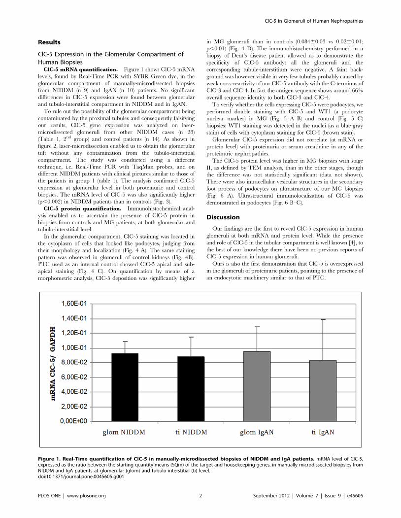
ClC-5 mRNA quantification. Figure 1 shows ClC-5 mRNA
levels, found by Real-Time PCR with SYBR Green dye, in the
glomerular compartment of manually-microdissected biopsies
from NIDDM (n 9) and IgAN (n 10) patients. No significant
differences in ClC-5 expression were found between glomerular
and tubulo-interstitial compartment in NIDDM and in IgAN.
To rule out the possibility of the glomerular compartment being
contaminated by the proximal tubules and consequently falsifying
our results, ClC-5 gene expression was analyzed on laser-
microdissected glomeruli from other NIDDM cases (n 28)
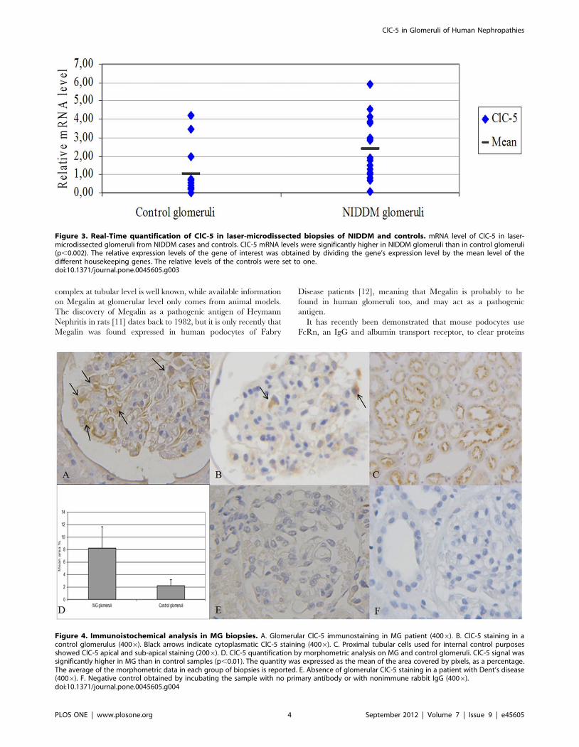
(Table 1, 2nd group) and control patients (n 14). As shown in
figure 2, laser-microdissection enabled us to obtain the glomerular
tuft without any contamination from the tubulo-interstitial
compartment. The study was conducted using a different
technique, i.e. Real-Time PCR with TaqMan probes, and on
different NIDDM patients with clinical pictures similar to those of
the patients in group 1 (table 1). The analysis confirmed ClC-5
expression at glomerular level in both proteinuric and control
biopsies. The mRNA level of ClC-5 was also significantly higher
(p,0.002) in NIDDM patients than in controls (Fig. 3).
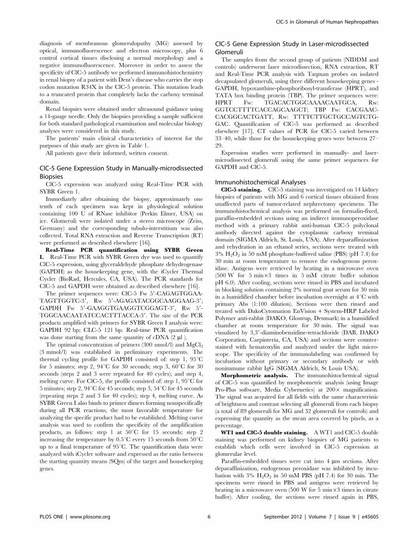
ClC-5 protein quantification. Immunohistochemical anal-
ysis enabled us to ascertain the presence of ClC-5 protein in
biopsies from controls and MG patients, at both glomerular and
tubulo-interstitial level.
In the glomerular compartment, ClC-5 staining was located in
the cytoplasm of cells that looked like podocytes, judging from
their morphology and localization (Fig. 4 A). The same staining
pattern was observed in glomeruli of control kidneys (Fig. 4B).
PTC used as an internal control showed ClC-5 apical and sub-
apical staining (Fig. 4 C). On quantification by means of a
morphometric analysis, ClC-5 deposition was significantly higher
in MG glomeruli than in controls (0.08460.03 vs 0.0260.01;
p,0.01) (Fig. 4 D). The immunohistochemistry performed in a
biopsy of Dent’s disease patient allowed us to demonstrate the
specificity of ClC-5 antibody: all the glomeruli and the
corresponding tubule-interstitium were negative. A faint back-
ground was however visible in very few tubules probably caused by
weak cross-reactivity of our ClC-5 antibody with the C-terminus of
ClC-3 and ClC-4. In fact the antigen sequence shows around 66%
overall sequence identity to both ClC-3 and ClC-4.
To verify whether the cells expressing ClC-5 were podocytes, we
performed double staining with ClC-5 and WT1 (a podocyte
nuclear marker) in MG (Fig. 5 A–B) and control (Fig. 5 C)
biopsies: WT1 staining was detected in the nuclei (as a blue-gray
stain) of cells with cytoplasm staining for ClC-5 (brown stain).
Glomerular ClC-5 expression did not correlate (at mRNA or
protein level) with proteinuria or serum creatinine in any of the
proteinuric nephropathies.
The ClC-5 protein level was higher in MG biopsies with stage
II, as defined by TEM analysis, than in the other stages, though
the difference was not statistically significant (data not shown).
There were also intracellular vesicular structures in the secondary
foot process of podocytes on ultrastructure of our MG biopsies
(Fig. 6 A). Ultrastructural immunolocalization of ClC-5 was
demonstrated in podocytes (Fig. 6 B–C).
Discussion
Our findings are the first to reveal ClC-5 expression in human
glomeruli at both mRNA and protein level. While the presence
and role of ClC-5 in the tubular compartment is well known [4], to
the best of our knowledge there have been no previous reports of
ClC-5 expression in human glomeruli.
Ours is also the first demonstration that ClC-5 is overexpressed
in the glomeruli of proteinuric patients, pointing to the presence of
an endocytotic machinery similar to that of PTC.
Figure 1. Real-Time quantification of ClC-5 in manually-microdissected biopsies of NIDDM and IgA patients. mRNA level of ClC-5,expressed as the ratio between the starting quantity means (SQm) of the target and housekeeping genes, in manually-microdissected biopsies fromNIDDM and IgA patients at glomerular (glom) and tubulo-interstitial (ti) level.doi:10.1371/journal.pone.0045605.g001
ClC-5 in Glomeruli of Human Nephropathies
PLOS ONE | www.plosone.org 2 September 2012 | Volume 7 | Issue 9 | e45605
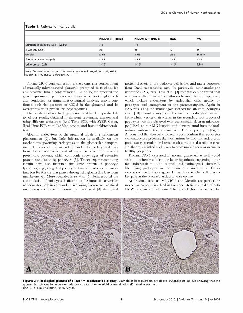
Finding ClC-5 gene expression in the glomerular compartment
of manually microdissected glomeruli prompted us to check for
any proximal tubule contamination. To do so, we repeated the
gene expression experiments on laser-microdissected glomeruli
and conducted an immunohistochemical analysis, which con-
firmed both the presence of ClC-5 in the glomeruli and its
overexpression in proteinuric nephropathies.
The reliability of our findings is confirmed by the reproducibil-
ity of our results, obtained in different proteinuric diseases and
using different techniques (Real-Time PCR with SYBR Green,
Real-Time PCR with TaqMan probes, and immunohistochemis-
try).
Albumin endocytosis by the proximal tubuli is a well-known
phenomenon [2], but little information is available on the
mechanisms governing endocytosis in the glomerular compart-
ment. Evidence of protein endocytosis by the podocytes derives
from the clinical assessment of renal biopsies from severely
proteinuric patients, which commonly show signs of extensive
protein vacuolation by podocytes [5]. Tracer experiments using
ferritin have also identified this large protein in podocyte
lysosomes, suggesting that podocytes have an endocytic recovery
function for ferritin that passes through the glomerular basement
membrane [6]. More recently, Eyre et al. [7] demonstrated the
accumulation of endocytosed albumin in the intracellular vesicles
of podocytes, both in vitro and in vivo, using fluorescence confocal
microscopy and electron microscopy. Koop et al. [8] also found
protein droplets in the podocyte cell bodies and major processes
from Dahl salt-sensitive rats. In puromycin aminonucleoside
nephrotic (PAN) rats, Tojo et al [9] recently demonstrated that
albumin is filtered via other pathways beyond the slit diaphragm,
which include endocytosis by endothelial cells, uptake by
podocytes and entrapment in the paramesangium. Again in
PAN rats, using the immunogold method for albumin, Kinugasa
et al [10] found many particles on the podocytes’ surface.
Intracellular vesicular structures in the secondary foot process of
podocytes was also observed with transmission electron microsco-
py (TEM) on our MG biopsies and ultrastructural immunolocal-
ization confirmed the presence of ClC-5 in podocytes (Fig.6).
Although all the above-mentioned reports confirm that podocytes
can endocytose proteins, the mechanisms behind this endocytotic
process at glomerular level remains obscure. It is also still not clear
whether this is linked exclusively to proteinuric disease or occurs in
healthy people too.
Finding ClC-5 expressed in normal glomeruli as well would
seem to indirectly confirm the latter hypothesis, suggesting a role
for endocytosis in both normal and pathological glomeruli.
Identifying podocytes as the main cells involved in ClC-5
expression would also suggested that this epithelial cell plays a
key part in the protein’s endocytotic re-uptake.
At proximal tubular level ClC-5 and Megalin are part of the
molecular complex involved in the endocytotic re-uptake of both
LMW proteins and albumin. The role of this macromolecular
Figure 2. Histological picture of a laser microdissected biopsy. Example of laser-microdissection pre- (A) and post- (B) cut, showing that theglomerular tuft can be separated without any tubulo-interstitial contamination (Ematoxilin staining).doi:10.1371/journal.pone.0045605.g002
Table 1. Patients’ clinical details.
NIDDM (1st group) NIDDM (2nd group) IgAN MG
Duration of diabetes type II (years) .5 .5 – –
Mean age (years) 52 45 30 56
Gender Male Male Male 10M/4F
Serum creatinine (mg/dl) ,1.8 ,1.8 ,1.8 ,1.8
Urine protein (g/l) 1–1.5 1–1.5 1–1.5 2.5–3
Note: Conversion factors for units: serum creatinine in mg/dl to mol/L, x88.4.doi:10.1371/journal.pone.0045605.t001
ClC-5 in Glomeruli of Human Nephropathies
PLOS ONE | www.plosone.org 3 September 2012 | Volume 7 | Issue 9 | e45605
complex at tubular level is well known, while available information
on Megalin at glomerular level only comes from animal models.
The discovery of Megalin as a pathogenic antigen of Heymann
Nephritis in rats [11] dates back to 1982, but it is only recently that
Megalin was found expressed in human podocytes of Fabry
Disease patients [12], meaning that Megalin is probably to be
found in human glomeruli too, and may act as a pathogenic
antigen.
It has recently been demonstrated that mouse podocytes use
FcRn, an IgG and albumin transport receptor, to clear proteins
Figure 4. Immunoistochemical analysis in MG biopsies. A. Glomerular ClC-5 immunostaining in MG patient (4006). B. ClC-5 staining in acontrol glomerulus (4006). Black arrows indicate cytoplasmatic ClC-5 staining (4006). C. Proximal tubular cells used for internal control purposesshowed ClC-5 apical and sub-apical staining (2006). D. ClC-5 quantification by morphometric analysis on MG and control glomeruli. ClC-5 signal wassignificantly higher in MG than in control samples (p,0.01). The quantity was expressed as the mean of the area covered by pixels, as a percentage.The average of the morphometric data in each group of biopsies is reported. E. Absence of glomerular ClC-5 staining in a patient with Dent’s disease(4006). F. Negative control obtained by incubating the sample with no primary antibody or with nonimmune rabbit IgG (4006).doi:10.1371/journal.pone.0045605.g004
Figure 3. Real-Time quantification of ClC-5 in laser-microdissected biopsies of NIDDM and controls. mRNA level of ClC-5 in laser-microdissected glomeruli from NIDDM cases and controls. ClC-5 mRNA levels were significantly higher in NIDDM glomeruli than in control glomeruli(p,0.002). The relative expression levels of the gene of interest was obtained by dividing the gene’s expression level by the mean level of thedifferent housekeeping genes. The relative levels of the controls were set to one.doi:10.1371/journal.pone.0045605.g003
ClC-5 in Glomeruli of Human Nephropathies
PLOS ONE | www.plosone.org 4 September 2012 | Volume 7 | Issue 9 | e45605

that would otherwise clog the slit diaphragm [13]. In polarized
epithelia, the FcRn receptor has been shown to traffic IgG from
the apical and basolateral membrane into the recycling endosomes
via a transcytotic mechanism that is still poorly understood. The
FcRn receptor has also been identified in the podocytes of adult
human glomeruli [14–15]. Which binding mechanism is used by
ClC-5 at glomerular level remains to be discovered.
Finding that ClC-5 is overexpressed in the glomeruli of patients
with proteinuric diseases, prompted us to hypothesize a role for
this protein in the pathophysiology of proteinuria, probably
relating to an effect that counteracts the larger quantity of filtered
proteins. It is hard to say whether a healthy cell function can
adapts to a pathological environment, or whether such an adaptive
process may become maladaptive in disease. The reason why no
correlation emerged between glomerular ClC-5 expression, at
mRNA or protein level, and proteinuria in all the proteinuric
nephropathies we studied (NIDDM, IgAN, MG) might lie in the
fact that not all our patients underwent ACE inhibitor therapy
wash-out, and in the small number of patients considered in this
study.
In conclusion, our data indicate that ClC-5 in glomeruli, and at
podocyte level in particular, may have a role in protein
endocytosis. This could be another route for glomerular albumin
clearance in proteinuric nephropathy.
Materials and Methods
Ethics StatementAll clinical investigations were conducted according to the
principles expressed in the Declaration of Helsinki and patients’
data were analyzed anonymously. All patients gave their informed,
written consent. The study was approved by Ethics Committee for
experimental studies of Azienda Ospedaliera of Padova protocol
number 0027778 (29/5/2012).
BiopsiesThree different groups of human renal biopsies were analyzed
for this study: the first included manually-microdissected glomeruli
from cortical biopsies of 10 patients with a diagnosis of IgA
nephropathy (IgAN) and 9 patients with diabetic nephropathy
(NIDDM); the second group contained laser-microdissected
glomeruli from biopsies of 28 NIDDM patients and 14 controls,
obtained from sites remote from the tumor-bearing renal tissue;
the third group consisted of 14 biopsies from patients with a
Figure 5. Double staining for ClC-5 and WT1 in MG and control biopsies. Co-localization of ClC-5 (cytoplasmatic brown stain) and WT1(nuclear blu-grey stain) in control (C) and in MG (A) glomeruli to identify podocytes (4006). B. Oil image immersion of a detail of panel A (10006).doi:10.1371/journal.pone.0045605.g005
Figure 6. Immunolocalization of ClC-5 in ultrastucture of MG biopsies. A. Transmission electron microscopy (TEM) of MG biopsy revealedsmall vesicles in the secondary foot processes of podocytes (direct magnification 90006). B–C. Ultrastructural immunolocalization of ClC-5 in MGbiopsy. Arrows indicate some of the gold particles located in podocytes (direct magnification 400006).doi:10.1371/journal.pone.0045605.g006
ClC-5 in Glomeruli of Human Nephropathies
PLOS ONE | www.plosone.org 5 September 2012 | Volume 7 | Issue 9 | e45605
diagnosis of membranous glomerulopathy (MG) assessed by
optical, immunofluorescence and electron microscopy, plus 6
control cortical tissues disclosing a normal morphology and a
negative immunofluorescence. Moreover in order to assess the
specificity of ClC-5 antibody we performed immunohistochemistry
in renal biopsy of a patient with Dent’s disease who carries the stop
codon mutation R34X in the ClC-5 protein. This mutation leads
to a truncated protein that completely lacks the carboxy terminal
domain.
Renal biopsies were obtained under ultrasound guidance using
a 14-gauge needle. Only the biopsies providing a sample sufficient
for both standard pathological examination and molecular biology
analyses were considered in this study.
The patients’ main clinical characteristics of interest for the
purposes of this study are given in Table 1.
All patients gave their informed, written consent.
ClC-5 Gene Expression Study in Manually-microdissectedBiopsies
ClC-5 expression was analyzed using Real-Time PCR with
SYBR Green 1.
Immediately after obtaining the biopsy, approximately one
tenth of each specimen was kept in physiological solution
containing 100 U of RNase inhibitor (Perkin Elmer, USA) on
ice. Glomeruli were isolated under a stereo microscope (Zeiss,
Germany) and the corresponding tubulo-interstitium was also
collected. Total RNA extraction and Reverse Transcription (RT)
were performed as described elsewhere [16].
Real-Time PCR quantification using SYBR Green
I. Real-Time PCR with SYBR Green dye was used to quantify
ClC-5 expression, using glyceraldehyde phosphate dehydrogenase
(GAPDH) as the housekeeping gene, with the iCycler Thermal
Cycler (BioRad, Hercules, CA, USA). The PCR standards for
ClC-5 and GAPDH were obtained as described elsewhere [16].
The primer sequences were: ClC-5 Fw 59-CAGAGTGGAA-
TAGTTGGTC-39, Rw 59-AGAGATACGGCAAGGAAG-39;
GAPDH Fw 59-GAAGGTGAAGGTCGGAGT-39, Rw 59-
TGGCAACAATATCCACTTTACCA-39. The size of the PCR
products amplified with primers for SYBR Green I analysis were:
GAPDH 92 bp; CLC-5 121 bp. Real-time PCR quantification
was done starting from the same quantity of cDNA (2 ml ).
The optimal concentration of primers (300 nmol/l) and MgCl2(3 mmol/l) was established in preliminary experiments. The
thermal cycling profile for GAPDH consisted of: step 1, 95uCfor 5 minutes; step 2, 94uC for 30 seconds; step 3, 60uC for 30
seconds (steps 2 and 3 were repeated for 40 cycles); and step 4,
melting curve. For ClC-5, the profile consisted of: step 1, 95uC for
5 minutes; step 2, 94uC for 45 seconds; step 3, 54uC for 45 seconds
(repeating steps 2 and 3 for 40 cycles); step 4, melting curve. As
SYBR Green I also binds to primer dimers forming nonspecifically
during all PCR reactions, the most favorable temperature for
analyzing the specific product had to be established. Melting curve
analysis was used to confirm the specificity of the amplification
products, as follows: step 1 at 50uC for 15 seconds; step 2
increasing the temperature by 0.5uC every 15 seconds from 50uCup to a final temperature of 95uC. The quantification data were
analyzed with iCycler software and expressed as the ratio between
the starting quantity means (SQm) of the target and housekeeping
genes.
ClC-5 Gene Expression Study in Laser-microdissectedGlomeruli
The samples from the second group of patients (NIDDM and
controls) underwent laser microdissection, RNA extraction, RT
and Real-Time PCR analysis with Taqman probes on isolated
decapsulated glomeruli, using three different housekeeping genes -
GAPDH, hypoxanthine-phosphoribosyl-transferase (HPRT), and
TATA box binding protein (TBP). The primer sequences were:
HPRT Fw: TGACACTGGCAAAACAATGCA, Rw:
GGTCCTTTTCACCAGCAAGCT; TBP Fw: CACGAAC-
CACGGCACTGATT, Rw: TTTTCTTGCTGCCAGTCTG-
GAC. Quantification of ClC-5 was performed as described
elsewhere [17]. CT values of PCR for ClC-5 varied between
33–40, while those for the housekeeping genes were between 27–
29.
Expression studies were performed in manually- and laser-
microdissected glomeruli using the same primer sequences for
GAPDH and ClC-5.
Immunohistochemical AnalysesClC-5 staining. ClC-5 staining was investigated on 14 kidney
biopsies of patients with MG and 6 cortical tissues obtained from
unaffected parts of tumor-related nephrectomy specimens. The
immunohistochemical analysis was performed on formalin-fixed,
paraffin-embedded sections using an indirect immunoperoxidase
method with a primary rabbit anti-human ClC-5 polyclonal
antibody directed against the cytoplasmic carboxy terminal
domain (SIGMA Aldrich, St. Louis, USA). After deparaffinization
and rehydration in an ethanol series, sections were treated with
3% H2O2 in 50 mM phosphate-buffered saline (PBS) (pH 7.4) for
30 min at room temperature to remove the endogenous perox-
idase. Antigens were retrieved by heating in a microwave oven
(500 W for 5 min63 times in 5 mM citrate buffer solution
pH 6.0). After cooling, sections were rinsed in PBS and incubated
in blocking solution containing 2% normal goat serum for 30 min
in a humidified chamber before incubation overnight at 4uC with
primary Abs (1:100 dilution). Sections were then rinsed and
treated with DakoCytomation EnVision + System-HRP Labeled
Polymer anti-rabbit (DAKO, Glostrup, Denmark) in a humidified
chamber at room temperature for 30 min. The signal was
visualized by 3,39-diaminobenzidine-tetrachloride (DAB, DAKO
Corporation, Carpinteria, CA, USA) and sections were counter-
stained with hematoxylin and analyzed under the light micro-
scope. The specificity of the immunolabeling was confirmed by
incubation without primary or secondary antibody or with
nonimmune rabbit IgG (SIGMA Aldrich, St Louis USA).
Morphometric analysis. The immunohistochemical signal
of ClC-5 was quantified by morphometric analysis (using Image
Pro-Plus software, Media Cybernetics) at 2006 magnification.
The signal was acquired for all fields with the same characteristic
of brightness and contrast selecting all glomeruli from each biopsy
(a total of 89 glomeruli for MG and 32 glomeruli for controls) and
expressing the quantity as the mean area covered by pixels, as a
percentage.
WT1 and ClC-5 double staining. A WT1 and ClC-5 double
staining was performed on kidney biopsies of MG patients to
establish which cells were involved in ClC-5 expression at
glomerular level.
Paraffin-embedded tissues were cut into 4 mm sections. After
deparaffinization, endogenous peroxidase was inhibited by incu-
bation with 3% H2O2 in 50 mM PBS (pH 7.4) for 30 min. The
specimens were rinsed in PBS and antigens were retrieved by
heating in a microwave oven (500 W for 5 min63 times in citrate
buffer). After cooling, the sections were rinsed again in PBS,
ClC-5 in Glomeruli of Human Nephropathies
PLOS ONE | www.plosone.org 6 September 2012 | Volume 7 | Issue 9 | e45605

blocked in 2% normal goat serum for 30 min, and incubated with
rabbit anti-human ClC-5 overnight at 4uC. The specimens were
rinsed in PBS, reacted with DakoCytomation EnVision + System-
HRP Labeled Polymer anti-rabbit in a humidified chamber for
30 min and stained with DAB (brown stain). Then they were
incubated again in 3% H2O2 in 50 mM PBS for 30 min and
rinsed in PBS. After blocking with 2% normal goat serum solution
for 30 min, they were incubated overnight at 4uC with rabbit anti-
human WT1 (1:100) polyclonal antibody (Santa Cruz Biotechnol-
ogy Inc, CA, USA). The specimens were rinsed in PBS and treated
with DakoCytomation EnVision + System-HRP Labeled Polymer
anti-rabbit (DAKO, Glostrup, Denmark) in a humidified chamber
for 30 min. The WT1 signal (blue-gray stain) was visualized with
the Vector SG substrate kit for peroxidase (VECTOR Laborato-
ries, Burlingame, USA). No hematoxylin counterstaining was
performed.
Transmission Electron Microscopy (TEM)TEM was performed on all MG biopsies. Semithin sections
(0.5 mm or less) were evaluated at light microscope first, before
proceeding to consider ultrathin sections. The sections were
stained with uranyl acetate and lead citrate and examined under a
Hitachi H-7000 electron microscope equipped with digital
camera.
Immunogold LabelingFor ultrastructural analysis, renal biopsies were fixed by
immersion in modified Karnovsky’s solution (2% paraformalde-
hyde/0.5%glutaraldehyde) in 0.1 M sodium phosfate buffer,
pH 7.2, dehydrated in crescent ethanol concentration and
embedded in LRWhite medium (EMS, Hatfield, PA, USA).
Biopsies ultrathin sections, picked up on nikel grids, underwent to
a post-embedding immunogold labeling by using 1:100 rabbit
anti-CLCN5 antibody (Sigma-Aldrich, Saint Louis, MO, USA) in
TBS-1%BSA buffer at 4uC overnight followed by 1 h of
incubation with (1:40) goat anti-rabbit IgG 15-nm gold particles
conjugated (BBInternational, Cardiff, UK). Control sections were
incubated with secondary antibodies alone. After staining with 1%
uranyl acetate in aqueous solution for five minutes, grids with
sections were observed on Hitachi H-7000 (Hitachi, Japan)
transmission electron microscope.
Statistical AnalysisStudent’s t-test and regression analysis were performed. A p-
value #0.05 was considered statistically significant.
Author Contributions
Analyzed the data: FA DDP. Wrote the paper: FA DDP. Performed the
research: MC ET HJB DV MV MDB. Contributed to acquisition of data:
GB. Contributed to clinical evaluations: AM LB AD. Designed the
research study: FA DDP.
References
1. Pavenstadt H, Kriz W, Kretzler M (2003) Cell biology of the glomerularpodocyte. Physiol Rev 83: 253–307.
2. Birn H, Christensen EI (2006) Renal albumin absorption in physiology and
pathology. Kidney Int 69: 440–449.3. Hryciw DH, Ekberd J, Pollock CA, Poronnik P (2006) ClC-5: a chloride channel
with multiple roles in renal tubular albumin uptake. Int J Biochem Cell Biol 38:1036–1042.
4. Wellhauser L, D’Antonio Christina, Bear CE (2010) ClC transporter: discoveriesand challenges in defining the mechanisms underlying function and regulation of
ClC-5. Eur J Physiol 460: 543–557.
5. Yoshika N, Ito H, Akamatsu R, Hazikano H, Okada S, et al. (1986) Glomerularpodocyte vacuolization in focal segmental glomerulosclerosis. Arch Pathol Lab
Med 110: 394–398.6. Graham RC Jr, Karnovsky MJ (1966) Glomerular permeability. Ultrastructural
cytochemical studies using peroxidases as protein tracers. J Exp Med 124: 1123–
1134.7. Eyre J, Ioannou K, Grubb BD, Saleem MA, Mathieson PW, et al. (2007) Statin-
sensitive endocytosis of albumin by glomerular podocytes. Am J Physiol RenalPhysiol. 292: F674–681.
8. Koop K, Eikmans M, Wehland M, Baelde H, Ijpelaar D, et al. (2008) Selectiveloss of podoplanin protein expression accompanies proteinuria and precedes
alterations in podocyte morphology in a spontaneous proteinuric rat model.
Am J Pathol 173: 315–326.9. Tojo A, Onozato ML, Kitiyakara C, Kinugasa S, Fukuda S, et al. (2008)
Glomerular albumin filtration through podocyte cell body in puromycinaminonucleoside nephrotic rat. Med Mol Morphol 41: 92–98.
10. Kinugasa S, Tojo A, Sakai T, Fujita T (2010) Silver-enhanced immunogold
scanning electron microscopy using vibratome sections of rat kidneys: detection
of albumin filtration and reabsorption. Med Mol Morphol 43: 218–225.
11. Kerjaschi D, Farquhar MG (1982) The pathogenic antigen of heymann nephritis
is a membrane glycoprotein of the renal proximal tubule brush border. Proc Natl
Acad Sci USA 79: 5557–5561.
12. Prabakaran T, Nielsen R, Larsen JV, Sorensen SS, Feldt-Rasmussen U, et al.
(2011) Receptor –Mediated Endocytosis of a-galactosidase A in human Podocytes
in Fabry Disease. PLoS ONE; e25065.doi:10.1371/journal.pone.0025065.
13. Christensen EI, Verroust PJ (2002) Megalin and cubilin, role in proximal tubule
function and during development. Pediatr Nephrol 17: 993–999.
14. Akilesh S, Huber T B, Wu H, Wang G, Hartleben B, et al. (2008) Podocytes use
FcRn to cleat IgG from the glomerular basement membrane. PNAS 105: 967–
972.
15. Haymann JP, Levraud JP, Bouet S, Kappes V, Hagege J, et al. (2000)
Characterization and localization of the neonatal Fc receptor in adult human
kidney. J Am Soc Nephrol 11: 632–639.
16. Del Prete D, Gambaro G, Lupo A, Anglani F, Brezzi B, et al. (2003) Precocious
activation of genes of the renin-angiotensin system and the fibrogenic cascade in
IgA glomerulonephritis. Kidney Int 64: 149–159.
17. Baelde HJ, Eikmans M, Lappin DW, Doran PP, Hohenadel D, et al. (2007)
Reduction of VEGF-A and CTGF expression in diabetic nephropathy is
associated with podocyte loss. Kidney Int 71: 637–645.
ClC-5 in Glomeruli of Human Nephropathies
PLOS ONE | www.plosone.org 7 September 2012 | Volume 7 | Issue 9 | e45605