Intranasal exposure of African green monkeys to SARS-CoV-2 ...
12
SHORT REPORT Open Access Intranasal exposure of African green monkeys to SARS-CoV-2 results in acute phase pneumonia with shedding and lung injury still present in the early convalescence phase Robert W. Cross 1,2 , Krystle N. Agans 1,2 , Abhishek N. Prasad 1,2 , Viktoriya Borisevich 1,2 , Courtney Woolsey 1,2 , Daniel J. Deer 1,2 , Natalie S. Dobias 1,2 , Joan B. Geisbert 1,2 , Karla A. Fenton 1,2 and Thomas W. Geisbert 1,2* Abstract We recently reported the development of the first African green monkey (AGM) model for COVID-19 based on a combined liquid intranasal (i.n.) and intratracheal (i.t.) exposure to severe acute respiratory syndrome coronavirus 2 (SARS-CoV-2). Here, we followed up on this work by assessing an i.n. particle only route of exposure using the LMA mucosal atomization device (MAD). Six AGMs were infected with SARS-CoV-2; three animals were euthanized near the peak stage of virus replication (day 5) and three animals were euthanized during the early convalescence period (day 34). All six AGMs supported robust SARS-CoV-2 replication and developed respiratory disease. Evidence of coagulation dysfunction as noted by a transient increases in aPTT and circulating levels of fibrinogen was observed in all AGMs. The level of SARS-CoV-2 replication and lung pathology was not quite as pronounced as previously reported with AGMs exposed by the combined i.n. and i.t. routes; however, SARS-CoV-2 RNA was detected in nasal swabs of some animals as late as day 15 and rectal swabs as late as day 28 after virus challenge. Of particular importance to this study, all three AGMs that were followed until the early convalescence stage of COVID-19 showed substantial lung pathology at necropsy as evidenced by multifocal chronic interstitial pneumonia and increased collagen deposition in alveolar walls despite the absence of detectable SARS-CoV-2 in any of the lungs of these animals. These findings are consistent with human COVID-19 further demonstrating that the AGM faithfully reproduces the human condition. Keywords: Coronavirus, SARS-CoV-2, COVID-19, Nonhuman primate, Animal models © The Author(s). 2020 Open Access This article is licensed under a Creative Commons Attribution 4.0 International License, which permits use, sharing, adaptation, distribution and reproduction in any medium or format, as long as you give appropriate credit to the original author(s) and the source, provide a link to the Creative Commons licence, and indicate if changes were made. The images or other third party material in this article are included in the article's Creative Commons licence, unless indicated otherwise in a credit line to the material. If material is not included in the article's Creative Commons licence and your intended use is not permitted by statutory regulation or exceeds the permitted use, you will need to obtain permission directly from the copyright holder. To view a copy of this licence, visit http://creativecommons.org/licenses/by/4.0/. The Creative Commons Public Domain Dedication waiver (http://creativecommons.org/publicdomain/zero/1.0/) applies to the data made available in this article, unless otherwise stated in a credit line to the data. * Correspondence: [email protected] 1 Department of Microbiology and Immunology, University of Texas Medical Branch, Galveston, TX 77555, USA 2 Galveston National Laboratory, University of Texas Medical Branch, Galveston, TX 77555, USA Cross et al. Virology Journal (2020) 17:125 https://doi.org/10.1186/s12985-020-01396-w
Transcript of Intranasal exposure of African green monkeys to SARS-CoV-2 ...

SHORT REPORT Open Access
Intranasal exposure of African greenmonkeys to SARS-CoV-2 results in acutephase pneumonia with shedding and lunginjury still present in the earlyconvalescence phaseRobert W. Cross1,2, Krystle N. Agans1,2, Abhishek N. Prasad1,2, Viktoriya Borisevich1,2, Courtney Woolsey1,2,Daniel J. Deer1,2, Natalie S. Dobias1,2, Joan B. Geisbert1,2, Karla A. Fenton1,2 and Thomas W. Geisbert1,2*
Abstract
We recently reported the development of the first African green monkey (AGM) model for COVID-19 based on acombined liquid intranasal (i.n.) and intratracheal (i.t.) exposure to severe acute respiratory syndrome coronavirus 2(SARS-CoV-2). Here, we followed up on this work by assessing an i.n. particle only route of exposure using the LMAmucosal atomization device (MAD). Six AGMs were infected with SARS-CoV-2; three animals were euthanized nearthe peak stage of virus replication (day 5) and three animals were euthanized during the early convalescence period(day 34). All six AGMs supported robust SARS-CoV-2 replication and developed respiratory disease. Evidence ofcoagulation dysfunction as noted by a transient increases in aPTT and circulating levels of fibrinogen was observed inall AGMs. The level of SARS-CoV-2 replication and lung pathology was not quite as pronounced as previously reportedwith AGMs exposed by the combined i.n. and i.t. routes; however, SARS-CoV-2 RNA was detected in nasal swabs ofsome animals as late as day 15 and rectal swabs as late as day 28 after virus challenge. Of particular importance to thisstudy, all three AGMs that were followed until the early convalescence stage of COVID-19 showed substantial lungpathology at necropsy as evidenced by multifocal chronic interstitial pneumonia and increased collagen deposition inalveolar walls despite the absence of detectable SARS-CoV-2 in any of the lungs of these animals. These findings areconsistent with human COVID-19 further demonstrating that the AGM faithfully reproduces the human condition.
Keywords: Coronavirus, SARS-CoV-2, COVID-19, Nonhuman primate, Animal models
© The Author(s). 2020 Open Access This article is licensed under a Creative Commons Attribution 4.0 International License,which permits use, sharing, adaptation, distribution and reproduction in any medium or format, as long as you giveappropriate credit to the original author(s) and the source, provide a link to the Creative Commons licence, and indicate ifchanges were made. The images or other third party material in this article are included in the article's Creative Commonslicence, unless indicated otherwise in a credit line to the material. If material is not included in the article's Creative Commonslicence and your intended use is not permitted by statutory regulation or exceeds the permitted use, you will need to obtainpermission directly from the copyright holder. To view a copy of this licence, visit http://creativecommons.org/licenses/by/4.0/.The Creative Commons Public Domain Dedication waiver (http://creativecommons.org/publicdomain/zero/1.0/) applies to thedata made available in this article, unless otherwise stated in a credit line to the data.
* Correspondence: [email protected] of Microbiology and Immunology, University of Texas MedicalBranch, Galveston, TX 77555, USA2Galveston National Laboratory, University of Texas Medical Branch,Galveston, TX 77555, USA
Cross et al. Virology Journal (2020) 17:125 https://doi.org/10.1186/s12985-020-01396-w

IntroductionThe unprecedented pandemic of COVID-19 caused bysevere acute respiratory syndrome coronavirus 2 (SARS-CoV-2) has had devastating effects on public health andthe global economy. Considerable resources have beenallocated by governments, philanthropic organizations,and private companies in an attempt to expedite thedevelopment of vaccines and treatments to combatCOVID-19. With the rapid development of 24 preventa-tive vaccines in clinical evaluation [1], and nearly 200more in the pipeline [2], coupled with the availability ofnearly 300 candidate antivirals and disease modulators[2] it is impossible to investigate the safety and efficacyof all of these various interventions in humans. Bothsmall animal models and nonhuman primates (NHP)may prove valuable in triaging the most promisingmedical countermeasures prior to use in humans.Hamsters and ferrets are currently being used as im-munocompetent small animal models of COVID-19[3–5] while several NHP models have been quicklydeveloped [6–12]. Among the nonhuman primatemodels evaluated the African green monkey (AGM)appears to best recapitulate the most salient featuresof human COVID-19 [10–12].We recently reported the development of the first
AGM model for COVID-19 and showed that back-challenge of animals with SARS-CoV-2 5 weeks after ini-tial exposure resulted in protection from reinfection[10]. In this study the AGMs were exposed to SARS-CoV-2 by a combination of the intranasal (i.n.) andintratracheal (i.t.) routes with the virus delivered in li-quid media. As a natural extension of this initial workwe sought to assess the pathogenesis of SARS-CoV-2in AGMs exposed by the i.n. route only using theLMA Mucosal Atomization Device (MAD). Previousstudies with another respiratory virus, Nipah virus,showed that there were no major differences in dis-ease pathogenesis when virus was delivered to AGMsby a combined liquid-based i.n. and i.t. delivery [13]or by the LMA MAD system [14]. The LMA MADwas developed for the efficient and safe delivery oftest particles and is currently employed to administerUS FDA approved drugs for i.n. delivery. The LMAMAD delivers atomized particles that range in sizefrom 30 to 100 μm, which is highly consistent withthe size of droplets exhaled by humans due to cough-ing [15]. In addition, in our previous work as theAGMs were back challenged with SARS-CoV-2 it wasimpossible to assess tissue pathology during convales-cence after primary challenge. Here, we focused onassessing the pathogenesis of SARS-CoV-2 infectionin AGMs when administered as 30 to 100 μm parti-cles and on evaluating virus shedding and lung path-ology during early convalescence.
Materials and methodsVirusThe virus (SARS-CoV-2/INMI1-Isolate/2020/Italy) wasisolated on January 30, 2020 from the sputum of the firstclinical case in Italy, a tourist visiting from the Hubeiprovince of China that developed respiratory illnesswhile traveling [16]. The virus was initially passagedtwice (P2) on Vero E6 cells; the supernatant and cell lys-ate were collected and clarified following a freeze/thawcycle. This isolate is certified mycoplasma and Foot-and-Mouth Disease virus free. The complete sequence wassubmitted to GenBank (MT066156) and is available onthe GISAID website (BetaCoV/Italy/INMI1-isl/2020:EPI_ISL_410545) upon registration. For in vivo chal-lenge, the P2 virus was propagated on Vero E6 cells andthe supernatant was collected and clarified by centrifu-gation making the virus used in this study a P3 stock.
Animal challengeSARS-CoV-2 seronegative AGMs (Chlorocebus aethiops)(6 females) (St Kitts origin, Worldwide Primates, Inc.)were randomized into two cohorts where one group(n = 3) was scheduled for euthanasia at 5 dpi and theother at 34 dpi. Animals were anesthetized with keta-mine and inoculated with a target dose of 3.0 × 10⁶ PFUof SARS-CoV-2 (SARS-CoV-2/INMI1-Isolate/2020/Italy)using the LMA MAD, with the dose being equally di-vided between each nostril. All animals were longitudin-ally monitored for clinical signs of illness includingtemperature (measured by surgically implanted DSTmicro-T small implantable thermo loggers (Star-Oddi,Gardabaer, Iceland)), respiration quality, and clinicalpathology. All measurements requiring physical manipu-lation of the animals were performed under sedation byketamine. Mucosal swabs were obtained using sterileswabs inserted into the mucosal cavity, gently rotated tomaximize contact with the mucosal surface, and depos-ited into 2.0 mL screw-top tubes containing sterile MEMmedia supplemented to 2% with FBS.
RNA isolation from SARS-CoV-2-infected AGMsOn specified procedure days (days 0, 2, 3, 4, 5, 7, 12, 15,21, 28, 34), 100 μl of blood was added to 600 μl of AVLviral lysis buffer (Qiagen) for virus inactivation and RNAextraction. Following removal from the high contain-ment laboratory, RNA was isolated from blood andswabs using the QIAamp viral RNA kit (Qiagen).
Detection of SARS-CoV-2 loadRNA was isolated from blood and mucosal swabs andassessed using the CDC SARS-CoV-2 N2 assay primers/probe for reverse transcriptase quantitative PCR (RT-qPCR) [17]. SARS-CoV-2 RNA was detected using One-step probe RT-qPCR kits (Qiagen) run on the CFX96
Cross et al. Virology Journal (2020) 17:125 Page 2 of 12

detection system (Bio-Rad), with the following cycle con-ditions: 50 °C for 10 min, 95 °C for 10 s, and 45 cycles of95 °C for 10 s and 55 °C for 30 s. Threshold cycle (CT)values representing SARS-CoV-2 genomes were ana-lyzed with CFX Manager Software, and data are pre-sented as GEq. To generate the GEq standard curve,RNA was extracted from supernatant derived from VeroE6 cells infected with SARS-CoV-2/INMI1-Isolate/2020/Italy was extracted and the number of genomes was cal-culated using Avogadro’s number and the molecularweight of the SARS-CoV-2 genome.Infectious virus was quantitated by plaque assay on Vero
E6 cells (ATCC CRL-1586) from all blood plasma andmucosal swabs, and bronchoalveolar lavage (BAL) sam-ples. Briefly, increasing 10-fold dilutions of the sampleswere adsorbed to Vero E6 cell monolayers in duplicatewells (200 μl). Cells were overlaid with EMEM mediumplus 1.25% Avicel, incubated for 2 days, and plaques werecounted after staining with 1% crystal violet in formalin.The limit of detection for this assay is 25 PFU/ml.
Hematology and serum biochemistryTotal white blood cell counts, white blood cell differen-tials, red blood cell counts, platelet counts, hematocritvalues, total hemoglobin concentrations, mean cell vol-umes, mean corpuscular volumes, and mean corpuscularhemoglobin concentrations were analyzed from bloodcollected in tubes containing EDTA using a VetscanHM5 hematologic analyzer (Abaxis). Serum sampleswere tested for concentrations of albumin, amylase, ala-nine aminotransferase (ALT), aspartate aminotransferase(AST), alkaline phosphatase (ALP), blood urea nitrogen(BUN), calcium, creatinine (CRE), C-reactive protein(CRP), gamma-glutamyltransferase (GGT), glucose, totalprotein, and uric acid by using a Piccolo point-of-careanalyzer and Biochemistry Panel Plus analyzer discs(Abaxis). Partial pressures of CO2 and O2 were obtainedusing an iSTAT Alinity hematological analyzer (Abbott).
Serum neutralization assayNeutralization titers were calculated by determining thedilution of serum that reduced 50% of plaques (PRNT50).A standard 100 PFU amount of SARS-CoV-2 was incu-bated with two-fold serial dilutions of serum samples for1 hour. The virus-serum mixture was then used to in-oculate Vero E6 cells for 60 min. Cells were overlaidwith EMEM medium plus 1.25% Avicel, incubated for 2days, and plaques were counted after staining with 1%crystal violet in formalin.
ELISASARS-CoV-2-specific IgG antibodies to nucleoproteinwere measured in sera by ELISA at the indicated timepoints. Nucleoprotein ELISA kits were kindly provided
by Zalgen Labs, LLC. Sera were initially diluted 1:100 andthen two-fold through 1:25,600 in 4 in (1 x PBS with 0.02%Tween-20). After a one-hour incubation, plates werewashed six times with wash buffer (1 x PBS with 0.2%Tween-20) and incubated for an hour with a 1:5000 dilu-tion of horseradish peroxidase conjugated anti-primate IgGantibody (Fitzgerald Industries International; Cat: 43R-IG020HRP). Tetramethylbenzidine was used to develop thereaction; the reaction was stopped with methane-sulfonicacid and plates were read at a wavelength of 450 nm. Ab-sorbance values were normalized by blank-subtractingvalues from wells incubated with sera from a SARS-CoV-2-naïve animal at the corresponding serum dilution. End-point titers were defined as the reciprocal of the last ad-justed serum dilution with a value ≥0.20.
Histopathology and immunohistochemistryNecropsy was performed on all subjects euthanized at 5dpi and 34 dpi. Tissue samples of all major organs werecollected for histopathologic and immunohistochemical(IHC) examination and were immersion-fixed in 10%neutral buffered formalin for > 7 days. Specimens wereprocessed and embedded in paraffin and sectioned at5 μm thickness. For IHC, specific anti-SARS immunoreac-tivity was detected using an anti-SARS nucleocapsid proteinrabbit primary antibody at a 1:800 dilution for 60min(Novusbio). The tissue sections were processed for IHCusing the ThermoFisher Scientific Lab Vision Autostainer360 (ThermoFisher Scientific). Secondary antibody usedwas biotinylated goat anti-rabbit IgG (Vector Laboratories)at 1:200 for 30min followed by Vector Streptavidin Alka-line Phosphatase at a dilution of 1:200 for 20min (VectorLaboratories). Slides were developed with Bio-Red (Bio-path) for 7min and counterstained with hematoxylin for 1minute. For IHC, specific anti-fibrin was detected using ananti-fibrin monoclonal mouse primary antibody at a 1:3200dilution for 60min (Sekisui Diagnostics). The tissue sec-tions were processed for IHC using the ThermoFisherScientific Lab Vision Autostainer 360 (ThermoFisher Scien-tific). Secondary antibody used was biotinylated goat anti-mouse IgG (Vector Laboratories) at 1:200 for 30minfollowed by Vector Streptavidin Alkaline Phosphatase at adilution of 1:200 for 20min (Vector Laboratories). Slideswere developed with Bio-Red (Biopath Laboratories) for 7min and counterstained with hematoxylin for 1 minute.Tissues were stained following package instructions for col-lagen with the Trichrome One-Step Blue & Red Stain Kit(American MasterTech Scientific Laboratory Supplies).
ResultsSARS-CoV-2 experimental infection of African greenmonkeys using the LMA MADWe challenged six healthy, adult AGMs with a targetdose of 3.0 × 10⁶ PFU of SARS-CoV-2 (SARS-CoV-2/
Cross et al. Virology Journal (2020) 17:125 Page 3 of 12

INMI1-Isolate/2020/Italy) via intranasal inoculation withthe LMA MAD (actual delivered dose of 2.8 × 10⁶ PFU).Three animals were euthanized at 5 days post-infection(dpi) which is thought to be the approximate time pointof peak disease in AGMs [10], while the remaining threeanimals were euthanized at 34 dpi during early convales-cence. Blood and mucosal swabs were sampled from allanimals on days 0, 2, 3, 4, 5, and additionally on days 7,9, 12, 15, 21, 28, and 34 for AGM-4, AGM-5, and AGM-6. BAL fluid collection was performed on days − 8, 3,and 5 for all animals, as well as 7 dpi for AGM-4, AGM-5 and AGM-6. Consistent with our previous report de-scribing the development of the combined intranasaland intratracheal SARS-CoV-2 challenge model inAGMs [10], we did not observe overt signs of clinical ill-ness in any AGMs in this study, other than decreasedappetite or brief (single day) anorexia (Supp Table 1).Temperature was longitudinally monitored in 15min in-crements for the entire study duration using surgicallyimplanted temperature loggers; several animals (AGM-4,AGM-6) experienced brief (< 2 h) periods of mildly ele-vated temperatures at 3 dpi, and two animals (AGM-2,AGM-3) exhibited an abnormal temperature cycling pat-tern at 3 dpi (Supp Fig. 1).As in our previous report, transient shifts in leukocyte
populations, predominately manifested as lymphocytope-nia (5/6 animals), thrombocytopenia (3/6 animals), andgranulocytosis (defined by neutrophilia, eosinophilia,and/or basophilia) (6/6 animals) were observed, whilemarkers for renal (BUN, CRE) and hepatic function(ALT, AST, ALP, GGT) remained unchanged for themost part, with the exception of mild (≤ 2-fold) in-creases in ALT (2/6 animals), and mild to moderate (1to 16-fold) increases in CRP, a marker of acute systemicinflammation (5/6 animals) (Supp Table 1), althoughstatistical significance was not reached for most parame-ters at most time points (Fig. 1). In addition, hypercapnia(defined here as ≥4 mmHg increase in dissolved CO2)was observed in 3/6 animals (Supp Table 1), which aswe observed previously [10], appeared to follow a bi-phasic pattern (Fig. 1a, data shown as fold-change frombaseline]).All animals exhibited normal prothrombin times (PT)
as compared to their individual baseline values; however,mild to moderate prolongation of the activated partialthromboplastin time (aPTT) was also observed in all ani-mals through the acute phase of disease, most promin-ently in AGM-1 and AGM-2, indicating possibledisorder of the intrinsic coagulation pathway (Fig. 1h, i);this was mirrored by increased levels of circulating fi-brinogen (Fig. 1j). We previously showed that the path-ways connected to IL-6 production are activated duringSARS-CoV-2 infection of AGMs [10], indicating possiblemechanisms of coagulopathy in the current study.
All animals seroconverted, with weakly neutralizing ti-ters (as quantified by PRNT50) being detected as early as5 dpi and gradually increasing in potency by 34 dpi, withterminal neutralizing antibody titers ranging from ~ 1:16–1:128 (Fig. 2a-e). We next quantified SARS-CoV-2nucleoprotein specific IgG by ELISA (Fig. 2f). Serocon-version was not detected until day 15 in two animals(AGM-4 & AGM-5). Interestingly, not until 34 dpi was amodest level (1:800) of seroconversion detected in thethird animal.
Quantification of viral load in blood, mucosal swabs, andlungsViral RNA (vRNA) was purified from whole blood, oral,nasal and rectal mucosa, and BAL fluid from all collec-tion days, as well as from lung tissue harvested at nec-ropsy. As we previously reported [10], we were unable todetect SARS-CoV-2 vRNA in whole blood by RT-qPCR,nor were we able to recover infectious virus in theplasma fraction by plaque assay, confirming a lack of ei-ther cell-associated or freely-circulating virus in the per-ipheral blood. SARS-CoV-2 vRNA and infectious viruswas detected in the nasal mucosa from all animals asearly as 2 dpi, with vRNA persisting in a single animalup to 15 dpi (Fig. 3a, b). Likewise, vRNA was detected inoral swabs from all animals beginning 2–3 dpi beforefalling below the limit of detection by 7 dpi, while lowquantities of infectious virus (1–2 log10 PFU/mL) wereonly isolated from three animals (AGM-4, AGM-5, andAGM-6) (Fig. 3c, d). Remarkably, vRNA was transientlyshed from the lower gastrointestinal tract up to 28 dpi(AGM-4 and AGM-6), although infectious virus couldonly be recovered from the rectal swab of a single ani-mal (AGM-3) 4–5 dpi (Fig. 3e, f). vRNA was detected inBAL fluid from 4/6 animals 3 dpi and up to 7 dpi in allthree animals held past 5 dpi, while infectious virus wasrecovered from 3/6 animals (Fig. 3g, h). Detectablequantities of vRNA were absent from lungs harvestedduring necropsy of AGMs euthanized 34 dpi, while 6–9log10 GEq/g were detected from all three animals eutha-nized at 5 dpi (Fig. 3i).
Gross pathology, histopathology, andimmunohistochemistryNecropsy was performed on all animals following eu-thanasia, and lungs were collected for gross examinationand histopathological analysis. Consistent with our pre-vious study utilizing a combined i.n. and i.t. inoculationroute [10], all AGMs displayed varying degrees of pul-monary consolidation with hyperemia and hemorrhage,characterized by depressed and patchy dark red to lightpink regions (Fig. 4, arrows). In all AGMs, the most se-vere lesions were located in the dorsal aspects of thelower lung lobes. A board-certified veterinary pathologist
Cross et al. Virology Journal (2020) 17:125 Page 4 of 12

Fig. 1 (See legend on next page.)
Cross et al. Virology Journal (2020) 17:125 Page 5 of 12

(See figure on previous page.)Fig. 1 Hematological features of SARS-CoV-2 infection in AGMs. Blood gas (a, b), selected leukocyte populations (c-g), and coagulation assays (h-j) are shown. For parameters where fold change is used, fold change was determined by baseline (0 dpi) subtraction of each time point for eachanimal. Statistical significance was determined in Graphpad Prism 8.4.3 by mixed-effects analysis with the Geisser-Greenhouse correction withoutthe assumption of sphericity, with multiple comparisons made using Dunnett’s post-hoc test and all comparisons made to baseline values (0 dpi).Asterisks denote significance: * = p ≤ 0.05, ** = p≤ 0.01, *** = p≤ 0.001. Two-tailed p-values were computed for all comparisons
Fig. 2 Serum neutralization and binding antibody titers in SARS-CoV-2 infected AGMs. Total anti-SARS-CoV-2 serum neutralization activity wasdetermined for each animal by PRNT50 at the indicated time points (a-e). Anti-SARS-CoV-2 N protein specific IgG endpoint titers were quantifiedfor each animal and time point by ELISA (f)
Cross et al. Virology Journal (2020) 17:125 Page 6 of 12

Fig. 3 (See legend on next page.)
Cross et al. Virology Journal (2020) 17:125 Page 7 of 12
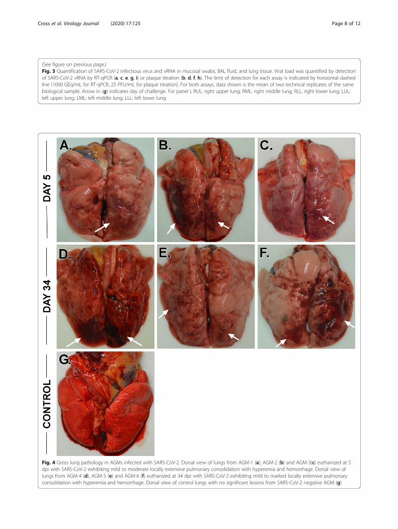
(See figure on previous page.)Fig. 3 Quantification of SARS-CoV-2 infectious virus and vRNA in mucosal swabs, BAL fluid, and lung tissue. Viral load was quantified by detectionof SARS-CoV-2 vRNA by RT-qPCR (a, c, e, g, i) or plaque titration (b, d, f, h). The limit of detection for each assay is indicated by horizontal dashedline (1000 GEq/mL for RT-qPCR, 25 PFU/mL for plaque titration). For both assays, data shown is the mean of two technical replicates of the samebiological sample. Arrow in (g) indicates day of challenge. For panel I, RUL: right upper lung; RML: right middle lung; RLL: right lower lung; LUL:left upper lung; LML: left middle lung; LLL: left lower lung
Fig. 4 Gross lung pathology in AGMs infected with SARS-CoV-2. Dorsal view of lungs from AGM-1 (a), AGM-2 (b) and AGM-3(c) euthanized at 5dpi with SARS-CoV-2 exhibiting mild to moderate locally extensive pulmonary consolidation with hyperemia and hemorrhage. Dorsal view oflungs from AGM-4 (d), AGM-5 (e) and AGM-6 (f) euthanized at 34 dpi with SARS-CoV-2 exhibiting mild to marked locally extensive pulmonaryconsolidation with hyperemia and hemorrhage. Dorsal view of control lungs with no significant lesions from SARS-CoV-2 negative AGM (g)
Cross et al. Virology Journal (2020) 17:125 Page 8 of 12

Fig. 5 (See legend on next page.)
Cross et al. Virology Journal (2020) 17:125 Page 9 of 12

approximated lesion severity for each lung lobe (SuppTable 2). All AGMs at 5 dpi also had segmentally flaccidand gas distention of small intestines. There were noother significant gross lesions.Histologically, all three AGMs euthanized at 5 dpi de-
veloped mild multifocal neutrophilic bronchointerstitialpneumonia (Fig. 5a-e, o). Histologic features includeacute inflammation centered within the airways of ter-minal bronchioles with occasional flooding of adjacentalveolar spaces with neutrophils, macrophages, fibrin,edema, hemorrhage, mucous and rarely multinucleatedgiant cells (5A, B). In lesser-affected regions alveolarseptate were expanded with mixed inflammatory cellsand alveolar spaces contain increased numbers of alveo-lar macrophages with scattered red blood cells. Ulcera-tive tracheobronchitis was also present in all threeAGMs and characterized by multifocal epithelial erosionassociated with underlying hemorrhage, fibrin accumula-tion and infiltrating acute inflammation. Polymerizedfibrin, highlighted by IHC, colocalized with acute inflam-mation within the bronchial lumen, alveolar spaces,alveolar walls and ulcerated regions of the trachea andbronchus (Fig. 5c). Fibrin was also present withinmedium and small caliber vessels but was not associatedwith an obvious adherent thrombus. Trichrome stain ofrepresentative lung sections identified modest collagendeposition within multifocal regions of alveolar septae(Fig. 5d). IHC for SARS-CoV-2 antigen was positive inall three AGMs associated with pulmonary lesions. Posi-tive IHC labeling was noted diffusely within the cyto-plasm of respiratory epithelium of the bronchus (Fig. 5o)and less in type I and type II pneumocytes (Fig. 5e).Histologically, all three AGMs euthanized at 34 dpi
developed moderate multifocal chronic interstitial pneu-monia Fig. 5f-j). Histologic features include expansion ofalveolar septae with macrophages, lymphocytes, and veryrarely neutrophils (Fig. 5f, g). Wispy, pale eosinophilic,acellular material also multifocally expanded the alveolarwalls and stained as immature collagen with trichromestaining (Fig. 5i). Polymerized fibrin was present withinmedium and small caliber vessels but was not associatedwith an obvious adherent thrombus (Fig. 5h). No immu-nolabeling for SARS-CoV-2 was noted with IHC in any
of the examined tissue sections from this 34 dpi cohort(Fig. 5j).
DiscussionWe previously reported the development of the AGM asa promising animal model of human COVID-19 [10].Other studies have subsequently reported similar find-ings [11, 12]. The focus of the current study was to as-sess a more natural route of human exposure,specifically an exposure mimicking an infection resultingfrom mucosal exposure to infectious droplets expelledfrom close quarter exposure to a sneeze, cough, or evenspeech in order to begin characterization of lung path-ology in the early convalescence phase of COVID-19.The disease resulting from the i.n. MAD challenge waslargely reflective of that observed with the combinationof the i.t. and i.n. routes except it appeared to be some-what milder in terms of length of any fever, less severesigns of pneumonia as evidenced by reduced alveolarflooding, and a lower prevalence of SARS-CoV-2 infec-tion [10]. However, the MAD-infected AGMs still devel-oped virus-induced pneumonia and viral shedding wasdetected into the early convalescence period. While itappears that inclusion of direct i.t. instillation of SARS-CoV-2 as an exposure route may result in a more severedisease in AGMs, it is also possible that animal to ani-mal variability may have contributed to the modest dif-ference between the studies. SARS-CoV-2 infection ofhumans results in a wide spectrum of disease rangingfrom asymptomatic to severe and fatal disease so it isnot unexpected that there could be variability amongAGMs as well. While the current study employed femaleAGMs because of animal availability at the time thework was initiated, gender did not affect the outcomewhen compared to similar studies [10–12].Coagulation dysfunction is a consistent observation in
human COVID-19 and has been associated with diseaseseverity [18–22]. Here, we performed a limited analysisof blood clotting times (PT and aPTT) and circulating fi-brinogen levels to begin to characterize the coagulopathyin SARS-CoV-2-infected AGMs. Transient increases inaPTT and in circulating fibrinogen levels were observedduring the acute phase of infection. Increases in PT and/
(See figure on previous page.)Fig. 5 Comparative pulmonary histologic lesions in AGMs infected with SARS-CoV-2. Representative tissues of AGM from 5 dpi (a-e & o) and 34dpi (f-j). SARS-CoV-2 naïve tissues from an AGM (k-n). H&E staining at low magnification (20x) (a, f, & k) and higher magnification (40x) (b, g, l,and f inset) of pulmonary alveolar septae and alveolar spaces. Moderate neutrophilic bronchiolitis and alveolitis and mild interstitial pneumoniawith congestion (a & b) Moderate lymphohistocytic interstitial pneumonia with congestion, mild alveolar wall fibrosis (f & g) and moderateperivascular lymphocytic cuffs (f inset). No significant lesions (k & l). IHC for anti-fibrin antigen (red) (c, h & m). Alveolar spaces are partially tocompletely flooded with fibrin (c) Minimal intravascular fibrin labeling (h) and no significant fibrin immunolabeling (m). Trichrome special stain forcollagen (blue) (d, i & n). Minimal to mild alveolar wall collagen deposition (d) moderate alveolar wall collagen deposition (i) and minimalcollagen staining of alveolar wall basement membranes (n). IHC labeling for anti-SARS-CoV2 antigen (red) (e, j & o). IHC positive type Ipneumoncytes (black arrows) and type II pneumoncytes (white arrow) localized with alveolar inflammation (e), No immonolabeling (j) and IHCpositive labeling of respiratory epithelium of the bronchus (o). Images captured at 20x (m, d, i, n, & j) and 40x (c, h, e, & o)
Cross et al. Virology Journal (2020) 17:125 Page 10 of 12
or aPTT have been linked to severe human COVID-19cases in some but not all studies [18–22]. However,nearly all severe COVID-19 cases have been associatedwith high levels of fibrinogen [20–22].Our findings regarding lung injury in the three AGMs
that were euthanized at 34 dpi during early convalescenceare consistent with the limited human COVID-19 studiesthat have been reported so far. For example, a recent studyof fifty-seven COVID-19 patients in China was completedduring the early convalescence phase, approximately 30days after discharge [23]. The study included 40 non-severe cases and 17 severe cases. Thirty-one patients(54.3%) had abnormal CT findings while abnormalitieswere detected in the pulmonary function tests in 43(75.4%) of the patients. In a second human study, 21 pa-tients recovering from COVID-19 (without severe respira-tory distress during the disease course), had lungabnormalities visible on chest CT at 10 days after initialonset of symptoms [24]. While other studies suggest thatsome of the abnormalities may be resolved over time [25,26] more research needs to be conducted in this area.Regarding histopathology, human data is particularly
sparse. One small study performed thoracoscopies withblebs resection and pleurectomies on performed on the16th and 23rd days from symptoms onset of two patients[27]. Despite well-known pulmonary damages inducedduring the acute phase of COVID-19, the late-phase grossand histological changes include nonspecific chronic rep-arative lesions, similarly to what we have described in theAGMs at 34 dpi. Grossly in the human study, there wasnon-specific diffuse pulmonary congestion, edema andhemorrhagic necrosis. Histologically, the main lesionswere focused on alveolar damage with mildly thickened al-veolar interstitial tissues with fibrosis and mononuclearcellular infiltration (lymphocytes, plasma cells and multi-nucleate giant cells). Intravascular hemorrhagic throm-bosis was also noted in these specimens.In summary, we have expanded on our previous devel-
opment of the combined i.n. and i.t. inoculation modelof SARS-CoV-2 in AGMs. Importantly, while AGMschallenged with SARS-CoV-2 via the LMA MAD exhib-ited apparently milder clinical illness and disease, hall-mark features from our previous study were stillapparent, notably the development of viral pneumoniaduring the acute phase. The AGM COVID-19 modelshould be useful in future studies to assess disease anddevelop interventions that improve recovery.
Supplementary informationSupplementary information accompanies this paper at https://doi.org/10.1186/s12985-020-01396-w.
Additional file 1: Figure S1: Longitudinal temperature monitoring ofSARS-CoV-2 infected AGMs. Temperature was longitudinally monitored
for all animals via surgically-implanted temperature loggers (see Materialsand methods). Data shown for all animals begins 1 day prior to infection(− 1) and terminates in the morning of day 5 post-infection, when AGM-1, AGM-2, and AGM-3 were euthanized. Periods of elevated temperatureare indicated in red. Arrows denote time of challenge.
Additional file 2: Table S1: Clinical description and outcome of Africangreen monkeys following SARS-CoV-2 challenge. Days after SARS-CoV-2challenge are in parentheses. All reported findings are in comparison tobaseline (d0) values. Decreased appetite is defined as some food but notall food consumed from the previous day. Anorexia is defined as no foodconsumed from the previous day. Lymphocytopenia, monocytopenia,erythrocytopenia, thrombocytopenia, neutropenia, eosinopenia, andbasopenia are defined by a ≥ 35% drop in numbers of lymphocytes,monocytes, erythrocytes, platelets, neutrophils, eosinophils, and basophils,respectively. Lymphocytosis, monocytosis, neutrophilia, eosinophilia, andbasophilia are defined by a 100% or greater increase in numbers of lym-phocytes, monocytes, neutrophils, eosinophils, or basophils, respectively.Hyperglycemia is defined as a 100% or greater increase in levels of glu-cose. Hypoglycemia is defined by a ≥ 25% decrease in levels of glucose.Hypoalbuminemia is defined by a ≥ 25% decrease in levels of albumin.Hypoproteinemia is defined by a ≥ 25% decrease in levels of total protein.Hypoamylasemia is defined by a ≥ 25% decrease in levels of serum amyl-ase. Hypocalcemia is defined by a≥ 25% decrease in levels of serum cal-cium. Hypercapnia was defined as having a partial CO2 > 4 mmHg overd0 baseline values. (ALT) alanine aminotransferase, (AST) aspartate amino-transferase, (ALP) alkaline phosphatase, (CRE) Creatinine, (CRP) C-reactiveprotein, (Hct) hematocrit, (Hgb) hemoglobin.
Additional file 3: Table S2: Gross lung lesion severity scores in AGMsinfected with SARS-CoV-2
AbbreviationsaPTT: Activated partial thromboplastin time; AGM: African green monkey;BAL: Bronchoalveolar lavage; COVID-19: Coronavirus Disease 2019;IHC: Immunohistochemistry; i.n.: Intranasal; i.t.: Intratracheal; MAD: MucosalAtomization Device; PFU: Plaque forming unit; PT: Prothrombin time; SARS-CoV-2: Severe acute respiratory syndrome coronavirus 2
AcknowledgmentsThe authors would like to thank the UTMB Animal Resource Center forveterinary support for surgery to implant temperature data loggers andhusbandry support of laboratory animals and Dr. Kevin Melody for assistancewith animal studies. The virus used in this publication was kindly providedby the European Virus Archive goes Global (EVAg) project that has receivedfunding from the European Union’s Horizon 2020 research and innovationprogram under grant agreement No 653316.
Authors’ contributionsRWC and TWG conceived and designed the study. DJD, JBG, and TWGperformed the SARS-CoV-2 challenge experiments. RWC, DJD, CW, JBG, andTWG performed animal procedures and clinical observations. KNA and VBperformed the clinical pathology assays. VB performed the SARS-CoV-2infectivity assays. KNA optimized and performed the PCR. NSD optimizedand performed the immunohistochemistry. CW performed ELISAs. KAFperformed necropsies and analysis of the gross pathology, histopathology,and immunohistochemistry. All authors analyzed the clinical pathology,virology, and immunology data. RWC, ANP, KAF, and TWG, wrote the paper.The authors had access to all of the data and approved the final version ofthe manuscript.
FundingThis study was supported by funds from the Department of Microbiologyand Immunology, University of Texas Medical Branch at Galveston,Galveston, TX to TWG. Operations support of the Galveston NationalLaboratory was supported by NIAID/NIH grant UC7AI094660.
Availability of data and materialsThe data supporting the conclusions of this article are included within thearticle.
Cross et al. Virology Journal (2020) 17:125 Page 11 of 12

Ethics approval and consent to participateAll animal studies were approved by the University of Texas Medical Branch(UTMB) Institutional Animal Care and Use Committee and adhere to the NIHGuide for the Care and Use of Laboratory Animals.
Consent for publicationNot applicable.
Competing interestsThe authors declare no competing interests.
Received: 27 July 2020 Accepted: 11 August 2020
References1. World Health Organization. DRAFT landscape of COVID-19 candidate
vaccines – 13 August 2020. Available from: https://www.who.int/publications/m/item/draft-landscape-of-covid-19-candidate-vaccines.
2. Milken Institute. COVID-19 treatment and vaccine tracker; 2020. Availablefrom: https://covid-19tracker.milkeninstitute.org/.
3. Chan JF, Zhang AJ, Yuan S, Poon VK, Chan CC, Lee AC, et al. Simulation ofthe clinical and pathological manifestations of coronavirus disease 2019(COVID-19) in golden Syrian hamster model: implications for diseasepathogenesis and transmissibility. Clin Infect Dis. 2020:ciaa325. https://doi.org/10.1093/cid/ciaa325.
4. Sia SF, Yan LM, Chin AWH, Fung K, Choy KT, Wong AYL, et al. Pathogenesisand transmission of SARS-CoV-2 in golden hamsters. Nature. 2020;583:834–8.
5. Richard M, Kok A, de Meulder D, Bestebroer TM, Lamers MM, Okba NMA,et al. SARS-CoV-2 is transmitted via contact and via the air between ferrets.Nat Commun. 2020;11:3496.
6. Yu P, Qi F, Xu Y, Li F, Liu P, Liu J, et al. Age-related rhesus macaque modelsof COVID-19. Animal Model Exp Med. 2020;3:93–7.
7. Munster VJ, Feldmann F, Williamson BN, van Doremalen N, Pérez-Pérez L,Schulz J, et al. Respiratory disease in rhesus macaques inoculated withSARS-CoV-2. Nature. 2020. https://doi.org/10.1038/s41586-020-2324-7.
8. Rockx B, Kuiken T, Herfst S, Bestebroer T, Lamers MM, Oude Munnink BB,et al. Comparative pathogenesis of COVID-19, MERS, and SARS in anonhuman primate model. Science. 2020;368:1012–5.
9. Lu S, Zhao Y, Yu W, Yang Y, Gao J, Wang J, et al. Comparison of SARS-CoV-2infections among 3 species of non-human primates. bioRxiv. 2020:2020.04.08.031807v2. https://doi.org/10.1101/2020.04.08.031807.
10. Woolsey C, Borisevich V, Prasad AN, Agans KN, Deer DJ, Dobias NS, et al.Establishment of an African green monkey model for COVID-19. bioRxiv.2020:2020.05.17.100289. https://doi.org/10.1101/2020.05.17.100289.
11. Blair R, Vaccari M, Doyle-Meyers LA, Roy CJ, Russell-Lodrigue K, Fahlberg M,et al. ARDS and cytokine storm in SARS-CoV-2 infected Caribbean Vervets.bioRxiv. 2020:2020.06.18.157933. https://doi.org/10.1101/2020.06.18.157933.
12. Hartman AL, Nambulli S, McMillen CM, White AG, Tilston-Lunel NL, Albe JR,et al. SARS-CoV-2 infection of African green monkeys results in mildrespiratory disease discernible by PET/CT imaging and prolonged sheddingof infectious virus from both respiratory and gastrointestinal tracts. bioRxiv.2020:2020.06.20.137687. https://doi.org/10.1101/2020.06.20.137687.
13. Mire CE, Satterfield BA, Geisbert JB, Agans KN, Borisevich V, Yan L, et al.Pathogenic differences between Nipah virus Bangladesh and Malaysiastrains in primates: implications for antibody therapy. Sci Rep. 2016;6:30916.
14. Geisbert JB, Borisevich V, Prasad AN, Agans KN, Foster SL, Deer DJ, et al. Anintranasal exposure model of lethal Nipah virus infection in African greenmonkeys. J Infect Dis. 2020;221(Supplement_4):S414–8.
15. Xie X, Li Y, Sun H, Liu L. Exhaled droplets due to talking and coughing. J RSoc Interface. 2009;6(Suppl 6):S703–14.
16. Capobianchi MR, Rueca M, Messina F, Giombini E, Carletti F, Colavita F, et al.Molecular characterization of SARS-CoV-2 from the first case of COVID-19 inItaly. Clin Microbiol Infect. 2020;26:954–6.
17. Centers for Disease Control and Prevention. Research use only 2019-novelcoronavirus (2019-nCoV) real-time RT-PCR primers and probes; 2020.Available from: https://www.cdc.gov/coronavirus/2019-ncov/lab/rt-pcr-panel-primer-probes.html.
18. Chen L, Yu J, He W, Chen L, Yuan G, Dong F, et al. Risk factors for death in1859 subjects with COVID-19. Leukemia. 2020;34:2173–83.
19. Bonetti G, Manelli F, Patroni A, Bettinardi A, Borrelli G, Fiordalisi G, et al.Laboratory predictors of death from coronavirus disease 2019 (COVID-19) inthe area of Valcamonica, Italy. Clin Chem Lab Med. 2020;58:1100–5.
20. Di Minno MND, Calcaterra I, Lupoli R, Storino A, Spedicato GA, ManiscalcoM, et al. Hemostatic changes in patients with COVID-19: a meta-analysiswith meta-regressions. J Clin Med. 2020;9:E2244.
21. Lin J, Yan H, Chen H, He C, Lin C, He H, et al. COVID-19 and coagulationdysfunction in adults: a systematic review and meta-analysis. J Med Virol.2020;1–11. https://doi.org/10.1002/jmv.26346.
22. Zhu J, Pang J, Ji P, Zhong Z, Li H, Li B, et al. Coagulation dysfunction isassociated with severity of COVID-19: a meta-analysis. J Med Virol. 2020.https://doi.org/10.1002/jmv.26336.
23. Huang Y, Tan C, Wu J, Chen M, Wang Z, Luo L, et al. Impact of coronavirusdisease 2019 on pulmonary function in early convalescence phase. RespirRes. 2020;21:163.
24. Pan F, Ye T, Sun P, Gui S, Liang B, Li L, et al. Time course of lung changes atchest CT during recovery from coronavirus disease 2019 (COVID-19).Radiology. 2020;295:715–21.
25. Liu C, Ye L, Xia R, Zheng X, Yuan C, Wang Z, et al. Chest CT and clinicalfollow-up of discharged patients with COVID-19 in Wenzhou City, Zhejiang,China. Ann Am Thorac Soc. 2020. https://doi.org/10.1513/AnnalsATS.202004-324OC.
26. Wang Y, Dong C, Hu Y, Li C, Ren Q, Zhang X, et al. Temporal changes of CTfindings in 90 patients with COVID-19 pneumonia: a longitudinal study.Radiology. 2020;296:E55–64.
27. Aiolfi A, Bruni B, Biraghi T, Montisci A, Miceli A, Baronio B, et al. Latehistological findings in symptomatic COVID-19 patients: a case report.Medicine (Baltimore). 2020;99:e21046.
Publisher’s NoteSpringer Nature remains neutral with regard to jurisdictional claims inpublished maps and institutional affiliations.
Cross et al. Virology Journal (2020) 17:125 Page 12 of 12