INTRAHEPATIC CHOLESTASIS OF PREGNANCY -...
58
Departments of Obstetrics and Gynecology, and Medical Genetics, Helsinki University Central Hospital, University of Helsinki, Finland INTRAHEPATIC CHOLESTASIS OF PREGNANCY Genetic background, epidemiology and hepatobiliary consequences Anne Ropponen Academic Dissertation To be presented by permission of the Medical Faculty of the University of Helsinki for public discussion in the Auditorium of the Department of Obstetrics and Gynecology, Helsinki University Central Hospital, Haartmanninkatu 2, Helsinki, on May 19 th , 2006, at 12 o’clock noon.
Transcript of INTRAHEPATIC CHOLESTASIS OF PREGNANCY -...

Departments of Obstetrics and Gynecology, and Medical Genetics,Helsinki University Central Hospital,
University of Helsinki, Finland
INTRAHEPATIC CHOLESTASIS OF PREGNANCY
Genetic background, epidemiology and
hepatobiliary consequences
Anne Ropponen
Academic Dissertation
To be presented by permission of the Medical Faculty of the Universityof Helsinki for public discussion in the Auditorium of the Department of
Obstetrics and Gynecology, Helsinki University Central Hospital,Haartmanninkatu 2, Helsinki, on May 19th, 2006,
at 12 o’clock noon.

Supervised by Kristiina Aittomäki, M.D., Ph.D. Department of Medical Genetics Helsinki University Central Hospital
Olavi Ylikorkala, Professor, M.D. Ph.D. Department of Obstetrics and Gynecology Helsinki University Central Hospital
Reviewed by Katriina Aalto-Setälä, M.D., Ph.D. Department of Medicine Tampere University Hospital
Päivi Polo-Kantola, M.D., Ph.D. Department of Obstetrics and Gynecology Turku University Central Hospital
Offi cial opponent Juha Tapanainen, Professor, M.D., Ph.D. Department of Obstetrics and Gynecology Oulu University Hospital
ISBN 952-92-0010-2 (paperback)
ISBN 952-10-2922-6 (PDF)
http://ethesis.helsinki.fi
Helsinki University Printing House
Helsinki 2006

CONTENTS
LIST OF ORIGINAL PUBLICATIONS ........................................................ 5
ABBREVIATIONS ......................................................................................... 6
ABSTRACT .................................................................................................... 8
INTRODUCTION ......................................................................................... 9
REVIEW OF THE LITERATURE ................................................................. 10
1. Physiology of bile acids ............................................................................. 101.2. Enterohepatic circulation .................................................................. 111.3. Transporter mechanisms ................................................................... 13
1.3.1. Basolateral membranes of hepatocytes .................................. 131.3.2. Canalicular membranes of hepatocytes ................................. 151.3.3. Intestinal transport ................................................................. 151.3.4. Regulation ............................................................................... 16
2. Intrahepatic cholestasis ............................................................................. 162.1. Intrahepatic cholestatic diseases ....................................................... 172.2. Familial cholestatic diseases .............................................................. 17
3. Liver during pregnancy ............................................................................. 213.1. Intrahepatic cholestasis of pregnancy .............................................. 21
3.1.1. Epidemiology .......................................................................... 223.1.2. Pathogenesis ............................................................................ 223.1.3. Genetics ................................................................................... 223.1.4. Clinical picture and treatment ............................................... 233.1.5. Associated hepatobiliary disorders ......................................... 24
3.2. Other liver diseases and pregnancy .................................................. 25
4. Postmenopausal hormone therapy and liver proteins ............................ 264.1. Sex hormone-binding globulin ........................................................ 264.2. C-reactive protein .............................................................................. 27

AIMS OF THE STUDY .................................................................................. 29
SUBJECTS AND METHODS ....................................................................... 30
1. Subjects ...................................................................................................... 302. Methods ..................................................................................................... 322.1.1. Linkage analysis ................................................................................... 322.1.2. Polymerase chain reaction .................................................................. 332.2. Hormone therapy protocol .................................................................... 332.3. Assays ...................................................................................................... 343. Statistical analyses ..................................................................................... 34
RESULTS ........................................................................................................ 36
1. Genetics ..................................................................................................... 362. Incidence and risk factors ......................................................................... 373. The effect of hormone therapy on sex hormone-binding globulin ....... 384. The effect of hormone therapy on C-reactive protein ............................ 405. Associated liver and biliary diseases ......................................................... 40
DISCUSSION ................................................................................................ 41
CONCLUSIONS ............................................................................................ 46
ACKNOWLEDGEMENTS ............................................................................ 47
REFERENCES ................................................................................................ 49
ORIGINAL PUBLICATIONS ....................................................................... 59

5
LIST OF ORIGINAL PUBLICATIONS
This thesis is based on the following original publications, which are referred to in the text by their Roman numerals:
I *Savander M, *Ropponen A, *Avela K, Weerasekera N, Cormand B, Hir-vioja ML, Riikonen S, Ylikorkala O, Lehesjoki AE, Williamson C, Ait-tomäki K. Genetic evidence of heterogeneity in intrahepatic cholestasis of pregnancy. Gut 2003;52:1025–1029.
II Ropponen A, Sund R, Ylikorkala O, Aittomäki K. A nation-wide study on intrahepatic cholestasis of pregnancy identifi es new risk factors. (sub-mitted)
III Ropponen A, Aittomäki K, Vihma V, Tikkanen MJ, Ylikorkala O. Effects of oral and transdermal estradiol administration on levels of sex hor-mone-binding globulin in postmenopausal women with and without a history of intrahepatic cholestasis of pregnancy. J Clin Endocrinol Me-tab 2005;90:3431–3434.
IV Ropponen A, Aittomäki K, Tikkanen MJ, Ylikorkala O. Levels of serum C-reactive protein during oral and transdermal estradiol in postmeno-pausal women with and without a history of intrahepatic cholestasis of pregnancy. J Clin Endocrinol Metab 2005;90:142–146
V Ropponen A, Sund R, Riikonen S, Ylikorkala O, Aittomäki K. Intrahepat-ic cholestasis of pregnancy as an indicator of liver and biliary diseases: a population based study. Hepatology 2006;43:723–728
* Contributed equally to this work.

6
ABBREVIATIONS
ALAT alanine aminotransferaseASAT aspartate aminotransferaseATP adenosine triphosphateBRIC benign recurrent intrahepatic cholestasisBSEP bile salt export pumpCA cholic acid (3α7α12α-trihydroxy-5β-cholanoic acid)CDCA chenodeoksycholic acid (3α7α-dihydroxy-5β-cholanoic acid)CRP C-reactive proteinDCA deoxycholic acid (3α12α-dihydroxy-5β-cholanoic acid)E1 estroneE2 estradiolEPT estrogen progestin therapyET estrogen therapyFIC1 a P-type ATPase, aminophospholipid transporterFXR farnesoid receptor XGT gammaglutamyl transferaseHELLP hemolysis, elevated liver enzymes, low platelet countHT hormone therapyICD International Classifi cation of DiseasesICP intrahepatic cholestasis of pregnancyIGF-1 insulin-like growth factor 1IL-6 interleukin-6, cytokineIVF in-vitro-fertilizationLCA lithocholic acid (3α-monohydroxy-5β-cholanoic acid)LOD logarithm of odds MDR multidrug resistance proteinMPA medroxyprogesterone acetateMRP multidrug resistance associated proteinNETA norethisterone acetateNTCP sodium-taurocholate cotransporterOAT/OCT transporters for the small organic compoundsOATP sodium-independent organic anion transporting polypeptidePBC primary biliary cirrhosisPCR polymerase chain reactionPFIC progressive familial intrahepatic cholestasisPSC primary sclerosing cholangitisSHBG sex hormone-binding globulinSHP small heterodimeric partner
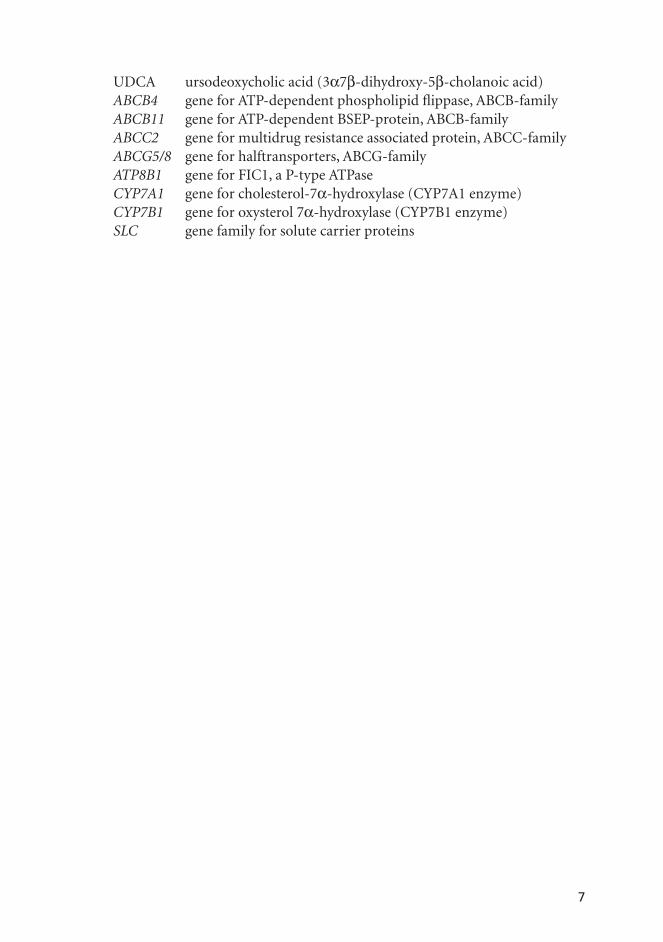
7
UDCA ursodeoxycholic acid (3α7β-dihydroxy-5β-cholanoic acid) ABCB4 gene for ATP-dependent phospholipid fl ippase, ABCB-familyABCB11 gene for ATP-dependent BSEP-protein, ABCB-familyABCC2 gene for multidrug resistance associated protein, ABCC-familyABCG5/8 gene for halftransporters, ABCG-familyATP8B1 gene for FIC1, a P-type ATPaseCYP7A1 gene for cholesterol-7α-hydroxylase (CYP7A1 enzyme)CYP7B1 gene for oxysterol 7α-hydroxylase (CYP7B1 enzyme)SLC gene family for solute carrier proteins

8
ABSTRACT
Intrahepatic cholestasis of pregnancy (ICP) is the most common cholestatic liver disease during pregnancy. The reported incidence varies from 0.4 to 15% of full-term pregnancies. The etiology is heterogeneous but familial clustering is known to occur. Here we have studied the genetic background, epidemiol-ogy, and long-term hepatobiliary consequences of ICP.
In a register-based nation-wide study (n=1 080 310) the incidence of ICP was 0.94% during 1987–2004. A slightly higher incidence, 1.3%, was found in a hospital-based series (n=5304) among women attending the University Hos-pital of Helsinki in 1992–1993. Of these 16% (11/69) were familial and showed a higher (92%) recurrence rate than the sporadic (40%) cases. In the register-based epidemiological study, advanced maternal age and, to a lesser degree, parity were identifi ed as new risk factors for ICP. The risk was 3-fold higher in women >39 years of age compared to women <30 years. Multiple pregnancy also associated with an elevated risk. In a genetic study we found no associa-tion of ICP with the genes regulating bile salt transport (ABCB4, ABCB11 and ATP8B1).
The livers of postmenopausal women with a history of ICP tolerated well the short-term exposure to oral and transdermal estradiol, although the doses used were higher than those in routine clinical use. The response of serum levels of sex hormone-binding globulin (SHBG) to oral estradiol was slightly reduced in the ICP group. Transdermal estradiol had no effect on C-reactive protein (CRP) or SHBG. A number of liver and biliary diseases were found to be associated with ICP. Women with a history of ICP showed elevated risks for non-alcoholic liver cirrhosis (8.2 CI 1.9–36), cholelithiasis and cholecystitis (3.7 CI 3.2–4.2), hepatitis C (3.5 CI 1.6–7.6) and non-alcoholic pancreatitis (3.2 CI 1.7–5.7).
In conclusion, ICP complicates around 1% of all full-term pregnancies in Finland and its incidence has remained unchanged since 1987. It is familial in 16% of cases with a higher recurrence rate. Although the cause remains unknown, several risk factors, namely advanced maternal age, parity and mul-tiple pregnancies, can be identifi ed. Both oral and transdermal regimens of postmenopausal hormone therapy (HT) are safe for women with a history of ICP when liver function is considered. Some ICP patients are at risk of other liver and biliary diseases and, contrary to what has been thought, a follow-up is warranted.

9
INTRODUCTION
Intrahepatic cholestasis of pregnancy (ICP) is the most common of familial cholestatic conditions. It usually manifests in the third trimester of pregnancy as skin itching and as elevation of the serum levels of bile acids and liver en-zymes (Reyes 1997, Lammert et al 2000). The levels of liver transaminases are usually 2–10-fold higher than the normal range. Generally, ICP is considered to be harmless to the mother, but preterm birth (12–44%) (Fisk 1988, Rioseco et al 1994, Glantz et al 2004), fetal distress (10–44%) (Laatikainen et al 1984, Fisk 1988, Alsulyman et al 1996, Glantz et al 2004), and intrauterine fetal death (1–3%) (Laatikainen et al 1984, Fisk 1988, Alsulyman et al 1996) may ensue. Biochemical cholestasis resolves within a couple of days after delivery (Elferink 2003), but ICP may recur in 40–60% of subsequent pregnancies (Reyes 1997, Germain et al 2002). There has been a wide variation in the reported incidence of ICP in different countries (0.4–15%) (Abedin et al 1999, Locatelli et al 1999, Germain et al 2002); in Finland and Sweden the incidence is 0.54–1.5% (Laa-tikainen et al 1984, Berg et al 1986, Heinonen et al 1999).
ICP is thought to be the result of insuffi cient liver capacity to metabolize high amounts of placenta-derived sex steroids during pregnancy (Reyes 1997, Davidsson 1998). The familial occurrence of ICP in some cases suggests he-reditary susceptibility (Holzbach et al 1983, Reyes et al 1993, Hirvioja et al 1993, Jacquemin 2001) and the increasing understanding of the genetic back-ground of cholestatic diseases in general has aroused interest in the search for genes and mutations predisposing also to ICP. Moreover, the increased rate of cholelithiasis in these women may imply that ICP is not specifi c to pregnancy (Reyes 1997). This hypothesis is further supported by data showing that the high amounts of synthetic estrogens, eg. previously used oral contraceptives, can trigger symptoms and signs of cholestasis in women with a history of ICP (Kreek et al 1967, Adlercreutz et al 1964, Drill 1974, Reyes et al 1981).
The present studies were designed to examine the genetic background, epi-demiology and hepatobiliary consequences of ICP.

10
REVIEW OF THE LITERATURE
Intrahepatic cholestasis of pregnancy is not well defi ned in the medical litera-ture and several names have been used for the condition. Icterus and pruri-tus gravidarum, recurrent jaundice of pregnancy, hepatosis gravidarum, and cholestasis of gravidarum have all been used as synonyms to the currently used intrahepatic cholestasis of pregnancy (ICP) (Reyes 1997) and obstetric cholestasis (Williamson et al 2004). Itching of healthy skin with elevated levels of liver transaminases and/or bile acids are the diagnostic criteria for ICP. In Finland, Sweden and Chile this disease entity has been well-known for a long time and in Finland at least three academic theses on ICP have been published previously (Ikonen 1964, Ylöstalo 1970, Heikkinen 1982).
1. Physiology of bile acids
Bile acids facilitate excretion, absorption, and transport of fats and sterols in the intestine and liver (Trauner et al 2003). Primary bile acids, cholic acid (CA) and chenodeoxycholic acid (CDCA), are synthesized from cholesterol by two pathways in the hepatocytes (Fuchs 2003). The two molecules are structurally very similar, differing by only one hydroxyl group at position 7 (Figure 1). For-mation of CA through the classic pathway accounts for 90% of the total bile acid synthesis, which is up to 0.5g daily. The synthesis is regulated by the mi-crosomal cholesterol-7α-hydroxylase (CYP7A1), which in turn is regulated by the availability of cholesterol and the concentrations of bile acids themselves. (Chiang 2003).
Figure 1. Structure of primarybile acids
Cholic acid Chenodeoxycholic acid
3a7a12a-trihydroxy-5ß-cholanoic acid 3a7a-dihydroxy-5ß-cholanoic acid

11
An alternative pathway leads to the formation of CDCA, where the 7α-hy-droxylation is preceded by the formation of oxysterols. These are metabolized by oxysterol 7α-hydroxylase (CYP7B1) enzyme (Chiang 2003).
The primary bile acids are conjugated in the hepatocytes with glycine or taurine to form bile salts, which are actively excreted to the bile through ATP-dependent membrane transporters (Fuchs 2003). The bile salts are then stored in the gallbladder as mixed micelles with phosphatidylcholine and cholesterol.
1.2. Enterohepatic circulation
The enterohepatic circulation ensures effi cient use of synthesized bile acids with minimal daily loss (Figure 2). The pool of human bile salts, 2–4g, circulates 3–15 times per day resulting in 20–40g of daily excretion. Only 3–5% of bile salts (0.5g)
95%
5% (0.5g/day)
5%(0.5g/day)
Figure 2. Enterohepatic circulation. Primary bile acids are synthesized in the liver and con-jugated with glycine or taurine. Bile salts are excreted into bile and stored in the gallblad-der. From the intestine bile acids are transported back to the liver via portal circulation.

12
are lost daily in faeces and are replaced by de novo synthesis from cholesterol. (Trauner et al 2003). The fi rst step of the enterohepatic circulation is the excretion of bile from the gallbladder to the intestine. This secretion into the duodenum occurs after each meal due to vagal stimulation and release of cholecystokinin from the mucosa of the duodenum, which causes contraction of the gallbladder. (Chiang 2003). In the intestine CA and CDCA are converted to secondary bileacids, deoxycholic acid (DCA, 3α12α-dihydroxy-5β-cholanoic acid) and litho-cholic acid (LCA, 3α-monohydroxy-5β-cholanoic acid), by bacteria (Chiang 2003) (Figure 3). Ursodeoxycholic acid (UDCA, 3α7β-dihydroxy-5β-chola-noic acid) is also formed by intestinal bacteria. It differs from CDCA by one hydroxyl group, which is in the β position at C-7. Only trace amounts of this bile acid are present in humans, whereas larger amounts are found at least in bears. (Trauner et al 2003, van Mil et al 2005).
Figure 3. Primary and secondary bile acids are synthesized from cholesterol.
CholesterolCholesterol
Cholic acidCholic acidChenodeoxycholic
acidChenodeoxycholic
acid
Deoxycholic acidDeoxycholic acid Litocholic acidLitocholic acid
Conjugated bile acids are absorbed from the intestine by at least two carrier-mediated processes (van Mil et al 2005). Bile acids, returned to the liver in portal venous blood, are actively taken up by the liver and, once again, secreted into the bile thus completing the enterohepatic circulation. The large major-ity of bile salts (95%) are reabsorbed from the intestine (Pauli-Magnus et al 2005) (Figure 2 and Table 1). Equal amounts of conjugated CA and CDCA (80%), and conjugated DCA (10%) form the main bile acid pool and only traces of conjugated LCA and UDCA are present in the bile. Bile salts that es-cape from the enterohepatic circulation to the systemic circulation are fi ltered

13
through the glomeruli in the kidneys and either reabsorbed from the tubules or excreted into urine. Normally the main excretion is through faeces, but in cholestatic conditions the amount excreted in urine increases (van Mil et al 2005). In blood bile salts are bound predominantly to albumin and high-den-sity lipoproteins (60–85%). The ultimate goal of enterohepatic circulation is to ensure that bile salts are rapidly available in suffi cient amounts when needed for digestion.
1.3. Transporter mechanisms
Hepatobiliary transport systems mediate hepatic uptake and biliary excretion of bile salts. Reduced or totally absent expression of these transporters has an important role in cholestasis (Chiang 2003) and for full comprehension of cholestatic disorders one must understand their function.
1.3.1. Transporters on basolateral membranes of hepatocytes
The basolateral membrane of the hepatocyte plays a key role in transporting bile acids and salts from venous blood to the liver (Figure 4). This is achieved by two major pathways. Firstly, the sodium-dependent sodium-taurocholate cotransporting polypeptide NTCP (a member of the solute carrier protein, the SLC-gene family, SLC10A1) accounts for more than 80% of conjugated, but for less than 50% of unconjugated bile salt uptake (Pauli-Magnus et al 2005). Secondly, the sodium-independent organic anion transporting polypeptides (OATPs) (members of the SLC-gene family) form another transporter system, which also transports bilirubin, bromsulphthalein, steroid sulphates and glu-
Table 1. Bile acids in the enterohepatic circulation.
Amounts produced and circulated
Total pool in body 2–4 g
Daily synthesis 0.5 g
Secretion 4–6 g/meal; 12–40 g/day
Recycling frequency 3–15 times/day
Concentrations
Biliary ductuli 20–50 × 10³µmol/l
Gallbladder up to 300 × 10³µmol/l
Venous blood Portal vein Systemic circulation
20–50µmol/l3–5 µmol/l

14
Basolateral transporters Canalicular transporters
Bile
can
alic
ulus
Cap
illar
yci
rcul
atio
nNTCP
OATPs
OCT1
OAT2
MDR3
BSEP
MRP2
ABCG5/8
FIC1
MRPs1 3 4 5 6
Nucleus
Nucleus
Hepatocyte
FXR
CYP7A1CYP7B1NTCP
Supression oftranscription
Activation oftranscription
BSEPMDR3
baba
baba ba
ba
Bileacids
FXRba
FXRba
Bileacids
FXR
XSHP
A
B
Figure 4. A) Bile acid transporter proteins. Sodium-taurocholate transporting protein (NTCP), or-ganic anion transporting polypeptides (OATPs) and transporter for small hydrophilic or-ganic compounds (OAT/OCT) transport bile acids across the basolateral membrane into the hepatocyte. Multidrug resistance protein 3 (MDR3), bile salt export pump (BSEP), multidrug associated protein 2 (MRP2), a P-type ATPase FIC1, and halftransporters ABCG5/8 transport across canalicular membrane to the bile canaliculi. MRPs are transporters of various organic anions, such as bile salts.
B) Schematic presentation of the regulation of intracellular bile acid concentration by nuclear receptors. When bile acid (ba) concentration rises in the hepatocyte, Farnesoid receptor X (FXR) binds to bile acids and inhibits via small heterodimeric partner (SHP) the transcription of CYP7A1 and CYP7B1 enzymes (inhibits synthesis of bile acids) and NTCP transporter (decreases uptake), but activates the transcription of BSEP and MDR3 (increases excretion). As the result, bile acid concentration decreases in the hepatocyte.

15
curonides, neutral steroids, and conjugated and unconjugated bile salts (Pauli-Magnus et al 2005). Bile acids and salts are highly toxic to cells (van Mill et al 2005) and they are bound to intracellular proteins in the hepatocytes (Ferenci et al 2002), although it is not known exactly how they are transported there. Anyway, the basolateral transporters regulate the uptake of bile salts from ve-nous blood to hepatocytes.
1.3.2. Transporters on canalicular membranes of hepatocytes
The canalicular membrane of the hepatocyte faces the bile ductulus (Figure 4). The transporter proteins on the canalicular membrane belong to the family of ATP-dependent P-glycoproteins (ATP-Binding-Cassette, ABC-superfamily) (Lee et al 2000). These include several subgroups of transporter proteins, such as multidrug-resistance proteins (ABCB-gene family), multidrug-resistance associated proteins (ABCC-gene family), and ABC-halftransporters (ABCG-gene family) (Pauli-Magnus et al 2005).
The bile salt export pump (BSEP, ABCB11) is primarily responsible for transporting the conjugated bile salts from the hepatocyte to the bile canaliculi. The multidrug resistance protein 3 (MDR3, ABCB4) functions as a phospholi-pid fl ippase translocating phospholipids from the inner to the outer leaf of the canalicular membrane (Pauli-Magnus et al 2005) (Figure 4). A P-type ATPase, the FIC1-protein (ATP8B1), is involved in bile acid transport. Its function is not well understood, but it is an ATP-dependent aminophospholipid trans-porter and thought to play a role in the regulation of the bile acid pool. (Pauli-Magnus et al 2005). The multidrug resistance-associated protein 2 (MRP2, ABCC2) is the only member of this MRP subgroup which is located on the canalicular membrane while the other MRPs are on the basolateral membrane (Pauli-Magnus et al 2005). It transports bile salt conjugates, bilirubin diglu-curonide, and glutathione conjugates. Halftransporters ABCG5 and ABCG8 are also expressed in the canalicular membrane and participate in cholesterol and plant sterol excretion (Yu et al 2002). Taken as a whole, the basolateral and canalicular membranes contain many transporter proteins which regulate bile acid and cholesterol homeostasis in man and which, if defective or absent, lead to cholestasis.
1.3.3. Intestinal transporters
Bile acids and salts are actively transported in the intestine. In the terminal ileum, the apical membrane of the enterocytes contains sodium-dependent bile salt transporters (SLC10A2). In the enterocytes, bile acid-binding protein transports bile acids to the basolateral membrane (van Mil et al 2005).The mechanism of bile salt transport to the venous blood and portal circulation is unknown (Pauli-Magnus et al 2005). A multidrug resistance-associated pro-

16
tein, MRP3, is expressed in the basolateral membrane of enterocytes (Pauli-Magnus et al 2005), and is a candidate transporter for this.
1.3.4. Regulation
The intracellular concentration of bile acids in the hepatocytes is tightly regulated as bile acids are highly toxic due to their detergent properties. The synthesis and excretion of bile acids and salts are controlled by their concentration in blood and hepatocytes. In response to high bile acid concentration, both the synthesis and absorption in the hepatocyte decrease while the secretion into the bile increases (van Mill et al 2005). Regulation of these processes is mediated by transcription factors called nuclear hormone receptors, which can activate or inactivate the transcription of relevant genes (Figure 4). Farnesoid X receptor (FXR), when bound to bile acids, activates the transcription of short heterodimeric partner (SHP), which leads to inactivation of the rate limiting step in bile acid synthesis (CYP7A1 and CYP7B1 enzymes) (Figure 4). At the same time, bile salt absorp-tion decreases from the capillary venous blood, because SHP also inhibits the expression of the NTCP-pump in the basolateral membrane. (Pauli-Magnus et al 2005, van Mil et al 2005). In contrast, the transcription of BSEP and MDR3 is increased by FXR, enhancing the excretion of bile acids. Thus, the concentration of bile acids in the hepatocyte is decreased by all these mechanisms. On the oth-er hand, liver X Receptor reacts to high cholesterol levels in the hepatocyte andCYP7A1 enzyme is induced to convert cholesterol to bile acids. (van Mil et al 2005). Summing up, the homeostasis of bile acids and cholesterol is effi ciently regulated in the hepatocytes, but the complex regulatory mechanisms are still incompletely understood.
2. Intrahepatic cholestasis
Cholestasis is defi ned as an impairment of bile secretion due to intrahepatic or extrahepatic causes. Any diseases which lead to failure in the synthesis or decreased excretion of bile acids within the liver can result in cholestasis (Bal-istreri et al 2005). Extrahepatic cholestasis is caused by any obstructing factor in the bile ducts outside the liver, of which cholelithiasis and pancreatic tumors are the two most common. Normally the ratio of bile salts bound to glycine or taurine is 3:1, but in cholestasis the concentrations of sulphate and glucuro-nide conjugates increase (Lammert et al 2000). Likewise, excretion of bile salts through the kidneys increases in cholestasis, which may explain the increased amounts of sulphate and glucuronide conjugates in urine (Trauner et al 2003). The ratio of CA to CDCA is normally less than 1.5, but it increases in cholestatic conditions (Lammert et al 2000). The increased concentration of bile acids in cholestasis inhibits their synthesis. This in turn leads to the accumulation of

17
cholesterol and hypercholesterolemia may ensue. (Chiang 2003). Lipoprotein X is an abnormal, low density lipoprotein which is present only in patients with cholestasis (Jacquemin 2001). These changes are typical to cholestasis, regard-less of the causative factor (Chiang 2003), and explain how the synthesis and homeostasis of bile acids and cholesterol are intertwined with each other and contribute to the clinical phenotypes.
2.1. Intrahepatic cholestatic diseases
Primary biliary cirrhosis (PBC) and primary sclerosing cholangitis (PSC) are the major chronic cholestatic diseases. Their prevalence is approximately 5 and 3 per 100 000, respectively, and over 90% of PBC patients are women (Kowdley 2000). Both diseases progress slowly to cirrhosis. It is established that infl am-matory factors are important in PBC and PSC and that in PSC the large and medium sized bile ductuli are destroyed while in PBC the small bile canaliculi are affected (Kowdley 2000). The risk of PBC is nearly 100-fold elevated in the fi rst-degree relatives of patients (Parikh-Patel et al 1999). No genetic defect has been found so far either in PBC or PSC (Pauli-Magnus et al 2005).
Sepsis and several viruses, such as Ebstein-Bar, cytomegalo, herpes and hep-atitis, may also cause cholestasis (Elferink 2003, Hinedi et al 2003). This may be a refl ection of infection-infl ammation cascade. In hepatitis C the down-regu-lation of the MRP2 expression may also be involved. (Hinoshita et al 2001).
The mechanism, by which drugs and alcohol induce cholestasis, is not known. They may inhibit the BSEP-pump, thus impairing the excretion of bile salts from the hepatocyte (Pauli-Magnus et al 2005), but they also impair the expression of NTCP and OATPs, decreasing the uptake of bile salts from the systemic circulation (Zollner et al 2001). As briefl y described above, much is known of the causes and development of intrahepatic cholestasis, but the regu-latory mechanisms behind these phenomena are still poorly understood.
2.2. Familial cholestatic diseases
Familial intrahepatic cholestases comprise a group of diseases which are mostly quite rare (Table 2). These include defects of synthesis and transport of bile ac-ids (Jacquemin 2001, van Mil et al 2005) and some other rare conditions (Table 2). Mutations behind this heterogeneous group of familial diseases have been identifi ed in recent years (Pauli-Magnus et al 2005). Some of them (such as be-nign recurrent intrahepatic cholestases, BRICs) are mild with long, symptom-free periods intervening with recurrences. Others are serious illnesses (such as progressive familial intrahepatic cholestases, PFICs) leading to progressive liver failure and cirrhosis and may necessitate liver transplantation already in childhood (Jacquemin 2001). Defects in the canalicular transporters (Figure 4) include three subtypes of recessively inherited progressive familial intrahepatic

18
Tabl
e 2.
Syn
drom
es o
f fam
ilial
intr
ahep
atic
cho
lest
asis
.
Dis
orde
rs o
f bile
aci
d sy
nthe
sis;
Phe
noty
peG
ene
locu
sG
ene
Def
ect
Hyp
erlip
idem
ia, p
rem
atur
e co
rona
ry a
ndpe
riph
eral
vas
cula
r di
seas
e, a
nd p
rem
atur
ega
llsto
ne d
isea
se
8q21
.13
CYP
7A1
CY
P7A
1 de
fi cie
ncy
Neo
nata
l cho
lest
asis
and
cir
rhos
is8q
21.3
CYP
7B1
CY
P7B1
defi
cie
ncy
Intr
ahep
atic
cho
lest
asis
, jau
ndic
e, p
ale
stoo
ls,
dark
uri
ne7q
32–3
3A
KR
1D1
3β-∆
5-C
27-h
ydro
xyst
eroi
d ox
idor
educ
tase
defi c
ienc
y
Xan
thom
as a
nd c
ardi
ovas
cula
r pr
oble
ms
2q33
qter
CYP
27A
1C
ereb
rote
ndin
ous x
anth
omat
osis
Adu
lt on
set s
enso
ry m
otor
neu
ropa
thy,
accu
mul
atio
n of
pri
stan
ic a
cids
and
bile
aci
d in
term
edia
tes
15p1
3.2–
5q11
.1A
MAC
R2-
Met
hyla
cyl C
oA r
acem
ase
defi c
ienc
y
Hep
atom
egal
y, d
evel
opm
enta
l def
ects
, hyp
oton
ia,
seiz
ures
5q2
HSD
17B4
D-b
ifunc
tiona
l pro
tein
defi
cie
ncy
Def
ects
of b
ile a
cid
synt
hesi
s/ti
ght j
unct
ion
Fam
ilial
hyp
erch
olan
emia
(FH
CA
)9q
12–1
3T
JPD
efec
tive
tight
junc
tion
prot
ein
Fat m
alab
sorp
tion,
vita
min
K-d
efi c
ienc
y,so
met
imes
cho
lest
asis
and
chr
onic
hep
atiti
s9q
22–2
3BA
ATD
efec
tive
tight
junc
tion
prot
ein

19
Dis
ord
ers
of b
ile
acid
syn
thes
is; P
hen
otyp
eG
ene
locu
sG
ene
Def
ect
Def
ects
in th
e ca
nal
icu
lar
tran
spor
t
Pro
gres
sive
fam
ilial
intr
ahep
atic
ch
oles
tasi
sty
pe 1
(P
FIC
1)18
q21-
22AT
P8B
1FI
C1
prot
ein
Ben
ign
rec
urr
ent
intr
ahep
atic
ch
oles
tasi
s ty
pe 1
(B
RIC
1)18
q21–
22AT
P8B
1FI
C1
prot
ein
Pro
gres
sive
fam
ilial
intr
ahep
atic
ch
oles
tasi
sty
pe 2
(P
FIC
2)2q
24A
BC
B11
Bile
sal
t ex
port
pu
mp
Ben
ign
rec
urr
ent
intr
ahep
atic
ch
oles
tasi
s ty
pe 2
(B
RIC
2)2q
24A
BC
B11
Bile
sal
t ex
port
pu
mp
Pro
gres
sive
fam
ilial
intr
ahep
atic
ch
oles
tasi
sty
pe 3
(P
FIC
3)7q
21A
BC
B4
Mu
ltid
rug
resi
stan
ce p
rote
in 3
Intr
ahep
atic
ch
oles
tasi
s of
pre
gnan
cy (
ICP
)7q
2118
q21–
22A
BC
B4
ATP
8B1
Mu
ltid
rug
resi
stan
ce p
rote
in 3
FIC
1 pr
otei
n
Oth
er s
ynd
rom
es o
f fa
mil
ial i
ntr
ahep
atic
chol
esta
sis
Atr
hro
gryp
osis
, ren
al t
ubu
lar
dysf
un
ctio
n,
and
chol
esta
sis
(AR
C-s
yndr
ome)
15q2
6.1
VSP
33B
Abn
orm
al in
trac
ellu
lar
prot
ein
traf
fi ck
ing
Lym
phoe
dem
a-ch
oles
tasi
s sy
ndr
ome
(LC
S)/
Aag
enae
s sy
ndr
ome
(Neo
nat
al in
trah
epat
icch
oles
tasi
s an
d ly
mph
oede
ma)
15q
7N
ot k
now
n
Nor
th A
mer
ican
In
dian
ch
ildh
ood
cirr
hos
is(N
AIC
C)
(Neo
nat
al ja
un
dice
, bili
ary
cirr
hos
is16
q22
Cir
hin
Cir
hin
defi
cie
ncy

20
cholestases (PFIC1, PFIC2 and PFIC3) and two subtypes of benign recurrent intrahepatic cholestasis (BRIC1 and BRIC2). Cholestasis presenting already during the fi rst year of life is characteristic for all PFIC types (Table2) (Jac-quemin 2001). In PFIC types 1 and 2 concentrations of bile salts in serum are high, while the levels of cholesterol and gamma-glutamyl transferase (GT) are normal. Steatorrea, diarrhea and jaundice belong to clinical features of PFIC1 (Pauli-Magnus et al 2005). These symptoms are not present in patients with PFIC2. Mutations in the ATP8B1 gene encoding the FIC1 transporter cause PFIC type 1 and cause total or almost total absence of functional FIC1 protein on the canalicular membrane (Figure 4) (Pauli-Magnus et al 2005, van Mil et al 2005). The same protein is also expressed on the cell membranes in the in-testine, pancreas and biliary tract cholangiocytes. This may explain the occur-rence of pancreatitis and diarrhea in these patients (Pauli-Magnus et al 2005). Mutations in the BSEP, coded by ABCB11, cause decreased bile salt export to the bile canaliculus in PFIC type 2 patients.
Only the liver is affected in PFIC type 3 and these patients have high levels of gamma GT (Jacquemin 2001). Phospholipids, which are important in the formation of micelles in biliary canaliculi, are partly or totally missing from bile in this disease depending on the type of mutation in the ABCB4 gene en-coding the MDR3 protein. Bile ducts are thus proliferated and typically these patients have jaundice, itching, hepatosplenomegaly, portal hypertension and ultimately liver function fails (van Mil et al 2005).
It has been thought that BRIC diseases are clinically benign diseases. They typically begin in adulthood with recurrent periods of cholestasis. Mutations in BRIC types 1 and 2 are in the same genes, ATP8B1 and ABCB11, which cause PFIC types 1 and 2. The differences of the mutations with varying re-sidual function seem to explain the varying phenotypes especially in PFIC1 and BRIC1 patients. (Pauli-Magnus et al 2005). A later developing progressive cholestasis has also been reported in patients who were thought to have the benign type of the disease. Cholelithiasis is associated with ABCB11 gene mu-tations in BRIC2 patients. As in BRIC1 some patients with BRIC2 manifest a more aggressive type of disease later in life (van Mil et al 2005). Not all families segregating the BRIC phenotype have mutations in these two genes, suggest-ing a third locus (Pauli-Magnus 2005). As we have seen different mutations in genes encoding the transporting proteins of bile acids lead to cholestasis.

21
3. Liver during pregnancy
Although human pregnancy is accompanied by signifi cant changes in cardio-vascular and other physiology, the liver and its function show relatively small alterations in normal pregnancy (Scherlock 1985). The size of the liver and its blood fl ow do not differ from those in the non-pregnant state. In liver biopsy, only mild lymphocytic infi ltration of the portal zones can be seen, but there are no changes specifi c for pregnancy. (Scherlock 1985). However, teleangiectasias and palmar erythema, which can be markers of liver dysfunction in non-preg-nant subjects, may appear in up to 60% of normal pregnancies without any evidence of liver disease (Knox 1998). The increasing levels of progesterone may relax the smooth muscle of the gallbladder, which does not contract ef-fi ciently during pregnancy (Riley 1992).
Changes seen in the function of the liver during normal pregnancy include reduced clearance of bromsulphtalein during the last trimester (Simcock et al 1967, Fulton et al 1983). Additionally the synthesis of albumin decreases pro-gressively, and its level in serum is around 30% lower at term as in non-pregnant women; this can of course be partly due to the relative hemodilution (Branch 1992). Likewise, the synthesis of globulins decline but not as much as albumin and therefore the albumin/globulin ratio decreases. Estrogen increases the syn-thesis of several proteins in the liver such as fi brinogen and ceruloplasmin, and the binding proteins for corticosteroids, sex steroids, and thyroid hormones. Increased fi brinogen concentrations result in the elevation of the erythrocyte sedimentation rate. (Branch 1992). Total and LDL-cholesterol levels increase by approximately 25% and 34%, respectively, from baseline to week 36 during normal pregnancy (Amundsen et al 2006). The levels of bile acids and liver transaminases do not change during normal pregnancy, but serum alkaline phosphatase levels rise mainly due to leakage of placental alkaline phosphatase (Laatikainen 1977, Carter 1991, Bacq et al 1996). However, the bile acid pool in fetal serum at term differs from the maternal pool. During normal pregnancy the main bile acid in the fetus is CDCA (Laatikainen 1977). Because in mater-nal blood the ratio of CA to CDCA is less than 1.5, the placenta maintains a concentration gradient for these bile acids between maternal and fetal plasma. Recently, some bile acid transporter proteins, namely MDR3, FIC1, and two members of OATP-protein family, have been found to be expressed in placenta (Patel et al 2003). Although OATPs and FIC1 are down-regulated in the third trimester, MDR3 protein expression is up-regulated four-fold compared to the fi rst trimester of normal pregnancy (Patel et al 2003).
3.1. Intrahepatic cholestasis of pregnancy
The most common form of intrahepatic cholestatic condition in general is ICP and it is the most common liver disorder during pregnancy.

22
3.1.1. Epidemiology
The incidence of ICP shows large variation between different countries and populations (Lammert et al 2000). In Finland and Sweden, ICP occurs in ap-proximately 1.0–1.5% of pregnancies (Laatikainen et al 1984, Berg et al 1986) while in other European countries the incidences have been lower e.g. in France and Italy 0.4–1% (Reyes 1997, Locatelli et al 1999, Roncaglia et al 2002). In the United Kingdom, ICP affects only 0.6% of pregnancies in white Caucasians, but 1.4% of pregnancies of Indian and Pakistani origin (Abedin et al 1999), suggesting ethnic origin as one determinant of the risk. The highest incidence of ICP (14%) has been reported from Chile (Reyes 1997), but a much lower in-cidence (2–4%) was found in the latest report (Germain et al 2002). The vary-ing incidences may be explained by differences in diagnostic criteria, genetic background, or in the environment.
Multiple pregnancy increases the risk of ICP 5-fold (Gonzalez et al 1989, Glantz et al 2004). The recurrence of ICP has been noted in 40–60% of patients (Reyes 1997). The data available so far imply that there are three risk factors for ICP multiple pregnancy, positive family history and a previous pregnancy with ICP (Davison 1998).
3.1.2. Pathogenesis
In spite of extensive studies, the cause of ICP is still unknown. Yet it is com-monly accepted that ICP could be the result of a relative incapacity of the liver to metabolize the high amounts of placenta-derived steroids during pregnancy (Reyes 1997). This is also supported by data showing that multiple pregnancies with higher steroid loads predispose to ICP (Laatikainen et al 1984, Gonzalez et al 1989, Glantz et al 2004). Accordingly, the previously used high-dose oral contraceptives have been shown to trigger cholestasis in women with a history of ICP (Kreek et al 1967, Adlercreutz et al 1964, Drill 1974, Reyes et al 1981). The liver histology in ICP reveals only dilated bile canaliculi without little or no evidence of hepatocellular change and no infl ammatory reaction.
3.1.3. Genetics
A genetic background is suggested by the familial occurrence of ICP. Several published pedigrees support the dominant mode of inheritance (Holzbach et al 1983, Hirvioja et al 1993, Eloranta et al 2001). The observation that het-erozygous mothers of children with recessively inherited progressive familial intrahepatic cholestasis type 3 (PFIC3) had experienced ICP during pregnancy implies that the genetic background, namely ABCB4 gene mutations, exists for ICP in these families (Dixon et al 2000, Jacquemin 2001, Gendrot et al 2003, Lucena et al 2003, Müllenbach et al 2003). A tendency to gallstone disease in these heterozygous parents could be explained by high cholesterol and low

23
phospholipid concentrations in bile. A tendency to develop ICP was proposed to be associated with a specifi c ABCB11 allele (Eloranta et al 2003), although later studies did not confi rm this (Painter et al 2004). Recently, several variants of the ATP8B1 gene (encoding the FIC1 protein) have been detected in women with ICP (Painter et al 2005, Müllenbach et al 2005), but the signifi cance of these fi ndings is still uncertain. Presently ICP is classifi ed as a cholestatic con-dition with defective bile acid transport, as is the case for those few patients in whom the molecular genetic background has been identifi ed, but other disease mechanisms are also possible.
3.1.4. Clinical picture and treatment
Typically ICP manifests in the third trimester with itching, which starts on the soles and palms. Subsequently it extends to the extremities and trunk, and is usually aggravated during the night disturbing sleep. Levels of liver transami-nases in serum are usually 2–10-fold increased and bile acids may be up to 100 times higher than the normal range (Reyes 1992). Typically, the levels of CA are at least twice as high as the levels of CDCA (Heikkinen et al 1981, Brites et al 1998) and taurine conjugates are increased. The glycine/taurine ratio in maternal blood during pregnancy with ICP is half of that in normal pregnancy (Brites et al 1998). Total and LDL-cholesterol concentrations are higher during pregnancy with ICP than in normal pregnancy (Dann et al 2006).
No anatomical changes have been detected in the liver or biliary tract com-pared to non-pregnant fi ndings in ultrasonography. Biochemical cholestasis resolves in a couple of days after delivery (Elferink 2003).
To the mother ICP does not impose a risk (Reyes 1997). However, it is as-sociated with increased fetal risks such as preterm birth (12–44%) (Fisk et al 1988, Rioseco et al 1994, Glantz et al 2004), fetal distress (10–44%) (Laatikai-nen et al 1975, 1984, Fisk et al 1988, Alsulyman et al 1996, Glantz et al 2004), or death of the fetus (1–3%) (Laatikainen et al 1975, Fisk et al 1988, Alsulyman et al 1996). It is not known by which mechanism ICP endangers fetal wellbe-ing, but in autopsies of stillbirth, signs of acute asphyxia (petechial bleeding in lungs, pericardium, and adrenal glands) are seen. Placental histology reveals only non-specifi c changes. (Williamson et al 2004). It has been proposed that maternal levels of bile acids exceeding 40µmol/l could be used as a marker of possible fetal risk (Glantz et al 2004). Although there is discrepancy over whether the fetal distress is the result of high levels of bile acids in the fetal blood (Laatikainen 1977, Heikkinen et al 1980, Shaw et al 1982, Alsulyman et al 1996, Laatikainen et al 1984), the levels of bile acids in fetal serum and amniotic fl uid are higher in ICP than in normal pregnancies (Laatikainen et al 1978, Rodrigues et al 1999). One mechanism by which bile acids conjugated with taurine could endanger fetal well-being is their arrhythmogenic effect on the fetal cardiac myocyte function (Gorelik et al 2004).

24
Various medications, such as antihistamines and benzodiazepines, have been used to relieve the itching. These do not affect the bile acid levels in the serum. Phenobarbital, when used for treatment, can induce the hepatic micro-somal enzymes and thereby it may increase the excretion of bile acids (Davids-son 1998). Intravenous S-adenosylmethionine (Frezza et al 1990, Ribalta et al 1991, Nicastri et al 1998), oral dexamethasone (Hirvioja et al 1992, Glantz et al 2005), cholestyramine (Laatikainen 1978, Heikkinen et al 1982), and guar gum (Riikonen et al 2000) have been shown to reduce the levels of bile acids or to relieve itching. Most studies have been small and there is insuffi cient evidence of the benefi ts (Burrows et al 2001).
A naturally occurring hydrophilic bile acid UDCA modifi es the bile acid pool. It is widely used in treatment of cholestatic diseases in general (James 1990). The concentrations of CA and CDCA decrease during UDCA treatment both in maternal and cord blood, in amniotic fl uid and in colostrum (Brites et al 1998, Berkane et al 2000, Mazzella et al 2001) but not in the meconium. In enterohepatic circulation UDCA replaces the CA and CDCA and CA/CDCA ratio returns to the same level as in normal pregnancy (Brites et al 1998). Dur-ing treatment, the concentration of UDCA increases in the maternal serum, but only very slightly in the amniotic fl uid (Mazzella et al 2001). In hepatocytes UDCA binds to FXR thereby decreasing the synthesis of bile acids and increas-ing excretion.
Tauroconjugates of bile acids, which are increased in ICP patients (Brites et al 1998), are also decreased during UDCA treatment. No maternal or fetal side effects have been described (Davies et al 1995, Davidsson 1998, Burrows et al 2001).
Fetal surveillance is necessary in pregnancies with ICP, at least at an outpa-tient clinic, as is the use of UDCA to lower the bile acid levels. Clinically, the lev-els of liver transaminases and bile acids are determined routinely once or twice weekly and fetal cardiotocography recorded weekly or even daily, depending on the bile acid levels in the maternal blood. Yet a normal fetal heart rate does not guarantee fetal well-being (Alsulyman et al 1996, Sentilhes et al 2006). As clini-cians do not have a totally reliable test for fetal risks, labour is often induced 1-2 weeks before term.
3.1.5. Associated hepatobiliary disorders
Although it has been established that a history of ICP predisposes to gallstone disease (Reyes 1997), other hepatobiliary consequences of ICP are scarcely known. It is of course possible that in some ICP cases an early form of PBC may have been present and misdiagnosed as ICP (Leevy et al 1997, Reyes 1997, Lucena et al 2003). It has also been reported that the incidence of ICP is el-evated in women with hepatitis C (Locatelli et al 1999) but this fi nding has not been confi rmed by others. Thus, except for an elevated risk of cholelithiasis, al-

25
most no data exist on the subsequent hepatobiliary or other diseases in women with a history of ICP.
3.2. Other liver diseases and pregnancy
Many chronic liver diseases do not prevent pregnancies and therefore, preg-nancies may be accompanied by these. Pruritus, nausea, jaundice, and acute abdominal pain during pregnancy all necessitate the testing of liver function and a search for the cause.
Pregnancy does not alter the course of hepatitis A, B or C (Floreani et al 1996, Hunt et al 1999). Hepatitis C, however, may be associated with ICP or an ICP-like condition (Locatelli et al 1999). Liver cirrhosis impairs fertility and, in case of a pregnancy, the risk of spontaneous abortion, hypertension, pre-ec-lampsia, and preterm birth may be increased (Armenti et al 2000). Pregnancy may also deteriorate the prognosis of cirrhosis (Marinaccio et al 1992, Armenti et al 2000). Inherited disorders of bilirubin metabolism are characterized by el-evated levels of serum conjugated or unconjugated bilirubin and they may fur-ther elevate during pregnancy especially in Dubin-Johnson syndrome, which is caused by defective function of the MRP2 (Knox 1998, Keppler et al 2000). Pregnancy does not, however, impair the liver function or prognosis of these patients.
Cholelithiasis may naturally occur at any time during pregnancy, and preg-nancy itself predisposes to cholelithiasis. It is known that 6–12% of women have asymptomatic gallstones immediately after delivery (Valdivieso et al 1993, Hunt et al 1999). Severe pre-eclampsia may also be accompanied by changes in the liver function (Knox et al 1996). Hemolysis, elevated liver enzymes, and a low platelet count constitute the HELLP syndrome accompanied with other symptoms of pre-eclampsia.
Acute fatty liver of pregnancy affects 1 in 13 000 pregnancies and occurs typically during the third trimester (Knox et al 1996, Hunt et al 1999). A he-matoma or rupture of the liver are encountered very rarely (1 in 40 000–250 000 pregnancies) (Hunt et al 1999). Acute hemorrhage and subcapsular hematoma of the liver are usually associated with severe pre-eclampsia (80%), but tumors and abscesses can also predispose to their occurrence (Knox 1998).
Severe liver diseases during pregnancy are not common, but they are clini-cally important as many of them are serious diseases which may even have fatal outcomes.

26
4. Postmenopausal hormone therapy and liver proteins
With increasing age the size of the liver and also its blood fl ow decreases and the repair of damaged cells is slower than in younger people. The synthesis of albumin does not change and the levels of liver transaminases stay within the normal range. The production and fl ow of bile decrease and this may contrib-ute to the increased risk of gallstone formation. (Vuoristo 2001).
Hormone therapy (HT), estrogen alone (ET) or in combination with pro-gestin (EPT), is commonly used to alleviate postmenopausal vasomotor symp-toms (MacLennan et al 2002). These regimens are usually administered orally or transdermally, although estrogens can be given also intranasally, subcutane-ously or intramuscularly (Samsioe 2002). Estrogens and progestins given orally undergo the fi rst pass metabolism in the liver, whereas when given parenter-ally these hormones escape this step (Nachtigall 1995). After oral administra-tion of estradiol (E2) approximately 70% is metabolized in the liver to estrone (E1), which is the main circulating estrogen during oral treatment. Further breakdown happens through two major pathways, which result in metabolites with decreasing activity, namely estriol (E3) and cathecol-estrogens (Samsioe 2002). In the liver E1 and E2 are conjugated mainly to glucuronide and sulfate and excreted through bile to the intestine, where they are deconjugated and 80% are reabsorbed into blood. Only a minor proportion (1–2%) of E1 and E2 circulate as free hormones, because they are bound to SHBG (40%), and to albumin (58%) (Dunn 1981, Plowchalk 2002).
The main circulating estrogen during transdermal E2 administration is E2 (Samsioe 2002). Transdermal HT does not affect the hepatic metabolism (Se-rin et al 2001). Whether postmenopausal HT affects liver function and the pro-duction of CRP and SHBG in women with a history of ICP is unknown.
4.1. Sex hormone-binding globulin
The synthesis of SHBG occurs in the liver (Jänne et al 1998). Plasma SHBG regulates the bioavailable fraction of steroids and also their access to target cells (Hryb et al 1990, Rosner et al 1991, Kahn et al 2002). The gene coding for SHBG resides in chromosome 17 and hepatocyte nuclear factor-4 controls the transcription (Jänne et al 1998). Mutations in this gene result in decreased excretion of SHBG (Hogeveen et al 2001, 2002). Estrogen and thyroid hor-mones increase, while androgens decrease the synthesis of SHBG (Anderson 1974, Loukovaara et al 1995, Nachtigall et al 2000). Hyperinsulinemia, insulin-like growth factor-1 (IGF-1) and hyperandrogenism are associated with low circulating SHBG (Pugeat et al 1995, Kalme et al 2003, Hogeveen et al 2002). Falling levels of estrogens are thought to explain the decrease in the levels of SHBG after menopause (Sarrel 2002).

27
Use of oral ET elevates the concentrations of SHBG (Samsioe 2002). Trans-dermal ET, however, has no effect on SHBG levels according to most stud-ies (Steingold KA et al 1991, Nachtigall et al 2000, Samsioe 2002), although small increases in SHBG levels have been reported (Kramer et al 2003). The effect of a progestin on serum SHBG levels depends on the androgenicity of a given therapy (Nugent et al 2003). Androgenic progestins, e.g. norethisterone acetate, decrease the synthesis of SHBG, whereas cyproterone acetate and dy-drogesterone have no effect (Nugent et al 2003). Changes in SHBG concentra-tions during HT use may be of clinical signifi cance because the higher the level of SHBG the smaller the biologically active free fraction of estrogen (Samsioe 2002). This may determine, at least in part, the clinical consequences of estro-gen use, both desired and undesired.
4.2. C-reactive protein
During the last few years CRP has become a focus of intense research in meno-pause. This protein has been used for years as a marker of infection or infl am-mation. In the tissue, infection causes the release of cytokines which then enter the liver stimulating the synthesis of CRP (Straub et al 2000). Recent data have, however, suggested that even subclinical elevations in the concentration of CRP may predict the risk of cardiovascular disease (Koenig et al 1999, Ridker et al 2000). This effect could be mediated through activation of the complement cascade in atherosclerotic plaques in blood vessels, which may ultimately result in a thrombosis-like event in arterial walls (Lagrand et al 1999).
Long-term use of oral HT has been associated with elevated levels of CRP whereas transdermal HT has had no effect (Table 3). It has been thought that this CRP rise may be responsible for the negative effects of HT in the trials where oral HT has been given either for primary (Manson et al 2003) or sec-ondary (Hulley et al 1998) prevention of cardiovascular diseases. It is diffi cult to explain why oral HT raises CRP because the synthesis of cytokine interleu-kin 6 (IL-6), the main stimulator of CRP synthesis, does not increase during oral HT. (Lakoski et al 2005). Although much data have been collected on CRP during HT use, we still do not know how soon the effect of oral HT becomes detectable or whether it is strictly dependent on the dose of estrogen (Lakoski et al 2005). Moreover, the effect of progestin on CRP is less clear and, fi nally, no data exist on the impact of HT on CRP in women with a history of ICP.

28
Table 3. Studies evaluating the effect of oral and transdermal hormone therapy on C-reactive protein with different progestins.
Study Estrogen + progestin Dose/day ** months CRP
van Baal Estradiol 2mg O 1–3 ↑1999 Estradiol +
Dydrogesterone (14days/monthly) orTrimegestone (continuous)
2mg10mg0.5mg
OOO
1–3 ↑
Cushman Conjugated estrogens 0.625mg O 12–36 ↑1999 Conjugated estrogens +
MPA (12days/monthly) orMPA (continuous) ormicronized progesterone (12days/monthly)
0.625mg10mg2.5mg200mg
OOOO
12–36 ↑
Wakatsuki Conjugated estrogens 0.625mg O 3 ↑2002 Conjugated estrogens +
MPA (continuous)0.625mg+ 2.5mg
OO
3 ↑
Conjugated estrogens +MPA (continuous)
0.625mg+5mg
OO
3 →
Post Estradiol 50µg T 4–13 →2002 Estradiol 1mg O 4–13 ↑
Estradiol +Gestodene (continuous)
1mg+25µg OO
4–13 →
Decensi 2002
Estradiol +MPA (12days/monthly)
50µg/d10mg
TO
6–12 ↑/→
Conjugated estrogens +MPA (12days/monthly)
0.625mg10mg
OO
6–12 ↑
Skouby 2002
Estradiol +Cyproterone acetate (10/28 days) orCyproterone acetate (10/21 days) orNETA (continuous) orLevonorgestrel locally (spiral) orMedroxyprogesterone acetate (14/91 days)
2mg1mg1mg1mg20µg/24h20mg
OOOOLO
6–12→→↑↑
↑ / →
Silvestri2003
Conjugated estrogens +MPA (continuous)
0.625 mg 2.5mg
OO
3–6 ↑
Strandberg2003
Estradiol 2mg (12 days) +Estradiol 2mg + NETA 1mg (10 days) +Estradiol 1mg (6 days)
O 6 ↑
Estradiol +NETA (14days/monthly)
50µg0.25mg
TT
6 →
Ylikorkala2002
Estradiol +NETA (continuous)
2mg1mg
OO
6–12 ↑
**O=oral; T=transdermal treatment, L=local, spiral in the uterusMPA=medroxyprogesterone acetateNETA= noretisterone acetate↑ = rise→ = no change↑ / → = rise during estrogen phase, but decrease during MPA phase

29
AIMS OF THE STUDY
The present studies were aimed to clarify
1. the genetic background of intrahepatic cholestasis of pregnancy (ICP) in Finnish families
2. the incidence and risk factors for developing ICP
3. the effect of oral or transdermal postmenopausal hormone therapy on SHBG in women with a history of ICP
4. the effect of oral or transdermal postmenopausal hormone therapy on CRP in women with a history of ICP
5. liver and biliary diseases associated with ICP

30
SUBJECTS AND METHODS
The studies were conducted with the approval of the Ethics Committee of the Department of Obstetrics and Gynecology of Helsinki University Central Hos-pital in 1992–2004. Informed consent was obtained from the patients and con-trols in studies I, III and IV after explaining the purpose, nature and possible risks of the study. Studies II and V were register-based epidemiological studies, which were conducted in collaboration with the National Research and Devel-opment Centre for Welfare and Health in Helsinki.
1. Subjects
Sixty-nine pregnant women fulfi lling the established criteria for ICP attended the Department of Obstetrics and Gynecology in Helsinki University Cen-tral Hospital during the years 1992–1993 (Study I) (Table 4). They were in-terviewed for a family history of ICP. The clinical features were compared in
Table 4. Number of patients and controls in each separate study.
Patients, controls by study Time period
Numberof subjects
Study Ia) Women with intrahepatic cholestasis of pregnancy 1992–1993 69b) All delivered women 1992–1993 5 304
Study IIa) Women with a history of intrahepatic cholestasis of pregnancy
1987–2004 8 380
b) All delivered women 1987–2004 1 080 310
Study III, IVa) Women with a history of intrahepatic cholestasis of pregnancy
2000–2001 20
b) Control women 2000–2001 20
Study Va) Women with a history of intrahepatic cholestasis of pregnancy
1972–2000 10 504
b) Control women 1972–2000 10 504
The data for women in studies II and V were extracted from the Finnish Hospital Discharge Register. In studies I, III and IV women were collected from the patient register of the Department of Obstet-rics and Gynecology Helsinki University Central Hospital.

31
patients with a verifi ed family history of ICP (n=11) and in patients with no such history (n=58).
For the genetic analysis, 21 individuals (15 affected with ICP and 6 unaf-fected) from two independently ascertained families were genotyped. In these two families ICP had occurred in three consecutive generations (Figure 5) (Study I).
Data from women with ICP (n=8380) were collected from the Finnish Hos-pital Discharge Register in years 1987–2004 (Study II). During the study years two versions of the International Classifi cation of Diseases, ICD-9 and ICD-10, were used and all women with the diagnosis numbers 6467A (n=4289) or 026.6 (n=3851) were enrolled. A proportion of women (n=240) had undergone their fi rst ICP pregnancy before 1987, but they had also experienced a pregnancy complicated with ICP during the study years.
To study the effect of postmenopausal HT on liver function we invited 20 healthy postmenopausal women with a history of ICP (age 53-64 years) and 20 age-matched controls to participate in study (Study III, IV). They had entered menopause approximately six years before the study. All ICP women had a history of at least one pregnancy complicated by ICP and 13 of them had expe-rienced ICP repeatedly. On average, ICP had occurred 30 years (26–33 years) before the study.
To evaluate the associations of liver and biliary diseases with ICP, we col-lected data from 10 504 women with a history of ICP and the same number of matched controls with no such history from the Finnish Hospital Discharge Register during the years 1987–2000 (Study V). Altogether 21008 women were included. Cumulative numbers of ICP women during these years are shown in Figure 6.
D2S2190 4 3BSEPD2S111 3 3
1 35 3
1 36 3
2 34 3
2 34 3
2 14 5
2 14 5
1 34 4
1 34 3
1 25 5
1 32 4
3 24 5
3 24 5
1 13 3
1 23 5
3 24 5
4 23 5
1 22 5
1 22 5
4 23 5
Figure 5. Two Finnish families with intrahepatic cholestasis of pregnancy in three pedi-grees. The fl anking markers for bile salt export pump (BSEP) in chromsome 2q24 are shown and respective alleles in genotyped individuals.

32
2. Methods
2.1.1. Linkage analysis
The genetic mapping of a disease with linkage analysis is based on the proximity of the disease locus to other previously known loci (marker loci) on the same chromosome. These loci are said to be linked. During meiosis each chromo-some replicates into two sister chromatids. Homologous parental chromosomes are then paired before division occurs. During this synapsis crossing over (or recombination) may happen. As a result, exchange of chromosome material between the two homologous parental chromosomes occurs. The likelihood of a crossing over occurring between two loci is dependent on the physical and genetic distance between the two, the likelihood being higher the greater the distance. Genetic distances are expressed as centiMorgans (cM). When two loci are 1cM apart, there is a 1% chance of recombination between these two loci per meiosis. One cM contains about one million base pairs in physical distance. When the disease locus is linked to a marker, whose inheritance can be studied in a family, the location of the disease gene can be deduced by studying the affected and unaffected individuals in the family. Linkage analysis tests the likelihood that the disease locus and the marker locus are inherited together by proximity
10002000300040005000600070008000900010000
1972-76 77-81 82-86 87-91 92-95 96-2000
ICD-8 ICD-9 ICD-10
0.61%0.55%
0.49%
1.05%0.87%
0.95%
Dark grey column: new ICP patient, age < 30 years Black column: new ICP patient, age ≥ 30 White column: cumulative ratio of women, ICP diagnosed during earlier yearsLight grey background: follow-up time during ICD-9 and-10
Figure 6. Cumulative numbers of women with intrahepatic cholestasis of pregnancy (ICP) during years 1972–2000 and during different International Classifi cation of Diseases ICD-8, ICD-9 and ICD-10. Incidence of ICP for each time period is shown above the column.

33
against the likelihood that they are inherited by chance. This is expressed by the logarithm of odds, or LOD, score. In different families the LOD scores are cal-culated independently and can be combined. The power of the linkage analysis is stronger the larger the number of families with affected individuals, although many other factors also infl uence the power of the analysis.
The same principal can be utilized to test the association of a known gene with a specifi c disease. In those cases markers fl anking the candidate locus or intragenic polymorphisms are used.
2.1.2. Polymerase chain reaction
Polymerase chain reactions (PCR) with primers for markers on chromosomes 2, 7 and 18 fl anking the BSEP, MDR3 and FIC1 loci, respectively, were per-formed (Study I). The primer sequences were obtained from the GDB Human Genome Database. Buffers and AmpliTaq-Gold polymerase from Applied Bio-systems (Roche, Branchburg, New Jersey, USA) were used during PCR.
2.2. Hormone therapy protocol
The women were randomized to receive increasing doses of estradiol (E2) orally (2mg for 14 days and 4mg for 14 days of estradiol valerate, Progynova ®, Schering AG, Berlin, Germany) or transdermally from a patch (50µg/24h for 14 days and 100µg/24h for 14 days of estradiol hemihydrate, FemSeven ®, Mer-ck KgaA, Darmstadt, Germany) (Figure 7). The study was double-blinded in that during both treatments each woman took tablets (active or placebo) and used patches (placebo or active). At the end of the latter E2 period, medroxy-
Figure 7. Oral and transdermal estradiol followed by addition of MPA were given in 6-week periods with placebo patches or placebo tablets, respectively. The subjects were ran-domized to groups A (fi rst treatment oral, solid line) and B (fi rst treatment transdermal, hatsched line).
al E2 with placebo patch
mg E2 4 mg E2 4 mg E2 2 mg E2 4 mg E2 4 mg E2 +10 mg MPA WASH-OUT +10 mg MPA
µg E2 100 µg E2 100 µg E2 50 µg E2 100 µg E2 100 µg E2 +10 mg MPA +10 mg MPA
ansdermal E2 with placebo tablets
weeks 2 weeks 2 weeks 4 weeks 2 weeks 2 weeks 2 weeks

34
3. Statistical analyses
The incidences of ICP were age-adjusted using the direct-standardization tech-nique and parous women during 1987–2004 as the standard population. Pois-son regression analysis was used for the calculation of relative risks (RR). Rates, rate ratios and confi dence intervals were calculated using standard techniques (Study II, V). Because the analyses incorporated signifi cance tests for multiple but equally important endpoints, the p-values were Bonferroni-adjusted in or-der to detect any possible bias. The software packages Survo MM (www.survo.fi ) and SAS v9.1 (www.sas.com) were used in the data processing and analysis.
In the crossover design (Study III, IV), the possibility of a period effect was tested by the Mann-Whitney test, where we compared the differences between the periods in the two groups of patients (those beginning with oral and those beginning with transdermal E2); no period effect was detected. The possibility
progesterone acetate (MPA) (10mg) (Lutopolar ®, Orion Pharma, Espoo, Fin-land) was given for 14 days concomitantly with E2. After a four-week wash-out period, the subjects were crossed over to the other treatment. Blood samples for alanine (ALAT) and aspartate (ASAT) transaminases, bile acids, GT, SHBG, CRP, E1 and E2 were drawn after an overnight fast and serum separated with centrifugation was stored –20°C until analyzed.
2.3. Assays
The levels of liver transaminases and bile acids were measured using auto-ana-lyzers. Estrone (E1), E2, SHBG and CRP were assayed with established meth-ods, the details of which are given in Table 5.
Table 5. Characteristics of the assays used.
Factor Principle of assay Source of reacent Intra-assay CV
Inter-assay CV
Estrone Radioimmunoassay (RIA) Estron, Diagnostic Systems Laboratories, Sinsheim, Germany
<8% <10%
Estradioli Time-resolved fl uoroimmunoassay (FIA)
Delfi a, Wallac, Turku, Finland
<10% <10%
SHBG Time-resolved fl uoroimmunoassay (FIA)
AutoDelfi a, Wallac, Turku, Finland
<5% <3%
CRP Sensitive CRP Immunoentzymometric assay (IEMA)
Medix Biochemica, Kauniainen, Finland
<5% <5%
CV=Coeffi cient of variation, SHBG=sex hormone-binding globulin, CRP=C-reactive protein

35
of a treatment-period interaction was investigated by the Mann-Whitney test, which compared the average responses to the two treatments. They were un-related to the order of treatment. Therefore, women using HT orally or trans-dermally were analyzed as separate groups. Because of the skewed distribution of SHBG, CRP, E2 and E1 data, non-parametric tests (Wilcoxon’s signed-rank test) were used for comparison. Non-parametric repeated measures ANOVA (Friedman test) were used to test variation among groups during treatment and Wilcoxon’s signed-rank test was used to test changes between the groups. Correlation analyses were performed using Spearman’s non-parametric cor-relation coeffi cient. A two-tailed p<0.05 was accepted as the level of signifi -cance.

36
RESULTS
Detailed results are given in the original publications and therefore, only the main results are summarized here.
1. Genetics
Positive family history for ICP was detected in 16% (11/69) of women. The levels of serum transaminases were higher in the familial than in the sporadic group (Table 6). Also the recurrence rate of ICP in familial cases (92%) was higher (p<0.05) as compared to that in sporadic cases (40%). ICP complicated 14% and 43% of twin and triplet pregnancies, respectively.
Table 6. Two highest alanine (ALAT) and aspartate (ASAT) aminotransferase and bile acid values (mean; SEM), and comparison of clinical features of int-rahepatic cholestasis of pregnancy (ICP) between familial and sporadic cases (number; %).
Normal range Familial (n=11) Sporadic (n=58)ASAT (U/l) 15–35 265.0 (66.0) 106.0 (9.0)**ALAT (U/l) 10–40 348.7 (85.5) 190.5 (22.3)**Bile acid (µmol/l) <6 29.4 (4.8) 32.0 (4.4)Number of multiple pregnancies
149/5304 (2.8%) 3/11 (27%) 22/58 (38%)
Occurrence of ICP in previous pregnancies
11/12 (92%) 18/45 (40%)**
**p<0.05
Three known cholestasis genes were studied with markers fl anking each lo-cus. Amplifi cation of microsatellite markers on chromosomes 2 (D2S2190, D2S111), 7 (D7S644, D7S2410) and 18 (D18S977, D18S849) fl anking the BSEP, MDR3 and FIC1 locus, respectively, was performed with polymerase chain reactions (PCR) (Study I). The distance between the markers D2S2190 and D2S111 at the gene locus ABCB11 coding for the bile salt export pump is 4.8cM. Multipoint linkage analysis gave positive LOD scores, maximum 1.12, over this region in family 2, but negative scores in family 1. Even though LOD score was positive it did not mean that the genes are linked. In haplotype analy-sis (Figure 5), the affected individuals in family 1 had different alleles, suggest-ing that mutations in this region are not associated with ICP in these women. The linkage analysis of the ABCB4 gene coding for MDR3 and the ATP8B1

37
gene coding for FIC1 gave negative LOD scores in both families. Additionally, two members from each of the two large families were screened for mutations in the ABCB4 gene by sequencing, but none were found.
2. Incidence and risk factors
The incidence of ICP in deliveries in Helsinki University Central Hospital (1.3%) (Study I) was slightly higher than that in the nation-wide study (1.0%) (Study II) and did not show any signifi cant changes during 1987–2004. For the register-based study, the crude and the age-adjusted incidences and the num-bers of women with ICP during three 6-year periods are shown in Table 7. The risk of ICP correlated with rising maternal age. The risk in women > 39 years of age was 3-fold higher than that in women < 30 years when adjusted for parity and multiple pregnancies (RR 2.98; 95% CI 2.73–3.25). The risk of ICP was also associated with maternal parity being higher in nulliparous com-pared to multiparous women (incidence 1.24% versus 0.80%). Likewise, the risk of ICP was higher in multiple versus singleton pregnancies (RR 4.54; CI 4.19–4.91). Multiple pregnancy was an independent risk factor, whereas there was a signifi cant interaction between maternal age and parity. Using parous women < 30 years of age as the reference group, the RR of ICP for nulliparous women was 1.34 (CI 1.26–1.42) in women < 30 years, 2.41 (CI 2.26–2.58) in women 30-39 years, and 5.52 (CI 4.73–6.44) in women > 39 years of age. The incidence was highest 16.9 % (CI 13.6–20.2) in the fi rst, multiple pregnancies of women > 39 years.
Table 7. The crude and age-adjusted incidences of ICP and number of preg-nancies with ICP (n) during three 6-year period 1987–2004.
Periods 1987–1992 1993–1998 1999–2004
Crude incidence 0.95 % 0.87 % 1.02 %
Age-adjusted incidence 0.98 % 0.87 % 1.00 %
Pregnancy n n n
Singleton, nulliparous women 1 571 1 222 1 362
Singleton, parous women 1 835 1 701 1 818
Multiple pregnancy, nulliparous women 116 131 120
Multiple pregnancy, parous women 87 116 106
Total number of ICP pregnancies 3 609 3 170 3 406

38
3. The effect of hormone therapy on sex hormone-binding globulin
Oral and transdermal E2 administration was not accompanied by signifi cant changes in levels of liver transaminases or bile acids. The levels fl uctuated but remained within the normal range in each subject.
Oral E2 therapy increased liver SHBG production in women, with and with-out a history of ICP, after two weeks of treatment (Table 8). In the ICP group, SHBG concentrations increased during 2mg and 4mg doses of E2 daily by 42% and 121% and in control women by 67% and 171%, respectively. The response of SHBG to increasing E2 was blunted in the ICP group (median rise SHBG 38.2 nmol/l) compared to controls (median 59.4 nmol/l) (p=0.006). The addition of MPA was associated with signifi cant decreases in SHBG levels both in ICP (14%) and control women (16%). In the control group, the SHBG responses correlated positively with E2 during weeks 0–2 (r=0.66, p=0.002), weeks 0–4 (r=0.49, p=0.03) and weeks 0–6 (r=0.51, p=0.02) whereas in ICP women such a correlation existed only during weeks 0-4 (r=0.63, p=0.003). Increases in E2 concentrations did not differ between the ICP and control women.
Transdermal E2 with higher dose (100µg/day) raised SHBG concentration only in the control group (Table 9). Addition of MPA orally to the transdermal regimen reduced SHBG production in both groups.

39
Table 8. Responses from baseline of sex hormone-binding globulin (SHBG)(nmol/l) and C-reactive protein (CRP)(mg/l) (median, 95% confi dence interval) to oral estradiol (E2) therapy alone and in combination with medroxyproges-terone acetate (MPA)(10mg/day) in women with a history of intrahepatic cholestasis of pregnancy (ICP) and control women.
E2 dose/day ∆ p E2 + MPA ∆ p
2 weeks 2mg +2weeks 4mg weeks 0–2 vs weeks 0-4
+2weeks4mg +MPA 10mg
weeks 0–4 vs weeks 0-6
SHBG
ICP(n=20)
25 (10–38) 62 (48–106) p<0.001 66 (34–71) p=0.010
Control (n=19)
30 (24–56) 81 (57–103) p<0.001 57 (44–90) p<0.001
CRP
ICP (n=20)
0.2 (0.1–1.1) 0.6 (0.3–1.6) p=0.020 0.4 (0.2–1.1) NS
Control (n=19)
0.4 (0.1–0.9) 0.7 (0.3–2.4) p=0.020 0.8 (–0.3–1.3) NS
∆ p= signifi cance to the responses
Table 9. Responses from baseline of sex hormone-binding globulin (SHBG)(nmol/l) and C-reactive protein (CRP)(mg/l) (median, 95% confi dence interval) to transdermal estradiol (E2) therapy alone and in combination with medroxy-progesterone acetate (MPA)(10mg/day) in women with a history of intrahe-patic cholestasis of pregnancy (ICP) and control women.
E2 dose/day ∆ p E2 + MPA ∆ p
2 weeks 50µg/day +2 weeks +100µg/
day
weeks 0–2
versus
weeks 0–4
+2 weeks +100µg/
day +MPA 10mg
weeks 0–4
versus weeks
0–6
SHBG
ICP
(n=20)
–0.4 (–3.4–3.9) 3.7 (–1.6–6.2) NS –6.6 (–17 – –2.0) p=0.003
Control
(n=19)
–1.5 (–8.9–4.7) 3.0 (–2.0–12) p=0.020 –9.5 (–16 – – 2.3) p<0.001
CRP
ICP
(n=20)
–0.2 (–0.8–0.2) –0.02 (–0.9– 0.4) NS –0.2 (-0.4–0.4) NS
Control
(n=19)
–0.03 (–0.1–0.2) 0.02 (–0.2–0.9) NS –0.2 (–0.5–0.1) NS
∆ p=signifi cance to the responses

40
4. The effect of hormone therapy on C-reactive protein
The levels of CRP rose within two weeks during oral E2 use. There were no dif-ferences in CRP rise during oral 2mg and 4mg estradiol dose between women with and without a history of ICP. When both women with and without ICP history were analyzed together the rise in CRP was dose-dependent (49% and 91%) (Table 8) and the changes in serum concentrations of CRP and E2 cor-related positively (r=0.326, p=0.007). The administration of MPA during the last two weeks with higher E2 dose had no effect further on CRP production (which was already stimulated by oral E2).
Transdermal E2 had no effect on the CRP production in either group al-though the dose was increased to 100µg/day. The administration of MPA to transdermal regimen had no effect on CRP (Table 9).
5. Associated liver and biliary diseases
Several liver and biliary diseases were associated with a history of ICP (Table 10). The rate ratio for non-alcoholic cirrhosis with liver fi brosis was 8.2 (CI 1.9–35.5) and for PBC alone 5.8 (CI 0.7–48). There were six cases of PBC in the ICP group, but none in the control group. A history of ICP was associ-ated with hepatitis C, and with non-specifi c hepatitis (including autoimmune hepatitis).
The incidence of cholecystitis and cholelithiasis was almost fourfold higher in ICP women with rate ratio 3.7 (CI 3.2–4.2). In an age-group analysis it was clearly seen that women with ICP had gallstones at a younger age than controls. Non-alcoholic pancreatitis was more common in ICP patients than in controls (3.2, CI 1.7–5.7), whereas the rate ratio for alcohol-induced pancreatitis was not signifi cantly different in patient and controls.
Table 10. Diseases of the liver and biliary system in women with a history of intrahepatic cholestasis of pregnancy (ICP) and in the control women. Rate ratio (cases/controls) and 95% confi dence interval (CI) is shown.
Disease ICP(n=10 504)
Control(n=10 504)
Rate/ ratio
95% CI p
Hepatitis C and non-A-non-B 29 8 3.5 1.6–7.6 <0.001
Hepatitis, non-specifi c 26 6 4.2 1.7–10.2 <0.001
Cirrhosis, non-alcoholic 17 2 8.2 1.9–35.5 <0.050
Cholelithiasis and cholecystitis 965 260 3.7 3.2–4.2 <0.001
Biliary system disorders 45 15 2.9 1.6–5.2 <0.001
Non-alcoholic pancreatitis 46 14 3.2 1.7–5.7 <0.001

41
DISCUSSION
Intrahepatic cholestasis of pregnancy (ICP) affects approximately 1% of preg-nancies in Finland (Laatikainen et al 1984), and is probably the most common intrahepatic cholestatic condition world-wide. The relative hepatic incapacity to metabolize large amounts of pregnancy-related steroids is one of the ma-jor contributing factors (Reyes 1993), but still the exact etiology has remained unknown. It has been noted that ICP occurs in families suggesting genetic susceptibility (Holzbach et al 1983, Hirvioja et al 1993). Recently, the genetic backgrounds of several types of progressive familial cholestasis were identifi ed (Jacquemin 2001). This has raised interest in the possible molecular genetic cause of ICP as well.
In a genetic isolate the frequency of certain genetic variants and mutations may be high due to geographical enrichment. In Finland this has been shown both for alleles predisposing to common, complex diseases (Vuorio et al 2001) and mutations causing less common single gene disorders (Aittomäki et al 1995). As ICP may behave as a single gene disorder in some families but as a complex trait in most cases, it should be possible to study both of these situa-tions in the Finnish population. To elucidate the clinical picture and heredity of ICP we studied a series of 69 patients from whom detailed family history of ICP was collected. To study the genetic background of ICP we performed molecular genetic studies to fi nd the possible genes behind ICP in two Finnish families.
Moreover, we wanted to determine the incidence of ICP, possible risk factors and associated liver and biliary diseases in Finland. For that purpose the rather uniform diagnosis of ICP and high standard register gave an excellent oppor-tunity. We collected patients and controls from the nation-wide data base, the Finnish Hospital Discharge Register. In register-based studies biases may arise from case-control collection and this may affect the results. To avoid this bias the controls and patients were matched for age as well as time and place of delivery. The personal identifi cation number, which was used as a linkage key, excludes the possibility of a double enrollment of the same individual. The va-lidity of the Finnish Hospital Discharge Register has been evaluated in several studies. Virtually all inpatient hospital care periods in Finland are recorded in the register because the reporting is compulsory. The accuracy of most vari-ables utilised in this study (personal and hospital ID-numbers, age, sex, and dates of admission and discharge) is at least 95%. The recorded main diagnosis is typically consistent (about 95%) with the medical record and for the subsidi-ary diagnoses the consistency is also good. (Mäkikyrö et al 1998, Leppälä et al 1999, Gissler et al 2004, Pajunen et al 2005).

42
Finally, we studied the effects of oral and transdermal HT on the produc-tion of SHBG and CRP. Liver dysfunction, even if subclinical, may have an effect on the protein production. The study design, a double-blind prospective cross-over study on 40 postmenopausal women, enabled the true comparison of the effects of oral and transdermal HT on SHBG and CRP in women with and without a history of ICP.
Incidence, risk factors and genetic background of ICP
The incidence of ICP was 1.1% in the Helsinki University Central Hospital in the 1970’s and 1980’s (Laatikainen et al 1984). In our hospital-based series the incidence of ICP was 1.3% in the 1992. A somewhat lower incidence (0.54%) was reported from Kuopio University Hospital in 1990 (Heinonen et al 1999) but that study had excluded all multiple pregnancies, which predispose to this disease. In the present nationwide study ICP occurred in 0.94% of pregnan-cies and this rate did not change markedly during 1987-2004. An unchanged incidence of ICP supports the idea that genetic predisposition could be one etiologic factor. However, as only inpatient care is recorded in our register the incidence of ICP we report is the minimum incidence. It is possible that some patients with ICP may have been treated as outpatients and were not included in our study. Moreover, some patients could be lost due to non-reporting of the diagnosis, because this risk appears higher for secondary diagnosis, while the fi rst diagnosis is known to be more reliable (Sund 2003, Gissler et al 2004).
We found that multiple pregnancy predisposes to the development of ICP, as has been shown in small series (Laatikainen et al 1984, Gonzalez et al 1989, Glantz et al 2004). The hospital-based study showed a higher risk of ICP (14% of twin pregnancies) than the register-based study (4.0%).
As a novel fi nding we present strong evidence that advanced maternal age predisposes to ICP. The risk of ICP showed a positive correlation with mater-nal age and is 3-fold higher in women over 39 years of age than in those under 30 years. The risk was further enhanced by nulliparity. These fi ndings may ex-plain, at least in part, why an elevated risk of ICP (RR 3.8) has been found in in-vitro-fertilization (IVF) pregnancies (Koivurova et al 2002). Women under-going IVF are typically older, nulliparous women.
We demonstrate that ICP is familial in 16% and sporadic in 84% of cases. The background of ICP may actually differ between these two groups of pa-tients, and this is also suggested by the differences in the clinical features such as signifi cantly higher levels of transaminases and higher rate of recurrence in familial cases. Recently the genetic backgrounds of several types of cholesta-sis, including PFIC types 1, 2, and 3 with mutations in ATP8B1, ABCC11, and ABCB4, respectively, have been identifi ed. The possible association of ICP with some of these genes was initially suspected, because same heterozygous moth-ers of children affected with PFIC3 had experienced ICP during pregnancy

43
(Jacquemin 2001). Although autosomal dominant inheritance has been sug-gested, it is uncommon to fi nd large families with many females affected with ICP. We were able to study two such families in whom ICP had occurred in at least 15 individuals in 3 generations (Figure 6). However, none of the above mentioned cholestasis genes were found to be implicated in ICP etiology in our study.
Hormone use and liver proteins
Oral and transdermal HT was well tolerated during this short study with in-creasing doses of E2. Liver transaminase activities remained within normal ranges in women with a history of ICP and controls. This can be seen as evi-dence that the liver can metabolize both oral and transdermal E2 normally in women with a history of ICP.
Postmenopausal oral HT has shown to increase the production of SHBG (Stomati et al 1996, Serin et al 2001, Samsioe 2002). Women with a history of ICP were characterized as having normal levels of SHBG before the initiation of HT. The levels of SHBG rose with increasing doses of E2 in both women with and without a history of ICP. This must be a refl ection of the stimulated synthesis of this protein in the liver. Compared to control women, the response of SHBG to oral E2 was blunted in ICP women. Because no data imply that ICP should affect the degradation of SHBG, our data suggest that it is an in-born feature of these women to have a suppressed response of SHBG to oral es-trogen. In theory, these women may therefore become exposed to higher levels of free estrogen during the use of oral E2, and this effect may have both positive (e.g. bone) and negative (e.g. breast, gallstones and cardiovascular) effects. As transdermal E2 does not stimulate the synthesis of SHBG the route of estrogen administration may have an important role. The higher dose of transdermal E2 and oral MPA induced decreases in SHBG concentrations. This may be medi-ated in part through a rise in insulin-like growth factor 1 (IGF-1), which has been shown to decrease SHBG production (Kalme et al 2003). High doses of MPA have been shown to increase the production of IGF-1 (Saaresranta et al 2002).
Long-term oral postmenopausal HT has been shown to be associated with elevated levels of CRP (van Baal et al 1999, Post et al 2002, Ylikorkala et al 2002, Silvestri et al 2003). This rise may account for the lack of effect of HT in primary (Manson et al 2003) and secondary (Hulley et al 1998) prevention of ischemic cardiac events.
Oral E2 raised the serum concentrations of CRP within two weeks both in women with a history of ICP and controls, whereas transdermal HT had no ef-fect. This is in accordance with previous studies (van Baal et al 1999, Post et al 2002, Ylikorkala et al 2002, Silvestri et al 2003). We could demonstrate that the rise in CRP was E2 dose-dependent, although it is possible that some of this ef-

44
fect could be due to a prolonged effect of the preceding 2mg treatment as pre-viously suggested (van Baal et al 1999). The increases in CRP concentrations were related to the rises in E2 only during oral use. Our data also show that the effect of oral E2 on CRP vanishes totally in four weeks.This supports the role of the liver in estrogen-induced elevation of CRP concentrations. We did not study the effect of HT on IL-6, which is suggested to be the primary stimulating factor for CRP synthesis in infl ammatory reactions (Straub et al 2000), but it has been shown that this cytokine is not increased during oral HT use (Lakoski et al 2005). It is also known that adiposity is correlated with higher CRP levels without HT (Barinas-Mirchell et al 2001). Obese postmenopausal women, due to higher aromatization in fat tissue (Ruggiero et al 2002, Tapanainen 2003), have higher E1 concentrations and this might be one explanation for CRP el-evation in obese women. On the other hand, both human intra-abdominal and subcutaneous abdominal fat is known to produce IL-6 (Fried et al 1998). Of course we must take into account that the highest transdermal dose was 100µg/day, and we do not know if higher doses of E2 would have stimulated CRP production.
It has also been thought that the rise in CRP during HT could be a sign of a proinfl ammatory effect of estrogen which may accelerate atherosclerosis and vascular occlusion (Lagrand et al 1999), yet most recent data do not uniformly support this hypothesis (Lakoski et al 2005). Anyway, our data can be seen as evidence that women with a history of ICP can use HT safely, both orally and transdermally, at least as it concerns liver function.
Consequences of ICP on liver and biliary tract
No later liver consequences, except for the risk for cholelithiasis, have been es-tablished for ICP and ICP has been considered to be specifi c to the pregnancy. Our study did not support this. Cirrhosis was more common in women with a history of ICP. Of course we could not exclude with full certainty the possibil-ity that these liver diseases could have preceded the onset of ICP subclinically, because these diseases may exist in a mild form and remain undiagnosed dur-ing pregnancy. We also found increased incidence of hepatitis C infection in ICP patients than in the controls, while there was no difference in the risk of A and B hepatitis. This was in line with the only previous study from Italy (Lo-catelli et al 1999), although the diagnostic criteria for ICP in the Italian study were not entirely similar to ours. It has been suggested that the hepatitis C virus could down-regulate the expression of the transporter MRP2 and this could cause the hepatocyte damage (Hinoshita et al 2001). The situation may re-semble that in Dubin-Johnson syndrome, which is caused by mutations in the MRP2 gene. Like ICP patients, patients with Dubin-Johnson syndrome may develop jaundice during the intake of high-dose oral contraceptives. (Pauli-Magnus et al 2005). A similar mechanism could trigger ICP, although we could

45
not demonstrate a connection between ICP and diseases of bilirubin metabo-lism in our study.
We could also confi rm that the risk of cholelithiasis and cholecystitis was elevated in women with a history of ICP. Mutations in the genes coding pro-teins MDR3 and BSEP have been associated to cholelithiasis (Jacquemin 2001, Rosmorduc et al 2003, van Mil et al 2005) We regard this higher rate as a true consequence of ICP, although a more frequent use of ultrasound examination in women with ICP during pregnancy may cause bias: asymptomatic patients with gallstones become more often detected in ICP group. It was also shown that women with ICP develop gallstones at a much younger age than controls. Both endogeneous and exogeneous estrogens and perhaps progestins may have a role in this (Dowling 2000, Simon et al 2001, Cirillo et al 2005). Gallstones and alcohol abuse are the most important causes of pancreatitis (van Brumme-len 2003). Cholelithiasis may explain the higher frequency of pancreatitis (not alcohol-induced). However, the ATP8B1 gene is strongly expressed in extrahe-patic tissues such as pancreas, small intestine and kidney (Pauli-Magnus et al 2005). The presence of diarrhea and pancreatitis in patients with PFIC1 suggest a role in extrahepatic tissues. Finally, a genetic factor connecting cholelithiasis with ICP are mutations in the ABCB4 gene (coding MDR3), which have been found in patients with gallstones and in patients with ICP (Jacquemin et al 2001, Rosmorduc et al 2001). To resolve the causes of associations between ICP and these diseases, further studies are clearly needed.

46
CONCLUSIONS
1. ICP was familial in at least 15% of patients and in some families showed autosomal dominant inheritance. The disease mechanisms in sporadic and familial cases might not be entirely similar as there were differences in the phenotypes between the two groups. Mutations in three genes (ABCB4, ABCB11 and ATP8B1) regulating bile acid transport were not encountered in two Finnish families with ICP.
2. The incidence of ICP in Helsinki University Central Hospital in 1992–1993 was 1.3% and ICP recurred in 92% of familial and in 40% of sporadic cases. In a nationwide study the incidence of ICP was slightly smaller (1.0%) and remained stable during the years 1987–2004.
3. Maternal age, parity, and multiple pregnancy were all risk factors for ICP. The recognition of these allows identifi cation of special risk pregnancies.
4. The livers of women with a history of ICP tolerated well the increasing doses
of estradiol, both orally and transdermally, although the response of SHBG was slightly blunted during the oral regimen. Likewise, the liver of these women could increase the synthesis of CRP in a dose-dependent manner in response to oral estradiol already within two weeks; this response van-ished in four weeks after the cessation of treatment. The activities of liver transaminases remained within the normal range. Transdermal estradiol had no effect on liver CRP or SHBG.
5. Women with a history of ICP were at a higher risk of some other liver and biliary diseases including non-alcoholic cirrhosis, non-specifi c hepatitis, hepatitis C, cholelithiasis and cholecystitis and pancreatitis in their later life.
6. Clinicians should inquire as to the history of ICP because it may have an infl uence on the health of a woman. Further studies are needed to resolve whether the women at risk of subsequent liver diseases could be identifi ed already during an ICP pregnancy.

47
ACKNOWLEDGEMENTS
These studies were carried out in the Department of Obstetrics and Gyne-cology and Medical Genetics in Helsinki University Central Hospital. I wish to express my deep gratitude to the Head of the Department of Obstetrics and Gynecology, Professor Olavi Ylikorkala and the Administrative Head of the Department, Docent Maija Haukkamaa, and Heads of the Department of Clinical Genetics for providing me with the working facilities.
I express my sincere gratitude to both of my supervisors and teachers:
Professor Olavi Ylikorkala for his great expertise and ideas. I admire his abil-ity to see the essentials. His outstanding guidance and effectiveness in helping with the manuscripts made this thesis possible.
Docent Kristiina Aittomäki for introducing me to the fascinating world of genetics. Her excellent expertise, intelligence, guidance and humanness have helped me during all these years.
I wish to express my sincere thanks to
Docent Päivi Polo-Kantola and Docent Katriina Aalto-Setälä, the offi cial re-viewers of this thesis for their positive attitude and constructive comments, which greatly improved the text.
I am very deeply grateful to my co-authors and collaborators:
Reijo Sund, for his enormous help in the epidemiologic studies and in helping me with my problems while doing statistics by myself.Professor Matti J. Tikkanen and Veera Vihma for their collaboration and valu-able contributions to this study.Seija Riikonen for helping me to understand liver and biliary diseases.Mia Savander, Kristiina Avela, Marja-Liisa Hirvioja, Anna-Elina Lehesjoki and Catharine Williamson for their excellent collaboration.Bruce Cormand for teaching and helping me with working in the laboratory at the very beginning of this work.Jodie Painter for encouragement and especially revising the language of this thesis.Nick Bolton for revising the language of manuscripts.

48
Ms. Leena Järvinen, Ms. Eira Halenius and everyone in the laboratory for their friendly and irreplaceable help in many ways during the studies. Ms. Laila Selkinen, Leena Vaara and Ms. Raili Alanne for their kind assistance in every possible way.
I gratefully acknowledge the volunteers for making these studies possible.
These studies were supported by the Sigrid Juselius Foundation, Maud Kuis-tila Foundation and Finnish State Funds TYH (TYH2210, TYH5102), and the Research Funds of the Helsinki University Central Hospital. I wish to thank Merck KGaA for providing the hormone patches used in clinical studies.
I wish to thank:
All my friends and colleagues at Helsinki University Central Hospital, Naisten-klinikka, Kätilöopisto and Jorvi, for their interest and support during all these years.
All my friends outside the fi eld of this study for encouragement.
My parents and my mother-in-law for helping in everyday life with kids.
Finally, my family, the most important people in my life. My husband Saku for his support and understanding. He organized a lot of things during these years to make this thesis possible. My children Kia and Pete, you are always on my mind.
Helsinki, May 2006

49
REFERENCES
Abedin P, Weaver JB, Egginton E. Intrahepatic cholestasis of pregnancy: prevalence and ethnic distribution. Ethnic Health1999;4:35–37
Adlercreutz H, Ikonen E. Oral contraceptives and liver damage. Brit Med J 1964;2:1133
Aittomäki K, Lucena JLD, Pakarinen P, Sistonen P, Tapanainen J, Gromoll J, Kaskikari R, Sankila EM, Lehväslaiho H, Engel AR, Nieschlag E, Huhtaniemi I, de la Chapelle A. Mutation in the follicle stimulating hormone receptor gene causes hereditary hypergo-nadotropic ovaran failure. Cell 1995;82:959–968
Alsulyman OM, Ouzounian JG, Ames-Castro M, Goodwin TM. Intrahepatic cholestasis of pregnancy: perinatal outcome associated with expectant management. Am J Obstet Gynecol 1996;175:957–960
Amundsen AL, Khoury J, Iversen PO, Bergei C, Ose L, Tonstad S, Retterstol K. Marked changes in plasma lipids and lipoproteins during pregnancy in women with familial hypercholesterolemia. Atherosclerosis doi:10.1016/j.atherosclerosis.2006.01.002
Anderson DC. Sex-hormone-binding globulin. Clin Endocrinol 1974;3:69–96
Armenti VT, Herrine SK, Radomski JS, Moritz MJ. Pregnancy after liver transplantation. Liver Transpl 2000;6:671–685
Baal van WM, Kenemans P, Mooren MJ, Kessel H, Emeis JJ, Stehouwer CD. Increased C-re-active protein levels during short-term hormone replacement therapy in healthy post-menopausal women. Thromb Haemost 1999;81:925–928
Bacq Y, Zarka O, Brechot JF, Mariotte N, Vol S, Tichet J, Weill J. Liver function tests in nor-mal pregnancy: a prospective study of 103 pregnant women and 103 matched controls. Hepatol 1996;23:1030–1034
Balistreri WF, Bezerra JA, Jansen P, Karpen SJ, Schneider BL, Suchy FJ. Intrahepatic cholestasis: summary of an American association for the study of liver diseases single-topic conference. Hepatol 2005;42:222–235
Barinas-Mitchell E, Cushman M, Meilahn EN, Tracy RP, Kuller LH. Serum levels of C-reactive protein are associated with obesity, weight gain, and hormone replacement therapy in healthy postmenopausal women. Am J Epidemiol 2001;153:1094–1101
Berg B, Helm G, Petersohn L, Tryding N. Cholestasis of pregnancy. Acta Obstet Gynecol Scand 1986;65:107–113
Berkane N, Cocheton JJ, Brehier D, Merviel P, Wolf C, Lefevre G, Uzan S. Ursodeoxycholic acid in intrahepatic cholestasis of pregnancy. A retrospective study of 19 cases. Acta Obstet Gynecol Scand 2000;79:941–946
Biglia N, Ambroggio S, Ponzone R, Sgro L, Ujcic E, Dato FA, Sismondi P. Modifi cation of serum IGF-1, IGFBPs and SHBG levels by different HRT regimens. Maturitas 2003;45:283–291
Branch DW, Physiologic adaptations of pregnancy. Am J Reprod Immunol 1992;28:120–122
Brites D, Rodrigues C, Oliveira N, Cardoso M, Graca LM. Correction of maternal serum bile acid profi le during ursodeoxycholic acid therapy in cholestasis of pregnancy. J Hepatol 1998;28:91–98

50
Brites D, Rodrigues CM. Elevated levels of bile acids in colostrum of patients with cho-lestasis of pregnancy are decreased following ursodeoxycholic acid therapy. J Hepatol 1998;29:743–751
Brummelen van SE, Venneman NG, Erpecum van KJ, Berge-Henegouwen van GP. Acute idiopathic pancreatitis: does it really exist or is it a myth? Scand J Gastroenterol 2003;239:117–122
Burrows RF, Clavisi O, Burrows E. Interventions for treating cholestasis in pregnancy. The Cochrane Database of Systematic Reviews. 2001:4:000493
Carter J. Serum bile acids in normal pregnancy. Br J Obstet Gynecol 1991;98:540–543
Chetkowski RJ, Meldrum DR, Steingold KA, Randle D, Lu JK, Eggena P, Hershman JM, Alkjaersig NK, Fletcher AP, Judd HL. Biologic effect of transdermal estradiol. NEJM 1986;314:1615–1620
Chiang JY. Bile acid regulation of hepatic physiology III. Bile acids and nuclear receptors. AJP Gastrointest Liver Physiol 2003;284:G349–356
Cirillo DJ, Wallace RB, Rodabough RJ, Greenland P, LaCroix A, Limacher M, Larson J. Ef-fect of estrogen therapy on gallbladder disease. JAMA 2005;293:330–339
Cushman M, Legault C, Barrett-Connor E, Stefanick ML, Kessler C, Judd HL, Sakkinen PA, Tracy RP. Effect of postmenopausal hormones on infl ammation-sensitive proteins. Circulation 1999;100:717–722
Dann AT, Kenyon AP, Wierzbicki AS, Seed PT, Shennan AH, Tribe RM. Plasma lipid profi les of women with intrahepatic cholestasis of pregnancy. Obstet Gynecol 2006;107:106–114
Davidson KM. Intrahepatic cholestasis of pregnancy. Semin Perinatol 1998;22:104–111
Davies MH, da Silva RCMA, Jones SR, Weaver JB, Elias E. Fetal mortality associated with cholestasis of pregnancy and the potential benefi t of therapy with ursodeoxycholic acid. Gut 1995;37:580–584
Decensi A, Omodei U, Robertson C, Bonanni B, Guerrieri-Gonzaga A, Ramazzotto F, Jo-hansson H, Mora S, Sandri MT, Cazzaniga M, Franchi M, Pecorelli S. Effect of transder-mal estradiol and oral conjugated estrogen on C-reactive protein in retinoid-placebo trial in healthy women. Circulation 2002;106:1224–1228
Dixon PH, Weerasekera N, Linton KJ, Donaldson O, Chambers J, Egginton E, Weaver J, Nelson-Piercy C, de Swiet M, Warnes G, Elias E, Higgins CF, Johnston DG, McCarthy MI, Williamson C. Heterozygous MDR3 missense mutation associated with intrahe-patic cholestasis of pregnancy: evidence for a defect in protein traffi cking. Hum Mol Genet 2000;9:1209–1217
Dowling RH. Review: pathogenesis of gallstones. Aliment Pharmacol Ther 2000;14:39-47
Drill VA. Benign cholestatic jaundice of pregnancy and benign cholestatic jaundice from oral contraceptives. Am J Obstet Gynecol 1974;119:165–174
Dunn JF, Nisula BC, Rodbard D. Transport of steroid hormones: binding of 21 endogene-ous steroids to both testosterone-binding globulin and corticosteroid-binding globulin in human plasma. J Clin Endocrinol Metab 1981;53:58–68
Elferink RO. Cholestasis. Gut 2003;52:42–48
Eloranta ML, Heinonen S, Mononen T, Saarikoski S. Risk of obstetric cholestasis in sisters of index patients. Clin Genet 2001;60:42–45

51
Eloranta ML, Hakli T, Hiltunen M, Helisalmi S, Punnonen K, Heinonen S. Association of single nucleotide polymorphisms of the bile salt export pump gene with intrahepatic cholestasis of pregnancy. Scand J Gastroenterol 2003;38:648–652
Ferenci P, Zollner G, Trauner M. Hepatic transport systems. J Gastroenterol Hepatol 2002;17:S105-112
Fisk NM, Storey GN. Fetal outcome in obstetric cholestasis. Br J Obstet Gynecol 1988;95:1137–1143
Floreani A, Paternoster D, Zappala F, Cusinato R, Bombi G, Grella P, Chiaramonte M. Hepatitis C virus infection in pregnancy. Br J Obstet Gynecol 1996;103:325–329
Frezza M, Centini G, Cammareri G, Le Grazie C, Di Padova C. S-adenosylmethionine for the treatment of intrahepatic cholestasis of pregnancy. Results of a controlled clinical trial. Hepato-gastroenterol 1990;37:122–125
Fried SK, Bunkin DA, Greenberg AS. Omental and subcutaneous adipose tissue of obese subjects release interleukin-6: depot difference and regulation by glucocorticoid. JCEM 1998;83:847–50
Fuchs M. Bile acid regulation of hepatic physiology III. Regulation of bile acid synthesis: past progress and future challenges. AJP-Gastrointest Liver Physiol 2003;284:G551–557
Fulton IC, Douglas JG, Hutchon DJ, Beckett GJ. Is normal pregnancy cholestatic? Clin Chim Acta 1983;130:171–176
Gendrot C, Bacq Y, Brechot MC, Lansac J, Andres C. A second heterozygous MDR3 non-sense mutation associated with intrahepatic cholestasis of pregnancy. J Med Genet 2003;40:32
Germain AM, Carvajal JA, Glasinovic JC, Kato S, Williamson C. Intrahepatic cholesta-sis of pregnancy: an intriguing pregnancy-specifi c disorder. J Soc Gynecol Investig 2002;9:10–14
Gissler M, Haukka J. Finnish health and social welfare registers in epidemiological re-search. Norsk Epidemiologi 2004;14:113–120
Glantz A, Marschall HU, Mattsson LÅ. Intrahepatic cholestasis of pregnancy: relationships between bile acid levels and fetal complication rates. Hepatology 2004;40:467–474
Glantz A, Marschall HU, Lammert F, Mattsson LÅ. Intrahepatic cholestasis of pregnancy: a randomized controlled trial comparing dexamethasone and ursodeoxycholic acid. Hepatology 2005;42:1399–1405
Gonzalez MC, Reyes H, Arrese M, Figueroa D, Lorca B, Andresen M, Segovia N, Molina C, Arce S. Intrahepatic cholestasis of pregnancy in twin pregnancies. J Hepatol 1989;9:84–90
Gorelik J, Shevchuk A, de Swiet M, Lab M, Korchev Y, Williamson C. Comparison of the arrhythmogenic effects of tauro- and glycoconjugates of cholic acid in an in vitro study of rat cardiomyocytes. Br J Obstet Gynecol 2004;111:867–870
Heikkinen J, Mäentausta O, Tuimala R, Ylöstalo P, Jänne O. Amniotic fl uid bile acids in normal and pathologic pregnancies. Obstet Gynecol 1980;56:60–64
Heikkinen J, Mäentausta O, Ylöstalo P, Jänne O. Changes in serum bile acid concentrations during normal pregnancy, in patients with intrahepatic cholestasis of pregnancy and in pregnant women with itching. Br J Obstet Gyn 1981;88:240–245
Heikkinen J. Bile acids in pregnancy. 1982. Acta Universitatis Ouluensis. Series D, Medi-ca:89 no.6

52
Heikkinen J, Mäentausta O, Ylöstalo P, Jänne O. Serum bile acid levels in intrahepatic cholestasis of pregnancy during treatment with phenobarbital or cholestyramine. Eur J Obstet Gynecol 1982;14:153–162
Heinonen S, Kirkinen P. Pregnancy outcome with intrahepatic cholestasis. Obstet Gynecol 1999;94:189–193
Hinedi TB, Koff RS. Cholestatic hepatitis induced by Epstein-Barr virus infection in an adult. Dig Dis Scienc 2003;48:539–541
Hinoshita E, Taguchi K, Inokuchi A, Uchiumi T, Kinukawa N, Shimada M, Tsuneyoshi M, Sugimachi K, Kuwano M. Decreased expression of an ATP-binding cassette transporter, MRP2, in human livers with hepatitis C virus infection. J Hepatol 2001;35:765–773
Hirvioja ML, Tuimala R, Vuori J. The treatment of intrahepatic cholestasis of pregnancy by dexamethasone. Br J Obstet Gynecol 1992;99:109–111
Hirvioja ML, Kivinen S. Inheritance of intrahepatic cholestasis of pregnancy in one kin-dred. Clin Genet 1993;43:315–317
Hogeveen KN, Talikka M, Hammond GL Human sex hormone-binding globulin promot-er activity is infl uenced by a (TAAAA)n repeat element within an Alu sequence. J Biol Chem 2001;276:36383–36390
Hogeveen KN, Cousin P, Pugeat M, Dewailly D, Soudan B, Hammond GL. Human sex hormone-binding globulin variants associated with hyperandrogenism and ovarian dysfunction. J Clin Invest 2002;109:973–981
Holzbach RT, Sivak DA, Braun WE. Familial recurrent intrahepatic cholestasis: a genetic study providing evidence for transmission of a sex-limited, dominant trait. Gastroen-terology 1983;85:175–179
Hryb DJ, Khan MS, Romas NA, Rosner W. The control of the interaction of sex hormone-binding globulin with its receptor by steroid hormones. J Biol Chem 1990;265:6048–6054
Hulley S, Grady D, Bush T, Furberg C, Herrington D, Riggs B, Vittinghoff E. Randomized trial of estrogen plus progestin for secondary prevention of coronary heart disease in postmenopausal women. JAMA 1998;280:605–613
Hunt C, Sharara AI. Liver disease in pregnancy. Amer Family Phys 1999;59:829–836
Ikonen E. Jaundice in late pregnancy. Doctoral thesis. Helsinki University 1964. Acta Ob-stet Gyn Scand 1964;43 no.5
Jacquemin E. Role of multidrug resistance 3 defi ciency in pediatric and adult liver disease: one gene for three diseases. Semin Liver Dis 2001;21:551–562
James OF. Ursodeoxycholic acid treatment for chronic cholestatic liver disease. J Hepatol 1990;11:5–8
Jänne M, Hammond GL. Hepatocyte nuclear factor-4 controls transcription from a TATA-less human sex hormone-binding globulin gene promoter. J Biol Chem 1998;273:34105–34114
Kahn SM, Hryb DJ, Nakhla AM, Romas NA, Rosner W. Sex hormone-binding globulin is synthesized in target cells. J Endocrinol 2002;175:113–120
Kalme T, Koistinen H, Loukovaara M, Koistinen R, Leinonen P. Comparative studes on the regulation of insulin-like growth factor-binding protein-1 (IGFBP-1) and sex hor-mone-binding globulin (SHBG) production by insulin and insulin-like growth factors in human hepatoma cells. J Ster Biochem Mol Biol 2003;86:197–200

53
Keppler D, König J. Hepatic secretion of conjugated drugs and endogeneous substances. Semin Liver Dis 2000;20:265–272
Knox TA, Olans LB. Liver diseases in pregnancy. NEJM 1996;335:569–576
Knox TA. Evaluation of abnormal liver function in pregnancy. Semin Perinatol 1998;22:98–103
Koenig W, Sund M, Fröhlich M, Fischer HG, Löwel H, Döring A, Hutchinson WL, Pepys MB. C-reactive protein, a sensitive marker of infl ammation, predicts future risk of coronary heart disease in initially healthy middle-aged men:results from the MONICA Augsburg Cohort Study, 1984 to 1992. Circulation 1999;99:237–242
Koivurova S, Hartikainen AL, Karinen L, Gissler M, Hemminki E, Martikainen H, Tuomivaara L, Järvelin MR. The course of pregnancy and delivery and the use of ma-ternal healthcare services after standard IVF in Nothern Finland 1990–1995. Hum. Re-prod. 2002;17:2897–2903
Kowdley KV. Ursodeoxycholic acid therapy in hepatobiliary disease. Am J Med 2000;108:481–486
Kraemer GR, Kraemer RR, Ogden BW, Kilpatrick RE, Gimpel TL, Castracane VD. Variabil-ity of serum estrogens among postmenopausal women treated with the same transder-mal estrogen therapy and the effect on androgens and sex hormone binding globulin. Fertil Steril 2003;79:534–542
Kreek MJ, Weser E, Sleisenger MH, Jeffries GH. Idiopathic cholestasis of pregnancy. The response to challenge with synthetic estrogen, ethinylestradiol. NEJM 1967;277:1391–1395
Laatikainen T, Ikonen E. Fetal prognosis in obstetric hepatosis. Ann Chir Gynaecol Fenn 1975;64:155–164
Laatikainen T. Fetomaternal relationships of serum bile acids in uncomplicated pregnan-cy. Scand J Clin Lab Invest. 1977;37:605–608
Laatikainen T, Ikonen E. Serum bile acids in cholestasis of pregnancy. Obstet Gyn 1977;50:313–318
Laatikainen T, Lehtonen P, Hesso A. Fetal sulfated and nonsulfated bile acids in intrahe-patic cholestasis of pregnancy. J Lab Clin Med 1978;92:185–193
Laatikainen T. Effect of cholestyramine and phenobarbital on pruritus and serum bile acid levels in cholestasis of pregnancy. Am J Obstet Gynecol 1978;132:501–506
Laatikainen T, Tulenheimo A. Maternal serum bile acid levels and fetal distress in cholesta-sis of pregnancy. Int J Gynecol Obstet 1984;22:91–94
Lagrand WK, Visser CA, Hermens WT, Niessen HW, Verheugt FW, Wolbink GJ, Hack CE. C-reactive protein as a cardiovascular risk factor: more than an epiphenomenon? Cir-culation 1999;100:96–102
Lakoski SG, Herrington DM. Effects of hormone therapy on C-reactive protein and IL-6 in postmenopausal women: a review article. Climacteric. 2005;8(4):317–26
Lakoski SG, Brosnihan B, Herrington DM. Hormone therapy, C-reactive protein, and pro-gression of atherosclerosis: data from the estrogen replacement study on progression of coronary artery atherosclerosis (ERA) trial. Am Heart J 2005;150:907–911
Lammert F, Marschall HU, Glantz A, Matern S. Intrahepatic cholestasis of pregnancy: mo-lecular pathogenesis, diagnosis and management. J Hepatol 2000;33:1012–1021

54
Lee J, Boyer JL. Molecular alterations in hepatocyte transport mechanisms in acquired cholestatic liver disorders. Semin Liver Dis 2000;20:373–384
Leevy CB, Koneru B, Klein KM. Recurrent familial prolonged intrahepatic cholestasis of pregnancy associated with chronic liver disease. Gastroenterology 1997;113:966–972
Leppälä JM, Virtamo J, Heinonen OP. Validation of stroke diagnosis in the National Hos-pital Discharge Register and the Register of causes of death in Finland. Eur J Epidemiol 1999;15:155–160
Locatelli A, Roncaglia N, Arreghini A, Bellini B, Vergani P, Ghidini A. Hepatitis C virus infection is associated with a higher incidence of cholestasis of pregnancy. Br J Obstet Gynecol 1999;106:498–500
Loukovaara M, Carson M, Adlercreutz H. Regulation of sex hormone-binding globu-lin secretion and gene expression by cycloheximide. J Steroid Biochem Molec Biol 1995;54:141–146
Lucena JF, Herrero JI, Quiroga J, Sangro B, Garcia-Foncillas J, Zabalegui N, Sola J, Her-raiz M, Medina JF, Prieto J. A multidrug resistance 3 gene mutation causing chole-lithiasis, cholestasis of pregnancy, and adulthood biliary cirrhosis. Gastroenterology 2003;124:1037–1042
MacLennan A, Lester S, Moore V. Oral estrogen replacement therapy versus placebo for hot fl ushes: a systematic review. Climacteric 2001;4:58–74
Manson JE, Hsia J, Johnson KC, Rossouw JE, Assaf AR, Lasser NL, Trevisan M, Black HR, Heckbert SR, Detrano R, Strickland OL, Wong ND, Crouse JR, Stein E, Cushman M. Es-trogen plus progestin and the risk of coronary heart disease. NEJM 2003;349:523–534
Marinaccio M, Pinto V, Sambati GS, Papandrea D, DeVeteris M. Uncomplicated pregnancy in patients with unrecognized primary biliary cirrhosis. Minerva Ginecol 1992;44:273–276
Mazzella G, Rizzo N, Azzarolli F, Simoni P, Bovicelli L, Miracolo A, Simonazzi G, Colec-chia A, Nigro G, Mwangemi C, Festi D, Roda E. Ursodeoxycholic acid administration in patients with cholestasis of pregnancy: effects on primary bile acids in babies and mothers. Hepatology 2001;33:504–508
Meng LJ, Reyes H, Axelsson M, Palma J, Hernandez I, Ribalta J, Sjövall J. Progesterone me-tabolites and bile acids in serum of patients with intrahepatic cholestasis of pregnancy: effect of ursodeoxycholic acid therapy. Hepatology 1997;26:1573–1579
Mil van SW, Houwen RH, Klomp LW. Genetics of familial intrahepatic cholestasis syn-dromes. J Med Genet 2005;42:449–463
Moore P. Maternal physiology during pregnancy. In book: Current Obstetrics & Gynecol-ogy, Diagnosis and Treatment. 8th Edition, p.147. Ed. DeCherney AH & Pernoll ML. Appleton & Lange, USA 1994
Müllenbach R, Linton KJ, Wiltshire S, Weerasekera N, Chambers J, Elias E, Higgins CF, Johnston DG, McCarthy MI, Williamson C. ABCB4 gene sequence variation in women with intrahepatic cholestasis of pregnancy. J Med Genet 2003;40:e70
Müllenbach R, Bennett A, Tetlow N, Patel N, Hamilton G, Cheng F, Chambers J, Howard R, Taylor-Robinson SD, Williamson C. ATP8B1 mutations in British cases with intra-hepatic cholestasis of pregnancy. Gut 2005;54:829–834
Mäkikyrö T, Isohanni M, Moring J, Hakko H, Hovatta I, Lonnqvist J. Accuracy of register-based schizophrenia diagnoses in a genetic study. Eur Psychiatry 1998;13:57–62

55
Nachtigall LE. Emerging delivery systems for estrogen replacement, aspects of transder-mal and oral delivery. Am J Obstet Gynecol 1995;173:993–997
Nachtigall LE, Raju U, Banerjee S, Wan L, Levitz M. Serum estradiol binding profi les in postmenopausal women undergoing three common estrogen replacement therapies: associations with sex hormone-binding globulin, estradiol, and estrone levels. Meno-pause 2000;7:243–250
Nicastri PL, Diaferia A, Tartagni M, Loizzi P, Fanelli M. A randomized placebo-controlled trial of ursodeoxycholic acid and S-adenosylmethionine in the treatment of intrahe-patic cholestasis of pregnancy. Br J Obstet Gynecol 1998;105:1205–1207
Nugent AG, Leung KC, Sullivan D, Reutens AT, Ho KKY. Modulation by progestogens of the effects of estrogen on hepatic endocrine function in postmenopausal women. Clin Endocrinol 2003;59:690–698
Painter JN, Savander M, Sistonen P, Lehesjoki AE, Aittomäki K. A known polymorphism in the bile salt export pump is not a risk allele for intrahepatic cholestasis of pregnancy. Scand J Gastroenterol 2004;39:694–695
Painter JN, Savander M, Ropponen A, Nupponen N, Riikonen S, Ylikorkala O, Lehesjoki AE, Aittomäki K. Sequence variation in the ATP8B1 gene and intrahepatic cholestasis of pregnancy. Eur J Hum Gen 2005;13:435–439
Pajunen P, Koukkunen H, Ketonen M, Jerkkola T, Immonen-Raiha P, Karja-Koskenkari P et al. The validity of the Finnish Hospital Discharge Register and causes of Death Reg-ister data on coronary heart disease. Eur J Cardiovasc Prev Rehabil 2005;12:132–137
Parikh-Patel A, Gold E, Mackay IR, Gerschwin ME. The geoepidemiology of primary bil-iary cirrhosis: contrasts and comparisons with the spectrum of autoimmune diseases. Clin Immunol 1999;91:206–218
Patel P, Weerasekera N, Hitchins M, Boyd CAR, Johnston DG, Williamson C. Semi quantitative expression analysis of MDR3, FIC1, BSEP, OATP-A, OATP-C, OATP-D, OATP-E and NTCP gene transcripts in 1st and 3rd trimester human placenta. Placenta 2003;24:39–44
Pauli-Magnus C, Meier PJ. Hepatocellular transporters and cholestasis. J Clin Gastroen-terol 2005;39:103–110
Pauli-Magnus C, Stieger B, Meier Y, Kullak-Ublick GA, Meier P. Enterohepatic transport of bile salts and genetics of cholestasis. J Hepatol 2005;43:342–357
Plowchalk DR, Teeguarden J. Development of a physiologically based pharmacokinetic model for estradiol in rats and humans: a biologically motivated quantitative frame-work for evaluating responses to estradiol and other endocrine compounds. Toxicol Scienc 2002;69:60–78
Post MS, van der Mooren MJ, Stehouwer CDA, van Baal WM, Mijatovic V, Schalkwijk CG, Kenemans P. Effects of transdermal and oral oestrogen replacement therapy on C-reac-tive protein levels in postmenopausal women: a randomized, placebo-controlled trial. Tromb Haemos 2002;88:605–610
Pugeat M, Moulin P, Cousin P, Fimbel S, Nicolas MH, Crave JC, Lejeune H. Interrelations between sex hormone-binding globulin (SHBG), plasma lipoproteins and cardiovas-cular risk. J Steroid Biochem Molec Biol 1995;53:567–572
Reyes H, Ribalta J, Gonzalez MC, Segovia N, Oberhauser E. Sulfobromophthalein clear-ance tests before and after ethinyl estradiol administration, in women and men with fa-milial history of intrahepatic cholestasis of pregnancy. Gastroenterol 1981;81:226–231

56
Reyes H. The spectrum of liver and gastrointestinal disease seen in cholestasis of preg-nancy. Gastroenterol Clin North Am 1992;21:905–921
Reyes H, Simon FR. Intrahepatic cholestasis of pregnancy: an estrogen-related disease. Semin Liver Dis 1993;13:289–301
Reyes H. Intrahepatic cholestasis. A puzzling disorder of pregnancy. J Gastroenterol Hepa-tol. 1997;12:211–216
Ribalta J, Reyes H, Gonzalez MC, Iglesias J, Arrese M, Poniachik J, Molina C, Segovia N. S-adenosyl-L-methionine in the treatment of patients with intrahepatic cholestasis of pregnancy: a randomized, double-blind, placebo-controlled study with negative re-sults. Hepatol 1991;13:1084–1089
Ridker PM, Hennekens CH, Rifai N, Buring JE, Manson JE. Hormone replacement therapy and increased plasma concentration of C-reactive protein. Circulation 1999;100:713–716
Ridker PM, Hennekens CH, Buring JE, Rifai N. C-reactive protein and other mark-ers of infl ammation in the prediction of cardiovascular disease in women. NEJM 2000;342:836–843
Riikonen S, Savonius H, Gylling H, Nikkilä K, Tuomi AM, Miettinen TA. Oral guar gum, a gel-forming dietary fi ber relieves pruritus in intrahepatic cholestasis of pregnancy. Acta Obstet Gynecol Scand 2000;79:260–264
Riley CA. Liver diseases in pregnancy. In book: Medicine of the fetus and mother, p. 1077–1085. Ed. Reece EA, Hobbins JC, Mahoney RH, Petrie RH. J.B.Lippincott, Philadelphia, 1992
Rioseco AJ, Ivankovic MB, Manzur A, Hamed F, Kato SR, Parer JT, Germain AM. Intrahe-patic cholestasis of pregnancy: a retrospective case-control study of perinatal outcome. Am J Obstet Gynecol 1994;170:890–895
Rodrigues CMP, Marin JJG, Brites D. Bile acid patterns in meconium are infl uenced by cholestasis of pregnancy and not altered by ursodeoxycholic acid treatment. Gut 1999;45:446–452
Roncaglia N, Arreghini A, Locatelli A, Bellini B, Andreotti C, Ghidini A. Obstetric cholesta-sis: outcome with active management. Eur J Obstet Gynecol Reprod Biol 2002;100:167–170
Rosmorduc O, Hermelin B, Boelle PY, Parc R, Taboury J, Poupon R. ABCB4 gene muta-tion-associated cholelithiasis in adults. Gastroenterology 2003;125:452–459
Rosner W, Hryb DJ, Khan MS, Nakhla AM, Romas NA. Sex hormone-binding globu-lin: anatomy and physiology of a new regulatory system. J Steroid Biochem Mol Biol 1991;40:813–820
Ruggiero RJ, Liks FE. Estrogen: physiology, pharmacology, and formulations for replace-ment therapy. J Midwifery Womens Health 2002;47:130–138
Saaresranta T, Irjala K, Polo-Kantola P, Polo O. Medroxyprogesterone-induced endocrine alterations after menopause. Menopause 2002;9:288–292
Samsioe G. The role of ERT/HRT. Best practice & Research in Clinical Obstetrics & Gynae-cology 2002;16:371–381
Sarrel PM. Androgen defi ciency: menopause and estrogen related factors. Fertil Steril 2002;77:S63–67
Sentilhes L, Verspyck E, Pia P, Marpeau L. Fetal death in a patient with intrahepatic cholestasis of pregnancy. Obstet Gynecol 2006;107:458–460

57
Serin IS, Ozcelik B, Basbug M, Aygen E, Kula M, Erez R. Long-term effects of continuous oral and transdermal estrogen replacement therapy on sex hormone binding globulin and free testosterone. Eur J Obstet, Gynecol & Reprod Biol 2001;99:222–225
Shaw D, Frohlich J, Wittmann BAK, Willms M. A prospective study of 18 patients with cholestasis of pregnancy. Am J Obstet Gynecol 1982;142:621–625
Scherlock S. The liver in pregnancy. In book: Diseases of the liver and biliary system. 7th Edition, p.420-428. Ed. Sherlock S. Blackwell Scientifi c Publications, London 1985.
Silvestri A, Gebara O, Vitale C, Wajngarten M, Leonardo F, Ramires JAF, Fini M, Mercuro G, Rosano GMC. Increased levels of C-reactive protein after oral hormone replacement therapy may not be related to an infl ammatory response. Circulation 2003;107:3165–3169
Simcock MJ, Forster FMC. Pregnancy is cholestatic. Med J Aust 1967;2:971–973
Simon JA, Hunninghake DB, Agarwal SK, Lin F, Cauley JA, Ireland CC, Pickar JH. Effect of estrogen plus progestin on risk for biliary tract surgery in postmenopausal women with coronar artery disease. Ann Int Med 2001;135:493–501
Skouby SO, Gram J, Andersen LF, Sidelmann J, Petersen KR, Jespersen J. Hormone replace-ment therapy: Estrogen and progestin effects on plasma C-reactive protein concentra-tions. Am J Obstet Gynecol 2002;186:969–977
Steingold KA, Matt DW, DeZiegler D, Sealey JE, Fratkin M, Reznikov S. Comparison of transdermal to oral estradiol administration on hormonal and hepatic parameters in women with premature ovarian failure. JCEM 1991;73:275–280
Stomati M, Hartmann B, Spinetti A, Mailand D, Rubino S, Albrecht A, Huber J, Petraglia F, Genazzani AR. Effects of hormonal replacement therapy on plasma sex hormone-binding globulin, androgen and insulin-like growth factor-1 levels in postmenopausal women. J Endocrinol Invest 1996;19:535–41
Strandberg TE, Ylikorkala O, Tikkanen MJ. Differing effects of oral and transdermal hor-mone replacement therapy on cardiovascular risk factors in healthy postmenopausal women. Am J Cardiol 2003;92:212–214
Straub RH, Hense HW, Andus T, Schölmerich J, Riegger GAJ, Schunkert H. Hormone replacement therapy and interrelation between serum interleukin-6 and body mass index in postmenopausal women: A population-based study. J Clin Endocrinol Metab 2000;85:1340–1344
Sund R. Utilisation of administrative registers using scientifi c knowledge discovery. Intel-ligent data analysis 2003;7:501–519
Tapanainen J. Estrogeenivaikutuksen monet kasvot. Duodecim 2003;119:2179–2181
Trauner M, Boyer JL. Bile salt transporters: molecular characterization, function, and reg-ulation. Physiol Rew 2003;83:633–671
Valdivieso V, Covarrubias C, Siegel F, Cruz F. Pregnancy and cholelithiasis: pathogenesis and natural course of gallstones diagnosed in early puerperium. Hepatology 1993;17:1–4
Vuorio AF, Aalto-Setälä K, Koivisto UM, Turtola H, Nissen H, Kovanen PT, Miettinen TA, Gylling H, Oksanen H, Kontula K. Familial hypercholesterolemia in Finland: common, rare and mild mutations of the LDL receptor and their clinical consequences. Ann Med 2001;33:410–421
Vuoristo M. Gastroenterologia. In book: Geriatria 1st Edition. p.142. Ed. Tilvis R, Hervo-nen A, Jäntti P, Lehtonen A, Sulkava R. 2001, Helsinki.

58
Wakatsuki A, Okatani Y, Ikenoue N, Fukaya T. Effect of medroxyprogesterone acetate on vascular infl ammatory markers in postmenopausal women receiving estrogen. Circu-lation 2002;105:1436-1439
Williamson C, Hems LM, Goulis DG, Walker I, Chambers J, Donaldson O, de Swient M, Johnston DG. Clinical outcome in a series of cases of obstetric cholestasis identifi ed via a patient support group. Br J Obstet Gynecol 2004;111:676-681
Ylikorkala O, Eviö S, Välimäki M, Tiitinen A. Effects of hormone therapy and alendronate on C-reactive protein, E-selectin, and sex-hormone binding globulin in osteoporotic women. Fertil Steril 2003;80:541-545
Ylöstalo P. Liver function in hepatosis of pregnancy and pre-eclampsia with special refer-ence to modifi ed bromsulphthalein tests. Doctoral thesis. Oulu Univeristy 1970. Acta Obstet Gyn Scand 1970;49 no.4
Yu L, Li-Hawkins J, Hammer RE, Berge KE, Horton JD, Cohen JC, Hobbs HH. Overexpres-sion of ABCG5 and ABCG8 promotes biliary cholesterol secretion and reduces frac-tional absorption of dietary cholesterol. J Clin Invest 2002;110:671-680
Zollner G, Fickert P, Zenz R, Fuchsbichler A, Stumptner C, Kenner L, Ferenci P, Stauber RE, Krejs GJ, Denk H, Zatloukal K, Trauner M. Hepatobiliary transporter expression in percutaneous liver biopsies of patients with cholestatic liver diseases. Hepatology 2001;33:633–646