VESTIBULAR SCHWANNOMA: …ethesis.helsinki.fi/julkaisut/laa/kliin/vk/levo/vestibul.pdfVESTIBULAR...
51
From the Departments of Otolaryngology & Head and Neck Surgery and Neurosurgery Helsinki University Central Hospital Finland VESTIBULAR SCHWANNOMA: POSTOPERATIVE RECOVERY Hilla Levo Academic dissertation To be presented with the consent of the Medical Faculty of the University of Helsinki for public examination in the Auditorium of the Otolaryngological Hospital, Haartmaninkatu 4 E, Helsinki, on April 20 th , 2001 at 12 o'clock noon. Helsinki 2001
-
Upload
nguyenthien -
Category
Documents
-
view
217 -
download
0
Transcript of VESTIBULAR SCHWANNOMA: …ethesis.helsinki.fi/julkaisut/laa/kliin/vk/levo/vestibul.pdfVESTIBULAR...

From the Departments of Otolaryngology & Head and Neck Surgery and Neurosurgery
Helsinki University Central Hospital
Finland
VESTIBULAR SCHWANNOMA:
POSTOPERATIVE RECOVERY
Hilla Levo
Academic dissertation
To be presented with the consent of the Medical Faculty of the University of Helsinki for public
examination in the Auditorium of the Otolaryngological Hospital, Haartmaninkatu 4 E,
Helsinki, on April 20th, 2001 at 12 o'clock noon.
Helsinki 2001

Supervised by
Professor Ilmari Pyykkö, M.D.Department of OtorhinolaryngologyKarolinska HospitalStockholm, Sweden
Göran Blomstedt, M.D.Department of NeurosurgeryHelsinki Univesity Central HospitalHelsinki, Finland
Reviewed by
Docent Kalle Aitasalo, M.D.Department of OtorhinolaryngologyTurku University Central HospitalTurku, Finalnd
Professor John Koivukangas, M.D.Department of NeurosurgeryOulu University Central HospitalOulu, Finland
Opponent
Docent Tapani Rahko, M.D.Department of AudiologyTampere University Central HospitalTampere, Finland
ISBN 952-91-3309-X (kirja)
ISBN 951-45-9911-X (PDF)© Hilla Levo
Yliopistopaino
Helsinki 2001

CONTENTS
ABSTRACT …………………………………………………………………………… 7
ACKNOWLEDGMENTS ……………………………………………………………….… 8
LIST OF ORIGINAL PUBLICATIONS ………………………………………………….. 9
ABBREVIATIONS ………………………………………………………………………... 10
1. INTRODUCTION …………………………………………………………………... 11
2. REVIEW OF THE LITERATURE ………………………………………………… 12
2.1. Tumor characteristics ……………………………………………………….. 12
Epidemiology …………………………………………………………… 12
Pathogenesis ………………………………………………………………. 12
Histopathology ……………………………………………………………. 13
Genetics …………………………………………………………………… 13
2.2. Symptoms …………………………………………………………………… 14
Hearing loss ……………………………………………………………….. 14
Tinnitus ……………………………………………………………………. 15
Vertigo …………………………………………………………………….. 15
Cerebellar dysfunction …………………………………………………….. 15
Trigeminal nerve dysfunction ……………………………………………… 15
Headache ………………………………………………………………….. 16
Facial nerve dysfunction …………………………………………………… 16
Cranial nerve IX-XI palsies and long tract signs ………………………….. 16
Asymptomatic tumors ……………………………………………………… 16
2.3. Diagnosis …………………………………………………………………. 16
2.3.1. Audiologic assessment ……………………………………………….. 17
Pure tone audiogram and speech discrimination …………………… 17
Auditory brain stem response ………………………………………… 17
2.3.2. Vestibular assessment ……………………………………………… 17
2.3.3. Imaging ………………………………………………………………. 17
2.4. Treatment ………………………………………………………………… 18
History …………………………………………………………….…. 18
2.4.1. Surgery …………………………………………………………... 18
Retrosigmoid (suboccipital) surgery ……….………………………… 18
Translabyrinthine surgery …………………………………………. 19
Middle cranial fossa surgery ……………...…………………………. 20
2.4.2. Nonsurgical management ………………………………………… 20
Conservative management ……………………………………………… 20
Stereotactic radiosurgery ……………………………………………… 21

2.5. Outcome ………………………………………………………………….. 21
Complete tumor removal ………………………………………………….. 21
Facial nerve ……………………………………………………………….. 22
Hearing preservation ………………………………………………………. 23
Contralateral hearing …………………………………………………… 24
Postural stability ……………………………………………………….. 24
Headache ……………………………………………………………. 25
3. PURPOSE OF THE STUDY ……………………………………………………. 27
4. MATERIAL AND METHODS …………………………………………………. 28
4.1. Patients ………………………………………………………………… 28
4.2. Methods ..………………………………………………………………. 29
4.3. Statistical methods ……………………………………………………….. 29
4.4. Ethics ……………………………………………………………………... 30
5. RESULTS …………………………………………………………………………. 31
5.1. Postoperative headache (I) ……………………………………………….. 31
5.2. Postoperative headache (II) ………………………………………………. 32
5.3. Cochleolabyrinthitis (III) …………………………………………………. 33
5.4. Hearing preservation (IV) ………………………………………………… 34
5.5. Postural stability (V) ………………………………………………………… 35
6. DISCUSSION ……………………………………………………………………… 38
6.1. Methodological aspects …………………………………………………… 38
6.2 . Postoperative headache …………………………………………………… 40
6.3 . Sympathetic cochleolabyrinthitis ………………………………………….. 40
6.4 . Hearing preservation ……………………………………………………… 40
6.5 . Postural stability ………………………………………………………….. 41
7. CONCLUSION ……………………………………………………………………. 42
APPENDIX ……………………………………………………………………………… 43
REFERENCES ………………………………………………………………………… 44
ORIGINAL PUBLICATIONS (I-V) …………………………………………………... 53

ABSTRACT
The purpose was to characterize the postoperative outcome of patients with vestibularschwannoma (VS) by evaluating especially postoperative headache, contralateral hearing loss,hearing preservation and recovery of postural stability. Altogether 384 VS patients in HelsinkiUniversity Central Hospital were included in the study in addition to a survey on 97 patientsoperated on at Sahlgrenska Hospital, Gothenburg, Sweden.
Almost 60% of the VS patients suffered from headache after surgery. One year laterheadache persisted in 40% and it was more common among those with also preoperative headache.If headache continued for one year, it tended to be permanent. The most important risk factor forheadache was retrosigmoid (RS) operation. No single mechanism for postoperative headache wasfound. However, a sumatriptan trial suggested that vascular structures may be involved in at leastsome cases.
After translabyrinthine (TL) operation contralateral hearing loss starting few week aftersurgery, was observed in 6% of the patients. After RS operation 1.8% of the patients developed asignificant contralateral hearing loss which started after two to four years.
Hearing was preserved in 40% of the patients. Sixty-six per cent of these patientsconsidered the preserved hearing a success, although in only 10 patients the preserved hearing wasbetter than 30 dB. Tinnitus did not interfere with speech understanding.
Subjectively 31% of the patients had problems with gait, but based on measurements ofpostural stability deterioration occurred in 68% of the patients. The impairment of postural stabilitywas usually slight. Most patients had good postural stability with eyes open, but with eyes closedthe postural stability was poor. Advanced age, depression and the RS approach were risk factorsfor postoperative gait problems. Also female gender had a negative influence on recovery. Elderlypatients had increased risk for poor vestibular compensation after surgery.
In conclusion, postoperative headache, although probably multietiological, may be causedby trigeminal neuropathy in the posterior fossa, where pain afferents trigger abnormal reflexesanalogously with local vascular headache. If postoperative headache has continued for more thanone year no significant reduction in the maximum pain intensity can be expected. The usefulness ofpreserved hearing was better than predicted and surgeons should continue to preserve hearing inVS surgery. Vestibular habituation therapy increased the use of proprioception and improvedpostural stability.

ACKNOWLEDGMENTS
This work was carried out in the Department of Otorhinolaryngology, Helsinki University CentralHospital. I am grateful to Professors Jukka Ylikoski and Pekka Karma for providing me with theopportunity to work there and for placing the facilities of the Department at my disposal. Most ofthe data recordings were made there.
My deepest gratitude is devoted to my supervisors, Professor Ilmari Pyykkö and GöranBlomstedt, M.D. Ilmari Pyykkö had visionary ideas and ability to provide expert guidance.Combined with his enthusiasm, patience and support throughout the course of this work, theymade this study possible. I received considerable help from Göran Blomstedt who had an essentialrole in helping me with the manuscripts. His fruitful scientific discussions and encouraging attitudetoward the problems that arose during the work, are especially appreciated.
I am grateful to Heikki Aalto, for helping me with the many questions concerning technicalproblems throughout the course of this work and Pekka Honkavaara, M.D. for helping me with thefinal technical problems.
I am grateful for the assistance offered by librarians Marja-Leena Yli-Vakkuri, M.A., andAino Nikupaavo, M.A., National Library of Health Sciencies, Department of Otolaryngology andMr Teo Hämäläinen from Helsinki University Central Hospital. They provided expert libraryassistance. The skillful linguistic revision has been done by Pekka Rikkonen.
The work was reviewed by Docent Kalle Aitasalo, M.D, of the Department ofOtorhinolaryngology, University of Turku and Professor John Koivukangas, M.D. of theDepartment of Neurosurgery, University of Oulu. Their constructive criticism and valuablecomments were of great help.
Helsinki 25th of March, 2001
Hilla Levo

LIST OF ORIGINAL PUBLICATIONS
This dissertation is based on the following articles, which will be referred to in the text by theRoman numerals I to V.
I Levo H, Blomstedt G, Pyykkö I. Postoperative headache after surgery for vestibularschwannoma. Ann Otol Rhinol Laryngol 2000; 109(9): 853-8.
II Levo H, Blomstedt G, Hirvonen H, Pyykkö I. Causes of persistent postoperative headacheafter surgery for vestibular schwannoma. Clin Otolaryngol (in press)
III Pyykkö I, Levo H, Blomstedt G, Rosenhall U. Sympathetic cochleolabyrinthitis - an occultdisease? J Audiol Med 1997; 6(1): 24-35.
IV Levo H, Blomstedt G, Pyykkö I. Is hearing preservation useful in vestibular schwannomasurgery? Ann Otol Rhinol Laryngol (in press)
V Levo H, Blomstedt G, Pyykkö I. Postural stability after vestibular schwannoma removal.Neurosurgery (submitted)
The publishers of these communications have kindly granted their permission to reproduce the
articles in this thesis.

ABBREVIATIONS
ABR Auditory brain stem response
AID Autoimmune inner-ear disease
CPA Cerebellopontine angle
CSF Cerebrospinal fluid
CT Computerized tomography
HL Hearing level
IAC Internal auditory canal
MPQ McGill Pain Questionnaire
MRI Magnetic resonance imaging
NF1 Neurofibromatosis 1
NF2 Neurofibromatosis 2
POH Postoperative headache
PTA Pure tone average hearing level
RS Retrosigmoid
SDS Speech discrimination scores
SO Suboccipital
TL Translabyrinthine
VAS Visual Analogue Scale
VAT Vestibular autorotation test
VS Vestibular schwannoma(s)

1. INTRODUCTION
Vestibular schwannoma (VS), previously called acoustic neuroma and neurinoma, is a benigngrowth of the eighth nerve sheath. The growth rate of VS is characteristically slow. The firstsymptom is usually a hearing loss indicating damage to the cochlear nerve and it may be due totumor compression or invasion (Tos et al. 1998, Selesnick and Jackler 1992, Matsunaga et al.1995). Vertigo is seldom present (Selesnick and Jackler 1992, Tos et al. 1998). The treatment ofVS is mainly surgical, although other alternatives, such as stereotactic radiation therapy andconservative management also exist.
Figure 1. Arthur af Forselles (1864-1953),the first Professor of oto-rhino-laryngologyin Helsinki University Central Hospital.
In 1913 af Forselles (Fig. 1) performed the first successful VS removal in Finland (afForselles 1916). Two years after operation the patient was able to work although he had total facialparalysis and spontaneous nystagmus to the right. Historically, discussion of postoperativemorbidity focused on complications such as meningitis, cerebrospinal fluid leak, intracranialhemorrhage, cerebral vascular accidents and brain compression injuries (Dandy 1934, Horrax andPoppen 1939, Driscoll et al. 1998). After the development of surgical technique postoperativemortality has decreased below 2%, facial nerve preservation has became routine and even hearingpreservation is obtained in selected cases. The quality of life after surgery has become an issue andquestions such as hearing preservation, tinnitus, dysequilibrium, headache, damage to the facialnerve and depression have arisen (Parving et al. 1992, Driscoll et al. 1998).
The object of the present work was to characterize the postoperative outcome and inparticular the following aspects: 1. headache, 2. contralateral hearing loss, 3. hearing preservationand 4. postural stability.

2. REVIEW OF THE LITERATURE
2.1. Tumor characteristics
Epidemiology. VS is reported to occur in all races (Lanser et al. 1992). The tumor accounts for 6to 10% of all primary intracranial tumors (Shiffman et al. 1973, Nager 1985, Tos et al. 1998), andfor about 71 to 90% of all cerebellopontine angle tumors (Nager 1985, Tos et al. 1998). Theincidence of occult VS estimated from autopsies of asymptomatic cases is about 1% (Karjalainen etal. 1984), but the real incidence of VS has been debated (Nestor et al. 1988, Lanser et al. 1992),and can even be lower (Selesnick et al. 1999). Tos et al. (1999) found a gradual increase in clinicalincidence of VS during the last 20 years, which can be due to better diagnostic tools. The majorityof the tumors were microscopic without clinical significance. Tos et al. (1999) suggest that theclinical incidence is 7.8 to 12.4 tumors per million per year.
Most commonly VS occurs during the fourth and fifth decades of life (Shiffman et al.1973), and it is about two times as common in females as in males (Nager 1985). There are twodistinct clinical presentations: sporadic unilateral VS and hereditary bilateral VS. Sporadic VS doesnot result in development of other tumors within the CNS or in transmission of the disease to theproband’s offspring (Lanser et al. 1992). Bilateral schwannoma of the internal auditory canal (IAC)usually manifests itself during the second or third decade of life (Shiffman et al. 1973), and is oftenan expression of neurofibromatosis 2 (NF2) (Tos et al. 1998). NF2 is an autosomal dominantlyinherited disorder with a high degree of penetrance, which predisposes to the development ofvestibular schwannomas (usually bilateral) to affected individuals; schwannomas of the othercranial, spinal or peripheral nerves; meningiomas, both intracranial and intraspinal; and some lowgrade central nervous system malignancies (ependymomas, gliomas) (Evans et al. 1999). Bilateraltumors account for about 4-5% of all VS (Nager 1985, Evans et al. 1999).
Neurofibromatosis 1 (NF1) or von Recklinghausen’s neurofibromatosis, a separate geneticdisease resembling NF2, extremely rarely causes VS (Lanser et al. 1992, Tos et al. 1998).
Pathogenesis. VS is a benign growth of the eighth nerve sheath, an overproliferation of Schwanncells, usually beginning along the vestibular nerve within the IAC at the junction of peripheral andcentral myelin sheath (Shiffman 1973). VS is initially located in the vestibular division of the eighthnerve in the IAC in the majority of the cases (Henschen 1916, Ylikoski et al. 1978, Nager 1985,Rhoton and Tedeschi 1992).
VS is a slowly growing neoplasm and insidiously progressive in its development (Shiffman1973). The tumor growth varies between -0.3 (shrinking) to 20 mm per year (Kawamoto et al.1995), and is generally slow (Lesser et al. 1991). The progression of VS growth may be extremelydifficult to determine (Lanser et al. 1992), as the VS may be dormant for several years and thengrow rapidly. The growth has been categorized into one of three growth patterns: slow, mediumand fast (Lanser et al. 1992). According to Rosenberg (2000) the vast majority of VS patients over65 years of age have an overall growth rate of 0.9 mm per year when followed with MRI scans,and therefore these patients do not require intervention.
The growth of VS may affect all four nerves (the facial, the superior and the inferiorvestibular, and the cochlear nerves) and blood vessels within the IAC (Shiffman 1973). In thebeginning the tumor mechanically displaces the nerves and the vessels that are ultimately flattenedagainst the canal walls. The expanding tumor widens or erodes the canal, and a portion of thetumor spills out of the porus and into the cerebellopontine angle. If the tumor growth progresses,the tumor finally contacts cranial nerves V and VI in the anteromedial direction and cranial nervesIX, X and XI in the inferior direction. The tumor compromises cerebrospinal fluid circulation when

it compresses the cerebellum and the brain stem. Tumor infiltration of the cochlear nerve has alsobeen verified (Ylikoski et al 1978), although some suggest (Perre et al. 1990) that onlyneurofibromas, not schwannomas, infiltrate the cochlear nerve.
The typical sporadic unilateral tumor with expansive growth affects adversely the cochlearnerve by compression (Lanser et al. 1992). With NF2 patients the tumors tend to be moreaggressive and will often infiltrate adjacent nerves (Lanser et al. 1992).
Bilateral eighth nerve tumors differ from unilateral ones in several ways. Bilateral lesionsobviously reveal a multicentric growth pattern (Nager 1985), which reflects more often a fastgrowth pattern (Lanser et al. 1992). They can reach a remarkable size and can cause severecompression of the brain stem. The bony changes are also more extensive, and the tumor tends toinvade the pneumatic cells and marrow spaces.
Histopathology. VSs are generally well demarcated and encapsulated, with a pale color and a firmconsistence initially (Nager 1985). The color and consistency of the tumor varies with the amountof regressive changes (Nager 1985). Schwannomas are comprised of streams of elongated spindlecells, somewhat fibrous in appearance, and arranged into a characteristic palisade (Shiffman 1973),fascicle, stream or loop pattern (Nager 1985).
Within the VS there can be distinguished two types of tissue: a dense, cellular, fibrillarstructure of the tissue type “A“ of Antoni, and a loose, less cellular, reticular structure of the tissuetype “B“ of Antoni (Nager 1985). The histologic distinction of these two cell types is of no knownclinical significance (Lanser et al. 1992). Regions of Antoni A and B may coexist within the sametumor.
Genetics. Neurofibromatosis type 2 (NF2) is an autosomal dominant disorder associated withbilateral VS. NF2s comprise about 5% of all VSs (Lanser et al. 1992). About half of the NF2s aresporadic and half of them are dominantly inherited. The genetic locus of NF2 has been localized inthe long arm of chromosome 22 (22q) (Rouleau et al 1987, Keiser-Kupfer et al. 1989). Thediagnosis of NF2 has been based on finding bilateral tumors of the 8th cranial nerves in MRI. Thegene defect is in chromosome 22 and is called NF2-gene which is also a tumor growth suppressinggene (Seizinger et al. 1987). It is distinguished from NF1 where the gene defect is in chromosome17 (Seizinger et al. 1987). NF1 presents with multiple peripheral neurofibromas with frequent cafe-au-lait spots on the skin. NF1 is not linked to vestibular schwannomas, except in some rare cases(Sadeh et al. 1989). Thus, it seems likely that VSs are all linked to the NF2 gene either by sporadicmutation or inherited defect of chromosome 22. Both NF1 and NF2 can also present with othertumors within the central nervous system (CNS) with somewhat different predilections. Thus NF2has frequently been combined with meningiomas, fifth cranial nerve gliomas, intramedullaryastrocytomas and spinal neurofibromas (Riccardi 1981, Evans et al. 1998).
NF2 was probably first reported by Wishart (1820) in a case with multiple intracranialtumors without cutaneous involvement but later confounded with findings of cases with both skinlesions and central nervous system tumors (see Evans et al. 1992). In the 1970's the distinctionbetween the two types of neurofibromatosis (NF1 and NF2) became accepted (Kanter et al. 1980,Riccardi 1981). Subsequent studies have shown that patients with NF1 do not have an increasedfrequency of vestibular schwannomas when compared to the normal population (Evans et al.1998). A further effort to separate the NF2 from the usual acoustic neuroma was made by theNational Institute Health Consensus Conference (Dec. 1991) where the NF2 tumors wereconsidered histologically analogous to schwannomas of the vestibular nerve. The conferencerecommended replacing the term acoustic neuroma in NF2 by VS.

Chromosome 22 and NF2-gene locus
Figure 2. Flanking markers indicating location of NF2-gene in chromosome 22. (Sainio et al.1995).
The location of the genetic defect of NF2 in the long arm of chromosome 22 lies in thevicinity of the meningioma translocation braking point and the crystalline gene (Fig. 2). Thespontaneous mutation rates and the braking point in the long arm produce various subtypes of thedisorders, some with frequent meningiomas, spinal tumors and posterior capsular cataracts, andsome without these signs. Based on clinical observations two subtypes have been proposed: the socalled Gardner and Wishart types (Evans et al. 1999). The Gardner type shows a relatively benigncourse and mainly bilateral schwannomas whereas the Wishart type shows early onset, rapid courseand multiple other tumors in addition to bilateral VS (Evans et al. 1999).
2.2. Symptoms
Symptoms of VS have been known for over 150 years (Lanser et al. 1992). VS is commonlyassociated with hearing loss, but may manifest with other symptoms, too, which depend on the siteof the origin and the growth pattern of the tumors (Tos et al. 1998).
Hearing loss. Hearing loss is the most common symptom of VS. It was the first symptom in 75%of the patients according to Thomsen and Tos (1990) as well as Rosenberg (2000). Hearing lossdevelops gradually over months or years and manifests as a high frequency unilateral sensorineuralhearing loss (Thomsen and Tos 1990). It is associated with impairment of speech discriminationmore than expected from the level of hearing loss (Johnson 1977, Schuknecht and Woellner 1955).Among patients with sudden sensorineural hearing loss only 2 to 5% have CPA tumors (Saunderset al. 1995, Doyle 1999), but among patients with VS, from 10 to 26% have had sudden hearingloss (Higgs 1973, Selesnick et al. 1993). According to Selesnick and Jackler (1993) in 15% ofpatients with VS hearing was subjectively normal at the time of the diagnosis, but only 4% hadnormal hearing in pure tone audiometry. Thomsen and Tos (1990) recorded in 3 % the patientswith VS normal hearing in audiometry at the time of the diagnosis. According to Selesnick et al.

(1993) the incidence of hearing loss as a complaint increased with the size of the tumor: 77% withthe tumor less than 1 cm, 88% with the tumor 1 - 3 cm and 95% with the tumor larger than 3 cm.The average duration of hearing loss was 3.9 years before diagnosis.
The exact mechanism causing hearing loss in VS is not certain, but compression of thecochleovestibular nerve by the tumor and compromise of the blood supply to the nerve or to thecochlea are possible explanations (Perre et al. 1990). Compression varies with the tumor growthrate, the location of the tumor within the IAC, and the neural origin of the tumor (Perre et al.1990). Vascular compromise can lead to either gradual or sudden hearing loss, whereascompression of the cochleovestibular nerve may lead to gradual hearing loss (Higgs 1973, Perre etal 1990). Vascular compromise of the cochlear nerve by the tumor mass or hemorrhage seems tobe the mechanism behind sudden hearing loss in VS (Higgs 1973), which was a presentingsymptom in 8.2% to 13.8% of the patients (Rosenberg 2000).
Tinnitus. Tinnitus is commonly present in VS and may vary in character. It is very seldom thereason for the investigations, and is often overshadowed by the hearing loss (Mathew et al. 1978).Rosenberg (2000) found tinnitus present in 10.2 to 17.5% of cases. According to Parving et al.(1992) 62% of the patients complained of tinnitus. Tinnitus was present in 53% of the patients withsmall tumors, in 59% of those with medium size tumors and in 52% of those with large tumors. Itwas an early symptom in 36% of the patients, but seldom the sole initial symptom (Selesnick et al.1993). From a database of 174 patients having unilateral tinnitus only one VS was detectedaccording to Dawes and Basiouny (1999). The mechanisms for tinnitus are thought to be the sameas those for hearing loss: neural compression or vascular derangement (Selesnick et al. 1993).
Vertigo. Vertigo can be described as an illusion of motion (Stewart et al. 1999) and it usuallyoccurs relatively early in the clinical course of VS, and after lasting for days or weeks thenspontaneously resolves (Selesnick and Jackler 1992). As vertigo resolves, dysequilibrium, a senseof floating or unsteadiness, may begin (Selesnick and Jackler 1992). Symptoms are usually mild ormoderate in intensity but rarely disabling. Severe vestibular symptoms begin relatively late in thecourse of the tumor growth and tend to be continuous and unremitting (Selesnick et al. 1993).Rosenberg (2000) found dysequilibrium present in 20% to 30.7% of the cases and vertigo in 16%of the cases. Fifty-seven percent of the patients had experienced dysequilibrium before surgeryaccording to Parving et al (1992). Selesnick et al. (1993) found 48% of the patients havingexperienced dysequilibrium: it was present in 37% of the patients with tumor less than 1 cm, in47% of the 1 to 3 cm tumors and in 71% of the tumors larger than 3 cm.
The etiology of vertigo is most probably tumor infiltration or compression of the vestibularnerve, or compromise of vascular structures or the cerebellum. The distortion of vestibularinformation from the side of the tumor is gradually compensated for (Magnusson et al. 1992).
Cerebellar dysfunction. Cerebellar dysfunction is usually caused by gradual lateral compression(Selesnick and Jackler 1992), so midline structures of the cerebellum are rarely involved. Cerebellarsymptoms are manifested as incoordination, ataxia, or dysequilibrium. Cerebellar dysfunction cancause dysequilibrium alone or in combination with vestibular function loss (Selesnick and Jackler1992). Symptoms begin late in the course of tumor growth. Thomsen and Tos (1990) foundcerebellar dysfunction in as many as 45% of the patients, but Rosenberg (2000) in 1.3% to 14.3%of the patients.
Trigeminal nerve dysfunction. Trigeminal dysfunction appears as hypoesthesia or paresthesia,most frequently in the midfacial region (Selesnick et al. 1993). It is thought that the earliesttrigeminal symptom is the absence of the corneal reflex. Further progression leads to midfacialtingling followed by upper and lower face tingling (Selesnick and Jackler 1992). Facial pain may

occur but is unusual (Snow and Frazer 1987).
In one series the incidence of trigeminal sensory symptoms was 20% of the patients andwas dependent on tumor size (Selesnick et al. 1993). No patients with tumor less than 1 cm hadtrigeminal symptoms, whereas 20% with tumor of 1 to 3 cm and 48% with the tumor larger than 3cm had trigeminal symptoms. Trigeminal nerve dysfunction was the initial symptom in 3% of thepatients (Selesnick et al. 1993). Thomsen and Tos (1990) found the trigeminal nerve affected in28% of the patients, and in tumors larger than 4 cm 53% were affected. Trigeminal nervesymptoms are thought to arise secondary to compression of the nerve in the CPA between thesuperior aspect of the nerve and tentorium (Selesnick and Jackler 1992).
Headache. Classically cephalalgia secondary to an intracranial lesion is constant, dull, diffuse painthat is gradual in onset and is usually apparent late in the course of the tumor growth as a sequel ofhydrocephalus (Selesnick and Jackler 1992). Headache due to VS occurs in a minority of thepatients. Pain is often found to be worst in the morning and precipitated by head motion andexertion (Edwards and Patterson 1951).
In one study headache was present in 19% of the patients and the frequency increased asthe tumor size increased (Selesnick et al. 1993). Patients with tumor less than 1 cm had noheadache, with tumor size 1 to 3 cm 20% had headache and when the tumor was larger than 3 cm43% of the patients had headache.
Headache, nausea, vomiting, decreased visual acuity, diplopia, anosmia, and obtundationare associated with increased intracranial pressure. They may occur late in the case of a largetumor, tend to be gradual in onset, and are persistent (Selesnick and Jackler 1992). Selesnick et al.(1993) found 4% of VS patients having hydrocephalus at the time of diagnosis, and none of themcomplained of headache.
Facial nerve dysfunction. Facial nerve dysfunction is rare. It manifests gradually, late in thecourse of the tumor growth, and is rarely seen with small or medium size tumors (Selesnick andJackler 1992). Dysfunction can be either hypofunction, manifested as facial weakness, and/orhyperfunction, manifested as facial twitch or spasms. Selesnick et al. (1993) found facial nervedysfunction in 10% of the patients with no relationship to tumor size. It was present in 7% ofpatients with small tumors, in 11% of patients with medium tumors, and in 10% of patients withlarge tumors. The average duration of facial nerve dysfunction was 0.6 years prior to diagnosis,and it was present as an initial symptom in only 2% of patients. Thomsen and Tos (1990) foundinvolvement of the intermedius nerve in 48% of patients. In their study facial nerve function wasnormal in 89%. Syms III et al. (1997) found normal facial nerve function in 96% of the patients.
Cranial nerve IX-XI palsies and long tract signs. Clinically palsies of cranial nerves IX-XIpresent as dysarthria, dysphagia, aspiration, or hoarseness. Long tract signs present as ipsilaterallower or upper extremity dysfunction. These lower cranial nerve palsies and long tract signs rarelyoccur in VS (Selesnick et al. 1993).
Asymptomatic tumors. A new asymptomatic group of patients with VS has appeared when theMRI has been taken for other reasons (Selesnick et al. 1999). With widespread use of MRIasymptomatic VSs are occasionally found (Anderson et al. 2000).
2.3. Diagnosis
In 1917 when Cushing reviewed his first series of VS, the landmarks for assessing VS were thehistory of unilateral hearing loss, cerebellar signs, and physical examination (Cushing 1917). At that

time audiometry was in its infancy. Cushing employed caloric testing with visual inspection on hispatients and found that 83% had evidence of vestibular dysfunction (Cushing 1917). He also notedthat all the patients had either marked hearing loss or complete deafness and frequently tinnitus. AfForselles (1916) described a patient with tinnitus in the right ear for 8 years, diplopia since theprevious spirng, headache more on the right side for 3 months, both hearing loss and visiondeterioration on the right, dysequilibrium for one month, stiffness on the right side of the face, andduring the last 3 weeks also digestion problems and vomiting. Af Forselles found trigeminalhypoesthesia, facial paralysis and hearing loss in the right, and spontaneous nystagmus both to theleft and right. The Caloric test to the right ear gave no reaction, the Romberg test was unstable, thetongue deviated to the right and the sensitivity was also reduced in the right side of the tongue. Thediagnosis was tumor of the acoustic nerve.
2.3.1. Audiologic assessment
Pure tone audiogram and speech discrimination. In pure tone audiometry the hearing loss inVS can be described by the shape of the audiogram: flat or high-tone loss (Selesnick and Jackler1992). Usually hearing loss in VS is characterized by poor speech discrimination (Johnson 1977,Schuknecht and Woellner 1955).
Auditory brain stem response. Auditory brain stem response (ABR) is the most reliableaudiologic examination in assessing VS (Ramsden 1995, Doyle 1999). The auditory stimulus elicitsthrough the auditory pathway distinct but weak electrical activation waves that are hidden behindnormal brain activity. The characteristic ABR waveform is amplified, filtered, and averaged over atime period. Electrical activity from the auditory nerve is represented in waves I and II. Waves IIIand V represent presumably synchronous discharge from the cochlear nuclei and the inferiorcolliculus in the midbrain. Waves III and V are the largest and most reliable waveforms in clinicaluse. Wave amplitude, latency, and interval between ears are the characteristics commonlymeasured. The interaural wave I-V latency differences are the most reliable parameters in thediagnosis of CPA tumors. When one ear has normal hearing and the hearing in the ear on the sideof the tumor is better than 50 dB at 4 kHz, the normal difference is less than 0.2-0.3 msec (Doyle1999). Small VS may be associated with normal ABR in 10% to 30% of patients. (Doyle 1999).Thus, ABR was the most accurate of the noninvasive tests in screening for VS before thedevelopment of magnetic resonance imaging (MRI) (Brackmann 1999). Robinette et al. (2000)recommend the use of ABR together with MRI, which allows considerable savings if the patienthas a low risk for VS.
2.3.2. Vestibular assessment
Selesnick and Jackler (1992) found spontaneous nystagmus uncommon among patients with VS.The pathology of unilateral involvement can be localized with the caloric tests if it is abnormal.Today, when even nonsymptomatic small tumors are discovered, ENG abnormalities are no longera golden standard for assessing VS (Daniels et al. 1996).
Other vestibular tests are available at present, such as the head autorotation test, therotatory chair tests, and the posturography test (O’Leary et al 1991, Johansson et al 1995). Thesetests evaluate simultaneously the function of both vestibular organs and have limited value inlocalizing unilateral CPA tumors (Selesnick and Jackler 1992).
2.3.3. Imaging
Roentgen introduced diagnostic imaging in 1895, and Henschen (1910) was the first to use it todetect the erosion of the porus acusticus (Selesnick and Jackler 1992).

Computerized tomography (CT) was previously used as a reliable examination (Curtin andHirsch 1992), but contrast medium is needed to detect a tumor on CT because of the minimaldensity differences between the VS and brain tissue (Curtin and Hirsch 1992). With intravenouscontrast medium approximately 70% of VS can be detected with CT. Intracanalicular VS aredifficult to detect even after contrast injection, since bone and enhancing tumor tissue areessentially equally dense. Only tumors projecting more than 5 mm into the CPA are regularlydetected (Brackmann 1999). The reliability of CT is reduced in the CPA due to artifacts from thesurrounding bony structures. MRI is today the standard in assessing patients with possible VS(Brackmann 1999, Doyle 1999). On MRI the VS has the same density as brain tissue. MRI issuperior to CT because on MRI bony structures appear black and cause less artifacts. The VSenhances intensely with contrast medium, such as gadolinium, and thus virtually all tumors largerthan 2 mm, including those in the IAC, appear brightly white against black bone or (on T1weighted images) black cerebrospinal fluid (CSF) (Doyle 1999, Brackmann 1999).
2.4. Treatment
History. The first successful removal of an acoustic tumor was accomplished by either Sir CharlesBallance in 1891 or Thomas Annandale in 1895 (Cushing 1917, Ramsden 1995). In the absence ofhistologic evidence the exact nature of either of the tumors is not known. At the turn of the centuryHorsely had a series of six surgically treated acoustic tumors and at least one of the patients madean excellent recovery (House and Graham 1973). Krause described unilateral suboccipital (SO)craniectomy in 1903 (Krause 1903, Krause 1906). In 1904 Panse suggested an approach throughthe labyrinth, that could be the more direct route to the tumor, because it avoided some of theproblems associated with cerebellar exposure (Panse 1904). Only a few successes were recordedbecause of the difficulty in controlling bleeding, the inability to cope with cerebellar swelling, andthe limitations imposed by the size of the opening of the skull itself (House and Graham 1973).
In 1917 Cushing described the bilateral suboccipital approach, in which the posterior fossawas widely exposed (Cushing 1917). By carrying out a subtotal enucleation of the tumor and usingwide bony decompression of the posterior fossa he was able to reduce surgical mortality to 10%.Dandy reintroduced in 1925 a concept of unilateral exposure of the cerebellopontine angle using abilateral suboccipital craniectomy as proposed earlier by Krause (Dandy 1925) with even lowermortality rates than Cushing.
2.4.1. Surgery
Retrosigmoid (suboccipital) surgery. The retrosigmoid approach to the posterior fossa is amodification of the neurosurgical suboccipital craniotomy or craniectiomy (for review see Cohen,1992). It gives a wide view of the posterior fossa. The approach was traditionally performed at thesitting position, but has undergone many modifications.
Retrosigmoid approach (Cohen 1992). During the operation the patient is in the supine position,with the head turned away from the operator. The skin incision is based anteriorly and centered atthe approximate location of the transverse sinus. After craniotomy, which measures 3 to 5 cm, isperformed, an anteriorly based dural flap is incised exposing the cerebellum. The cerebellumgenerally tends to fall away from the tumor, allowing the opening into the cerebellopontinecistern, with release of cerebrospinal fluid.
The cerebellum is then retracted medially to expose the tumor. The tumor may bedecompressed with the use of an ultrasonic aspirator, if it is bulky. In the case of intracanaliculartumor when cerebellar retraction is minimal, the tumor can usually be excised in one piece. Ifnecessary the tumor is dissected off the cerebellum and brain stem depending on the size of thetumor. The facial nerve can be identified near or at the brain stem. Every attempt is made not to

open the posterior semicircular canal or vestibule if hearing preservation is a goal. Up to 7 mmof the posterior IAC wall can generally be removed without endangering the posteriorsemicircular canal. It is usually possible to recognize the lateral end of the tumor. It is importantalso to drill superior and inferior to the canal to expose the circumference of the canal, leavingonly the anterior wall intact. This allows much easier and less traumatic dissection of the tumoroff the nerves.
Dissection of the lateral end of the tumor should reveal the origin from the superior orinferior vestibular nerve. When the vestibular nerves are cut laterally, a plane is establishedbetween the tumor and the facial nerve superiorly and the cochlear nerve inferiorly. Dissectioncan proceed either from lateral to medial or medial to lateral. A plane is developed between thetumor and the cochlear nerve and dissection proceeds to remove the entire tumor withpreservation of both facial and cochlear nerves.
Translabyrinthine surgery. Approaches to the cerebellopontine angle through the temporal bonehad been advocated already in 1904 by Panse (House and House 1964). The route is generallycalled translabyrinthine since it passes through the mastoid and the labyrinth. In 1964 Housereported 54 patient cases of whom 41 underwent the translabyrinthine approach. Thetranslabyrinthine approach is the most direct route to the CPA. Large tumors can be resectedthrough the translabyrinthine approach (Jackler 1994). Sacrifice of residual hearing is adisadvantage of this approach. This method seemingly has the lowest mortality, approximately0.4%, and possibly also the lowest morbidity (Brackmann and Green 1992).
Translabyrinthine approach (House and House 1964, Brackmann and Green 1992). In thetranslabyrinthine approach the patient is placed in the supine position with the head turned to theopposite side. A complete mastoidectomy is performed down to the level of the horizontalsemicircular canal after which labyrinthectomy is begun. The opening along the superior petrosalsinus is gradually deepened and widened until the labyrinthine bone is encountered. The lateraland posterior semicircular canals are progressively removed, and the facial nerve, which liesanteriorly, is carefully approached. The superior semicircular canal is followed to its ampulla.The vestibule is opened, and the facial nerve skeletonized from the genu inferiorly to near thestylomastoid foramen. The skeletonizing is done from the posterior direction allowing access tothe CPA.
Dissection of bone surrounding the IAC is started after the labyrinthine bone has beenremoved to the level of the vestibule. It is started along the superior petrosal sinus and graduallyenlarged in all directions toward the IAC. The dura is identified posteriorly. Dissection is carriedinferior to the labyrinth until the blueness of the dome of the jugular bulb is seen through theoverlying bone.
The vestibular aqueduct and the beginning of the endolypmhatic sac are encised and boneis further removed along the posterior fossa dura beneath the sigmoid sinus. After the cochlearaqueduct is identified, bone removal is continued around the IAC to the porus acusticus until theentire posterior lip of the IAC is removed. The bone of the posterior fossa dura is removedinferiorly until the sigmoid sinus is skeletonized. Dura is opened anterior to the sigmoid sinus infront of the IAC.
Dissection is carried superiorly and anteriorly around the IAC leaving facial nerve intact.The superior lip of the porus acusticus is removed, but nor entirely. Dissection of the lateral endof the IAC begins inferiorly. The transverse crest is identified. After the dissection around the IACis completed, the posterior fossa dura is opened to expose the CPA.
The dura is incised over the midpoint of the IAC. The vessels on the surface of the tumorare avoided. The tumor removal can begin intracapsularly to reduce the tumor size if the tumor islarge, and if no nerve bundles are inspected on the posterior surface. During intracapsularremoval extensive movement of and pressure on the tumor must be avoided, to avoid injury to thefacial nerve.
After the inferior end of the tumor has been gutted the surface of the capsule is followed tothe brain stem. Inferiorly the IXth cranial nerve and often large vessels are localized. They are

separated from the tumor capsule. The plane of the facial nerve is established at the lateral end ofthe IAC. The facial nerve can often be followed without difficulties even past the area ofadhesions.
After the facial nerve has been separated from the tumor all the way to the brain stem, thebulk of the tumor is removed, leaving only a small bulk attached to the brain stem, which is themremoved.
Middle cranial fossa surgery. The middle cranial fossa approach was reported for vestibularnerve section in 1904 (House and Shelton 1992). The primary indication for the middle fossaapproach in VS surgery is a small tumor that is confined to the IAC or protrudes only slightly intothe posterior fossa (House and Shelton 1992), but Wiegand (1989) has extended those criteria tolarger tumors. This route allows the removing of laterally placed tumors with visual control,preservation of hearing, preservation of function of the facial nerve, avoidance of injury to the brainstem or cerebellum, a minimal amount of vertigo, and a low postoperative morbidity (House andShelton 1992, House and Graham 1973). With the middle cranial fossa approach there is a smallrecess at the fundus that cannot be exposed (Haberkamp et al. 1998).
Many authors (Shelton 1992, Fisch 1970, Glasscock 1969, House 1961) favor the use ofthe middle cranial fossa approach.
Middle fossa approach (Shelton 1992). During the operation the patient is placed in the supineposition with head turned to the side. The incision is in the pretragal area extending superiorly ina gentle curve fashion. An approximately 2.5 cm square craniotomy located two-thirds anteriorand one-third posterior to the external auditory canal, is made.
The dura from the floor of the middle fossa is elevated. The middle meningeal artery is theinitial landmark, marking the anterior extent of the dissection. The dural dissection proceeds in aposterior and anterior fashion. The petrous ridge is identified while avoiding injury to thepetrosal sinus. The arcuate eminence and greater superior petrosal nerve are identified; they arethe major landmarks to the subsequent intratemporal dissection.
After the dura is elevated, a retractor supports the temporal lobe. The blue line of thesemicircular canal is identified by drilling at the arcuate eminence. The greater superior petrosalnerve medial to the middle meningeal artery is followed posteriorly to the geniculate ganglion.The labyrinthine portion of the facial nerve is medial to the ganglion. Bone is removed from thesuperior surface of the IAC down to the porus acusticus internus. The lateral end of the IAC isdissected. The dura of the IAC is divided along the posterior aspect. The facial nerve is followedin the anterior portion of IAC. The superior vestibular nerve is cut at its lateral end andvestibulofacial anastomotic fibers are divided. The tumor is separated from the facial andcochlear nerves. The inferior vestibular nerve is divided, and the tumor is gently freed from thelateral end of the IAC. The tumor is then separated from the cochlear and facial nerves andremoved. The internal auditory artery is preserved if hearing is to be preserved.
2.4.2. Nonsurgical management
Conservative management. Improved diagnostic screening with MRI and also a better informedpopulation have resulted in the diagnosis of smaller and even nonsymptomatic VS (Selesnick et al.1999, Rosenberg 2000). Surgical removal may pose a risk for complications requiring aconservative management strategy (Nedzelski et al. 1992, Fucci et al 1999, Rosenberg 2000). Thispolicy assumes that according to its pattern of growth the tumor will not result in either mortalityor significant morbidity within the expected life span of a given patient (Levo et al. 1997).Rosenberg (2000) advises that the majority of the VS patients over 65 years of age do not requireintervention. The indication for an intervention should be based on a combination of tumor growthpattern and the severity of symptoms (Rosenberg 2000).

Stereotactic radiosurgery. Stereotactic radiosurgery or gamma knife radiosurgery was first timeused to treat VS patient in 1969 (Leksell 1971) at the Karolinska Institutet in Stockholm, Sweden.In stereotactic radiosurgery intracranial targets are destroyed using single high dose of focusedionizing radiation. Radiation is administered using stereotactic guidance (Leksell 1969). Tumorgrowth control was achieved, but incidence of facial or trigeminal dysfunction was also apparent(Pollock et al. 1998). MRI, improvement of dose-planning software and the gradual reduction ofradiation dose are important changes in VS radiosurgery (Pollock et al. 1998). The development ofa Linac-based system, in which multiple radiation arcs are used to cross-fire photon beams at atarget defined in stereotactic space, has improved the accuracy and precision of dose delivery(Niranjan and Lundsford 2000), although also the cost is higher. Although surgery is the treatmentof choice for most patients with VS, radiosurgery can be good alternative for those with tumorrecurrence (Pollock et al. 1998) or with bilateral tumors (Poen et al. 1999).
2.5. Outcome
Cushing (1917) described his mortality rates: “after the first operation the surgical mortality was100%. After the first 10 cases it was lowered to 40%, after 15 cases it had dropped to 33.3%,after 20 cases to 30%, after 25 cases to 24%, and after 30 cases to 20%; and it must continue tofall until it drops to 10 or to 5% or better, even though the total figures must carry the burden ofearly inexperience.”
Microscopic VS surgery when performed by well-qualified surgeons is associated withrelatively low morbidity and mortality rates (Irving et al. 1995, Wiet et al. 1992). Withmicrosurgical technique and modern anesthesia, mortality has fallen from 20% (Hardy et al. 1989).In the 1950s mortality rates were 16% and normal facial nerve function rates 3% (Irving et al.1995). In 1985 postoperative mortality was 5%, all among patients with large tumor (Welch andDawes 1985). In 1995 the mortality was reported to be 2-3% with normal facial function in 48-67% of the patients (Irving et al. 1995).
House and House (1964) reported three major complications: hemorrhage (4.9%),meningitis (5.7%), and cerebrospinal fluid leak (9.4% required secondary surgical closure), themortality being 5.4%. Although the permanent complication rate is very low, cerebrospinal fluidleakage can occur in up to 20% of patients, the majority owing to poor intraoperative identificationof exposed air cells within the temporal bone (Wackym et al. 1999). In the patient series of Hardy(1989) 13% of the patients had CSF leakage, part of them developing meningitis (Hardy et al.1989).
The quality of life after VS surgery is an important measure of outcome of surgery,although various results of facial nerve function following surgery suggest that an increase in tumorsize increases morbidity, and that postoperative facial nerve function directly relates to tumor size(Irving et al. 1995). The patient’s perspective has been considered in only a few studies (Irving etal. 1995). Poor outcome appears to be relatively independent of the age of the patient according toIrving et al. (1995), and age alone should not be considered a contraindication to surgery.
Complete tumor removal. According to Welch and Dawes (1985) complete tumor excision withsuboccipital operation was possible in 84% of the patients with no recurrences seen in 5 to 7 years.In 1989 Hardy et al. reported 3 perioperative deaths among 100 TL operations, and thepostoperative morbidity was low. Complete tumor excision was achieved in 97% and norecurrences were seen during follow up of 1 to 7 years (Hardy et al. 1989).
A question of debate is subtotal tumor removal (Wiet et al. 1992). Subtotal removal hasbeen used in patients with advanced age or in those who wish to decrease complication risksassociated with the VIIth cranial nerve (Wiet et al. 1992). Lownie and Drake (1991) found a

recurrence rate of 18%. The surgical route can impose anatomic limitations (Wackym et al. 1999).Incomplete tumor removal especially within the IAC is possible after suboccipital ortranslabyrinthine resection (Wackym et al. 1999). With the retrosigmoid approach Haberkamp etal. (1998) had a recurrence rate of 2.4%, compared with 44% for El-Kashlan et al. (2000) afterincomplete resection.
Facial nerve. Historically, discussion of postoperative morbidity has focused on complicationssuch as meningitis, cerebrospinal fluid leaks, intracranial hemorrhage, cerebral vascular accidents,and compression injuries (Driscoll et al. 1998). With improvement of surgical managementmortality has dropped under 2%, the facial nerve is routinely saved, and hearing preservation isobtained in selected cases. Thus quality of life issues such as dysequilibrium, headache, anddepression become more important (Driscoll et al. 1998).
Anatomical preservation of the facial nerve with complete tumor removal, especially inpatients with large tumors, is still a challenge (Wiet et al. 1992). Facial nerve monitoring hasgreatly aided separation of the facial nerve from the tumor (Wiet et al. 1992). When 10% of thefunctioning motoneurons are intact normal facial nerve function is preserved (Axon and Ramsden1999). In a study by Hardy et al. (1989) the facial nerve was preserved in 82% of the 98 patientswith intact nerve preoperatively, and postoperatively 77 patients assessed their life quality to beexcellent (Hardy et al. 1989).
The results of postoperative facial nerve function in VS surgery from different surgicalteams in Helsinki University Central Hospital (Fig. 3) show the time before the operatingmicroscope era, and the time with facial monitoring (Blomstedt, unpublished data).
Figure 3. Postoperative facial nerve function after vestibular schwannoma surgery from threedifferent surgical teams in Helsinki University Central Hospital.(* = House- Brackmann scale (House and Brackmann 1985)
The facial nerve is sometimes severed during the removal of VS, either intentionally toensure complete tumor removal, or unintentionally because of difficulties in identification of thenerve. The risk for permanent facial paralysis increases with the size of the tumor (Harner andEbersold 1985). In a study by Jääskeläinen et al. (1990) altogether 36 facial nerves out of 219patients were severed during years 1979 to 1987. Twenty-five facial nerves were cut intentionally
Facial function after vestibular schwannoma surgery from different surgical teams in Helsinki University Central Hospital
0 %
20 %
40 %
60 %
80 %
100 %
1962-1973(n=133)
1979-1987(n=182)
1990-1992(n=47)
Perc
enta
ge
*VI
*IV-V
*II-III
*I
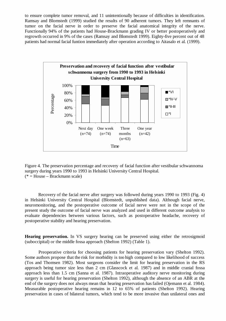
to ensure complete tumor removal, and 11 unintentionally because of difficulties in identification.Ramsay and Blomstedt (1999) studied the results of 90 adherent tumors. They left remnants oftumor on the facial nerve in order to preserve the facial anatomical integrity of the nerve.Functionally 94% of the patients had House-Brackmann grading IV or better postoperatively andregrowth occurred in 9% of the cases (Ramsay and Blomstedt 1999). Eighty-five percent out of 48patients had normal facial funtion immediately after operation according to Aitasalo et al. (1999).
Figure 4. The preservation percentage and recovery of facial function after vestibular schwannomasurgery during years 1990 to 1993 in Helsinki University Central Hospital.(* = House – Brackmann scale)
Recovery of the facial nerve after surgery was followed during years 1990 to 1993 (Fig. 4)in Helsinki University Central Hospital (Blomstedt, unpublished data). Although facial nerve,neuromonitoring, and the postoperative outcome of facial nerve were not in the scope of thepresent study the outcome of facial nerve was analyzed and used in different outcome analysis toevaluate dependencies between various factors, such as postoperative headache, recovery ofpostoperative stability and hearing preservation.
Hearing preservation. In VS surgery hearing can be preserved using either the retrosigmoid(suboccipital) or the middle fossa approach (Shelton 1992) (Table 1).
Preoperative criteria for choosing patients for hearing preservation vary (Shelton 1992).Some authors propose that the risk for morbidity is too high compared to low likelihood of success(Tos and Thomsen 1982). Most surgeons consider the limit for hearing preservation in the RSapproach being tumor size less than 2 cm (Glasscock et al. 1987) and in middle cranial fossaapproach less than 1.5 cm (Sanna et al. 1987). Intraoperative auditory nerve monitoring duringsurgery is useful for hearing preservation (Shelton 1992), although the absence of an ABR at theend of the surgery does not always mean that hearing preservation has failed (Ojemann et al. 1984).Measurable postoperative hearing remains in 12 to 65% of patients (Shelton 1992). Hearingpreservation in cases of bilateral tumors, which tend to be more invasive than unilateral ones and
Preservation and recovery of facial function after vestibularschwannoma surgery from 1990 to 1993 in Helsinki
University Central Hospital
0%
20%
40%
60%
80%
100%
Next day(n=74)
One week(n=74)
Threemonths(n=63)
One year(n=42)
Time
Perc
enta
ge
*VI
*IV-V
*II-III
*I

tend to involve the cochlear nerves, is difficult (Bance and Ramsden 1999), but is not impossible(Samii and Matthies 1997, Glasscock et al. 1987).
Table 1. Hearing preservation after vestibular schwannoma surgery surgery according to theliterature.
Year Patients Approach Tumor size Hearing preserved
(number) (mean)____________________________________________________________________________________
Harner et al 1984 119 MCF 17mm 12%
Palva et al 1985 30 RS <20mm 43%
Silverstein et al 1985 13 RS 38%
Kemink et al 1990 20 RS 15.5mm 65%
Hoehmann 1991 41 MCF 17<10mm, 22 10-20mm, 46%2>20mm
Cohen et al 1993 146 RS 10.2mm 26%
Glasscock et al 1993 MCF, RS
unilateral 136 49 IC, 75<10mm,12<20mm 35%
bilateral 22 9 IC, 13<10mm, 3<20mm,1>20mm 44%
Dornhoffer et al 1995 93 MCF (65<5mm, 11 5-10mm, 58%17 10-15mm)
Umezu et al 1996 51 RS transmeatal 15.7 mm/22.8 mm 41.2%
Samii and Matthies1997 732 RS 39.5%
Kanzaki et al 1998 94 MCF 6.9 mm 50%
Ishikawa et al 1998 43 MCF 1 IC, 18<10mm,10 10-20mm, 41%
10>20mm
Slattery et al 1998 18 MCF 11mm 65%____________________________________________________________________________________
Abbreviations: MCF = middle cranial fossa, RS = retrosigmoid, IC = intracanalicular
Contralateral hearing. Contralateral hearing loss after surgery is a rare complication that canhave immediate or delayed onset (Wiet et al. 1992). It has been reported to occur in the recoveryroom, during the first postoperative week, months after surgery and several years after surgery(Greiner et al. 1971, Clemis et al. 1982). Harris et al (1985) have proposed that sympatheticcochleolabyrinthitis may occur in up to 1.3% of the patients.
Postural stability. Vertigo and imbalance postoperatively may be caused by vestibular orcerebellar dysfunction (Wiet et al. 1992). Vestibular disability may persist in elderly patients (Wietet al. 1992). According to Wiet et al. (1992) most of VS arise from the vestibular nerve, andtherefore vestibular function has already been considerably reduced or even totally lost beforesurgery and immediate postoperative vertigo is usually minimal. It is also thought that ifpostoperative imbalance does occur from absence of vestibular compensation, it is usually transientand diminishes within some weeks, and although it may persist longer in some older patients, itseldom causes significant disability (Wiet et al. 1992) (Table 2).

Table 2. Postoperative balance problems after vestibular schwannoma surgery reported in theliterature.
Year Patients Approach Balance problems
(number)
______________________________________________________________________________
Magnusson et al 1991 57 TL stiffness increased witheyes open
Parving et al 1992 273 TL 56% dizziness 6 months after surgery
Jorgensen and Pedersen 1994 46 TL 30 out of 44 vertigo
13 RS 8 out of 9 vertigo
Pyykkö et al 1994 224 RS/TL 41% slight postural instability
41% moderate posturalinstability
3% could not walkwithout help
van Leeuwen et al 1996 134 TL/RS more vertigo after TL
Rigby et al 1997 130 TL,RS,MCF 14.3% balance troubles
Andersson et al 1997 141 TL 45% balance problems19% dizziness
Driscoll et al 1998 210 RS 31% balance problems
Lynn et al 1999 237 RS 65% balance problems________________________________________________________________________
Abbreviations: TL = translabyrinthine, RS = retrosigmoid, MCF = middle cranial fossa
Headache. Severe postoperative headache is associated with VS surgery (Vijayan 1995, Schesselet al. 1992) and it may be related to surgical bone debris in the subarachnoidal space, residualintracranial air, or a transient elevation in intracranial pressure (Wiet et al. 1992).
Postoperative headache can be very severe in nature and last for many years after surgery.Despite the use of nonsteroidal anti-inflammatory drugs or even narcotic analgesics the headachemay be persistent (Hanson et al. 1998). Many studies have confirmed a high incidence of headache(Table 3.) and some have also postulated possible mechanisms and means of prevention with varieddegrees of success (Hanson et al. 1998).

Table 3. Postoperative headache after vestibular schwannoma surgery according to the literature.
Year Patients Approach Headache
(number)
_______________________________________________________________________________
Parving et al 1992 273 TL 29%
Cohen et al 1993 146 RS 14.3%
Schessel et al 1992 58 RS 64%40 RS 67% (sigmoid incision, craniectomy)13 RS 69% (triradiate incision, craniectomy)19 RS 5% (triradiate incision, craniotomy)
Harner et al 1993 331 RS 9% (two years postoperatively)
Harner et al 1995 24 RS 4% (cranioplasty)24 RS 17% (no cranioplasty)
Pedrosa et al 1994 155 RS 73%
TL 53%
Vijayan 1995 280 75%
van Leeuwen et al 1996 134 TL/RS more pain after RS
Ruckenstein et al 1996 18 TL no difference between TL and35 RS SO groups one year postoperatively
Catalano et al 1996 84 RS 64% (excision)81% (excision and cranioplasty)
10% (excision, cranioplasty, residue trapping)
Andersson et al 1997 141 TL 22%______________________________________________________________________________
Abbreviations: TL = translabyrinthine, RS = retrosigmoid

3. PURPOSE OF THE STUDY
The purpose of this study was to characterize the postoperative outcome of patients operated onfor VS and to evaluate some quality of life aspects of VS patients after surgery. The specific aimswere:
1. To characterize the frequency, type, and severity of postoperative headache in a retrospectivestudy of patients after VS removal with special reference to possible risk factors andrecovery.
2. To study the etiology of postoperative headache with special reference to vestibular function.
3. To evaluate the occurrence of hearing loss in the contralateral ear following cerebellopontineangle tumor surgery through the TL and the RS approach.
4. To evaluate the usefulness of preserved hearing including the role of tinnitus, and to evaluatepre- and postoperative factors predicting successful outcome in hearing preservation.
5. To evaluate postural stability after VS removal.

4. MATERIAL AND METHODS
4.1. Patients
The main study group consisted of 384 patients with unilateral or bilateral VS operated on duringyears 1979 to 1994 in Helsinki University Central Hospital (Table 4.). In addition, 97 unilateral VScases operated on during the years 1978 to 1988 at Sahlgrenska Hospital, Gothenburg, Swedenwere also included in one part of the main study.
Table 4. Patient data (N = 348) of vestibular schwannoma patients operated on during years 1979to 1994 in Helsinki University Central Hospital.
Bilateral / Unilateral 19 / 365
Operation route (retrosigmoid / translabyrinthine) 348 / 36
Tumor size (mm) mean 22.0 (range 3-58 mm)
Age (years) mean 47.7 (range 7.9-73.4 years)
Gender (Male / female) 158 / 226
Operation radicality (yes / no) 325 / 59
Follow up time (years) 8.9 (range 1-16 years)
Tumor side (right / left) 178 / 206
.
Study I included charts from 359 patients. A questionnaire, based on the McGill PainQuestionnaire (MPQ) (Melzack 1975, Melzack 1987) and the Finnish Pain Questionnaire(Ketovuori and Pöntinen 1981, Ketovuori et al. 1984), was mailed to 317 patients with unilateraltumor, and altogether 251 (79.2%) responded. Ninety-two patients out of these 251 reportedhaving postoperative headache, but only 27 patients considered it a major problem. These 27patients, 11 men and 16 women, were entered further into Study II.
Study III consisted of 364 patients with unilateral VS. In addition, a survey was performedon cases operated on during the years 1978 to 1988 at Sahlgrenska Hospital, Gothenburg, Sweden.There were altogether 114 patients of whom 97 had unilateral VS, 14 cerebellopontine anglemeningeoma and 3 bilateral tumors. Only the 97 patients with unilateral cerebellopontine angletumors were included. All cases with postoperative hearing loss of contralateral ear exceeding 20dB HL were evaluated (n = 9).
Study IV included 119 unilateral VS patients in whom the cochlear nerve was preserved inorder to preserve hearing.
In Study V the retrospective group consisted of 177 patients with unilateral VS operatedon during 1979 to 1987. The RS approach in the sitting position was used in 166 patients, and TLapproach in the supine position in 11 patients. Active postoperative habituation was started in1988, and the prospective group consisted of 44 patients with unilateral VS operated on after1988. In 40 patients the approach was RS in the lateral recumbent position and in 4 patients TL inthe supine position.

4.2. Methods
The intensity of postoperative headache was expressed using the Visual Analogue Scale (VAS)ranging from “no pain“ (0) to “worst possible pain“ (100) (Huskisson 1974). The scale is a 10 cmlong line and patients choose the intensity of their headache between the two points “no pain” ja“worst possible pain”. The number of words chosen in the MPQ or more clearly in the Finnish PainQuestionnaire (Ketovuori et al. 1981) was also used to characterize pain intensity (Melzack 1975).Using a VAS, the patients were grouped into the categories of “no or slight headache“ (VAS<3.3cm), “moderate headache“ (VAS between 3.3 and 6.6 cm), or “severe headache“ (VAS>6.6 cm).
The tinnitus questionnaire was based on the Tinnitus Handicap Questionnaire (Tyler et al.1989, Kuk et al. 1990). The hearing questionnaire was based on the Social Hearing HandicapIndex (Ewertsen and Birk-Nielsen 1973) (see Appendix).
Pure-tone audiograms and speech audiometries were performed to evaluate hearing.
The Vestibulo-ocular reflex (VOR), producing compensatory eye movements during headmovements in the operative frequency range of 0.1 to 5 Hz (Juhola et al. 1997), was evaluatedwith the Vestibular Autorotation Test (VAT). In the VAT the patient is asked to rotate his or herhead 10 degrees from side to side in pace with a sound signal while gazing at a stationary red spoton the wall 150 cm to the front (Hirvonen et al. 1995). The head movement accelerates graduallyover 20 seconds from 0.5 Hz to as fast as possible (about 5 Hz). The VOR is measured at differentfrequencies; the highest frequency reached and also the gain and phase shift values are measured.Gain is computed for each sine wave of the signals as the ratio of the eye movement amplitude andthe head movement amplitude. The phase shift is computed between the maxima or minima of thesine waves of the eye and head movement signals.
In posturography the force platform is constructed according to the strain gauge principle(Aalto et al. 1988). The vertical force distribution over the platform surface is measured and thecoordinates of the center point of the force in the forward-backward and lateral directions aremeasured and analyzed. The posturography measurement lasted for 180 seconds. Stabilograms arerecorded during two tests, one with (eyes open) and one without (eyes closed) visual control. Theprogram calculates the sway velocity (SV). The Romberg quotient (RQ) describes the ratiobetween SV with eyes closed and SV with eyes open.
Postural perturbation is induced with vibrators, bilaterally on the calf muscles or on theneck muscles. Vibration is generated by means of a revolving DC motor (Escap, Switzerland) witha 5 g unbalanced weight at one end. The vibrator is built in a metal cylinder, length 50 mm anddiameter 18 mm. Varying the input voltage of the DC motor controls the frequency of thevibration. The waveform of the vibration is sinusoidal. Eklund (1971) has provided a detaileddescription of this kind of vibrator system. Frequencies of 20, 40, 60, 80 and 100 Hz are used anddelivered in pseudo-random fashion. The stimulator unit is controlled with a microcomputer(Hewlett Packard 75 C). The amplitude of the vibration is 0.4 mm (peak-to-peak) and is constantat all frequencies when measured unloaded. Two vibrators were held firmly against the calf musclesor the neck muscles with straps. The Proprioceptive index is the ratio between the SV duringvibration perturbation on the calf muscles at 80 Hz and the baseline SV, both with eyes closed.
During the testing of the 27 patients with POH the headache was provoked with both theVAT and the posturography measurement. When the headache occurred the patient was given100mg sumatriptan.
4.3. Statistical methods
In Study I the variables characterizing the patients’ headache were analyzed by a factor analysisusing simple correlation and varimax rotation. To find variables suggesting a risk of postoperative

headache a linear discriminant function analysis and logistic regression analysis were used.Duncan’s multiple range test was used to compare the groups.
In Study II the Chi-square test with Yates correction was used for comparing the maximalfrequency in the VAT. For the continuous variables of the VAT and the posturography, an analysisof variance was used, and the differences were gauged with Bonnferoni’s test.
In Study IV linear regression analysis was used for the comparison between the outcome ofvarious factors influencing the success of hearing preservation. Stepwise logistic regression analysiswas used when evaluating factors determining the outcome of hearing preservation surgery. Instudying internal correlations Kendall’s linear correlation analysis was used. Student’s t-test wasused for the statistical calculations between the groups of patients.
In Study V factors influencing the postural stability were assessed with linear regressionanalysis and logistic regression analysis. The patients and controls were compared with Student's t-test.
4.4. Ethics
The ethics committee of Helsinki University Central Hospital approved the present study(KO11/94, KO 41/95).

5. RESULTS
5.1 Postoperative headache (I)
Headache that commenced only after surgery was regarded postoperative headache (POH). In theresults the subjects with headache were classified into two groups: the POH group and those withpreoperative and postoperative headache, the prePOH group.
The patients with postoperative headache experienced their headache mainly in the area ofthe neck (71%) and the occiput (76%) regardless of the surgical approach. In both groups the painwas typically unilateral and felt on the side of the surgery. The changes of postoperative headacheafter surgery in both prePOH and POH groups is shown in the Figures 5 and 6. The severity of thepostoperative headache declined more among the POH patients than among the prePOH patients
(X2, p<0.05). (I)
Figure 5. Continuing headache among the patients with headache pre- and postoperatively(prePOH) and among the patients with no headache preoperatively (POH) (N = 251).
The model consisting of preoperative headache, surgical approach, postoperative gaitdisorders, and tumor size predicted headache postoperatively for 68.1% of the patients, and wasstatistically highly significant (p<0.001) when analyzed with logistic regression analysis. Thesurgical approach (p<0.05) was the most important single risk factor linked to severe postoperativeheadache, the odds ratio (OR) being 3.9 for the retrosigmoid approach. Preoperative headache wasalso a statistically significant risk factor (p<0.05) for postoperative headache (OR 2.5), andpostoperative gait disorders increased the probability of postoperative headache (OR 2.4, p<0.05).Also small tumor size was a risk factor (OR 1.1, p<0.01).
When only the patients with postoperative headache were studied, the logistic regressionanalysis showed that the surgical approach (RS) was the single most important risk factor forpostoperative headache (OR 5.8, p<0.05).
0%
20%
40%
60%
80%
100%
Preoperat Postoperat 1 year postop 8.9 yearspostop
Continuing headache
Perc
enta
ge
Headachepreoperatively(prePOH)n=122No headachepreoperatively(POH) n=129

Figure 6. Maximum pain 1 year after operation and a mean of 8.9 years after operation. Patientswith postoperative headache are grouped as those with headache preoperatively (prePOH) andthose with no headache preoperatively (POH). N = 251.
5.2. Postoperative headache (II)
All 27 patients with severe headache were able to carry out the VAT; there was no difference inperformance among patients with or without headache. The difference in gain was significantbetween the patients and normal controls (p<0.01).
Body sway in posturography was significantly increased among the patients with headacheduring the baseline stance, and during the vibration of neck muscles, when comparing the patientswith the healthy controls (p<0.01). No differences were observed between the patients with andwithout headache.
After anesthetising the neck muscles, 25 patients were able to rotate their head at afrequency of 1 Hz, 17 at 3 Hz and 7 patients at 4 Hz. All 9 normal controls reached 4 Hz (Fig. 7).
Of the 27 patients 26 reported headache during the three days of testing and provocation,only one experiencing no pain or discomfort at all. In the 26 patients in whom headache occurred100mg sumatriptan alleviated pain in 9 patients and completely abolished the pain in 1 patient.
0%
20%
40%
60%
80%
100%
1 Year 8.9 years
Time after operation
Pai
n m
axim
um
Headache preoperat.(prePOH) n=122
No headachepreoperat.(POH)n=129

Figure 7. Highest performed VAT (percentage) frequencies after neck muscle anesthesia. Resultsof vestibular schwannoma patients with headache and the normal controls.
5.3. Cochleolabyrinthitis (III)
Altogether 478 patients with VS or cerebellopontine angle meningeomas were studied, 32 ofwhom were operated on using the TL approach and 446 the RS approach. Two patients with theTL approach developed vertigo and prominent hearing loss in the contralateral ear (Table 5). Thehearing loss started within two weeks of the surgery. Eight patients with the RS approachdeveloped a significant hearing loss of the contralateral ear that started two to four years afteroperation, and in three patients this was identical to delayed hydrops (Table 5).
Table 5. The development of cochleolabyrinthitis. Contralateral hearing loss, tumor size andvertigo in patients with removed vestibular schwannoma and one cerebellopontine anglemeningeoma.
Case Operationroute
Meantumor size
(mm)
Preop.HL(dB)
Post-opHL(dB)
Time toonset of HL
Onset and course ofHL
Vertigo
1 TL 30 7 35 1 week fast, stabilized yes2 TL 13 10 50 2 weeks fast, stabilized yes3 RS * 17 90 4 years fast, progressive no4 RS 25 15 55 2 years fast, fluctuant yes5 RS 10 10 45 2 years fast, fluctuant no6 RS 27 10 35 2 years fast, fluctuant yes
7 RS 37 7 30 2 years slow, fluctuant no
8 RS 27 10 47 5 to 7 years slow, progressive no
9 RS 35 15 35 3 to 6 years slow, stabilized no
10 RS 30 12 35 3 to 6 years slow, stabilized no
TL = translabyrinthine, RS = retrosigmoid, * = meningeoma
0%
20%
40%
60%
80%
100%
1Hz 2Hz 3Hz 4Hz 5Hz
Frequency
VA
T (P
erce
ntag
e)Patients with headache(n=27)
Normal controls (n=9)

5.4. Hearing preservation (IV)
Hearing was preserved in 47 out of 119 patients (Table 6). In 10 patients with preserved hearingtumor size was larger than 15 millimeters. Ten patients had hearing better than 30 dB. Hearing wasfollowed on average 7.3 years, range 1 to 16 years. There was gradual deterioration (about 5 dB)during the follow-up time. Hearing was lost in four patients during the follow up period (threemonths, one year, and in two patients two years after surgery).
Table 6. Vestibular schwannoma patients grouped to those with and without successful hearingpreservation surgery.
Cochlear nerve intactFactor
Hearing lost n = 72 Hearing preserved
n = 47
Mean age (yr) 45 49.2
Mean tumor size (mm) 15.0 12.6
Mean preoperative PTA (dBHL)
37.1 31.0
Mean preoperativeDiscrimination (%)
76.2 82.9
Mean postoperative PTA (dBHL)
>120 54.7
Mean postoperativeDiscrimination (%)
0 64.6
Mean facial function* 1.3 1.1
* = House and Brackmann scale, PTA = pure tone average, HL = hearing level
The model for predicting successful hearing preservation consisted of age, tumor size,PTA, SDS and hearing level at 1000 Hz in pure-tone audiometry (p<0.003). In the model age(p<0.008) and SDS (p<0.06) were the strongest predictors for preserved hearing. Tumor size,PTA and hearing at 1000 Hz did not reach the level of statistical significance. During surgery theinterface between the nerve and the tumor seemed to be the most important factor for hearingpreservation, an easily identifiable cleavage plane enabling less traumatic tumor removal.

Table 7. Vestibular schwannoma patients’subjective evaluation of the usefulness of their preservedhearing.
Rating scale
Usefulness of hearingpreservation*
(n=39)
Hearingof sound
(n=39)
Speechdiscrimination
(n=39)
Soundlocalization
(n=39)
None 8% (16dB / 97%) 8% 15% 28%
Undecided 20% (10dB / 100%) 26% 23% 23%
Some 23% (3dB / 100%) 20% 21% 15%
Moderate 13% (3dB / 97%) 13% 18% 21%
Very 36% (11dB / 97%) 33% 23% 13%
*=Mean contralateral PTA / SDS
PTA = Pure tone average hearing level, SDS = Speech discrimination scores
The questionnaire indicated that 70% of the patients found their preserved hearing useful orvery useful (Table 7). Sound distortion was not experienced to be a problem in the majority of thepatients (65%), but was distressing in 15%. Tinnitus was present in 68% of the patients. Itlocalized to the tumor ear in all but 2 patients. Table 8 shows the subjective handicap caused bytinnitus.
Table 8. Subjective disability, and use of sleeping pills due to tinnitus according the vestibularschwannoma patients with preserved hearing.
Rating severity oftinnitus
Subjectivedisability
(n=39)
Interference withsleep
(n=39)
Use of sleepingpills
(n=39)
No tinnitus 38% 38% 38%
Very mild 5% 38% 54%
Slight 33% 21% 8% (occasionally)
Moderate 21% 3% 0 (weekly)
Severe 3% 0 0 (every night)
5.5. Postural stability (V)
In the retrospective study 122 out of 177 patients considered their moving ability normal, 27 hadslight difficulties, 7 needed a cane, 3 were able to move only with human support and 1 patient wastotally unable to walk. Postural stability, based on posturography measured 5 or 7 years aftersurgery, was normal in 122 patients (69%) under visual control and in 67 patients (38%) undernon-visual control (Fig. 8). Vibration in non-visual control increased body sway more than in visualcontrol (p<0.001).

Figure 8. Sway velocity under visual and non-visual control among the patients in the prospectiveand the retrospective groups, and among normal controls.
In the prospective patient group, under visual control 30 patients (68%) had normalpostural stability on posturography one year after the operation. Under non-visual control 23patients (52%) had normal postural stability. The difference between the retrospective and theprospective patient groups regarding SV was statistically significant (p<0.05) under non-visualcontrol (Fig. 8).
Figure 9. Sway velocity (cm/s) both with visual- and non-visual control measured preoperativelyand during the first postoperative year. The prospective patient group. (N = 44)
0
0.5
1
1.5
2
2.5
3
3.5
4
4.5
5
Retrospectivegroup (n=177)
Prospective group(n=44)
Controls (n=100)
Sway
vel
ocity
(cm
/s
Eyes closed
Eyes open
00.5
11.5
22.5
33.5
44.5
5
Preope
rative
ly
1 wee
k pos
topera
tively
3 mon
ths po
stope
rative
ly
1 yea
r pos
topera
tively
Sway
vel
ocity
(cm
/s)
eyesopeneyesclosed

The changes in the mean SV before and after operation under visual and non-visual controlare shown in Figure 9. Three months after surgery the mean SV had returned almost to thepreoperative value; however, one year after surgery the mean SV was still not better than the valuebefore surgery.
The retrospective and prospective groups were combined to study the role of the posturalactive habituation and surgical position for outcome of postural recovery. In the logistic regressionanalysis the surgical position and habituation are heavily intercorrelated as both changes took placeat the same time. With gait problems the model consisting of surgical position (p<0.01), gender(p<0.01), and outcome of posturography (p<0.001) as the independent variables was statisticallysignificant and could explain 65% of the variance of the gait problems. The odds ratios for surgicalposition and female gender were 6 and 2.5, respectively, making them the most important riskfactors for poor postural stability.
The role of proprioception and visual control as compensatory vestibular mechanisms wasalso studied. A high proprioceptive index indicates relative reliance on the former (p< 0.001), and ahigh Romberg quotient on the latter (p<0.001). Those who relied more on proprioception faredbetter than those who relied on visual control.

6. DISCUSSION
In the present study we examined headache after vestibular schwannoma surgery. If headache waspresent before the operation it tended to continue also postoperatively, although 20% of patientswho had headache preoperatively did not complain of headache postoperatively. Sixty (23.9%)patients developed POH without preoperative headache. Headache declined during the first yearafter operation, but if it persisted for a year, it seemed to become permanent.
There was no difference in vestibular function when the POH patients were compared withVS patients without POH. Differences were not observed between these groups in vestibulo-ocularor vestibulo-spinal reflexes, either. Therefore the POH may not be explained by the vestibularimbalance. The postoperative headache, although probably multietiological, may be caused bytrigeminal neuropathy in the posterior fossa, where pain afferents trigger abnormal reflexesmimicking local vascular headache (Vass et al.1997, Vass et al. 1998). Bone dust irritation ormanipulation of the sensitive duro-periosteal complex may cause this trigeminal neuropathy.
Ten out of 478 patients in study III developed hearing impairment of the non-operated side:two patients directly after TL operation (6%) and eight patients after RS operation (1.8%) with alate onset. One explanation could be AID (autoimmune inner-ear disease) (Hariri 1993). Thecontralateral hearing loss in the RS cases with an intact labyrinth might be due to the removal ofthe endolymphatic sac. It is therefore speculated that even in the RS cases, AID may be the reasonfor contralateral hearing loss.
In the present study hearing was preserved in 39% of patients in whom the cochlear nervewas preserved. The result is very similar to that of other studies (Cohen et al. 1993, Samii andMatthies 1997). Four patients (8.5%) with preserved hearing later experienced significant hearingimpairment. Similar impairment has been reported to occur in other series with the same frequency(Shelton and House 1990). The preserved hearing was considered beneficial by 66% of the patientscontrary to the results of Tos et al. (1988), although the preserved hearing was better than 30 dB inonly 10 patients. Preserved hearing was perceived as valuable for speech discrimination duringconversation. We found that neither distortion of speech nor aggravation of tinnitus caused majordisability after surgery.
Vestibular compensation after removal of VS takes place during the first months aftersurgery (Haid et al. 1991), even without active habituation (Mruzek et al. 1995). However, Lynn etal. (1999) reported persistent dysequilibrium in up to 65% of patients. In the present series theresults of retrospective study showed that 69% of the patients considered their gait normal, whilein posturography only 32% had postural stability within normal limits, indicating that subjectiveevaluation of gait and measured postural stability are evaluating different aspects of bodyequilibrium. The impairment of postural stability was usually slight, as only four patients had severedifficulties with postural stability.
The VS patients in present study were all in all quite satisfied with their postoperativecondition. Although headache was frequent for majority of the patients it was a minor complaint.Only one patient had headache which did not relieve with any medication, but headache appearedvery seldom. Hearing preservation was experienced beneficial. The contralateral hearing loss wasrare. Patients did not complain of balance difficulties spontaneously, but they had motivation toactive postoperative habituation. Although the patients succeeded in their every day life, theretiring tendency was apparent.
6.1. Methodological aspects
The diagnosis of VS has changed during the last years considerably as MRI has become the mostpreferred diagnostic tool (Möller 2000, Robinette 2000). The incidence of VS increases with age

(Möller 2000) and even tumors of only a few millimeters in size are diagnosed with MRI.Nowadays the patients present with minor symptoms and smaller tumors than in the beginning ofthe present study, which causes differences within the patient material. The total patient material inthis study is large, including all the VS patients treated at Helsinki University Central Hospitalduring 1979 to 1994. The study was done in a tertiary center to which the most of the VS patientsin the southern Finland are referred. Mainly two operative methods were used, TL operation andRS operation, although the group of TL patients was altogether small.
In a study by Blomstedt et al. (1991) in which the VS operations were done before theoperating microscope era, the mortality was quite obviously higher among VS patients than amongthe matched general population during the first year after surgery, but after the first year themortality rates became equal (Blomstedt et al. 1991). In 1985 Welch and Dawes reported 5%postoperative mortality, all among patients with large tumors. In 1995 Irving et al. reported apostoperative mortality rate of 2-3%, and Guerin et al. in 1999 a rate of 0.3%. Even results with nopostoperative mortality have been reported (Jung et al. 2000, Lanman et al. 1999). NF2 patientshave a larger risk for postoperative mortality because of the more progressive tumor growth (Samiiet al. 1997). The development of the operative technique during the years of present study togetherwith the presenting smaller tumor size have influenced the postoperative results The tumor sizesduring years 1962 to 1973 and 1990 to 1992 were compared in Helsinki University CentralHospital (Fig. 10).There are today about equally frequently giant tumors but fewer large and mid-size tumors than at the early 70’s. The dominance is shifting towards small tumors. When the majorcomplications hemorrhage, meningitis, and cerebrospinal fluid leak are becoming rare, quality oflife issues such as dysequilibrium, headache, and hearing preservation are more important.
Figure10. The size of vestibular schwannoma during years 1962 to 1973 (N = 133) and 1990 to1992 (N = 47) in Helsinki University Central Hospital.
Several questionnaires were used in present study. VAS (Huskisson 1974), MPQ (Melzack1975), Finnish Pain Questionnaire (Ketovuori et al. 1981), Tinnitus Handicap Questionnaire (Tyleret al. 1989, Kuk et al. 1990) and Social Hearing Handicap Index (Ewertsen and Birk-Nielsen 1973)are all validated international questionnaires. Only the questionnaires concerning gait, depression,and quality of life were not international.
0%
10%
20%
30%
40%
50%
60%
GIANT BIG MID-SIZE SMALL
Size
Per
cen
tag
e
1962-1973 (n=133)
1990-1992 (n=47)

6.2. Postoperative headache
Postoperative headache is usually connected with the SO or RS approach in surgery for VS(Schessel et al. 1992), and up to 73% of patients suffering from POH have undergone thesuboccipital approach (Pedrosa et al. 1994). The incidence of headache after the TL approachvaries in the literature from 0% (Schessel et al. 1992) to 75% (Vijayan 1995). In the present studythe incidence of headache after the TL approach was 39%. Thus none of the operative approachesare without risk for headache, but in the present study the RS approach carried a 3.8 times higherrisk for headache than did the TL procedure.
Mechanical, electrical or chemical stimulation of the trigeminal sensory nerves may produceneurogenic inflammation and intense head pain (Hargreaves et al. 1999). Vass et al. (1997, 1998)demonstrated that the first branch of the trigeminal ganglion sends projections to the cochlea andalso to the dura of the posterior fossa (Keller et al. 1985). Vass et al. (1997) suggested that thetrigeminal sensory innervation of the cochlea plays an important role in cochlear blood flowregulation. Stimulation of trigeminal perivascular nerve endings causes vasodilatation and edemaleading to further sensory nerve activation and neurogenic inflammation (Vass et al 1998).
Sumatriptan interacts with the trigemino-vascular system, possibly through direct cerebralvessel constriction or through the inhibition of sensory neuropeptide release or both (Moskowitzand Buzzi 1991). In present study one patient responded to sumatriptan with complete pain relief,and nine others considered it helpful to some extent. The sumatriptan trial was not randomized andtherefore our observation is not reliable. However this result suggests that vascular structures maybe involved in at least some cases. More research is needed to solve the etiology of headache.
6.3. Sympathetic cochleolabyrinthitis
AID can be induced experimentally by infection (Gloddek et al. 1992), trauma ((Jackson 1994) orvaccination (Harris 1984). Recent animal models using immunization to inner ear extracts(Gloddek et al. 1994) or adjuvant therapy (Harris 1984) indicate that the pathological changes inthe inner ear are characteristic of a disease like Ménière`s disease, with autoimmune cellularresponse against the endolymphatic sac (Soliman 1996).
Sympathetic ophthalmia may be used as an analogy for sympathetic cochleolabyrinthitis. Inthe recent literature the incidence of sympathetic ophthalmia varies between 0.2% and 0.5% amongsubjects with a penetrating eye wound, although in the older literature an incidence as high as 16%has been reported (Towler and Lightman 1996, Power and Foster 1996). After eye surgery theincidence is significantly lower, being about 0.007% (Tower and Lightman 1996). The latency ofonset of sympathetic ophthalmia varies from 10 days to 50 years from the original traumaticincident. In present study the onset of hearing loss after operation varied from 2 weeks to fouryears.
A test to prove the presence of antibodies to inner ear proteins in affected patients, andtheir absence from patients without contralateral hearing loss, is needed before the etiology ofsympathetic cochleolabyrinthitis can be confirmed.
6.4. Hearing preservation
Hearing was preserved in 40% of the 119 attempted cases. The preservation of hearing wasconsidered a success by 66% of the patients, although in only 10 patients the preserved hearingwas better than 30 dB. There is controversy as to what should be considered useful, acceptable orserviceable hearing (Shelton and House 1990). Usually when audiometric criteria are advocated, it

is said that the 50/50 rule should apply to serviceable hearing (Glasscock et al. 1983). Our patients’subjective evaluation of the usefulness of their residual hearing indicates that the levels are beyondthese limits, and there was no correlation between the measured PTA levels and speechdiscrimination. The second general belief is that if the hearing level between the ears differs bymore than 20 dB the weaker ear does not aid in conversation (Glasscock et al 1983). It is alsoclaimed that leaving the cochlear nerve increases the risk of handicapping tinnitus (Harcourt et al.1997). In 5 patients with a deaf ear and tinnitus, the non-functioning cochlear nerve was removed.In 2 cases of them tinnitus was significantly relieved, but in 3 cases it was unchanged. Thisexperience has discouraged us from attempts to eliminate tinnitus by re-operation. Among thepatients with residual hearing tinnitus was a major problem in only one patient. Thus the argumentthat leaving the cochlear nerve will worsen the tinnitus seems unfounded. The results of the presentstudy encourage surgeons to continue efforts to preserve hearing in VS surgery.
6.5. Postural stability
Herdman et al. (1995) and Lynn et al. (1999) found no relationship between age and posturalinstability after VS removal, but in their study the surgical approach in the experimental group wasTL (Herdman et al. 1995). Tumor size was not related to postural stability (Herdman et al. 1995,Lynn et al. 1999) either,as was also found in the present study, also. In the present study elderlypatients showed increased risk for poor vestibular compensation. In these patients postural stabilityis influenced by poor vision, diminishing of stretch reflexes, and changes in the vestibular part ofthe labyrinth (Hytönen et al. 1993), which results in decreased adaptive capability.
Depression seems to be associated with VS surgery (Blomstedt et al. 1996). Postoperativedepression was associated with a significant risk for poor postural recovery. The reason remainsunclear. The RS approach had 6.8 times greater risk for poor postural recovery than the TLapproach. This may be linked to the sitting position in RS during which the cerebellum must befirmly retracted and the subject is exposed to air embolism. The patients’ position during RSsurgery was changed to the supine in 1988.
Female gender was a risk factor for postoperative gait problems as shown in other studies(Driscoll et al. 1998). Postural imbalance may be linked to reduction of estrogen levels with ageing(Naessen et al. 1997). Postural balance is better preserved in long-term estrogen users than in non-users (Naessen et al. 1997). In the present study the postmenopausal female patients did not useestrogen, which may be one reason for the female patients being at risk for postoperative gaitproblems.
The results of the present study show that not all of the VS patients compensate thevestibular damage after VS surgery, and that they benefit from the use of vestibular activehabituation to improve their postural stability.

7. CONCLUSIONS
1. At an average of 9 years after surgery, 93 out of 251 patients with headache postoperativelystill reported headache, which was a major problem for 27 of them. Headache declined duringthe first year after operation, but if it persisted for a year, it seemed to become permanent. Theretrosigmoid approach, postoperative gait problems, preoperative headache and small tumorsize predicted problematic headache after surgery.
2. Vestibular imbalance or abnormal activation of neck muscles did not explain postoperativeheadache. Occipital nerve entrapment or craniotomy-related dural retraction explainedheadache in a few patients. POH was alleviated by sumatriptan in eleven of the 27 patientswith persistent headache. The sumatriptan effect may suggest a vascularly mediated trigeminalneuropathy in the etiology of postoperative headache. Trigeminal irritation in the inner ear orthe posterior fossa may cause neurogenic inflammation mediated through vascular structures.
3. Contralateral hearing loss was observed to occur in 6% of the TL and 1.8% of the RS cases,indicating that the TL approach may be related with more complications than are presentlyknown.
4. Hearing preservation was successful in 40% of the cases with intact cochlear nerve. Of thosepatients 66% rated preserved hearing valuable. Neither tinnitus nor speech distortion wereconsidered disadvantageous for hearing preservation. The vascular structure of the inner ear ispossibly the most significant factor for hearing preservation.
5. After operation 69% of patients spontaneously regained normal postural stability in visualconditions, but only 37% in non-visual conditions. Vestibular active habituation increased theuse of proprioception in mainteining postural control. Advanced age was a risk factor for poorpostural recovery. Female gender and surgical position were risk factors for postoperative gaitproblems.
More research is needed to solve the etiology of postoperative headache. A test proving thepresence of antibodies to inner ear proteins in affected patients, and their absence from patientswithout contralateral hearing loss is needed before the etiology of sympatheticcochleolabyrinthitis can be confirmed. Hearing preservation is worthwhile after VS surgery.Not all of VS patients can compensate for vestibular damage after VS surgery, but manypatients benefit from the use of active habituation to improve their postural stability.
Overall impression of the VS patients was satisfaction with postoperative condition in spite ofdifferent minor or even major problems. Although the patients succeeded in their every daylife, the retiring tendency was apparent.

APPENDIX
The Social Hearing Handicap Questionnaire (Ewertsen and Birk-Nielsen 1973)______________________________________________________________________________
1. If you are sitting opposite a person at a distance about 1 m, will you be able to understandeverything if the person speaks in a normal tone of voice?
2. Do you have problems in understanding when members of the family are gathered together?
3. Is it difficult for you to hear in the kitchen when the water is running?
4. Do you find it difficult to hear what the shop assistant says to you?
5. Is it difficult for you to carry on a telephone conversation?
6. Can you follow the conversation when you are talking to several people?
7. Is it difficult for you to follow a conversation at a tea table with several persons?
8. Can you hear most of what is said when sitting at the back of a lecture room, a theatre or achurch?
9. Can you carry on a conversation with someone sitting at the other end of the room andspeaking in a normal tone of voice?
10. Can you hear the radio well when it is set at normal volume?
11. Can you carry on a conversation easily in a car, tram or bus?
12. Can you carry on a conversation easily at the dinner table at home?
13. Do you understand what is said to you when there is noise from other voices, typewriters,traffic, music?
14. Do you recognize persons by their voices?
15. Is it difficult for you to hear people speaking on TV when it is not turned up?
16. Is it difficult for you to follow a conversation at a large dinner table?
17. Can you carry on a conversation with someone when the radio or TV is not turne d down?
18. Is it difficult for you to carry on a conversation with someone in a busy street?
19. Is it difficult for you to understand what is said to you from an adjoining room?
20. Can you carry on a conversation with a person where there is noise, for instance at arestaurant or at a party?
21. Do you often misunderstand other people’s conversation?
______________________________________________________________________________

REFERENCES
Aalto H, Pyykkö I, Starck J. Computerized posturography, a development of measuring system. ActaOtolaryngol Suppl (Stockh) 1988; 449: 71-5.
Af Forselles A. Ett translabyrintärt opererat fall av akustikustumör. Nord Tidskr Otorhinolaryngol 1916; 1:29-46.
Aitasalo K, Suonpää J, Jääskeläinen S. Intraoperative monitoring of the facial nerve during acousticneurinoma surgery. In: Sanna M, Taibah A, Russo A, Mancini F (editors): Acoustic neurinoma andother CPA tumors. Bologny: Monduzzi Editore 1999, 433-7.
Andersson G, Ekvall L, Kinnefors A, Nyberg G, Rask-Andersen H. Evaluation of quality of life andsymptoms after translabyrinthine acoustic neuroma surgery. Am J Otol 1997; 18: 421-6.
Anderson TD, Loevner LA, Bigelow DC, Mirza N. Prevalence of unsuspected acoustic neuroma found bymagnetic resonance imaging. Otolaryngol Head Neck Surg 2000; 122: 643-6.
Axon PR, Ramsden RT. Facial nerve injury caused by vestibular schwannoma compression: severity andadaptation to maintain normal clinical facial function. Am J Otol 1999; 20: 763-9.
Bance M, Ramsden TR. Management of neurofibromatosis Type 2. ENT Ear Nose Throat J 1999; 78 (2):91-6.
Blomstedt GC, Jääskeläinen JE, Pyykkö IV, Sankila RJ. Recurrence of acoustic neurinomas after subtotalremoval: a comparison of the observed and expected survival rates of 139 patients operated on from1962 to 1973. In: Tos M, Thomsen J (editors): Acoustic Neuroma. Prodeecings of the FirstInternational Conference of acoustic neuroma. Amsterdam, New York: Kugler Publications 1991, 709-12.
Blomstedt GC, Katila H, Henriksson M, Ekholm A, Jääskeläinen J, Pyykkö I.: Depression after surgery foracoustic neuroma. J Neurol Neurosurg Psychiatry 1996; 61: 403-6.
Brackmann DE. Current status of ABR audiometry in acoustic neuroma diagnosis. Arch Otolaryngol HeadNeck Surg 1999; 125: 235.
Brackmann DE, Green JD. Translabyrinthine approach for acoustic tumor removal. Otolaryngol Clin NorthAm 1992; 25: 311-29.
Catalona PJ, Jacobowitz O, Post KD. Prevention of headache after retrosigmoid removal of acoustic tumors.Am J Otol 1996; 17: 904-8.
Clemis JD, Mastricola PG, Schuler-Vogler M. Sudden hearing loss in the contralateral ear in postoperativeacoustic tumor: three case reports. Laryngoscope 1982; 92: 76-9.
Cohen NL. Retrosigmoid approach for acoustic tumor removal. Otolaryngol Clin N Am 1992; 125: 295-310.
Cohen NL, Lewis WS, Ransohoff J. Hearing preservation in cerebellopontine angle tumor surgery: the NYUexperience 1974-1991. Am J Otol 1993; 14: 423-33.
Curtin HD, Hirsch WL. Imaging of acoustic neuromas. Otolaryngol Clin North Am 1992; 25(3): 553-607.
Cushing H. Tumors of the nervus acusticus and the syndrome of the cerebellopontine angle. Philadelphia:WB Saunders, 1917.
Dandy WE. An operation for total removal of cerebellopontile (acoustic) tumors. Surg Gynec Obst 1925;41: 129-48.
Daniels RL, Shelton C, Harnsberger R. Ultra high resolution nonenhanced fast spin echo magneticresonance imaging: cost-effective screening for acoustic neuroma in patients with sudden sensorineuralhearing loss. Otolaryngol Head Neck Surg 1998; 119: 364-9.
Dawes PJD, Basiouny HE. Outcome of using magnetic resonance imaging as an initial screen to excludevestibular schwannoma in patients presenting with unilateral tinnitus. J Laryngol Otol 1999; 113: 818-22.

Dornhoffer JL, Helms J, Hoehmann DH. Hearing preservation in acoustic tumor surgery: results andprognostic factors. Laryngoscope 1995; 105: 184-7.
Doyle KJ. Is there still a role in auditory brain stem response audiometry in the diagnosis of acousticneuroma? Arch Otolaryngol Head Neck Surg 1999; 125(2): 232-4.
Driscoll CL, Lynn SG, Harner G, Beatty CW, Atkinson EJ. Preoperative identification of patients at risk ofdeveloping persistent dysequilibrium after acoustic neuroma removal. Am J Otol 1998; 19 (4): 491-5.
Edwards CH, Patterson JH. A review of the symptoms and signs of acoustic neurofibromata. Brain 1951; 4:144-90.
Eklund G. Some physical properties of muscle vibrators used to elicit tonic proprioceptive reflexes in man.Acta Soc Med Ups 1971; 76: 271-80.
El-Kashlan HK, Zeitoun H, Arts HA, Hoff JT, Telian SA. Recurrence of acoustic neuroma after incompleteresection. Am J Otol 2000; 21: 389-92.
Evans DGR, Huson SM, Donnai D, Neary W, Blair V, Newton V, Harris R. A clinical study of type 2neurofibromatosis. Q J Med 1998; 42: 603-18.
Evans DGR, Lye R, Neary W, Black G, Strachan T, Wallace A, Ramsden RT. Probability of bilateraldisease in people presenting with a unilateral vestibular schwannoma. J Neurol Neurosurg Psychiatry1999; 66: 764-7.
Ewertsen HW, Birk-Nielsen H. Social hearing handicap index. Audiology 1973; 12: 180-7.
Fisch U. Transtempora surgery of the internal auditory canal. Adv Otorhinolaryngol 1970; 17: 202-39.
Fucci MJ, Buchman CA, Brackmann DE, Berliner KI. Acoustic tumor growth: implications for treatmentchoices. Am J Otol 1999; 20: 495-9.
Glasscock ME, McKennan KX, Levine SC. Acoustic neuroma surgery: the results of hearing preservationsurgery. Laryngoscope 1987; 97: 785-9.
Glasscock ME, Hays JW, Minor LB, Haynes DS, Carrasco VN. Preservation of hearing in surgery foracoustic neuromas. J Neurosurg 1983; 78: 864-70.
Gloddek B, Lamm K, Haslov K. Influence of middle ear immune response on the immunological state andfunction of the inner ear. Laryngoscope 1992; 102: 177-81.
Gloddek B, Rogowski M, Arnold W. Adoptive transfer of autoimmunological labyrinthitis in the guinea pig;animal model for sympathetic cochleolabyrinthitis. Clin Exp Immunol 1994; 97: 133-7.
Greiner GF, Philippides D, Collard M, Conraux C, Buchheit F, Maitrot D. Hypoacousie controlaterale dansle neurinome du VIII. Evolution post-operatoire. Rev Oto Neuro Ophtalmol 1971; 43: 54-8.
Guerin C, Sampath P, Long DM. Acoustic neuroma: outcome of surgical resection and study on theanatomy of facial and cochlear nerves. Ann Acad Med Singapore 1999; 28: 402-8.
Haberkamp TJ, Meyer GA, Fox M. Surgical exposure of the fundus of the internal auditory canal: anatomiclimits of the middle fossa versus the retrosigmoid transcanal approach. Laryngoscope 1998; 108: 1190-4.
Haid CT, Goertzen W, Christ P, Schuster B. Posture and central compensation after acoustic neurinomasurgery. Acta Oto-rhino-laryngol Belg 1991; 45: 349-54.
Hanson MB, Glasscock III ME, Brandes JL, Jackson CG. Medical treatment of headache after suboccipitalacoustic tumor removal. Laryngoscope 1998; 108: 1111-4.
Harcourt J, Thomsen J, Tos M. Translabyrinthine vestibular schwannoma surgery: postoperative tinnitusand cochlear nerve integrity. Auris Nasus Larynx 1997; 24: 21-6.
Hardy DG, MacFarlane R, Baguley D, Moffat DA. Surgery for acoustic neuroma. An analysis of 100translabyrinthine operations. J Neurosurg 1989; 71: 799-804.
Hargreaves R, Williamson D, Shepheard S. Neurogenic inflammation: relation to novel antimigraine drugs.In: Edvinsson A (editor) Migraine and Headache Pathophysiology. London: Martin Dunitz, 1999: 93-101.
Hariri MA. Autoimmune inner ear disease. J Audiol Med 1993; 2: 41-52.

Harner SG, Beatty CW, Ebersold MJ. Headache after acoustic neuroma excision. Am J Otol 1993; 14: 552-5.
Harner SG, Beatty CW, Ebersold MJ. Impact of cranioplasty on headache after acoustic neuroma removal.Neurosurgery 1995; 36: 1097-100.
Harner SG, Ebersold MJ. Management of acoustic neuromas, 1978-1983. J Neurosurg 1985; 63: 175-9.
Harner SG, Laws ER Jr, Onofrio BM. Hearing preservation after removal of acoustic neurinoma.Laryngoscope 1984; 94: 1431-4.
Harris JP: Immunology of the inner ear: evidence of local antibody production. Ann Otol Rhinol Laryngol1984; 93: 157-62.
Harris JP, Low NC, House WF. Contralateral hearing loss following inner ear injury: sympatheticcochleolabyrinthitis? Am J Otol 1985; 6: 371-7.
Henschen F. Zur Histologie und Pathogenese der Kleinhirnbruchenwinkeltumoren. Arch Psychiatr 1916; 56:21-122.
Herdman S, Clendaniel R, Mattox D, Holliday M, Niparko J. Vestibular adaptation exercises and recovery:acute stage after acoustic neuroma resection. Otolaryngol Head Neck Surg 1995; 113: 77-7.
Higgs WA. Sudden deafness as the presenting symptom of acoustic neurinoma. Arch Otolaryngol 1973; 98:73-6.
Hirvonen T, Pyykkö I, Juhola M, Aalto H. High frequency deficit of the vestibular system after acousticneurinoma surgery. Acta Otolaryngol Suppl (Stockh) 1995; 520: 16-8.
Hoehmann D. Pre- and postoperative hearing thresholds and brainstem responses in patients with acousticneuroma: follow-up study using the middle fossa approach. Am J Otol 1991; 12: 172-8.
Horrax G, Poppen JL. Experiences with the total and intracapsular extirpation of acoustic neuromata. AnnSurg 1939; 110: 513-24.
House HP, House WF. Historical review and problems of acoustic neuroma: monograph - transtemporalbone microsurgical removal of acoustic neuromas. Arch Otolaryngol 1964; 80: 601-4.
House JW, Brackmann DE. Facial nerve grading system Otolaryngol Head Neck Surg 1995; 93: 146-7.
House WF. Surgical exposure to the internal auditory canal and its contents through the middle cranialfossa. Laryngoscope 1961; 71: 1363-85.
House WF, Graham MD. Surgery of acoustic tumors. Otolaryngol Clin North Am 1973; 6: 245-66.
House WF, Hitselberger WE. Morbidity and mortality of acoustic neuromas; monograph - transtemporalbone microsurgical removal of acoustic neuromas. Arch Otolaryngol 1964; 80: 745-54.
House WF, Shelton C. Middle fossa approach for acoustic tumor removal. Otolaryngol Clin North Am1992; 25: 347-59.
Huskisson EC. Measurement of pain. Lancet 1974; 2: 1127-31.
Hytönen M, Pyykkö I, Aalto H, Starck J. Postural control and age. Acta Otolaryngol (Stockh) 1993; 113:119-22.
Irving RM, Beynon GJ, Viani L, Hardy DG, Baguley DM, Moffat DA. The patient’s perspective aftervestibular schwannoma removal: quality of life and implications for management. Am J Otol 1995; 16:331-7.
Ishikawa K, Yasui N, Mineura K, Sasajima H, Togawa K. Significance of hearing preservation in acousticneuroma surgery. Acta Otolaryngol Suppl (Stockh) 1998; 537: 16-9.
Jackler RK. Acoustic neuroma (vestibular schwannoma). In: Jackler RK, Brackmann DE (editors):Neurotlogy. St Louis: Mosby 1994, 729-85.
Jackson CA. Autoimmune inner ear disease. In: Jackler RK, Brackman DE (editors) Neurotology. St Louis:Mosby 1994, 517-25.
Johansson R, Magnusson M, Fransson PA, Karlberg M. Discrimination of patients with acoustic neuromaand peripheral vestibular lesion with human posture dynamics. Acta Otolaryngol Suppl (Stockh) 1995;

520: 27-8.
Johnson EW. Auditory test results in 500 cases of acoustic neuroma. Arch Otolaryngol 1977; 103:152-8.
Jorgensen BG, Pedersen CB. Acoustic neuroma: follow-up of 78 patients. Clin Otolaryngol 1994; 19: 478-84.
Juhola M, Hirvonen T, Aalto H, Pyykkö I. Computional problems in the analysis of eye movement signals inthe determination of vestibulo-ocular reflex. Comput Biomed Res 1997; 30: 49-60.
Jung S, Kang S, Kim T, Kim H, Jeong S, Seok-Chul, Lee J, Kim J, Kim S. Current surgical results ofretrosigmoid approach in extralarge vestibular schwannomas. Surg Neurol 2000; 53: 370-7.
Jääskeläinen J, Pyykkö I, Blomstedt G, Porras M, Palva T, Troupp H. Functional results of facial nervesuture after removal of acoustic neurinoma: Analysis of 25 cases. Neurosurgery 1990; 27: 408-11.
Kanter WR, Eldringe R, Fabricant R, Allen JC, Koerber T. Central neurofibromatosis with bilateralacoustic neurinoma: Genetic, clinical and biochemical distinction from peripheral neurofibromatosis.Neurology 1980; 30: 851-9.
Kanzaki J, Ogawa K, Inoue Y, Shiobara R, Toya S. Quality of hearing preservation in acoustic neuromasurgery. Am J Otol 1998; 19: 644-8.
Karjalainen S, Nuutinen J, Neittaanmäki H, Naukkarinen A, asikainen R. The incidence of acoustic neuromain autopsy material. Arch Otorhinolaryngol 1984; 240: 91-3.
Kawamoto Y, Uozumi T, Kiya K, Fujioka Y, Kurisu K, Kawamoto K, Sugiyama K. Clinicopathologicalgrowth factors of acoustic neuromas. Surg Neurol 1995; 43: 546-52.
Keiser-Kupfer MI, Freidlin V, Datiles MB et al. The association of posterior capsular lens opacities withbilateral acoustic neurinomas in patients with neurofibromatosis type 2. Arch Ophthalmol 1989; 107:541-544.
Keller JT, Saunders MC, Beduk A, Jollis JG. Innervation of the posterior fossa dura of the cat. Brain ResBull 1985; 14: 97-102.
Kemink JL, LaRouere MJ, Kileny PR, Telian SA, Hoff JT. Hearing preservation following suboccipitalremoval of acoustic neuromas. Laryngoscope 1990; 100: 597-602.
Ketovuori H, Kohvakka A, Hurri H, Eskelinen L. The Finnish pain questionnaire: comparison ofdescriptions of pain in coronary heart disease and low back pain. Scand J Rehab Med 1984; 16:71-6.
Ketovuori H, Pöntinen PJ. A pain vocabulary in Finnish - the Finnish pain questionnaire. Pain 1981; 11:247-53.
Krause F. Zur Freilegung der hinteren Felsenbeinfläche und des Kleinhirns. Beitr Klin Chir 1903; 37: 728-64.
Krause F. Operationen in der hinteren Schädelgrube. Arch Klin Chir 1906; 81: 40-60.
Kuk FK, Tyler RS, Russell HJ. The psychometric properties of a tinnitus handicap questionnaire. Ear Hear1990; 11: 434-45.
Lanman TH, Brackmann DE, Hitselberger WE, Subin B. Report of 190 consecutive cases of large acoustictumors (vestibular schwannoma) removed via the translabyrinthine approach. J Neurosurg 1999; 90:617-23.
Lanser MJ, Sussman SA, Frazer K. Epidemiology, pathogenesis, and genetics of acoustic tumors.Otolaryngol Clin North Am 1992; 25: 499-520.
Leksell L A note on the treatment of acoustic tumours. Acta Chir Scand 1971; 137: 673-5.
Lesser THJ, Janzer RC, Kleihues P, Fish U. Clinical growth rate of acoustic schwannomas: Correlation withthe growth fraction as defined by the monoclonal antibody Ki-67. Skull Base Surg 1991; 1: 11-5.
Levo H, Pyykkö I, Blomstedt G. Non-surgical treatment of vestibular schwannoma patients. ActaOtolaryngol Suppl (Stockh) 1997; 529: 56-8.
Lownie SP, Drake CG. Radical intracapsular removal of acoustic neuromas. J Neurosurg 1991; 74: 422.
Lynn SG, Driscoll CLW, Harner SG, Beatty CW, Atkinson EJ. Assessment of dysequilibrium after acousticneuroma removal. Am J Otol 1999; 20: 484-94.
Magnusson M, Johansson R, Merche U, Harris S, Sundbärg G. Postural control in subjects with acousticneurinoma and effects of surgery. In: Tos M, Thomsen J (editors). Acoustic Neuroma. Proceedings ofFirst Acoustic Neurinoma Conference 1991. Amsterdam, New York: Kugler Publications, 1992: 921-3.
Mathew GD, Fager GW, Suh KW, Houser OW, O’Brien PC. Symptoms, findings, and methods ofdiagnosis in patients with acoustic neuroma. Laryngoscope 1978; 88: 1893-903.
Matsunaga T, Kanzaki J, Igarashi M. The limitations of hearing preservation in acoustic neuroma surgery:histological study of the interface between the eighth cranial nerve and the tumor. Acta Otolaryngol(Stockh) 1995; 115: 269-72.
Melzack R. The short-form McGill pain questionnaire. Pain 1987; 30: 191-7.
Melzack R. The McGill pain questionnaire: major properties and scoring methods. Pain 1975; 1: 277-99.
Moskowitz MA, Buzzi MG. Neuroeffector function of sensory fibers: implications for headache mechanismsand drug actions. J Neurol 1991; 238: S18-S22.
Mruzek M, Barin K, Nichols DS, Burnet CN, Welling DB. Effects of vestibular rehabilitation and socialreinforcement on recovery following ablative vestibular surgery. Laryngoscope 1995; 105: 686-92.
Möller P, Myrseth E, Pedersen PH, Larsen JL, Krakenes J, Moen G. Acoustic neuroma treatmentmodalities. Surgery, gamma-knife or observation? Acta Otolaryngol Suppl (Stockh) 2000; 543: 34-7.
Naessen T, Lindmark B, Larsen HC. Better postural balance in elderly women receiving estrogens. Am JObstet Gynecol 1997;177: 412-6.
Nager GT. Acoustic neurinomas. Acta Otolaryngol (Stockh) 1985; 99: 245-61.
Nedzelski JM, Schessel DA, Pfleiderer A, Kassel EE, Rowed DW. Conservative management of acousticneuromas. Otolaryngol Clin North Am 1992; 25: 691-728.
Nestor JJ, Korol HW, Nutik SL, Smith R. The incidence of acoustic neuromas. Arch Otolaryngol HeadNeck Surg 1988; 114: 680.
Niranjan AM, Lunsford LD. Radiosurgery: where we were, are, and may be in the third millenium.Neurosurgery 2000; 46: 531-43.
Ojemann RG, Levine RA, Montgomery W, McGaffigan PM. Use of intraoperative auditory evokedpotentials to preserve hearing in unilateral acoustic neuroma removal. J Neurosurg 1984; 61: 938-48.
O’Leary DP, Davis LL, Maceri DR. Vestibular autorotation test asymmetry analysis of acoustic neuromas.Otolaryngol Head Neck Surg 1991; 104: 103-9.
Palva T, Troupp H, Jauhiainen T. Hearing preservation in acoustic neurinoma surgery. Acta Otolaryngol(Stockh) 1985; 99: 1-7.
Panse R. Klinische und pathologische Mitteilungen. IV Ein Glioms des Akustikus. Arch Ohrenheilk 1904;61: 251-5.
Parving A, Tos M, Thomsen J, Moller H, Buchwald C. Some aspects of life quality after surgery of acousticneuroma. Arch Otolaryngol Head Neck Surg 1992; 118: 1061-4.
Pedrosa CA, Ahern DK, McKenna MJ, Ojemann RG, Acquadro MA. Determinants and impact of headacheafter acoustic neuroma surgery. Am J Otol 1994; 15: 793-7.
Perre J, Viala P, Foncin J-F. Involvement of cochlear nerve in acoustic tumors. Acta Otolaryngol (Stockh)1990; 110:245-52.
Poen JC, Golby AJ, Forster KM, Martin DP, Chinn DM, Hancock SL, Adler JR. Fractionated stereotacticradiosurgery and preservation of hearing in patients witn vestibular schwannoma: a preliminary report.Neurosurgery 1999; 45: 1299-307.
Pollock BE, Lundsford DL, Norén G. Vestibular schwannoma management in the next century: aradiosurggical perspective. Neurosurgery 1998; 43: 475-81.
Power WJ, Foster CS. Update of sympathetic ophthalmia. Int Ophthalmol Clin 1996; 35: 127-37.

Pyykkö I, Ishizaki H, Blomstedt G, Jääskeläinen J, Levo H. Further studies on postural stability in patientswith removed acoustic neurinoma. In: Tagushi M, Igarashi M, Mori S (editors): Vestibular and NeuralFront. Amsterdam: Elsevier Science BV, 1994, 291-4.
Ramsay H, Blomstedt G. Near total removal of acoustic neurinoma. Results in 90 cases. In: Sanna M,Taibah A, Russo A, Mancini F (editors): Acoustic neurinoma and other CPA tumors. Bologny:Monduzzi Editore 1999, 201-4.
Ramsden RT. The bloody angle: 100 years of acoustic neuroma surgery. J Royal Soc Med 1995; 88: P64-P68.
Ramsden RT. ‘A brilliant surgical result, the first recorded’: Annandale’s case, 3 May 1895. J LaryngolOtol 1995; 109: 369-73.
Rhoton AL, Tedeschi H. Microsurgical anatomy of acoustic neuroma. Otolaryngol Clin North Am 1992; 25:257-94.
Riccardi VM. Von Recklinghausens neurofibromatosis. N. Engl. J. Med. 1981; 305: 1617-27.
Rigby PL, Shah SB, Jackler RK, Chung JH, Cooke DD. Acoustic neuroma surgery: outcome analysis ofpatient-perceived disability. Am J Otol 1997; 18: 427-35.
Robinette MS, Bauch CD, Olsen WO, Cevette MJ: Auditory brainstem response and magnetic resonanceimaging for acoustic neuromas. Arch Otolaryngol Head Neck Surg 2000; 126: 963-6.
Rosenberg SI. Natural history of acoustic neuromas. Laryngoscope 2000;110: 497-508.
Rouleau GA, Wertelecki W, Haines JL, Hobbs WJ, Trofatter JA, Seizinger BR, Martuza RL, SuperneauDW, Conneally PM, Gusella JF. Genetic linkage of bilateral acoustic neurofibromatosis to a DNAmarker on chromosome 22. Nature 1987; 329: 246-8.
Ruckenstein MJ, Harris JP, Cueva RA, Prioleau G, Alksne J. Pain subsequent to resection of acousticneuromas via suboccipital and translabyrinthine approaches. Am J Otol 1996; 17: 620-4.
Sadeh M, Martinovits G, Goldhammer Y. Occurrence of both neurofibromatosis 1 and 2 in the sameindividual with a rapidly progressive course. Neurology 1989; 39: 282-3.
Sainio M, Straham T, Blomstedt G, Salonen O, Setälä K, Palotie A, Palo J, Pyykkö I, Peltonen L,Jääskeläinen J. Presymptomatic DNA and MRI diagnosis of neurofibromatosis 2 (NF2) with mildclinical course in an extended pedigree. Neurology 1995; 45: 1314-22.
Samii M, Matthies C. Management of 1000 vestibular schwannomas (acoustic neuromas): hearing functionin 1000 tumor resections. Neurosurgery 1997; 40: 248-62.
Samii M, Matthies C, Tatagiba M. Management of vestibular schwannomas (acoustic neuromas): auditoryand facial function after resection of 120 vestibular schwannomas in patients with neurofibromatosis 2.Neurosurgery 1997; 40: 696-705.
Sanna M, Zini C, Mazzoni A, Gandolfi A, Parechi R, Pasani E, Gamoletti R. Hearing preservation inacoustic neuroma surgery. Middle cranial versus suboccipital approach. Am J Otol 1987; 8: 500-6.
Saunders JE, Luxford WM, Devgan KK, Fetterman BL. Sudden hearing loss in acoustic neuroma patients.Otolaryngol Head Neck Surg 1995; 113: 23-31.
Schessel DA, Nedzelski JM, Rowed D, Feghali JG. Pain after surgery for acoustic neuroma. OtolaryngolHead Neck Surg 1992; 107: 424-9.
Schessel DA, Rowed DW, Nedzelski JM, Feghali JG. Postoperative pain following excision of acousticneurinoma by the suboccipital approach: observations of possible cause and potential amelioration. AmJ Otol 1993; 14: 491-4.
Schuknecht HF, Woellner RC. An experimental and clinical study of deafness from lesions of the cochlearnerve. J Laryngol Otol 1955; 69: 75-97.
Seizinger BR, Rouleau G, Ozelius LJ, Lane AH, Faryniarz AG, Chao MV, Huson S, Parry DM, Pericak-Vance MA et al. Genetic linkage of von Recklinghausen neurofibromatosis to the nerve growth factorreceptor gene. Cell 1987; 49: 589-94.

Seizinger BR, Rouleau G, Ozelius LJ, Lane AH, St George-Hyslop P, Huson S, Gusella JF, Martuza RL.Common pathogenetic mechanism for three tumour types in bilateral acoustic neurofibromatosis.Science 1987; 236: 317-9.
Selesnick SH, Deora M, Drotman MB, Heier LA. Incidental discovery of acoustic neuromas. OtolaryngolHead Neck Surg 1999; 120:815-8.
Selesnick SH, Jackler RK. Atypical hearing loss in acoustic neuroma patients. Laryngoscope 1993; 103:437-41.
Selesnick SH, Jackler RK. Clinical manifestations and audiologic diagnosis of acoustic neuromas.Otolaryngol Clin N Am 1992; 25: 521-51.
Selesnick SH, Jackler RK, Pitts LH. Clinical presentation of acoustic neuroma in the MRI era.Laryngoscope 1993; 103: 431-6.
Shelton C. Hearing preservation in acoustic tumor surgery. Otolaryngol Clin North Am 1992; 25: 609-21.
Shelton C, House WF. Hearing improvement after acoustic neuroma tumor removal. Otolaryngol HeadNeck Surg 1990; 103: 963-5.
Shiffman F, Dancer J, Rothballer AB, Berrett A, Baum S. The diagnosis and evaluation of acousticneuromas. Otolaryngol Clin North Am 1973; 6: 189-228.
Silverstein H, McDaniel A, Norrell H, Haberkamp T. Hearing preservation after acoustic neuroma surgerywith intraoperative direct eighth cranial nerve monitoring: Part II. A classification of results.Otolaryngol Head Neck Surg 1986; 95: 285-91.
Slattery WH 3rd, Brackmann DE, Hitselberger W. Hearing preservation in neurofibromatosis type 2. Am JOtol 1998; 19: 638-43.
Snow RB, Frazer RAR. Cerebellopontine angle tumor causing contralateral trigeminal neuralgia: a casereport. Neurosurgery 1987; 21: 84-6.
Soliman AM. A subpopulation of Meniere´s patients produce antibodies that bind to endolymphatic sacantigen. Am J Otol 1996; 17: 76-80.
Stewart MG, Chen AY, Wyatt JR, Favrot S, Beinart S, Coker NJ, Jenkins HA. Cost-effectiveness of thediagnostic evaluation of vertigo. Laryngoscope 1999; 109: 600-5.
Syms III CA, House III JR, Luxford WM, Brackmann DE. Preoperative electroneurography and facialnerve outcome in acoustic neuroma surgery. Am J Otol 1997; 18: 401-3.
Thomsen J, Tos M. Acoustic neuroma: clinical aspects, audiovestibular assessment, diagnostic delay, andgrowth rate. Am J Otol 1990; 11: 12-9.
Tos M, Charabi S, Thomsen J. Clinical experience with vestibular schwannomas: epidemiology,symptomatology, diagnosis, and surgical results. Eur Arch Otorhinolaryngol 1998; 255: 1-6.
Tos M, Charabi S, Thomsen J. Incidence of vestibular schwannomas. Laryngoscope 1999; 109: 736-40.
Tos M, Thomsen J. The price of preservation of hearing in acoustic neuroma surgery. Ann Otol RhinolLaryngol 1982; 91: 240-5.
Tos M, Thomsen J, Harmsen Å. Is preservation of hearing in acoustic neuroma worthwhile? ActaOtolaryngol Suppl (Stockh) 1988; 452: 57-68.
Towler HMA, Lightman S. Sympathetic ophthalmia. Int Ophthalmol Clin 1996; 35: 31-42.
Tyler S, Stouffer J, Schum R. Audiological rehabilitation of the tinnitus client. JARA 1989; 22: 30-42.
Umezu H, Aiba T, Tsuchida S, Seki Y. Early and late postoperative hearing preservation in patients withacoustic neuromas. Neurosurgery 1996; 39: 267-72.
Van Leeuwen JPPM, Branspenning JCC, Meiejer H, Cremers CWRJ. Quality of life after acoustic neuromasurgery. Ann Otol Rhinol Laryngol 1996; 105: 423-30.
Vass Z, Shore S, Nuttall A., Jancso G, Brechtelsbauer PB, Miller J. Trigeminal ganglion innervation of thecochlea - a retrograde transport study. Neuroscience 1997; 79: 605-15.

Vass Z, Shore S, Nuttall A, Miller J. Direct evidence of trigeminal innervation of the cochlear blood vessels.Neuroscience 1998; 84: 559-67.
Vijayan N. Postoperative headache in acoustic neuroma. Headache 1995; 35: 98-100.
Wackym PA, King WA, Poe DS, Meyer GA, Ojemann RG, Barker FG, Walsh PR, Staecker H. Adjunctiveuse of endoscopy during acoustic neuroma surgery. Laryngoscope 1999; 109: 1193-201.
Welch AR, Dawes JDK. Suboccipital approach in the removal of acoustic neuromas. J Laryngol Otol 1985;99: 1217-23.
Wiet RJ, Teixido M, Liang JG. Complications in acoustic neuroma surgery. Otolaryngol Clin North Am1992; 125: 389-412.
Wiegand DA, Fickel V. Acoustic neuroma - the patient s perspective: subjective assessment of symptoms,diagnosis, therapy and outcome in 541 patients. Laryngoscope 1989; 99: 179-87.
Ylikoski J, Palva T, Collan Y. Eighth nerve in acoustic neuromas. Arch Otolaryngol 1978; 104: 532-7.


ORIGINAL PUBLICATIONS (I-V)