Intestinal and Multiple Organ Transplantationd-scholarship.pitt.edu/5002/1/31735062125731.pdf ·...
10
1678 TRANSPLANT A TION euglycemia and survive longer than 200 islets in allogeneic and xenogeneic diabetic hosts. Transplant Proc 1993; 25:953-954. 67. Gotoh M, Maki T, Satomi S, et al: Immunological characteristics of purified islet grafts. Transplantation 1986; 42j387. 68. Klima G, Konigsrainer A, Schmid T, et al: Is the pancreas reo jected independently of the kidney after combined pancreatic- renal transplantation? Transplant Proc 1988; 20:665. 69. Prowse 5J, Bellgrau D, Lafferty KJ: Islet allografts are destroyed by disease recurrence in the spontaneously diabetic BB rat. Di- abetes 1986; 35:110. 70. Markmann JF, Posselt AM, Bassiri H, et al: Major-histocompat- ibility-complex restricted and nonrestricted autoil1'\mune effec- tor mechanisms in BB rats. Transplantation 1991; 52:662-667. 71. Navarro X, Kennedy WR, Loewenson RB, et al: Influence of pancreas transplantation on cardiorespiratory reflexes, nerve conduction, and mortality in diabetes mellitus. Diabetes 1990; 39:802. 72. Weber q, Silva FG, Hardy MA, et al: Effect of islet transplan- tation on renal function and morphology of short- and long- term diabetic rats. Transplant Proc 1979; 11:549. 73. Gotzche 0, Gunderson HJ, Osterby R: Irreversibility of glomer- ular basement membrane accumulation despite reversibility of renal hypertrophy with islet transplantation in early diabetes. Diabetes 1981; 30:481. 74. Fung H, Alessini M, Abu-Elmagd K, et al: Adverse effects asso- ciated with the use ofFK 506. Transplant Proc 1991; 23:3105. 75. Tzakis AG: Personal communication, 1991. I CHAPTER 185 Intestinal and Multiple Organ Transplantation Jorge Reyes. MD . Robert Selby. MD • Kareem Abu-Elmagd, MD Andreas C. Tzakis, MD • Saloru Todo. MD Adriall Casavi/la, MD • Thomas E. Starz/, MD. PhD The concept and practice of intestinal transplantation were born together with those for kidney and liver transplanta- tion. After the introduction of cyclosporin A, transplantation of these other organs enjoyed rapid clinical applicability. Success with intestinal transplantation, however, remained blurred because of a high incidence of graft loss due to rejection. infection, and technical complications.! The first experimental attempts at intestinal transplanta- tion were reported by Lillehei and coworkers in 1959 as an isolated organ graft in dogs. 2 One year later, Starzl and Kaupp included the small bowel as part of a multi visceral graft in dogs (liver, stomach, pancreaticoduodenal complex, small and large intestine).3 The first human application of a modified form of this operation was the transplantation of a "cluster" of organs in 1989. 4 This allograft consisted of liver and pancreaticoduodenal complex after upper abdom- inal exenteration for malignancy (Fig. 185-1). Viability of varying lengths of intestine with these clusters was proven, as was evidence of regeneration after severe rejection-in- duced injury. The inclusion of the liver in this type of graft was believed to protect the other organs transplanted from the same donor against rejection. J · S.' In 1987, a 3-year-old girl received a multivisceral abdom- inal graft that contained the stomach. duodenum, pancreas, small bowel. colon. and liver. She had an extended survival of 6 months with intestinal graft function.' An even longer survival of 1 year was obtained in a recipient of a liver and small bowel graft treated by Grant and associates· Until Figure 185-1. Cluster allograft (shaded portion), including the liver, pancreas, and duodenal segment of small intestine. (From Starzl TE, Todo S. Tzakis A. et al: Abdominal organ cluster transplanta- tion for the treatment of I,lpper abdominal malignancies. Ann 5urg 1989; 210:374-386.) 1990, there had been only two survivors of isolated cadav- eric intestinal grafting.'· !O INDICATIONS Loss of intestinal function may be acute (e.g., necrotizing enterocolitis, volvulus. mesenteric thrombosis) or chronic (e.g., Crohn's disease, radiation enteritis). Candidate classi- fication can be approached with an arbitrary division of surgical and nonsurgical cause. Patients with surgical causes generally suffer from loss of bowel length after resec- tions for atresias, infarctions (e.g., volvulus, vascular catas- trophes, necrotizing enterocolitis), or strictures and fistulas as with Crohn's disease. With nonsurgical causes of intes- tinal failure, the anatomic length and gross morphology may be normal. These causes include motility disorders (e.g., intestinal pseudo-obstruction, Hirschsprung's disease). absorptive insufficiencies (e.g., microvillus inclusion dis- ease), polyposis syndromes, and incarcerating desmoid tu- mors. Total parenteral nutrition has become the standard of care for patients who are unable to maintain a normal nutritional state by use of the gastrointestinal tract alone (intestinal failure).'! Transplantation of the intestine either alone or accompanied by other intra-abdominal organs (liver, stom- ach, pancreas) may be beneficial in these patients. since the stability and duration of total parenteral nutrition therapy varies, depending on complicating factors such as infection, metabolic disorders. difficulty with vascular access, and liver dysfunction. I1 The decision regarding allograft composition focuses on the integrity of the remaining gut and other abdominal or- gans. both functionally ,md anatomically. Guidelines used ,'t, " , .:1/ :l
Transcript of Intestinal and Multiple Organ Transplantationd-scholarship.pitt.edu/5002/1/31735062125731.pdf ·...

1678 TRANSPLANT A TION
euglycemia and survive longer than 200 islets in allogeneic and xenogeneic diabetic hosts. Transplant Proc 1993; 25:953-954.
67. Gotoh M, Maki T, Satomi S, et al: Immunological characteristics of purified islet grafts. Transplantation 1986; 42j387.
68. Klima G, Konigsrainer A, Schmid T, et al: Is the pancreas reo jected independently of the kidney after combined pancreaticrenal transplantation? Transplant Proc 1988; 20:665.
69. Prowse 5J, Bellgrau D, Lafferty KJ: Islet allografts are destroyed by disease recurrence in the spontaneously diabetic BB rat. Diabetes 1986; 35:110.
70. Markmann JF, Posselt AM, Bassiri H, et al: Major-histocompatibility-complex restricted and nonrestricted autoil1'\mune effector mechanisms in BB rats. Transplantation 1991; 52:662-667.
71. Navarro X, Kennedy WR, Loewenson RB, et al: Influence of pancreas transplantation on cardiorespiratory reflexes, nerve conduction, and mortality in diabetes mellitus. Diabetes 1990; 39:802.
72. Weber q, Silva FG, Hardy MA, et al: Effect of islet transplantation on renal function and morphology of short- and longterm diabetic rats. Transplant Proc 1979; 11:549.
73. Gotzche 0, Gunderson HJ, Osterby R: Irreversibility of glomerular basement membrane accumulation despite reversibility of renal hypertrophy with islet transplantation in early diabetes. Diabetes 1981; 30:481.
74. Fung H, Alessini M, Abu-Elmagd K, et al: Adverse effects associated with the use ofFK 506. Transplant Proc 1991; 23:3105.
75. Tzakis AG: Personal communication, 1991.
I CHAPTER 185
Intestinal and Multiple Organ Transplantation
Jorge Reyes. MD . Robert Selby. MD • Kareem Abu-Elmagd, MD Andreas C. Tzakis, MD • Saloru Todo. MD Adriall Casavi/la, MD • Thomas E. Starz/, MD. PhD
The concept and practice of intestinal transplantation were born together with those for kidney and liver transplantation. After the introduction of cyclosporin A, transplantation of these other organs enjoyed rapid clinical applicability. Success with intestinal transplantation, however, remained blurred because of a high incidence of graft loss due to rejection. infection, and technical complications.!
The first experimental attempts at intestinal transplantation were reported by Lillehei and coworkers in 1959 as an isolated organ graft in dogs.2 One year later, Starzl and Kaupp included the small bowel as part of a multi visceral graft in dogs (liver, stomach, pancreaticoduodenal complex, small and large intestine).3 The first human application of a modified form of this operation was the transplantation of a "cluster" of organs in 1989.4 This allograft consisted of liver and pancreaticoduodenal complex after upper abdominal exenteration for malignancy (Fig. 185-1). Viability of varying lengths of intestine with these clusters was proven, as was evidence of regeneration after severe rejection-induced injury. The inclusion of the liver in this type of graft was believed to protect the other organs transplanted from the same donor against rejection.J · S.'
In 1987, a 3-year-old girl received a multivisceral abdominal graft that contained the stomach. duodenum, pancreas, small bowel. colon. and liver. She had an extended survival of 6 months with intestinal graft function.' An even longer survival of 1 year was obtained in a recipient of a liver and small bowel graft treated by Grant and associates· Until
Figure 185-1. Cluster allograft (shaded portion), including the liver, pancreas, and duodenal segment of small intestine. (From Starzl TE, Todo S. Tzakis A. et al: Abdominal organ cluster transplantation for the treatment of I,lpper abdominal malignancies. Ann 5urg 1989; 210:374-386.)
1990, there had been only two survivors of isolated cadaveric intestinal grafting.'· !O
INDICATIONS
Loss of intestinal function may be acute (e.g., necrotizing enterocolitis, volvulus. mesenteric thrombosis) or chronic (e.g., Crohn's disease, radiation enteritis). Candidate classification can be approached with an arbitrary division of surgical and nonsurgical cause. Patients with surgical causes generally suffer from loss of bowel length after resections for atresias, infarctions (e.g., volvulus, vascular catastrophes, necrotizing enterocolitis), or strictures and fistulas as with Crohn's disease. With nonsurgical causes of intestinal failure, the anatomic length and gross morphology may be normal. These causes include motility disorders (e.g., intestinal pseudo-obstruction, Hirschsprung's disease). absorptive insufficiencies (e.g., microvillus inclusion disease), polyposis syndromes, and incarcerating desmoid tumors.
Total parenteral nutrition has become the standard of care for patients who are unable to maintain a normal nutritional state by use of the gastrointestinal tract alone (intestinal failure).'! Transplantation of the intestine either alone or accompanied by other intra-abdominal organs (liver, stomach, pancreas) may be beneficial in these patients. since the stability and duration of total parenteral nutrition therapy varies, depending on complicating factors such as infection, metabolic disorders. difficulty with vascular access, and liver dysfunction. I1
The decision regarding allograft composition focuses on the integrity of the remaining gut and other abdominal organs. both functionally ,md anatomically. Guidelines used
.t~
,'t,
"
,.:1/ ~. :l
~ ~

-------------------------------
INTESTINAL AND MULTIPLE ORGAN TRANSPLANTATION 1679
TABLE 185-1. Partial and Complete Intestinal Allografts
Organ Transplanted
Multivisceral (stomach, duodenum, pancreas, liver, small bowel, colon)
Liver and small intestine
Liver, duodenum, and pancreas (organ cluster transplantation)
Small intestine
Indication
Pseudo-obstruction/' aganglionosis syndrome with hepatic failure; diffuse splanchnic venous thrombosis and hepatic failure
Hepatic failure after prolonged hyperalimentation for short gut syndrome
After upper abdominal exenteration for malignancy
Congenital or acquired absence or dysfunction
in substantiating the need for concomitant liver replacement in these intestinal transplantation candidates are biochemical dysfunction (hyperbilirubinemia, transaminase abnormalities, hypoalbuminemia, and coagulopathy), pathologic processes (fibrosis or cirrhosis on liver biopsy), and the clinical presence of portal hypertension as manifested by hepatosplenomegaly, ascites, or esophageal varices. Patients deficient in protein 5, protein C, and antithrombin III (liverderived) should receive a combined liver-small intestine allograft. Recipients lacking these substances develop diffuse thromboses within the splanchnic system and undergo transplantation for mesenteric venous hypertension rather than for intestinaJ insufficiency.12 Patients with motility disorders that involve the entire gastrointestinal tract are candidates for replacement of this entire system (Table 185-1).
Table 185-2 lists the causes for intestinal failure in patients who underwent transplantation at the University of Pittsburgh. Inability to continue total parenteral nutrition because of the development of hepatic cirrhosis and venous access limitations were the most frequent indications for transplantation.
ABDOMINAL VISCERAL PROCUREMENT
The safe procurement of multiple visceral organs, either en bloc or as separate components, hinges on a few fundamental precepts. Conceptually, the focus is to isolate and cool the organs, thus preserving their vascular and parenchymal anatomy. Multivisceral en bloc retrieval including the stomach, duodenum, pancreas, liver, and small intestine is the parent operation, and the assembled components have been likened by Starzl and colleagues to a large clump of individual grapes from the whole.13 An appreciation of the funda-
TABLE 185-2. Indications for Composite and Isolated Intestinal Transplantation in 33 Patients at the University of Pittsburgh from May 1990 to January 1993
Pediatric Patients (48%) Adult Patients (52%)
Necrotizing enterocolitis 29% Crohn's disease 31% Gastroschisis 21% Thrombolytic disorder 25% Volvulus 17% Trauma 16% Pseudo-obstruction 13% Pseudo-obstruction 9% Hirschsprung's disease 7% Radiation enteritis 6% Intestinal atresia 7% Desmoid tumor 5% Microvillus inclusion 3% Familial polyposis 4%
disease Malrotation 3% Volvulus 4%
mental strategy of multivisceral organ retrieval leads to an understanding of the lesser variant operations-that is, liver, small intestine, combined liver-small intestine, and organ cluster (liver, duodenum, and pancreas) transplantation. A more complete discussion of the specifics of organ procurement is presented in Chapter 178.
RECIPIENT OPERA nONS
Most patients who need intestinal or multiorgan replacements have had multiple forays into the abdominal cavity for intestinal resections, lengthening procedures, and treatment of complications. This results in volume contraction of the abdominal cavity and severe adhesions. For this reason, the organs of the donor need to be smaller than those of the recipient to allow proper abdominal closure.
Previous operations may complicate the removal of the recipient's organs, especially if cirrhosis, portal hypertension, or inferior vena caval thromboses are present, all of which may be sequelae of the original disease or of prior operations. The recipient's operation consists of removal of the failed organs with exposure of the vascular anatomy and, finally, allograft implantation. Following is a brief description of the salient features of the recipient operations.
Multivisceral Transplantation
After essentially abdominal exenteration and exposure of the retroperitoneal aorta and inferior vena cava have been performed, the multivisceral graft (Fig. 185-2) is connected by its vascular attachments: first the suprahepatiC attachment, then infrahepatic vena caval connections, and finally the arterioaortic anastomosis. The recipient's portal vein and its outflow and inflow organs (gastrointestinal tract, pancreas, and liver) are removed with the enterectomy. The donor portal vein retains its continuity via the liver in the procurement of the allograft; thus, no portal vein anastomosis is required in this procedure.
Restoration of intestinal continuity requires an esophagogastric anastomosis and a coloenteric anastomosis with the distal ileum allograft. Initially, the patient also receives an ileostomy (see Fig. 185-2). Takedown of the ileostomy can be performed after several months, when oral nutrition is consistently adequate, a stable immunosuppressant regimen has been achieved, and the need for frequent endoscopic surveillance has lessened.
Liver-Small Bowel
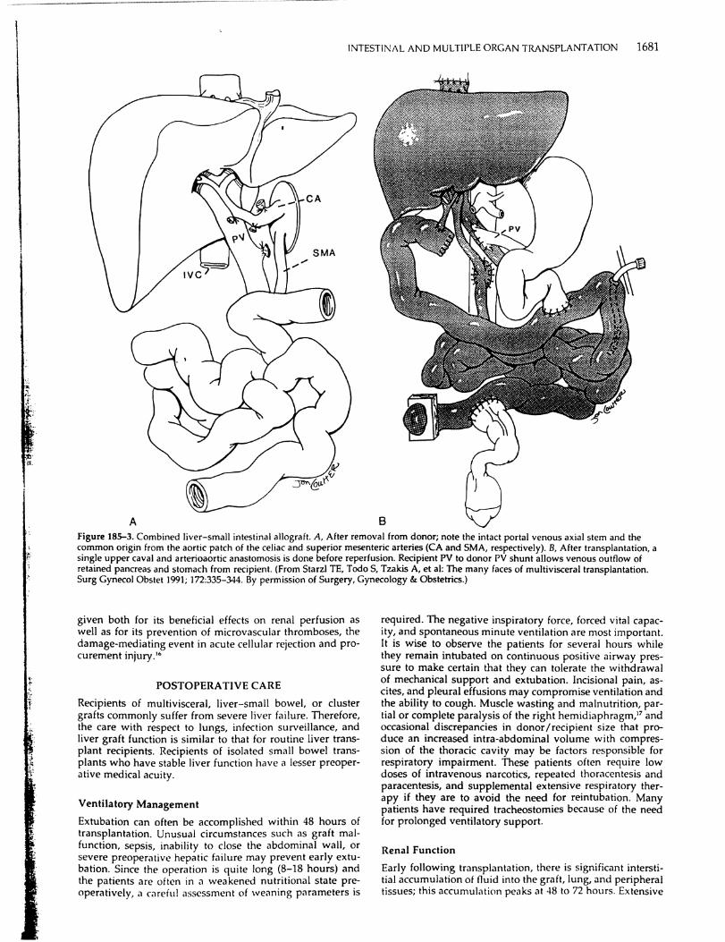
Liver and small intestine are removed in these patients, but the remainder of the foregut (stomach, duodenum, pancreas) is retained. When possible, the liver is removed with the retrohepatic vena cava preserved in situ.14 After the enterectomy, the composite allograft is implanted by anastomosing the suprahepatic vena cava of the donor (including the hepatic veins) end-ta-side to the recipient's vena cava. The donor infrahepatic vena cava can then be ligated. The double arterial stem of the celiac and superior mesenteric arteries (using the Carrel button technique) are connected to the infra renal aorta with subsequent graft reperfusion. Since the axial stem of the portal vein between the donor organs has remained intact, all that is required for the completion of portal flow is attachment of the portal vein of the remnant foregut in the recipient to the intact portal stem of the donor (Fig. 185-3). This may not be possible, however, beca'use of size discrepancy or difficult anatomic relationships between donor and recipient portal veins. In this case, a permanent portocaval shunt is per-
-

1680 TRANSPLANTATION
A
Figure 185-2. Diagrams of muitivisceral donor organs before (Al and after (8) implantation show the suprahepatic and infrahepatic vena caval and arterioaortic anastomoses. Abbreviations: Ive = inferior vena cava; PV = portal vein. (From Starzl TE, Todo S, Tzakis A, el al: The many faces of multivisceral transplantation. Surg Gynecol Obstet 1991; 172:335-344. By permission of Surgery, Gynecology & Obstetrics. )
formed. The intestinal anastomoses are then completed with a proximal jejunojejunostomy, ileocolostomy, and Roux-enY biliary anastomoses (see Fig. 185-3). A temporary distal ileostomy is also provided.
Isolated Small Bowel
After wide exposure, the recipient's small intestine is removed from the ligament of Treitz to the colon. The superior mesenteric artery of the donor bowel is sewn to the infra rena I aorta, and the donor superior mesenteric vein to the recipient portal vein or, alternatively, the donor superior mesenteric vein is anastomosed to the recipient's inferior vena cava (Fig. 185-4). Reperfusion of the graft is effected after the vascular anastomoses. Intestinal continuity is completed with proximal and distal anastomoses, and access to the ileum for endoscopic examination is provided by a tem-porary chimney ileostomy.'5 '
Cold ischemia refers to the time between procurement and implantation of the allograft; for best results, whenever a segment of the intestine is included, this period should be less than 10 hours to avoid preservation injury. Warm ischemic time for the allograft (sewing-in time) is about 30 minutes and is also a determinant of preservation injury to the intestine.
IMMUNOSUPPRESSION
Immunosuppression is similar in recipients of small bowel, liver-small bowel, cluster, and multivisceral transpl;mts. One gram of intravenous hydrocortisone (for children) or methylprednisolone (for adults) is given immediately J Itn graft reperfusion. Administration of FK-506 (0.15-0.2 Ill)j .
kg- J • d- I ) is then begun by continuous intravenous infusion, with steady-state plasma levels between 2 and 3 ng/ mL as targets. A steroid taper of methylprednisolone is started at a dose of 100 mg (for children) or 200 mg (for adults) and reduced over a period of 5 days to 10 mg (for children) or 20 mg (for adults) per day. In some cases, azathioprine may be added to mitigate the nephrotoxicity <lnd neurotoxicity of the FK-506. As gastrointestinal motilitv resumes, oral FK-S06 given twice daily may be used to supplement the intravenous regimen, which is gradually tJpered.
Induction therapy as well as chronic maintenance therapy involves the use of two and often three drugs. However, If organ tolerance with minimal rejection episodes is demonstrated, gradual reduction ilnd even cessation of sterOid therapy may be possible.
Prostaglandin EI (Prostin) is administered, 0.003 to 0.009 mg . kg - I . min - I, for the first 5 postoperative days. This IS
INTESTINAL AND MULTIPLE ORGAN TRANSPLANTATION 1681
A Figure 185-3. Combined liver-small intestinal allograft. A, After removal from donor; note the intact portal venous axial stem and the common origin from the aortic patch of the celiac and superior mesenteric arteries (CA and SMA, respectively). B, After transplantation, a single upper caval and arterioaortic anastomosis is done before reperfusion. Recipient PV to donor PY shunt allows venous outflow of retained pancreas and stomach from recipient. (From Starzl TE, Todo S, Tzakis A, et a1: The many faces of multivisceral transplantation. Surg Gynecol Obstet 1991; 172:335-344. By permission of Surgery, Gynecology & Obstetrics.)
given both for its beneficial effects on renal perfuSion as well as for its prevention of microvascular thromboses, the damage-mediating event in acute cellular rejection and procurement injury.'·
POSTOPERATIVE CARE
Recipients of multivisceral, liver-small bowel, or cluster grafts commonly suffer from severe liver failure. Therefore, the care with respect to lungs, infection surveillance, and liver graft function is similar to that for routine liver transplant recipients. Recipients of isolated small bowel transplants who have stable liver function have a lesser preoperative medical acuity.
Ventilatory Management
Extubation can often be accomplished within 48 hours of transplantation. Unusual circumstances such as graft malfunction, sepsis, inability to close the abdominal wall, or severe preoperative hepatic failure may prevent early extubation. Since the operation is quite long (8-18 hours) and the patients are often in <I weakened nutritional state preoperatively, a c<lreful assessment of weaning parameters is
required. The negative inspiratory force, forced vital capacity, and spontaneous minute ventilation are most important. It is wise to observe the patients for several hours while they remain intubated on continuous positive airway pressure to make certain that they can tolerate the withdrawal of mechanical support and extubation. Incisional pain, ascites, and pleural effusions may compromise ventilation and the ability to cough. Muscle wasting and malnutrition, partial or complete paralysis of the right hemidiaphragm,17 and occasional discrepancies in donor/recipient size that produce an increased intra-abdominal volume with compression of the thoracic cavity may be factors responsible for respiratory impairment. These patients often require low doses of intravenous narcotics, repeated thoracentesis and paracentesis, and supplemental extensive respiratory therapy if they are to avoid the need for reintubation. Many patients have required tracheostomies because of the need for prolonged ventilatory support.
Renal Function
Early follOWing transplantation, there is significant interstitial accumulation of fluid into the graft, lung, and peripheral tissues; this accumulation peaks at 48 to 72 hours. Extensive

1682 TRANSPLANTATION
volume shifts into the transplanted bowel (re~4ted to preservation injury) and heavy ascites production. (related to mesenteric lymphatic leakage) lead to intravascular volume depletion, which can exacerbate the nephrotoxicity of FK-506 and certain antibiotics. Continuous central venous pressure measurement, often for weeks following transplantation, provides important information for maximizing graft perfusion and preserving the integrity of the kidneys.
Infection Control
Recipients of isolated or composite small bowel grafts receive prophylactic, broad-spectrum intravenous antibiotics. Any history of recent nosocomial infections before transplantation should be addressed with the administration of appropriate specific antibiotics. Colonizing organisms growing from enterocutaneous fistulous tracts should be treated perioperatively.
All recipients are given a "cocktail" of oral nonabsorbable antibiotics every 6 hours for 1 month; the mix includes amphotericin B, gentamicin, and polymixin E and is intended to achieve selective bowel decontamination.'" Surveillance stool cultures are performed every 3 days. If any organisms grow in quantitative cultures to colonies of greater than 10" organisms and if the patient demonstrates signs of systemic sepsis, or if there is ongoing acute cellular rejection of the allograft, then specifically directed intravenous antibiotics are added to the regimen to treat the presumed translocating organisms. This most commonly occurs during episodes of acute rejection, when the mucosal barrier of the allograft has been immunologically damaged.
Routine prophylaxis to prevent cytomegalovirus infection includes a 2-week course of intravenous ganciclovir while
Figure 185-4. lllustr<1tion of an isolated small bowel graft shOWing mesenteric venous and arterial connections. The distal ileal chimney allows easy access to bowel mucosa. (From Todo S, Tzakis A, AbuElmagd K, et al: Intestinal transplantation in composite visceral grafts or alone. Ann Surg 1992; 216:223-234.)
lifetime oral trimethoprim-sulfamethoxazole is used as prophylaxis for pneumocystis pneumonia.
Nutritional Support
Standard total parental nutrition formulas are tapered gradually as oral or enteral feedings (via gastric or jejunal tube) are advanced. Tube feedings are initiated with an isotonic dipeptide formula containing medium-chain triglycerides and glutamine. This is later converted to a lactose- and gluten-free diet that contains dietary fibers to promote normalization of intestinal motility and function. Most patients do not voluntarily eat adequate amounts early after the operation; therefore, enteral supplementation is required when the intestinal tract becomes functional. This resistance to resumption of oral feedings has been particularly impressive in pediatric recipients."
Assessment of Graft Status
A judgment of the anatomic and functional integrity of the graft begins in the operating room. The normal <lppe<lrancl' of the mesentery and intestine is pink and nonedematous with the intestine occasionally demonstrating contractions. Alterations from this appearance can be observed in the operating room and in the ileal stoma postoperatively.
Surveillance for intestinal graft rejection focuses on clinical evaluation and gross morphologic examination of the stoma and the distal ileum with enteroscopy. EndoscopIC evaluations are performed routinely twice a week through the allograft ileostomy, whereas upper endoscopy is performed as indicated. Grossly, the bowel reacts to insult .IIl
nonspeCific ways with edema, cyanosis, congestion, and Ill'

INTESTINAL AND MULTIPLE ORCAN TRANSI'LANTAflON 1683
creased stomal output; these alterations should signal a broad differential to include preservation injury, systemic sepsis, rejection, and enteritis.
The stomal output is assessed for \'olume, consistency, and the presence of reducing substances, which can be seen in the event of rejection, bacterial overgrowth, or malabsorption. Typical stomal output of a clear, watery effluent within the first week of implantation is 1 to 2 Lid for adults and 40 to 60 mLlkg per day for children. If these volumes are exceeded and no significant pathology is present, paregoric, loperamide, pectin, somatostatin, or oral antibiotics may be utilized singly or in combination to control the diarrhea. The presence of blood in the stool is always an ominous sign and must be assumed to indicate rejection until it is proved otherwise.
Serum tests are important to give indications of possible anatomic injury to the liver (bilirubin, aspartate aminotransferase, and alanine aminotransferase), but no such tests exist for the intestinal grafts. Serum markers for nutritional adequacy and anabolic status (transferrin, albumin, retinoic acid) are of limited value, whereas specific tests of the absorptive ability of the graft are good measures of overall function. Assessment of small bowel function relies on absorption studies of D-xylose and FK-506 and on th~ quantitation of fat in the stool. Most patients develop satisfactory absorption curves for D-xylose within the first postoperative month, with absorption improving over time. Abnormal results obtained after 1 month should always prompt an aggressive search for underlying pathology, especially rejection. The maintenance of satisfactory FK-506 plasma trough levels of 2 to 3 nglmL on oral therapy alone is a good indicator of adequate absorption. In our patients, this has occurred at a mean of 28 days after transplantation and tends to be delayed longer in recipients of multivisceral grafts.'· The excretion of fat in the stool has been abnormal in almost all patients. However, clinical steatorrhea has not been a problem.
Figure 185-5. Severely damaged allograft intestine in J recipient of a liver-small bowel after multiple episodes of rejection. Diffuse tubulized gut, strictures, and significant distention of the native duodenum are seen.
Radiologic evaluations by standard barium gastrointestinal examination are valuable in assessing mucosal pattern and motility and are performed routinely ilfter the first postoperative week. A normill mucosal p,lItern is expected. Intestinal transit time is around 2 hours. Intestinal gr,lft rejection, when mild, can be suspected whell evidence of mucosal edema exists. Severe rejection, with exfoliiltion of the mucosa, ablates the normal mucosal pattern ,lOd can be seen as segments of "tubulized" intestine and strictures (Fig. 185-5).
COMPLICA nONS
Before a description of the variety of potential complications, it is important to have a general perspective on the care of these patients. Comprehensive management of intestinal recipients requires a multidisciplinary approach by surgeons, anestheSiologists, nurses, critical care physicians, pathologists, and a host of internal medicine subspecialists. Easy access to diagnostic and therapeutic modalities, including mechanical ventilation, hemodialysis, bronchoscopy, gastrointestinal endoscopy, thromboelastography, percutaneous cholangiography, ultrasonography, invasive and noninvasive contrast radiography, and sophisticated hemodynamic monitoring systems, is paramount.
More important than the above, however, is a vigilance about patient care and attention to detail, on the part of both physicians and nurses. Problems in these patients can originate from a multiplicity of sources. Several assumptions can be made in these patients based on our experience:
1. Preoperative deterioration of physical performance status predisposes to various organ system failure that persists in the postoperative period even though allograft function may be acceptable.
2. Transplant patients are labor intensive and require aggressive respiratory therapy, nutritional and antibiotic sup-

II ,
I
'I Ii
I II
16H41 Ri\NSJ'L ANT,\T10N
port. Iluid management, and nursing care, often for prolonged periods in the intensive care unit.
1. lInmunother,lpv dose's in patients with Illultivisceral transpl,l11ts tend to be hIgher than in patients with smgll' orgilll trilllsplilnts. . . .
-I. The majority oi patIents develop episodes 01 intectlOn ilnd rejection after transplantation, often concOimtantly. Any subjective complaints or objective ilbnormalities should be vigorously pursued until a Ciluse is found or until they resolve.
Graft Rejection
Intestinal allograft rejection can present ilS an array of symptoms that include iever, "l,dominal pain, distention, nausea, vomiting, and a sudden increase in stomal output. The stoma may become edematous, erythematous, and friable. Gastrointestinal bleeding can occur in cases of severe uncontrollable rejection in which ulcerations and sloughing of the intestinal mucosa occur. Septic shock or acute respiratory distress syndrome'may develop. Bacterial or fungal translocation can occur during intestinal allograft rejection due to disruption of the intestinal mucosal barrier. Gut decontamination must be in,stituted during these episodes.20
Endoscopically, the transplanted intestinal mucosa loses its velvety appearance. It may become hyperemic or dusky, as well as hypoperistaltic. Erythema may be focal or diffuse. The mucosa becomes friable, and diffuse ulcerations appear (Fig. 185-6).
Histologically, there is a variable presence of lamina propria edema and villous blunting. However, the mononuclear cell infiltrates and cryptitis with apoptosis and regeneration are necessary for establishing the diagnosis of rejection. Neutrophils, e05inllphi\S, ;l!1d macrophages may be seen tril\'ersing the mlls(ulMis mucosa.'1 The degree of epithelial and crypt cell damage \·,lries. Complete mucosal sloughing and crypt destruction are seen in grafts with severe rejection. The mucosal surface is partially replaced by inflammatory pseudomembranes and granulation tissue (Fig. 185-7). This may precipitate continuous blood loss as well as intermittent septic episodes from the damaged intestine.
Chronic rejection has been observed in patients with persistent intractable rejection episodes. Clinically progressive weight loss, chronic diarrhea, intermittent fever, and gastrointestinal bleeding dominate the presentation, Histologically, villous blunting, focal ulcerations, epithelial metapl,lsia and scant cellular infiltrate are present on endoscopic mucosal biopsies. Full-thickness intestin;d biopSIes show obliterative thickening of intestinal arterioles.
The incidence of acute intestinal allograft rejection during the first 90 days after transplantation is reported to be 80% in isolated small bowel recipients and 77% in liver-small
I I 1-' <' _.
bowel recipients. The incidenn' of acute liver allograit reJection in li\'l'r-small bowel recipients is 55%.21
Crilft rl'jectilln is tTl'alec! initially with bolus steroid ther,'py (intr,l\'en()l1s hydrocorl!~o,1l' or methylprednisolone) ill cases ot mild rejection, with a ~tl'roid taper in cases of moderate to se\'ere rejection. The FK-506 trough levels in plasm<, should reach 2 to 3 ng/mL by either the oral or intravenous routes. OKT3 is used when rejection has progressed with a steroid taper; however, it should be entertained as the initial therapeutic agent in cases of severe mucosal injury and crypt damage.
Postoperative Hemorrhage
Coagulopathy is more often illl intraoperative problem tklt relates to li\'er dysfunctJOIl, qualit,ltive <md quantitilti\'l' platelet defects, and fibrinolysis." Intraoperative bleeding is furthered by vascularized adhesions from previous surgery and portal hypertenSion. Temporary graft reperfusion coagulopathy mediated by plasminogen activators from the graft may occurY Efforts are taken to normalize these globill aspects of coagulation by the end of the operati\'e procedure so that in the absence of liver dysfunction, the coagulopathy is usually minor in the postoperative period. Postoperatin' intra-abdominal bleeding is most often a technical problem, arising from vascular anastomoses or extensive, raw peritoneal surfaces. Certainly, coagulation should be normalized if postoperative bleeding occurs; if bleeding is proved, the origin should be presumed surgical and managed as such by early exploration.
Biliary Complications
Continuity of the biliary axis is preserved in multiviscerill and cluster grafts, whereils li\'er-'Illilll bowel and isolMni small bowel grafts require Roux'l'n- Y choledochojejul111'tomy. Correspondingly, these gratts (,111 dewlop biliJrY S\S'
tem-related surgical complicatlolls (I.e., leilks and obstructions).
Biliary leaks usually occur within the first 2 weeks ,lttl'r small bowel transplantation and may herald their presellll' with bilious drainage from the abdominal wound or dr,'!II' or merely with unexplained sepsis. The response to ex telll,d bilious drainage should be immediate exploration WIth surgical revision of the biliarv dehiscellCl'. In the case oi unexplained sepsis in <Illy intestin,,1 trilllsplant recipient, all surgical anastomoses should be r'ldiogrilphically inspectl'd (with percutaneous cholanglllgr<lphv), <1I1d it leilkage is suspected, they should be openly rc\·isl'd. There is no place tor percutaneous diversion of biliary or intestinal leakage ill these patients, since both wound healing <,nd antimicf(li",,1 immunity are impaired by multInlOdal iml11l1llotherap\'
Biliary obstruction generallv follows <In anastomotic strIC' ture and is a delayed complication, but any clinical picture
Figure 185-6. A, Normall'ndoscopic i'ppear.lncl' "I transplanted small intestine. Il, 'vloderate acutl' cl'liul,\f rejection of an intestinal <lliograft lit-ll1onstr,lting dillu'" edem., and focalervthL'ma. (:;l'l' Color 1'1,)((, Sellion "I this tl'xtbo"k) .

INTESTI\!;\L AN)) MULTIPLE ORC;\N TRAI\:SI'L-\\:TATlOi\ 1685
Figure 185-7. Acute c~II~Jar rejection. A, Endoscopic biopsy obtained 14 days after transplantation showed widening of the lamina propria with increased mononuclear cells, which were often cuffed around small vessels and infiltrating the crypt epithelium (arrow; hematoxylin and eosin, 140x). B, The reaction was more intense in biopsies that contained lymphoid nodules and where blastogenesis, focal ulcerations, congestion, and neutrophil plugging of capillaries were also seen (moderate acute cellular rejection; hematoxylin and eosin, 140 x). C. Uncontrolled acute rejection eventually resulted in widespread mucosal destruction; the mucosa was replaced by granulation tissue. Note the overlying inflammatory pseudomembrane (arrow; hematoxylin and eosin, 350 X).
that resembles cholangitis or biliary obstruction should be followed with cholilngiography to prm'e piltency, regardless of the timing ,liter tr,lnsplantiltion.
Vascular Complications
Major arterial thrombosis is a disastrous complication that leads to massive necrosis of the organs correspondingly supplied. Elevation of hepatic enzymes and pallor of the intestinal stoma are accompanied by clinical deterioration, fulminant sepsis, and hepatic coma. Isolated small bowel grafts can be removed with the expectation of patient recovery, but in patients with composite grilfts, the event is usually fatal. Piltency of the arteries Can be rapidly confirmed with Doppler ultrasound examiniltion.
Since the superior mesenteric vein-portal vein ilxis is preserved in the composite grilfts, \'enous outflow thrombosis is less likely to occur in piltients with them. Isolilted small bowel grafts hine iln anastomosis of these veins that can potentially occlude. Ascites, stomal congestion, and, ultimatelv, mesenteric infarction would be the ultimate result.
\Jetther of these problems produces subtle clinical signs, ilnd diilgnosls should be prompt and obvious. In our series, thrombosis of the hepatIC artery hilS occurred in a pediatric recipient of a liver-small bowel grilft, with consequent hepatic g;mgrene. This patient required retransplilntiltion of the liver component of the grilft, even though a full liversmall bowel graft was desirable.
Incomplete obstruction of major inflow or outflow vessels may be suspected on biopsy or based on clinical and laboriltory evidence of organ dysfunction. Contrast vascular studies are conhrmiltorv, and the correction is surgical or, in some GISeS, With balloon dilatation.
Gastrointestinal Complications
C,)stro111testll1al hlel'ding ,lttl'r intestinJI trJnspl,)J)tJtion is .li1 llJ))1J)Ol1S sign 1\1,)\ requires prompt .1ttentioll. Rl'jection lS
the most probable cause and should be immediately diagnosed or ruled ou t on the basis of enteroscopic biopsy results. The di,1gnosis of rejection relies not only on histologic evidence but also on the endoscopic ilppearance (see Figs. 185-6 ilnd 185-7). However, in the ,1bsence of any pathology on endoscopy and with other supporting clinical evidence of rejection, the patient should still be treated for acute rejection.
Leakage of either the proximal or distal gastrointestinal anastomosis can occur in any recipient, but it is more common in pediatric patients than in adults. Any fresh surgical margin, including native duodenal ilnd colonic stumps and gastrostomy sites, ilre vulnerable to poor wound healing and subsequent leJkage. Presentation is often dramatic (florid sepsis), and confirmation is with radiologic contrast imaging. Surgical revision, evacuation of peritoneal soilage, and often re-exploration Me required to eliminate the contilmination effectively. Again, sepsis without an obvious source should prompt the performance of contrast studies to document the integrity of all gastrointestinal anastomoses; if the findings are inconclusive, diilgnostic laparotomy is indicated.
Native gastric iltony and pylorospilsm that produce carly satiety or vomiting are common and self-limiting. Hypermotility of the allograft intestine occurs early after trilnsplantation; in the absence of rejection or bacteriill overgrowth, it can be controlled with agents such as paregoric, loperamide, or pectin. Sudden changes in intestinal motility, particularly when accompanied by abdominal distention ilnd vomiting in the case of decreased motility, should initiilte a search for rejection.
Infections
The trequency of infectious complic;)tions is high and is rl'sponsibiL> for signiiicant morbidity and mort;)lity Jfter intestinill transpl;mtJtion. This retlL'cts the rclati\'eiv higher Il'velll! Immunosuppression requirl'd to milint,llll thl' gr,1!t

1686 TRAN~I'Li\NI:\ IIO\:
Figure 185-8. A, Endoscopic appearance of cytomegaloviral enteritis is characterized by hyperemic erosions. B, The diagnosis was confirmed histologically by the presence of characteristic inclusions, by staining for viral antigens, or both. Note the focal neutrophilic inflammation (immunopefoxidase for cytomegalovirus antigens, 350 x). (See Color Plate Section of this textbook.)
in these intestinal redpients. '8 Other predisposing factors include the severity of the preoperative liver failure (in multivisceral and liver-small bowel reCipients) as well as the presence of intra-abdominal, pulmonary, or intravenous line-induced sepsis before transplantation, Also, technically more difficult transplantation procedures with increased operative time, transfusion requirements, and likelihood of reexploration reflect the advanced disease of these patients, Recipients of small bowel grafts have the lowest incidence of complications because of the more elective nature of their candidacv.
Infectious pilthogens include bacteria, fungi, and viruses. Infections that present clinically relate (in order of frequency) to intravenous lines, the abdominal wound, deep abdominal abscesses, peritonitis, and pneumonia. Bacterial translocation in grafts damaged by rejection illustrates the need for concomitant antirejection and antimicrobial therapy and is a frequent source of infection.
Of the bacterial pathogens, staphylococci and enterococci are common, whereas gram-negative rods usually accompany polymicrobial infections. Not uncommonly, separate sources of infection occur Simultaneously, or mixed infections from the same source are present. This leads to multiple antibiotic regimens and sets the stage for the development of resistant organisms. Particularly problematic has been the nascent strain of panresistant enterococci. Persistence of a physiologic hyperdynamic state in a patient being treated for proven infection should raise the suspicion of retained phlegmonous material in the abdomen or the possibility of rejection.
Fungal infcctions become problematic ilfter heavy treatment for rCJection, massive antibiotic uSilge, intestinal leaks, and multiple surgical explorations. The authors routinely employ low-dose amphotericin B prophylaxis in patients with these complications. Established fungal infections rcquire long-tcrm, full-dose antibiotic therilpy ilnd reduction or cessation of immunotherapy. All persistently septic recipients are potential candidates for moderation of immunosuppressant dosages if no coexistent cellular rejection is present.
Clinical cytomegalovirus infection is commonly seen in adult intestinal gr<lft recipients and often involves the allograft intestine (65% of the time). A cytomegalovirus-positive donor gr<llt trzlI1splanted into a cytomegalovirus-negative reCIpient is a significant risk factor, but intense bilseline
immunosuppression with high FK-S06 levels and cumulative doses of pulse steroids is a constant feature. Clinical presentation has generally been enteritis of variable severity with focal ulcerations and bleeding (Fig. 185-8). Neither prophylactic courses of ganciclovir therapy nor passive administration of immunoglobulin has been effective in preventing cytomegalovirus infection. A voidance of cytomegalovirus-positive grafts in cytomegalovirus-negative recipients and the reduction of immunosuppression are oLir present strategy for controlling cytomegalovirus infections in intestinal graft recipients.
Less commonly, respiratory syncytial virus, adenO\·irus, and parainfluenza virus have occurred in the pediatric pop· ulation. All viral infections are opportunistic and hilve as ,1
"common denominator" the need for ilggressive treatment of rejection episodes in complicated patients with high Acute PhysiologiC and Chronic Health Evaluation scores.
Post-transplantation lymphoproliferative disease associ· ated with the Epstein-Barr virus has occurred in three children (6% of all patients and 11 % of pediatric patients) and resulted in one fatality. These patients presented with mul· tifocal disease and were treated with intravenous acyclovir or ganciclovir as well as with cessation of immunosl;ppres· sion. Rejection of the intestinal allograft may occur durin h the recovery phase and should be treated with steroids and reinstitution of FK-506 immunosuppression.'·
Under the best of circumstances, the outlilY of fin<lnci,)i and time expenditures in composite and isolated small bowel transplant recipients is impressive. For best possible results, candidates who are nutritionally optimal and free III
active infection should be selected. Donor organs should be discarded if they are less thJn perfect. Even with technicallv perfect operations, the managing physicians should expect iI panoply of postoperiltive difficulties Jnd be prepared to support these pJtients in all ways for an mdefinite peTioli <,I time. ManJging the balance between exccssive Jnd inildl'· quate immunosuppression in the face of potentially virull'nt infections, the pursuit of rejection and sources of infection, and maintenance of comprehensive critical care support arc the most chilllenging tilsks.
References
1. Grant D: Intl'stinJI tr"nspl"nlat;on: Current sl,)llIS rr,lIlspl,II11 I'roc 11)89; 21:2R61)-287J.

2. Liilehel IK. Cooll B. Miller FA: The phy~iologlc response of the small bowel of the dog to ischemia including prolonged in vitro preservation of the bowel with successful repl;lcement and survival. Ann Surg 1959; 150:5-13.
3. Starzl TE. K,lUPP IIA Jr: Mass homotr.1n~pi.1ntatiop of abdominal organs in dogs. Surg Forum 1%0; 11:1S-:\l1
4. Starzl TE. Todo S. Tzakis A. d al: ,\bdominal organ cluster transplantation for the treatment of upper abdommal malignancies. Ann Surg 1989; 210:374-386.
5. Caine RY. Sells RA. Pena JR. et al: Introduction of immunologic tolerance by porcine liver allograft. Nature 1969; 223:472-474.
6. Kamada N. Davies HS, Wight 0, et al: Liver transplantation in the rat: Biochemical and histological evidence of complete tolerance induction in nonrejector strains. Transplantation 1983; 35:304-311.
7. Starzl TE, Rowe M, Todo S, ct al: Tr<lnsplantation of multiple abdominal \'iscera. JAMA 1989; 261:1449-1457.
8. Grant 0, Wall W, Mimeault R, et al: Successful small-bowell liver transplantation. Lancet 1990; 335:181-184.
9. Goulet OK, Revillon Y, Jan 0, et al: Small-bowel transplantation in children. Transplant Proc 1990; 22:2499.
10. Deitz E, Schroeder P, Gebhard H, et al: Successful clinical small bowel transplantation: Report of a case. Clin Transplant 1989; 3:89.
11. Oasis, Home Nutrition Support Patient Registry Annual Report-1989 Data. Albany, NY, The Oley Foundation, 1991.
12. Casella JF, Lewis JH, Bontl!mpo FA, et al: Successful treatment for homozygous protein C deficiency by hepatic transplantation. Lancet 1988; i:435-438.
I3. StaTZI TE, Todo S, Tzakis A, et al: The manv faces of multivisceral transplantation. Surg Gynecol Obstet 1991; 172:335-344.
14. Tzakis A, Todo S, Starzl TE: Piggyback orthotopic liver transplantation with preservation of the inferior vena cava. Ann Surg 1989; 210:649-652.
15. Todo S, Tzakis A, Abu-Elmagd K, et al: Intestinal transplantation in composite visceral grafts or alone. Ann Surg 1992; 216:223-234.
16. Takaya S, Iwaki Y, Starzl TE: Liver tran~pl~ntati"n in positive cytotoxic crossmatch cases using FK50(,. high dosl' ,teroids, and prostaglandin E,. Transplant,ltion 1991; 54:9~7-933.
17. Karavias 0, Jabbour N, Felekouras E. l'I al: Right diaphragmatiC paralysis following orthotopic liver transplantation. Submitted.
18. Reyes J, Abu-Elmagd K, Tzakis A. et al: InfectiOUs complications after human small bowel transplantation. Transplant Proc 1992; 24:1249-1250.
18a. Green M, Reyes J, Nour B, et al: Earlv infectious complications of liver-intestinal transplantation in children: Preliminary analysis. Transplant Proc 1994; 26:1420-1421.
19. Reyes J, Tzakis AG, Todo S, et al: Nutritional management of intestinal transplant reCipients. Transplant Proc 1993; 25:1200-1201.
20. Abu·Elmagd K, Tzakis A, Todo S, et .11: Monitoring and trl'atment of intestinal allograft rejection in humans. Tr,lnsplant I'roc 1993; 25: 1202-1203.
21. Nak;lnlUra K. Abu-Elmagd K. Todo S. l't al: [)athlllogv of human small intestinal transpluntation: Alonl' or In combination with the liver. Submitted.
22. Kang YG, Martin OJ, Marquez jM, et ill: Intraoperative changes in blood coagulation and thromboelastogruphic monitoring in liver transplantation. Anesth Analg 1985; 64:888
23. Stahl RL, Duncan A, Hooks MA, et al: A hvpl'rcoagulable statl' follows orthotopic liver transplantation. f lepatology 1990; 12:553.
24. Reyes J, Tzakis A, Bonet H, et al: Lymphoprnlifl'rati\"l' disease ,lfter intestinal transplantation under FK50h immunosuppression. Transplant Proc 1994; 26:1426-1427.
BONE \lARROW TRANSI'L;\NTATION 1687
.-CHAPTER 186
Bone Marrow Transplantation
£dlPard D. Ball, MD . flalln). BloolII. MD Alber/ D. DOllnellberg, PIID • S/t't'ell M. PillCIIs. MD. PhD Wi/old B. Rybka, MD • Margarida deMagallwl's-Sihwlllall. MD
Bone marrow transplantation (BMT) is increasingly used in the treatment of certain malignant as well as nonmalignant disorders (Table 186-1). The basic strategy is to give very high doses of drugs or radiation or both to ablilte malignant cells or the host's Iymphohematopoietic system, iollowed by infusion of normal stem cells. The source oi these cells can be a human leukocyte antigen (HLA)-identical sibling or a partially or fully HLA-identical related or matched unrelated donor (MUD). These are called allogeneic BMT. A twin donor is sometimes used (syngeneic transplant). In autologous BMT, the recipient's bone marrow (BM) or blood-derived stem cells are used. The choice between an autologous or allogeneic BMT depends on many issues, including the disease state, the availability of a suitable donor, and the risk-benefit analysis for eac;, patient. The age of BMT recipients has been increasing. Allogeneic BMT is now performed in patients up to 55 years of age. Autologous BMT can probably be performed up to age 70. This chapter reviews the major indications and problems associated with BMT and attempts to address the main issues in treatment of patients undergoing BMT.
INDICA TIONS
The indicJtions for BMT arc steadily illcrt.'Jsillg. 13\1T \VJS first applied to the leukemias and immune deiiciency disorders (including aplastic anemia), but the indications have expanded to include lymphoma, solid tumors, <1nd genetic diseases. As BMT becomes safer, the indications and the numbers of patients treated will most likely continue to increase. Some of the current indications for BMT are shown in Table 186-1.
ACUTE LEUKEMIA. Complete remission (CR) rates of 70% to 80% are achieved in the majority of piltients with i1cute myelogenous leukemia (AML). Cure of AML can only be obtained with additional therapy. Direct comparisons of the different modalities of treatment for p<1tients with AML in first CR have been difficult to assess. It is vet unclciIT whether BMT (autologous or allogeneic from HLA-identic<11 sibling) would result in long-term leukemia-free sur\'iv<11 superior to that with chemotherapy in patients with AML in first CR when various prognostic variables are tilken into account. I Studies addressing this issue are under way.
TABLE 186-1. Disorders Treated with Bone Marrow Transplantation
Maligllallt Disorders Acute leukemia Chronic myelogenous leukemia Myelodysplastic syndrome Lymphomas Solid tumors NOlllllll/igllllllt Disorders Aplastic anemia Immull()lo~ic diSlucil'rs ~lemogl()blnopathi<'s Metabolic disorders