Initiation of Meiotic Homologous Recombination: Flexibility, Impact...
17
Initiation of Meiotic Homologous Recombination: Flexibility, Impact of Histone Modifications, and Chromatin Remodeling Lo ´ ra ´ nt Sze ´ kvo ¨ lgyi 1 , Kunihiro Ohta 2 , and Alain Nicolas 3 1 Department of Biophysics and Cell Biology, Faculty of Medicine, University of Debrecen, 4032 Debrecen, Hungary 2 Department of Life Sciences, The University of Tokyo, 113-8654 Tokyo, Japan 3 Institut Curie Centre de Recherche, UMR3244 CNRS, Universite ´ Pierre et Marie Curie, 75248 Paris CEDEX 05, France Correspondence: [email protected]; [email protected] Meiotic recombination is initiated by the formation of DNA double-strand breaks (DSBs) catalyzed by the evolutionary conserved Spo11 protein and accessory factors. DSBs are nonrandomly distributed along the chromosomes displaying a significant ( 400-fold) var- iation of frequencies, which ultimately establishes local and long-range “hot” and “cold” domains for recombination initiation. This remarkable patterning is set up within the chro- matin context, involving multiple layers of biochemical activity. Predisposed chromatin accessibility, but also a range of transcription factors, chromatin remodelers, and histone modifiers likely promote local recruitment of DSB proteins, as well as mobilization, sliding, and eviction of nucleosomes before and after the occurrence of meiotic DSBs. Here, we assess our understanding of meiotic DSB formation and methodsto change its patterning. We also synthesize current heterogeneous knowledge on how histone modifications and chro- matin remodeling may impact this decisive step in meiotic recombination. S exual reproduction depends on halving the genome content of germ line cells and faith- ful chromosome transmission during meiosis to yield viable gametes. Meiosis comprises one round of DNA replication and two successive rounds of chromosome segregation, allowing the reduction of a diploid genome to produce haploid gametes (Fig. 1A). Central to meiosis is the process of recom- bination between the paternal and maternal chromosomes (interhomolog recombination), which is crucial to enhance the genetic di- versity of the gametes, but also for providing physical connections among homologs. These connections (i.e., chiasmata) ensure proper alignment of homologous chromosome pairs on the spindle, promoting proper reductional segregation following the regulated release of the sister chromatid cohesion among the dupli- cated chromosomal arms (Watanabe 2012). Defective meiotic recombination is a source of de novo germline mutations, abnormal genome content in gametes (the source of Down’s syn- drome), and infertility. Not surprisingly, cells have developed a variety of mechanisms and tight controls to ensure sufficient and well-dis- Editors: Stephen Kowalczykowski, Neil Hunter, and Wolf-Dietrich Heyer Additional Perspectives on DNA Recombination available at www.cshperspectives.org Copyright # 2015 Cold Spring Harbor Laboratory Press; all rights reserved; doi: 10.1101/cshperspect.a016527 Cite this article as Cold Spring Harb Perspect Biol 2015;7:a016527 1 on February 13, 2020 - Published by Cold Spring Harbor Laboratory Press http://cshperspectives.cshlp.org/ Downloaded from
Transcript of Initiation of Meiotic Homologous Recombination: Flexibility, Impact...

Initiation of Meiotic HomologousRecombination: Flexibility, Impact of HistoneModifications, and Chromatin Remodeling
Lorant Szekvolgyi1, Kunihiro Ohta2, and Alain Nicolas3
1Department of Biophysics and Cell Biology, Faculty of Medicine, University of Debrecen,4032 Debrecen, Hungary
2Department of Life Sciences, The University of Tokyo, 113-8654 Tokyo, Japan3Institut Curie Centre de Recherche, UMR3244 CNRS, Universite Pierre et Marie Curie,75248 Paris CEDEX 05, France
Correspondence: [email protected]; [email protected]
Meiotic recombination is initiated by the formation of DNA double-strand breaks (DSBs)catalyzed by the evolutionary conserved Spo11 protein and accessory factors. DSBs arenonrandomly distributed along the chromosomes displaying a significant (�400-fold) var-iation of frequencies, which ultimately establishes local and long-range “hot” and “cold”domains for recombination initiation. This remarkable patterning is set up within the chro-matin context, involving multiple layers of biochemical activity. Predisposed chromatinaccessibility, but also a range of transcription factors, chromatin remodelers, and histonemodifiers likely promote local recruitment of DSB proteins, as well as mobilization, sliding,and eviction of nucleosomes before and after the occurrence of meiotic DSBs. Here, weassess our understanding of meiotic DSB formation and methods to change its patterning. Wealso synthesize current heterogeneous knowledge on how histone modifications and chro-matin remodeling may impact this decisive step in meiotic recombination.
Sexual reproduction depends on halving thegenome content of germ line cells and faith-
ful chromosome transmission during meiosisto yield viable gametes. Meiosis comprises oneround of DNA replication and two successiverounds of chromosome segregation, allowingthe reduction of a diploid genome to producehaploid gametes (Fig. 1A).
Central to meiosis is the process of recom-bination between the paternal and maternalchromosomes (interhomolog recombination),which is crucial to enhance the genetic di-versity of the gametes, but also for providing
physical connections among homologs. Theseconnections (i.e., chiasmata) ensure properalignment of homologous chromosome pairson the spindle, promoting proper reductionalsegregation following the regulated release ofthe sister chromatid cohesion among the dupli-cated chromosomal arms (Watanabe 2012).Defective meiotic recombination is a source ofde novo germline mutations, abnormal genomecontent in gametes (the source of Down’s syn-drome), and infertility. Not surprisingly, cellshave developed a variety of mechanisms andtight controls to ensure sufficient and well-dis-
Editors: Stephen Kowalczykowski, Neil Hunter, and Wolf-Dietrich Heyer
Additional Perspectives on DNA Recombination available at www.cshperspectives.org
Copyright # 2015 Cold Spring Harbor Laboratory Press; all rights reserved; doi: 10.1101/cshperspect.a016527
Cite this article as Cold Spring Harb Perspect Biol 2015;7:a016527
1
on February 13, 2020 - Published by Cold Spring Harbor Laboratory Press http://cshperspectives.cshlp.org/Downloaded from

1: Initiation
B
Spo11
2: DSB processing
3: DSB repair
4: Resolution/ dissolution
Crossover Noncrossover Noncrossover
Double-Holliday junctionpathway
Synthetic-dependentstrand-annealing pathway
A
S DSB CO/SCMI
MII
Figure 1. The stages and mechanisms of meiosis. (A) Diploid yeast cells initiate meiosis on nutrient depletionand in the presence of a nonfermentable carbon source. In mammals, the process is started by endocrine/paracrine/juxtacrine stimuli from the surrounding cell and tissue environment. Relevant molecular stages areindicated: S, meiotic replication; DSB, double-strand breaks; CO, crossover; SC, synaptonemal complex; MI,and MII (first and second meiotic divisions, respectively). Colors: parental homologous chromosomes (greenand black), sister chromatid cohesion (red), SC (pink). (B) The mechanism of meiotic recombination. Stage 1:Initiation. DSBs are introduced by the Spo11 protein. Stage 2: DSB processing. Strand resection initiates to yield30 single-stranded DNA (ssDNA) overhangs. One of the 30 ssDNA tails engages in strand invasion and ahomology search of the homologous chromosome, resulting in single-end invasion (SEI) and D-loop interme-diates. Stage 3: DSB repair. (Legend continues on following page.)
L. Szekvolgyi et al.
2 Cite this article as Cold Spring Harb Perspect Biol 2015;7:a016527
on February 13, 2020 - Published by Cold Spring Harbor Laboratory Press http://cshperspectives.cshlp.org/Downloaded from

tributed meiotic recombination events withintheir genomes.
At the DNA level, meiotic recombinationcan be divided in four successive stages (Fig.1B): (1) initiation, which consists in the for-mation of programmed DNA DSBs; (2) DSBprocessing, which yields the recombinogenicsingle-strand tails; (3) homologous DSB repair,which involves the homologous recombinationpathway and several meiosis-specific differ-entiation modulations, which facilitate inter-homolog interactions; and (4) intermediateresolution and dissolution, which allow the for-mation of local (up to a few kb in length) non-crossover (NCO) gene conversion events andreciprocal crossovers (COs).
Recently, local and genome-wide studies ofnormal and mutant cells have uncovered a re-markable variability in the number and posi-tioning of recombination events per chromo-some and cell, which reveals an impressive levelof flexibility (Szekvolgyi and Nicolas 2010). Inthis review, we outline our understanding ofthe control of the initiation events, and howhistone modifications and chromatin remodel-ing impact this initial step of meiotic recombi-nation. The process of meiotic recombinationand its relationship to change in chromosomestructures and movements, allowing homologalignment, pairing,andsynapsis, isalso reviewedin Zickler and Kleckner (2015) and Lam andKeeney (2015).
MEIOTIC DSBs AND RECOMBINATIONEVENTS ARE NONRANDOMLY DISTRIBUTED
In all organisms, meiotic recombination is ini-tiated by the formation of a large number of
programmed DNA DSBs per cell, which are re-paired primarily via recombination betweennonsister chromatids (recombination initiationoccurs after replication) to generate NCO andCO recombinant products (Fig. 1B). TheseDSBs are catalyzed by the evolutionarily con-served Spo11 protein (Bergerat et al. 1997). Be-sides Spo11, a number of accessory proteins arerequired for DSB formation. In Saccharomycescerevisiae, 10 DSB proteins are known and func-tional, but sometimes sequence-divergent or-thologs are gradually being identified in otherorganisms, including mammals. Yeast two-hy-brid and co-immunoprecipitation (IP) studiesidentified several multiprotein subcomplexes,which are successively recruited to the meioticchromosomes before DSB formation in trig-gered (Arora et al. 2004; Panizza et al. 2011).The biochemical/structural functions of theseDSB protein complexes remains poorly under-stood. They play a role to select the potentialDSB regions along the chromosomes, and ulti-mately contribute to the recruitment of Spo11and trigger cleavage.
Spo11 is orthologous to the topoVI familyof topoisomerase discovered in archaea, andconsistently introduces DSBs by coupled trans-esterification reactions to form covalent tyrosyl-DNA linkages at the 50 termini of the brokenDNA. Spo11 is then removed byendonucleolyticcleavage (Neale et al. 2005), liberating shortSpo11-DNA oligonucleotide complexes andresected strands, which are further extendedto generate recombinogenic 30 single-strandedtails. Over the years, meiotic DSBs have beenmapped and quantified in yeast genomic DNAusing a variety of approaches, including South-ern blot analysis of chromosomal fragments
Figure 1. (Continued) In the double-Holliday junction (dHJ) pathway, the opposite DSB end is captured byannealing to the displaced strand of the D-loop, leading to the formation of a dHJ. In the synthesis-dependentstrand-annealing (SDSA) pathway, repair of DSBs occurs without the formation of a dHJ. Stage 4: Resolution/dissolution. In the dHJ pathway, after gap-filling DNA synthesis and nick ligation, the dHJ is cleaved onopposing single DNA strands, generating products that can be ligated. Depending on cleavage, the patternsdHJ resolution produces either CO recombinants (associated or not with gene conversion that results from HJmigration) or noncrossover (NCO) (gene conversions). In the SDSA pathway, the SEI intermediate undergoesDNA synthesis by extension of the invading DNA strand with D-loop dissolution, and the extended ssDNAultimately reanneals to its original complementary ssDNA strand on the opposite side of the DSB. An intactduplex is then produced by gap-filling DNA synthesis and nick ligation, which gives rise to NCO recombinants.
Meiotic DSBs: Toward Chromatin Architecture
Cite this article as Cold Spring Harb Perspect Biol 2015;7:a016527 3
on February 13, 2020 - Published by Cold Spring Harbor Laboratory Press http://cshperspectives.cshlp.org/Downloaded from

or full-length chromosomes, PCR, chromatinimmunoprecipitation (ChIP)-Chip, and ChIP-seq analyses of enriched single-stranded DNA(ssDNA) (Buhler et al. 2007) and, most recently,via the purification and sequencing of Spo11-associated oligos, providing exquisite resolutionboth in terms of mapping and quantification(Pan et al. 2011). The most amenable methodto estimate the number of DSBs on a per-cellbasis is to count the number of Rad51/Dmc1immunostaining foci on spread meiotic nuclei(Bishop 1994). On a broad scale, these methodshave provided consistent general conclusions.They show the evolutionary conservation ofthe mechanism of DSB formation and the essen-tial role of interhomolog recombination for theproduction of viable, euploid meiotic products.Depending on the organism and mutant defect,a lack or reduction in DSB frequency can lead toarrest of meiotic progression and apoptosis, orprogression and formation of inviable aneu-ploid gametes.
To address whether Spo11-catalyzed DSBsplay a unique role in meiosis, the functionalityof DSBs introduced by other nucleases and clas-togenic agents has been examined. Interhomo-log recombination was promoted by ionizingradiation (Bowring et al. 2006), indicating thatSpo11-catalyzed DSBs are not unique in theirability to promote recombination during meio-sis. Other studies used the expression of a heter-ologous endonuclease, such as HO (Kolodkinet al. 1986; Malkova et al. 2000), VDE (Hodgsonet al. 2011), or I-SceI (Farah et al. 2009) in yeast,and the Mos1 transposase in Caenorhabditis el-egans (Robert and Bessereau 2007). In all cases,interhomolog recombination was induced atthe break sites. However, in several respects,the DSBs and recombinants induced by thesesystems are different than those resulting fromSpo11. These site-specific DSBs are not bonafide Spo11 breaks because of (1) the uncon-trolled timing of DSB induction, (2) the abnor-mal and dangerous cleavage of both sister chro-matids, and (3) alteration in recombinationefficiencies when assayed in the wild-type orSpo11-deficient strain background. This relatesto the role of the Spo11 DSBs in the pairing of thehomologs. The uniqueness of the Spo11 breaks
might reside in its mode of cleavage that allows,like in other site-specific recombination pro-cesses, to intimately link break formation andprocessing, and also to avoid extensive DSB sig-naling associated with accidental DSBs (Bordeet al. 2004). Furthermore, Spo11 break forma-tion is intimately linked to the process of chro-mosome pairing that enforces interhomolograther than intersister recombinational repair.
Extensive studies with S. cerevisiae (Baudatand Nicolas 1997; Gerton et al. 2000) and laterwith Schizosaccharomyces pombe (Cromie et al.2007), Arabidopsis (Drouaud et al. 2013), mice(Smagulova et al. 2011), chimpanzee, and hu-mans (Myers et al. 2010; Auton et al. 2012)showed that the distribution of meiotic DSBsis not random along chromosomes, explainingthe longtime noted discrepancy between physi-cal and genetic map distances. Nowadays, genet-ic distances are measured by high-throughputmicroarray or genome-wide sequencing analy-ses of meiotic progenies, starting from diploidheterozygous strains carrying thousands of sin-gle-nucleotide polymorphisms (SNPs). In a pi-oneer study in S. cerevisiae, Mancera et al. (2008)genotyped the four spores of several tetrads toreconstitute the meiotic recombination eventsper meiotic cell and, on average, found �90COs and 46 NCOs (66 after correction for unde-tected NCO) per meiosis. Consistently, these re-combination events are located in correlationwith the preferred sites of DSBs and, in a num-ber, suggesting that a large majority of the DSBs(estimated to �150 per meiosis) are repairedon the homologous chromosome. This is con-sistent with the single-site genetic and physi-cal analysis of recombination intermediatesat the most studied artificial HIS4::LEU2 hot-spot, indicating that �80% of the meioticDSBs are repaired using a nonsister chromatidrather than the sister chromatid as template (Laoet al. 2013). Genome-wide analysis of meioticproducts in other organisms confirms thatmost DSBs are repaired by interhomolog recom-bination (Cole et al. 2010), but the large excess ofDSBs relative to COs—identified directly or bycounting the Rad51 and Dmc1 foci on meioticprophase spreads over the final recombinationproducts—can be explained also if meiotic DSB
L. Szekvolgyi et al.
4 Cite this article as Cold Spring Harb Perspect Biol 2015;7:a016527
on February 13, 2020 - Published by Cold Spring Harbor Laboratory Press http://cshperspectives.cshlp.org/Downloaded from

repair frequently occurs among sister chroma-tids. This raises the key question of how and towhat extent the interhomolog bias is imple-mented and, globally, to what extent the meioticversus mitotic differentiation of DSB repair tem-plate choice is organism specific.
MULTIPLE LAYERS OF CONTROL SHAPE THEDSB DISTRIBUTION: CLUSTERING,INTERFERENCE, AND REDISTRIBUTION
If recombination-initiation sites were randomlydistributed, DSBs would be equally likely to oc-cur at any location along the chromosomes andwould not influence one another (Fig. 2A). Thisis clearly not the case because (1) DSB frequen-cies vary greatly from site to site (at least 400-foldin S. cerevisiae, S. pombe, and Arabidopsis) (Panet al. 2011; Choi et al. 2013; Fowler et al. 2014).(2) DSB sites within a hotspot region are stronglylocalized. In S. cerevisiae, they are preferentiallylocalized in intergenic promoter-containing in-tervals, but depleted from other regions (Baudatand Nicolas 1997). In S. pombe and mice, en-hanced DSB formation occurs in various re-gions, but also alternate with regions showinglow DSB activity. (3) Over longer distances,DSB-prone and -repressed regions are clusteredin subchromosomal domains. The cold regionsare found in interstitial coding regions, showingno specific pattern along the chromosomes, butDSB formation is always strongly suppressed inthe telomere- and centromere-proximal regions,as well as within the recombinant DNA (rDNA).These quantitative and region-specific varia-tions are indeed strong evidence for the nonran-dom nature of the DSB distribution. The contri-bution of rare and, possibly, random (stochastic)DSBs is not excluded, but remains difficult tomeasure and map.
Figure 2A illustrates two hypothetical non-random modes of DSB distribution: (1) a uni-form distribution, when every DSB falls as farfrom its neighbors as possible, and (2) clustered,when most DSBs are concentrated close togetherand large regions contain very few, if any, breaks.The observed DSB pattern in S. cerevisiae (Bordeet al. 2009) illustrated for chromosome III fallsbetween these two extremes; with some devia-
tion, it falls closer to the clustered arrangement,indicating that the contribution of stochasticitymight be significant. It is noteworthy that thecurrent genome-wide DSB-mapping tech-niques (Spo11-oligo sequencing, Dmc1/Rpa/ssDNA ChIP) obscure cell-to-cell variations inDSB formation (as do all mass-biochemicalmethods that investigate cell populations).
Other strong driver elements that shape thenonrandom distribution of DSBs have been doc-umented in S. cerevisiae, but also remain poorlyunderstood. These phenomena are referred toas “DSB interference” and appear to drive evenspacing of DSBs and limit simultaneous break-age at allelic positions. First, the occurrence ofDSBs reduces the probability of nearby DSBs tooccur (Fig. 2B). This is a “cis-inhibition” process,in which strong DSB hotspots suppress the ac-tivity of nearby recombination-initiation siteson the same homolog (Xu and Kleckner 1995;Fan et al. 1997). This inhibitory effect of naturalor targeted DSBs on adjacent hotspots is knownto spread over significant distances along thechromosomes (up to 25–100 kbp in S. cerevi-siae) (Robine et al. 2007). The second interfer-ence phenomenon is that DSB formation on onehomolog decreases DSB formation on the otherhomolog (trans-inhibition) at the cognate allelicposition (Rocco et al. 1992; Zhang et al. 2011a).Whether these cis- and trans-regulatory eventsoccur independently remains to be elucidated,but, importantly, the trans-modulation appearsto be genetically controlled, involving the twosignal transduction kinases Mec1 (ATR) andTel1 (ATM) acting as potential direct effectorsof the trans-DSB interference (Zhang et al.2011a; Blitzblau and Hochwagen 2013; Gray etal. 2013). The benefit to locally regulate DSBformation between the sister and nonsister chro-matids is to prevent simultaneous DSB forma-tion at the same place on more than one chro-matid. This would generate poorly interactingbroken molecules, perturb the efficiency ofDSB repair, and, finally, facilitate mutagenicend-joining and out-of-register interactions be-tween repeats, as well as genome rearrangementson ectopic interactions. Rare but naturally oc-curring events of this type may contribute tothe arising of de novo germline mutations, a
Meiotic DSBs: Toward Chromatin Architecture
Cite this article as Cold Spring Harb Perspect Biol 2015;7:a016527 5
on February 13, 2020 - Published by Cold Spring Harbor Laboratory Press http://cshperspectives.cshlp.org/Downloaded from

Clustered
Uniform
Real
Cold Cold Cold
Chromosome lll coordinates (bp)
Cold ColdHot
50,000 100,000 150,000 200,000 250,000 300,000
Hot Hot Hot
1
100 lb 100 lb
A
Early DSBs
CEN
Late DSBs
x
x x x x x
xEvenly spaced interhomolog interactions
DSB 2.
DSB 5.
DSB
3.
*
B
DSB 1. *
*
DSB 4. *
*
Chromosome coordinates (bp)
Wild type
DS
B p
rofil
e Mutant 1 (e.g., UASGAL80)
Mutant 2 (e.g., set1Δ)
CThreshold
Figure 2. Spatial distribution of meiotic DNA double-strand breaks (DSBs) along the chromosomes. (A)Hypothetical (blue) and real (black) DSB patterns, representing random and nonrandom distributions. RealDSBs (Borde et al. 2009) establish “cold” and “hot” domains, which alternate along the chromosomes length.(B) DSB interference establishes even spacing among interhomolog crossovers (COs). Interference nucleates atactivated DSB sites (yellow rectangles) cleaved by Spo11 (red star) and spreads over significant distances alongthe sister/nonsister chromatids or homologous chromosome (trans-inhibition, blue triangles), or along thesame sister chromatid (cis-inhibition, green triangles). DSBs form sequentially as meiosis progresses such that“late” DSBs fill in the gaps among “early” DSBs. A subset of DSBs matures into COs (blue crosses). (C) DSBredistribution can shuffle recombination-initiation events to reinforce the “obligatory” number of COs perchromosome. Mutant 1 can be, for example, an upstream activating sequence (UAS)GAL80, mutant, in which theGal4 binding site was inactivated to prevent DNA cleavage by the Gal4BD-Spo11 fusion protein. New DSBsreadily appear nearby the UAS. Mutant 2 corresponds to, for instance, a set1D mutant, in which the disappear-ance of “canonical” hotspots often results in the activation of latent hotspots.
L. Szekvolgyi et al.
6 Cite this article as Cold Spring Harb Perspect Biol 2015;7:a016527
on February 13, 2020 - Published by Cold Spring Harbor Laboratory Press http://cshperspectives.cshlp.org/Downloaded from
source of human diseases (Campbell and Eichler2013). Also, cells with broken chromosomes mayprogress further into the meiotic prophase, and,unless they are arrested by the DNA damagecheckpoint before MI, the chromosomes wouldsegregate abnormally, yielding unbalanced ge-nomes.
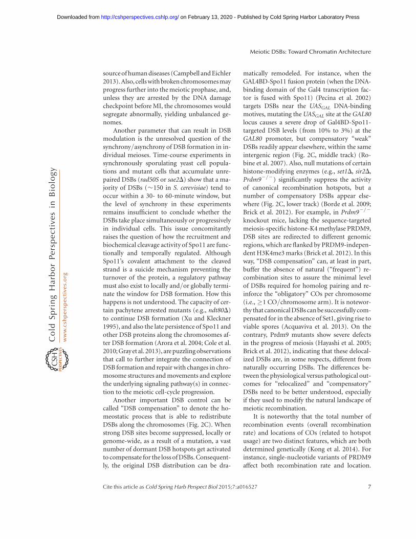
Another parameter that can result in DSBmodulation is the unresolved question of thesynchrony/asynchrony of DSB formation in in-dividual meioses. Time-course experiments insynchronously sporulating yeast cell popula-tions and mutant cells that accumulate unre-paired DSBs (rad50S or sae2D) show that a ma-jority of DSBs (�150 in S. cerevisiae) tend tooccur within a 30- to 60-minute window, butthe level of synchrony in these experimentsremains insufficient to conclude whether theDSBs take place simultaneously or progressivelyin individual cells. This issue concomitantlyraises the question of how the recruitment andbiochemical cleavage activity of Spo11 are func-tionally and temporally regulated. AlthoughSpo11’s covalent attachment to the cleavedstrand is a suicide mechanism preventing theturnover of the protein, a regulatory pathwaymust also exist to locally and/or globally termi-nate the window for DSB formation. How thishappens is not understood. The capacity of cer-tain pachytene arrested mutants (e.g., ndt80D)to continue DSB formation (Xu and Kleckner1995), and also the late persistence of Spo11 andother DSB proteins along the chromosomes af-ter DSB formation (Arora et al. 2004; Cole et al.2010; Grayet al. 2013), are puzzling observationsthat call to further integrate the connection ofDSB formation and repair with changes in chro-mosome structures and movements and explorethe underlying signaling pathway(s) in connec-tion to the meiotic cell-cycle progression.
Another important DSB control can becalled “DSB compensation” to denote the ho-meostatic process that is able to redistributeDSBs along the chromosomes (Fig. 2C). Whenstrong DSB sites become suppressed, locally orgenome-wide, as a result of a mutation, a vastnumber of dormant DSB hotspots get activatedto compensate for the loss of DSBs. Consequent-ly, the original DSB distribution can be dra-
matically remodeled. For instance, when theGAL4BD-Spo11 fusion protein (when the DNA-binding domain of the Gal4 transcription fac-tor is fused with Spo11) (Pecina et al. 2002)targets DSBs near the UASGAL DNA-bindingmotives, mutating the UASGAL site at the GAL80locus causes a severe drop of Gal4BD-Spo11-targeted DSB levels (from 10% to 3%) at theGAL80 promoter, but compensatory “weak”DSBs readily appear elsewhere, within the sameintergenic region (Fig. 2C, middle track) (Ro-bine et al. 2007). Also, null mutations of certainhistone-modifying enzymes (e.g., set1D, sir2D,Prdm92/2) significantly suppress the activityof canonical recombination hotspots, but anumber of compensatory DSBs appear else-where (Fig. 2C, lower track) (Borde et al. 2009;Brick et al. 2012). For example, in Prdm92/2
knockout mice, lacking the sequence-targetedmeiosis-specific histone-K4 methylase PRDM9,DSB sites are redirected to different genomicregions, which are flanked by PRDM9-indepen-dent H3K4me3 marks (Brick et al. 2012). In thisway, “DSB compensation” can, at least in part,buffer the absence of natural (“frequent”) re-combination sites to assure the minimal levelof DSBs required for homolog pairing and re-inforce the “obligatory” COs per chromosome(i.e., �1 CO/chromosome arm). It is notewor-thy that canonical DSBs can be successfully com-pensated for in the absence of Set1, giving rise toviable spores (Acquaviva et al. 2013). On thecontrary, Prdm9 mutants show severe defectsin the progress of meiosis (Hayashi et al. 2005;Brick et al. 2012), indicating that these delocal-ized DSBs are, in some respects, different fromnaturally occurring DSBs. The differences be-tween the physiological versus pathological out-comes for “relocalized” and “compensatory”DSBs need to be better understood, especiallyif they used to modify the natural landscape ofmeiotic recombination.
It is noteworthy that the total number ofrecombination events (overall recombinationrate) and locations of COs (related to hotspotusage) are two distinct features, which are bothdetermined genetically (Kong et al. 2014). Forinstance, single-nucleotide variants of PRDM9affect both recombination rate and location.
Meiotic DSBs: Toward Chromatin Architecture
Cite this article as Cold Spring Harb Perspect Biol 2015;7:a016527 7
on February 13, 2020 - Published by Cold Spring Harbor Laboratory Press http://cshperspectives.cshlp.org/Downloaded from

Other genetic variants have been identified thatalter the genetic map in humans: (1) CTCFL, thetestis-specific paralog of CTCF, a Zn-finger pro-tein organizing chromatin loops with cohesins(Sleutels et al. 2012), (2) the meiosis-specificcohesin RAD21L, which plays a role in the for-mation of meiotic chromosome axis and synap-sis, (3) the SUMO ligase RNF212, ubiquitinligase HEI10 (CCNB1IP1), and MutS homo-log MSH4, all related to members of the ZMMclass of pro-CO factors identified in S. cerevisiae,although with different synapsis defects (Rey-nolds et al. 2013; Kong et al. 2014; Qiao et al.2014). Strikingly, significant differences are seenamong these genetic variants, as those havinga large impact on recombination rates have noeffect on hotspot usage. This implies that, inmost cases, genetic variants affect the CO orNCO decision rather than directly touching onDSB formation. Accordingly, the yeast Zip3 pro-tein preferentially localizes to DSB hotspots thattend to be repaired as COs (whereas Zip3 is de-pleted from NCO-biased hotspots) (Serrentinoet al. 2013), pointing to the existence of distincttypes of DSB sites with regard to CO and NCOformation.
CHROMATIN REMODELING AND HISTONEMODIFICATIONS IMPACT MEIOTIC DSBFORMATION
Overall, heritable polymorphisms can be a ma-jor source of quantitative genetic variation shap-ing the recombination landscape among indi-viduals, but circumstantial and epigeneticallyinherited elements of the chromatin structurecan also significantly contribute to the plasticityof DSB formation. DNA cleavage by Spo11requires an accessible DNA template; hence,the energy barrier inherently exerted by nu-cleosomes and other chromatin-packaging pro-teins must be overcome. Because spontaneousrates of dissociation and sliding of nucleosomecore particles vastly exceed biologically relevanttimescales, diffusion-driven processes alonecannot account for the extensive reorganizationseen at DSB sites in vivo. In mitotically dividingcells, the concerted action of histone-modifyingactivities and nucleosome remodelers stimu-
lates nucleosome mobilization, end resection,and strand invasion at DSBs to finally restorethe original chromatin structure. Although di-rect evidence is missing, the same processes areexpected to hold for meiotic cells.
Histone Modifications
The chromatin-flanking DSB hotspots have aclear spatial organization of histone marks(Fig. 3A) such that H3K9ac falls closest to hot-spots, followed by H3K4me3 concentrated at theþ1, þ2, þ3 nucleosomes, whereas H3K4me1/me2, H3K36me3, H3K79me2, and H3R2me/as are mainly present inside or at the 30 ends ofopen reading frames (ORFs) (Zhang et al.2011b). Histone H3K56 acetylation is a transientmark enriched on nucleosomes that show rapidturnover kinetics (Rufiange et al. 2007; Wata-nabe et al. 2013). These chemical labels can im-pact meiotic recombination (1) by affectingthe structure and mobility of nucleosome coreparticles, and (2) by interacting with chromatin-signaling proteins that use histone modifica-tions as docking sites. Mishaps in the properwriting, reading, and erasing of these biochem-ical tags can interfere with recombination, lead-ing to the formation of pathological diseases,such as infertility or carcinogenesis (Schwartz-entruber et al. 2012). Here, we mention a fewremarkable examples from various model or-ganisms that exemplify the association betweenhistone modifications and meiotic DSBs. (1) InC. elegans, acetylation of histone H2A at lysine5 (H2AK5ac) seems to play a crucial role inmeiotic DSB and CO formation (Wagner et al.2010). Deletion of the XND-1 gene (X nondis-junction factor 1) disrupts H2AK5 acetylationand induces a significant change in the meioticDSB and CO landscape, with most COs occur-ring abnormally within the gene-rich regions ofautosomes. (2) In fission yeast, H3K9ac is spe-cifically enriched at recombination hotspots,but the “prototypical” DSB mark H3K4me3 isless relevant (Yamada et al. 2013). Mutating theH3K9 residue mildly reduced levels of Rec12binding (homologue of Spo11) and DSBs, indi-cating that H3K9ac may facilitate recombina-tion initiation by stabilizing the contact between
L. Szekvolgyi et al.
8 Cite this article as Cold Spring Harb Perspect Biol 2015;7:a016527
on February 13, 2020 - Published by Cold Spring Harbor Laboratory Press http://cshperspectives.cshlp.org/Downloaded from

H3R2me/asA
B
H3K4me1
H3K79me2
H3K36me3
H3R2me/s
His
tone
mod
ifica
tions
H3K4me3
H3K4ac
H3K9ac
SWI/SNF
ISWI
Arp5
Ino80
INO80
Rsc8
Snf2
Isw1
loc3
loc4
lsw2
–1 +1 +2–2NDR
Figure 3. Predominant positions of histone modifications and chromatin remodelers relative to nucleosomedepleted regions (NDRs). (A) Histone modifications, and (B) chromatin remodeling factors flanking NDRs areshown. Arrowheads point to the net directionality of nucleosome movement performed by the correspondingremodeler protein. Ino80 spreads across many positions, but the Arp5 subunit is enriched at the þ1 position.Rsc8 and Snf2 are enriched predominantly at the first three genic nucleosomes, but Snf2 is largely depleted at the21 position and enriched at 22. Isw2 maps mainly to theþ1 position and the regulatory subunit Ioc3 (ISW1a)is particularly enriched at the þ1 position, whereas Ioc4 (ISW1b) is enriched at positions þ2, þ3, and þ4.
Meiotic DSBs: Toward Chromatin Architecture
Cite this article as Cold Spring Harb Perspect Biol 2015;7:a016527 9
on February 13, 2020 - Published by Cold Spring Harbor Laboratory Press http://cshperspectives.cshlp.org/Downloaded from

Rec12 and DSB sites. (3) In budding yeast,deletion of the Sir2 histone deacetylase causesvariable, but genome-wide changes in DSB for-mation, reducing or elevating DSB levels at�12% of hotspots (Mieczkowski et al. 2007).Sir2 could actively repress DSBs within naturallycold regions (such as the rDNA cluster, telo-meres, centromeres) to prevent nonallelic ho-mologous recombination. Interestingly, evenin the presence of active Sir2, the edges of therDNA array remain exceptionally susceptible tomeiotic DSBs (Vader et al. 2011). It has turnedout that a secondary border-specific system,involving the meiotic ATPase Pch2 and Orc1,operates at heterochromatin–euchromatin junc-tions to shield the edges of the rDNA array.Pch2 is an evolutionarily conserved AAA-AT-Pase protein, which appears to influence theinitiation of recombination and timely meioticprogression (Borner et al. 2008).
A number of studies indicate that histoneH3K4me is a fundamental mark of meioticDSB formation, which is conserved from yeastto human: (1) deletion of the H3K4 methyl-transferase Set1 or point mutation of the mod-ifiable histone H3 lysine 4 residue severely re-duce meiotic DSB levels at canonical hotspotsin S. cerevisiae (Acquaviva et al. 2013; Sommer-meyer et al. 2013); (2) absence of Set1 in thedistantly related fission yeast partially reducesDSB formation at various loci, in which Set1and H3K9 acetylation redundantly regulatedmeiotic DSB formation (Yamada et al. 2013);(3) deletion of RAD6, as well as the substitutionof the ubiquitylation site on histone H2B, bothof which affect H3K4 methylation, decrease DSBfrequencies at various hotspots (Yamashita et al.2004); (4) H3K4me3 levels were significantlyenriched at several yeast (Robine et al. 2007)and mouse (Smagulova et al. 2011) DSB hot-spots, as well as at Arabidopsis thaliana CO hot-spots (Choi et al. 2013); and (5) from mice tohuman, hotspot activity is largely dependent onPrdm9, the sequence-targeted meiosis-specificH3K4 methyltransferase (Grey et al. 2011).
Our latest work (Acquaviva et al. 2013) hasrevealed a causative and unexpected link be-tween the presence of histone H3 lysine 4 meth-ylation and DSB formation. A genetically engi-
neered Set1 histone methyltransferase (Gal4BD-Set1) targeted to recombination cold regionsreadily induces DSB formation at these sites(Fig. 4A). The DSB-inducing effect of Gal4BD-Set1 depends on the presence of the modifi-able lysine 4 residue (because DSBs at thesesites were abolished in the H3K4R mutant), re-vealing a cause–effect relationship between thepresence of histone H3K4me and meiotic DSBformation. Unexpectedly, although Gal4BD-Set1-induced DSBs were strongly dependenton Spp1, the PHD-finger subunit of COMPASS(Fig. 4A), the reverse was not true: tetheringof Spp1 to recombination cold spots (Gal4BD-Spp1) strongly induced DSB formation, butthese breaks were largely independent of Set1and histone H3K4me (Fig. 4B). Moreover, theGal4BD-Spp1-induced DSBs were maintainedin both set1D and H3K4R mutants, indicatingthat Spp1 on its own is able to initiate meioticrecombination when recruited to the chromo-somes. The DSB-promoting effect of Spp1 is me-diated via its PHD finger domain, binding toH3K4 trimethylated nucleosomes, and by itsphysical interaction with the “core” DSB proteinMer2 (Acquaviva et al. 2013; Sommermeyeret al. 2013). Collectively, these findings suggestthat Spp1 makes a contact bridge between DSBhotspots and the chromosomal axis via contactswith Mer2 and histone H3K4me3, and contrastthe situation in fission yeast, which uses a mei-osis-specific bridge protein (liaisonin) to medi-ate axis–DSB hotspot interaction (Miyoshi etal. 2012).
It should also be noted that a subclass ofbudding yeast DSB cold spots, localized proxi-mal to chromosomal axes, are associated withlower histone H3K4me3. In budding yeast,binding of Rec8, the kleisin subunit of meioticcohesin, along chromosome axes plays a criticalrole in determining the canonical distribution ofmeiotic DSB sites (Kugou et al. 2009). Regionsspanning +0.8 kbp around axial Rec8 bindingsites show lower Spo11-oligo frequency (Ito etal. 2014). Moreover, Spo11 fused with the Gal4DNA-binding domain (Gal4BD-Spo11) cannotform meiotic DSBs efficiently when targeted tosites adjacent to Rec8 binding sites. In addition,H3K4me3 levels are remarkably lower in Rec8
L. Szekvolgyi et al.
10 Cite this article as Cold Spring Harb Perspect Biol 2015;7:a016527
on February 13, 2020 - Published by Cold Spring Harbor Laboratory Press http://cshperspectives.cshlp.org/Downloaded from

binding sites. It is, thus, suggested that reducedhistone H3K4me3 down-regulates Spo11 activ-ity on sequences proximal to the axes. One pos-sible mechanism is that the absence of H3K4me3around the axis hampers formation of tetheredaxis-loop complexes resulting in local inhibitionof DSBs.
Collectively, these recent studies stronglyimplicate COMPASS, H3K4me3, and Mer2 inthe determination of DSB sites. However, thereare several key unknowns to understand therelationship among meiotic DSB sites, histonemodifications, and higher-order genome archi-tecture. For example, (1) existence of axis-loopcontacts and their role in DSB formation havenot been proven at the molecular levels (physicalevidence is missing); (2) it is not known whichchromatin proteins and histone modifications,
in particular, COMPASS, Mer2, and H3K4me3,make or participate in mediating these contacts(causality is missing); and (3) mechanisms forDSB site selection in the absence of H3K4 meth-ylation and repression of Spo11 activity in Rec8binding sites with lower H3K4me3 remain un-known. Even if not absolutely required, thebroadly localized H3K4me3 modification hasthe virtue of permitting the initiation of recom-bination at numerous places of the genome, amolecular strategy that provides flexibility andensures a large diversity of recombinant haplo-types to be transmitted by the gametes.
CHROMATIN REMODELERS AND HISTONEMODIFICATIONSChromatin remodeling factors (Table 1) cansignificantly accelerate the dynamics of nucleo-
GAL4BD
GAL4BD
DNA
Gal4BD-Spp1
Parental
DSB
H3
H3K4R
set1
Δ
Gal4BD-Set1A
B
Parental
DSB
H3
H3K4R
spp1
Δ
CGCNNNNNNNNNNNCGCCGCNNNNNNNNNNNCGC
UASGAL
Set1
DNACGCNNNNNNNNNNNCGCCGCNNNNNNNNNNNCGC
UASGAL
Spp1
Figure 4. Modulating the double-strand break (DSB) pattern by targeting COMPASS proteins to ectopicchromosomal regions. (A) Gal4BD-Set1, and (B) Gal4BD-Spp1 fusion proteins induce meiotic DSB formationwithin recombinationally cold regions. Gal4BD-Set1-targeted DSBs depend on the presence of the modifiablehistone H3 lysine 4 residue (H3K4R mutant), as well as the presence of Spp1 (spp1D mutant), whereas Gal4BD-Spp1-targeted DSBs are independent of the H3 lysine 4 residue and Set1 (set1D mutant). UAS, upstreamactivating sequence.
Meiotic DSBs: Toward Chromatin Architecture
Cite this article as Cold Spring Harb Perspect Biol 2015;7:a016527 11
on February 13, 2020 - Published by Cold Spring Harbor Laboratory Press http://cshperspectives.cshlp.org/Downloaded from

somes to allow for more rapid and localizedaccess of Spo11 to meiotic DSB sites. For in-stance, the fission yeast SWI/SNF-type ATP-de-pendent remodeler SNF22 and CHD-1-typeATP-dependent remodeler Hrp3 activate theM26 recombination hotspot before DSB forma-tion (Yamada et al. 2004). During DSB repair inmitotically cycling cells, chromatin remodelersexhaustively participate in disrupting and mo-bilizing nucleosomes (Mellor and Morillon2004; Seeber et al. 2013). (1) Physical tetheringof INO80 (LexA-Ino80) to lacO-tagged DSBsenhances the mobility of the breaks, causingincreased gene conversion rates at ectopic donorsites (Neumann et al. 2012); also, INO80 is re-cruited to DSBs by carboxy-terminally phos-phorylated gH2AX, in which it evicts/remodelsthe H2A.Z variant nucleosomes (Htz1 in yeast)to facilitate end resection. (2) Fun30 mediatesthe process of end resection after DSB induction(Chen et al. 2012). (3) Rad54 mediates strand-exchange reactions during Holliday junctionformation and resolution (Nimonkar et al.2012). Whether the same remodeling factorsfunction in meiotic recombination remains un-clear.
A number observation points toward afunctional link between chromatin remodelersand histone modifications (however, these rela-tionships can be rather complex as the latteraffects the sites of chromatin remodeling, andvice versa). (1) On meiotic DSB formation,there is the rapid appearance of open chroma-tin—showing increased DNaseI and MNasesensitivity at hotspots—promptly followedby gH2AX phosphorylation and histone H4
acetylation spreading over hundreds of kbpsaway from breaks (Ohta et al. 1994; Downset al. 2004). (2) Histone H3K56 acetylation(H3K56ac) and SWR1 are mechanistically cou-pled as acetylated H3K56 modulates the specif-icity of SWR1 to remove the histone variantH2A.Z from gene regulatory regions (Watanabeet al. 2013). It is also known that A. thalianameiotic DSB sites significantly overlap withH2A.Z nucleosomes and H3K4me3 (similarlyto S cerevisiae) and the SWRI-deposited H2A.Znucleosomes promote meiotic DSB formationand repair (Choi et al. 2013). (3) Members ofthe SWI/SNF family have carboxy-terminalbromodomains, which interact with acetylatedhistones, to preferentially target the acetylatednucleosomes for eviction (Yodh 2013). (4) TheCHD remodelers have two tandem chromodo-mains, recognizing methylated histone H3 tails.
Bai et al. 2011 has screened �6000 S. cerevi-siae nucleosome-depleted regions (NDRs) forconsensus binding sites of known nucleosome-depleting factors (NDFs), including remodelers(e.g., Rsc3) and transcription factors (e.g., Abf1,Rap1, Reb1). A significant fraction of NDRs(30%) contained at least one NDF binding site.Similar observations were published in S. pombe(De et al. 2012), which together suggest that dif-ferent NDRs depend on the modular binding ofNDFs (remodelers, transcription factors, andhistone modifiers). As not all NDRs constitutea functional recombination hotspot (Pan et al.2011), it is easy to envisage that only a combina-torial association of these factors makes an NDRcompetent for recruiting the Spo11 machineryto initiate homologous recombination.
Table 1. Chromatin remodeling factors
SWI/SNF ISWI CHD INO80/SWRI
Saccharomycescerevisiae
SWI/SNFRSC
ISWI1aISWI1bISW2
CHD1 INO80SWRI
Homo sapiens SWI/SNF ACF/WCFRCHRACRSFWICH
NuRD INO80SRCAP
Yeast and human chromatin remodeling complexes classified by their ATPase subunits. CHD, chromo-helicase/ATPase
DNA binding.
L. Szekvolgyi et al.
12 Cite this article as Cold Spring Harb Perspect Biol 2015;7:a016527
on February 13, 2020 - Published by Cold Spring Harbor Laboratory Press http://cshperspectives.cshlp.org/Downloaded from
In a systematic analysis, Yen et al. (2013) hasrevealed that remodelers bind to DNA in anucleosome-position and orientation-specificmanner. Nucleosomes are mobilized with apredefined net directionality relative to NDRs(toward or away from them), such that (1) re-modelers bind predominantly to the þ1 nucle-osome, or (2) they are located at multiple posi-tions along the ORF (Fig. 3B). For example,Ino80 spreads across many positions, but theArp5 subunit of the same INO80 complex isenriched at the þ1 position, moving this nucle-osome 50 to 30. In line with the above, the RSC(remodels the structure of chromatin) remod-eling complex plays a highly specific role in theprecise positioning of nucleosomes over yeastpromoter, a function that cannot be replacedby other closely related remodeling enzymes(Wippo et al. 2011). All of the above collective-ly suggest that +1 nucleosome-flanking NDRsare differentially processed by remodelers suchthat the initial positioning of this particular nu-cleosome may automatically cause the position-ing of adjacent nucleosomes. Therefore, NDRs,in which most meiotic DSBs fall in S. cerevisiaeand Arabidopsis, might be the active organizingcenters (not just simple bystanders) of DNAcleavage in meiosis.
CONCLUDING REMARKS
Numerous studies have suggested that post-translational histone modifications do not rep-resent a “code” (Sims and Reinberg 2008; Leeet al. 2010), but that the majority of these chem-ical tags mobilize or immobilize, rather thanmark, the nucleosomes. Because of the stringentspatial order of histone modifications that flankmeiotic DSB sites, many aspects of meiotic DSBcontrol, including hotspot intensity, clustering/interference, and compensation, might operateat the level of the well-positioned +1 nucleo-somes bordering DSB hotspots. Indeed, bindingof Prdm9 to DSB sites actively reorganizes theflanking+1 nucleosomes, creating a symmetri-cal pattern of NDRs centered over the 13-merconsensus binding motif of the PRDM9 zinc-finger array (Baker et al. 2014). In this way,H3K4 methylated nucleosomes establish a per-
missible chromatin structure for meiotic DSBformation, dependingon the activityofPRDM9.
In conclusion, the +1 nucleosome-flank-ing meiotic DSB hotspots seem to be criticalfor meiotic recombination initiation and repair.We propose that a combinatorial associationof histone modifications and nucleosome re-modelers positively (or negatively) regulatesthe turnover/mobility of the critical +1 nucle-osome. This, in turn, automatically positionsthe adjacent nucleosomes to establish a permis-sive (or restrictive) chromatin context for re-combination. The resultant effect of these ac-tivities is expected to modulate the efficiencyof Spo11-mediated DNA cleavage at meioticrecombination hotspots.
ACKNOWLEDGMENTS
Work in the A. Nicolas laboratory was support-ed by a grant from Meiogenix SA. L.S. was sup-ported by the European Union (FP7/MCA-CIG), CRP-ICGEB (Trieste, Italy), and theHungarian Scientific Research Fund (OTKA-PD), cofinanced by the European Social Fundin the framework of TAMOP-4.2.4.A/2-11/1-2012-0001 National Excellence Program. K.O.was supported by a Grant-in-Aid for ScientificResearch on Innovative Areas and Platform forDynamic Approaches to Living System fromThe Ministry of Education, Culture, Sports, Sci-ence and Technology (MEXT) and the JapanSociety for the Promotion of Science (JSPS).
REFERENCES�Reference is also in this collection.
Acquaviva L, Szekvolgyi L, Dichtl B, Dichtl BS, de La RocheSaint Andre C, Nicolas A, Geli V. 2013. The COMPASSsubunit Spp1 links histone methylation to initiation ofmeiotic recombination. Science 339: 215–218.
Arora C, Kee K, Maleki S, Keeney S. 2004. Antiviral proteinSki8 is a direct partner of Spo11 in meiotic DNA breakformation, independent of its cytoplasmic role in RNAmetabolism. Mol Cell 13: 549–559.
Auton A, Fledel-Alon A, Pfeifer S, Venn O, Segurel L, StreetT, Leffler EM, Bowden R, Aneas I, Broxholme J, et al.2012. A fine-scale chimpanzee genetic map from popu-lation sequencing. Science 336: 193–198.
Bai L, Ondracka A, Cross FR. 2011. Multiple sequence-spe-cific factors generate the nucleosome-depleted region onCLN2 promoter. Mol Cell 42: 465–476.
Meiotic DSBs: Toward Chromatin Architecture
Cite this article as Cold Spring Harb Perspect Biol 2015;7:a016527 13
on February 13, 2020 - Published by Cold Spring Harbor Laboratory Press http://cshperspectives.cshlp.org/Downloaded from

Baker CL, Walker M, Kajita S, Petkov PM, Paigen K. 2014.PRDM9 binding organizes hotspot nucleosomes andlimits Holliday junction migration. Genome Res 24:724–732.
Baudat F, Nicolas A. 1997. Clustering of meiotic double-strand breaks on yeast chromosome III. Proc Natl AcadSci 94: 5213–5218.
Bergerat A, de Massy B, Gadelle D, Varoutas PC, Nicolas A,Forterre P. 1997. An atypical topoisomerase II from Ar-chaea with implications for meiotic recombination. Na-ture 386: 414–417.
Bishop DK. 1994. RecA homologs Dmc1 and Rad51 interactto form multiple nuclear complexes prior to meioticchromosome synapsis. Cell 79: 1081–1092.
Blitzblau HG, Hochwagen A. 2013. ATR/Mec1 prevents le-thal meiotic recombination initiation on partially repli-cated chromosomes in budding yeast. eLife 2: e00844.
Borde V, Lin W, Novikov E, Petrini JH, Lichten M, Nicolas A.2004. Association of Mre11p with double-strand breaksites during yeast meiosis. Mol Cell 13: 389–401.
Borde V, Robine N, Lin W, Bonfils S, Geli V, Nicolas A. 2009.Histone H3 lysine 4 trimethylation marks meiotic recom-bination initiation sites. EMBO J 28: 99–111.
Borner GV, Barot A, Kleckner N. 2008. Yeast Pch2 promotesdomainal axis organization, timely recombination pro-gression, and arrest of defective recombinosomes duringmeiosis. Proc Natl Acad Sci 105: 3327–3332.
Bowring FJ, Yeadon PJ, Stainer RG, Catcheside DE. 2006.Chromosome pairing and meiotic recombination inNeurospora crassa spo11 mutants. Curr Genet 50: 115–123.
Brick K, Smagulova F, Khil P, Camerini-Otero RD, Petu-khova GV. 2012. Genetic recombination is directedaway from functional genomic elements in mice. Nature485: 642–645.
Buhler C, Borde V, Lichten M. 2007. Mapping meiotic sin-gle-strand DNA reveals a new landscape of DNA double-strand breaks in Saccharomyces cerevisiae. PLoS Biol 5:e324.
Campbell CD, Eichler EE. 2013. Properties and rates ofgermline mutations in humans. Trends Genet 29: 575–584.
Chen X, Cui D, Papusha A, Zhang X, Chu CD, Tang J, ChenK, Pan X, Ira G. 2012. The Fun30 nucleosome remodellerpromotes resection of DNA double-strand break ends.Nature 489: 576–580.
Choi K, Zhao X, Kelly KA, Venn O, Higgins JD, Yelina NE,Hardcastle TJ, Ziolkowski PA, Copenhaver GP, FranklinFC, et al. 2013. Arabidopsis meiotic crossover hot spotsoverlap with H2A.Z nucleosomes at gene promoters. NatGenet 45: 1327–1336.
Cole F, Keeney S, Jasin M. 2010. Comprehensive, fine-scaledissection of homologous recombination outcomes at ahot spot in mouse meiosis. Mol Cell 39: 700–710.
Cromie GA, Hyppa RW, Cam HP, Farah JA, Grewal SI, SmithGR. 2007. A discrete class of intergenic DNA dictatesmeiotic DNA break hotspots in fission yeast. PLoS Genet3: e141.
de Castro E, Soriano I, Marin L, Serrano R, Quintales L,Antequera F. 2012. Nucleosomal organization of replica-
tion origins and meiotic recombination hotspots in fis-sion yeast. EMBO J 31: 124–137.
Downs JA, Allard S, Jobin-Robitaille O, Javaheri A, Auger A,Bouchard N, Kron SJ, Jackson SP, Cote J. 2004. Binding ofchromatin-modifying activities to phosphorylated his-tone H2A at DNA damage sites. Mol Cell 16: 979–990.
Drouaud J, Khademian H, Giraut L, Zanni V, Bellalou S,Henderson IR, Falque M, Mezard C. 2013. Contrastedpatterns of crossover and non-crossover at Arabidopsisthaliana meiotic recombination hotspots. PLoS Genet9: e1003922.
Fan QQ, Xu F, White MA, Petes TD. 1997. Competitionbetween adjacent meiotic recombination hotspots inthe yeast Saccharomyces cerevisiae. Genetics 145: 661–670.
Farah JA, Cromie GA, Smith GR. 2009. Ctp1 and Exonucle-ase 1, alternative nucleases regulated by the MRN com-plex, are required for efficient meiotic recombination.Proc Natl Acad Sci 106: 9356–9361.
Fowler KR, Sasaki M, Milman N, Keeney S, Smith GR. 2014.Evolutionarily diverse determinants of meiotic DNAbreak and recombination landscapes across the genome.Genome Res 24: 1650–1664.
Gerton JL, DeRisi J, Shroff R, Lichten M, Brown PO, PetesTD. 2000. Global mapping of meiotic recombinationhotspots and coldspots in the yeast Saccharomyces cere-visiae. Proc Natl Acad Sci 97: 11383–11390.
Gray S, Allison RM, Garcia V, Goldman AS, Neale MJ. 2013.Positive regulation of meiotic DNA double-strand breakformation by activation of the DNA damage checkpointkinase Mec1(ATR). Open Biol 3: 130019.
Grey C, Barthes P, Chauveau-Le FG, Langa F, Baudat F, deMassy B. 2011. Mouse PRDM9 DNA-binding specificitydetermines sites of histone H3 lysine 4 trimethylationfor initiation of meiotic recombination. PLoS Biol 9:e1001176.
Hayashi K, Yoshida K, Matsui Y. 2005. A histone H3 meth-yltransferase controls epigenetic events required for mei-otic prophase. Nature 438: 374–378.
Hodgson A, Terentyev Y, Johnson RA, Bishop-Bailey A, An-gevin T, Croucher A, Goldman AS. 2011. Mre11 and Exo1contribute to the initiation and processivity of resectionat meiotic double-strand breaks made independently ofSpo11. DNA Repair (Amst) 10: 138–148.
Ito M, Kugou K, Fawcett JA, Mura S, Ikeda S, Innan H, OhtaK. 2014. Meiotic recombination cold spots in chromo-somal cohesion sites. Genes Cells 19: 359–373.
Kolodkin AL, Klar AJ, Stahl FW. 1986. Double-strand breakscan initiate meiotic recombination in S. cerevisiae. Cell46: 733–740.
Kong A, Thorleifsson G, Frigge ML, Masson G, Gudbjarts-son DF, Villemoes R, Magnusdottir E, Olafsdottir SB,Thorsteinsdottir U, Stefansson K. 2014. Common andlow-frequency variants associated with genome-wide re-combination rate. Nat Genet 46: 11–16.
Kugou K, Fukuda T, Yamada S, Ito M, Sasanuma H, Mori S,Katou Y, Itoh T, Matsumoto K, Shibata T, et al. 2009. Rec8guides canonical Spo11 distribution along yeast meioticchromosomes. Mol Biol Cell 20: 3064–3076.
L. Szekvolgyi et al.
14 Cite this article as Cold Spring Harb Perspect Biol 2015;7:a016527
on February 13, 2020 - Published by Cold Spring Harbor Laboratory Press http://cshperspectives.cshlp.org/Downloaded from

� Lam I, Keeney S. 2015. Mechanism and regulation of mei-otic recombination initiation. Cold Spring Harb PerspectBiol 7: a016634.
Lao JP, Cloud V, Huang CC, Grubb J, Thacker D, Lee CY,Dresser ME, Hunter N, Bishop DK. 2013. Meiotic cross-over control by concerted action of Rad51-Dmc1 in ho-molog template bias and robust homeostatic regulation.PLoS Genet 9: e1003978.
Lee JS, Smith E, Shilatifard A. 2010. The language of histonecrosstalk. Cell 142: 682–685.
Malkova A, Klein F, Leung WY, Haber JE. 2000. HO endo-nuclease-induced recombination in yeast meiosis re-sembles Spo11-induced events. Proc Natl Acad Sci 97:14500–14505.
Mancera E, Bourgon R, Brozzi A, Huber W, Steinmetz LM.2008. High-resolution mapping of meiotic crossoversand non-crossovers in yeast. Nature 454: 479–485.
Mellor J, Morillon A. 2004. ISWI complexes in Saccharomy-ces cerevisiae. Biochim Biophys Acta 1677: 100–112.
Mieczkowski PA, Dominska M, Buck MJ, Lieb JD, Petes TD.2007. Loss of a histone deacetylase dramatically alters thegenomic distribution of Spo11p-catalyzed DNA breaksin Saccharomyces cerevisiae. Proc Natl Acad Sci 104:3955–3960.
Miyoshi T, Ito M, Kugou K, Yamada S, Furuichi M, Oda A,Yamada T, Hirota K, Masai H, Ohta K. 2012. A centralcoupler for recombination initiation linking chromo-some architecture to S phase checkpoint. Mol Cell 47:722–733.
Myers S, Bowden R, Tumian A, Bontrop RE, Freeman C,MacFie TS, McVean G, Donnelly P. 2010. Drive againsthotspot motifs in primates implicates the PRDM9 genein meiotic recombination. Science 327: 876–879.
Neale MJ, Pan J, Keeney S. 2005. Endonucleolytic processingof covalent protein-linked DNA double-strand breaks.Nature 436: 1053–1057.
Neumann FR, Dion V, Gehlen LR, Tsai-Pflugfelder M,Schmid R, Taddei A, Gasser SM. 2012. Targeted INO80enhances subnuclear chromatin movement and ectopichomologous recombination. Genes Dev 26: 369–383.
Nimonkar AV, Dombrowski CC, Siino JS, Stasiak AZ, StasiakA, Kowalczykowski SC. 2012. Saccharomyces cerevisiaeDmc1 and Rad51 proteins preferentially function withTid1 and Rad54 proteins, respectively, to promote DNAstrand invasion during genetic recombination. J BiolChem 287: 28727–28737.
Ohta K, Shibata T, Nicolas A. 1994. Changes in chromatinstructure at recombination initiation sites during yeastmeiosis. EMBO J 13: 5754–5763.
Pan J, Sasaki M, Kniewel R, Murakami H, Blitzblau HG,Tischfield SE, Zhu X, Neale MJ, Jasin M, Socci ND, etal. 2011. A hierarchical combination of factors shapes thegenome-wide topography of yeast meiotic recombina-tion initiation. Cell 144: 719–731.
Panizza S, Mendoza MA, Berlinger M, Huang L, Nicolas A,Shirahige K, Klein F. 2011. Spo11-accessory proteins linkdouble-strand break sites to the chromosome axis in earlymeiotic recombination. Cell 146: 372–383.
Pecina A, Smith KN, Mezard C, Murakami H, Ohta K,Nicolas A. 2002. Targeted stimulation of meiotic recom-bination. Cell 111: 173–184.
Qiao H, Prasada Rao HB, Yang Y, Fong JH, Cloutier JM,Deacon DC, Nagel KE, Swartz RK, Strong E, et al. 2014.Antagonistic roles of ubiquitin ligase HEI10 and SUMOligase RNF212 regulate meiotic recombination. Nat Ge-net 46: 194–199.
Reynolds A, Qiao H, Yang Y, Chen JK, Jackson N, Biswas K,Holloway JK, Baudat F, de Massy B, Wang J, et al. 2013.RNF212 is a dosage-sensitive regulator of crossing-overduring mammalian meiosis. Nat Genet 45: 269–278.
Robert V, Bessereau JL. 2007. Targeted engineering of theCaenorhabditis elegans genome following Mos1-triggeredchromosomal breaks. EMBO J 26: 170–183.
Robine N, Uematsu N, Amiot F, Gidrol X, Barillot E, NicolasA, Borde V. 2007. Genome-wide redistribution of meioticdouble-strand breaks in Saccharomyces cerevisiae. MolCell Biol 27: 1868–1880.
Rocco V, de Massy B, Nicolas A. 1992. The Saccharomycescerevisiae ARG4 initiator of meiotic gene conversion andits associated double-strand DNA breaks can be inhibitedby transcriptional interference. Proc Natl Acad Sci 89:12068–12072.
Rufiange A, Jacques P.E., Bhat W, Robert F, Nourani A. 2007.Genome-wide replication-independent histone H3 ex-change occurs predominantly at promoters and impli-cates H3 K56 acetylation and Asf1. Mol Cell 27: 393–405.
Schwartzentruber J, Korshunov A, Liu XY, Jones DT, Pfaff E,Jacob K, Sturm D, Fontebasso AM, Quang DA, Tonjes M,et al. 2012. Driver mutations in histone H3.3 and chro-matin remodelling genes in paediatric glioblastoma. Na-ture 482: 226–231.
Seeber A, Hauer M, Gasser SM. 2013. Nucleosome remod-elers in double-strand break repair. Curr Opin Genet Dev23: 174–184.
Serrentino ME, Chaplais E, Sommermeyer V, Borde V. 2013.Differential association of the conserved SUMO ligaseZip3 with meiotic double-strand break sites reveals re-gional variations in the outcome of meiotic recombina-tion. PLoS Genet 9: e1003416.
Sims RJ III, Reinberg D. 2008. Is there a code embedded inproteins that is based on post-translational modifica-tions? Nat Rev Mol Cell Biol 9: 815–820.
Sleutels F, Soochit W, Bartkuhn M, Heath H, Dienstbach S,Bergmaier P, Franke V, Rosa-Garrido M, van de NobelenS, Caesar L, et al. 2012. The male germ cell gene regulatorCTCFL is functionally different from CTCF and bindsCTCF-like consensus sites in a nucleosome composi-tion-dependent manner. Epigenetics Chromatin 5: 8.
Smagulova F, Gregoretti IV, Brick K, Khil P, Camerini-OteroRD, Petukhova GV. 2011. Genome-wide analysis revealsnovel molecular features of mouse recombination hot-spots. Nature 472: 375–378.
Sommermeyer V, Beneut C, Chaplais E, Serrentino ME,Borde V. 2013. Spp1, a member of the Set1 Complex,promotes meiotic DSB formation in promoters by teth-ering histone H3K4 methylation sites to chromosomeaxes. Mol Cell 49: 43–54.
Szekvolgyi L, Nicolas A. 2010. From meiosis to postmeioticevents: Homologous recombination is obligatory butflexible. FEBS J 277: 571–589.
Vader G, Blitzblau HG, Tame MA, Falk JE, Curtin L, Hoch-wagen A. 2011. Protection of repetitive DNA borders
Meiotic DSBs: Toward Chromatin Architecture
Cite this article as Cold Spring Harb Perspect Biol 2015;7:a016527 15
on February 13, 2020 - Published by Cold Spring Harbor Laboratory Press http://cshperspectives.cshlp.org/Downloaded from
from self-induced meiotic instability. Nature 477: 115–119.
Wagner CR, Kuervers L, Baillie DL, Yanowitz JL. 2010. xnd-1regulates the global recombination landscape in Caeno-rhabditis elegans. Nature 467: 839–843.
Watanabe Y. 2012. Geometry and force behind kinetochoreorientation: Lessons from meiosis. Nat Rev Mol Cell Biol13: 370–382.
Watanabe S, Radman-Livaja M, Rando OJ, Peterson CL.2013. A histone acetylation switch regulates H2A.Z dep-osition by the SWR-C remodeling enzyme. Science 340:195–199.
Wippo CJ, Israel L, Watanabe S, Hochheimer A, PetersonCL, Korber P. 2011. The RSC chromatin remodelling en-zyme has a unique role in directing the accurate position-ing of nucleosomes. EMBO J 30: 1277–1288.
Xu L, Kleckner N. 1995. Sequence non-specific double-strand breaks and interhomolog interactions prior todouble-strand break formation at a meiotic recombina-tion hot spot in yeast. EMBO J 14: 5115–5128.
Yamada T, Mizuno K, Hirota K, Kon N, Wahls WP, Hart-suiker E, Murofushi H, Shibata T, Ohta K. 2004. Roles ofhistone acetylation and chromatin remodeling factor in ameiotic recombination hotspot. EMBO J 23: 1792–1803.
Yamada S, Ohta K, Yamada T. 2013. Acetylated histoneH3K9 is associated with meiotic recombination hotspots,and plays a role in recombination redundantly with otherfactors including the H3K4 methylase Set1 in fissionyeast. Nucleic Acids Res 41: 3504–3517.
Yamashita K, Shinohara M, Shinohara A. 2004. Rad6-Bre1-mediated histone H2B ubiquitylation modulates the for-mation of double-strand breaks during meiosis. Proc NatlAcad Sci 101: 11380–11385.
Yen K, Vinayachandran V, Pugh BF. 2013. SWR-C andINO80 chromatin remodelers recognize nucleosome-free regions near þ1 nucleosomes. Cell 154: 1246–1256.
Yodh J. 2013. ATP-dependent chromatin remodeling. AdvExp Med Biol 767: 263–295.
Zhang L, Kim KP, Kleckner NE, Storlazzi A. 2011a. Meioticdouble-strand breaks occur once per pair of (sister) chro-matids and, via Mec1/ATR and Tel1/ATM, once perquartet of chromatids. Proc Natl Acad Sci 108: 20036–20041.
Zhang L, Ma H, Pugh BF. 2011b. Stable and dynamic nu-cleosome states during a meiotic developmental process.Genome Res 21: 875–884.
� Zickler D, Kleckner N. 2015. Recombinatin, pairing, andsynapsis of homologs during meiosis. Cold Spring HarbPerspect Biol doi: 10.1101/cshperspect.a016626.
L. Szekvolgyi et al.
16 Cite this article as Cold Spring Harb Perspect Biol 2015;7:a016527
on February 13, 2020 - Published by Cold Spring Harbor Laboratory Press http://cshperspectives.cshlp.org/Downloaded from

2015; doi: 10.1101/cshperspect.a016527Cold Spring Harb Perspect Biol Lóránt Székvölgyi, Kunihiro Ohta and Alain Nicolas Histone Modifications, and Chromatin RemodelingInitiation of Meiotic Homologous Recombination: Flexibility, Impact of
Subject Collection DNA Recombination
Meiotic Recombination: The Essence of HeredityNeil Hunter Recombinational DNA Repair
An Overview of the Molecular Mechanisms of
Stephen C. Kowalczykowski
MaintenanceRegulation of Recombination and Genomic
Wolf-Dietrich HeyerHomologs during MeiosisRecombination, Pairing, and Synapsis of
Denise Zickler and Nancy Kleckner
Chromatin RemodelingFlexibility, Impact of Histone Modifications, and Initiation of Meiotic Homologous Recombination:
Lóránt Székvölgyi, Kunihiro Ohta and Alain Nicolas
MeiosisDNA Strand Exchange and RecA Homologs in
M. Scott Brown and Douglas K. Bishop
Recombination InitiationMechanism and Regulation of Meiotic
Isabel Lam and Scott KeeneyAneuploid Oocytes and Trisomy BirthsMeiosis and Maternal Aging: Insights from
al.Mary Herbert, Dimitrios Kalleas, Daniel Cooney, et
ProteinsThe Roles of BRCA1, BRCA2, and Associated Homologous Recombination and Human Health:
Rohit Prakash, Yu Zhang, Weiran Feng, et al.
Homeologous RecombinationMismatch Repair during Homologous and
Maria Spies and Richard Fishel
Cell Biology of Mitotic RecombinationMichael Lisby and Rodney Rothstein Amplification
Mechanisms of Gene Duplication and
Andrew B. Reams and John R. Roth
Homology-Directed RepairHomologous Recombination and DNA-Pairing and Annealing Processes in
Scott W. Morrical
at Functional and Dysfunctional TelomeresThe Role of Double-Strand Break Repair Pathways
Ylli Doksani and Titia de Lange
Mediators of Homologous DNA PairingAlex Zelensky, Roland Kanaar and Claire Wyman Recombination
Regulation of DNA Pairing in Homologous
Kwon, et al.James M. Daley, William A. Gaines, YoungHo
http://cshperspectives.cshlp.org/cgi/collection/ For additional articles in this collection, see
Copyright © 2015 Cold Spring Harbor Laboratory Press; all rights reserved
on February 13, 2020 - Published by Cold Spring Harbor Laboratory Press http://cshperspectives.cshlp.org/Downloaded from