Genetic recombination: 1.Homologous Recombination 2. Site-Specific Recombination 3. DNA...
57
Genetic recombination: 1.Homologous Recombination 2. Site-Specific Recombination 3. DNA Transposition
-
Upload
norman-wilson -
Category
Documents
-
view
242 -
download
3
Transcript of Genetic recombination: 1.Homologous Recombination 2. Site-Specific Recombination 3. DNA...
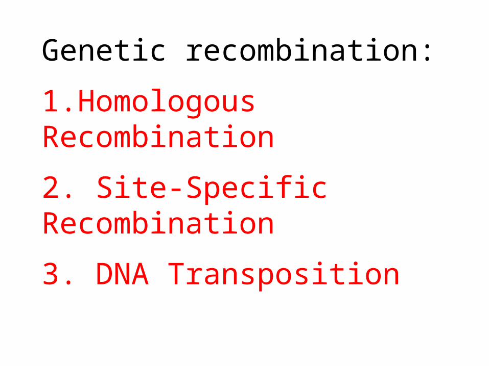
Genetic recombination:
1.Homologous Recombination
2. Site-Specific Recombination
3. DNA Transposition
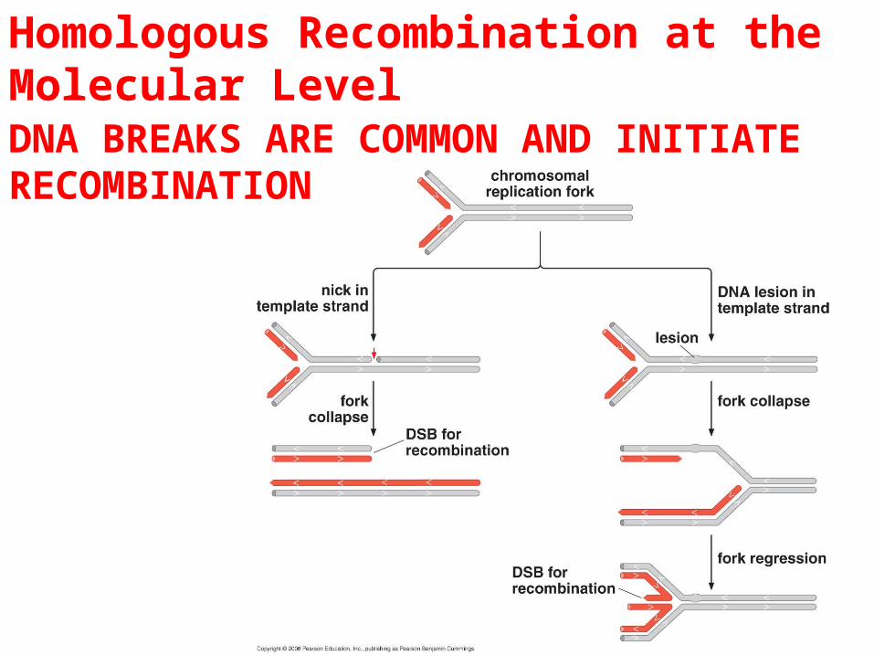
Homologous Recombination at the Molecular LevelDNA BREAKS ARE COMMON AND INITIATE RECOMBINATION
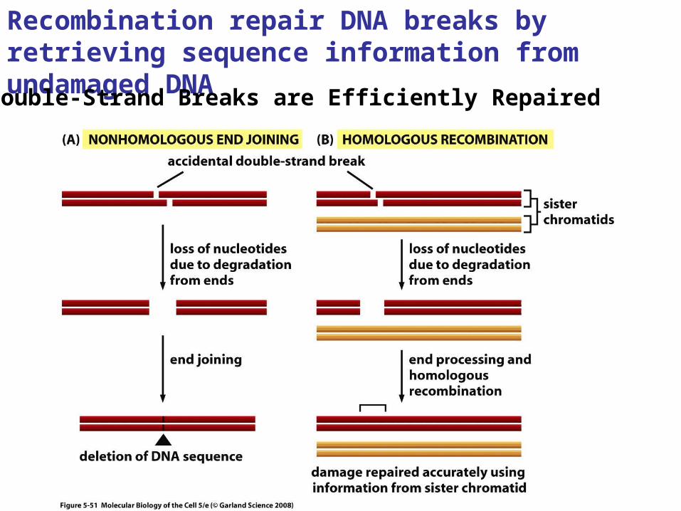
Recombination repair DNA breaks by retrieving sequence information from undamaged DNA
Double-Strand Breaks are Efficiently Repaired
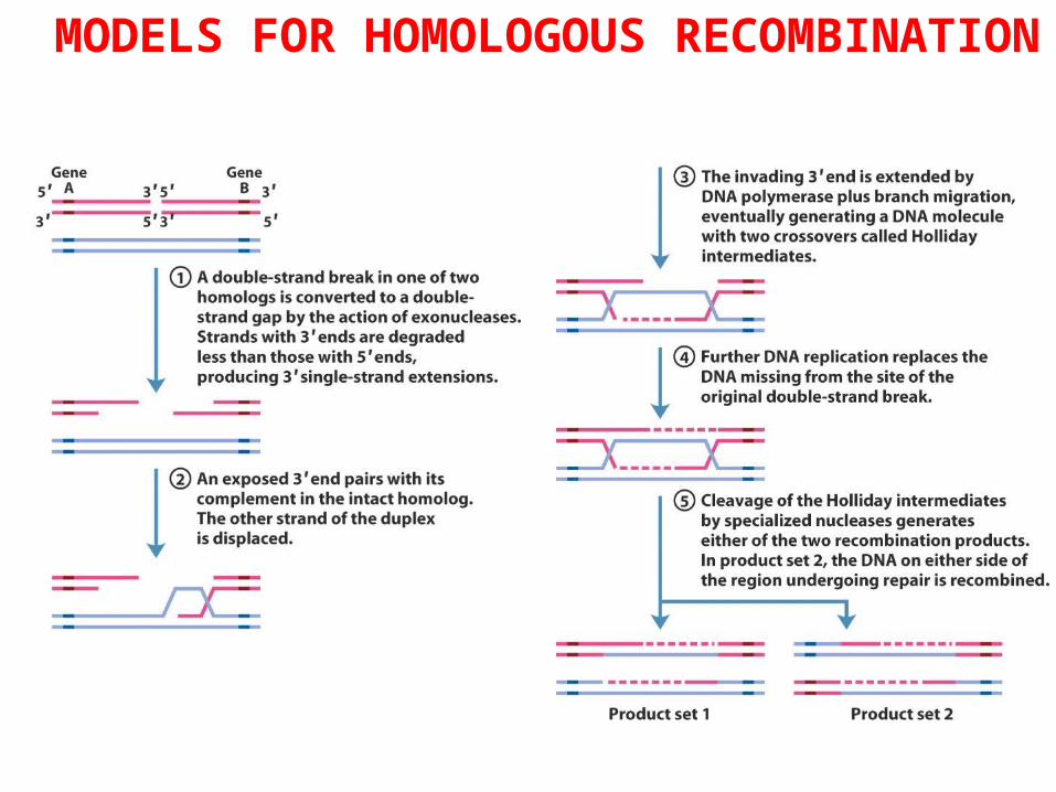
MODELS FOR HOMOLOGOUS RECOMBINATION
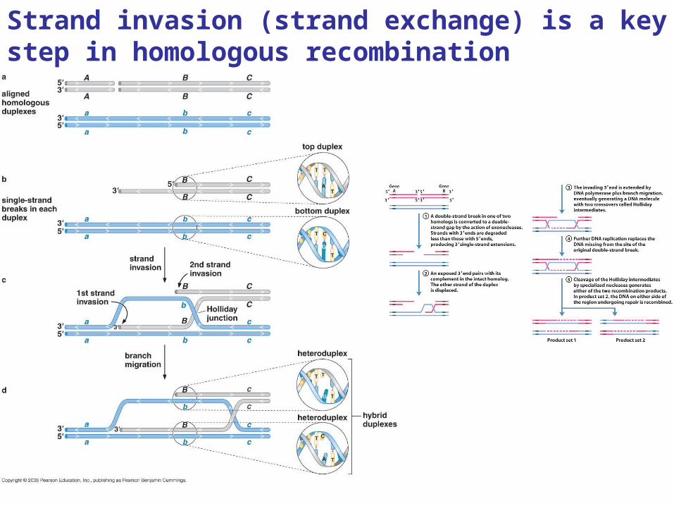
Strand invasion (strand exchange) is a key step in homologous recombination
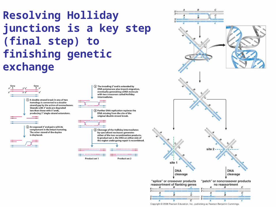
Resolving Holliday junctions is a key step (final step) to finishing genetic exchange
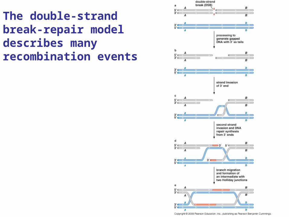
The double-strand break-repair model describes many recombination events
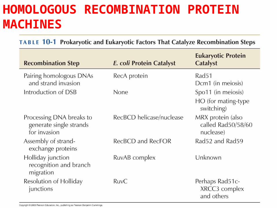
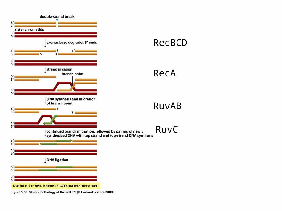
HOMOLOGOUS RECOMBINATION PROTEIN MACHINES
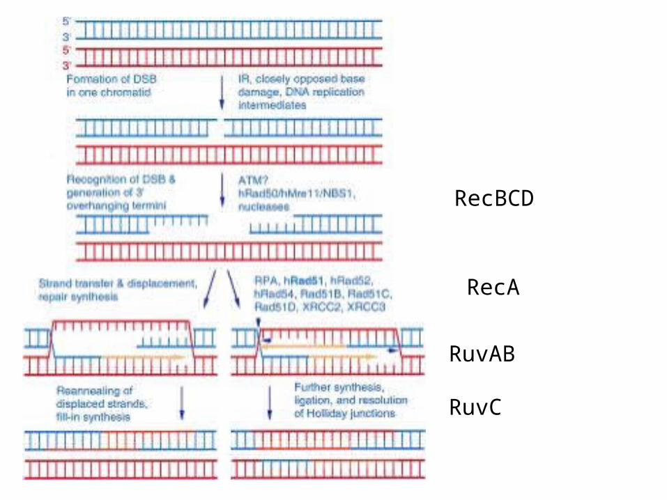
RecBCD
RecA
RuvAB
RuvC
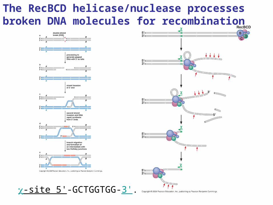
The RecBCD helicase/nuclease processes broken DNA molecules for recombination
c-site 5'-GCTGGTGG-3'.
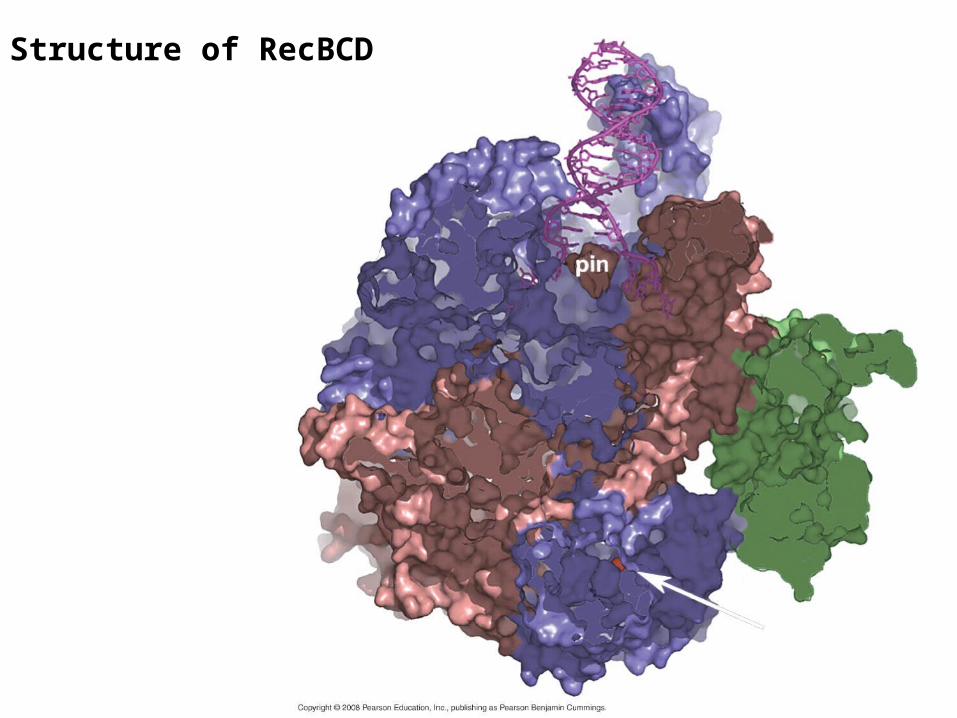
Structure of RecBCD
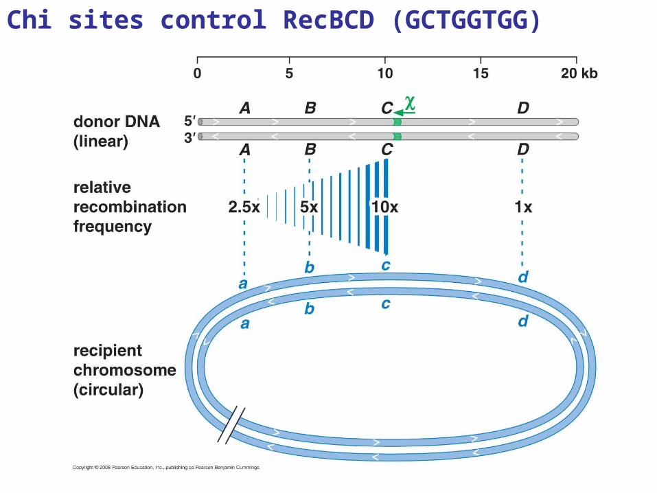
Chi sites control RecBCD (GCTGGTGG)
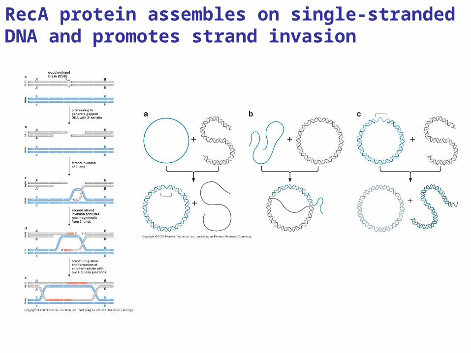
RecA protein assembles on single-stranded DNA and promotes strand invasion
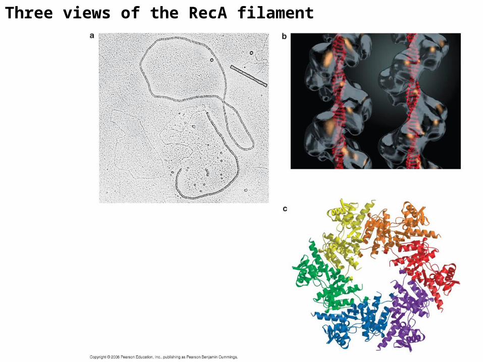
Three views of the RecA filament
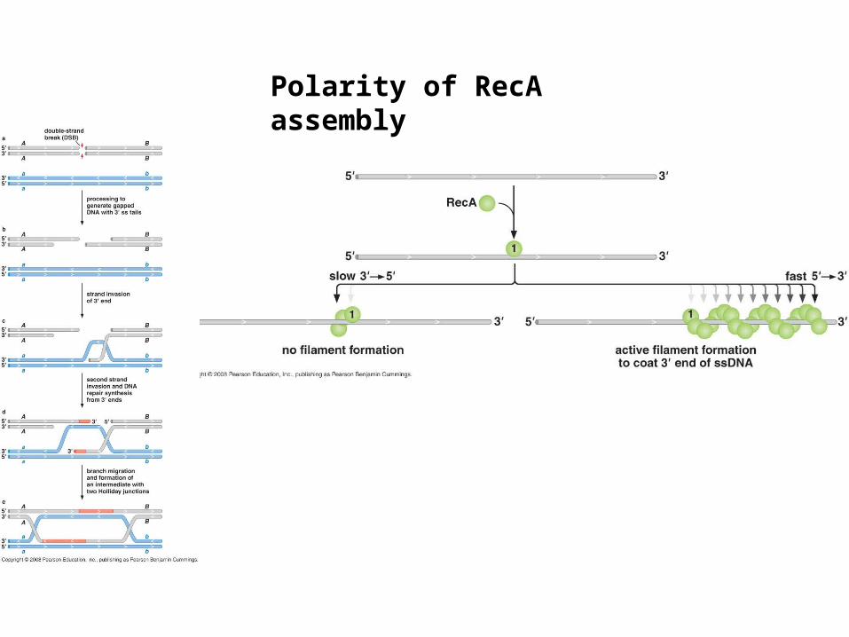
Polarity of RecA assembly
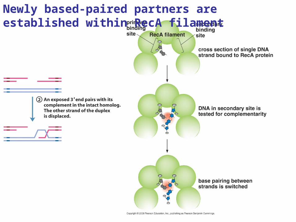
Newly based-paired partners are established within RecA filament
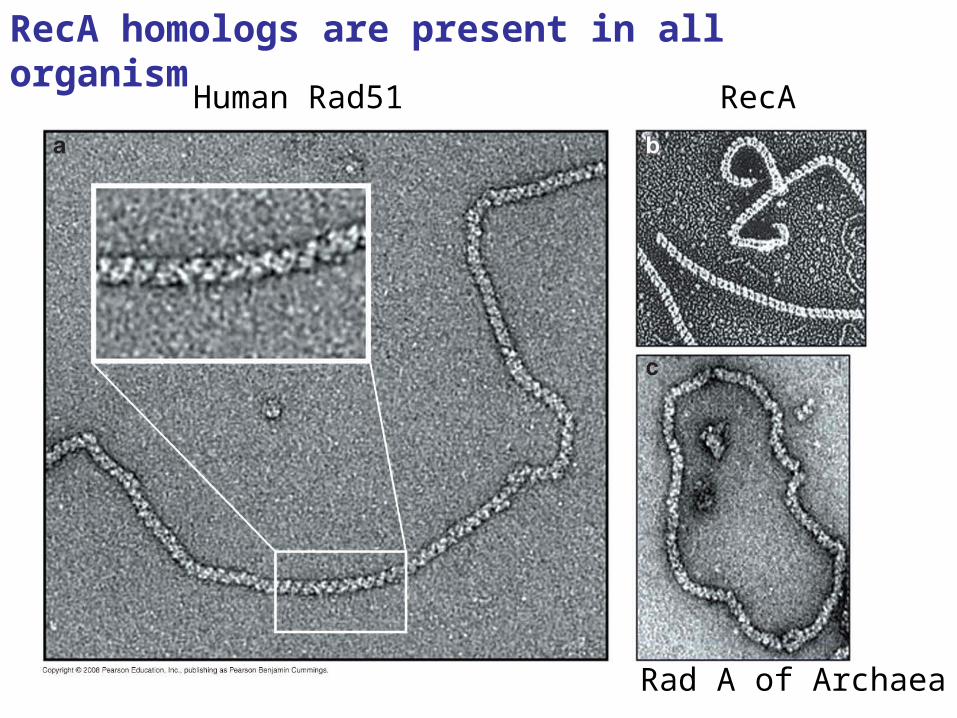
RecA homologs are present in all organism
Human Rad51 RecA
Rad A of Archaea
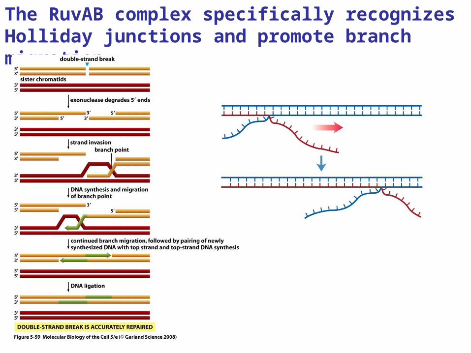
The RuvAB complex specifically recognizes Holliday junctions and promote branch migration
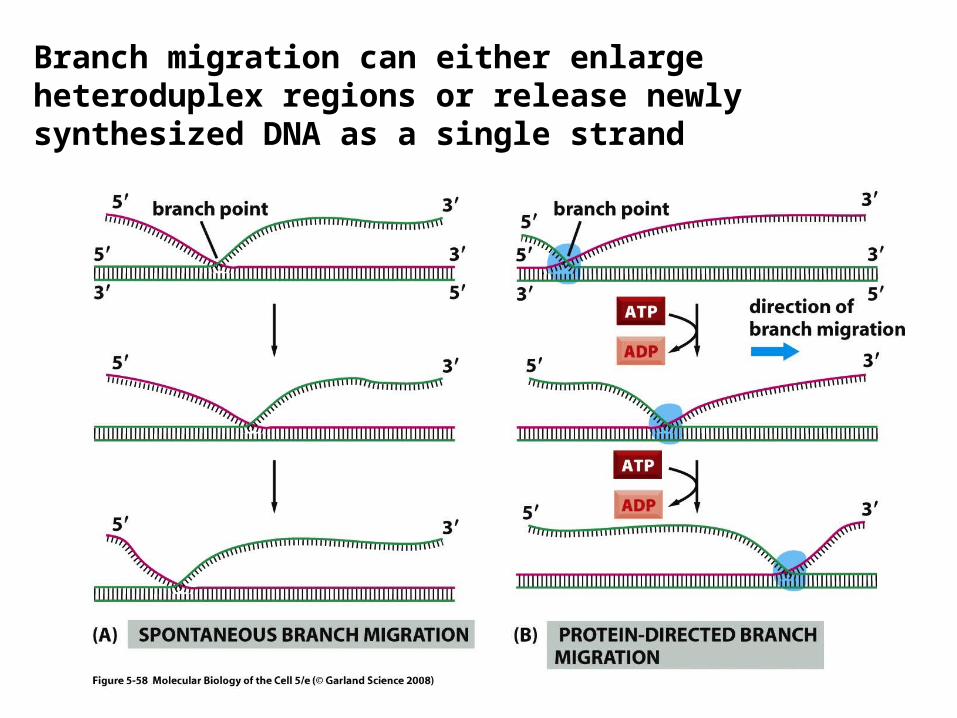
Branch migration can either enlarge heteroduplex regions or release newly synthesized DNA as a single strand
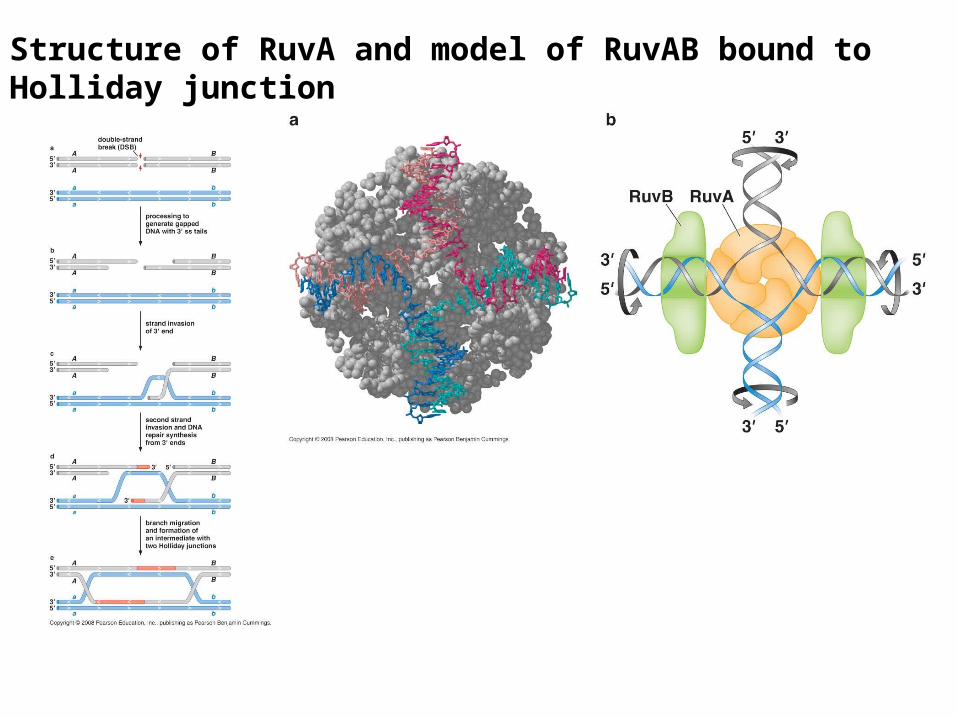
Structure of RuvA and model of RuvAB bound to Holliday junction
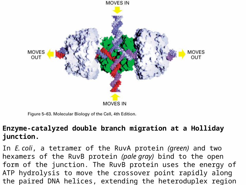
Enzyme-catalyzed double branch migration at a Holliday junction.
In E. coli, a tetramer of the RuvA protein (green) and two hexamers of the RuvB protein (pale gray) bind to the open form of the junction. The RuvB protein uses the energy of ATP hydrolysis to move the crossover point rapidly along the paired DNA helices, extending the heteroduplex region as shown. There is evidence that similar proteins perform this function in vertebrate cells. (Image courtesy of P. Artymiuk; modified from S.C. West, Cell 94:699–701, 1998.)
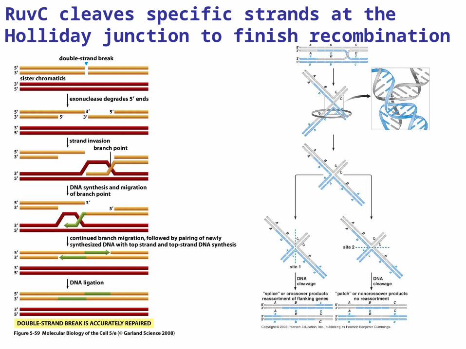
RuvC cleaves specific strands at the Holliday junction to finish recombination
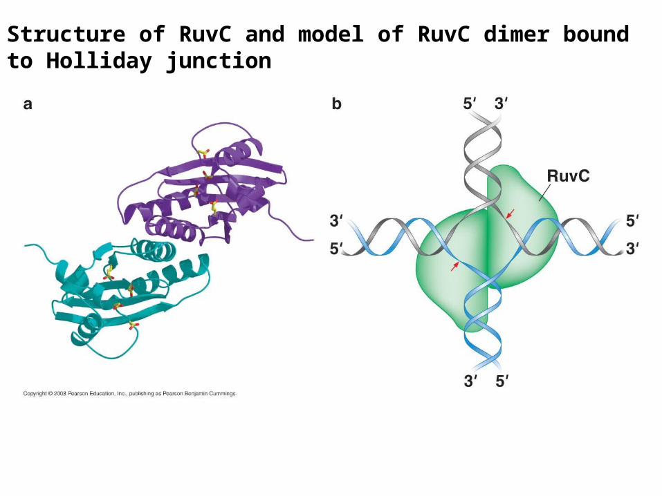
Structure of RuvC and model of RuvC dimer bound to Holliday junction
RecBCD
RecA
RuvAB
RuvC
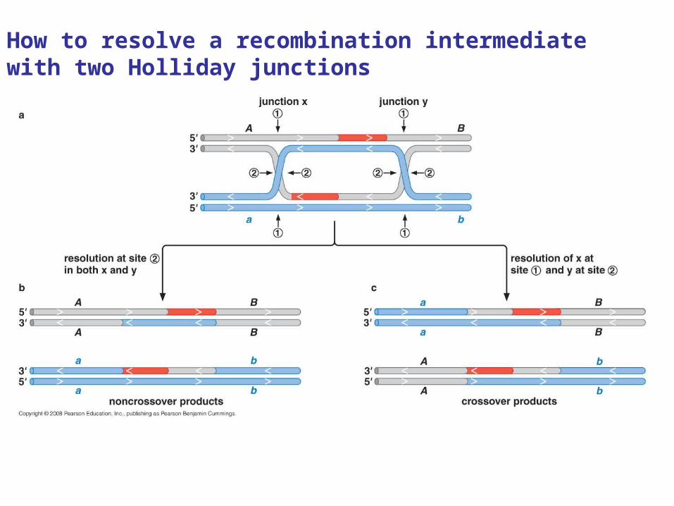
How to resolve a recombination intermediate with two Holliday junctions
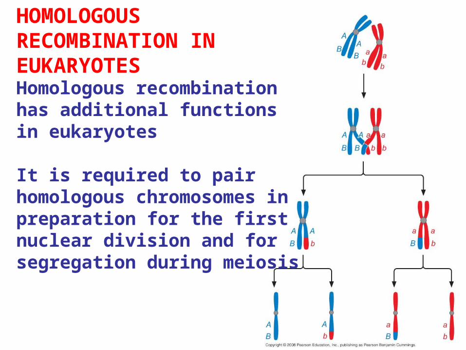
HOMOLOGOUS RECOMBINATION IN EUKARYOTESHomologous recombination has additional functions in eukaryotes
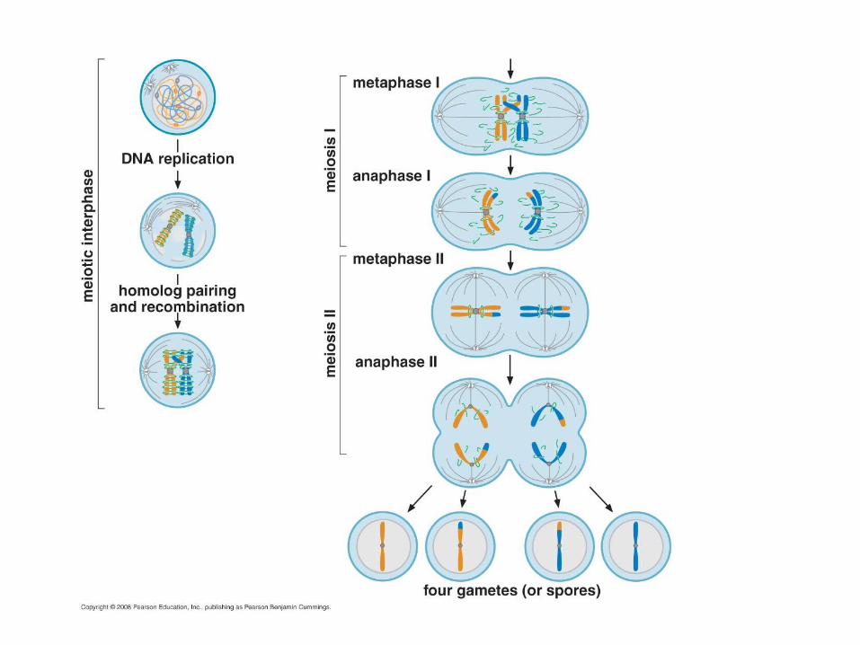
It is required to pair homologous chromosomes in preparation for the first nuclear division and for segregation during meiosis
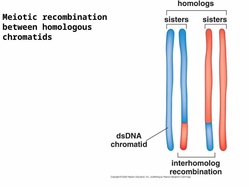
Meiotic recombination between homologous chromatids
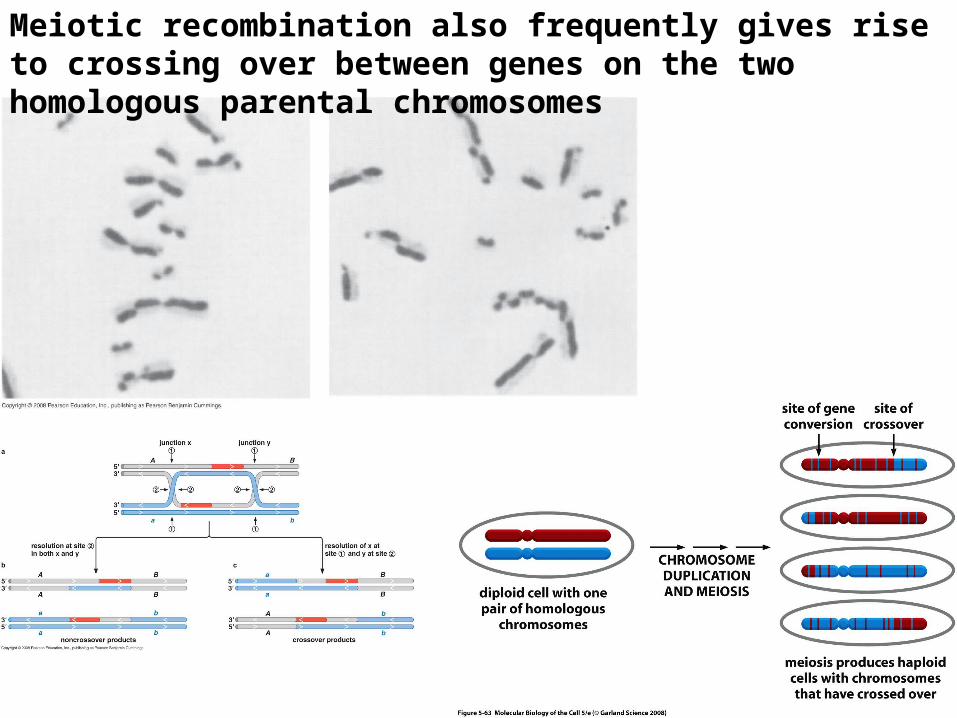
Meiotic recombination also frequently gives rise to crossing over between genes on the two homologous parental chromosomes
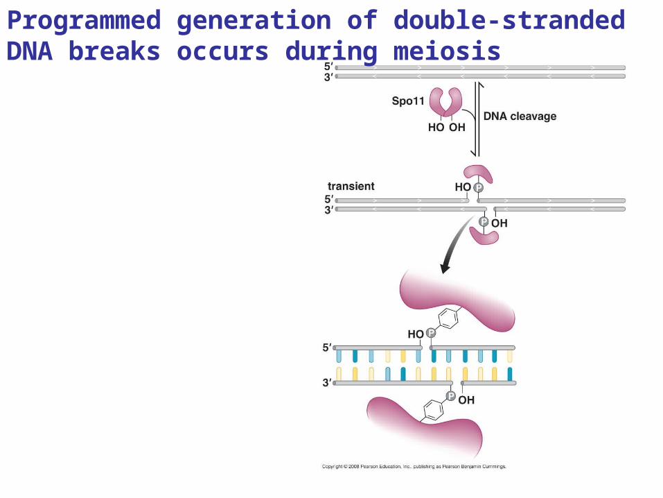
Programmed generation of double-stranded DNA breaks occurs during meiosis
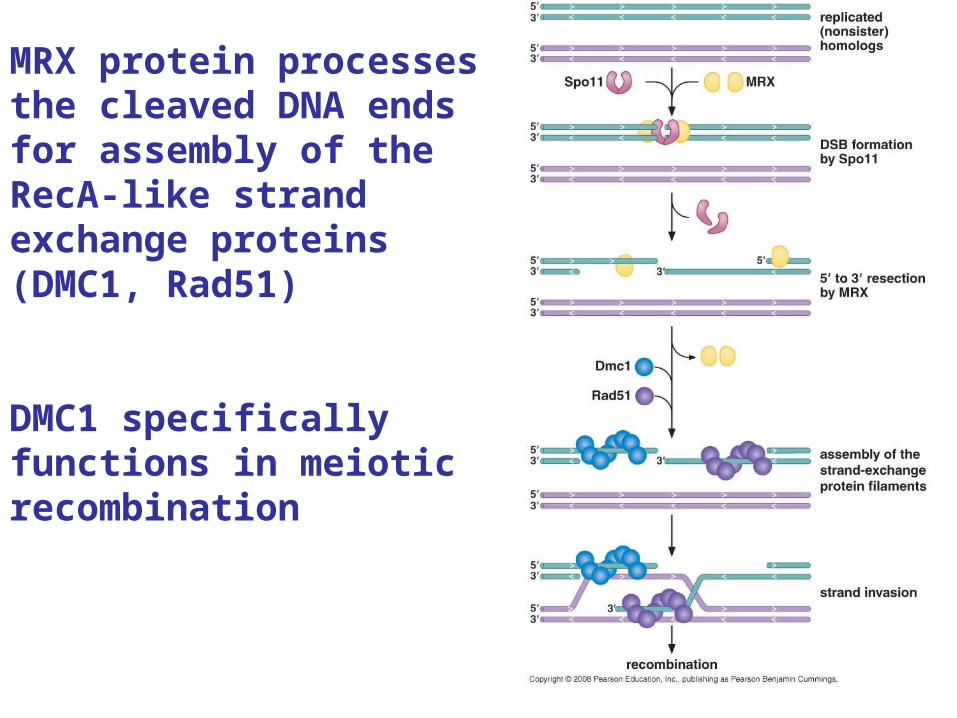
MRX protein processes the cleaved DNA ends for assembly of the RecA-like strand exchange proteins (DMC1, Rad51)
DMC1 specifically functions in meiotic recombination
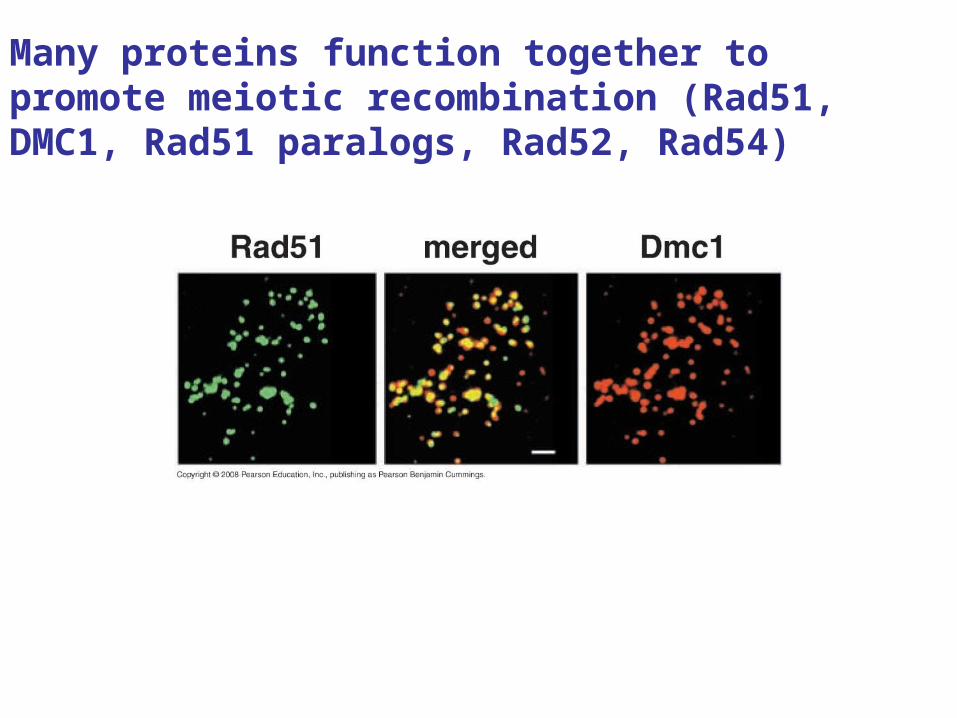
Many proteins function together to promote meiotic recombination (Rad51, DMC1, Rad51 paralogs, Rad52, Rad54)
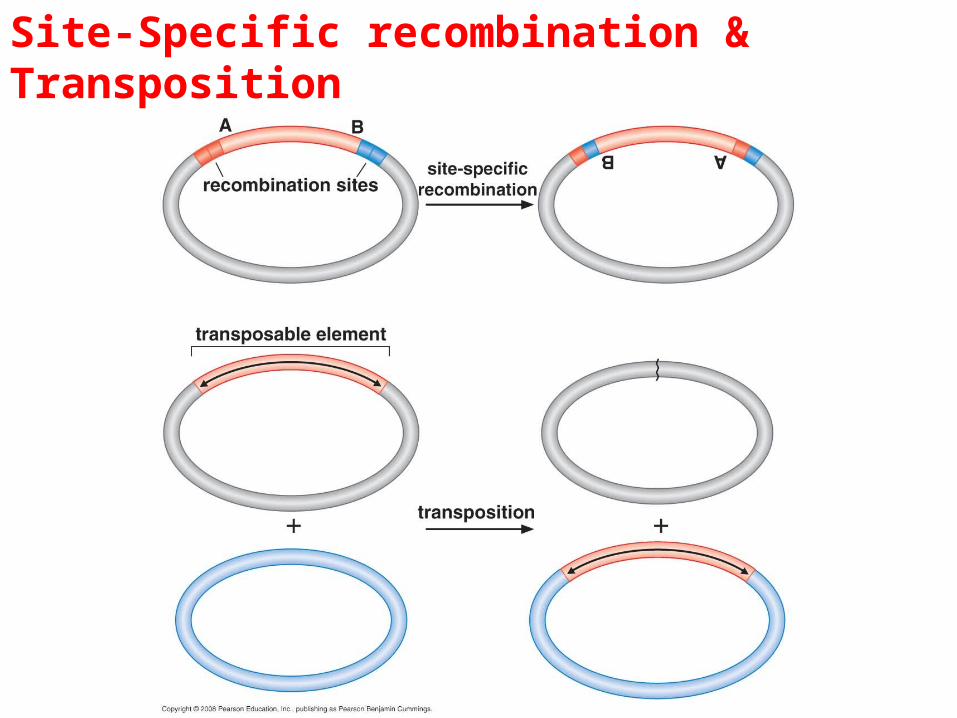
Site-Specific recombination & Transposition
Conservative site-specific recombination
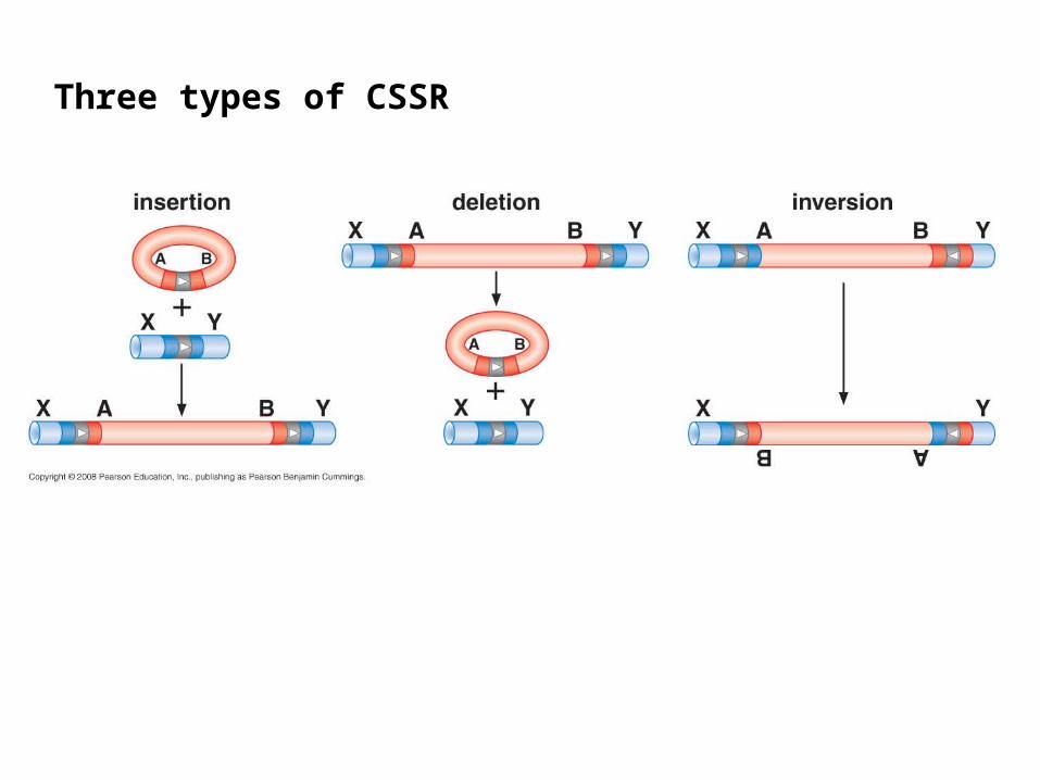
Three types of CSSR
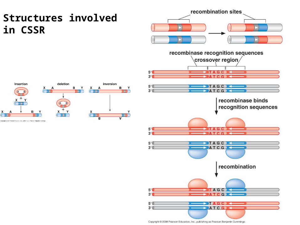
Structures involved in CSSR
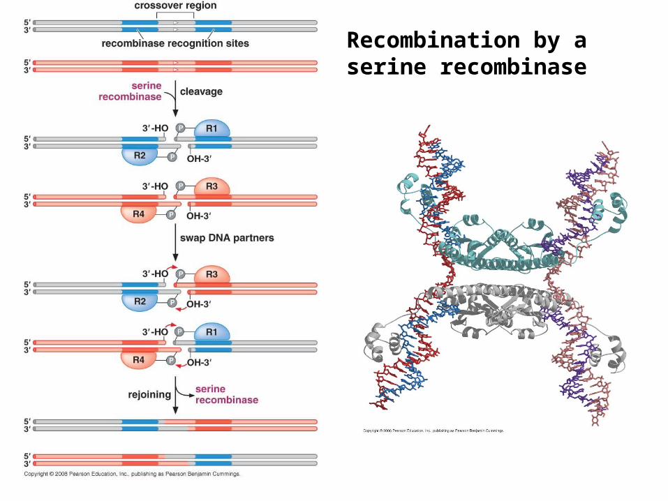
Recombination by a serine recombinase
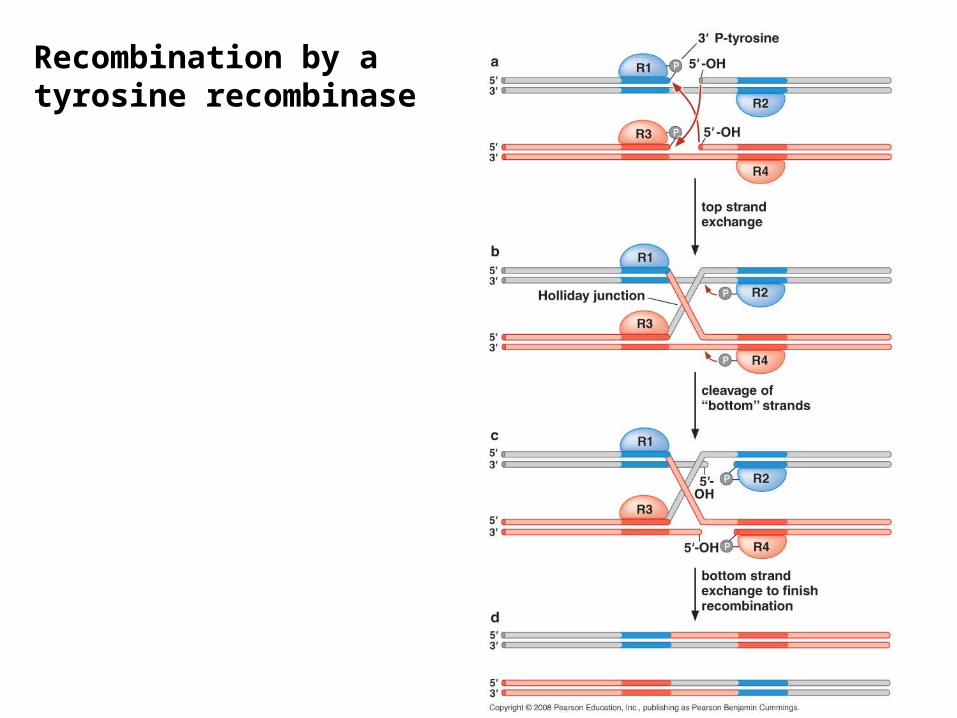
Recombination by a tyrosine recombinase
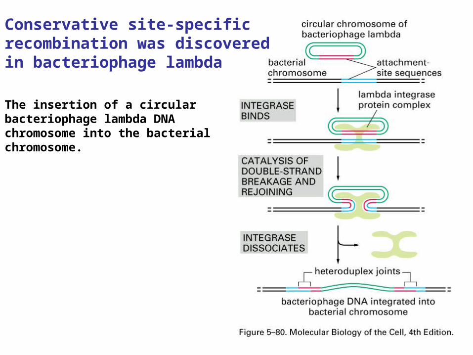
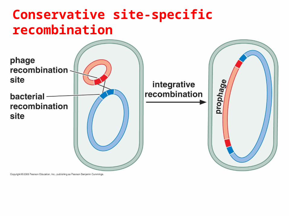
The insertion of a circular bacteriophage lambda DNA chromosome into the bacterial chromosome.
Conservative site-specific recombination was discovered in bacteriophage lambda
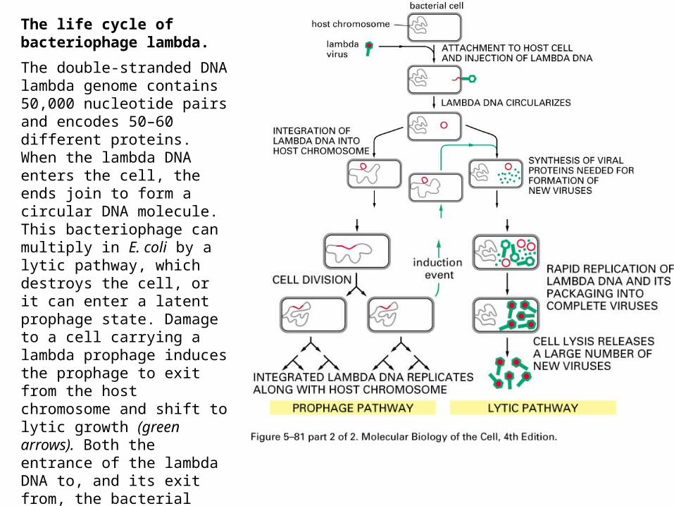
The life cycle of bacteriophage lambda.
The double-stranded DNA lambda genome contains 50,000 nucleotide pairs and encodes 50–60 different proteins. When the lambda DNA enters the cell, the ends join to form a circular DNA molecule. This bacteriophage can multiply in E. coli by a lytic pathway, which destroys the cell, or it can enter a latent prophage state. Damage to a cell carrying a lambda prophage induces the prophage to exit from the host chromosome and shift to lytic growth (green arrows). Both the entrance of the lambda DNA to, and its exit from, the bacterial chromosome are accomplished by a conservative site-specific recombination event, catalyzed by the lambda integrase enzyme (see Figure 5–80).
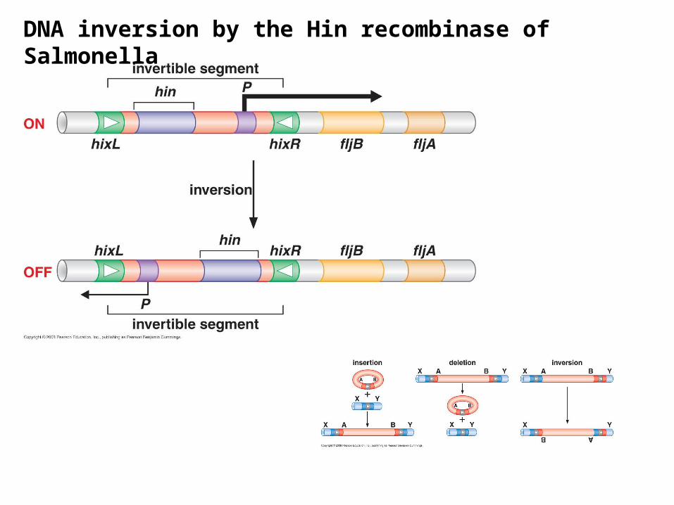
DNA inversion by the Hin recombinase of Salmonella
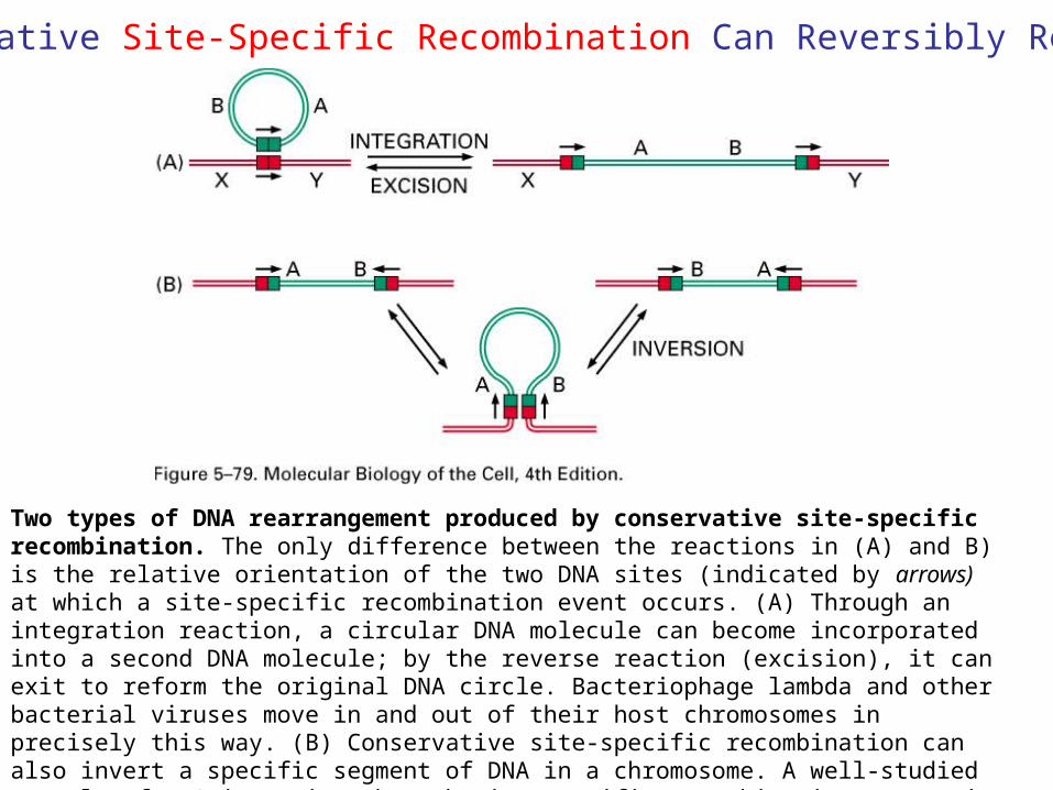
Two types of DNA rearrangement produced by conservative site-specific recombination. The only difference between the reactions in (A) and B) is the relative orientation of the two DNA sites (indicated by arrows) at which a site-specific recombination event occurs. (A) Through an integration reaction, a circular DNA molecule can become incorporated into a second DNA molecule; by the reverse reaction (excision), it can exit to reform the original DNA circle. Bacteriophage lambda and other bacterial viruses move in and out of their host chromosomes in precisely this way. (B) Conservative site-specific recombination can also invert a specific segment of DNA in a chromosome. A well-studied example of DNA inversion through site-specific recombination occurs in the bacterium Salmonella typhimurium, an organism that is a major cause of food poisoning in humans; the inversion of a DNA segment changes the type of flagellum that is produced by the bacterium (see Figure 7–64).
Conservative Site-Specific Recombination Can Reversibly RearrangeDNA
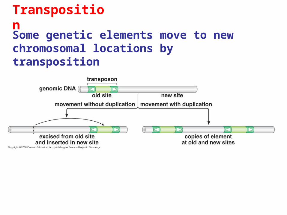
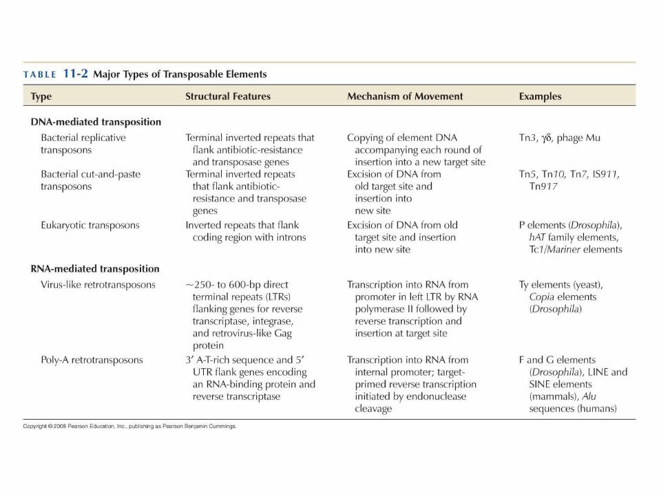
Transposition
Some genetic elements move to new chromosomal locations by transposition
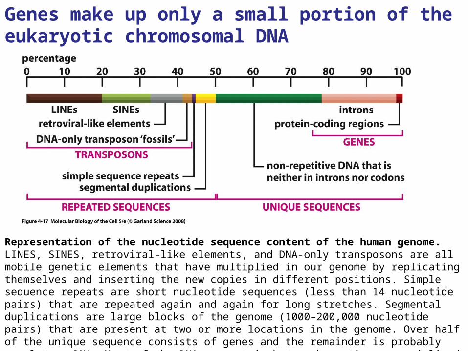
Representation of the nucleotide sequence content of the human genome.LINES, SINES, retroviral-like elements, and DNA-only transposons are all mobile genetic elements that have multiplied in our genome by replicating themselves and inserting the new copies in different positions. Simple sequence repeats are short nucleotide sequences (less than 14 nucleotide pairs) that are repeated again and again for long stretches. Segmental duplications are large blocks of the genome (1000–200,000 nucleotide pairs) that are present at two or more locations in the genome. Over half of the unique sequence consists of genes and the remainder is probably regulatory DNA. Most of the DNA present in heterochromatin, a specialized type of chromatin that contains relatively few genes, has not yet been sequenced
Genes make up only a small portion of the eukaryotic chromosomal DNA
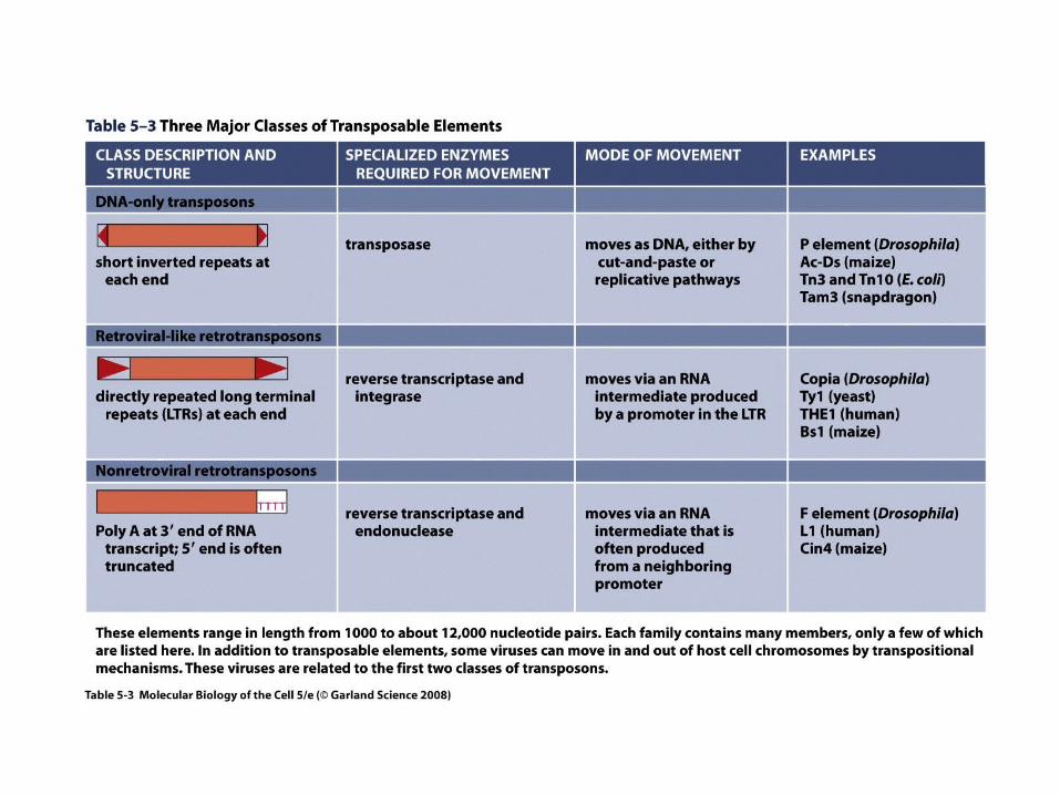
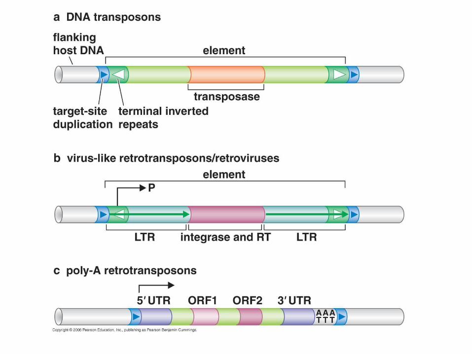
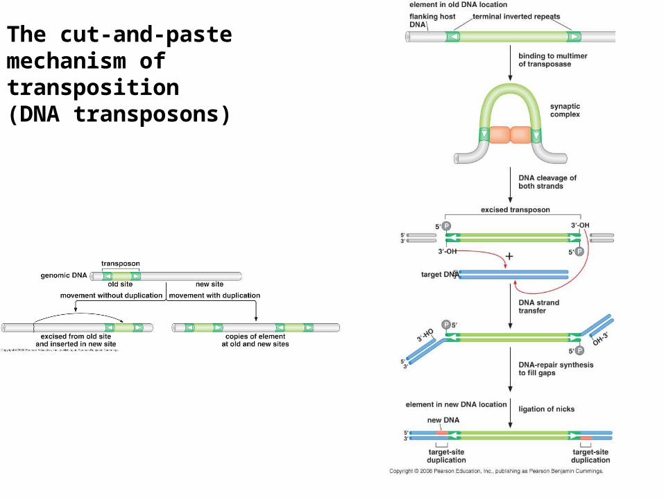
The cut-and-paste mechanism of transposition(DNA transposons)
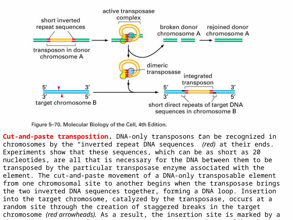
Cut-and-paste transposition. DNA-only transposons can be recognized in chromosomes by the “inverted repeat DNA sequences” (red) at their ends. Experiments show that these sequences, which can be as short as 20 nucleotides, are all that is necessary for the DNA between them to be transposed by the particular transposase enzyme associated with the element. The cut-and-paste movement of a DNA-only transposable element from one chromosomal site to another begins when the transposase brings the two inverted DNA sequences together, forming a DNA loop. Insertion into the target chromosome, catalyzed by the transposase, occurs at a random site through the creation of staggered breaks in the target chromosome (red arrowheads). As a result, the insertion site is marked by a short direct repeat of the target DNA sequence, as shown. Although the break in the donor chromosome (green) is resealed, the breakage- and-repair process often alters the DNA sequence, causing a mutation at the original site of the excised transposable element (not shown).
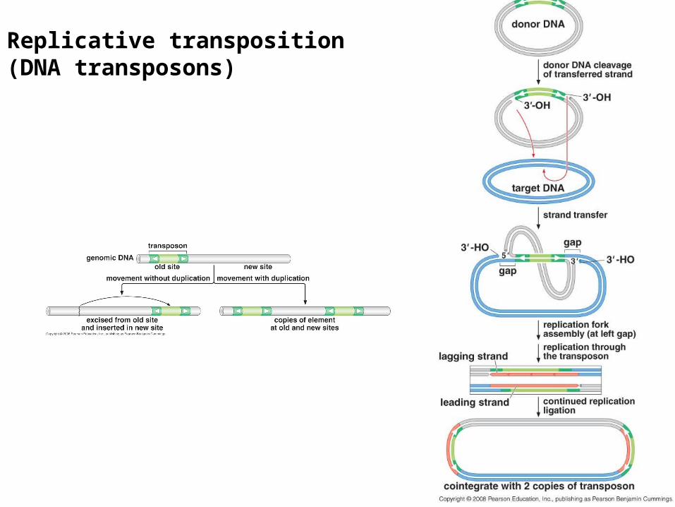
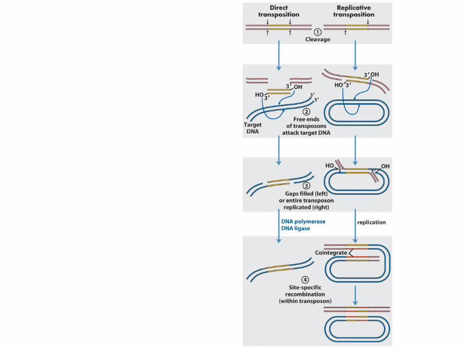
Replicative transposition(DNA transposons)
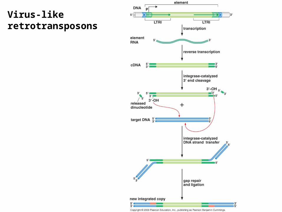
Virus-like retrotransposons
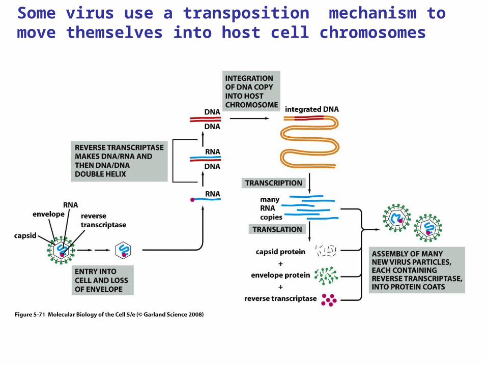
Some virus use a transposition mechanism to move themselves into host cell chromosomes

Reverse transcriptase
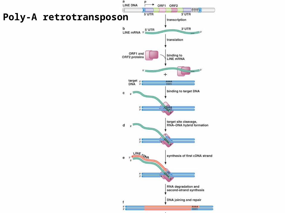
Poly-A retrotransposon
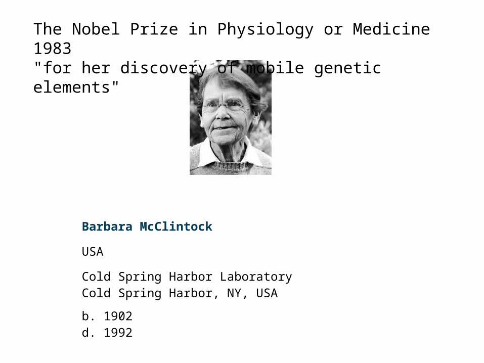
The Nobel Prize in Physiology or Medicine 1983"for her discovery of mobile genetic elements"
Barbara McClintock
USA
Cold Spring Harbor Laboratory Cold Spring Harbor, NY, USA
b. 1902d. 1992