Homologous features = similar structure, different functions.
February 3, 2004
Homologous Recombination by the RecBCD and RecF Pathways.
Maria Spies and Stephen C. Kowalczykowski*
Sections of Microbiology and of Molecular and Cellular Biology, Center for Genetics
and Development, University of California, Davis, CA, 95616-8665
*CORRESPONDING AUTHOR:
University of California, Davis,
Section of Microbiology
Center for Genetics and Development
One Shields Ave.
Davis, CA 95616-8665
E-mail: [email protected]
Tel.: 530-752-5938
Fax: 530-752-5939
1
Abstract
In all cells, genetic recombination is used to repair DNA breaks and, as a result, genetic
information is exchanged between homologous DNA molecules. Discontinuities in DNA strands,
specifically double-strand DNA breaks and single-strand DNA gaps, attract the enzymes
responsible for the initiation of homologous recombination. In wild-type Escherichia coli, two
distinct pathways are responsible for the repair of DNA by recombination: the RecBCD- and the
RecF-pathways. The RecBCD-pathway is specific to recombination initiated at double-strand
DNA breaks, whereas the RecF-pathway is primarily responsible for recombination initiated at
single-strand DNA gaps, although it can also act at double-strand breaks. This review
summarizes the biochemical mechanisms of homologous recombination in E. coli.
2
Recombinational repair of DNA damage.
Homologous, or general, recombination is a crucial biological process that involves the
pairing and transfer of strands between DNA molecules that share a region of significant
sequence homology. Since its discovery in 1946 by Lederberg and Tatum (48), homologous
recombination in bacteria was associated with the sexual process of conjugation and was viewed
as an evolutionary mechanism both for shuffling the genome and for spreading favorable alleles.
However, more recently, a more immediate function of homologous recombination was
recognized: namely, it is a mechanism for the maintenance of chromosomal integrity that acts to
repair DNA lesions, both double-strand DNA breaks (DSBs) and single-strand DNA gaps
(SSGs), generated during the course of DNA replication. The intimate connection between the
processes of replication and recombination was initially appreciated in the life-cycle of
bacteriophage T4 (56) and then later recognized as an important determinant of viability in
bacteria (39, 45). In T4 phage, recombination is linked to replication to produce a high yield of
phage DNA; in E. coli, recombination is linked to replication to permit its completion when
interrupted by DNA damage, and also to initiate DNA replication in the absence of origin
function. This view of recombination as an integral part of efficient chromosome duplication also
reconciled the high level of inviability (up to 95%) of recombination-deficient cells (12).
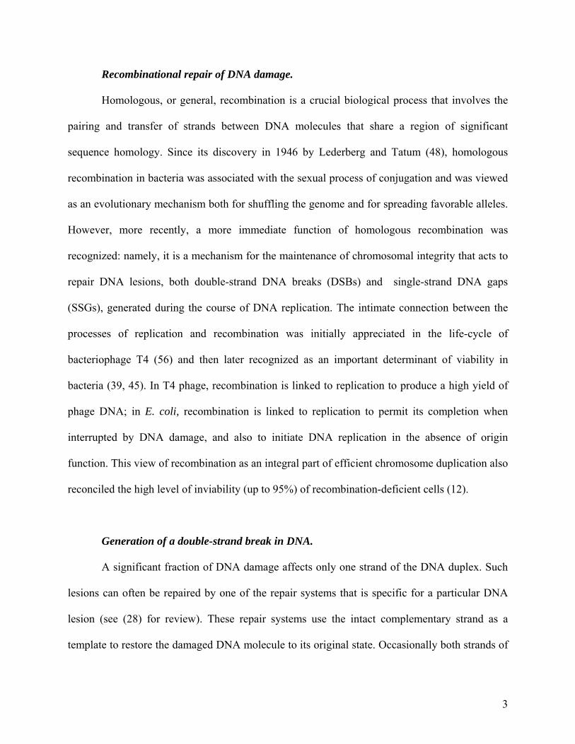
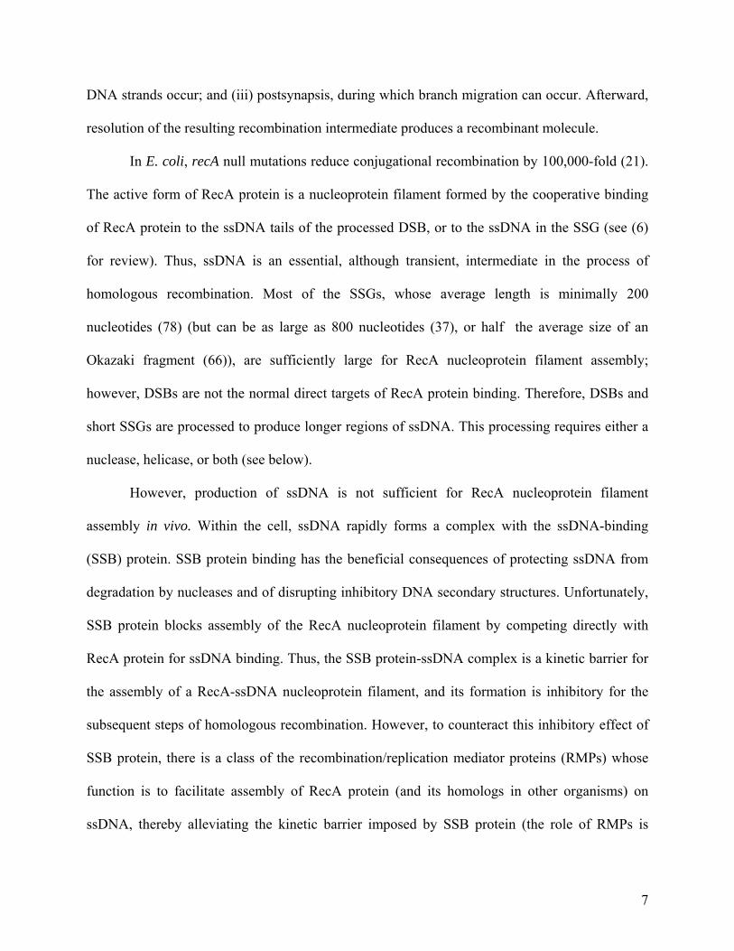
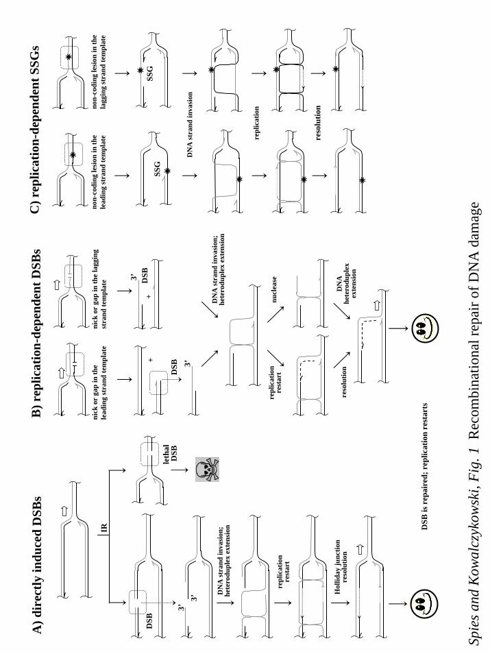
Generation of a double-strand break in DNA.
A significant fraction of DNA damage affects only one strand of the DNA duplex. Such
lesions can often be repaired by one of the repair systems that is specific for a particular DNA
lesion (see (28) for review). These repair systems use the intact complementary strand as a
template to restore the damaged DNA molecule to its original state. Occasionally both strands of
3
the dsDNA can be broken opposite to each other, resulting in a double-strand break. DSBs can
be produced, for example, as a direct consequence of ionizing radiation (Figure 1a). However,
the bulk of the DSBs in bacteria are generated indirectly as the result of DNA replication through
an unrepaired break in just a single-strand of DNA (Figure 1b). Replication of DNA containing a
single-strand nick or a gap in the leading-strand results in the dissociation of the DNA
polymerase holoenzyme complex, and the generation of one blunt DSB (Figure 1b), whereas
replication of DNA with a nick in lagging-strand produces a DSB with an 3’-terminated ssDNA
tail.
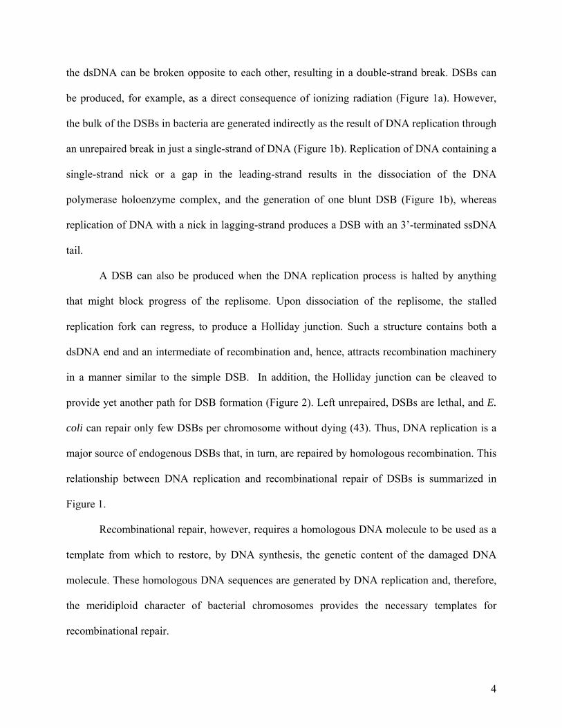
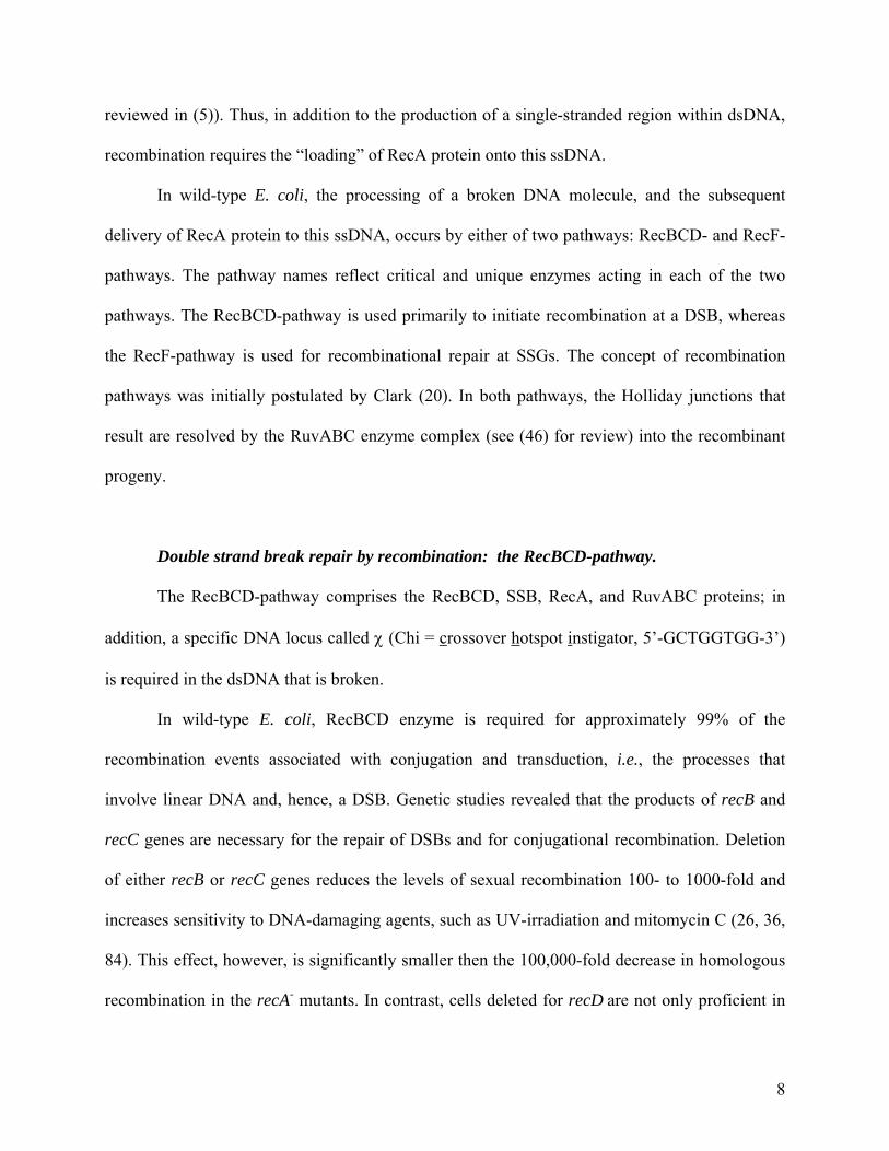
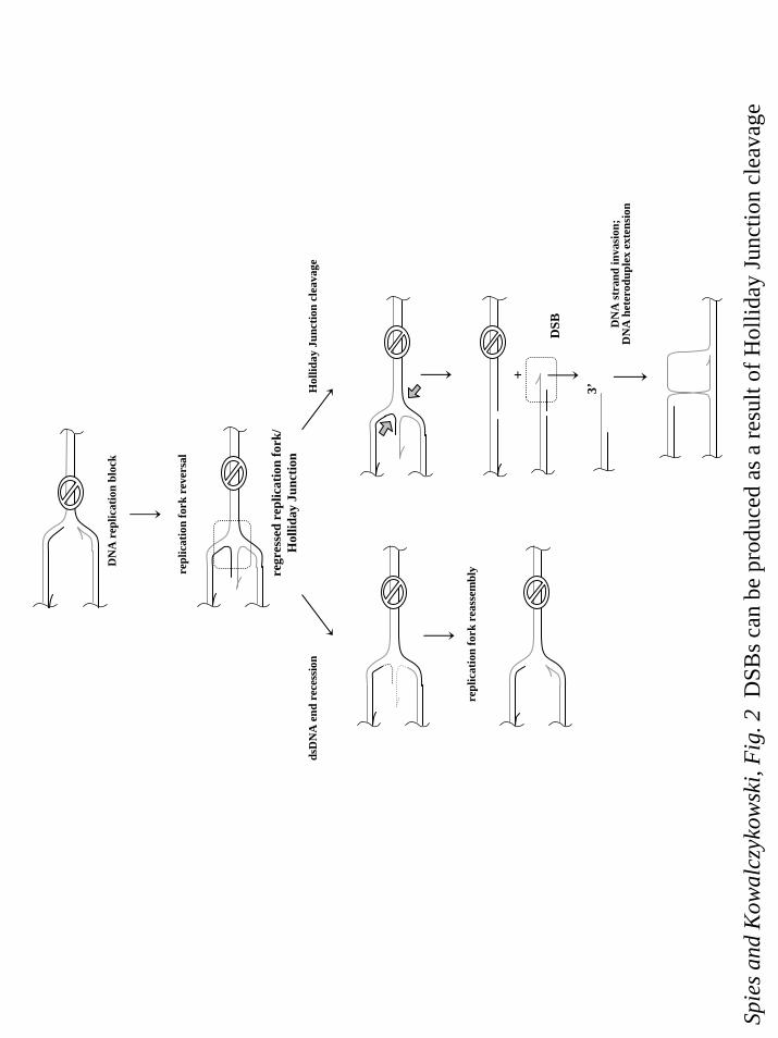
A DSB can also be produced when the DNA replication process is halted by anything
that might block progress of the replisome. Upon dissociation of the replisome, the stalled
replication fork can regress, to produce a Holliday junction. Such a structure contains both a
dsDNA end and an intermediate of recombination and, hence, attracts recombination machinery
in a manner similar to the simple DSB. In addition, the Holliday junction can be cleaved to
provide yet another path for DSB formation (Figure 2). Left unrepaired, DSBs are lethal, and E.
coli can repair only few DSBs per chromosome without dying (43). Thus, DNA replication is a
major source of endogenous DSBs that, in turn, are repaired by homologous recombination. This
relationship between DNA replication and recombinational repair of DSBs is summarized in
Figure 1.
Recombinational repair, however, requires a homologous DNA molecule to be used as a
template from which to restore, by DNA synthesis, the genetic content of the damaged DNA
molecule. These homologous DNA sequences are generated by DNA replication and, therefore,
the meridiploid character of bacterial chromosomes provides the necessary templates for
recombinational repair.
4
Generation of a single-strand gap in DNA.
When synthesis of a DNA strand is blocked by a non-coding lesion (for example, an
abasic site or an intra-strand crosslink, such as a thymine dimer), continued replication of the
flawless strand beyond the lesion produces a single-strand DNA gap (Figure 1c) (42, 54). SSGs
are also known as daughter-strand gaps, since they appear on one of the newly synthesized
daughter strands during semi-conservative DNA replication.
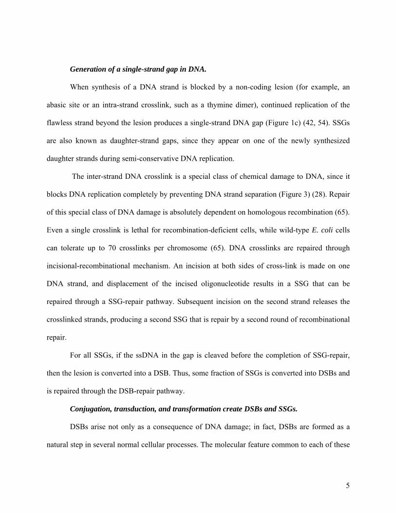
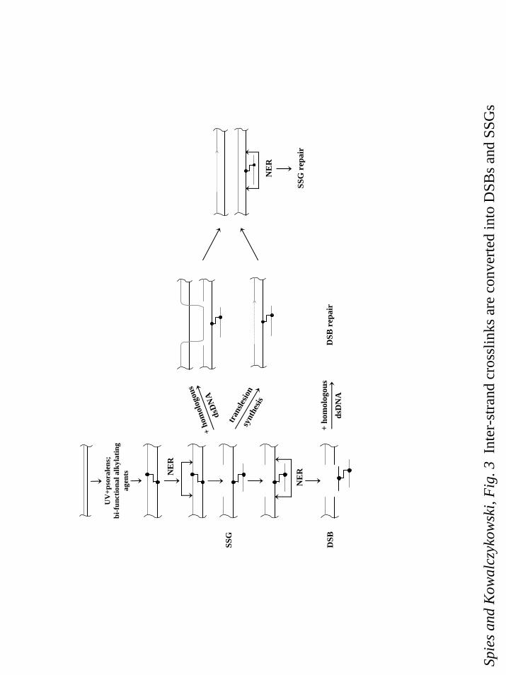
The inter-strand DNA crosslink is a special class of chemical damage to DNA, since it
blocks DNA replication completely by preventing DNA strand separation (Figure 3) (28). Repair
of this special class of DNA damage is absolutely dependent on homologous recombination (65).
Even a single crosslink is lethal for recombination-deficient cells, while wild-type E. coli cells
can tolerate up to 70 crosslinks per chromosome (65). DNA crosslinks are repaired through
incisional-recombinational mechanism. An incision at both sides of cross-link is made on one
DNA strand, and displacement of the incised oligonucleotide results in a SSG that can be
repaired through a SSG-repair pathway. Subsequent incision on the second strand releases the
crosslinked strands, producing a second SSG that is repair by a second round of recombinational
repair.
For all SSGs, if the ssDNA in the gap is cleaved before the completion of SSG-repair,
then the lesion is converted into a DSB. Thus, some fraction of SSGs is converted into DSBs and
is repaired through the DSB-repair pathway.
Conjugation, transduction, and transformation create DSBs and SSGs.
DSBs arise not only as a consequence of DNA damage; in fact, DSBs are formed as a
natural step in several normal cellular processes. The molecular feature common to each of these
5
processes is the cellular acquisition of a linear segment of dsDNA. The end of this linear DNA
molecule is seen as a DSB, and the recombinational repair of the DSB is initiated. Bacteria can
acquire linear DNA by any of three different routes. First, during conjugation, a copy of the
chromosome is transferred from one bacterium, which possesses a fertility factor (the F’
plasmid) to another. Genetic studies of this process led to isolation of the first recombination-
deficient mutants (21). Transformation is a second route by which some bacteria take-up a
segment of DNA from the environment; unrelated in process, but not in the form of the DNA
involved, artificial transformation is widely used as a laboratory technique to introduce foreign
dsDNA into cells. Finally, infection by bacteriophage also results in the introduction of linear
DNA into bacteria. Note, however, that in addition to DSBs, the processes of conjugation and
natural transformation also produce SSGs due to incomplete replication of the DNA
intermediates that form during these processes.
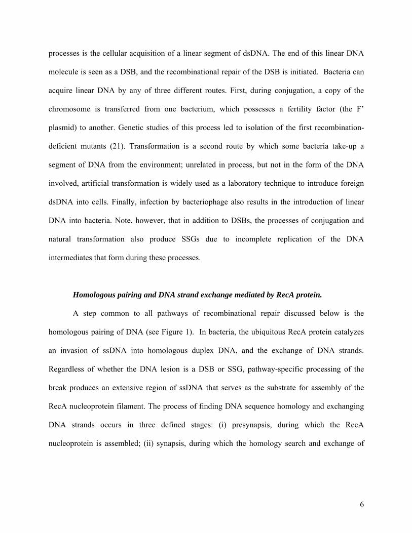
Homologous pairing and DNA strand exchange mediated by RecA protein.
A step common to all pathways of recombinational repair discussed below is the
homologous pairing of DNA (see Figure 1). In bacteria, the ubiquitous RecA protein catalyzes
an invasion of ssDNA into homologous duplex DNA, and the exchange of DNA strands.
Regardless of whether the DNA lesion is a DSB or SSG, pathway-specific processing of the
break produces an extensive region of ssDNA that serves as the substrate for assembly of the
RecA nucleoprotein filament. The process of finding DNA sequence homology and exchanging
DNA strands occurs in three defined stages: (i) presynapsis, during which the RecA
nucleoprotein is assembled; (ii) synapsis, during which the homology search and exchange of
6
DNA strands occur; and (iii) postsynapsis, during which branch migration can occur. Afterward,
resolution of the resulting recombination intermediate produces a recombinant molecule.
In E. coli, recA null mutations reduce conjugational recombination by 100,000-fold (21).
The active form of RecA protein is a nucleoprotein filament formed by the cooperative binding
of RecA protein to the ssDNA tails of the processed DSB, or to the ssDNA in the SSG (see (6)
for review). Thus, ssDNA is an essential, although transient, intermediate in the process of
homologous recombination. Most of the SSGs, whose average length is minimally 200
nucleotides (78) (but can be as large as 800 nucleotides (37), or half the average size of an
Okazaki fragment (66)), are sufficiently large for RecA nucleoprotein filament assembly;
however, DSBs are not the normal direct targets of RecA protein binding. Therefore, DSBs and
short SSGs are processed to produce longer regions of ssDNA. This processing requires either a
nuclease, helicase, or both (see below).
However, production of ssDNA is not sufficient for RecA nucleoprotein filament
assembly in vivo. Within the cell, ssDNA rapidly forms a complex with the ssDNA-binding
(SSB) protein. SSB protein binding has the beneficial consequences of protecting ssDNA from
degradation by nucleases and of disrupting inhibitory DNA secondary structures. Unfortunately,
SSB protein blocks assembly of the RecA nucleoprotein filament by competing directly with
RecA protein for ssDNA binding. Thus, the SSB protein-ssDNA complex is a kinetic barrier for
the assembly of a RecA-ssDNA nucleoprotein filament, and its formation is inhibitory for the
subsequent steps of homologous recombination. However, to counteract this inhibitory effect of
SSB protein, there is a class of the recombination/replication mediator proteins (RMPs) whose
function is to facilitate assembly of RecA protein (and its homologs in other organisms) on
ssDNA, thereby alleviating the kinetic barrier imposed by SSB protein (the role of RMPs is
7
reviewed in (5)). Thus, in addition to the production of a single-stranded region within dsDNA,
recombination requires the “loading” of RecA protein onto this ssDNA.
In wild-type E. coli, the processing of a broken DNA molecule, and the subsequent
delivery of RecA protein to this ssDNA, occurs by either of two pathways: RecBCD- and RecF-
pathways. The pathway names reflect critical and unique enzymes acting in each of the two
pathways. The RecBCD-pathway is used primarily to initiate recombination at a DSB, whereas
the RecF-pathway is used for recombinational repair at SSGs. The concept of recombination
pathways was initially postulated by Clark (20). In both pathways, the Holliday junctions that
result are resolved by the RuvABC enzyme complex (see (46) for review) into the recombinant
progeny.
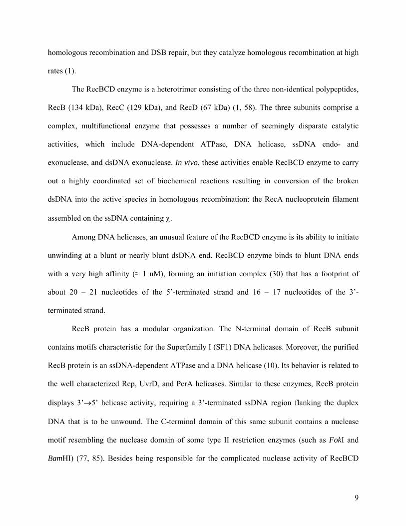
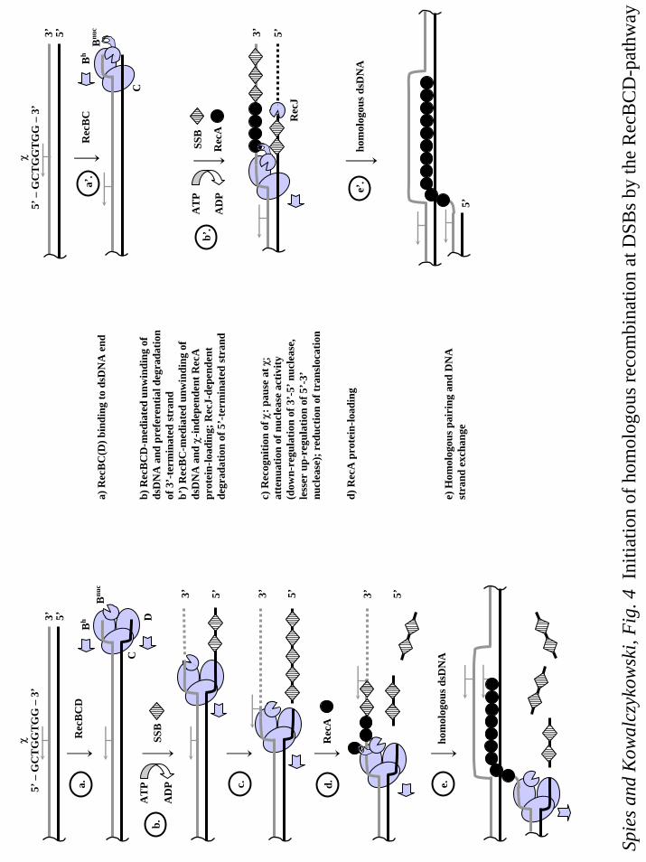
Double strand break repair by recombination: the RecBCD-pathway.
The RecBCD-pathway comprises the RecBCD, SSB, RecA, and RuvABC proteins; in
addition, a specific DNA locus called χ (Chi = crossover hotspot instigator, 5’-GCTGGTGG-3’)
is required in the dsDNA that is broken.
In wild-type E. coli, RecBCD enzyme is required for approximately 99% of the
recombination events associated with conjugation and transduction, i.e., the processes that
involve linear DNA and, hence, a DSB. Genetic studies revealed that the products of recB and
recC genes are necessary for the repair of DSBs and for conjugational recombination. Deletion
of either recB or recC genes reduces the levels of sexual recombination 100- to 1000-fold and
increases sensitivity to DNA-damaging agents, such as UV-irradiation and mitomycin C (26, 36,
84). This effect, however, is significantly smaller then the 100,000-fold decrease in homologous
recombination in the recA- mutants. In contrast, cells deleted for recD are not only proficient in
8
homologous recombination and DSB repair, but they catalyze homologous recombination at high
rates (1).
The RecBCD enzyme is a heterotrimer consisting of the three non-identical polypeptides,
RecB (134 kDa), RecC (129 kDa), and RecD (67 kDa) (1, 58). The three subunits comprise a
complex, multifunctional enzyme that possesses a number of seemingly disparate catalytic
activities, which include DNA-dependent ATPase, DNA helicase, ssDNA endo- and
exonuclease, and dsDNA exonuclease. In vivo, these activities enable RecBCD enzyme to carry
out a highly coordinated set of biochemical reactions resulting in conversion of the broken
dsDNA into the active species in homologous recombination: the RecA nucleoprotein filament
assembled on the ssDNA containing χ.
Among DNA helicases, an unusual feature of the RecBCD enzyme is its ability to initiate
unwinding at a blunt or nearly blunt dsDNA end. RecBCD enzyme binds to blunt DNA ends
with a very high affinity (≈ 1 nM), forming an initiation complex (30) that has a footprint of
about 20 – 21 nucleotides of the 5’-terminated strand and 16 – 17 nucleotides of the 3’-
terminated strand.
RecB protein has a modular organization. The N-terminal domain of RecB subunit
contains motifs characteristic for the Superfamily I (SF1) DNA helicases. Moreover, the purified
RecB protein is an ssDNA-dependent ATPase and a DNA helicase (10). Its behavior is related to
the well characterized Rep, UvrD, and PcrA helicases. Similar to these enzymes, RecB protein
displays 3’→5’ helicase activity, requiring a 3’-terminated ssDNA region flanking the duplex
DNA that is to be unwound. The C-terminal domain of this same subunit contains a nuclease
motif resembling the nuclease domain of some type II restriction enzymes (such as FokI and
BamHI) (77, 85). Besides being responsible for the complicated nuclease activity of RecBCD
9
enzyme, the C-terminal domain of RecB subunit has another crucial function: it harbors a site for
interaction with RecA protein (19).
The amino acid sequence of RecC protein provides no clue as to its role in the
holoenzyme. However, the existence of the RecC mutants that enable the holoenzyme to
recognize an altered χ-sequence suggests a significant role in χ-recognition (3). Interaction with
RecC subunit greatly stimulates the weak nuclease activity of the RecB helicase, and increases
its affinity for dsDNA ends (61). The resulting RecBC enzyme is a fast, processive helicase that
can initiate homologous recombination. However, RecBC enzyme displays negligible nuclease
activity. As result, homologous recombination in the recD- cells strongly depends on the function
of RecJ nuclease (51).
The RecD subunit also contains SF1 helicase motifs. The purified RecD protein is a
DNA-dependent ATPase that was recently shown to be a 5’→3’ helicase (23) similar to the
closely related TraI and Dda helicases. Like these other helicases, RecD protein unwinds
substrates containing a 5’-terminated ssDNA flanking the duplex DNA. Interaction with the
RecD subunit further stimulates both the helicase activity and the dsDNA end-binding affinity of
RecBC enzyme. Given that the RecB subunit is bound to the 3’-terminated strand; the RecD
subunit is bound to the 5’-terminated strand (27); and each motor subunit moves with an
opposite polarity, the resulting bipolar RecBCD enzyme translocates with each motor subunit
moving in the same direction relative to the DSB. In addition, the resulting RecBCD holoenzyme
now manifests its vigorous nuclease activity, implying that the RecD subunit also activates the
nuclease contained within the RecB subunit (41).
The activities described above permit the following description for the mechanism of
action of the RecBCD helicase/nuclease (Figure 3). Upon binding to the DNA end (Figure 4
10
(a.)), the enzyme uses energy of ATP hydrolysis to translocate along and unwind the dsDNA
molecule, consuming approximately two to three molecules of ATP per 1 base pair unwound
(63) (Figure 4 (c.)). RecBCD enzyme unwinds, on average, 30,000 bps of dsDNA per binding
event (62) at a rate of approximately 1,000-1,300 bps/sec (at 37 ºC). DNA unwinding by
RecBCD enzyme is accompanied by endonucleolytic cleavage of newly produced ssDNA. The
nuclease activity of RecBCD enzyme is asymmetric, with digestion occurring preferentially on
the 3’-terminated strand relative to the DSB (25).
The recombination hotspot, Chi.
Originally, χ was identified as a cis-acting mutation in bacteriophage λ that allowed its
more efficient growth in E. coli by stimulating the host’s recombination system. Stimulation of
recombination is approximately 10-fold (47), and it is confined to loci that are downstream of χ
(70): stimulation is highest at χ and then decreases exponentially (17, 47). The χ sequence is
over-represented in E. coli; there are 1009 χ sequences found in the 4.6 Mb genome of the
MG1655 strain (9). Furthermore, over 60% of χ sequences are oriented towards the replication
origin. Such an orientation would facilitate RecBCD-mediated recombination repair of DSBs
created during DNA replication (11).
Biochemically, the activities of RecBCD enzyme are altered upon recognition of χ.
Alteration of RecBCD enzyme activity is manifest only when the enzyme approaches χ, 5’-
GCTGGTGG-3’, from its 3’-side. In the schematic depiction (Figure 4), χ, which is the sequence
in the “top strand” (7), is recognized by an enzyme moving only from the right to the left. In
vivo, interaction with χ results in the stimulation of homologous recombination downstream of χ
(69, 71). In vitro, recognition of the χ-sequence causes RecBCD enzyme to switch the polarity
11
of its nuclease activity (Figure 4 (d.)): upon interaction with χ, degradation of the 3’-terminated
strand is down-regulated, while degradation of the 5’-terminated strand is up-regulated (24, 25).
Consequently, the enzyme produces a lengthy ssDNA tail with χ at the 3’-terminated end. As
determined from production of the χ-specific ssDNA fragments, the probability of recognizing a
single χ is about 30 – 40% (3, 72). Interaction with χ also affects the helicase activity of
RecBCD enzyme. Recognition of χ causes the enzyme to pause briefly (typically, a few seconds)
at χ and to resume translocation after the χ site, but at a rate that is reduced by approximately 2-
fold (68). Another consequence of χ-modification is that the RecBCD enzyme gains the ability
to “load” RecA protein onto the newly produced ssDNA (2) (Figure 4 (e.)). Thus, in response to
χ, the RecBCD enzyme accomplishes both tasks essential for initiation of homologous
recombination: 1) it recesses the DSB to produce an ssDNA-tailed duplex DNA with χ at its
terminus; and 2) it catalyzes formation of the RecA nucleoprotein filament on the ssDNA
produced. This RecA nucleoprotein filament now can search for homology, promote invasion of
the homologous recipient, and exchange the DNA strands (Figure 4f.).
The exact molecular mechanism, by which χ-recognition is translated into the observed
changes in the activities of the RecBCD enzyme, remains unknown. Most models propose either
dissociation or inactivation of RecD subunit (2, 19, 57, 73). Indeed, as mentioned previously, the
RecBC enzyme (lacking the RecD subunit) is recombinationally proficient both in vivo (13) and
in vitro (18) (Figure 4a’). The RecBC enzyme is a processive helicase but with little or no
nuclease activity (41), which is in contrast to the χ-activated form of RecBCD enzyme; however,
this distinction is consistent with the requirement for RecJ nuclease activity in recD- cells in vivo
(51). Similar to RecBCD enzyme, RecBC helicase facilitates asymmetric assembly of RecA
protein only onto ssDNA that is 3’-terminal at the enzyme entry site (Figure 4 (c’)). The RecBC-
12
mediated loading of RecA protein is constitutive and is independent of χ, consistent with the
phenotypic behavior of recD- cells in vivo.
Regulated helicases/nucleases in other bacterial species.
For decades, the interaction between RecBCD enzyme and χ was known to exist only in
the species of enteric bacteria closely related to E. coli. But relatively recently, short (5 – 8 bps)
sequences, which protect linear dsDNA from degradation by attenuation of the nuclease activity
of RecBCD-like helicase/nuclease enzymes, were found in several distantly related bacteria (8,
16, 67). This finding indicates that, although apparently not universal, the regulation of
recombinational helicases/nucleases by specific DNA sequences is widely spread among
prokaryotes. While some bacteria possess clear homologues of RecBCD enzyme, other species
contain its functional equivalent, the AddAB enzyme (reviewed in (15)). AddAB
helicase/nuclease is constituted of two subunits encoded by addA and addB genes. The sole
motor subunit of AddAB enzyme, AddA protein, contains an SF1-helicase and a nuclease
domain, which display high degree of similarity to those in RecB protein (32). Also similar to
RecB protein, AddA is a 3’→5’ helicase, and its 3’→5’ nuclease activity is down-regulated upon
interaction with the cognate recombination hotspot of Bacillus subtilis, χBs (5’-AGCGG-3’) (14,
16). The AddB subunit has no substantial similarity to either the RecC or RecD subunits, but it
does contain a putative ATPase motif and a second nuclease site similar to that of the AddA
protein. The AddB subunit is responsible for the degradation of the 5’-terminated strand. In spite
of the limited sequence similarity to the RecBCD enzyme, AddAB enzyme is functional in E.
coli: its expression overcomes the recombination and repair defects of recBC-deficient cells (40).
Similar to RecBCD enzyme, AddAB enzyme binds to the blunt-ended dsDNA, and uses the
energy of ATP hydrolysis to translocate along and unwind dsDNA. However, where RecBCD
13
enzyme degrades the dsDNA asymmetrically, the AddAB enzyme degrades both strands of the
DNA duplex equally. Interaction of the translocating AddAB enzyme with a correctly oriented
χB results in down-regulation of only the 3’→5’ nuclease activity of the enzyme. The outcome,
therefore, is the same as that occurring for the E. coli enzyme: namely, the production of ssDNA-
tailed dsDNA with χ at the 3’-terminus. Homologues of AddAB enzyme are found in 12
different species of gram-positive bacteria, and χ-homologs were identified in several bacterial
species (reviewed in (15)).
Single-strand gap repair by recombination: the RecF-pathway.
The conjugal recombination deficiency of recB- or recC- mutants can be overcome by the
combined effect of two extragenic suppressor mutations, sbcB and either sbcC or sbcD
(suppressor of recBC). The sbcB mutation disables the nuclease activity of Exonuclease I (44),
while sbcC (31) disable one of the two subunits of the SbcCD nuclease which, in wild-type E.
coli, cleaves DNA hairpin and cruciform structures formed during replication of palindromic
sequences (22, 49). The combined effect of these mutations is the full activation of an alternative
pathway of sexual homologous recombination, referred to as the RecF-pathway. Interestingly,
the efficiency of conjugational and transductional recombination by the RecF-pathway in the
recBC sbcBC cells is similar to that of the RecBCD-pathway in wild-type cells, showing that the
machinery of this pathway can be as productive as that of the RecBCD-pathway. Moreover,
some bacterial species, whose survival depends on homologous recombination (such as
Deinococcus radiodurans), do not possess obvious RecBCD or AddAB enzymes, implying that a
RecF-like pathway is the wild-type pathway in those bacteria.
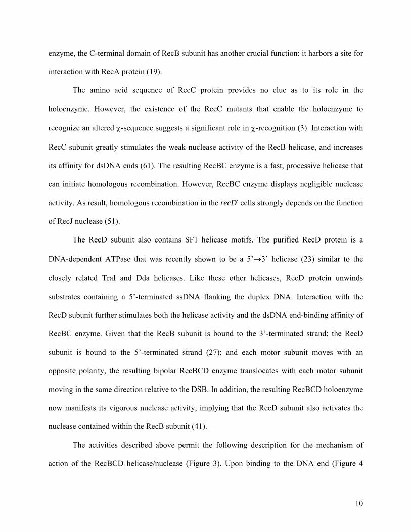
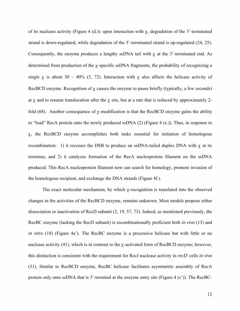
Homologous recombination in the recBC- sbcB sbcC- background depends on RecF,
RecJ, RecN, RecO, RecQ, RecR, and SSB proteins. The processing of a DSB is likely achieved
14
by the combined action of the RecQ helicase and RecJ nuclease. Although RecQ helicase is
responsible for about 75% of conjugal recombination events occurring in recBC sbcB sbcC-
cells, the remaining 25% require either UvrD (helicase II) or HelD (helicase IV) (53). The RecJ
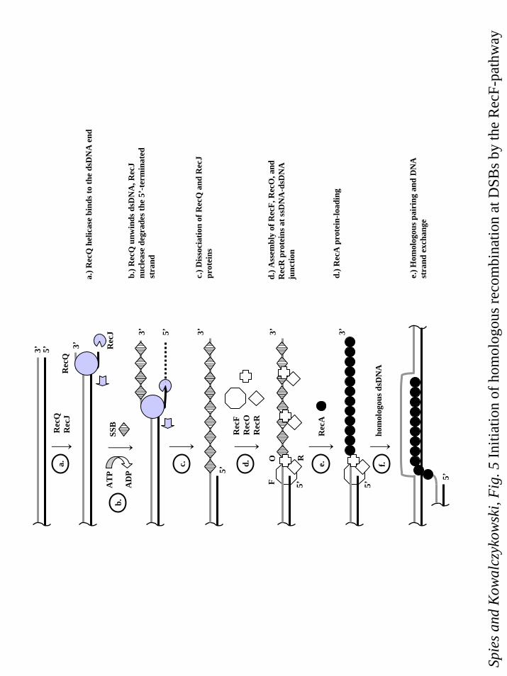
protein is an exonuclease that degrades ssDNA in the 5’→ 3’ direction (Figure 5 (a – c)). RecQ
protein is an SF2 DNA helicase with a 3’→ 5’ polarity. Similar to RecBCD enzyme, RecQ
helicase can unwind blunt ended dsDNA (76). The helicase activity of RecQ protein is not
limited to blunt dsDNA ends: the enzyme can also unwind ssDNA-dsDNA junction with a 3’-
ssDNA overhang (76), and even internal regions of dsDNA (33). It is hypothesized that DNA
unwinding by RecQ helicase is coupled to the degradation of the 5’-terminated strand by RecJ
nuclease, resulting in the production of the 3’-terminated ssDNA overhang, which can then be
used as a substrate for RecA nucleoprotein assembly (Figure 5 (c.)). In contrast to RecBCD
enzyme, RecQ does not facilitate RecA nucleoprotein filament assembly. Therefore, the ssDNA
produced by RecQ is bound by SSB protein and must be protected from degradation by
nucleases, which explains the requirement for the sbcB mutation, since the preferred substrate for
Exonuclease I is SSB-complexed ssDNA. Intriguingly, two major classes of mutations in the
structural gene for Exonuclease I were found. One class of mutations, referred to as sbcB,
restores both recombination and the UV-resistance of recBC cells. In contrast, the other class,
known as xonA mutants, suppresses only the UV-sensitivity but not the recombination deficiency
of the recBC- bacteria (44). Exonuclease I activity is significantly reduced in both classes of
mutants; moreover UV-sensitivity directly correlates with the amount of residual activity of the
enzyme (60). The sbcB mutations are not the same as null mutations and, therefore, likely
represent gain of function mutations. The molecular mechanism distinguishing the two types of
15
mutations still remains to be elucidated, but protection of the 3’-end of ssDNA by the sbcB
mutations is envisioned.
The loading of RecA protein is an essential aspect of recombination in the RecBCD-
pathway (4). Not unexpectedly, the RecF-pathway provides a RecA-loading activity in the form
of the RecFOR complex. The genetic data had suggested that the loading of RecA protein onto
SSB-coated ssDNA depends on the concerted action of RecF, RecO, and RecR proteins. Firstly,
mutant RecA proteins that suppress the UV-sensitivity of recF mutations (such as the RecA803
protein) displace SSB protein from ssDNA much faster and more extensively than wild-type
RecA protein (52), implying that RecF protein plays a role in SSB-displacement. Secondly, a
number of mutant recA alleles co-suppress mutations in recF, recO, and recR genes. Finally,
suppression of mutations in any of these three genes is dependent on recJ function, suggesting
that RecF, RecO, and RecR proteins may function together as a complex (79). This view is
strongly supported by biochemical observations showing that these proteins interact with one
another (34, 74).
The RecFOR proteins form a number of complexes with different activities: RecO
protein interacts with both RecR and SSB proteins (34), and facilitates RecA nucleoprotein
filament assembly on SSB-coated ssDNA (75). RecO protein also promotes the annealing of
ssDNA, and of SSB-ssDNA complexes (38). Finally, RecR protein interacts with RecF protein to
form a complex (34, 81) that will bind to an ssDNA-dsDNA junction (55). Biochemical analysis
revealed that RecF protein binds preferentially to the ssDNA-dsDNA junction (35), and that
DNA binding by RecF protein is controlled by ATP hydrolysis (80). The RecFOR complex will
bind to an ssDNA-dsDNA junction with a base-paired 5’-terminus at the junction region, and it
will facilitate assembly of RecA protein onto ssDNA adjacent to the junction (55). RecF protein
16
(or RecFR complex) recognizes an ssDNA-dsDNA junction with a 5’-end, which is the structure
that should be produced by RecQ and RecJ proteins. The RecOR complex (or RecO protein)
binds to the DNA-RecF(R) complex, which alters SSB-ssDNA complex nearby and allows RecA
protein nucleation; subsequent nucleoprotein filament extension permits assembly of RecA
protein on the entire ssDNA tail.
Another component of the RecF-pathway, RecN protein, has not yet been assigned any
biochemical function. However, the slight recombination deficiency and mild UV sensitivity of
recJ recN double mutant, combined with severe recombination defect (50- to 100-fold reduction)
of recD recJ recN mutant, suggest that RecN protein might be a functional equivalent of the
RecJ nuclease (50).
In spite of its apparent complexity, the enzymatic machinery of the RecF pathway is as
functional in DSB repair as RecBCD enzyme. Moreover, the components of the RecF pathway
have functional homologs or paralogs in all organisms from bacteriophage to human (5).
Single-strand gap repair.
Because conjugation (and transduction) involve a DSB as the initiating site for
recombination, it should not be surprising that the genes which emerged from a screen for
mutants defective in sexual recombination, are essential for the repair of DSBs. However, the
consequence of this nearly singular focus on conjugational recombination events led to the
erroneous conclusion that the RecF-pathway serves only a minor function in recombination in
the wild-type cells. Similarly incorrect, the discovery of the RecF pathway as the set of genes
that permitted recombination in the absence of the primary RecBCD-pathway implied that the
RecF-pathway was a cryptic recombination pathway that could be activated to compensate for
the loss of RecBCD enzyme function. Rather, in wild-type cells, the RecF-pathway is
17
responsible for the repair of all SSGs. This conclusion emerged from many studies, but was
made clearest from recombination assays that did not employ sexual events.
A genetic assay that measures recombination between direct repeats of a chromosomal
segment allows detection of sister-chromosome exchanges that are required for the repair of
DSBs (29). This assay was developed to approximate the function of homologous recombination
in the repair of the DNA lesions produced during replication. The recombination events
occurring between direct chromosomal repeats are detected in the colony-sectoring
recombination assay, since the detected recombination events eliminate the joint-point markers
located between the repeats. Recombination events detected in this assay, are absolutely
dependent on the RecA protein function. The recF and recJ mutants display a rec+ phenotype;
recB mutants show only slight defect; and recB recJ double mutants are capable of supporting
duplication segregation. On the other hand, recB recF double mutants are deficient in the
recombination between chromosomal direct repeats, suggesting that both RecBCD and RecF
pathways play major roles in recombination.
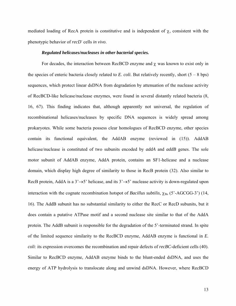
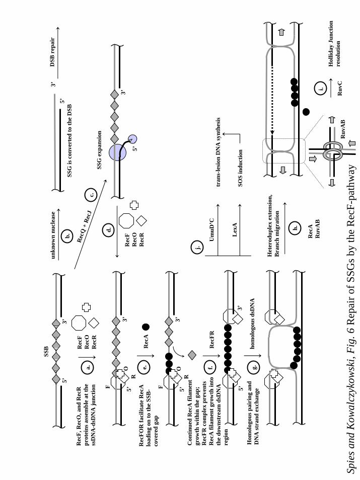
Initiation of homologous recombination on SSGs is presented in Figure 6. First, RecFOR
complex assembles at the ssDNA-dsDNA junction at the end of SSG containing an
complementary 5’-end (Figure 6 (a.)) and facilitates RecA nucleoprotein assembly in the ssDNA
region of the gap (55) (Figure 6 (b.)). RecFR complex can bind to the 3’-containing end of SSG
limiting RecA filament extension into the dsDNA region (82) (Figure 6 (c.)). The resulting RecA
nucleoprotein filament formed on the SSG can then invade a homologous dsDNA molecule
(Figure 6 (d.)). DNA strand exchange followed by heteroduplex extension results in the
formation of two Holliday junctions. To complete the repair, RecA protein must be removed
from the junctions and the Holliday junctions themselves need to be resolved. The processing of
18
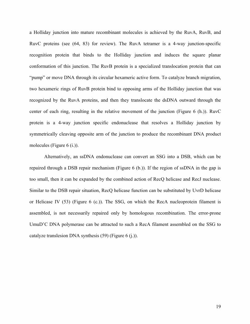
a Holliday junction into mature recombinant molecules is achieved by the RuvA, RuvB, and
RuvC proteins (see (64, 83) for review). The RuvA tetramer is a 4-way junction-specific
recognition protein that binds to the Holliday junction and induces the square planar
conformation of this junction. The RuvB protein is a specialized translocation protein that can
“pump” or move DNA through its circular hexameric active form. To catalyze branch migration,
two hexameric rings of RuvB protein bind to opposing arms of the Holliday junction that was
recognized by the RuvA proteins, and then they translocate the dsDNA outward through the
center of each ring, resulting in the relative movement of the junction (Figure 6 (h.)). RuvC
protein is a 4-way junction specific endonuclease that resolves a Holliday junction by
symmetrically cleaving opposite arm of the junction to produce the recombinant DNA product
molecules (Figure 6 (i.)).
Alternatively, an ssDNA endonuclease can convert an SSG into a DSB, which can be
repaired through a DSB repair mechanism (Figure 6 (b.)). If the region of ssDNA in the gap is
too small, then it can be expanded by the combined action of RecQ helicase and RecJ nuclease.
Similar to the DSB repair situation, RecQ helicase function can be substituted by UvrD helicase
or Helicase IV (53) (Figure 6 (c.)). The SSG, on which the RecA nucleoprotein filament is
assembled, is not necessarily repaired only by homologous recombination. The error-prone
UmuD’C DNA polymerase can be attracted to such a RecA filament assembled on the SSG to
catalyze translesion DNA synthesis (59) (Figure 6 (j.)).
19
Conclusion
Homologous recombination can be initiated either at DSBs or SSGs in duplex DNA. Two
major pathways are responsible for homologous recombination in wild-type E. coli: the
RecBCD- and RecF-pathways. The RecBCD-pathway is specific for the recombinational repair
of DSBs, and in the wild-type cells, the RecF-pathway is primarily used for recombination that
initiates at SSGs. However, with appropriate suppressor mutation in E. coli, and presumably in
bacteria that lack a RecBCD-pathway, the RecF-pathway can efficiently act at DSBs as well.
Despite the different initiating lesions, both pathways have the same subsequent step: conversion
of the broken DNA molecule into a central intermediate of recombination, which is the RecA
protein nucleoprotein filament assembled along the ssDNA. In the RecBCD pathway, this
process is carried out by a combined helicase/nuclease activity of RecBCD enzyme, and depends
on the presence of the recombination hotspot, χ. In the RecF-pathway, the combined efforts of
RecQ helicase and RecJ ssDNA nuclease are needed in combination with the RecFOR complex.
In the RecBCD pathway, RecBCD enzyme facilitates assembly of the RecA protein onto SSB-
coated ssDNA; while in the RecF pathway, this task is accomplished by RecF, RecO, and RecR
proteins. RecA nucleoprotein filament can then initiate invasion of ssDNA into homologous
dsDNA, progressing into the final stage of homologous recombination which is resolution by
RuvABC proteins.
20
References
1. Amundsen, S. K., A. F. Taylor, A. M. Chaudhury, and G. R. Smith. 1986. recD: The gene for an essential third subunit of exonuclease V. Proc. Natl. Acad. Sci. USA 83:5558-62.
2. Anderson, D. G., J. J. Churchill, and S. C. Kowalczykowski. 1997. Chi-activated RecBCD enzyme possesses 5'→3' nucleolytic activity, but RecBC enzyme does not: Evidence suggesting that the alteration induced by Chi is not simply ejection of the RecD subunit. Genes Cells 2:117-28.
3. Arnold, D. A., P. R. Bianco, and S. C. Kowalczykowski. 1998. The reduced levels of χ recognition exhibited by the RecBC1004D enzyme reflect its recombination defect in vivo. J Biol Chem 273:16476-86.
4. Arnold, D. A., and S. C. Kowalczykowski. 2000. Facilitated loading of RecA protein is essential to recombination by RecBCD enzyme. J Biol Chem 275:12261-5.
5. Beernink, H. T., and S. W. Morrical. 1999. RMPs: recombination/replication mediator proteins. Trends Biochem Sci 24:385-9.
6. Bianco, P. R., and S. C. Kowalczykowski. 1999. RecA protein, Encyclopedia of Life Sciences, vol. http://www.els.net. Nature Publishing Group, London.
7. Bianco, P. R., and S. C. Kowalczykowski. 1997. The recombination hotspot Chi is recognized by the translocating RecBCD enzyme as the single strand of DNA containing the sequence 5'-GCTGGTGG-3'. Proc Natl Acad Sci U S A 94:6706-11.
8. Biswas, I., E. Maguin, S. D. Ehrlich, and A. Gruss. 1995. A 7-base-pair sequence protects DNA from exonucleolytic degradation in Lactococcus lactis. Proc Natl Acad Sci U S A 92:2244-8.
9. Blattner, F. R., G. Plunkett, 3rd, C. A. Bloch, N. T. Perna, V. Burland, M. Riley, J. Collado-Vides, J. D. Glasner, C. K. Rode, G. F. Mayhew, J. Gregor, N. W. Davis, H. A. Kirkpatrick, M. A. Goeden, D. J. Rose, B. Mau, and Y. Shao. 1997. The complete genome sequence of Escherichia coli K-12. Science 277:1453-74.
10. Boehmer, P. E., and P. T. Emmerson. 1992. The RecB subunit of the Escherichia coli RecBCD enzyme couples ATP hydrolysis to DNA unwinding. J Biol Chem 267:4981-7.
11. Burland, V., G. d. Plunkett, D. L. Daniels, and F. R. Blattner. 1993. DNA sequence and analysis of 136 kilobases of the Escherichia coli genome: organizational symmetry around the origin of replication. Genomics 16:551-61.
12. Capaldo-Kimball, F., and S. D. Barbour. 1971. Involvement of recombination genes in growth and viability of Escherichia coli K-12. J. Bacteriol. 106:204-212.
13. Chaudhury, A. M., and G. R. Smith. 1984. A new class of Escherichia coli recBC mutants: Implications for the role of recBC enzyme in homologous recombination. Proc. Natl. Acad. Sci. USA 81:7850-7854.
14. Chedin, F., S. D. Ehrlich, and S. C. Kowalczykowski. 2000. The Bacillus subtilis AddAB helicase/nuclease is regulated by its cognate Chi sequence in vitro. J Mol Biol 298:7-20.
15. Chedin, F., and S. C. Kowalczykowski. 2002. A novel family of regulated helicases/nucleases from Gram-positive bacteria: insights into the initiation of DNA recombination. Mol Microbiol 43:823-34.
21
16. Chedin, F., P. Noirot, V. Biaudet, and S. D. Ehrlich. 1998. A five-nucleotide sequence protects DNA from exonucleolytic degradation by AddAB, the RecBCD analogue of Bacillus subtilis. Mol Microbiol 29:1369-77.
17. Cheng, K. C., and G. R. Smith. 1989. Distribution of Chi-stimulated recombinational exchanges and heteroduplex endpoints in phage lambda. Genetics 123:5-17.
18. Churchill, J. J., D. G. Anderson, and S. C. Kowalczykowski. 1999. The RecBC enzyme loads RecA protein onto ssDNA asymmetrically and independently of chi, resulting in constitutive recombination activation. Genes Dev 13:901-11.
19. Churchill, J. J., and S. C. Kowalczykowski. 2000. Identification of the RecA Protein-loading Domain of RecBCD Enzyme. J Mol Biol 297:537-542.
20. Clark, A. J. 1973. Recombination deficient mutants of E. coli and other bacteria. Annu. Rev. Genet. 7:67-86.
21. Clark, A. J., and A. D. Margulies. 1965. Isolation and characterization of recombination-deficient mutants of Escherichia coli K12. Proc. Natl. Acad. Sci. USA 53:451-459.
22. Connelly, J. C., L. A. Kirkham, and D. R. Leach. 1998. The SbcCD nuclease of Escherichia coli is a structural maintenance of chromosomes (SMC) family protein that cleaves hairpin DNA. Proc Natl Acad Sci U S A 95:7969-74.
23. Dillingham, M. S., M. Spies, and S. C. Kowalczykowski. 2003. RecBCD enzyme is a bipolar DNA helicase. Nature 423:893-897.
24. Dixon, D. A., J. J. Churchill, and S. C. Kowalczykowski. 1994. Reversible inactivation of the Escherichia coli RecBCD enzyme by the recombination hotspot χ in vitro: evidence for functional inactivation or loss of the RecD subunit. Proc Natl Acad Sci U S A 91:2980-2984.
25. Dixon, D. A., and S. C. Kowalczykowski. 1993. The recombination hotspot χ is a regulatory sequence that acts by attenuating the nuclease activity of the E. coli RecBCD enzyme. Cell 73:87-96.
26. Emmerson, P. T. 1968. Recombination deficient mutants of Escherichia coli K12 that map between thyA and argA. Genetics 60:19-30.
27. Farah, J. A., and G. R. Smith. 1997. The RecBCD enzyme initiation complex for DNA unwinding: enzyme positioning and DNA opening. J Mol Biol 272:699-715.
28. Friedberg, E. C., G. C. Walker, and W. Siede. 1995. DNA Repair and Mutagenesis. ASM Press, Washington, D.C.
29. Galitski, T., and J. R. Roth. 1997. Pathways for homologous recombination between chromosomal direct repeats in Salmonella typhimurium. Genetics 146:751-67.
30. Ganesan, S., and G. R. Smith. 1993. Strand-specific binding to duplex DNA ends by the subunits of Escherichia coli recBCD enzyme. J Mol Biol 229:67-78.
31. Gibson, F. P., D. R. Leach, and R. G. Lloyd. 1992. Identification of sbcD mutations as cosuppressors of recBC that allow propagation of DNA palindromes in Escherichia coli K-12. J Bacteriol 174:1222-8.
32. Haijema, B. J., G. Venema, and J. Kooistra. 1996. The C terminus of the AddA subunit of the Bacillus subtilis ATP-dependent DNase is required for the ATP-dependent exonuclease activity but not for the helicase activity. J Bacteriol 178:5086-91.
33. Harmon, F. G., and S. C. Kowalczykowski. 2001. Biochemical Characterization of the DNA Helicase Activity of the Escherichia coli RecQ Helicase. J Biol Chem 276:232-243.
22
34. Hegde, S. P., M. H. Qin, X. H. Li, M. A. Atkinson, A. J. Clark, M. Rajagopalan, and M. V. Madiraju. 1996. Interactions of RecF protein with RecO, RecR, and single-stranded DNA binding proteins reveal roles for the RecF-RecO-RecR complex in DNA repair and recombination. Proc Natl Acad Sci U S A 93:14468-73.
35. Hegde, S. P., M. Rajagopalan, and M. V. Madiraju. 1996. Preferential binding of Escherichia coli RecF protein to gapped DNA in the presence of adenosine (gamma-thio) triphosphate. J Bacteriol 178:184-90.
36. Howard-Flanders, P., and L. Theriot. 1966. Mutants of Escherichia coli K-12 defective in DNA repair and in genetic recombination. Genetics 53:1137-1150.
37. Iyer, V. N., and W. D. Rupp. 1971. Usefulness of benzoylated naphthoylated DEAE-cellulose to distinguish and fractionate double-stranded DNA bearing different extents of single-stranded regions. Biochim Biophys Acta 228:117-26.
38. Kantake, N., M. V. Madiraju, T. Sugiyama, and S. C. Kowalczykowski. 2002. Escherichia coli RecO protein anneals ssDNA complexed with its cognate ssDNA-binding protein: A common step in genetic recombination. Proc Natl Acad Sci U S A 99:15327-32.
39. Kogoma, T. 1997. Stable DNA replication: interplay between DNA replication, homologous recombination, and transcription. Microbiol Mol Biol Rev 61:212-38.
40. Kooistra, J., B. J. Haijema, and G. Venema. 1993. The Bacillus subtilis addAB genes are fully functional in Escherichia coli. Mol Microbiol 7:915-23.
41. Korangy, F., and D. A. Julin. 1993. Kinetics and processivity of ATP hydrolysis and DNA unwinding by the RecBC enzyme from Escherichia coli. Biochemistry 32:4873-4880.
42. Kowalczykowski, S. C. 2000. Initiation of genetic recombination and recombination-dependent replication. Trends Biochem Sci 25:156-165.
43. Krasin, F., and F. Hutchinson. 1977. Repair of DNA double-strand breaks in Escherichia coli, which requires recA function and the presence of a duplicate genome. J. Mol. Biol. 116:81-98.
44. Kushner, S. R., H. Nagaishi, and A. J. Clark. 1972. Indirect suppression of recB and recC mutations by exonuclease I deficiency. Proc. Natl. Acad. Sci. USA 69:1366-1370.
45. Kuzminov, A. 1995. Collapse and repair of replication forks in Escherichia coli. Mol Microbiol 16:373-84.
46. Kuzminov, A. 1999. Recombinational repair of DNA damage in Escherichia coli and bacteriophage lambda. Microbiol Mol Biol Rev 63:751-813, table of contents.
47. Lam, S. T., M. M. Stahl, K. D. McMilin, and F. W. Stahl. 1974. Rec-mediated recombinational hot spot activity in bacteriophage lambda. II. A mutation which causes hot spot activity. Genetics 77:425-33.
48. Lederberg, J., and E. L. Tatum. 1953. Sex in bacteria; genetic studies, 1945-1952. Science 118:169-75.
49. Lloyd, R. G., and C. Buckman. 1985. Identification and genetic analysis of sbcC mutations in commonly used recBC sbcB strains of Escherichia coli K-12. J. Bacteriol. 164:836-844.
50. Lloyd, R. G., and C. Buckman. 1991. Overlapping functions of recD, recJ and recN provide evidence of three epistatic groups of genes in Escherichia coli recombination and DNA repair. Biochimie 73:313-20.
23
51. Lloyd, R. G., M. C. Porton, and C. Buckman. 1988. Effect of recF, recJ, recN, recO and ruv mutations on ultraviolet survival and genetic recombination in a recD strain of Escherichia coli K12. Mol Gen Genet 212:317-24.
52. Madiraju, M. V. V. S., A. Templin, and A. J. Clark. 1988. Properties of a mutant recA-encoded protein reveal a possible role for Escherichia coli recF-encoded protein in genetic recombination. Proc. Natl. Acad. Sci. USA 85:6592-6.
53. Mendonca, V. M., H. D. Klepin, and S. W. Matson. 1995. DNA helicases in recombination and repair: construction of a ∆uvrD ∆helD ∆recQ mutant deficient in recombination and repair. J Bacteriol 177:1326-35.
54. Michel, B., S. D. Ehrlich, and M. Uzest. 1997. DNA double-strand breaks caused by replication arrest. Embo J 16:430-8.
55. Morimatsu, K., and S. C. Kowalczykowski. 2003. RecFOR proteins load RecA protein onto gapped DNA to accelerate DNA strand exchange: a universal step of recombinational repair. Mol Cell 11:1337-47.
56. Mosig, G. 1987. The essential role of recombination in phage T4 growth. Annu Rev Genet 21:347-71.
57. Myers, R. S., A. Kuzminov, and F. W. Stahl. 1995. The recombination hot spot χ activates RecBCD recombination by converting Escherichia coli to a recD mutant phenocopy. Proc Natl Acad Sci U S A 92:6244-8.
58. Myers, R. S., and F. W. Stahl. 1994. Chi and the RecBC D enzyme of Escherichia coli. Annu Rev Genet 28:49-70.
59. Pham, P., E. M. Seitz, S. Saveliev, X. Shen, R. Woodgate, M. M. Cox, and M. F. Goodman. 2002. Two distinct modes of RecA action are required for DNA polymerase V-catalyzed translesion synthesis. Proc Natl Acad Sci U S A 99:11061-6.
60. Phillips, G. J., D. C. Prasher, and S. R. Kushner. 1988. Physical and biochemical characterization of cloned sbcB and xonA mutations from Escherichia coli K-12. J. Bacteriol. 170:2089-94.
61. Phillips, R. J., D. C. Hickleton, P. E. Boehmer, and P. T. Emmerson. 1997. The RecB protein of Escherichia coli translocates along single-stranded DNA in the 3' to 5' direction: a proposed ratchet mechanism. Mol Gen Genet 254:319-29.
62. Roman, L. J., A. K. Eggleston, and S. C. Kowalczykowski. 1992. Processivity of the DNA helicase activity of Escherichia coli recBCD enzyme. J Biol Chem 267:4207-4214.
63. Roman, L. J., and S. C. Kowalczykowski. 1989. Characterization of the adenosinetriphosphatase activity of the Escherichia coli RecBCD enzyme: Relationship of ATP hydrolysis to the unwinding of duplex DNA. Biochemistry 28:2873-2881.
64. Shinagawa, H., and H. Iwasaki. 1995. Molecular mechanisms of Holliday junction processing in Escherichia coli. Adv Biophys 31:49-65.
65. Sinden, R. R., and R. S. Cole. 1978. Repair of cross-linked DNA and survival of Escherichia coli treated with psoralen and light: effects of mutations influencing genetic recombination and DNA metabolism. J Bacteriol 136:538-47.
66. Smith, C. L., J. G. Econome, A. Schutt, S. Klco, and C. R. Cantor. 1987. A physical map of the Escherichia coli K12 genome. Science 236:1448-53.
67. Sourice, S., V. Biaudet, M. El Karoui, S. D. Ehrlich, and A. Gruss. 1998. Identification of the Chi site of Haemophilus influenzae as several sequences related to the Escherichia coli Chi site. Mol Microbiol 27:1021-9.
24
68. Spies, M., P. R. Bianco, M. S. Dillingham, N. Handa, R. J. Baskin, and S. C. Kowalczykowski. 2003. A molecular throttle: The recombination hotspot, χ, controls DNA translocation by the RecBCD helicase. Cell 114:647-654.
69. Stahl, F. W., J. M. Crasemann, and M. M. Stahl. 1975. Rec-mediated recombinational hot spot activity in bacteriophage lambda. III. Chi mutations are site-mutations stimulating rec-mediated recombination. J Mol Biol 94:203-12.
70. Stahl, F. W., K. D. McMilin, M. M. Stahl, J. M. Crasemann, and S. Lam. 1974. The distribution of crossovers along unreplicated lambda bacteriophage chromosomes. Genetics 77:395-408.
71. Stahl, F. W., and M. M. Stahl. 1975. Rec-mediated recombinational hot spot activity in bacteriophage lambda. IV. Effect of heterology on Chi-stimulated crossing over. Mol Gen Genet 140:29-37.
72. Taylor, A. F., D. W. Schultz, A. S. Ponticelli, and G. R. Smith. 1985. RecBC enzyme nicking at Chi sites during DNA unwinding: Location and orientation-dependence of the cutting. Cell 41:153-163.
73. Taylor, A. F., and G. R. Smith. 1999. Regulation of homologous recombination: Chi inactivates RecBCD enzyme by disassembly of the three subunits. Genes Dev 13:890-900.
74. Umezu, K., N. W. Chi, and R. D. Kolodner. 1993. Biochemical interaction of the Escherichia coli RecF, RecO, and RecR proteins with RecA protein and single-stranded DNA binding protein. Proc Natl Acad Sci U S A 90:3875-79.
75. Umezu, K., and R. D. Kolodner. 1994. Protein interactions in genetic recombination in Escherichia coli. Interactions involving RecO and RecR overcome the inhibition of RecA by single-stranded DNA-binding protein. J Biol Chem 269:30005-13.
76. Umezu, K., K. Nakayama, and H. Nakayama. 1990. Escherichia coli RecQ protein is a DNA helicase. Proc Natl Acad Sci U S A 87:5363-7.
77. Wang, J., R. Chen, and D. A. Julin. 2000. A single nuclease active site of the Escherichia coli RecBCD enzyme catalyzes single-stranded DNA degradation in both directions. J Biol Chem 275:507-13.
78. Wang, T. C., and S. H. Chen. 1992. Similar-sized daughter-strand gaps are produced in the leading and lagging strands of DNA in UV-irradiated E. coli uvrA cells. Biochem Biophys Res Commun 184:1496-503.
79. Wang, T.-C. V., H. Y. Chang, and J. L. Hung. 1993. Cosuppression of recF, recR and recO mutations by mutant recA alleles in Escherichia coli cells. Mutat Res 294:157-66.
80. Webb, B. L., M. M. Cox, and R. B. Inman. 1999. ATP hydrolysis and DNA binding by the Escherichia coli RecF protein. J Biol Chem 274:15367-74.
81. Webb, B. L., M. M. Cox, and R. B. Inman. 1995. An interaction between the Escherichia coli RecF and RecR proteins dependent on ATP and double-stranded DNA. J Biol Chem 270:31397-404.
82. Webb, B. L., M. M. Cox, and R. B. Inman. 1997. Recombinational DNA repair: the RecF and RecR proteins limit the extension of RecA filaments beyond single-strand DNA gaps. Cell 91:347-56.
83. West, S. C. 1996. The RuvABC proteins and Holliday junction processing in Escherichia coli. J Bacteriol 178:1237-41.
25
84. Willetts, N. S., and D. W. Mount. 1969. Genetic analysis of recombination-deficient mutants of Escherichia coli K-12 carrying rec mutations cotransducible with thyA. J Bacteriol 100:923-34.
85. Yu, M., J. Souaya, and D. A. Julin. 1998. Identification of the nuclease active site in the multifunctional RecBCD enzyme by creation of a chimeric enzyme. J Mol Biol 283:797-808.
26
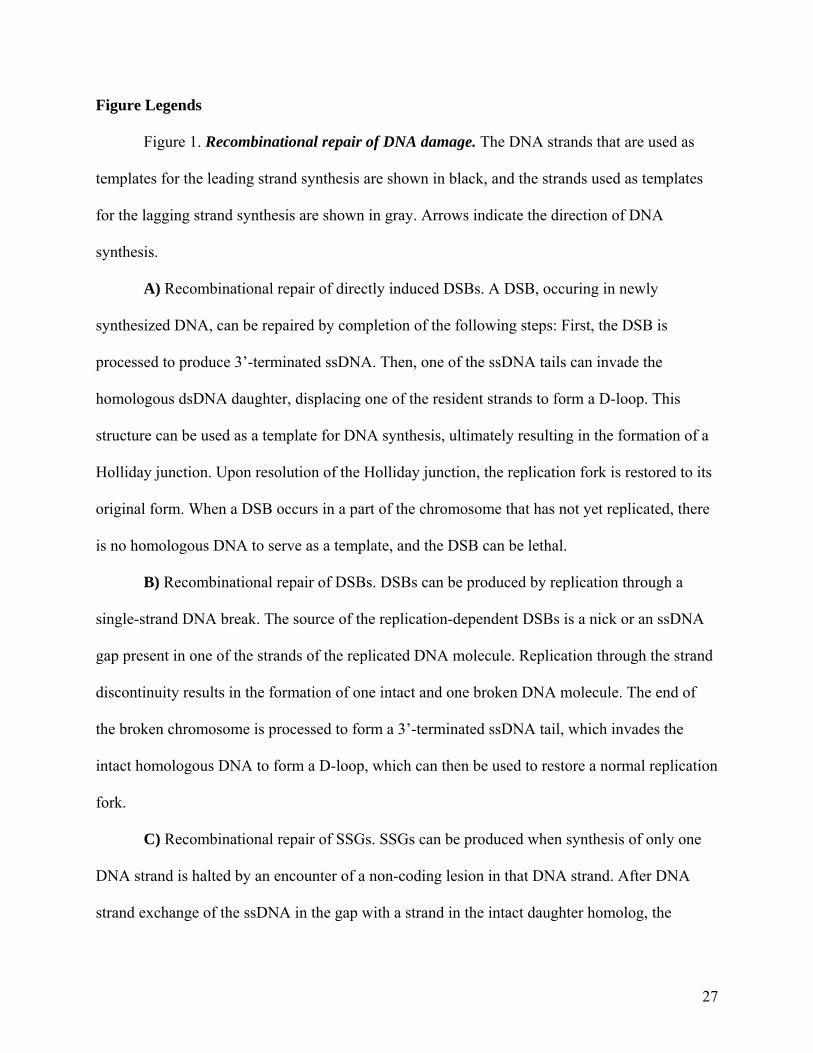
Figure Legends
Figure 1. Recombinational repair of DNA damage. The DNA strands that are used as
templates for the leading strand synthesis are shown in black, and the strands used as templates
for the lagging strand synthesis are shown in gray. Arrows indicate the direction of DNA
synthesis.
A) Recombinational repair of directly induced DSBs. A DSB, occuring in newly
synthesized DNA, can be repaired by completion of the following steps: First, the DSB is
processed to produce 3’-terminated ssDNA. Then, one of the ssDNA tails can invade the
homologous dsDNA daughter, displacing one of the resident strands to form a D-loop. This
structure can be used as a template for DNA synthesis, ultimately resulting in the formation of a
Holliday junction. Upon resolution of the Holliday junction, the replication fork is restored to its
original form. When a DSB occurs in a part of the chromosome that has not yet replicated, there
is no homologous DNA to serve as a template, and the DSB can be lethal.
B) Recombinational repair of DSBs. DSBs can be produced by replication through a
single-strand DNA break. The source of the replication-dependent DSBs is a nick or an ssDNA
gap present in one of the strands of the replicated DNA molecule. Replication through the strand
discontinuity results in the formation of one intact and one broken DNA molecule. The end of
the broken chromosome is processed to form a 3’-terminated ssDNA tail, which invades the
intact homologous DNA to form a D-loop, which can then be used to restore a normal replication
fork.
C) Recombinational repair of SSGs. SSGs can be produced when synthesis of only one
DNA strand is halted by an encounter of a non-coding lesion in that DNA strand. After DNA
strand exchange of the ssDNA in the gap with a strand in the intact daughter homolog, the
27
displaced DNA strand can be used as a template for DNA synthesis resulting in restoration of the
replication fork. Upon completion of replication, one of the DNA molecules will still contain the
original DNA damage. If this damage is not repaired by the appropriate repair system, then an
SSG will be formed again by the next round of DNA replication.
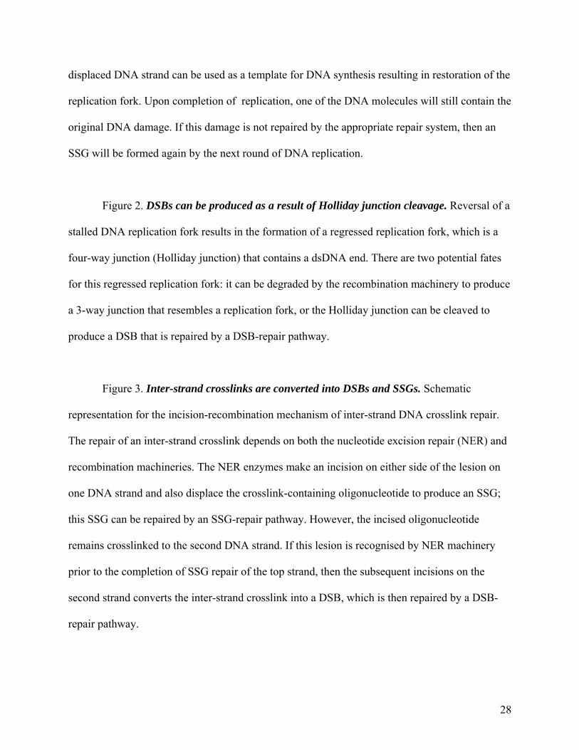
Figure 2. DSBs can be produced as a result of Holliday junction cleavage. Reversal of a
stalled DNA replication fork results in the formation of a regressed replication fork, which is a
four-way junction (Holliday junction) that contains a dsDNA end. There are two potential fates
for this regressed replication fork: it can be degraded by the recombination machinery to produce
a 3-way junction that resembles a replication fork, or the Holliday junction can be cleaved to
produce a DSB that is repaired by a DSB-repair pathway.
Figure 3. Inter-strand crosslinks are converted into DSBs and SSGs. Schematic
representation for the incision-recombination mechanism of inter-strand DNA crosslink repair.
The repair of an inter-strand crosslink depends on both the nucleotide excision repair (NER) and
recombination machineries. The NER enzymes make an incision on either side of the lesion on
one DNA strand and also displace the crosslink-containing oligonucleotide to produce an SSG;
this SSG can be repaired by an SSG-repair pathway. However, the incised oligonucleotide
remains crosslinked to the second DNA strand. If this lesion is recognised by NER machinery
prior to the completion of SSG repair of the top strand, then the subsequent incisions on the
second strand converts the inter-strand crosslink into a DSB, which is then repaired by a DSB-
repair pathway.
28
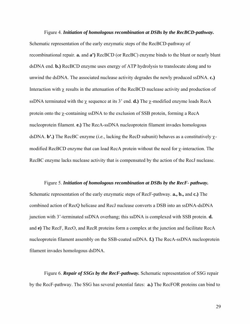
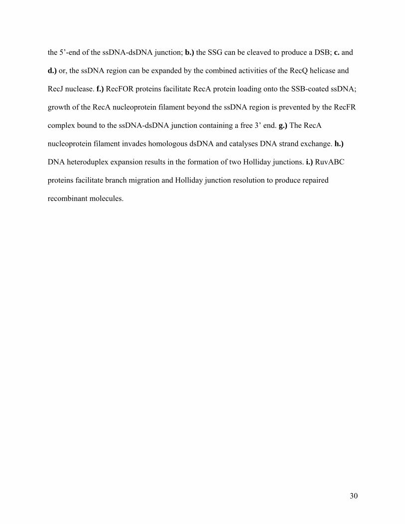
Figure 4. Initiation of homologous recombination at DSBs by the RecBCD-pathway.
Schematic representation of the early enzymatic steps of the RecBCD-pathway of
recombinational repair. a. and a’) RecBCD (or RecBC) enzyme binds to the blunt or nearly blunt
dsDNA end. b.) RecBCD enzyme uses energy of ATP hydrolysis to translocate along and to
unwind the dsDNA. The associated nuclease activity degrades the newly produced ssDNA. c.)
Interaction with χ results in the attenuation of the RecBCD nuclease activity and production of
ssDNA terminated with the χ sequence at its 3’ end. d.) The χ-modified enzyme loads RecA
protein onto the χ-containing ssDNA to the exclusion of SSB protein, forming a RecA
nucleoprotein filament. e.) The RecA-ssDNA nucleoprotein filament invades homologous
dsDNA. b’.) The RecBC enzyme (i.e., lacking the RecD subunit) behaves as a constitutively χ-
modified RecBCD enzyme that can load RecA protein without the need for χ-interaction. The
RecBC enzyme lacks nuclease activity that is compensated by the action of the RecJ nuclease.
Figure 5. Initiation of homologous recombination at DSBs by the RecF- pathway.
Schematic representation of the early enzymatic steps of RecF-pathway. a., b., and c.) The
combined action of RecQ helicase and RecJ nuclease converts a DSB into an ssDNA-dsDNA
junction with 3’-terminated ssDNA overhang; this ssDNA is complexed with SSB protein. d.
and e) The RecF, RecO, and RecR proteins form a complex at the junction and facilitate RecA
nucleoprotein filament assembly on the SSB-coated ssDNA. f.) The RecA-ssDNA nucleoprotein
filament invades homologous dsDNA.
Figure 6. Repair of SSGs by the RecF-pathway. Schematic representation of SSG repair
by the RecF-pathway. The SSG has several potential fates: a.) The RecFOR proteins can bind to
29
the 5’-end of the ssDNA-dsDNA junction; b.) the SSG can be cleaved to produce a DSB; c. and
d.) or, the ssDNA region can be expanded by the combined activities of the RecQ helicase and
RecJ nuclease. f.) RecFOR proteins facilitate RecA protein loading onto the SSB-coated ssDNA;
growth of the RecA nucleoprotein filament beyond the ssDNA region is prevented by the RecFR
complex bound to the ssDNA-dsDNA junction containing a free 3’ end. g.) The RecA
nucleoprotein filament invades homologous dsDNA and catalyses DNA strand exchange. h.)
DNA heteroduplex expansion results in the formation of two Holliday junctions. i.) RuvABC
proteins facilitate branch migration and Holliday junction resolution to produce repaired
recombinant molecules.
30
C) r
eplic
atio
n-de
pend
ent S
SGs
B) r
eplic
atio
n-de
pend
ent D
SBs
A) d
irec
tly in
duce
d D
SBs
nick
or
gap
in th
e le
adin
g st
rand
tem
plat
eIR
DSB
leth
al
DSB
3’3’
DN
A st
rand
inva
sion
;he
tero
dupl
ex e
xten
sion
non-
codi
ng le
sion
in th
e le
adin
g st
rand
tem
plat
eno
n-co
ding
lesi
on in
the
lagg
ing
stra
nd te
mpl
ate
nick
or
gap
in th
e la
ggin
g st
rand
tem
plat
e
SSG
SSG
reso
lutio
n
Spie
s and
Kow
alcz
ykow
ski,
Fig.
1R
ecom
bina
tiona
l rep
air o
f DN
A d
amag
e
DN
A st
rand
inva
sion
repl
icat
ion
repl
icat
ion
rest
art
Hol
liday
junc
tion
reso
lutio
n
+
DSB 3’
+
3’ DSB
DN
A st
rand
inva
sion
;he
tero
dupl
ex e
xten
sion
repl
icat
ion
rest
art
nucl
ease
DN
A
hete
rodu
plex
ex
tens
ion
reso
lutio
n
DSB
is r
epai
red;
rep
licat
ion
rest
arts
DN
A r
eplic
atio
n bl
ock
repl
icat
ion
fork
rev
ersa
l
regr
esse
d re
plic
atio
n fo
rk/
Hol
liday
Jun
ctio
nH
ollid
ay J
unct
ion
clea
vage
ds
DN
A e
nd r
eces
sion
repl
icat
ion
fork
rea
ssem
bly
DSB
+
3’
DN
A st
rand
inva
sion
;D
NA
het
erod
uple
x ex
tens
ion
Spie
s and
Kow
alcz
ykow
ski,
Fig.
2D
SBsc
an b
e pr
oduc
ed a
s a re
sult
of H
ollid
ay Ju
nctio
n cl
eava
ge
UV
+pso
rale
ns;
bi-f
unct
iona
l alk
ylat
ing
agen
ts NE
R
+ ho
mol
ogou
s
dsD
NA
NE
R
NE
R
SSG
rep
air
SSG
tran
slesio
n sy
nthe
sis
+ ho
mol
ogou
s
dsD
NA
DSB
rep
air
DSB
Spie
s and
Kow
alcz
ykow
ski,
Fig.
3In
ter-
stra
nd c
ross
links
are
con
verte
d in
to D
SBs a
nd S
SGs
χ5’
–G
CT
GG
TG
G –
3’χ
5’–
GC
TG
GT
GG
–3’
3’3’
5’5’
Rec
BC
DB
h DC
Bnu
c
SSB
AT
P
AD
P3’ 5’
Spie
s and
Kow
alcz
ykow
ski,
Fig.
4In
itiat
ion
of h
omol
ogou
s rec
ombi
natio
n at
DSB
sby
the
Rec
BC
D-p
athw
ay
3’ 5’
a.
b.
c.
C
Bh
Bnu
cR
ecB
Ca’
.a)
Rec
BC
(D) b
indi
ng to
dsD
NA
end
b) R
ecB
CD
-med
iate
d un
win
ding
of
dsD
NA
and
pre
fere
ntia
l deg
rada
tion
of 3
’-te
rmin
ated
stra
ndb’
) Rec
BC
-med
iate
d un
win
ding
of
dsD
NA
and
χ-in
depe
nden
t Rec
A
prot
ein-
load
ing;
Rec
J-de
pend
ent
degr
adat
ion
of 5
’-te
rmin
ated
stra
nd
SSB
Rec
A
AT
P
AD
Pb’
.
3’ 5’R
ecJ
c) R
ecog
nitio
n of
χ: p
ause
at χ
;at
tenu
atio
n of
nuc
leas
e ac
tivity
(d
own-
regu
latio
n of
3’-
5’nu
clea
se,
less
er u
p-re
gula
tion
of 5
’-3’
nucl
ease
); r
educ
tion
of tr
ansl
ocat
ion
d.R
ecA
d) R
ecA
pro
tein
-load
ing
e’.
3’ 5’
hom
olog
ous d
sDN
A
e.ho
mol
ogou
s dsD
NA
e) H
omol
ogou
s pai
ring
and
DN
A
stra
nd e
xcha
nge
5’
3’ 5’R
ecQ
Rec
J
Rec
J
Rec
Q3’
3’ 5’ 3’
5’R
ecF
Rec
OR
ecR
3’
5’F
RO
Rec
A
3’
5’
5’
SSB
AD
P
AT
P
a.
b.
c.
a.) R
ecQ
hel
icas
e bi
nds t
o th
e ds
DN
A e
nd
b.) R
ecQ
unw
inds
dsD
NA
, Rec
J nu
clea
se d
egra
des t
he 5
’-te
rmin
ated
st
rand
c.) D
isso
ciat
ion
of R
ecQ
and
Rec
J pr
otei
ns
d. e.
d.) A
ssem
bly
of R
ecF,
Rec
O, a
nd
Rec
R p
rote
ins a
t ssD
NA
-dsD
NA
ju
nctio
n
d.) R
ecA
pro
tein
-load
ing
f.ho
mol
ogou
s dsD
NA
e.) H
omol
ogou
s pai
ring
and
DN
A
stra
nd e
xcha
nge
Spie
s and
Kow
alcz
ykow
ski,
Fig.
5In
itiat
ion
of h
omol
ogou
s rec
ombi
natio
n at
DSB
sby
the
Rec
F-pa
thw
ay
SSB
DSB
rep
air
Spie
s and
Kow
alcz
ykow
ski,
Fig.
6R
epai
r of S
SGsb
y th
e R
ecF-
path
way
Um
uD’C
5’
3’
3’
unkn
own
nucl
ease
5’
Rec
FR
ecO
Rec
R
5’3’
F
RO
Rec
A
5’3’
F
RO
Rec
FR
5’3’
5’
3’
Rec
Q +
Rec
J
hom
olog
ous d
sDN
A
Rec
FR
ecO
Rec
R Lex
ASO
S in
duct
ion
tran
s-le
sion
DN
A sy
nthe
sis
a.
b.
c.
d.
e. f. g.
j.
Rec
F, R
ecO
, and
Rec
R
prot
eins
ass
embl
e at
the
ssD
NA
-dsD
NA
junc
tion
Rec
FOR
faci
litat
e R
ecA
lo
adin
g on
to th
e SS
B-
cove
red
gap
Con
tinue
d R
ecA
fila
men
t gr
owth
with
in th
e ga
p;
Rec
FR c
ompl
ex p
reve
nts
Rec
A fi
lam
ent g
row
th in
to
the
dow
nstr
eam
dsD
NA
re
gion
Hom
olog
ous p
airi
ng a
nd
DN
A st
rand
exc
hang
e
SSG
is c
onve
rted
to th
e D
SB
SSG
exp
ansi
on
h.
Het
erod
uple
x ex
tens
ion,
B
ranc
h m
igra
tion
Rec
AR
uvA
Bi.
Hol
liday
Jun
ctio
n re
solu
tion
Ruv
CR
uvA
B