Double-strand break repair by homologous recombination is ...

;. ... '
Chapter 7
Homologous Recombination Proteins and their Potential Applications in Gene Targeting Technology
Stephen C. KowalczykowskP and David A. Zarling2
1 Division of Biological Sciences
Sections of Microbiology and of Molecular and Cellular Biology
University of California, Davis
Davis, CA
2Cell and Molecular Biology Laboratory
SRI International
Menlo Park, CA
Department of Laboratory Medicine
University of California, San Francisco
San Francisco, CA
0-8493-8950-X/951$0.00+$.50 © 1995 by CRC Press, Inc. 167

168 Gene Targeting
Contents
L Introduction ........................................................................................ 169
II. Biochemical Activities of Key Components in Homologous Recombination ........................................................... 170
A. RecA Protein and Homologous Pairing Proteins ....................... 173 B. RecBCD Enzyme and DNA Helicases ....................................... 176 C. Nucleases ..................................................................................... 178 D. SSB Protein .................................................................................. 179 E. Recombination Hotspots .............................................................. 180
III. Coordination of Biochemical Functions In Vitro ............................. 180 A. Homologous Pairing Dependent on RecA, RecBCD,
and SSB Proteins and x ............................................................. 181 B. Resolution of Holliday Junctions ................................................ 183
IV. Applications of RecA Protein-Mediated Homologous DNA Targeting .......................................................... 183
A. Isolation of Homologous DNA Targets by a RecA Protein-Mediated D-Loop .......................................................... 183
B. Homologous Targeting, Detection, and Purification of Linear DNA Duplex Molecules With Two Complementary DNA Probe Strands ........................................ 185
C. RecA Protein-Assisted Restriction Endonuclease (RARE) Cleavage of Chromosomal DNA ................................ 188
D. RecA Protein-Stabilized D-Loops Inhibit Transcription of DNA by RNA Polymerase ................................................... 188
E. RecA Protein-Mediated Targeting and Mapping of Homologous ssDNA Probes to Sites on Individual DNA Molecules Analyzed by High-Resolution Darkfield Electron Microscopy .................................................................. 190
F. RecA Protein-Mediated Targeting of Cellular and Viral Chromosomal DNA in Fixed and Metabolically Active Nuclei Analyzed by Native FISH ............................................................................... 192
G. Potential Applications of RecA Protein-Mediated Homologous DNA Targeting .................................................... 196
V. Conclusions ........................................................................................ 197
Acknowledgments ....................................................................................... 198
References .................................................................................................... 198

Homologous Recombination Proteins 169
I. Introduction
Homologous recombination is the natural means of targeting a gene for replacement by a homologue. By definition, the recombination process is specific, being limited to homologous target sites that exceed a species-imposed minimum length of DNA sequence similarity. Most naturally occurring recombination events take place at the homologous locus, and in vivo it is usually efficient. In organisms such as Escherichia coli and Saccharomyces cerevisiae, foreign DNA introduced into cells by the artificial means of transformation is also integrated homologously. Thus, though seemingly ideal, the use of homologous recombination to target experimental genes in mammalian organisms has met with only partial success. Attempts to homologously target genes in mammalian cells have revealed that the majority of integration events occurs at nonhomologous sites.1-3 To overcome these limitations, researchers in the field of gene therapy, for instance, have turned to relatively higher efficiency, but nonhomologous, integration systems employing retroviral or adeno-associated viral vectors to stably introduce DNA into the genome. However, these systems suffer from their own problems. The foremost is that, since non-homologous retroviral recombination is approximately random, the sites of integration cannot be controlled, possibly resulting in the accidental knockout of important functional cellular genes and unpredictable control of target gene expression. Because adeno-associated virns or retroviral vectors cannot be targeted to the homologous locus, gene therapies based on these viral systems cannot correct dominant mutations, and their integration may result in activation of endogenous genes. Retroviral vectors are also limited to use only in replicating cells and they have a tendency to recombine. Finally, target cell specificity is often low in organs receiving the adeno-associated viruses, and retroviruses are innnunogenic. Thus, homologous recombination systems possess a number of conceptual advantages.
Unfortunately, the utility of homologous recombination is limited because of its low efficiency, relative to non-homologous recombination, when mammalian cells are transfected with foreign DNA. The reasons for this limitation is not clear and may result from a combination (1) of the complexity of the recombination reaction, (2) of competing processes that attempt to repair (nonhomologously) DNA strand breaks, and (3) of potential constraints imposed by nucleosome structure and/or chromatin condensation. It appears that many of the important variables that affect efficient gene targeting are just being described. Rather than discuss potential barriers to efficient homologous recombination in mannnalian cells, this article will focus on the biochemistry of the recombination apparatus. Low efficiency DNA transfer and targeting can result from a rate-limiting impediment in any one of a number of key steps in the recombination process. Through an appreciation of the entire process, rational procedures can be designed to overcome the inhibitory process(es) encountered in mammalian cells. Therefore, this article will provide an overview of the salient features of homologous recombination in E. coli, the system which

170 Gene Targeting
is the most biochemically refined. The nature of the enzymatic activities and the substrates that they act upon will be summarized; in addition, the current and future potential application of these enzymes will be highlighted. We hope that appreciation of the enzymology of this process in a relatively simple organism and in vitro will guide the design of experiments in more complex and experimentally less tractable systems. For a comprehensive review of these subjects, readers are referred to References l through 17.
11. Biochemical activities of key components in homologous recombination
The complexity of homologous recombination in E. coli is revealed in Table l, where the proteins that play a role in recombination are summarized.10 Over 25 proteins are needed, and their enzymatic activities include DNA strand exchange, DNA renaturation, DNA helicase, nuclease, A TPase, topoisomerase, and DNA-binding activities. Most of these activities are also commonly encountered in other biological processes, but one activity listed in Table 1 is unique to recombination: the DNA strand exchange activity of the recA protein. This activity was anticipated by nearly all models of recombination, but it was surprising that the ability to homologously pair and exchange DNA sequences would reside in a single, relatively small (Mr 38 k:Da) polypeptide.
To put the activities of this diverse set of enzymes into context requires appreciation of one key property of recA protein-promoted DNA strand exchange.4-17 RecA protein will pair two homologous DNA molecules only if one of them is at least partially single-stranded. Since DNA is normally doublestranded, this requirement imposes a necessary initiation step in any recA protein-promoted process: one of the DNA substrates must be processed to reveal ssDNA. This consideration is reflected in Figure 1, which illustrates the plausible function in the recombination mechanism for each of the proteins listed in Table 1. In nearly all models of homologous recombination, the initial event is the generation of a ssDNA or dsDNA break; in E. coli, a dsDNA break is created in all of the processes (conjugation, transduction, and transformation) that result in recombination.18•19 The first step envisioned in Figure 1 is the processing of one of the linear DNA molecules to create ssDNA. This can occur by a variety of biochemical means, but the primary method operative in wild-type E. coli is the combined unwinding and degradation of dsDNA by the recBCD enzyme.10•19•20 This enzyme, in conjunction with the recombination hotspot x, acts to create ssDNA that can be used by recA protein for the subsequent homologous pairing phase. An alternative approach for the production of ssDNA uses either a strand-specific dsDNA exonuclease or a DNA helicase, or the action of both.
The recA protein, assisted by the ssDNA-binding (SSB) protein (and, perhaps, facilitated by the reeF, recO, and recR proteins), promotes invasion of the homologous target. The recA protein is remarkably catholic about the types

Homologous Recombination Proteins 171
Table 1. Proteins and DNA sites involved in genetic recombination in Escherichia coli 10
Protein
RecA
RecBCD (exonuclease V)
RecBC RecE (sheA*) (exonuclease Vlll) ReeF RecG
RecJ RecN RecO RecQ RecR RecT RuvA
RuvB
RuvC SbcB (exonuclease I) (xonA)
SbcC SheD SSB DNA topoisomerase I (topA) DNA gyrase (gyrA & gyrB) DNA ligase (Zig)
DNA polymerase I (polA)
Helicase ll (uvrD, uvrE, reeL, mutU)
Helicase IV (helD)
Chi (X)
Activity
DNA strand exchange; DNA renaturation; DNAdependent ATPase; DNA- and ATP-dependent coprotease
DNA helicase; A TP-dependent ssDNA and dsDNA exonuclease; ATP-stimulated endonuclease; Xhotspot recognition
DNA helicase dsDNA exonuclease, 5' ~3' specific ssDNA and dsDNA binding; ATP binding Branch migration of Holliday junctions;
DNA helicase ssDNA exonuclease, 5' ~3' specific Unknown, A TP binding consensus sequence Interaction with recR and (possibly) reeF proteins DNA helicase Interaction with recO and (possibly) reeF proteins DNA renaturation Holliday-, crucifonn-, and4-way junction binding; interaction with ruvB protein
Branch migration of Holliday junctions; DNA helicase; interaction with ruvA protein
Holliday junction cleavage; 4-way junction binding ssDNA exonuclease, 3' ~5' specific;
deoxyribophosphodiesterase Unknown, ATP binding consensus sequence Unknown ssDNA binding ro protein, type I topoisomerase DNA gyrase, type II topoisomerase DNA ligase DNA polymerase; 5' ~3' exonuclease; 3' ~5'
exonuclease
DNA helicase DNA helicase Recombination hotspot: 5'-GCTGGTGG-3';
regulator of recBCD enzyme nuclease activity
From Kowalczykowski, S.C., Dixon, D.A., Eggleston, A.K., Lauder, S.D., and Rehrauer, W.M., Microbiol. Rev., 58, 3, 1994 (in press). With pennission.
* sbcA mutations are regulatory mutations that activate RecE function.

172 Gene Targeting
S' + 3' INITIATION 3' 5'
(PROCESSING) '?.::~VrecEj \[ecO ~ rec()_ (ssy II ~(SSB) ~
--<:· - ....... ' ~ --<:' ~SSB) \ {i~( I (S~~f/ ••
~R)~FOR~(Foy
HOMOLOGOUS PAIRING Q & DNA STRAND
EXCHANGE Joint Molecule
l ~~ ;~lymerase I ~es ~- HollidayJunction
DNA HETERODUPLEx recA 1 j recG EXTENSION ruvAB t ruvAB
~ RESOLUTION
DNA lig;;;:~ I \ rgvN~ ligase
.5.EWrm % \_'C _EAIOHE.D. 7T ,' - ~
+ ~:::::===
+
Figure 1. Biochemical model for homologous recombination in E. coli based on the enzymatic activities of proteins known to be involved based on genetic analysis. (From Kowalczykowski, S.C., Dixon, D.A., Eggleston, A.K., Lauder, S.D., and Rehrauer, W.M., MicrobioL Rev., 58, 3, 1994 (in press). With permission.)
of DNA molecules that it will pair, provided that one of them is at least partially single-stranded. For the reaction illustrated in 1, which is the invasion of a negatively supercoiled DNA recipient by a linear ssDNA fragment, there is a distinct bias for promoting insertion of the 3'-end of the ssDNA. 21 This 3'end can serve as a primer for any needed repair by replication.
The resulting joint molecule serves as the common intermediate in each of the biochemical pathways. Conversion of this structure to the obligatory Holliday junction, which is common to all mechanisms of reciprocal recombination, could occur by simple pairing of the displaced ssDNA with the other strand of the invading DNA molecule. The Holliday junction is capable of undergoing DNA heteroduplex extension either by a thermally driven branch migration process or by a protein promoted process. Although the thermal process seemingly might suffice for extension through regions of DNA sequence homology, thermally driven extension would terminate at DNA sequence heterologies or by the presence of DNA-binding proteins. Thus, it is not

Homologous Recombination Proteins 173
surprising to find a class of proteins that can catalyze this extension process. RecA, ruvAB, and recG proteins all possess this novel activity. 10•12•22
Finally, resolution of the Holliday junction by symmetric cleavage results in two types of recombinant products: the spliced recombinants, containing the reciprocal halves of the parental molecules, and the patched recombinants, containing nearly intact parental information, but possessing the DNA heteroduplex patch characteristic of a recombination event.22
Each of the activities mentioned will be described in more detail. However, only the most important properties of each are described The reader seeking more indepth discussion should consult the many fine reviews written on this subject.
A. RecA protein and homologous pairing proteins
The DNA strand exchange activity of E. coli recA protein is biochemically unique, yet ubiquitous in nature.4•5•7•9-11•13-17 All prokaryotes examined possess a gene that bears a high degree of homology to the E. coli gene, and this universality appears to extend to eukaryotes as wel1. 11•1523•24 RecA protein homologues have been purified from sources as divergent as the closely related organism Proteus mirabilis, the gram positive Bacillus subtilis, the bacteriophage T4, and the chloroplasts of pea plants.5•11 Each of these proteins possess the unvarying hallmarks of recA protein-promoted DNA strand exchange: ATP- and ssDNAdependence. Although ATP-independent homologous pairing proteins have been isolated, these proteins do not pair DNA by the same mechanism employed by recA protein; instead, they require ssDNA in both of the DNA molecules and rely on DNA reannealing as the homology recognition step.11
The DNA strand exchange reaction promoted by recA protein is comprised of at least three major phases: presynapsis, synapsis, and DNA heteroduplex extension (Figure 2); each of these major phases can be further dissected into more basic biochemical steps.
The presynaptic step involves the binding and polymerization ofrecAprotein onto the requisite ssDNA. The ssDNA is fully coated by recA protein, requiring about one recA protein monomer for every three to four nucleotides of ssDNA.25
RecA protein can also bind to dsDNA, but with rather slow kinetics;26-28 the rate of binding to dsDNA is increased either when ssDNA regions are present29•30 or when distortions are introduced in the form of either DNA lesions, intercalating dyes, or non-B-DNA character.31-35 ATP binding, but not hydrolysis, is required for this and the resultant ternary complex of A TP, recA protein, and ssDNA (or dsDNA) forms a distinctive right-handed helical filament.16 This nucleoprotein filament is about 100 A in diameter, has a characteristic 95 A pitch, and extends the DNA by about 50% over the normal dimension of B-form dsDNA. This distinctive ternary complex defines the structure referred to as the presynaptic complex. It is this nucleoprotein filament, rather than a monomer or any other limited aggregate of recA protein, that is the functional entity responsible for the homology search process.

174
PRESYNAPSIS
substrates
+
preeynspt/o complex
SYNAPSIS
cantacts
(pJectonemlcJ
Gene Targeting
DNA HETERODUPLEX EXTENSION
strand dii1JI/acemertl
5' ..
•
products
0 Figure 2. Mechanism of homologous pairing and DNA strand exchange promoted by recA protein. The reaction between circular ssDNA and linear dsDNA is depicted. (From Kowalczykowski, S.C., Annu. Rev. Biophys. Biophys. Chern., 20, 539, 1991. With permission.)
Once formed, the presynaptic complex is capable of identifying DNA sequence homology in dsDNA. This process is rapid, occurring in vitro in just a few minutes; specific, needing less than 50 nucleotide ofhomology;38-40 discriminating, rejecting as great as a 200,000-fold excess of non-homologous sequences;41 and completely independent of ATP hydrolysis.25·42-47 All of this is made even more remarkable by the fact that the presynaptic filament is 50% more extended than the duplex target homologous sequences.16 Although many details of the homology search remain unclear, several of the reaction characteristics are known. The search process is first order rather than second order in DNA concentration, implying that the rate-limiting step in the pairing process occurs within non-homologously paired complexes of ssDNA and dsDNA.48 Homologous recognition does not occur by repeated denaturation of the dsDNA,49-51 but rather, through transient non-Watson-Crick hydrogen bonding interactions between ssDNA and dsDNA, leading to a 3' -strand-containing structure, 52-54 at least transiently.44 During the homology search, the dsDNA is topologically unwound, presumably due to distortion required to transiently align the dsDNA with the extended dimensions of the presynaptic fl.lament.55-59
Once homologous alignment is achieved, invasion of the dsDNA by the ssDN A and exchange of DNA strands results in formation of a joint molecule or D-loop (displacement loop) structure. Depending on where pairing initiates, two kinds of joint molecules can be detected. If pairing occurs at the ends of a linear molecule, the exchanged DNA strands are free to intertwine, resulting in the formation of a plectonemic joint molecule; if pairing occurs at internal sites where topological constraints limit intertwining, then a paranemic joint molecule forms. Though paranemic joint molecules formed with a single-stranded DNA are unstable in the absence of recA protein, they can convert to plectonemic joint

Homologous Recombination Proteins 175
by extending the region of pairing to a free DNA end43•60 or by the more likely in vivo pathway, probably involving a topoisomerase.56•61.62 As will be discussed in more detail below, protein-free D-loop structures formed at internal dsDNA sites are kinetically unstable in the absence of negative supercoiling due to branch migration; however, the stability of these joint molecules can be increased substantially by hybridization of complementary ssDNA to the displaced strand of the D-loop.63,64
The joint molecule is a structure central to the recombination process. The stability of this structure is due to conventional base-pairing. DNA heteroduplex extension, the third phase of this recA protein-promoted process, increases its stability. RecA protein, itself, is capable of promoting DNA heteroduplex extension at a rate of about lO to 20 bp/sec.Z5 This process requires ATP hydrolysis and is unidirectional, occurring 3' -t5' relative to the ssDNA strand displaced. It can proceed for at least 7 kb and can traverse heterologies in the DNA as large as 1308 nucleotides.65
These fundamental properties of DNA strand exchange promoted by recA protein were determined using model DNA substrates (6 to 7 kb) in vitro that were totally devoid of protein. However, genomic DNA is clearly coated with polyamines and proteins, among them histones and chromatin-associated proteins in eukaryotic cells and histone-like proteins (e.g., HU) in prokaryotic cells. Not surprisingly, these proteins inhibit the pairing reactions of recA protein. 66•68 The binding of E. coli HU protein to dsDNA blocks formation of plectonemic, but not paranemic, joint molecules. Similarly, in a heterologous in vitro reaction, eukaryotic chromatin substrates formed by reconstitution of histones with dsDNA were not efficiently utilized by E. coli recA protein. Reconstitution at histone/DNA mass ratios of 0.8 and 1.6 suppressed the DNA heteroduplex extension phase, but had no effect on the initial homologous pairing step; however, at a higher mass ratio of 9.0, no pairing was detected. These effects were exacerbated by addition of histone H 1 to approximately physiological levels. Thus, the removal of histones and other DNA-binding proteins may represent an important aspect of efficient homologous recombination in the cell. Such clearing may be aided by DNA helicases (see below).
The only other homologous pairing protein described in E. coli is the recT protein, a protein encoded by a cryptic lambdoid phage, rae, and, for that reason, bearing functional resemblance to the ~-pairing protein from bacteriophage A..69 The recT protein binds to ssDNA (but not to dsDNA) and promotes the ATP-independent renaturation of complementary single-stranded DNA.70
The recT protein can also promote homologous pairing between circular ssDNA and linear dsDNA, provided that the linear dsDNA is resected by an exonuclease to expose homologous ssDNA.71 In the absence of nuclease activity, no pairing is detected; the recE protein (see below) of E. coli can provide this needed nuclease function in vitro. This recT protein-promoted pairing reaction also requires no ATP and at least one recT monomer per 13 nucleotides of ssDNAY The pairing reaction likely initiates via reannealing of the
176 Gene Targeting
homologous ssDN A regions, consistent with the known renaturation activity of recT protein; however, heteroduplex formation is not limited to the region of resected DNA, since ssDNA is displaced from duplex region of the linear dsDNA. This strand displacement or DNA heteroduplex extension is recT protein specific,71 since a protein proficient in DNA renaturation (histone Hl protein) can promote the initial pairing (reannealing) step, but cannot produce DNAheteroduplex products possessing displaced ssDNA.71•72 These properties of recT protein resemble those of the A TP-independent class of homologous pairing proteins isolated from many eukaryotic cells, 11 suggesting that ssDNA annealing may be an important pathway for recombination in eukaryotic cells.3
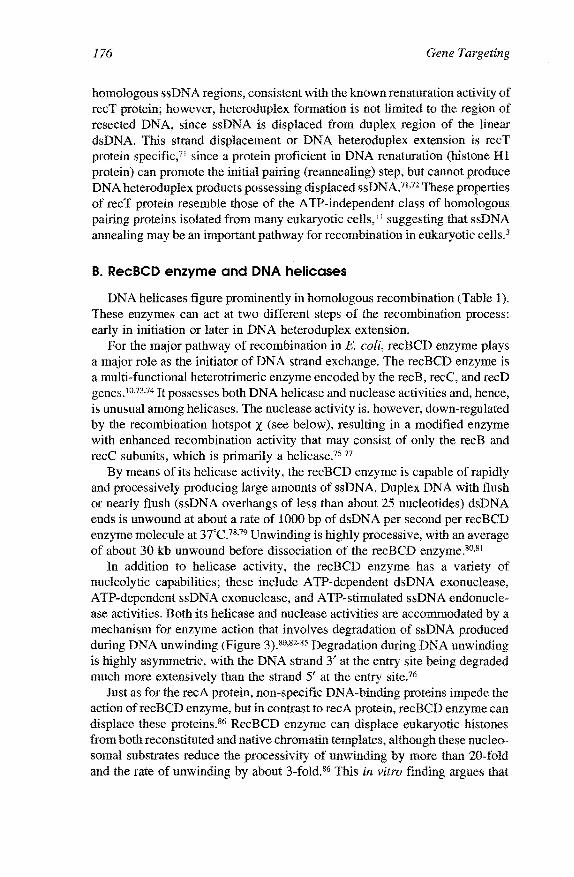
B. RecBCD enzyme and DNA helicases
DNA helicases figure prominently in homologous recombination (Table 1). These enzymes can act at two different steps of the recombination process: early in initiation or later in DNA heteroduplex extension.
For the major pathway of recombination in E. coli, recBCD enzyme plays a major role as the initiator of DNA strand exchange. The recBCD enzyme is a multi-functional heterotrimeric enzyme encoded by the recB, recC, and reeD genes.10•73•74 It possesses both DNA helicase and nuclease activities and, hence, is unusual among helicases. The nuclease activity is, however, down-regulated by the recombination hotspot X (see below), resulting in a modified enzyme with enhanced recombination activity that may consist of only the recB and recC subunits, which is primarily a helicase.75-77
By means of its helicase activity, the recBCD enzyme is capable of rapidly and processively producing large amounts of ssDNA. Duplex DNA with flush or nearly flush (ssDNA overhangs of less than about 25 nucleotides) dsDNA ends is unwound at about a rate of 1000 bp of dsDNA per second per recBCD enzyme molecule at 3TC.78•79 Unwinding is highly processive, with an average of about 30 kb unwound before dissociation of the recBCD enzyme.80•81
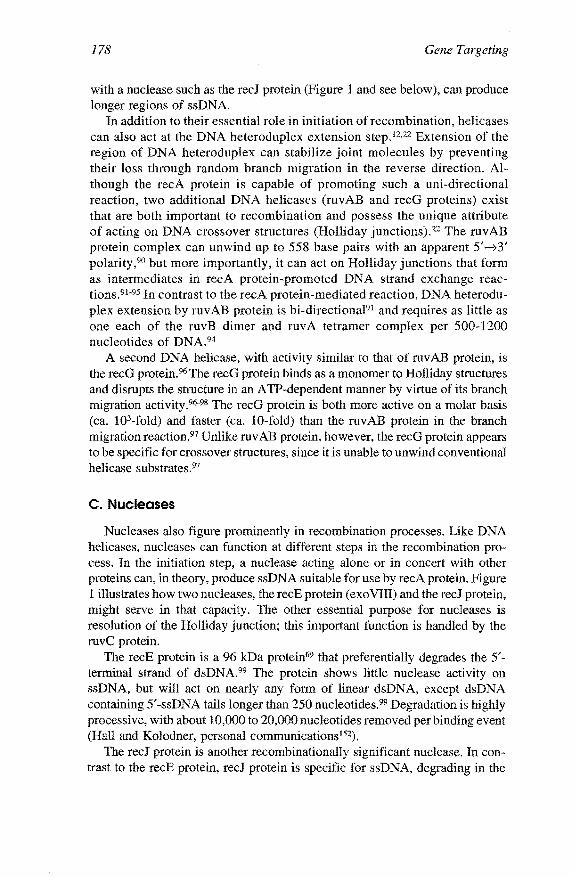
In addition to helicase activity, the recBCD enzyme has a variety of nucleolytic capabilities; these include A TP-dependent dsDNA exonuclease, ATP-dependent ssDNA exonuclease, and ATP-stimulated ssDNA endonuclease activities. Both its helicase and nuclease activities are accommodated by a mechanism for enzyme action that involves degradation of ssDNA produced during DNA unwinding (Figure 3).80•82-85 Degradation during DNA unwinding is highly asymmetric, with the DNA strand 3' at the entry site being degraded much more extensively than the strand 5' at the entry site.76
Just as for the recA protein, non-specific DNA-binding proteins impede the action of recBCD enzyme, but in contrast to recA protein, recBCD enzyme can displace these proteins.86 RecBCD enzyme can displace eukaryotic histones from both reconstituted and native chromatin templates, although these nucleosomal substrates reduce the processivity of unwinding by more than 20-fold and the rate of unwinding by about 3-fold.86 This in vitro finding argues that

Homologous Recombination Proteins 177
A
5' 35'' +.a 3' illllll!!li;jjj)jjj ~
! 8 5' ::d~ 3, I I I II !I II I I I II• II I,.
! ~ ,,,,,,,,,,,,,Ar 3~,
~5'
E
5' 3'
!
!
-3'
' / 1'\
Figure 3. Model for the mechanism of DNA
unwinding and degradation of linear dsDNA by recBCD enzyme. (From Kowalczykowski, S.C.,
Experientia, 50, 204, 1994. With permission.)
the enzyme should be capable of unwinding prokaryotic chromosomal DNA, which is condensed by histone-like proteins such as HU and IHF to form a "nucleoid" structure in E. coli and raises the possibility that specific helicases in eukaryotic cells may serve a similar function.
In addition to recBCD enzyme, there is at least one other DNA helicase that potentially functions at the initiation step of recombination. This is the recQ helicase, and its need in recombination is manifested in cells defective for recBCD function and containing needed suppressor mutations.87 As for all helicases, hydrolysis of either ATP or dATP is required for dsDNA unwinding by recQ protein. 88 In the absence of SSB protein, the recQ protein is a relatively poor helicase, unwinding only about 143 base pairs and requiring high concentrations of protein. However, SSB protein stimulates its DNA helicase activity, presumably by binding to the unwound strands. Under these conditions, up to 343 base pairs are unwound and unwinding approaches catalytic behavior.89
Presumably this limited level of DNA unwinding suffices for initiation of DNA strand exchange by recA protein; alternatively, the recQ protein, in concert
178 Gene Targeting
with a nuclease such as the recJ protein (Figure 1 and see below), can produce longer regions of ssDNA.
In addition to their essential role in initiation of recombination, helicases can also act at the DNA heteroduplex extension step.12·22 Extension of the region of DNA heteroduplex can stabilize joint molecules by preventing their loss through random branch migration in the reverse direction. Although the recA protein is capable of promoting such a uni-directional reaction, two additional DNA helicases (ruvAB and recG proteins) exist that are both important to recombination and possess the unique attribute of acting on DNA crossover structures (Holliday junctions).22 The ruvAB protein complex can unwind up to 558 base pairs with an apparent 5' ~3' polarity,90 but more importantly, it can act on Holliday junctions that form as intermediates in recA protein-promoted DNA strand exchange reactions.91·95 In contrast to the recA protein-mediated reaction, DNA heteroduplex extension by ruvAB protein is bi-directional91 and requires as little as one each of the ruvB dimer and ruvA tetramer complex per 500-1200 nucleotides of DNA.94
A second DNA helicase, with activity similar to that of ruvAB protein, is the recG protein. 96 The recG protein binds as a monomer to Holliday structures and disrupts the structure in an A TP-dependent manner by virtue of its branch migration activity.96·98 The recG protein is both more active on a molar basis (ca. 103-fold) and faster (ca. 10-fold) than the ruvAB protein in the branch migration reaction.97 Unlike ruv AB protein, however, the recG protein appears to be specific for crossover structures, since it is unable to unwind conventional helicase substrates.97
C. Nucleases
Nucleases also figure prominently in recombination processes. Like DNA helicases, nucleases can function at different steps in the recombination process. In the initiation step, a nuclease acting alone or in concert with other proteins can, in theory, produce ssDNA suitable for use by recA protein. Figure 1 illustrates how two nucleases, the recE protein ( exo VIII) and the recJ protein, might serve in that capacity. The other essential purpose for nucleases is resolution of the Holliday junction; this important function is handled by the ruvC protein.
The recE protein is a 96 kDa protein69 that preferentially degrades the 5'terminal strand of dsDNA.99 The protein shows little nuclease activity on ssDNA, but will act on nearly any form of linear dsDNA, except dsDNA containing 5'-ssDNA tails longer than 250 nucleotides.99 Degradation is highly processive, with about 10,000 to 20,000 nucleotides removed per binding event (Hall and Kolodner, personal communications152).
The recJ protein is another recombinationally significant nuclease. In contrast to the recE protein, recJ protein is specific for ssDNA, degrading in the

Homologous Recombination Proteins 179
5' --73' direction at a maximal rate of about 1000 nucleotides per min per protein molecule. 100 Its precise role in recombination is unknown, but genetic analyses suggest that recJ protein may work in concert with a helicase such as the recBC (i.e., without reeD) or recQ helicases,101 degrading one of the DNA strands produced by DNA unwinding (Figure 1). This degradation may prevent reannealing of the unwound ssDNA strands and has the net effect of producing ssDNA with a 3'-terminal end, which is the preferred substrate for recA protein-promoted invasion of negatively supercoiled DNA.21
The last nuclease discussed in this section has the remarkable property of recognizing and symmetrically cleaving Holliday junctions, arguing that this protein catalyzes the last step depicted in Figure 1. This nuclease is the ruvC protein. RuvC is a homodimeric protein composed of subunits with an estimated Mr of 18,747 Daltons.102 In the absence ofMg2+ or ATP, ruvC protein will bind to Holliday junctions, but not cleave the DNA.103 In the presence of Mg2+, ruvC protein will cleave at the junction, provided that at least six base pairs are fully homologous. 103 Cleavage is symmetric, occurring on the 3' side of thymine residues, and shows no bias, producing equal amounts of patched and spliced recombinant products.102.104 This unbiased pattern changes somewhat in the presence of recA protein, with spliced products appearing more frequently than patched products. 103•105 Unexpectedly, the reaction is apparently not catalytic, requiring as much as one ruvC monomer per 80 nucleotides of DNA.103
D. SSB protein
DNA strand exchange promoted by recA protein is stimulated by the SSB protein. SSB protein is a representative of a class of ssDNA-binding proteins that has no enzymatic activity, but binds ssDNA cooperatively and nonspecifically. SSB protein (Mr18.8 kDa106) binds both single-stranded DNA and RNA, resulting in a destabilization (i.e., lowering of the Tm) of duplex nucleic acid structures.107
The stimulatory effects of SSB protein are manifested both pre- and postsynaptically (Figure 4). In the presynaptic phase, SSB protein, by virtue of its helix-destabilization properties, removes the structural impediment to complete presynaptic complex formation by removing DNA secondary structure.108•109 The amount of SSB protein required for optimal presynaptic complex formation is about 1 SSB monomer per about 15 to 20 nucleotides of ssDNA.
Post-synaptically, SSB protein serves two functions. The first is to prevent formation of homologously paired networks of DNA that result from intermolecular re-invasion events; SSB protein can bind to the displaced linear ssDNA and hinder its utilization by recA protein. 110 The second role is more direct, requiring sufficient SSB protein to bind the ssDNA produced by DNA strand exchange; the binding of SSB protein to the displaced ssDNA directly stimulated the observed rate of joint molecule formation, presumably because it limits reversal of DNA strand exchange.lll

180
low [tv!g] A) PRESYNAPSIS $ --------::----
B) SYNAPSIS I< DNA STRANO EXCHANGE
0 -- --+
joint molecule
Gene Targeting
+
0
Figure 4. Role of SSB protein in recA protein-promoted DNA strand exchange: presynaptic (A) and synaptic (B) functions. (From Lavery, P.E., and Kowalczylwwski, S.C.,
J. Bioi. Chern., 267, 9315, 1992. With permission.)
E. Recombination hotspots
The frequency of recombination is not uniform across the genome, but certain regions have increased probabilities of crossing-over. 112 These sites are referred to as recombination hotspots, and E. coli possesses a specific hotspot known as X· Though initially identified in bacteriophage A, X sites are believed to be responsible for most, if not all, recombination in E. coli. Thus, although initially identified as a specialized site of recombination, it appears as though all recombination in wild-type cells is mediated by X sites, and therefore, they are general rather than a specialized feature of recombination.
Recombination is 5 to 10 times more frequent near x; is dependent on recB and recC; is stimulated primarily to the 5' side of the X site; and is detectable as far away as 10 kb. 113· 116 The DNA sequence 5' -GCTGGTGG-3' constitutes the X site117• In vitro, recBCD enzyme recognizes X only when unwinding DNA from the 3' -side of the X sequence, and recognition of X produces a ssDNA fragment that terminates near x; cleavage occurs in the DNA strand containing the X sequence, 4 to 6 nucleotides to the 3' side of x. 118•119
Ill. Coordination of biochemical functions in vitro
The detailed biochemical characterization of many of the proteins listed in Table I has permitted limited reconstitution of the recombination process. In

Homologous Recombination Proteins 181
one reaction, the initial steps of homologous pairing depicted in Figure 1 have been reconstituted in vitro. In this reaction, homologous pairing is dependent on the concerted action of recA, recBCD, and SSB proteins. In addition, homologous pairing in this reaction is stimulated by the presence of the recombination hotspot X· In a second reaction, Holliday junctions are formed in vitro by recA protein and are specifically resolved by the ruvC protein to produce the expected recombinant progeny.
A. Homologous pairing dependent on recA, recBCD, and SSB proteins and x
The first in vitro experiments were designed to test whether recBCD enzyme could generate ssDNA that was a suitable substrate for recA proteindependent homologous pairing reactions. 120•121 These reactions used DNA molecules that were not substrates for recA protein; however, one of the participants was a linear DNA molecule that could be unwound by the helicase activity of recBCD enzyme. These recombination reactions showed that the recBCD enzyme could, indeed, initiate recombinational events by producing ssDNA suitable for recA protein-dependent DNA heteroduplex formation, 120-123 demonstrating in vitro that recA, recBCD, and SSB proteins were sufficient to reconstitute the first two steps depicted in Figure 1.
Successful reconstitution of the initiation phase of the recombination reaction permitted an evaluation of the effect of the recombination hotspot X on the joint molecules formed in vitro. The nick-initiation model of Smith and colleagues124
predicted the formation of joint molecules containing ssDNA originating from the donor DNA (i.e., the dsDNA unwound by recBCD enzyme and the source of ssDNA for subsequent strand invasion) and derived from DNA downstream of X· In vitro, the prediction was not only verified, but DNA heteroduplex formation was significantly enhanced when x-containing DNA was used as the donor DNA.75 1n addition, these reactions revealed an unanticipated feature of x: the X recombination hotspot is also a regulatory sequence. It effects a change in the enzymatic behavior of recBCD enzyme, converting this multifunctional enzyme from a combined helicase-nuclease to a modified enzyme possessing essentially only helicase activity. 75•76 Interaction with X attenuates the nuclease activity of recBCD enzyme, an effect that lasts as long as the enzyme is associated with the DNA molecule that contained X· Upon dissociation, nuclease function is normally restored and the cycle of attenuation and reactivation can continue indefmitely. These observations provide a simple rationale for the recombination hotspot activity of X· In the absence of x. recBCD enzyme degrades and destroys the ssDNA needed for homologous pairing by recA protein. However, upon recognition of x, this needed ssDNA is spared from subsequent degradation, allowing an increased probability of a productive pairing event.
The precise nature of the molecular event responsible for nuclease attenuation is unclear, but both in vivo and in vitro experiments argue for loss or

182 Gene Targeting
modification of the reeD subunit as the cause.77•125•126 ln vivo, both the phenotype and distribution of crosses of certain reeD mutants imitate the behavior of wild-type, x-stimulated, reeBCD-dependent recombination.125·126 ln vitro, the recBC enzyme possesses characteristics similar to the recBCD enzyme that has interacted with x; most notably, the recBC enzyme has essentially no nuclease activity, but possesses significant helicase activity.77•127 Thus, the recBC enzyme most likely represents the modified enzyme that is constitutively proficient in recombination.n,Jzs
The nuclease attenuation model for initiation of genetic recombination illustrated in Figure 5 summarizes the salient features of the in vitro data. 75.76
The recombination reactions initiate by the unwinding and degradation of dsDNA by recBCD enzyme. The combined unwinding and nucleolytic action produce ssDNA fragments that are adequate substrates for recA protein-dependent reactions in vitro, but do not provide the continuity needed to produce crossover recombinants in vivo. RecBCD enzyme degrades the dsDNA asymmetrically, with the DNA strand 3' at the entry site being nicked more frequently than the 5' strand. Unwinding and degradation continue up to X· Interaction with X causes the recBCD enzyme to pause, securing a high
X
A) 3·======::!======~: •
B)
C)
unwinding of dsDNA and J asymmetric nuclease action
(3' strand » 5' strand)
=========±1 ===~~,. trapping of ssONA by J
RecA and SSB proteins
=======xi:::, :::lic---Chi recognition;
pausing and cutting; inactivation of ReeD results in
attenuation of nuclease activity 1 ,. __
D)
_____2lf' -------~5-
continued unwinding '
RecBCD
E)
F)
.8 ;:------.,i_C --------joint molecule I
formation'
-:-~-------G) ~ -------
Mg+2 dependent~ reactivation of
RecBCD enzyme
H) •
Figure 5. Nuclease attenuation model for initiation of recombination by recBCD
enzyme, X sites, and both recA and SSB proteins. (From Kowalczykowski, S.C., Dixon, D.A., Eggleston, A.K., Lauder, S.D., and Rehrauer, W.M., Microbiol. Rev., 58, 3, 1994 (in press). With permission.)

Homologous Recombination Proteins 183
probability of an endonucleolytic cleavage in the vicinity of X· Recognition of X causes modification or loss of the reeD subunit, resulting in at least a 500-fold reduction of the nuclease activity without loss of the helicase activity.15·76
The net effect is the creation of an enzyme that is primarily a helicase. Without its nuclease activity, recBCD enzyme continues to unwind the dsDNA, producing an intact ssDNA fragment downstream of X· This ssDNA strand is the substrate for recA protein-promoted DNA strand invasion of the supercoiled DNA recipient (chromosome). The nuclease function is attenuated as long as the x-containing DNA that produced the attenuation. This attenuation is fully reversible and catalytic because when the modified enzyme dissociates from the DNA, its nuclease activity is reactivated.75·76
B. Resolution of Holliday junctions
A second reconstituted in vitro reaction reproduces the Holliday junction formation, DNA heteroduplex extension, and resolution steps of the in vivo process (Figure 1).22 This reaction bypasses the initiation step by using processed DNA substrates suitable for the DNA strand exchange activity of recA protein. 128 Using circular dsDNA with a ssDNA gap and homologous linear dsDNA, recA protein can catalyze formation of a Holliday junction, and by virtue of its DNA heteroduplex extension activity, it can promote a proteindependent migration of the crossover point. 129
The ruvC protein can resolve the Holliday structure created by recA protein in vitro to yield both kinds of recombinant progeny (both sliced and patched molecules).103·105 On DNA strand exchange intermediates (i.e., Holliday junctions) stripped of recA protein, production of either spliced or patched products is unbiased; however, in the presence of recA protein, production is biased towards formation of splice products over patch products.103·105 The ruvC protein is the only known activity in E. coli that demonstrates specificity for such unique structures, 130 arguing that ruvC is the protein charged with the responsibility of resolving of Holliday junctions.
Thus, in separate reactions, both the initiation and resolution phases of homologous recombination have been reconstituted in vitro.
IV. Applications of recA protein-mediated homologous DNA targeting
A. Isolation of homologous DNA targets by a recA proteinmediated D-loop
RecA protein can efficiently coat short ssDNA oligonucleotides or ssDNA up to several kilobases in length to form a nucleoprotein filament that is commonly believed to be the active substrate for homologous pairing_?,9,1l,13,l6,!3l RecA

184 Gene Targeting
protein-coated ssDNA probes can interact with homologous DNA sequences in a dsDNA target by forming recombination intermediates containing hybridized DNA molecules. These DNA hybrids are believed to exist in unique structures commonly referred to as D-loops or DNA displacement loops.132•133
The probe-target hybrid molecules are stable when recA protein is removed from D-loops formed with negatively supercoiled DNA targets.21 •134•135 However, deproteinized D-loops are unstable at internal positions in linear dsDNA targets.43·6o.63.!3l,l 36-138 The presence of recA protein stabilizes the D-loop in linear dsDNA target molecules.139
RecA protein-coated ssDNA can be used to specifically search for and isolate rare sequences in mixtures of homologous and nonhomologous target dsDNA.41
For example, recA protein-coated circular M13 bacteriophage ssDNA was reacted with a mixture oflinear M13 dsDNA and a 1000-fold excess of linearized nonhomologous A, phage DNA. Heterologous "carrier" ssDNA was then added to allow aggregate formation with the nucleoprotein filaments. Next, NaCl was added to a final concentration of 50 mM to dissociate the heterologous duplex DNA from the nucleoprotein filament networks. Centrifugation of the reaction allowed purification of the target-homologous DNA hybrid containing the nucleoprotein filaments from the non-sedimenting nonhomologous duplex DNA targets. After 3 additional precipitations, approximately 70% of the homologous duplex target DNA and only 0.04% of the heterologous DNA was recovered. Therefore, the recA protein-mediated targeting provided at least a 1000-fold enrichment procedure for separating molecules containing homologous DNA sequences that were able to efficiently transform competent E. coli cells.41
DNA as short as a single-stranded 43-mer oligonucleotide could react with negatively supercoiled homologous plasmid DNA targets to form D-loop structures that were stable following removal of the recA protein. 135 This D-loop hybridization reaction using recA protein-coated biotinylated DNA probes was developed into a rapid method for screening supercoiled plasmid libraries.135
Biotinylated DNA probes coated with recA protein were used to search for homologous sequences in recombinant plasmid libraries consisting of heterogeneous mixtures of negatively supercoiled molecules. Homologous complexes were purified from uncomplexed plasmids by streptavidin-agarose chromatography or with a cupric imidodiacetic acid column. These recovered plasmids were then transformed into E. coli and purified. The specific isolation of homologous recombinant plasmid DNA molecules from complex mixtures allowed the recovery of approximately 10 to 20% of the homologous plasmid DNAs and provided a 10,000- to 100,000-fold DNA enrichment of homologous plasmid DNA.l35
This recA protein-mediated D-loop reaction is highly sequence specific; no interaction occurred between the DNA probe and nonhomologous plasmid sequences. Thus, the use of recA protein-mediated D-loop targeting techniques, coupled with affinity selection of the hybrids, permits the isolation of any chosen DNA sequence from complex mixtures of negatively supercoiled recombinant molecules.135 In contrast to negatively supercoiled DNA targets, neither relaxed

Homologous Recombination Proteins 185
circular nor linear duplex DNA targets were present in stable hybrids following removal of recA protein with ssDNA probes targeted to internal positions.
B. Homologous targeting, detection, and purification of linear DNA duplex molecules with two complementary DNA probe strands
A new reaction resulting in formation of a double D-loop structure efficiently targets two relatively short complementary recA protein-coated ssDNA probes to homologous sequences at any position in a linear duplex DNA molecule. 63 Stable hybrids were obtained after recA protein removal when both complementary probe strands were present (4-stranded DNA hybrid), but not when one probe strand was present (3-stranded DNA hybrid). This recA protein-mediated doubleD-loop targeting reaction provides a new and efficient method to target and detect DNA duplexes, isolate nondenatured duplexes, and map any desired sequence on a duplex DNA or native (nondenatured) chromosomal DNA fragment.63•139 The native DNA targeting technique has been used for homologous DNA hybridization in vitro and in fixed or metabolically active cell nuclei in situ.140
Double D-loop formation with bacteriophage 'A or other linear duplex targets requires the addition of both complementary DNA probe strands for stable hybrid formation; both a Watson and a Crick strand are required for hybrid stability after deproteinization of recA protein. Stable hybrids are formed when the two recA protein-coated complementary probes are added either sequentially or sirnultaneously.63 Hybrids formed at internal positions in linear duplex DNA targets with complementary single-stranded DNA probes are stable after recA protein removal, as monitored by co-electrophoresis of 32P-labeled probe and target DNA separated by agarose gel electrophoresis. When only one ssDNA probe strand was used, no hybrid formation was observed.63
The specificity of the reaction was demonstrated by hybridizing 32P-radiolabeled recA protein-coated complementary DNA probes to a mixture of restriction endonuclease digested linear duplex fragments of 'A DNA The labeled, recA protein-coated complementary single-stranded DNA probes only hybridized to the expected fragment containing the homologous target region, demonstrating targeting specificity. Hybrid formation was directly proportional to the amount of target fragment added over a 100- to 450-ng range and allowed recA protein to DNA probe nucleotide ratios of 6: I to I :3. In addition, the reaction can utilize ATP, ATPyS, or GTPyS as cofactors. The hybrids are stable following SDS, proteinase K and SDS, and phenol extractions.63
Biotinylation of one of the complementary single strands allowed the isolation of all the hybrid molecules by affinity capture with streptavidin-coated magnetic beads. Using one strand labeled with 32P and the complementary strand labeled with biotin permits simultaneous detection and capture of native duplex targets.63 Because both the biotinylated and 32P-labeled strands of the

186
Complementary ssDNA Probe
bio
L----, '32p Heat denature DNA probe
and coat with AecA protein
bio
~ ~
~ • 32p
• 32p
Gene Targeting
Linear dsDNA Target
=:::::::::== Homologous target site
Hybridize AecA protein-coated ssDNA probes and dsDNA target
Formation of a double-D-loop containing biotin- and p32 labeled probes hybridized with homologous target DNA
Detection of radiolabel and affinity capture of complexes with streptavidin linked magnetic beads
Selective removal of complementary probe strands; recovery of homologous duplex target DNA molecules
Figure 6. Targeting, detection, and selective isolation of homologous DNA sequences from heterogeneous mixtures of DNA molecules by recA protein-mediated hybridization of complementary ssDNA probes with target dsDNA. One strand of any desired DNA probe sequence is labeled with biotin (bio), and the complementary DNA strand is radiolabeled with 32P(*). The DNA is heat denatured, coated with recA protein, and hybridized to a complex mixture of homologous and nonhomologous target DNA containing the desired homologous sequence (thick black lines). Hybrids containing both 32P-labeled and biotin-labeled complementary strands of recA protein-coated probe DNA and target DNA were then purified by binding to streptavidin-linked magnetic beads (hatched ellipse labeled "SA-bio"). The amount of 32P-labeled DNA was quantitated by liquid scintillation counting. Duplex target DNA molecules are then recovered intact by selective thermal dissociation of the two probe strands at melting temperatures significantly lower than the duplex target melting temperature.
probe were present on the probe-target complex, it was proposed that the hybrid molecules contain four DNA strands referred to as a "double-D-loop" structure.63
Figure 6 shows the simultaneous detection and capture of native (nondenatured) DNA targets. These double-D-loop hybrids were stable at 75°C and most of the

Homologous Recombination Proteins 187
probe DNA was removed at 80°C; all the probes dissociated from the duplex after 5 min at 85°C. Because the probe melting temperature is significantly lower than the duplex melting temperature, the can be affinity isolated as a probe-target complex and separated from unhybridized probe; this permits· recovery of target DNA as a native duplex without ever being heat denatured. The efficiency of deproteinized hybrid recovery generally decreases with complementary DNA probe sizes in the order 500 > 280 > 121 > 79. Approximately 90% of the 500-mer, 65% ofthe 280-mer, and 5 to 20% of the 121-mer exist in stable double-D-loop hybrids.63 Therefore, this recA protein-mediated double-D-loop targeting technique allows stabilization of hybrids on linear duplex targets, simultaneous detection and affinity capture of the duplex target, and the recovery of the isolated target DNA as a duplex molecule without any denaturation.
The double-D-loop reaction also allows targeting, identification, and affinity selection of DNA probe sequences homologous with either negatively supercoiled, relaxed, or linear double-stranded DNA target molecules at any position and sequence in the DNA molecules. There, reaction depends on recA protein and a nucleoside triphosphate cofactor, reaction time (60 min), pH (7.5), and temperature (37°C).63 Double-D-loop formation is sufficiently efficient for targeting complementary ssDNA probes to duplexes identified by only short sequences such as oligonucleotides or sequence-tagged sites (STSs ). 141
Furthermore, stable hybrids may be formed with only partial homology, allowing the isolation of homologous DNA containing mutations. Finally, relatively large native chromosomal DNA targets, such as whole 48.5-kb A, phage embedded in agarose, can be recovered as duplex DNA.63
Double-D-loop formation in linear DNA duplexes with two complementary recA protein-coated ssDNA strands has been confirmed.64 The structure of the double-D-loop consists of two duplexes, possibly resembling a DNA replication bubble.63•64 It was suggested that the double-D-loop may be relevant in recombination reactions involving the combined activities of a helicase and a pairing protein to form intermediates (joint molecules) in homologous recombination,64 for example, as in the nuclease attenuation model for initiation of homologous recombination by recBCD enzyme.75•76 The complementary DNA strand that is not involved in the initiation of joint molecule formation in such reactions could, nevertheless, be important in stabilizing D-loop structures by pairing with the displaced strand. In the recA protein and recBCD proteinstimulated pairing reaction (see Figure 5), the intact complementary DNA strand derived from the non-x-containing strand could stabilize joint molecules as hypothesized.64
The double-D-loop technology can be further developed to isolate target sequences from heterogeneous populations and/or sequences with only limited homology to the target, possibly allowing the identification of gene families containing uncharacterized mutations. Large duplex molecules can be isolated intact; thus, there is strong potential for recA protein-mediated hybridization to isolate novel genes from genomic or possibly YAC libraries.63

~--~··------~
188 Gene Targeting
C. RecA protein-assisted restriction endonuclease (RARE) cleavage of chromosomal DNA
The RARE technique may become an important new DNA cloning and mapping method.137•142 It couples the exquisite homologous targeting specificity ofrecA protein-mediated D-loop formation to protect any chosen site on a duplex DNA molecule against methylation, allowing subsequent unique cleavage of this desired site. ArecA protein-coated single-stranded 30-mer DNA probe can be targeted to a specific A phage DNA sequence containing an EcoRI restriction site centered in the homologous probe-target site.137
Incubation of the recA protein-coated synaptic complex with EcoRI methylase and S-adenosylmethionine resulted in the methylation of all the available EcoRI restriction sites, with the important exception of the one protected by the recA protein-coated probe hybridized at the target hybrid site. When recA protein was removed, the oligonucleotide probe dissociated from the target, allowing EcoRI restriction endonuclease cleavage specifically at the homologous site that was protected from methylation, with an overall yield of about 80%. 137
Specifically chosen DNA fragments were also cleaved from the E. coli or human genome using two separate single-stranded recA protein-coated oligonucleotides spanning two different EcoRI sites. These two specific DNA target sequences, located 520 kb apart on the E. coli chromosome, were protected by recA protein-mediated hybridization.137 After methylation and removal of recA protein, cleavage of the 520-kb fragment was observed with a yield of 40% and 60% using 30-mer and 60-merprobes, respectively. Furthermore, a preselected 48- or 180-kb fragment of the human cystic fibrosis transmembrane regulator protein gene (CFTR) was specifically and efficiently cleaved out of human genomic DNA.137
RARE cleavage could be expected to occur at approximately every 32 bp given 8 "available" different methyltransferases, each with a different cleavage specificity of 4 base pairs. 142 This could permit precise genomic map distance measurements, as well as the isolation and cloning of any chosen chromosomal DNA fragment. In general, this homologous DNA targeting technique allows recA protein-stabilized single D-loops to specifically and efficiently target any predetermined restriction site for RARE cleavage. 137•
142
D. RecA protein-stabilized D-loop inhibits transcription of DNA by RNA polymerase
RecA protein-coated ssDNA also can be targeted to homologous internal promoter sequences in linear dsDNA to form protein-stabilized D-loops. Such D-loops were positioned in a promoter region to block the transcription of a linear dsDNA target by RNA polymerase.

Homologous Recombination Proteins 189
RecA protein-coated joint molecule complexes covering all or a portion of the T7 or T3 phage promoter blocked RNA transcription by T7 or T3 RNA polymerases, respectively. 143 The inhibition of T7 transcription with a recA protein-coated ssDNA 42-mer homologously targeted to either DNA strand inhibited RNA transcription by more than 98% in each case. No inhibition of transcription occurred with recA protein-coated nonhomologous DNA or with arecA protein-stabilized D-loop formed 93 bases upstream of the T3 promoter, indicating that the D-loop may provide a direct barrier to the polymerase. A homologous recA protein-coated 70-mer DNA covering the promoter inhibited transcription by >99%, and a 20-mer covering either 12 or 17 bases of the T3 promoter inhibited transcription by about 93%.
The elongation of transcription was also inhibited when the recA proteinstabilized D-loop was positioned 276 to 317 bp downstream from the T7 promoter.143 The formation of this D-loop resulted in a truncated RNA transcript of the expected size with both T3 and T7 RNA polymerases. Furthermore, in addition to truncated DNA transcripts, the RNA polymerase molecules may be physically retained by the recA protein-stabilized D-loop because the D-loop-arrested RNA polymerase molecules appear not to be involved in any further transcriptional events.
These studies were extended to test for transcriptional inhibition of human RNA polymerase II by recA protein-stabilized D-loops.144 RecA protein-coated D-loops formed with ssDNA homologous to a viral TATA box (which interacts with cellular factor TFIID) or to three GC boxes (which bind cellular factor Spl) inhibited transcription by RNA polymerase II. Complete inhibition of the eukaryotic RNA polymerase was observed with oligonucleotides 33 nucleotides in length, but was less complete with 22 nucleotides, whereas 20 nucleotides were sufficient with the phage RNA polymerase.143•144 Experiments with T3 and T7 promoters showed D-loops targeted downstream of the RNA polymerase promoter inhibited transcription elongation and produced truncated transcripts;143 however, D-loops formed downstream of the HIV -1 promoter did not block elongation of RNA144 by RNA polymerase II. The stability of this recA protein-stabilized D-loop was demonstrated by specific inhibition of restriction endonuclease cleavage at the Dral restriction endonuclease site.144 Thus, recA protein-hybridized oligonucleotides can be used to block both eukaryotic and prokaryotic RNA polymerases, as well as prokaryotic methylases or restriction endonucleases.
It was suggested that the recA protein-stabilized singleD-loop may be used as an alternative method to the mutational analysis of promoters of transcription.144 It will also be interesting to determine whether recA protein-stabilized D-loop hybrids can also be used to probe DNA sites that interact with different sequence-specific DNA proteins other than those involved in RNA transcription. It is not known whether the use of recA protein-stabilized D-loops may provide a general method for targeting homologous single-stranded oligonucleotides to any sequence to inhibit other important DNA enzymes, for example, DNA polymerase.

190 Gene Targeting
E. RecA protein~mediated targeting and mapping of homologous ssDNA probes to sites on individual DNA molecules analyzed by high-resolution darkfield electron microscopy
RecA protein-coated DNA probes have been successfully used for directly visualizing and mapping the sites of homologous probe-target hybrids on individual linear dsDNA molecules by high-resolution darkfield electron microscopy (EM). Linear DNA was reacted with recA protein protein-coated complementary ssDNA probes that were 446 or 222 bases in length to measure the accuracy of the targeting reaction as evidenced by the position of the recA protein-coated DNA probe on individual target molecules.139 This is possible because the recA protein-coated ssDNA nucleoprotein filament is approximately five times the diameter of uncoated dsDNA target molecules. In reactions using recA protein-coated complementary pairs of ssDNA, the proteincoated target sites appear as distinct thick products on the DNA strand.
Figure 7 shows the progression of the targeting reaction at 37°C and the mapping of a 446-base-long probe on individual linear duplex molecules analyzed by darkfield EM.139 Figure 7, Panel A, shows the short duplex 446-mer DNA probe. Following heat denaturation and rapid cooling, single strands of the 446-mer are separated and appeared as bushes or filaments in the background (Panel B). After 5 to 15 min, the ssDNA probes covered with recA protein were clearly visible and the nucleoprotein filaments were more rigid and thicker than uncoated DNAI6•145.I46 (Figure 7, Panel C). The arrow points to a minor population of DNA probe molecules that probably renatured before they could be coated with recA protein. Coating is complete after 15 min at 37°C, as judged by dark:field EM. Approximately 20 min after incubation of target duplex DNA with the recA protein-coated 446-mer, large DNA synaptic complexes are formed. These web-like synaptic complexes are composed of molecules with DNA probe homologies located at either end of the target molecules (Panel D). Panel E shows the 446-mer hybridized to homologous sites at either end of the target after 45 to 60 min at 37°C. At higher resolution, a target molecule is shown with the probe at a homologous end site. The arrow shows the position of a physically bent DNA marker near the other end of this target (Panel F). Panel G shows the 446-mer after 15 to 20 min reaction positioned to a homologous site in another target near the bent DNA marker. Finally, Figure 7, Panel H, shows the 446-mer probe precisely paired with the homologous target site adjacent to the bent DNA marker.
In general, hybridization is complete in 45 min and all the DNA probes are bound. No hybrids are formed with nonhomologous DNA targets. Thus, darkfield EM mapping of recA protein-coated ssDNA probes on individual linear duplex molecules allows direct visualization of individual hybrids at high resolution without chemical fixation or sample shadowing.139 Therefore, there is no DNA distortion or shrinkage. The hybridization reaction is highly efficient and

Homologous Recombination Proteins 191
Figure 7. Homologous targeting on linear duplex molecules with complementary ssDNA probes coated with recA protein and analysis by annular darlifield electron microscopy. A 446-base-lang ssDNA probe was heat denatured, coated with recA protein, and reacted with a linear duplex plasmid DNA target. The DNA target ends were defined by restriction endonuclease cleavage sites and contain a physically bent DNA marker far mapping the DNA probe position to the homologous target site. Panels A-Hare described in the text. The lengths of the bars representO.l mm. (From Revet, B.M., Sena, E.P., and Zarling, D.A., J. Mol. Biol., 232, 779, 1993. With permission.)
mapping of probes is extremely precise, to within 40 nucleotides, as judged by high-resolution darkfield EM. This targeting and mapping technique should

192 Gene Targeting
allow the use of short probes, including STSs, to visually map sequences on cloned DNA and large chromosomal DNA fragments using either electron or laser microscopic analyses.
Homologous targeting was also monitored at two different sites separated by 900 bp in the DNA target.139 Two different 446- and 222-base-long probes can both specifically target their homologous target sequences on the same molecule, but not as frequently as expected. When added at equimolar concentrations, the 446- and 222-base-long probes were both observed at each respective site (separated by the expected 900 bases) on the same molecule in only 2% of the total DNA molecules. About 40% of the targets hybridized the 446-mer alone, and 40% of the targets hybridized the 222-mer alone, but only 2% of the targets had both the 446-mer and the 222-mer probes positioned together (900 bp apart) on the same molecule. It was hypothesized that topological strains may be imposed in the target molecules by the directionality of probe movement in the homology search and/or separation of DNA strands in the target region.l39
In summary, the DNA probe-target hybrids can be visualized on individual DNA molecules at any sequence, and the recA protein serves as a marker for the probe's position on the target. These recA protein-mediated targeting reactions and direct visual mapping techniques should allow rapid and precise DNA sequence mapping on individual DNA molecules, on large chromosomal DNA fragments, or even on whole chromosomes.
F. RecA protein-mediated targeHng of cellular and viral chromosomal DNA in fixed and metabolically active nuclei analyzed by naHve FISH
RecA protein-mediated fluorescence in situ hybridization (FISH) methods could allow homologous DNA targeting to nondenatured chromosomal DNA targets. A new and powerful native FISH method has been developed for targeting of complementary ssDNA probes coated with recA protein to homologous nondenatured human viral and cellular chromosomal DNA sequences.I40,l47
In classic FISH reactions, methanol:acetic fixed and permeabilized cellular DNA is denatured and hybridized with ssDNA probes in mass action driven DNA-DNA hybridization reactions.l48 The kinetics of the hybridization reaction is determined by the DNA probe and target DNA concentrations, reaction time, ionic concentration of the buffer, and temperature. Hybridization requires thermal denaturation of the chromosomal target DNA.148
Native FISH methods eliminate the requirement for denaturing the duplex DNA to produce single-stranded target sequences for hybridization. Classic FISH methods treat target DNA in fixed cells by either heat denaturation, chemical denaturation, or endonuclease digestion to prepare the DNA target for hybridization. These severe denaturation treatments may produce spurious and unwanted changes in metaphase or interphase DNA structure and repeated
Homologous Recombination Proteins 193
sequence random reassociations, and the lengthy hybridization reaction times add significant time to the FISH procedure.
A sequence-specific, rapid, and efficient recA protein-mediated nativeDNA FISH reaction has been developed that is fully capable of detecting either multiple-copy or single-copy gene sequences in fixed cells probed on slides or in suspension.140•147 The recA protein-mediated native FISH reactions were observed with chromosome-specific satellite centromeric DNA probes specific for human chromosome X and 7 DNA sequences in HEp-2 human epitheloid carcinoma cells, and chromosomes 1 and 17 in human hepatitis B virus (HBV)infected hepatocellular carcinoma cells (HCC). Native chromosome 17 p53 tumor suppressor genes in HEp-2 or HCC cells were detected with p53 eDNA as well as with human chromosome 17 alpha satellite DNA probes. Also, HBV DNA targets were observed in HCC cells. These DNA target sequences were visualized with 100- to 600-base-long or larger biotinylated (nick-translated with bio-14-dUTP) probes by FISH and analyzed by fluorescent microscopy or confocal laser scaling microscopy (CLSM). In these reactions, there was no amplification of the FITC-labeled biotinylated DNA probe.148
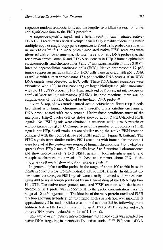
Figure 8, top, shows nondenatured acetic acid:ethanol fixed HEp-2 cells hybridized with human chromosome 7 specific alpha satellite centromeric DNA probe coated with recA protein. Under these conditions most of the interphase HEp-2 nuclei cell on slides showed about 2 FITC-labeled FISH signals. No FISH signals were obtained in reactions without recA protein or without incubation at 37°C. Comparisons of the number of chromosome FISH signals per HEp-2 cell nucleus were similar using the native FISH reaction compared with the control denatured FISH reaction (Figure 8, bottom). The FITC signals from similar native FISH reactions with human chromosome 1 were located at the centromere region of human chromosome 1 in metaphase spreads from HEp-2 nuclei. HEp-2 cells have 2 to 3 number 1 chromosomes and show approximately 2 to 3 FISH signals in both interphase nuclei and metaphase chromosome spreads. In these experiments, about 73% of the interphase cell nuclei showed hybridization signals.147
In general, alpha satellite probes in the range of about 100 to 600 bases in length produced recA protein-mediated native FISH signals. In different experiments, the strongest FISH signals were usually obtained with probes averaging 400 bases in length produced by nick translation of the DNA with bio-14-dUTP. The native recA protein-mediated FISH reaction with the human chromosomal 1 probe was proportional to the probe concentration over the range of 10 to 50 ng/reaction. The kinetics of the recA protein-mediated FISH reaction showing hybridization with fixed nuclei in solution was maximal at approximately 2 hr, and on slides was optimal at about 2.5 hr, following probe addition. Native FISH reactions required an ATPyS or ATP cofactor and recA protein/DNA probe nucleotide ratios of 1:1 or 1:2.
This native in situ hybridization technique with fixed cells was adapted for native DNA targeting in metabolically active nuclei.140·147 Efficient dsDNA

194 Gene Targeting
Figure 8. Comparison of recA protein-mediated fluorescence in situ hybridization of human chromosome 7 alpha satellite probe DNA with nondenatured HEp-2 target cell nuclei (top), with classic FISH (not recA protein-mediated) in heat denatured target cell nuclei (bottom). HEp-2 interphase nuclei were fixed with acetic acid and methanol ( 3:1) and probed on slides with biotinylated (nick-translated) ssDNA probes specific for human chromosome number 7 alpha satellite DNA sequences. In the reaction shown in the top of Figure 8 the recA protein coated complementary single-stranded chromosome 7 DNA probes were reacted with nondenatured nuclei for 2.5 hr at 37°C, followed by washing, blocking, and adding FJTC-avidin as the reporter. In the reaction shown in the bottom, heat-denatured nuclei were hybridized overnight with heat-denatured chromosome 7 alpha satellite DNA. washed, and blocked, and F/TC-avidin was also used as the reporter. In the denatured FISH reaction, the signal was amplified by the addition of a biotinylated goat anti-avidin antibody.
targeting techniques were developed in cell nuclei embedded in agarose. Human cell nuclei permeabilized with the non-ionic detergent Triton-100 and encapsulated in agarose remain morphologically intact, synthesize both DNA

Homologous Recombination Proteins 195
and RNA, and permit entry of recA protein-coated ssDNA probes. 14° FISH in metabolically active nuclei was tested using recA protein-coated ssDNA complementary 200- to 800-base-long biotinylated repeated satellite or unique sequence DNA probes with a streptavidin-FITC reporter and viewed by fluorescence microscopy or CLSM.140,l47
Figure 9 shows p53 DNA probes targeted in metabolically active human HEp-2 cell nuclei embedded in agarose. In these targeting reactions, 60 ng of recA protein-coated single-stranded complementary biotinylated (nick-translated) p53 eDNA was reacted for 3 hr at 37°C. The two FITC-labeled nuclear FISH signals are overlaid onto the phase image of the nuclei in the top left panel (Figure 9).
The native DNA targeting and hybridization reaction with biotinylated (nick-translated) p53 eDNA probes in nuclei embedded in agarose required
Figure 9. Targeting of recA protein-coated complementary single-stranded p53 DNA to native (nondenatured) chromosomal DNA sequences in metabolically active human cell
nuclei. Nuclei of HEp-2 cells encapsulated in agarose151 were reacted with recA protein
coated biotinylated p53 eDNA probes for 3 hr at 3JOC and washed. f1TC-avidin was added as a reporter and the nuclei were washed and immediately visualized by confocal laser scanning microscopy. The phase image of a metabolically active cell nuclei in agarose is shown in the upper right panel. The lower right panel shows a digital image
of a nucleus with two p53 DNA signals as monitored by laser scanning microscopy. This digital image was processed and the processed image is shown in the lower left panel. The
processed digital image was overlaid onto the phase image in the top left panel.

-- --~-~--------
196 Gene Targeting
incubation with recA protein at 37°C, was directly proportional to DNA probe concentration in the range of 30 to 90 ng of DNA probe per reaction, pH (7 .4 ), and required a nucleoside triphosphate cofactor (ATP or ATPyS). Targeting required a 3-h incubation at 37°C of recA protein-coated complementary ssDNA DNA probes with the permeabilized nuclei. Similar numbers of native FISH signals were observed in metabolically active nuclei as in interphase fixed cells permeabilized with acetic acid and methanol using either recA protein-mediated nondenatured DNA FISH, classic heat-denatured FISH in fixed interphase nuclei, or metaphase chromosomes using either unique or repeated sequence-specific DNA probes. When human satellite DNA probes coated with recA protein were hybridized with metabolically active nuclei and treated with hypotonic buffers to decondense the chromatin, repeated chromosomal specific-satellite FISH signals were specifically localized along the chromatin fiber. In intact nuclei-repeated satellite or unique p53 gene, FISH signals are often paired following nuclear DNA replication, as observed by confocal laser scanning microscopy and digital image analysis. 140•147
In summary, the recA protein-mediated in situ targeting reaction uses complementary nick-translated DNA probes with native (nondenatured) target sequences. The hybridization reaction depends on recA protein cofactor (ATP or ATPyS, 0.48 mM) and reaction time (2 to 2.5 hr), pH (7 .5), and temperature (37°C). Nick-translated eDNA probes efficiently form hybrids in about 75 to 85% of the nuclei. The recA protein-mediated targeting to DNA in fixed cells can be detected without signal amplification. Similar numbers of nondenatured FISH signals compared with heat-denatured FISH signals are observed. The specificity of the reaction was evidenced by homologous competition, copy number, linkage to well-established chromosomal markers, and metaphase cytogenetics.147
G. Potential applications of RecA protein~mediated homologous DNA targeting
These recA protein-mediated homologous DNA targeting reactions may be applied to target, detect, isolate, clone, and map any desired duplex gene or homologous chromosomal DNA fragment.41 •63•64•135•139 Novel gene mapping methods are evolving either by microscopy of large molecules or whole chromosomes, which may be physically oriented and stretched. RecA proteinmediated native chromosome walking techniques would permit more rapid gene discovery and mapping procedures. Furthermore, targeting of ssDNA probes to specific homologous sites by recA protein might block transcription in vivo as well as in vitro143•144 DNA methylation137•142 or DNA cleavage.l43 RecA protein-mediated targeting has been used to detect chromosomal targets in both fixed and metabolically active nuclei. This important new in situ DNA targeting technique could possibly be also used for gene tracking studies. Lastly, it may be possible to use recA protein-mediated DNA targeting and
Homologous Recombination Proteins 197
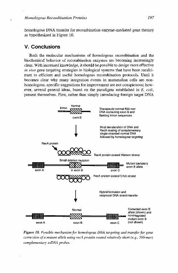
homologous DNA transfer for recombination enzyme-mediated gene therapy as hypothesized in Figure 10.
V. Conclusions
Both the molecular mechanisms of homologous recombination and the biochemical behavior of recombination enzymes are becoming increasingly clear. With increased knowledge, it should be possible to design more effective in vivo gene targeting strategies in biological systems that have been recalcitrant to efficient and useful homologous recombination protocols. Until it becomes clear why many integration events in mammalian cells are nonhomologous, specific suggestions for improvement are not conspicuous; however, several general ideas, based on the paradigms established in E. coli, present themselves. First, rather than simply introducing foreign target DNA
. Normal
~ exon B
RecA protein
'~ Small deletion mutation
exon A in exon B
Normal
exon A exon B
Therapeutic normal 500-mer DNA containing exon B and flanking intron sequences
Heat denaturation of DNA and RecA coating of complementary single-stranded normal DNA followed by homologous targeting
RecA protein-coated Watson strand
exon C
Mutant (deletion) exon B allele
RecA protein-coated Crick strand
Hybrid formation and reciprocal DNA strand transfer
exon C
Corrected exon B allele (shown) and nonintegrated mutant exon B (not shown)
Figure 10. Possible mechanism for homologous DNA targeting and transfer for gene correction of a mutant allele using recA protein coated relatively short (e.g., 500-mer)
complementary ssDNA probes.

~-~··~·-----
198 Gene Targeting
into cells, it might be advantageous to co-introduce enzymes that enhance various steps of the recombination mechanism. If, for example, dsDNA break sites are limiting, then an endonuclease can be introduced. Similarly, if generation of ssDNA from the donor DNA is limiting, then either ssDNA itself, or helicases or nucleases to maintain the single-stranded form can be in introduced or co-expressed. Recombination hotspots can be introduced into the donor DNA, or alternatively, mutant or modified recombination proteins with constitutively activated or enhanced recombination functions can be used. Alternatively, the over-expression of normally limiting endogenous cellular recombination enzymes, in concert with other normal cellular components, may be harnessed to increase recombination efficiency.
The applications of recA protein-targeted events present themselves as immediate options (for example, see Figure 6). Interestingly, when a modified E. coli recA protein containing a nuclear localization signal is expressed in mammalian cells, it localizes to the nucleus and binds to chromosomes.149
Perhaps, similar expression vectors can be used to introduce other recombination enzymes into the nucleus of mammalian cells. The continued taming and manipulation of the homologous recombination process can afford a variety of new options for more effective gene targeting protocols.
Acknowledgments
The authors are indebted to Angela Eggleston for providing Figure 3 and Elissa Sena and Sushma Patel for helpful discussions. The work in the authors' laboratories is supported by grants from the National Institutes of Health (AI-18987 and GM-41347 to S.C.K. and GM-38424 to D.A.Z.).
References
1. Gottesman, M.E., and Vogel, H.J., Ed., Mechanisms of Eukaryotic DNA Recombination, Academic Press, Inc., San Diego, 1992.
2. Sedivy, J.M., and Joyner, A.L., Gene Targeting, W.H. Freeman and Company, New York, 1992, 184.
3. Lin, M., Sperle, K., and Sternberg, N., Studies on extrachromosomal homologous recombination in mammalian cells: implications for chromosomal recombination and gene targeting, in Mechanisms ofEukaryotic DNA Recombination, Gottesman, M.E., and Vogel, H.J., Eds., Academic Press, Inc., San Diego, 1992, 15.
4. Cox, M.M., and Lehman, I.R., Enzymes of general recombination, Annu. Rev. Biochem., 56, 229, 1987.
5. Eggleston, A.K., and Kowalczykowski, S.C., An overview of homologous pairing and DNA strand exchange proteins, Biochimie, 73, 163, 1991.

Homologous Recombination Proteins 199
6. Fishel, R., General genetic recombination, inModernMicrobial Genetics, WileyLiss, New York, 1991, 91.
7. Griffith, J.D., and Harris, L.D., DNA strand exchanges, CRC Crit. Rev. Biochem., 23 Suppl. 1, S43, 1988.
8. W.-D., and Kolodner, R.D., Enzymology of homologous recombina-tion in Saccharomyces cerevisiae, Pro g. Nucleic Acid Res. Mol. Biol., 46, 221, 1993.
9. Kowalczykowski, S.C., Biochemistry of genetic recombination: energetics and mechanism of DNA strand exchange, Annu. Rev. Biophys. Biophys. Chern., 20, 539, 1991.
10. Kowalczykowski, S.C., Dixon, D.A., Eggleston, A.K., Lauder, S. D., and Rehrauer, W.M., Biochemistry of homologous recombination in Escherichia coli, Microbial. Rev., 58, 3, 1994.
11. Kowalczykowski, S.C., and Eggleston, A.K., Homologous pairing and DNA strand exchange proteins, Annu. Rev. Biochem., 63, 991, 1994.
12. Lloyd, R.G., and Sharples, G.J., Genetic analysis of recombination in prokaryotes, Curr. Opin. Genet. Dev., 2, 683, 1992.
13. Radding, C.M., Helical RecA nucleoprotein filaments mediate homologous pairing and strand exchange, Biochim. Biophys. Acta, 1008, 131, 1989.
14. Radding, C.M., Helical interactions in homologous pairing and strand exchange driven by RecA protein, J. Biol. Chern., 266, 5355, 1991.
IS. Roca, A.I., and Cox, M.M., The RecA protein: structure and function, Crit. Rev. Biochem. Mol. Biol., 25, 415, 1990.
16. Stasiak, A., and Egelman, E.H., Visualization of recombination reactions, in Genetic Recombination, Kucherlapati, R., and Smith, G.R., Eds., American Society for Microbiology, Washington, DC, 1988, 265.
17. West, S.C., Enzymes and molecular mechanisms of genetic recombination, Annu. Rev. Biochem., 61, 603, 1992.
18. Thaler, D.S., and Stahl, F.W., DNA double-chain breaks in recombination of phage lambda and of yeast, Annu. Rev. Genet., 22, 169, 1988.
19. Smith, G.R., Homologous recombination in procaryotes, Microbial. Rev., 52, 1, 1988.
20. Smith, G.R., Conjugational recombination in E. coli: myths and mechanisms, Cell, 64, 19, 1991.

200 Gene Targeting
21. Konforti, B.B., and Davis, R.W., 3' homologous free ends are required for stable joint molecule formation by the RecA and single-stranded binding proteins of Escherichia coli., Proc. Nat!. Acad. Sci. U.S.A., 84, 690, 1987.
22. West, S.C., and Connolly, B., Biological roles of the Escherichia coli RuvA, RuvB and RuvC proteins revealed, Mol. Microbial., 6, 2755, 1992.
23. Shinohara, A., Ogawa, H., and Ogawa, T., Rad51 protein involved in repair and recombination in S. cerevisiae is a RecA-like protein, Cell, 69, 457, 1992.
24. Shinohara, A., Ogawa, H., Matsuda, Y., Ushio, N., Ikeo, K., and Ogawa, T., Cloning of human, mouse and fission yeast recombination genes homologous to RAD51 and recA, Nat. Genet., 4, 239, 1993.
25. Cox, M.M., and Lehman, I.R., RecA protein of Escherichia coli promotes branch migration, a kinetically distinct phase of DNA strand exchange, Proc. Natl. A cad. Sci. U.S.A., 78, 3433, 1981.
26. Kowalczykowski, S.C., C1ow, J., and Krupp, R.A., Properties of the duplex DNA-dependent ATPase activity of Escherichia coli recA protein and its role in branch migration, Proc. Natl. Acad. Sci. U.S.A., 84, 3127, 1987.
27. Pugh, B.F., and Cox, M.M., Stable binding of recA protein to duplex DNA. Unraveling a paradox, J. Biol. Chern., 262, 1326, 1987.
28. Pugh, B.F., and Cox, M.M., General mechanism for RecA protein binding to duplex DNA, J. Mol. Biol., 203, 479, 1988.
29. West, S.C., Cassuto, E., Mursalim, J., and Howard-Flanders, P., Recognition of duplex DNA containing single-stranded regions by recA protein, Proc. Natl. Acad. Sci. U.S.A., 77, 2569, 1980.
30. Shaner, S.L., and Radding, C.M., Translocation of Escherichia coli recA protein from a single-stranded tail to contiguous duplex DNA, J. Bioi. Chern., 262, 9211, 1987.
31. Blaho, J.A., and Wells, R.D., Left-handed Z-DNA binding by the recA protein of Escherichia coli, J. Bioi. Chern., 262, 6082, 1987.
32. Kim, J.I., Heuser, J., and Cox, M.M., Enhanced recA protein binding to Z DNA represents a kinetic perturbation of a general duplex DNA binding pathway, J. Biol. Chern., 264, 21848, 1989.
33. Thresher, R.J., and Griffith, J.D., lnterca1ators promote the binding of RecA protein to double-stranded DNA, Proc. Natl. Acad. Sci. U.S.A., 87, 5056, 1990.

Homologous Recombination Proteins 201
34. Kojima, M., Suzuki, M., Morita, T., Ogawa, T., Ogawa, H., and Tada, M., Interaction of Re"A protein with pBR322 DNA modified by N-hydroxy-2-acetylaminofluorene and 4-hydroxyaminoquinoline 1-oxide, Nucleic Acids Res., 18, 2707, 1990.
35. Dianov, G.L., Saparbaev, M.K., Mazin, A.V., and Salganik, R.I., The chemical mutagen dimethyl sulphate induces homologous recombination of plasmid DNA by increasing the binding ofRecA protein to duplex DNA, Mutat. Res., 249, 189, 1991.
36. Stasiak, A., DiCapua, E., and Koller, T.H., Elongation of duplex DNA by recA proteins, J. Mol. Biol., 151, 557, 1981.
37. Menetski, J.P., and Kowalczykowski, S.C., Interaction of recA protein with single-stranded DNA. Quantitative aspects of binding affinity modulation by nucleotide cofactors, J. Mol. Biol., 181, 281, 1985.
38. Conley, E. C., and West, S.C., Underwinding of DNA associated with duplexduplex pairing by RecA protein, J. Biol. Chern., 265, 10156, 1990.
39. Lindsley, J.E., and Cox, M.M., On RecA protein-mediated homologous alignment of two DNA molecules. Three strands versus four strands, J. Biol. Chern., 265, 10164, 1990.
40. Hsieh, P., Camerini-Otero, C.S., and Camerini-Otero, R.D., The synapsis event in the homologous pairing of DNAs: RecA recognizes and pairs less than one helical repeat of DNA, Proc. Natl. Acad. Sci. U.S.A., 89, 6492, 1992.
41. Honigberg, S.M., Rao, B.J., and Radding, C.M., Ability of RecA protein to promote a search for rare sequences in duplex DNA, Proc. Nat/. Acad. Sci. U.S.A., 83, 9586, 1986.
42. Honigberg, S.M., Gonda, D.K., Flory, J., and Radding, C.M., The pairing activity of stable nucleoprotein filaments made from recA protein singlestranded DNA and adenosine 5' -r-thiotriphosphate, J. Biol. Chern., 260, 11845, 1985.
43. Riddles, P.W., and Lehman, I.R., The formation of plectonemic joints by the recA protein of Escherichia coli, J. Biol. Chern., 260, 170, 1985.
44. Menetski, J.P., Bear, D.G., and Kowalczykowski, S.C., Stable DNA heteroduplex formation catalyzed by the Escherichia coli RecA protein in the absence of ATP hydrolysis, Proc. Natl. Acad. Sci. U.S.A., 87, 21, 1990.
45. Rosselli, W., and Stasiak, A., Energetics of RecA-mediated recombination reactions: without ATP hydrolysis RecA can mediate polar strand exchange but is unable to recycle, J. Mol. Biol., 216, 335, 1990.

202 Gene Targeting
46. Kim, J.I., Cox, M.M., and Inman, R.B., On the role of ATP hydrolysis in RecA protein-mediated DNA strand exchange. I. Bypassing a short heterologous insert in one DNA substrate, J. Biol. Chern., 267, 16438, 1992.
47. Kowalczykowski, S.C., and Krupp, R.A., DNA strand exchange promoted by Escherichia coli recA protein in the absence of ATP: implications for the mechanism of energy transduction, submitted, 1994.
48. Gonda, D.K., and Radding, C.M., The mechanism of the search for homology promoted by recA protein: facilitated diffusion within nucleoprotein networks, J. Biol. Chern., 261, 13087, 1986.
49. West, S.C., Cassuto, E., and Howard-Flanders, P., Homologous pairing can occur before DNA strand separation in general genetic recombination, Nature (London), 290, 29, 1981.
50. Bianchi, M., Riboli, B., and Magni, G., Escherichia coli recA protein possesses a strand separating actvity on short duplex DNA, EMBO 1., 4, 3025, 1985.
51. Hsieh, P., and Camerini-Otero, R.D., Formation of joint DNA molecules by two eukaryotic strand exchange proteins does not require melting of a DNA duplex, I. Biol. Chem, 264, 5089, 1989.
52. Howard-Flanders, P., West, S.C., and Stasiak, A., The spiral protein filaments of genetic recombination, Nature (London), 309, 1, 1983.
53. Stasiak, A., Three-stranded DNA structure; is this the secret of DNA homologous recognition?, Mol. Microbial., 6, 3267, 1992.
54. Camerini-Otero, R.D., and Hsieh, P., Parallel DNA triplexes, homologous recombination, and other homology-dependent DNA interactions, Cell, 73, 217, 1993.
55. Stasiak, A., DiCapua, E., and Koller, T., Unwinding of duplex DNA in complexes with recA protein, Cold Spring Harbor Symp. Quant. Biol., 47, 811, 1983.
56. Wu, A.M., Bianchi, M., DasGupta, C., and Radding, C.M., Unwinding associated with synapsis of DNA molecules by recA protein, Proc. Natl. Acad. Sci. U.S.A., 80, 1256, 1983.
57. Schutte, B.C., and Cox, M.M., Homology-dependent underwinding of duplex DNA in recA protein generated paranernic complexes, Biochemistry, 27, 7886, 1988.
58. Honigberg, S.M., and Radding, C.M., The mechanics of winding and unwinding helices in recombination: torsional stress associated with strand transfer promoted by RecA protein., Cell, 54, 525, 1988.
Homologous Recombination Proteins 203
59. Rould, E., Muniyappa, K., and Radding, C.M., Unwinding of heterologous DNA by RecA protein during the search for homologous sequences, J. Mol. Biol., 226, 127, 1992.
60. Riddles, P.W., and Lehman, I.R., The formation of paranemic and plectonemic joints between DNA molecules by the recA and single-stranded DNA-binding proteins of Escherichia coli, J. Biol. Chern., 260, 165, 1985.
61. Cunningham, R.P., Wu, A.M., Shibata, T., DasGupta, C., and Radding, C.M., Homologous pairing and topological linkage of DNA molecules by combined action of E. coli recA protein and topoisomerase I, Cell, 24, 213, 1981.
62. Cassuto, E., Formation of covalently closed heteroduplex DNA by the combined action of gyrase and RecA protein, EMBO J., 3, 2159, 1984.
63. Sena, E.P., and Zarling, D.A., Targeting in linear DNA duplexes with two complementary probe strands for hybrid stability, Nat. Genet., 3, 365, 1993.
64. Jayasena, V.K., and Johnston, B.H., Complement-stabilized D-loop. RecAcatalyzed stable pairing of linear DNA molecules at internal sites, J. Mol. Biol., 230, 1015, 1993.
65. Bianchi, M.E., and Radding, C.M., Insertions, deletions and mismatches in heteroduplex DNA made by recA protein, Cell, 35, 511, 1983.
66. Ramdas, J., Mythili, E., and Muniyappa, K., RecA protein promoted homologous pairing in vitro. Pairing between linear duplex DNA bound to HU protein (nucleosome cores) and nucleoprotein filaments ofrecA protein-single-stranded DNA, J. Biol. Chern., 264, 17395, 1989.
67. Ramdas, J., Mythili, E., and Muniyappa, K., Nucleosomes on linear duplex DNA allow homologous pairing but prevent strand exchange promoted by recA protein, Proc. Natl. Acad. Sci. U.S.A., 88, 1344, 1991.
68. Muniyappa, K., Ramdas, J., Mythili, E., and Galande, S., Homologous pairing between nucleosome cores on a linear duplex DNA and nucleoprotein filaments of RecA protein-single stranded DNA, Biochirnie, 73, 187, 1991.
69. Clark, A.J., Sharma, V., Brenowitz, S., Chu, C.C., Sandler, S., Satin, L., Templin, A., Berger, I., and Cohen, A., Genetic and molecular analyses of the C-terminal region of the recE gene from the rae prophage of Escherichia coli K-12 reveal the recT gene, J. Bacterial., 175, 7673, 1993.
70. Hall, S.D., Kane, M.F., and Kolodner, RD., Identification and characterization of the Escherichia coli RecT protein, a protein encoded by the recE region that promotes renaturation of homologous single-stranded DNA, J. Bacterial., 175, 277, 1993.

204 Gene Targeting
71. Hall, S.D., and Kolodner, R.D., Homologous pairing and strand exchange promoted by the Escherichia coli RecT protein, Proc. Nat!. Acad. Sci. U.S.A., 91, 3205, 1994.
72. Sander, M., Carter, M., and Huang, S.M., Expression of Drosophila Rrpl protein in Escherichia coli. Enzymatic and physical characterization of the intact protein and a carboxyl-terminally deleted exonuclease-deficient mutant, J. Biol. Chem., 268, 2075, 1993.
73. Taylor, A.F., The recBCD enzyme of Escherichia coli, in Genetic Recombination, Kucherlapati, R., and Smith, G.R., Eds., American Society for Microbiology, Washington, D.C., 1988, 231.
74. Smith, G.R., RecBCD enzyme, in Nucleic Acids and Molecular Biology, Vol. 4, Eckstein, F. and Lilley, D.M.J., Eds., Springer-Verlag, Berlin, 1990, 78.
75. Dixon, D.A., and Kowalczykowski, S.C., Homologous pairing in vitro stimulated by the recombination hotspot, Chi, Cell, 66, 361, 1991.
76. Dixon, D.A., and Kowalczykowski, S.C., The recombination hotspot, x,, is a regulatory sequence that acts by attenuating the nuclease activity of the Escherichia coli recBCD enzyme, Cell, 73, 87, 1993.
77. Dixon, D.A., Churchill, J.J., and Kowalczykowski, S.C., Reversible inactivation of the Escherichia coli recBCD enzyme by the recombination hotspot, x,, in vitro: evidence for functional inactivation or loss of the reeD subunit, Proc. Natl. Acad. Sci., U.S.A., 91, 2980, 1993.
78. Taylor, A.F., and Smith, G.R., Substrate specificity of the DNA unwinding activity of the recBC enzyme of E. coli, J. Mol. Biol., 185, 431, 1985.
79. Roman, L.J., and Kowalczykowski, S.C., Characterization of the helicase activity of the Escherichia coli recBCD enzyme using a novel helicase assay, Biochemistry, 28, 2863, 1989.
80. Taylor, A., and Smith, G.R., Unwinding and rewinding of DNA by the recBC enzyme, Cell, 22, 447, 1980.
81. Roman, L.J., Eggleston, A.K, and Kowalczykowski, S.C., Processivity of the DNA helicase activity of Escherichia coli recBCD enzyme, J. Bioi. Chem., 267, 4207, 1992.
82. Telander-Muskavitch, KM., and Linn, S., Electron microscopy of E. coli recBC enzyme reaction intermediates, in Mechanistic Studies of DNA Replication and Genetic Recombination, Vol. 19, Alberts, B., Ed., Academic Press, New York, 1980, 901.
83. Telander-Muskavitch, K., and Linn, S., RecBC-like enzymes: exonuclease V deoxyribonucleases, in The Enzymes, Boyer, P.D., Ed., Academic Press, New York, 1981, 233.

Homologous Recombination Proteins 205
84. Telander-Muskavitch, K., and Linn, S., A unified mechanism for the nuclease and unwinding activities of the recBC enzyme of E. coli, J. Biol. Chern., 257, 2641, 1982.
85. Roman, L.J., and Kowalczykowski, S.C., Characterization of the adenosinetriphosphatase activity of the Escherichia coli recBCD enzyme: relationship of ATP hydrolysis to the unwinding of duplex DNA, Biochemistry, 28, 2873, 1989.
86. Eggleston, A.K., O'Neill, T.E., Bradbury, E.M., and Kowalczykowski, S.C., Unwinding of nucleosomal DNA by the Escherichia coli recBCD helicase, submitted, 1993.
87. Nakayama, H., Nakayama, K., Nakayama, R., Irino, N., Nakayama, Y., and Hanawalt, P.C., Isolation and genetic characterization of a thymineless deathresistant mutant of Escherichi coli K12: identification of a new mutation (recQI) that blocks the reeF recombination pathway, Mol. Gen. Genet., 195, 474, 1984.
88. Umezu, K., Nakayama, K., and Nakayama, H., Escherichia coli RecQ protein is a DNA helicase, Proc. Natl. Acad. Sci. U.S.A., 87, 5363, 1990.
89. Umezu, K., and Nakayama, H., The recQ DNA helicase of Escherichia coli. Characterization of the helix-unwinding activity with emphasis on the effect of single-stranded DNA binding protein, J. Mol. Biol., 230, 1145, 1993.
90. Tsaneva, I.R., Muller, B., and West, S.C., Ruv A and RuvB proteins of Escherichia coli exhibit DNA helicase activity in vitro, Proc. Natl. Acad. Sci. U.S.A., 90, 1315, 1993.
91. Tsaneva, I.R., Muller, B., and West, S.C., ATP-dependent branch migration of Holliday junctions promoted by the RuvA and RuvB proteins of E. coli, Cell, 69, 1171, 1992.
92. Tsaneva, I.R., Illing, G., Lloyd, R.G., and West, S.C., Purification and properties of the RuvA and RuvB proteins of Escherichia coli, Mol. Gen. Genet., 235, 1, 1992.
93. Iwasaki, H., Takahagi, M., Nakata, A., and Shinagawa, H., Escherichia coli Ruv A and RuvB proteins specifically interact with Holliday junctions and promote branch migration, Genes Dev., 6, 2214, 1992.
94. Miiller, B., Tsaneva, I.R., and West, S.C., Branch migration of Holliday junctions promoted by the Escherichia coli ruvA and ruvB proteins. I. Comparison of ruv AB- and ruvB-mediated reactions, .!. Bioi. Chern., 268, 17179, 1993.
95. Miiller, B., Tsaneva, I.R., and West, S.C., Branch migration of Holliday junctions promoted by the Escherichia coli ruvA and ruvB proteins. II. Interaction of ruvB with DNA, J. Biol. Chern., 268, 17185, 1993.

206 Gene Targeting
96. Lloyd, R.G., and Sharples, G.J., Dissociation of synthetic Holliday junctions by E. coli recG protein, EMBO 1., 12, 17, 1993.
97. Lloyd, R.G., and Sharples, G.J., Processing of recombination intermediates by the recG and ruvAB proteins of Escherichia coli, Nucleic Acids Res., 21, 1719, 1993.
98. Whitby, M.C., Ryder, L., and Lloyd, R.G., Reverse branch migration of Holliday junctions by RecG protein: a new mechanism for resolution of intermediates in recombination and DNA repair, Cell, 75, 341, 1993.
99. Joseph, J.W., and Kolodner, R., Exonuclease VIII of Escherichia coli. II. Mechanism of action, J. Biol. Chem., 258, 10418, 1983.
100. Lovett, S.T., and Ko1odner, R.D., Identification and purification of a singlestranded-DNA-specific exonuclease encoded by the reel gene of Escherichia coli, Proc. Natl. Acad. Sci. U.S.A., 86, 2627, 1989.
101. Lovett, S.T., DeLuca, C.L., and Kolodner, R.D., The genetic dependence of recombination in reeD mutants of Escherichia coli, Genetics, 120, 37, 1988.
102. Iwasaki, H., Takahagi, M., Shiba, T., Nakata, A., and Shinagawa, H., Escherichia coli RuvC protein is an endonuclease that resolves the Holliday structure, EMBO J., 10, 4381, 1991.
103. Dunderdale, H.J., Benson, F.E., Parsons, C.A., Sharples, G.J., Lloyd, R.G., and West, S.C., Formation and resolution of recombination intermediates by E. coli RecA and RuvC proteins, Nature (London), 354, 506, 1991.
104. Bennett, R.J., Dunderdale, H.J., and West, S.C., Resolution of Holliday junctions by ruvC resolvase: cleavage specificity and DNA distortion, Cell, 74, 1021, 1993.
105. Connolly, B., and West, S.C., GeneticrecombinationinEscherichia coli: Holliday junctions made by RecA protein are resolved by fractionated cell-free extracts, Proc. Natl. Acad. Sci. U.S.A., 87, 8476, 1990.
106. Sancar, A., Williams, K.R., Chase, J.W., and Rupp, W.O., Sequences of the ssb gene and protein, Proc. Nat!. Acad. Sci. U.S.A., 78, 4274, 1981.
107. Kowalczykowski, S.C., Bear, D.O., and von Hippel, P.H., Single-stranded DNA binding proteins, in The Enzymes, Boyer, P. D., Ed., Academic Press, New York, 1981, 373.
108. Muniyappa, K., Shaner, S.L., Tsang, S.S., andRadding, C.M., Mechanism of the concerted action of recA protein and helix destabilizing proteins in homologous recombination, Proc. Natl. Acad. Sci. U.S.A., 81, 2757, 1984.

Homologous Recombination Proteins 207
109. Kowalczykowski, S.C., and Krupp, R.A., Effects of the Escherichia coli SSB protein on the single-stranded DNA- dependent ATPase activity of Escherichia coli recA protein: evidence that SSB protein facilitates the binding of recA protein to regions of secondary structure within single-stranded DNA, J. Mol. Bioi., 193, 97, 1987.
110. Chow, S.A., Rao, B.J., and Radding, C.M., Reversibility of strand invasion promoted by recA protein and its inhibition by Escherichia coli single-stranded DNA-binding protein or phage T4 gene 32 protein,./. Bioi. Chern., 263,200, 1988.
111. Lavery, P.E., and Kowalczykowski, S.C., A postsynaptic role for single-stranded DNA-binding protein in recA protein-promoted DNA strand exchange, J. Biol. Chern., 267, 9315, 1992.
112. Smith, G.R., Hotspots of homologous recombination, Experientia, 50, 234, 1994.
113. Lam, S.T., Stahl, M.M., McMilin, K.D., and Stahl, F.W., Rec-mediated recombinational hot spot activity in bacteriophage lambda. II. A mutation which causes hot spot activity, Genetics, 77, 425, 1974.
114. Stahl, F.W., Crasemann, J.M., and Stahl, M.M., Rec-mediated recombinational hot spot activity in bacteriophage lambda. III. Chi mutations are site-mutations stimulating rec-mediated recombination, J. Mol. Biol., 94, 203, 1975.
115. Dower, N.A., and Stahl, F.W., Chi activity during transduction-associated recombination, Proc. Nat!. Acad. Sci. U.S.A., 78, 7033, 1981.
116. Cheng, K.C., and Smith, G.R., Distribution of Chi-stimulated recombinational exchanges and heteroduplex endpoints in phage lambda, Genetics, 123, 5, 1989.
117. Smith, G.R., Comb, M., Schultz, D.W., Daniels, D.L., and Blattner, F.R., Nucleotide sequence of the Chi recombinational hotspot Chi+D in bacteriophage lambda, J. Viral., 37, 336, 1981.
118. Ponticelli, A.S., Schultz, D.W., Taylor, A.F., and Smith, G.R., Chi-dependent DNA strand cleavage by recBC enzyme, Cell, 41, 145, 1985.
119. Taylor, A.F., Schultz, D.W., Ponticelli, A.S., and Smith, G.R., RecBC enzyme nicking at Chi sites during DNA unwinding: location and orientation-dependence of the cutting, Cell, 41, 153, 1985.
120. Roman, L.J., and Kowalczykowski, S.C., Formation of heteroduplex DNA promoted by the combined activities of Escherichia coli recA and recBCD proteins, ./. Biol. Chern., 264, 18340, 1989.
121. Roman, L.J., Dixon, D.A., and Kowalczykowski, S.C., RecBCD-dependent joint molecule formation promoted by the Escherichia coli recA and SSB proteins, Proc. Natl. Acad. Sci. U.S.A., 88, 3367, 1991.

208 Gene Targeting
122. Wang, T.C., and Smith, K.C., The roles of RecBCD, Ssb and RecA proteins in the formation of heteroduplexes from linear-duplex DNA in vitro, Mol. Gen. Genet., 216, 315, 1989.
123. Kowalczykowski, S.C., and Roman, L.J., Reconstitution of homologous pairing activity dependent upon the combined activities of purified E. coli recA, recBCD, and SSB proteins, in Molecular Mechanisms in DNA Replication and Recombination, Vol. 127, Richardson, C.C., and Lehman, I. R., Eds., Wiley-Liss, New York, 1990, 357.
124. Smith, G.R., Schultz, D.W., Taylor, A.F., and Triman, K., Chi sites, recBC enzyme, and generalized recombination, Stadler Symp., 13, 25, 1981.
125. Thaler, D.S., Sampson, E., Siddiqi, I., Rosenberg, S.M., Stahl, F. W., and Stahl, M., A hypothesis: Chi-activation of recBCD enzyme involves removal of the reeD subunit, in Mechanisms and Consequences of DNA Damage Processing, Friedberg, E., and Hanawalt, P., Eds., Alan R. Liss, Inc., New York, 1988, 413.
126. Stahl, F.W., Thomason, L.C., Siddiqi, I., and Stahl, M.M., Further tests of a recombination model in which chi removes the ReeD subunit from the RecBCD enzyme of Escherichia coli, Genetics, 126, 519, 1990.
127. Korangy, F., and Julin, D.A., Kinetics and processivity of ATP hydrolysis and DNA unwinding by the recBC enzyme from Escherichia coli, Biochemistry, 32, 4873, 1993.
128. West, S.C., Cassuto, E., and Howard-Flanders, P., RecA protein promotes homologous-pairing and strand-exchange reactions between duplex DNA molecules, Proc. Natl. Acad. Sci. U.S.A., 78, 2100, 1981.
129. West, S.C., Countryman, J.K., and Howard-Flanders, P., Enzymatic formation of biparental figure-eight molecules from plasmid DNA and their resolution in E. coli, Cell, 32, 817, 1983.
130. Connolly, B., Parsons, C.A., Benson, F.E., Dunderdale, H.J., Sharples, G.J., Lloyd, R.G., and West, S.C., Resolution of Holliday junctions in vitro requires the Escherichia coli ruvC gene product., Proc. Natl. Acad. Sci. U.S.A., 88, 6063, 1991.
131. Hsieh, P., Camerini-Otero, C.S., and Camerini-Otero, R.D., Pairing of homologous DNA sequences by proteins: evidence for three-stranded DNA, Genes Dev., 4, 1951, 1990.
132. Shibata, T., DasGupta, C., Cunningham, R.P., and Radding, C.M., Purified E. coli recA protein catalyzes homologous pairing of superhelical DNA and singlestranded fragments, Proc. Natl. Acad. Sci. U.S.A., 76, 1638, 1979.

Homologous Recombination Proteins 209
133. McEntee, K., Weinstock, G.M., and Lehman, I.R., Initiation of general recombination catalysed in vitro by the recA protein of Escherichia coli, Proc. Natl. Acad. Sci. U.S.A., 76, 2615, 1979.
134. Radding, C.M., Beattie, K.L., Hoffman, W.K., and Wiegand, R.C., Uptake of homologous single-stranded fragments by superhelical DNA IV branch migration, J. Mol. Bioi., 116, 825, 1977.
135. Rigas, B., Welcher, A.A., Ward, D.C., and Weissman, S.M., Rapid plasmid library screening using RecA-coated biotinylated probes, Proc. Natl. Acad. Sci. U.S.A., 83, 9591, 1986.
136. Konforti, B.B., and Davis, R.W., DNA substrate requirements for stable joint molecule formation by the RecA and single-stranded DNA-binding proteins of Escherichia coli, J. Bioi. Chern., 266, 10112, 1991.
137. Ferrin, L.J., and Camerini-Otero, R.D., Selective cleavage of human DNA: RecA-assisted restriction endonuclease (RARE) cleavage, Science, 254, 1494, 1991.
138. Adzuma, K., Stable synapsis of homologous DNA molecules mediated by the Escherichia coli RecA protein involves local exchange of DNA strands, Genes Dev., 6, 1679, 1992.
139. Revet, B.M., Sena, E.P., and Zarling, D.A., Homologous DNA targeting with RecA protein-coated short DNA probes and electron microscope mapping on linear duplex molecules, J. Mol. Bioi., 232, 779, 1993.
140. Zarling, D.A., and Arboleda, M.J., RecA protein-dependent homologous DNA targeting in metabolically active human nuclei monitored by fluorescent in situ hybridization and laser microscopy, in Fourth Int. Cent. Biotechnol. Symp. Gene Transfer Gene Ther., Stockholm, Sweden, 1993.
141. Olson, M., Hood, L., Cantor, C., and Botstein, D., New game plan for genome mapping, Science, 245, 1434, 1989.
142. Koob, M., Burkiewicz, A., Kur, J., and Szybalski, W., RecA-AC: single-site cleavage of plasmids and chromosomes at any predetermined restriction site, Nucleic Acids Res., 20, 5831, 1992.
143. Golub, E.l., Ward, D.C., and Radding, C.M., Joints formed by RecA protein from oligonucleotides and duplex DNA block initiation and elongation of transcription, Nucleic Acids Res., 20, 3121, 1992.
144. Golub, E.I., Radding, C.M., and Ward, D.C., Inhibition of RNA polymerase II transcription by oligonucleotide-RecA protein filaments targeted to promoter sequences, Proc. Natl. Acad. Sci. U.S.A., 90, 7186, 1993.

210 Gene Targeting
145. Koller, T., DiCapua, E., and Stasiak, A., Complexes of recA protein with singlestranded DNA, in Mechanisms of DNA Replication and Recombination, Cozzarelli, N. R., Ed., Alan R Liss, New York, 1983, 723.
146. Flory, J., Tsang, S.S., and Muniyappa, K., Isolation and visualization of active presynaptic filaments of recA protein and single-stranded DNA, Proc. Natl. Acad. Sci. U.S.A., 81, 7026, 1984.
147. Zarling, D.A., Homologous DNA targeting, In Second Int. Conf. Gene Ther. Cancer, San Diego, 1993.
148. Pinke1, D., Straume, T., and Gray, J., Cytogenetic analysis using quantitative, high-sensitivity, fluorescence hybridization, Proc. Natl. Acad. Sci. U.S.A., 83, 2934, 1986.
149. Kido, M., Yoneda, Y., Nakanishi, M., Uchida, T., and Okada, Y., Escherichia coli RecA protein modified with a nuclear location signal binds to chromosomes in living mammalian cells, Exp. Cell Res., 198, 107, 1992.
150. Kowalczykowski, S.C., In vitro reconstitution of homologous recombination reactions, Experientia, 50, 204, 1994.
151. Jackson, D.A., and Cook, P.R., A general method for preparing chromatin, EMBO J., 4, 913, 1985.
152. Hall, S.D., and Kolodner, RD., personal communications.