01 - Vaste Inbouwspots - Spots encastrés fixes - Recessed Spots Fixed - Einbauleuchten Fix

Intersubunit signaling in RecBCDenzyme, a complex protein machineregulated by Chi hot spotsSusan K. Amundsen,1 Andrew F. Taylor,1 Manjula Reddy,2 and Gerald R. Smith3
Division of Basic Sciences, Fred Hutchinson Cancer Research Center, Seattle, Washington 98109, USA
The Escherichia coli RecBCD helicase–nuclease, a paradigm of complex protein machines, initiateshomologous genetic recombination and the repair of broken DNA. Starting at a duplex end, RecBCD unwindsDNA with its fast RecD helicase and slower RecB helicase on complementary strands. Upon encountering aChi hot spot (5�-GCTGGTGG-3�), the enzyme produces a new 3� single-strand end and loads RecA proteinonto it, but how Chi regulates RecBCD is unknown. We report a new class of mutant RecBCD enzymes thatcut DNA at novel positions that depend on the DNA substrate length and that are strictly correlated with theRecB:RecD helicase rates. We conclude that in the mutant enzymes when RecD reaches the DNA end, itsignals RecB’s nuclease domain to cut the DNA. As predicted by this interpretation, the mutant enzymes cutcloser to the entry point on DNA when unwinding is blocked by another RecBCD molecule traveling in theopposite direction. Furthermore, when RecD is slowed by a mutation altering its ATPase site such that RecBreaches the DNA end before RecD does, the length-dependent cuts are abolished. These observations lead usto hypothesize that, in wild-type RecBCD enzyme, Chi is recognized by RecC, which then signals RecD tostop, which in turn signals RecB to cut the DNA and load RecA. We discuss support for this “signal cascade”hypothesis and tests of it. Intersubunit signaling may regulate other complex protein machines.
[Keywords: Homologous recombination; E. coli; RecBCD enzyme; Chi sites; complex protein machines]
Supplemental material is available at http://www.genesdev.org.
Received August 17, 2007; revised version accepted October 16, 2007.
Multistep processes in living cells, such as replication,transcription, and genetic recombination, are often car-ried out by complex protein “machines” (Alberts 1998).The multiple activities of each machine must be prop-erly regulated for the process to be successful, but thebasis of the regulation is in many cases unclear. We pre-sent here experimental results leading to a new hypoth-esis for how one such machine, the RecBCD enzyme ofEscherichia coli, is regulated by Chi, a special nucleotidesequence in the DNA on which RecBCD acts during ge-netic recombination and the repair of DNA double-strand breaks (DSBs).
The faithful repair of DSBs is crucial for living cells.Failure to repair such breaks can result in the loss ofgenetic information, and incorrect repair can yield del-eterious genome rearrangements. Repair using homolo-gous but genetically different DNA as a template canproduce genetic recombinants, thereby increasing ge-
netic diversity and aiding evolution. Recombinationthus provides both short-term and long-term benefits toliving organisms.
Recombination is a complex process that requiresmultiple proteins and enzymatic activities. At the mo-lecular level, one of the best understood paradigms is themajor (RecBCD) pathway of DSB repair and recombina-tion in E. coli. Essential for this pathway is the RecBCDenzyme, a protein machine with multiple activities onDNA that promote the initial stages of recombination(Smith 2001). These multiple activities are regulated byChi sites, 5�-GCTGGTGG-3�, which are hot spots of re-combination by the RecBCD pathway (Stahl and Stahl1977). The physical basis of Chi’s regulation of RecBCDis, however, unknown. Beginning at a double-strand (ds)end in broken DNA, RecBCD rapidly unwinds DNAwith its fast RecD helicase moving on the 5�-endedstrand and slower RecB helicase moving on the 3�-endedstrand (Taylor and Smith 2003). A single-stranded (ss)loop thus accumulates on the 3�-ended strand and growsas the reaction proceeds (Fig. 1A,B; see also Fig. 3A, be-low).
When RecBCD encounters Chi from the right, as writ-ten here, the activities of the enzyme change dramati-cally. In reactions with excess ATP over Mg2+ ions,
1These authors contributed equally to this work.2Present address: Center for Cellular and Molecular Biology, Hyderabad500 007, India.3Corresponding author.E-MAIL [email protected]; FAX (206) 667-6497.Article is online at http://www.genesdev.org/cgi/doi/10.1101/gad.1605807.
3296 GENES & DEVELOPMENT 21:3296–3307 © 2007 by Cold Spring Harbor Laboratory Press ISSN 0890-9369/07; www.genesdev.org
Cold Spring Harbor Laboratory Press on April 10, 2019 - Published by genesdev.cshlp.orgDownloaded from

the enzyme’s exonuclease activity is low, but its endo-nuclease activity makes, at high frequency, a ss nick afew nucleotides to the 3� side of the Chi sequence (Tay-lor et al. 1985); subsequently, the three subunits disas-semble, perhaps at the end of the DNA, and the enzymeremains inactive (Taylor and Smith 1999). In reactionswith excess Mg2+ ions over ATP, RecBCD’s exonucleaseactivity is high, and the enzyme degrades the 3�-ended
strand up to Chi (Dixon and Kowalczykowski 1993),nicks the complementary strand (Taylor and Smith1995b), and then degrades the 5�-ended strand duringcontinued unwinding (Anderson and Kowalczykowski1997a). At least under this condition, the enzyme beginsto load RecA protein onto the 3�-ended strand to the left(“downstream”) of Chi (Anderson and Kowalczykowski1997b), and the single-stranded DNA (ssDNA)-RecA fila-
Figure 1. DNA unwinding by RecBCD enzyme and structure of the enzyme bound to a dsDNA end. The RecB subunit is orange, RecCis blue, and RecD is green. (A) Loop–tail structure formed during DNA unwinding by RecBCD. The lengths of the short (x) and long(y) tails are proportional to the rates of the slower (RecB) and faster (RecD) helicases, respectively (Taylor and Smith 2003). See Figure3 for examples of electron micrographs of such structures. (B) Model for Chi-stimulated recombination. RecBCD binds a duplex DNAend (a) and unwinds the DNA with the formation of a loop–tail structure (b). (c) The loop and tails enlarge as RecBCD unwinds; thetails can anneal to form a twin-loop structure. Upon encountering Chi, the enzyme cuts the top strand (d) and loads RecA onto the3�-ended strand (e). (f) The RecA-ssDNA filament forms a D-loop with a homologous duplex. (g) The DNA loop can be cut, with theformation of a Holliday junction, which can be resolved into crossover-type recombinants. (h) Alternatively, the D-loop can primeDNA synthesis, with the formation of a replication fork and a break-induced recombinant (BIR). For discussion of alternative modelssee Amundsen and Smith (2007) (adapted with permission from the Annual Review of Genetics, Volume 35 © 2001 by AnnualReviews, http://www.annualreviews.org) and Smith (2001) (adapted with permission), from which this figure is adapted. (C) Surfacerepresentation of RecBCD-dsDNA complex. The four terminal base pairs of DNA are unwound, and the 3� end lies within RecB.During unwinding, this strand is postulated to pass through a tunnel in RecB and RecC on its way to the nuclease domain of RecB.Chi is postulated to be recognized by parts of the tunnel in RecC (red arrow). The 5� end of the DNA lies within RecC and extendstoward an unordered part of RecD (amino acids 245–255) lying behind the surface shown. Data from Singleton et al. (2004). (D) Ribbonrepresentation of part of the RecBCD-dsDNA complex. Helicase motifs are in red. The RecB �-helix composed of residues 785–807contains the conserved motif VI common to helicases and the amino acid substitutions Y803H and V804E described here. Data fromSingleton et al. (2004).
Intersubunit signaling in RecBCD enzyme
GENES & DEVELOPMENT 3297
Cold Spring Harbor Laboratory Press on April 10, 2019 - Published by genesdev.cshlp.orgDownloaded from

ment undergoes strand exchange with a homologous du-plex (Dixon and Kowalczykowski 1991). This joint mol-ecule has been postulated to form recombinants by abreak-copy scheme (break-induced replication) or by for-mation and resolution of a Holliday junction (Fig. 1B;Smith 1991).
Crucial to the production of recombinants is the alter-ation of RecBCD’s activities at and by Chi. The stimu-lation of recombination by Chi can be up to 30-fold(Stahl and Stahl 1977; Schultz et al. 1983), and recBCDmutants specifically lacking the ability to respond to Chihave reduced recombination proficiency (Schultz et al.1983; Lundblad et al. 1984). Two classes of mutantslacking Chi hot spot activity have mutations in recC(see Discussion). The amino acids altered in these mu-tants (Arnold et al. 2000; S.K. Amundsen, unpubl.) linepart of a tunnel in the structure of RecBCD cocrystal-lized with hairpin DNA (Fig. 1C,D). It has been postu-lated that RecC recognizes Chi as the 3�-ended strandmoves from the RecB helicase domain through the tun-nel in RecC on its way to the nuclease domain of RecB(Singleton et al. 2004). The steps between Chi recogni-tion and alteration of the nuclease and RecA loadingactivities are unknown. We describe here a novel class ofrecB mutant enzymes whose properties indicate thatthe RecD subunit signals the RecB subunit to cutDNA. These observations lead us to propose a new hy-pothesis for the regulation of wild-type RecBCD by Chi:a cascade of intersubunit signals from Chi–RecC toRecD to RecB.
Results
Isolation of a novel class of Rec− Nuc+ recBCDmutants
Previous studies of recBCD mutants that lack some butnot all RecBCD activities have helped to elucidate howChi regulates RecBCD enzyme (e.g., Schultz et al. 1983;
Lundblad et al. 1984; Amundsen et al. 1990, 2002; Yu etal. 1998b; Amundsen and Smith 2007). To find addi-tional novel mutants, we targeted mutations in DNAencoding the C-terminal 381 amino acids, residues 800–1180, of RecB. This region contains the nuclease andRecA loading domains (Yu et al. 1998b; Spies and Kowal-czykowski 2006), two activities altered by Chi. Using amutagenic PCR and colony-screening procedure, wefound 11 isolates that were recombination deficient(Rec−) in Hfr crosses but retained RecBCD exonucleaseactivity (Nuc+) as indicated by resistance to phage infec-tions (see below; Schultz et al. 1983) or by assay of cell-free extracts (S.K. Amundsen, unpubl.). Each isolate con-tained two to 10 missense mutations, or 57 mutations inall. Twelve of these mutations were clustered in codons800–810, of which five were in codon Y803 and two incodon V804. For further analysis, we made single codonmutations, each of which was among the initial 57 mu-tations, to create two new alleles: recB2732 (Y803H) andrecB2734 (V804E). These altered amino acids are in theconserved helicase motif VI of RecB (Fig. 1D; see Discus-sion). The cellular phenotypes and enzymatic activitiesin extracts of these mutants were similar to those of theoriginal isolates containing the corresponding muta-tions. The data presented here were obtained with thesingle codon mutations.
The two new mutants were nearly as Rec− as strainswith a �recBCD-null allele. In Hfr crosses, the recombi-nation proficiency of V804E was reduced by a factor of∼500, like �recBCD, and that of Y803H by a factor of∼200 (Table 1). In phage � crosses, in which recombina-tion is less dependent on RecBCD (Stahl and Stahl 1977),the proficiencies were reduced by a factor of ∼7, similarto that of the �recBCD null. In these � crosses, we mea-sured Chi hot spot activity, the ratio of the recombinantfrequency in an interval with Chi to that in the sameinterval without Chi (Stahl and Stahl 1977). recBCD+
cells gave a Chi activity of 5.3, whereas a recBCD-nullmutant gave no Chi activity (ratio of 1), as reported pre-
Table 1. recB helicase motif VI mutants are recombination deficient and lack Chi hot spot activity but retain intracellularexonuclease activity
recBCD alleleaChi
activityb
� Recombinantfrequency(% J+ R+)b
Hfr recombinantfrequency
(% His+ [StrR])b
Efficiency ofplaque formationc
T4 T4 2
+ 5.1 ± 0.3 7.1 ± 0.7 5.3 ± 1.5 0.9 3 × 10⁻⁶
− 1.0 ± 0.1 0.7 ± 0.2 0.009 ± 0.002 ≡1 ≡1recB2732 (Y803H) 0.95 ± 0.1 0.9 ± 0.4 0.03 ± 0.009 0.9 2.9 × 10⁻⁶
recB2734 (V804E) 1.1 ± 0.2 1.1 ± 0.3 0.01 ± 0.001 1.0 1.3 × 10⁻⁶
�recD 1.0 ± 0.1 5.3 ± 0.2 3.2 ± 0.1 1.0 0.8recD2177 (K177Q) 3.3 ± 0.2 4.2 ± 0.7 3.0 ± 0.3 0.9 1.9 × 10⁻⁵
recB2732 (Y803H) recD2177 (K177Q) 2.7 ± 0.1 0.5 ± 0.1 0.01 ± 0.002 0.9 2.1 × 10⁻⁵
recB2734 (V804E) recD2177 (K177Q) 0.9 ± 0.1 0.4 ± 0.1 0.009 ± 0.001 1.0 1.8 × 10⁻⁴
aStrains are transformants of strain V2831 (�recBCD2731) with derivatives of plasmid pMR3 (recBCD+–argA+) containing the indicatedrec alleles. “−” contains pBR322, and �recD contains pSA198 (recBC+).bData are the mean ± SEM from two to 11 independent experiments.cPhage titer on the indicated strain divided by that on strain V2831 (pBR322). At least 84 plaques were counted for each determination.Similar results were obtained in two other experiments.
Amundsen et al.
3298 GENES & DEVELOPMENT
Cold Spring Harbor Laboratory Press on April 10, 2019 - Published by genesdev.cshlp.orgDownloaded from

viously (Stahl and Stahl 1977). Like recBCD-null mu-tants, the new mutants lacked detectable Chi activity(i.e., they were Rec− Chi−) (Table 1).
Two assays indicated that the mutants retainedRecBCD exonuclease activity. This activity degradesthe DNA of phage T4 lacking the gene 2 protein, whichis thought to bind to the ends of the linear DNA inthe virion and thereby protect the DNA from RecBCDexonuclease upon injection into an E. coli cell (Oliverand Goldberg 1977). T4 gene 2 mutant phage formedplaques with the same low efficiency (∼10−6) on thenew mutants as on recBCD+ cells (Table 1) but formedplaques with near unit efficiency on previously isolatedmutants lacking RecBCD exonuclease (�recBCD or�recD).
We next tested the exonuclease activity of RecBCDenzymes purified from the mutants. As expected fromthe resistance of the mutants to T4 gene 2 mutant phage(Table 1), the mutant enzymes had nearly wild-type lev-els of ATP-dependent ds exonuclease activity (Table 2),the hallmark of RecBCD enzyme (Smith 1990). Wenoted, however, that at very low ATP concentration (25µM), the enzymes had little ds exonuclease activity(Supplementary Fig. S1). Half maximal ds exonucleaseactivity required ∼0.5–2 mM ATP (Supplementary Fig.S1), closer to the intracellular ATP concentration of ∼3mM than the standard assay concentration of 25 µM (Ei-chler and Lehman 1977). The ATP-dependent ssDNAexonuclease activity of the mutant enzymes, comparedwith that of the wild-type enzyme, was indistinguish-able at low ATP concentration (A.F. Taylor, unpubl.) andsimilar at high ATP concentration (Table 2). Thus, these
mutants are Rec− Nuc+ Chi− and may have alterations inChi’s regulation of RecBCD enzyme.
Mutant RecBCD enzymes cut DNA at a positiondependent on the DNA substrate length
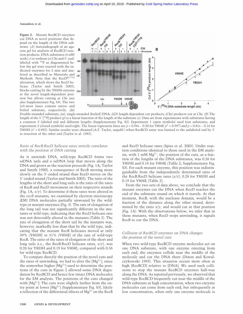
Wild-type RecBCD enzyme cuts DNA a few nucleotidesto the 3� side of the Chi sequence 5�-GCTGGTGG-3�(Taylor et al. 1985). When we tested the mutant enzymesfor this activity, we observed that they failed to cut atChi (Fig. 2A), as expected from the lack of Chi hot spotactivity in the mutants (Table 1). Instead, each mutantenzyme cut at a novel position, different for each mutantenzyme (Fig. 2A). Remarkably, these novel positions de-pended on the length of the DNA substrate. Eight sub-strates, each with the same 5�-labeled end and ranging inlength from 1.1 kb to 4.4 kb, were made and reacted witheach enzyme. The lengths of the novel 5�-labeled prod-ucts were determined by gel electrophoresis (Supplemen-tary Fig. S2).
The results of these experiments showed that thelength of the product fragment was a linear function ofthe length of the substrate for each mutant (Fig. 2B).Linear regression of the length of the product vs. thelength of the substrate gave a straight line with a slope of0.81 for the Y803H enzyme and 0.94 for the V804Eenzyme. Similar results were obtained with sub-strates whose nucleotide sequence was a circular per-mutation of that in Figure 2B, confirming that the novelcuts are not sequence dependent (A.F. Taylor, unpubl.).Our interpretation of these results will be clear after thenext section.
Table 2. RecBCD mutant enzymes retain exonuclease activity and cut DNA at novel positions strictly correlated with the ratioof the RecB:RecD helicase rates
RecBCD enzyme
dsExonuclease
activitya
ssExonuclease
activityb
Rate of helicase (bp/sec)c
Position ofnovel cuteRecB RecD
Ratiod
(RecB/RecD)
+ 100 100 316 574 0.56 ± 0.05 —f
RecBY803HCD 76 65 122 565 0.27 ± 0.03 0.260.30 ± 0.02 0.25
RecBV804ECD 53 49 97 600 0.19 ± 0.02 0.180.20 ± 0.01 0.18
RecBCDK177Q 11 ND 82 32 5.1 ± 2.8 —RecBY803HCDK177Q 56 ND 61 ND ND —RecBV804ECDK177Q 32 ND ND ND ND —
aPercentage of wild-type ds exonuclease-specific activity (9.2 × 10⁴ U/mg protein) under standard assay conditions but with 2 mMATP.bPercentage of wild-type ss exonuclease-specific activity (1.26 × 10⁵ U/mg protein) under standard assay conditions but with 2 mMATP.cThe rate of elongation of the short tail (by RecB) or the long tail (by RecD; the converse for RecBCDK177Q) in loop–tail unwindingstructures observed by EM (x and y, respectively, in Fig. 1A). For each rate, SEM was 5%–16% of the mean (n = 15–37, except 5 forRecBCD and 6 for RecBCDK177Q, previously analyzed by Taylor and Smith [2003]). Data from two experiments, on different days, areshown for RecBY803HCD and RecBV804ECD.dMean ± SEM of the ratio of the short to long tail lengths on individual molecules.eDistance of the novel cut from the 3� end at the RecBCD entry site divided by the length of the substrate in reactions with 5 mM ATPand 1 mM Mg2+ (see Fig. 2B; Supplementary S3).fNo novel cuts observed. RecBCD and RecBY803HCDK177Q cut at Chi (see Fig. 2A; Supplementary S4).(ND) Not determined.
Intersubunit signaling in RecBCD enzyme
GENES & DEVELOPMENT 3299
Cold Spring Harbor Laboratory Press on April 10, 2019 - Published by genesdev.cshlp.orgDownloaded from
Ratio of RecB:RecD helicase rates strictly correlateswith the position of DNA cutting
As it unwinds DNA, wild-type RecBCD forms twossDNA tails and a ssDNA loop that moves along theDNA and grows as the reaction proceeds (Fig. 1A; Taylorand Smith 1980), a consequence of RecB moving moreslowly on the 3�-ended strand than RecD moves on the5�-ended strand (Taylor and Smith 2003). The ratio of thelengths of the short and long tails is the ratio of the ratesof RecB and RecD movement on their respective strands(Fig. 1A, x/y). To determine if these rates were altered inthe recB mutants, we examined by electron microscopy(EM) DNA molecules partially unwound by the wild-type or mutant enzymes (Fig. 3). The rate of elongation ofthe long tail was not significantly different in the mu-tants or wild type, indicating that the RecD helicase ratewas not detectably altered in the mutants (Table 2). Therate of elongation of the short tail by the mutants was,however, markedly less than that by the wild type, indi-cating that the mutant RecB helicases moved at only39% (Y803H) or 31% (V804E) of the rate of wild-typeRecB. The ratio of the rates of elongation of the short andlong tails (i.e., the RecB:RecD helicase rates, x/y), was0.28 for Y803H and 0.19 for V804E, compared with 0.56for wild-type RecBCD.
To compare directly the position of the novel cuts andthe rates of unwinding, we had to alter the [Mg2+], sincethe somewhat higher [Mg2+] used to determine the posi-tions of the cuts in Figure 2 allowed some DNA degra-dation by RecBCD and hence few intact DNA moleculesfor the EM analysis. The positions of the cuts changedwith [Mg2+]: The cuts were slightly farther from the en-try point at lower [Mg2+] (Supplementary Fig. S3), likelya reflection of the differential effects of Mg2+ on the RecB
and RecD helicase rates (Spies et al. 2005). Under reac-tion conditions identical to those used in the EM analy-sis, with 1 mM Mg2+, the position of the cuts, as a frac-tion of the lengths of the DNA substrates, was 0.26 forY803H and 0.18 for V804E (Table 2; Supplementary Fig.S3). For each mutant enzyme, this position was indistin-guishable from the independently determined ratio ofthe RecB:RecD helicase rates (x/y), 0.28 for Y803H and0.19 for V804E (Table 2).
From the two sets of data above, we conclude that themutant enzymes cut the DNA when RecD reaches theend of the substrate strand on which it travels. At thatmoment, RecB, with the nuclease domain, would be afraction of the distance along the other strand, deter-mined by the ratio x/y, and would cut at that position(Fig. 1A). With the observations below, we infer that inthese mutants, when RecD stops unwinding, it signalsRecB to cut the DNA.
Collision of RecBCD enzymes on DNA changesthe position of the novel cuts
When two wild-type RecBCD enzyme molecules act onone DNA substrate, with one enzyme entering fromeach end, the enzymes collide near the middle of themolecule and cut the DNA there (Dixon and Kowal-czykowski 1993). This situation occurs most often athigh [RecBCD] relative to [DNA]. We used such colli-sions to stop the mutant RecBCD enzymes half-wayalong the DNA. As reported previously, we observed thatwild-type RecBCD frequently cut near the middle of theDNA substrate at high concentration, when two enzymemolecules can come from each end, but infrequently atlow concentration, when only one enzyme molecule is
Figure 2. Mutant RecBCD enzymescut DNA at novel positions that de-pend on the length of the DNA sub-strate. (A) Autoradiograph of an aga-rose gel for analysis of RecBCD reac-tion products. DNA substrates (4 nM)with (+) or without (o) Chi and 5�-end-labeled with 32P as diagrammed be-low the gel were reacted with the in-dicated enzymes for 2 min and ana-lyzed as described in Materials andMethods. Note that the RecDK177Q
alteration, which slows the RecD he-licase (Taylor and Smith 2003),blocks cutting by the Y803H enzymeat the novel length-dependent posi-tion but allows cutting at Chi (seealso Supplementary Fig. S4). The twoleft-most lanes contain native andboiled substrate, respectively. (ds)Double-stranded substrate; (ss), single-stranded (boiled) DNA; (LD) length-dependent cut products; (Chi) products cut at Chi. (B) Thelength of the 5� [32P] product (p) is a linear function of the length of the substrate (s). Data are from experiments with substrates havinga common 5�-labeled end and different lengths (Supplementary Fig. S2). Experiment 1 (open symbols) used four substrates, andexperiment 2 (closed symbols) used eight. The linear regression lines are p = 0.94s − 0.20 for V804E (r2 = 0.997) and p = 0.81s − 0.16 forY803H (r2 = 0.992). Similar results were obtained (A.F. Taylor, unpubl.) when RecBCD entry was limited to the unlabeled end by 3�
ss resection of the other end (Taylor et al. 1985).
Amundsen et al.
3300 GENES & DEVELOPMENT
Cold Spring Harbor Laboratory Press on April 10, 2019 - Published by genesdev.cshlp.orgDownloaded from

present on the DNA (Fig. 4). Instead, at low concentra-tion, wild-type RecBCD cut at Chi, which was locatedbeyond the mid-point as RecBCD approached Chi in theactive orientation.
As shown above (Fig. 2A), the mutant enzymes did notcut at Chi, but as predicted by our hypothesis, they cutcloser to the entry point at high enzyme concentrationthan at low concentration (Fig. 4). On three differentlength substrates, the distance from the entry site to thecut site was about one-half of the distance at low enzymeconcentration. Collision at the midpoint of the DNA isthus equivalent to using a half-length DNA substrate.These results show that the position of the novel cut isaltered when the distance that the enzyme travels is al-tered. Since RecD is the faster helicase (Table 2; Taylorand Smith 2003), we infer that termination of RecD’stravel induces RecD to signal cutting by the RecB sub-unit. (It is not clear why wild-type RecBCD at high con-centration cuts in the middle of the substrate rather thanat one-half of its characteristic x/y ratio of RecB:RecD
helicase rates. Failure to cut there may be related to itsfailure to cut at low concentration at any point in theabsence of Chi [Fig. 2A].)
Slowing RecD helicase abolishes cutting at novelpositions and can revive Chi cutting
To test more directly the hypothesis that the mutantenzymes cut DNA when RecD translocation terminatesat the end of its substrate strand, we coupled therecD2177 (K177Q) mutation with the new recB2732(Y803H) or recB2734 (V804E) mutation to make doubly
Figure 4. The position of the novel length-dependent cutchanges when two mutant RecBCD enzymes collide. Productsof reactions with the DNA substrate (0.5 nM) diagrammed be-low the gel were analyzed by gel electrophoresis. At high mu-tant RecBCD concentration (four RecBCD molecules per DNAmolecule), the novel cuts (bullets on gel and thick arrows ondiagram) are closer to the RecBCD entry site than at lowRecBCD concentration (one RecBCD molecule per DNA mol-ecule; open circles on gel and thin arrows on diagram). Note thatwild-type RecBCD cuts at Chi at low concentration but in themiddle of the substrate at high concentration. Markers in theleft-most lane are boiled (ss) samples of the [5�-32P] DNA sub-strate and subfragments of it. These reactions did not containSSB. Data at the bottom are the lengths of the cut products as afraction of the length of three different [5�-32P]-labeled sub-strates at low and high enzyme concentration; only data for the4.36-kb substrate are shown here. In each case, cutting occurscloser to the entry site at high enzyme concentration.
Figure 3. Mutant RecBCD enzymes make ss loop–tail struc-tures during unwinding similar to those of wild-type RecBCDenzyme. Phage � DNA was reacted with the indicated enzymesfor the stated time, fixed, and examined by EM. ssDNA is boundby SSB protein and appears thicker than dsDNA. Bar is 0.5 µm(∼1.4 kb of dsDNA; ∼4.8 kb of ssDNA) and applies to all panels.For G, the substrate was cut with a restriction enzyme to pro-duce a blunt end.
Intersubunit signaling in RecBCD enzyme
GENES & DEVELOPMENT 3301
Cold Spring Harbor Laboratory Press on April 10, 2019 - Published by genesdev.cshlp.orgDownloaded from

mutant RecBCD enzymes. The recD2177 (K177Q) mu-tation, in the ATPase site, slows RecD to 32 nucleotides(nt) per second, ∼5% of the wild-type rate (Taylor andSmith 2003). For the Y803H and Y804E singly mutantenzymes, the rates of RecB-mediated unwinding are 122and 97 base pairs (bp) per second, respectively (Table 2).Consequently, in the doubly mutant enzymes, RecB isexpected to move more rapidly than RecD; RecB wouldreach the end of the substrate before RecD, and the sig-nal to cut the DNA might not be generated. As predicted,the novel length-dependent cuts made by the single recBmutant enzymes were not detectably made by the dou-bly mutant enzymes (Fig. 2A; Supplementary Fig. S4).Less than 2% of the DNA was cut by the doubly mutantenzymes, whereas ∼35% of the DNA was cut by thesingly mutant enzymes. These results indicate that theRecD subunit is involved in generating the novel length-dependent cuts on DNA.
Remarkably, altering the RecD subunit, in theRecBY803HCDK177Q enzyme, restored Chi-dependentcuts (Fig. 2A). This is consistent with cells bearing thisdoubly mutant enzyme showing Chi hot spot activity(Table 1) and with RecBY803H and likely RecC encoun-tering Chi before the very slow RecDK177Q subunit getsto the end of the DNA (Table 2). The RecBV804ECDK177Q
enzyme did not cut at Chi (Supplementary Fig. S4), con-sistent with the lack of Chi hot spot activity in cellswith this doubly mutant enzyme (Table 1).
Mutant enzymes do not load RecA onto the novel cutproducts
After encountering Chi, wild-type RecBCD loads RecAprotein onto the newly generated 3� end (Anderson andKowalczykowski 1997b). To determine if this Chi-de-pendent alteration occurs in the mutant enzymes afterthey generate their length-dependent cuts, we testedRecA loading onto DNA substrates with or without Chi.Each mutant RecBCD enzyme made the expected novelcut fragments, but in neither case was there detectableloading of RecA onto this product (Supplementary Fig.S5). RecA loading was measured by resistance of the cutproduct to digestion by exonuclease I, which is specificfor 3� ssDNA ends and is inhibited by RecA protein onthe DNA. The failure to load RecA can account for theRec− phenotype of these mutants (Table 1), the basis oftheir isolation.
Discussion
We describe here novel mutant RecBCD enzymes whosebehavior, both in cells and when purified, suggests thatthe RecD subunit signals the RecB subunit to cut DNA.These results lead to a new hypothesis of how Chi sitesregulate wild-type RecBCD enzyme, the complex pro-tein machine that initiates the major pathway for DSBrepair and homologous recombination in E. coli. Below,we discuss this hypothesis, support for it, and tests of thehypothesis.
Amino acid substitutions in helicase motif VI slowRecB and confer a novel DNA cutting activitydependent on the substrate length
The two mutants described here change two highly con-served amino acids in helicase motif VI, whose consensussequence in 39 bacterial RecB proteins is RLLYVA-TR,where “-” is a not-well-conserved amino acid. In E. coli’sRecB, this sequence is RLLYVALTR; the mutants de-scribed here are altered in the amino acids underlined. InRecBCD this sequence is part of a 24-amino-acid-long�-helix that does not appear to contact DNA during un-winding but lies close and parallel to another short helixthat likely does (Singleton et al. 2004). Studies of mu-tants with alterations in motif VI in several superfamilyI helicases suggest that amino acids in this motif arerequired to couple ATP hydrolysis to DNA movement(e.g., Graves-Woodward et al. 1997). In one case, E. coliUvrD (helicase II), the T → A amino acid alteration inthis motif changes the conformation of the protein, asindicated by increased sensitivity to limited proteolysis(Hall et al. 1998). Thus, this long �-helix may be impor-tant in transducing information, via a conformationalchange, between the ATPase and helicase active sites.This interpretation is consistent with the increased ap-parent KM for ATP and the decreased RecB helicase ratesin the RecBCD mutants studied here (Table 2; Supple-mentary Fig. S1). We suggest that in wild-type RecBCD,this helix is important also for the transduction of theChi-dependent signal that alters the activities of the en-zyme after acting at Chi (Taylor and Smith 1992).
The mutations studied here impart a novel phenotypeto RecBCD—the ability to determine a fraction of thelength of the DNA substrate and to cut the DNA at thatpoint. The simplest interpretation is that this “calcula-tion” reflects the ratio of the rates of movement of theRecB and RecD helicases (Fig. 1A, x/y; Table 2). In themutants, the RecD helicase moved at the same rate as itdoes in wild-type RecBCD, but the RecB helicase wasslower than that in wild type, 31% of the wild-type ratefor V804E and 39% for Y803H. The near equality of theRecB:RecD ratio and the position of the cut for eachenzyme (Table 2) supports this simple interpretation.Furthermore, since RecB has the nuclease domain (Yu etal. 1998b) and the position of the cut is indistinguishablefrom the point at which RecB’s nuclease domain wouldbe when RecD reaches the end of the DNA, we concludethat cutting, by RecB, is induced when RecD reaches theend of the DNA and stops unwinding DNA.
We tested the interpretation that the mutant enzymescut DNA when RecD stops unwinding in two ways. (1)Introduction of the recD2177 (K177Q) mutation in theRecD ATP site (Korangy and Julin 1992) slows the RecDhelicase to ∼5% of the wild-type RecD rate (Taylor andSmith 2003). This rate is slower than that of theRecBY803H or RecBV804E mutant subunit (Table 2). Thus,in the doubly mutant enzymes RecB is expected to reachthe DNA end before RecD does. As predicted, these dou-bly mutant enzymes did not cut at the novel length-dependent position (Fig. 2A; Supplementary Fig. S4). (2)
Amundsen et al.
3302 GENES & DEVELOPMENT
Cold Spring Harbor Laboratory Press on April 10, 2019 - Published by genesdev.cshlp.orgDownloaded from
When two wild-type RecBCD molecules simultaneouslyunwind a DNA molecule, one from each end of theDNA, the enzymes cut when they collide near themiddle of the DNA (Dixon and Kowalczykowski 1993).When the new mutant enzymes were similarly tested,cutting occurred at approximately one-half the distancefrom the entry end to the position of the novel cut ob-served at low enzyme concentration, when only one en-zyme molecule is present on the DNA (Fig. 4). Thus,whether RecD stops at the end of the DNA or upon col-lision with another RecBCD molecule, cutting is in-duced at the position where RecB is expected to be lo-cated.
Wild-type RecBCD generates a 3� DNA end a fewnucleotides 3� of the Chi sequence. With excess ATP,this occurs by a simple nick (Taylor et al. 1985), whereaswith excess Mg2+, degradation of the 3�-ended strandceases at or near Chi (Dixon and Kowalczykowski 1993;Taylor and Smith 1995b). The mutants studied here ap-pear to generate 3� ends in a similar manner under thesetwo reaction conditions (Fig. 2; Supplementary Fig. S6;S.K. Amundsen and A.F. Taylor, unpubl.) but at novellength-dependent positions. We suppose that the basicmechanism that induces the cut is the same as that inwild-type RecBCD but that the signal for this inductionis different, as discussed below. Although new 3� ssDNAends were produced by both the mutant and wild-typeenzymes, the mutants were recombination deficient(Table 1). This deficiency is likely due to the mutants’inability to load RecA protein onto the newly generated3� end (Supplementary Fig. S5). Thus, the mutantsmimic only part of the change at Chi—they cut DNA butdo not load RecA.
Our results indicate that in the mutant enzymes,RecD signals RecB to cut the DNA. Below, we extendthis explanation into a new hypothesis for how, in wild-type enzyme, Chi signals RecB to cut the DNA and toload RecA protein to initiate strand exchange.
A ‘signal transduction cascade’ hypothesis for Chi’sregulation of wild-type RecBCD enzyme
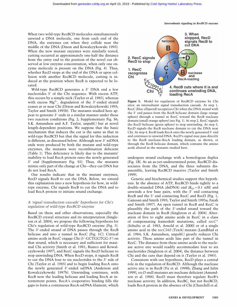
Based on these and other observations, especially theRecBCD crystal structure and its interpretation (Single-ton et al. 2004), we propose the following hypothesis forChi’s regulation of wild-type RecBCD enzyme (Fig. 5).The 3�-ended strand of DNA passes through the RecBhelicase and into a tunnel in RecC (Fig. 1C). Criticalamino acids in RecC engage Chi (5�-GCTGGTGG-3�) onthat strand, which is necessary and sufficient for maxi-mal Chi activity (Smith et al. 1981; Bianco and Kowal-czykowski 1997), and RecC signals the RecD helicase tostop unwinding DNA. When RecD stops, it signals RecBto cut the DNA four to six nucleotides to the 3� side ofChi (Taylor et al. 1985) and to begin loading RecA ontothe newly generated 3�-ended ssDNA (Anderson andKowalczykowski 1997b). Unwinding continues, withRecB now the leading helicase and loading RecA at in-termittent points. RecA’s cooperative binding fills thegaps to form a continuous RecA-ssDNA filament, which
undergoes strand exchange with a homologous duplex(Fig. 1B). At an as-yet-undetermined point, RecBCD dis-sociates from the DNA, and the three subunits dis-assemble, leaving RecBCD inactive (Taylor and Smith1999).
Genetic and biochemical studies support this hypoth-esis. In the absence of ATP, RecBCD binds tightly to adouble-stranded DNA (dsDNA) end (KD ≈ 0.1 nM) andunwinds a few base pairs, with the 3� end contactingRecB and the 5� end contacting RecC and RecD (Fig. 1;Ganesan and Smith 1993; Taylor and Smith 1995a; Farahand Smith 1997). An open tunnel in RecB and RecC isplausibly the path of the 3�-ended strand toward thenuclease domain in RecB (Singleton et al. 2004). Alter-ation of five to eight amino acids in RecC in a classof compensating frameshift mutations, called recC*(Schultz et al. 1983; Arnold et al. 2000) or of a singleamino acid in the recC343 (TexA) mutant (Lundblad etal. 1984; S.K. Amundsen, unpubl.) greatly reduces Chiactivity. These amino acids line part of the tunnel inRecC. The distance from these amino acids to the nucle-ase active site would readily accommodate four to sixnucleotides (Singleton et al. 2004), the distance betweenChi and the cuts that depend on it (Taylor et al. 1985).
Consistent with our hypothesis, RecD plays a centralrole in the regulation of RecBCD. Although the nucleaseactive site is in RecB (Yu et al. 1998b; Zhang and Julin1999), recD-null mutants are nuclease deficient (Amund-sen et al. 1986). RecD must therefore regulate RecB’snuclease activity. In addition, RecBC, but not RecBCD,loads RecA protein in the absence of Chi (Churchill et al.
Figure 5. Model for regulation of RecBCD enzyme by Chisites: an intersubunit signal transduction cascade. At step 1,RecC (blue ellipsoid) recognizes Chi when the DNA strand withthe 3� end passes from the RecB helicase domain (large orangesphere) through a tunnel in RecC toward the RecB nucleasedomain (small orange sphere) (see Fig. 1). At step 2, RecC signalsthe RecD helicase (green sphere) to stop unwinding. At step 3,RecD signals the RecB nuclease domain to cut the DNA nearChi. At step 4, RecB loads RecA onto the newly generated 3� endand continues to unwind DNA. RecD’s signal may pass directlyto the RecB nuclease–RecA loading domain, as shown, orthrough the RecB helicase domain, which contains the aminoacids altered in the mutants studied here.
Intersubunit signaling in RecBCD enzyme
GENES & DEVELOPMENT 3303
Cold Spring Harbor Laboratory Press on April 10, 2019 - Published by genesdev.cshlp.orgDownloaded from
1999); RecD must therefore inhibit the loading of RecA,which may be directed by part of RecB (Spies and Kowal-czykowski 2006). recB mutants altered in the nucleaseactive site are Rec− and do not load RecA unless theRecD subunit is removed (Anderson et al. 1999; Amund-sen et al. 2000). It is therefore plausible that the Chisignal, which alters the nuclease and RecA loading ac-tivities, is transmitted via RecD. In the crystal structure,the ordered part of RecD does not contact RecB (Fig. 1C;Singleton et al. 2004) but does come within ∼0.8 nm ofthe RecB nuclease domain, which may overlap the RecAloading domain (Spies and Kowalczykowski 2006). Wepropose that Chi alters the conformation of RecC, whichdoes contact RecD, and moves RecD against the RecBnuclease domain, thereby altering its activity. Alterna-tively, RecD may contact RecB during unwinding and,after receiving the Chi signal from RecC, alter the RecBhelicase domain, which in turn affects the RecB nucleasedomain via the ∼30-amino-acid-long tether connectingthese domains (Fig. 1C); this possibility is consistentwith our interpretation of the recB helicase domain mu-tants described in this study.
Studies of single RecBCD molecules by fluorescencemicroscopy also support this hypothesis. Unwinding ofthe DNA duplex pauses at or near Chi, and the length ofthe pause is proportional to the distance of the Chi sitefrom the DNA end at which RecBCD initiated unwind-ing (Spies et al. 2003). We interpret these results to meanthat RecD, the leading helicase before Chi (Taylor andSmith 2003), stops unwinding at or near Chi, in accordwith our hypothesis (Fig. 5). RecB continues, perhapswithout any pause, to travel along the ss loop for a timeproportional to the loop length (i.e., also proportional tothe distance of Chi from the DNA end) (Fig. 1A). Aftertraversing the loop, RecB becomes the leading (unwind-ing) helicase after Chi but at a rate slower than RecD wasbefore Chi, as observed (Spies et al. 2003). We supposethat the slowing or elimination of the RecD helicasereflects a conformational change as part of the Chi signaltransduction.
Our hypothesis is distinct from previous hypotheses ofhow Chi affects RecBCD enzyme. According to one hy-pothesis (Thaler et al. 1988), RecD is ejected at Chi. Earlyenzymatic studies argued against this possibility, how-ever: After acting at Chi, the enzyme retains nucleaseactivity (Taylor and Smith 1992), whereas RecBC (i.e.,without RecD) lacks nuclease activity (Amundsen et al.1986). Furthermore, subsequent light microscopy studiesof single RecBCD molecules showed that RecD remainswith the enzyme after Chi (Dohoney and Gelles 2001;Handa et al. 2005). According to another hypothesis (Yuet al. 1998a), the RecB nuclease domain “swings” fromone side of the enzyme, where it digests the 3� → 5�strand, to the other side, where it digests the 5� → 3�strand. Singelton et al. (2004) modified this view andhypothesized that at Chi the 3� → 5� strand, the one withChi, moves from a channel in RecC aimed toward thenuclease site into another channel aimed away from thenuclease site. This change might be effected by a RecB�-helix swinging to block the first channel. This hypoth-
esis is consistent with ours, which in addition specifieshow Chi effects this and other changes in the enzyme.
Although aspects of our hypothesis (Fig. 5) are specu-lative and require further support, it is consistent withcurrent observations, as noted above, and makes testablepredictions. Specific mutations in each gene should dis-rupt the signal transduction cascade. In addition to therecC* mutations that appear to abolish the Chi–RecCinteraction (see above), there should be mutant forms ofRecC that cannot transmit the signal to RecD, mutantforms of RecD that cannot receive the signal from RecCor that cannot transmit the signal to RecB, and mutantforms of RecB that cannot receive the signal from RecD.Previously described recC and recB mutations may cor-respond to these classes: recC2145, recB2154, andrecB2155 are Rec− Nuc+ Chi−, like the mutants describedhere, and may be unable to transmit or receive the Chisignal (Amundsen et al. 1990). Our model predicts a classof recD mutations that cannot receive or transmit thesignal and would be Rec−. Such mutations have not beenreported to date, but recD-null mutations are Rec+ Nuc−
Chi−, as predicted if Chi annuls the regulatory roles ofRecD (Amundsen et al. 2000). We suppose that the trans-duction of the Chi signal involves conformationalchanges in each of the RecBCD subunits; such changesmight be detectable by limited proteolysis or spectros-copy of fluorescently labeled subunits.
Intersubunit signaling in other complex proteinmachines
The conceptual model of intersubunit signaling, such asthat proposed here for RecBCD, may be applicable to abroad range of complex protein machines. Particularlyrelevant here are two examples of enzymes with threetypes of subunits and multiple activities on DNA, likeRecBCD. (1) Mismatch correction in E. coli depends onthe MutS, MutH, and MutL proteins. MutS binds to mis-matched bases in DNA, the MutH latent endonucleasebinds to a distant hemimethylated DNA site, and MutLappears to connect MutS and MutH (Iyer et al. 2006). TheMutH nuclease is activated by MutS and MutL in thepresence of ATP and a mismatch. The mechanism of theactivation is unclear but likely involves transduction ofa signal from MutS to MutH, perhaps via MutL. (2) TypeI restriction enzymes bind to a specific DNA sequence,travel along the DNA, and cut at a distant site whentravel stops, due to collision between two enzymes or astructural constraint in the DNA (Murray 2000). In theseenzymes, the HsdS subunit binds a specific DNA se-quence but is aided by the HsdM subunit, which con-tains the methyltransferase domain; the HsdR subunitcontains the endonuclease domain. If the DNA sequenceis hemimethylated, the HsdM subunit acts before travelis initiated, but if the sequence is unmethylated, travelcommences and the HsdR subunit acts. Signaling be-tween the subunits therefore must regulate modificationversus restriction. Another related example is the type IIDNA topoisomerases, in which there appears to be sig-naling between the ATPase site in one subunit and the
Amundsen et al.
3304 GENES & DEVELOPMENT
Cold Spring Harbor Laboratory Press on April 10, 2019 - Published by genesdev.cshlp.orgDownloaded from

DNA breaking–rejoining “gate” in another subunit(Bates and Maxwell 2007). Mutational alterations ofthese proteins may help elucidate the putative inter-subunit signal transduction, as reported here for RecBCDenzyme.
Materials and methods
Bacterial strains, phage, and plasmids
Bacterial strains are listed in Supplementary Table S1 with theirgenotypes and sources. Plasmids are listed in SupplementaryTable S2. For visual clarity, allele numbers and polypeptide des-ignations are expressed as superscripts when more than onerecBCD gene or RecBCD polypeptide are designated. Bacterialstrains were constructed by phage P1 transduction, CaCl2-me-diated transformation, electroporation, or “recombineering”(Ausubel et al. 2003; Thomason et al. 2005). Plasmids were con-structed by standard procedures (Ausubel et al. 2003).
Culture media and genetic assays
Culture media have been described (Cheng and Smith 1989).Chi hot spot activity and recombination proficiency were mea-sured in � vegetative crosses (Stahl and Stahl 1977), and E. colirecombination proficiency in Hfr crosses (Schultz et al. 1983).
Mutant isolation
Mutations in the C-terminal part of recB were generated bymutagenic PCR (Ausubel et al. 2003). Two primer pairs wereused—one amplifying codons 712–1181, and the other paircodons 794–1181 (Supplementary Table S3); recB has 1181codons including that for termination. Each PCR contained, in100 µL, 20 fmol of plasmid pDWS2 DNA, 30 pmol of eachprimer, 10 mM Tris-HCl (pH 8.3), 50 mM KCl, 0.5 mM MnCl2,0.01% gelatin, 1 mM dCTP and dTTP, 0.2 mM dATP and dGTP,and 5 U of Taq polymerase (Boehringer Mannheim Biochemi-cals) and ran for 30 cycles (1 min at 94°C, 1 min at 50°C, and1 min at 72°C). The PCR products were digested with BglII,which cleaves between recB codons 797 and 798, and withBseRI, which cleaves four nucleotides after the recB termina-tion codon. The largest fragment (1.1 kb) was “swapped” withthe corresponding recB+ fragment of pMR3. Approximately5000 ampicillin-resistant (AmpR) transformants of strain V2959[�recBCD2731�kan �(lacIZYA–argF)U169] were screened forrecombination deficiency (Rec−) by toothpick transfer of colo-nies to minimal lactose agar plates containing ampicillin (100µg/mL) and kanamycin (25 µg/mL) and spread with ∼108 sta-tionary-phase cells of strain KL226 (lac+ Hfr PO 12.2). Approxi-mately 1000 Rec− colonies (those unable to generate Lac+ AmpR
KanR recombinant colonies) were tested by cross-streaking colo-nies from an LB-ampicillin master plate onto LB agar with phageP2 (applied as lines of ∼20 µL of 108 phage per milliliter); growthof P2 appears to require RecBCD exonuclease (Nuc+) (Amund-sen et al. 1990). (Many of the Rec− Nuc− isolates had plasmidswithout the BglII–BseRI fragment, reflecting inefficient swap-ping.) Lawns of ∼200 Rec− Nuc+ candidates were tested by spottests for growth of � �(red–gam) �+ and �o phages, which do notmake plaques on Rec− Nuc+ strains, and of phage P1, which doesnot make plaques on Rec− strains (Schultz et al. 1983; Amund-sen et al. 1990, 2000). Plasmids from ∼50 stable Rec− Nuc+ can-didates were isolated and introduced into strain V2831(�recBCD2731�kan); these transformants were tested quanti-tatively as in Table 1. The recB nucleotide sequence was deter-
mined for 11 of the most Rec− candidates. The recB2732(Y803H) and recB2734 (V804E) mutations were introduced intoplasmid pSA124 using the QuikChange kit (Stratagene) andmutant oligonucleotides (Supplementary Table S3). These mu-tations were transferred to plasmid pMR3 by fragment swap-ping as described above. Double recB recD mutants were con-structed by site-directed mutagenesis of pMR3 to introduce therecD2177 mutation, followed by swapping of the BseRI–BglIIfragment from pSA176 or pSA178 to introduce the recB2732 orrecB2734 mutation.
Enzyme purification and assays
The purification of wild-type RecBCD enzyme has been de-scribed (Taylor and Smith 2003). Mutant RecBCD enzymeswere purified from strain V2831 (�recBCD2731) containingderivatives of pMR3 by similar methods. In brief, cells from a12 L of culture in Terrific Broth (Fisher) were lysed, and en-zymes were purified by column chromatography—HiTrap QSepharose, HiPrep Sephacryl S-300 HR, and HiTrap Heparin (allfrom GE Lifesciences), followed by CHTII hydroxyapatite (Bio-Rad) for the single mutants or ssDNA agarose (GE Lifesciences)for the double mutants. The final product (∼1 mg) was judged tobe ∼80% pure by staining with SimplyBlue (Invitrogen) an SDS–polyacrylamide gel loaded with 0.5 µg of protein.
Assays for RecBCD ds and ss exonuclease used native andboiled [3H] T7 DNA, respectively (Eichler and Lehman 1977).Gel-electrophoretic assays for DNA unwinding, Chi cutting,and RecA loading were as described (Taylor et al. 1985; Amund-sen et al. 2000; Taylor and Smith 2003) with 5 mM ATP, 3 mMMg(OAc)2, and 1 µM SSB (Promega), except as noted in Figure 4and Supplementary Figures S3 and S5. Agarose gels (0.7%; 22cm long) in TBE buffer (Ausubel et al. 2003) were run at roomtemperature for 2.5 h at 100 V (Fig. 2A; Supplementary Figs. S4,S5) or for ∼16 h at ∼50 V (Figs. 2B, 4; Supplementary Figs. S2, S3).Analysis of Typhoon Trio PhosphorImage files (GE Lifesciences)used ImageQuant TL software (Amersham); size markers werefit to a log-linear straight line (r2 > 0.997). EM assays for DNAunwinding (Taylor and Smith 2003) contained 5 mM ATP,1 mM Mg2+, and 1 µM SSB; rates were calculated from mol-ecules whose complementary unwound strands differed inlength by <33%.
For the experiments in Figures 2A and 4 and SupplementaryFigures S4 and S5, DNA substrates were prepared from pBR322�+F or �o (4361 bp) by cutting with HindIII, treating with phos-phatase, labeling the 5� ends using polynucleotide kinase and[�-32P] ATP (Amundsen et al. 2000), and cutting with ClaI,which produces a 4355-bp fragment with one of the two 32Plabels and two short fragments (five and seven nucleotides) notseen in our analyses. For the experiments in Figure 2B andSupplementary Figures S2 and S3, the DNA substrates weresimilarly prepared from pBR322 �o DNA by cutting with StyI,labeling the 5� ends, cutting with BsmI, and separating the 4351-bp fragment from a short fragment using an S200 spin column.Subfragments for RecBCD reactions and for size markers wereproduced by subsequent digestion of the 4351-bp fragment withNruI, SalI, HindIII, PvuI, AlwNI, AflIII, NdeI, or Tth111I.
Acknowledgments
We thank Harvey Eisen, Martin Gellert, Paul Modrich, and Nor-een Murray for helpful discussions of nucleic acid enzymes, andGareth Cromie and Luther Davis for helpful comments on themanuscript. This research was supported by research grantGM031693 from the National Institutes of Health to G.R.S.,
Intersubunit signaling in RecBCD enzyme
GENES & DEVELOPMENT 3305
Cold Spring Harbor Laboratory Press on April 10, 2019 - Published by genesdev.cshlp.orgDownloaded from

and institutional support from the Division of Basic Sciences ofthe Fred Hutchinson Cancer Research Center.
References
Alberts, B. 1998. The cell as a collection of protein machines:Preparing the next generation of molecular biologists. Cell92: 291–294.
Amundsen, S.K. and Smith, G.R. 2007. Chi hotspot activity inEscherichia coli without RecBCD exonuclease activity: Im-plications for the mechanism of recombination. Genetics176: 41–54.
Amundsen, S.K., Taylor, A.F., Chaudhury, A.M., and Smith,G.R. 1986. recD: The gene for an essential third subunit ofexonuclease V. Proc. Natl. Acad. Sci. 83: 5558–5562.
Amundsen, S.K., Neiman, A.M., Thibodeaux, S.M., and Smith,G.R. 1990. Genetic dissection of the biochemical activitiesof RecBCD enzyme. Genetics 126: 25–40.
Amundsen, S.K., Taylor, A.F., and Smith, G.R. 2000. The RecDsubunit of the Escherichia coli RecBCD enzyme inhibitsRecA loading, homologous recombination and DNA repair.Proc. Natl. Acad. Sci. 97: 7399–7404.
Amundsen, S.K., Taylor, A.F., and Smith, G.R. 2002. A domainof RecC required for assembly of the regulatory RecD sub-unit into the Escherichia coli RecBCD holoenzyme. Genet-ics 161: 483–492.
Anderson, D.G. and Kowalczykowski, S.C. 1997a. The recom-bination hot spot � is a regulatory element that switches thepolarity of DNA degradation by the RecBCD enzyme. Genes& Dev. 11: 571–581.
Anderson, D.G. and Kowalczykowski, S.C. 1997b. The translo-cating RecBCD enzyme stimulates recombination by direct-ing RecA protein onto ssDNA in a � regulated manner. Cell90: 77–86.
Anderson, D.G., Churchill, J.J., and Kowalczykowski, S.C.1999. A single mutation, RecBD1080A, eliminates RecA pro-tein loading but not Chi recognition by RecBCD enzyme. J.Biol. Chem. 274: 27139–27144.
Arnold, D.A., Handa, N., Kobayashi, I., and Kowalczykowski,S.C. 2000. A novel, 11 nucleotide variant of �, �*: One of aclass of sequences defining the Escherichia coli recombina-tion hotspot �. J. Mol. Biol. 300: 469–479.
Ausubel, F.M., Brent, R., Kingston, R.E., Moore, D.D., Seidman,J.G., Smith, J.A., and Struhl, K., eds. 2003. Current protocolsin molecular biology. John Wiley & Sons, New York.
Bates, A.D. and Maxwell, A. 2007. Energy coupling in Type IItopoisomerases: Why do they hydrolyze ATP? Biochemistry46: 7929–7941.
Bianco, P.R. and Kowalczykowski, S.C. 1997. The recombina-tion hotspot � is recognized by the translocating RecBCDenzyme as the single strand of DNA containing the sequence5�-GCTGGTGG-3�. Proc. Natl. Acad. Sci. 94: 6706–6711.
Cheng, K.C. and Smith, G.R. 1989. Distribution of Chi-stimu-lated recombinational exchanges and heteroduplex end-points in phage �. Genetics 123: 5–17.
Churchill, J.J., Anderson, D.G., and Kowalczykowski, S.C.1999. The RecBC enzyme loads RecA protein onto ssDNAasymmetrically and independently of � resulting in consti-tutive recombination activation. Genes & Dev. 13: 901–911.
Dixon, D.A. and Kowalczykowski, S.C. 1991. Homologous pair-ing in vitro stimulated by the recombination hotspot, Chi.Cell 66: 361–371.
Dixon, D.A. and Kowalczykowski, S.C. 1993. The recombina-tion hotspot � is a regulatory sequence that acts by attenu-ating the nuclease activity of the E. coli RecBCD enzyme.
Cell 73: 87–96.Dohoney, K.M. and Gelles, J. 2001. �-sequence recognition and
DNA translocation by single RecBCD helicase/nucleasemolecules. Nature 409: 370–374.
Eichler, D.C. and Lehman, I.R. 1977. On the role of ATP inphosphodiester bond hydrolysis catalyzed by the RecBC de-oxyribonuclease of Escherichia coli. J. Biol. Chem. 252: 499–503.
Farah, J.A. and Smith, G.R. 1997. The RecBCD enzyme initia-tion complex for DNA unwinding: Enzyme positioning andDNA opening. J. Mol. Biol. 272: 699–715.
Ganesan, S. and Smith, G.R. 1993. Strand-specific binding toduplex DNA ends by the subunits of Escherichia coliRecBCD enzyme. J. Mol. Biol. 229: 67–78.
Graves-Woodward, K.L., Gottlieb, J., Challberg, M.D., andWeller, S.K. 1997. Biochemical analyses of mutations in theHSV-1 helicase–primase that alter ATP hydrolysis, DNA un-winding, and coupling between hydrolysis and unwinding. J.Biol. Chem. 272: 4623–4630.
Hall, M.C., Ozsoy, A.Z., and Matson, S.W. 1998. Site-directedmutations in motif VI of Escherichia coli DNA helicase IIresult in multiple biochemical defects: Evidence for the in-volvement of motif VI in the coupling of ATPase and DNAbinding activities via conformational changes. J. Mol. Biol.277: 257–271.
Handa, N., Bianco, P.R., Baskin, R.J., and Kowalczykowski, S.C.2005. Direct visualization of RecBCD movement reveals co-translocation of the RecD motor after Chi recognition. Mol.Cell 17: 745–750.
Iyer, R.R., Pluciennik, A., Burdett, V., and Modrich, P.L. 2006.DNA mismatch repair: Functions and mechanisms. Chem.Rev. 106: 302–323.
Korangy, F. and Julin, D.A. 1992. Alteration by site-directedmutagenesis of the conserved lysine residue in the ATP-binding consensus sequence of the RecD subunit of theEscherichia coli RecBCD enzyme. J. Biol. Chem. 267: 1727–1732.
Lundblad, V., Taylor, A.F., Smith, G.R., and Kleckner, N. 1984.Unusual alleles of recB and recC stimulate excision of in-verted repeat transposons Tn10 and Tn5. Proc. Natl. Acad.Sci. 81: 824–828.
Murray, N.E. 2000. Type I restriction systems: Sophisticatedmolecular machines (a legacy of Bertani and Weigle). Micro-biol. Mol. Biol. Rev. 64: 412–434.
Oliver, D.B. and Goldberg, E.B. 1977. Protection of parental T4DNA from a restriction exonuclease by the product of gene2. J. Mol. Biol. 116: 877–881.
Schultz, D.W., Taylor, A.F., and Smith, G.R. 1983. Escherichiacoli RecBC pseudorevertants lacking Chi recombinationalhotspot activity. J. Bacteriol. 155: 664–680.
Singleton, M.R., Dillingham, M.S., Gaudier, M., Kowalczykow-ski, S.C., and Wigley, D.B. 2004. Crystal structure ofRecBCD enzyme reveals a machine for processing DNAbreaks. Nature 432: 187–193.
Smith, G.R. 1990. RecBCD enzyme. In Nucleic acids and mo-lecular biology (eds. F. Eckstein, and D.M.J. Lilley), pp. 78–98. Springer-Verlag, Berlin.
Smith, G.R. 1991. Conjugational recombination in E. coli:Myths and mechanisms. Cell 64: 19–27.
Smith, G.R. 2001. Homologous recombination near and far fromDNA breaks: Alternative roles and contrasting views. Annu.Rev. Genet. 35: 243–274.
Smith, G.R., Kunes, S.M., Schultz, D.W., Taylor, A., and Tri-man, K.L. 1981. Structure of Chi hotspots of generalized re-combination. Cell 24: 429–436.
Spies, M. and Kowalczykowski, S.C. 2006. The RecA binding
Amundsen et al.
3306 GENES & DEVELOPMENT
Cold Spring Harbor Laboratory Press on April 10, 2019 - Published by genesdev.cshlp.orgDownloaded from

locus of RecBCD is a general domain for recruitment ofDNA strand exchange proteins. Mol. Cell 21: 573–580.
Spies, M., Bianco, P.R., Dillingham, M.S., Handa, N., Baskin,R.J., and Kowalczykowski, S.C. 2003. A molecular throttle:The recombination hotspot Chi controls DNA translocationby the RecBCD helicase. Cell 114: 647–654.
Spies, M., Dillingham, M.S., and Kowalczykowski, S.C. 2005.Translocation by the RecB motor is an absolute requirementfor �-recognition and RecA protein loading by RecBCD en-zyme. J. Biol. Chem. 280: 37078–37087.
Stahl, F.W. and Stahl, M.M. 1977. Recombination pathwayspecificity of Chi. Genetics 86: 715–725.
Taylor, A. and Smith, G.R. 1980. Unwinding and rewinding ofDNA by the RecBC enzyme. Cell 22: 447–457.
Taylor, A.F. and Smith, G.R. 1992. RecBCD enzyme is alteredupon cutting DNA at a Chi recombination hotspot. Proc.Natl. Acad. Sci. 89: 5226–5230.
Taylor, A.F. and Smith, G.R. 1995a. Monomeric RecBCD en-zyme binds and unwinds DNA. J. Biol. Chem. 270: 24451–24458.
Taylor, A.F. and Smith, G.R. 1995b. Strand specificity of nick-ing of DNA at Chi sites by RecBCD enzyme: Modulation byATP and magnesium levels. J. Biol. Chem. 270: 24459–24467.
Taylor, A.F. and Smith, G.R. 1999. Regulation of homologousrecombination: Chi inactivates RecBCD enzyme by disas-sembly of the three subunits. Genes & Dev. 13: 890–900.
Taylor, A.F. and Smith, G.R. 2003. RecBCD enzyme is a DNAhelicase with fast and slow motors of opposite polarity. Na-ture 423: 889–893.
Taylor, A.F., Schultz, D.W., Ponticelli, A.S., and Smith, G.R.1985. RecBC enzyme nicking at Chi sites during DNA un-winding: Location and orientation dependence of the cut-ting. Cell 41: 153–163.
Thaler, D.S., Sampson, E., Siddiqi, I., Rosenberg, S.M., Stahl,F.W., and Stahl, M. 1988. A hypothesis: Chi-activation ofRecBCD enzyme involves removal of the RecD subunit. InMechanisms and Consequences of DNA Damage Process-ing, (eds. E. Friedberg, and P. Hanawalt), pp. 413–422. Alan R.Liss, New York.
Thomason, L., Court, D.L., Bubunenko, M., Costantino, N.,Wilson, H., Datta, S., and Oppenheim, A. 2005. Recom-bineering: Genetic engineering in bacteria using homolo-gous recombination. In Current Protocols in Molecular Bi-ology (eds. F.M. Ausubel, et al.), unit 1.16. Wiley, New York.
Yu, M., Souaya, J., and Julin, D.A. 1998a. The 30-kDa C-termi-nal domain of the RecB protein is critical for the nucleaseactivity, but not the helicase activity, of the RecBCD en-zyme from Escherichia coli. Proc. Natl. Acad. Sci. 95: 981–986.
Yu, M., Souaya, J., and Julin, D.A. 1998b. Identification of thenuclease active site in the multifunctional RecBCD enzymeby creation of a chimeric enzyme. J. Mol. Biol. 283: 797–808.
Zhang, X.J. and Julin, D.A. 1999. Isolation and characterizationof the C-terminal nuclease domain from the RecB protein ofEscherichia coli. Nucleic Acids Res. 27: 4200–4207.
Intersubunit signaling in RecBCD enzyme
GENES & DEVELOPMENT 3307
Cold Spring Harbor Laboratory Press on April 10, 2019 - Published by genesdev.cshlp.orgDownloaded from

10.1101/gad.1605807Access the most recent version at doi: 21:2007, Genes Dev.
Susan K. Amundsen, Andrew F. Taylor, Manjula Reddy, et al. regulated by Chi hot spotsIntersubunit signaling in RecBCD enzyme, a complex protein machine
Material
Supplemental
http://genesdev.cshlp.org/content/suppl/2007/11/28/21.24.3296.DC1
References
http://genesdev.cshlp.org/content/21/24/3296.full.html#ref-list-1
This article cites 47 articles, 22 of which can be accessed free at:
License
ServiceEmail Alerting
click here.right corner of the article or
Receive free email alerts when new articles cite this article - sign up in the box at the top
Copyright © 2007, Cold Spring Harbor Laboratory Press
Cold Spring Harbor Laboratory Press on April 10, 2019 - Published by genesdev.cshlp.orgDownloaded from