INCIDENCE, CHARACTERISTICS AND OUTCOME OF...
63
i Academic Year 2012 - 2013 INCIDENCE, CHARACTERISTICS AND OUTCOME OF PRIMARY NEUTROPENIA IN PEDIATRIC PATIENTS Celine DETREMERIE Promotor: Prof. Dr. Geneviève Laureys Dissertation presented in the 2 nd Master year in the program of Master of Medicine in Medicine
Transcript of INCIDENCE, CHARACTERISTICS AND OUTCOME OF...

i
Academic Year 2012 - 2013
INCIDENCE, CHARACTERISTICS AND
OUTCOME OF PRIMARY NEUTROPENIA
IN PEDIATRIC PATIENTS
Celine DETREMERIE
Promotor: Prof. Dr. Geneviève Laureys
Dissertation presented in the 2nd
Master year
in the program of
Master of Medicine in Medicine
ii
“The author and the promotor give the permission to use this thesis for consultation and to
copy parts of it for personal use. Every other use is subject to the copyright laws, more
specifically the source must be extensively specified when using results from this thesis.”
18/04/2013
Celine Detremerie Prof. Dr. Geneviève Laureys

iii
Table of Contents ABSTRACT (English) ............................................................................................................... 1
ABSTRACT (Nederlands) ......................................................................................................... 2
PART A ...................................................................................................................................... 4
1. Introduction ......................................................................................................................... 4
2. Materials and methods ........................................................................................................ 4
3. Neutrophils .......................................................................................................................... 5
3.1. Production .................................................................................................................... 5
3.2. Function ....................................................................................................................... 5
3.3. Normal values .............................................................................................................. 6
4. Neutropenia ......................................................................................................................... 7
4.1. Definition ..................................................................................................................... 7
4.2. Clinical ......................................................................................................................... 7
5. Diagnosis ............................................................................................................................. 8
6. Differential diagnosis/ Classification ................................................................................ 10
6.1. Congenital .................................................................................................................. 11
6.1.1 Disorders of granulocytopoiesis ......................................................................... 11
6.1.1.1. Severe congenital neutropenia (SCN) ......................................................... 11
6.1.1.2. Cyclic neutropenia....................................................................................... 15
6.1.1.3. Speculative model ....................................................................................... 17
6.1.2. Disorders of ribosomal dysfunction ................................................................... 18
6.1.2.1. Shwachman-Diamond syndrome ................................................................ 18
6.1.3. Disorders of metabolism ..................................................................................... 19
6.1.3.1. Glycogen-storage disease type 1b(GSD 1b) ............................................... 19
6.1.4. Disorders of vesicular transport .......................................................................... 20
6.1.4.1. Cohen syndrome .......................................................................................... 20
6.1.4.2. Griscelli syndrome ...................................................................................... 20
6.1.5. Disorders of immune function ............................................................................ 20
6.1.5.1. Cartilage-hair hypoplasia ............................................................................ 20
6.1.5.2. Fanconi anemia ........................................................................................... 20
6.1.6. Rarer causes ........................................................................................................ 21
6.2. Acquired ..................................................................................................................... 21
6.2.1. Post-infectious ......................................................................................................... 21

iv
6.2.2. Drug-induced neutropenia .................................................................................. 21
6.2.3.1. Neonatal alloimmune neutropenia .............................................................. 23
6.2.3.2. Primary autoimmune neutropenia............................................................... 23
6.3. Idiopathic neutropenia: .............................................................................................. 24
7. Treatment .......................................................................................................................... 24
7.1. Treatment and prevention of infections: ................................................................... 24
7.2. Treatment of neutropenia: ......................................................................................... 25
7.2.1. Granulocyte colony stimulating factor (G-SCF):............................................... 25
7.2.2. Haematopoietic stem cell transplant: ................................................................. 26
7.2.3 Obsolete treatment options: ............................................................................... 26
8. Follow-up .......................................................................................................................... 26
9. Outlook ............................................................................................................................. 26
PART B .................................................................................................................................... 28
1. Introduction ....................................................................................................................... 28
2. Materials and methods ...................................................................................................... 28
2.1. Patients ...................................................................................................................... 28
2.2. Data collection ........................................................................................................... 28
2.3. Statistical Methods .................................................................................................... 30
3. Results............................................................................................................................... 30
3.1. Demographics ............................................................................................................ 30
3.2. Diagnosis ................................................................................................................... 31
3.3. History of infections .................................................................................................. 32
3.4. Additional symptoms ................................................................................................ 33
3.5. Physical examination ................................................................................................. 33
3.5.1 Weight and length .................................................................................................... 33
3.5.2. Liver and spleen size .............................................................................................. 34
3.6. Blood counts .............................................................................................................. 35
3.7. Anti-neutrophil antibodies ......................................................................................... 36
3.8. Bone marrow aspirates .............................................................................................. 36
3.9. Treatment................................................................................................................... 37
3.9.1. G-CSF ................................................................................................................ 37
3.9.2. Bone marrow transplantation ............................................................................. 38
3.10. Outcome ................................................................................................................. 38

v
4. Discussion ......................................................................................................................... 39
4.1. Demographics ............................................................................................................ 39
4.2. Diagnosis ................................................................................................................... 39
4.3. Physical examination ................................................................................................. 43
4.3.1 Weight and length .................................................................................................... 43
4.3.2.Liver and spleen size ................................................................................................ 44
4.4. Blood counts .............................................................................................................. 44
4.5. Bone marrow aspirates ............................................................................................... 46
4.6. Treatment ................................................................................................................... 47
4.6.1. G-CSF ................................................................................................................. 47
4.6.2. Bone marrow transplantation ............................................................................. 47
4.7. Outcome ..................................................................................................................... 48
5. Conclusion ......................................................................................................................... 49
REFERENCES ......................................................................................................................... 51
APPENDICES .......................................................................................................................... 53


1
ABSTRACT (English)
Neutropenia is defined as an absolute neutrophil count (ANC) lower than 1500/µL. It is a
serious disorder because it makes the body vulnerable to bacterial and fungal infections. The
risk of serious infection increases as the absolute neutrophil count falls below 500/µL. The
duration and severity of neutropenia directly correlate with the incidence of all infections and
of those infections that are life threatening. Neutropenia might be an acute self resolving
problem, caused by acute infections, medication, chemotherapy.
This study focuses on chronic or recurrent neutropenia, not related to a defined external cause.
Patients with neutropenia require a search for a correct diagnosis and close follow up. History,
clinical examination and further investigations are performed in the search for an underlying
cause. However, the cause of neutropenia is often difficult to determine. Neutropenia can be
categorized in four major groups: congenital, cyclic, autoimmune and idiopathic neutropenia.
Close follow up, prompt treatment of febrile episodes and repeated blood counts are necessary.
Neutropenia itself may be treated with G-CSF to obtain an increase of ANC, to decrease the
incidence, duration and severity of infections and to increase the quality of life.
Retrospective analysis of pediatric patients with neutropenia admitted at the Department of
Pediatric Hematology, Oncology and Stem Cell Transplant at the University Hospital Ghent
was performed. Patients with acute or chemotherapy induced neutropenia were excluded. The
main objectives of this study are to give an overview of the symptoms, specific diagnoses,
treatment and complications in this patient group and to compare these results with literature
data. Data was collected on 69 pediatric patients who were diagnosed with neutropenia. The
mean age at presentation was 2,4 years, the mean ANC was 456/µl. Almost all patients
presented with a history of recurrent infections. Mean weight and height percentiles in this
patient group are lower than normal. Of all patients, 12 % is diagnosed with congenital
neutropenia, 4 % with cyclic neutropenia, 44% is diagnosed with autoimmune neutropenia,
33% with idiopathic neutropenia and other diagnoses are found in 7%. G-CSF therapy was
given in 38% of all patients, a bone marrow transplant was performed in 4 out of 69 (6%) of
the patients. Five patients died, none of them had received a bone marrow transplant. The
majority of patients is doing well.

2
ABSTRACT (Nederlands)
Neutropenie wordt gedefinieerd door een neutrofielen aantal onder 1500 /µL. Neutropenie is
een ernstige ziekte want het maakt het lichaam kwetsbaar voor bacteriële infecties en
schimmelinfecties. Het risico op een ernstige infectie stijgt wanneer het absolute neutrofiel
aantal onder 500/µL vallen. De duur en ernst van de neutropenie correleren met de totale
incidentie van infecties en de incidentie van levensbedreigende infecties. Neutropenie kan een
zelflimiterend probleem zijn, veroorzaakt door acute infecties, medicatie of chemotherapie. In
patiënten met chronische of recidiverende neutropenie die niet gerelateerd is aan een externe
oorzaak, dient gezocht te worden naar een correcte diagnose en is nabije opvolging
noodzakelijk. Door middel van anamnese, klinisch onderzoek en verdere onderzoeken wordt
gezocht naar een onderliggende oorzaak, maar de oorzaak van neutropenie is vaak moeilijk te
vinden. Neutropenie kan ingedeeld worden in vier grote groepen: congenitale, cyclische, auto-
immune en idiopathische neutropenie. Goede opvolging van patiënten, snelle behandeling van
febriele episodes en herhaalde bloedonderzoeken zijn nodig. De neutropenie zelf kan
behandeld worden met G-CSF om een stijging te verkrijgen in de absolute neutrofiel aantallen,
om de incidentie, duur en ernst van infecties te verminderen en de levenskwaliteit van de
patiënt te verhogen.
Deze studie focust op een groep pediatrische patiënten met neutropenie opgenomen op de
dienst Pediatrische Hemato-oncologie en Stamceltransplantatie van het Universitaire
Ziekenhuis Gent. Patiënten met acute neutropenie of neutropenie veroorzaakt door
chemotherapie werden geëxcludeerd. Het doel van deze studie is om een overzicht te geven
van de symptomen, specifieke diagnosen, therapie en complicaties in deze patiëntengroep en
dit te vergelijken met data gevonden in de literatuur. Deze studie rapporteert data van 69
pediatrische patiënten die gediagnosticeerd zijn met neutropenie. De gemiddelde leeftijd bij
presentatie was 2,4 jaar, het gemiddelde absolute neutrofiel aantal was 456/µl. Bijna alle
patiënten presenteerden zich met een voorgeschiedenis van recidiverende infecties. De
gemiddelde lengte- en gewichtspercentielen in deze groep patiënten liggen lager dan normaal.
Van alle patiënten is 12% gediagnosticeerd met congenitale neutropenie, 4% met cyclische
neutropenie, 44% met auto-immune neutropenie, 33% met idiopathische neutropenie. In 4%
is de diagnose van myelodysplasia of aplastische anemie later gemaakt, en één patient met een
overgroeisyndroom en één patiënt met een mobilisatiestoornis vormen de overige 3%. In de
totale groep patiënten, kreeg 38% een behandeling met G-CSF, een beenmergtransplantatie
werd uitgevoerd in 4 van de 69 (6%) patiënten. Vijf patiënten zijn overleden, geen enkel van

3
hen had een beenmergtransplantatie ondergaan. De meerderheid van de patiënten stelt het
goed.
4
PART A
1. Introduction
Neutropenia is a rare disorder but it is important to search for the underlying cause. The cause
of the neutropenia however is often difficult to establish. To understand the pathogenesis of
neutropenia it is important to have insight in the normal process of myelopoiesis,
differentiation and maturation. What are neutrophils, how do they develop and where can this
process go wrong? What are the consequences of this lack of neutrophils and how does this
show up? clinically? When has a patient true neutropenia? We discuss the different clinical
presentations and the different types of neutropenia. The difference should be made between
congenital and acquired neutropenia. Finding the cause of neutropenia is a difficult task and it
is important to emphasize that a definitive cause cannot always be found. The available
diagnostic tools are reviewed and guidelines about diagnosis are inserted as well. Therapeutic
options, complications of these therapies and long term complications of neutropenia will be
discussed. The importance of long term follow up will be underlined.
The insight into neutropenia is rapidly evolving, particularly the understanding of the genetic
and molecular causes congenital neutropenia. Options for gene therapy arise and an outlook
for the future will be made.
2. Materials and methods
In the first part we reviewed the literature concerning primary neutropenia; many articles were
collected. We searched through the Pubmed database and through Web of Science. The search
terms included: neutropenia, congenital, acquired, classification, etiology, children,
childhood, pediatric patients and SCN. Using combination of words and the Pubmed fields
‘Title’ and ‘Abstract’, we found 400 articles. The references of our found articles and the
‘related citations’ function, led us to a total of 500 articles. Further selection now was required
to choose the most suitable articles for our work. That is why we decided not to use articles
dated before 1980, with a few exceptions. We also collected many ‘reviews’, because usually
more information is provided. The language was limited to English, Dutch and German. This
led us to a remaining sum of 200 articles. Next we evaluated the papers according to the

5
importance of the journal in which published, by the Journal citation index. After reading of
the abstracts, our final dataset was composed of 87 articles.
In addition we used the textbook ‘Hematology in infancy and childhood’, particularly chapter
23 ‘The phagocyte system: structure and function’ and chapter 24 ‘Disorders of granulocyte
function and granulopoiesis.’
3. Neutrophils
3.1. Production
Neutrophils, also known as polymorphonuclear cells, are the largest group of leukocytes in
the blood. They are made in the bone marrow in a process called granulopoiesis. In the bone
marrow they differentiate from multipotent myeloid stem cells to mature neutrophils. This
process covers several generations: from myeloblast, over promyelocyte, myelocyte,
metamyelocyte to band neutrophil and segment neutrophil. Each of these are morphologically
recognizable. One process from myeloblast to mature neutrophil takes up 10 to 12 days. Then
mature neutrophils are being released from the bone marrow into the blood circulation, which
is called myelokathexis. The cells only briefly circulate in the blood stream, where they have
a half-life of 6 to 8 hours. Afterwards, they are cleared from the circulation and exit into the
peripheral tissue (1). This is called extravasation or diapedesis. Both processes are regulated
by chemotactic factors, mostly by complement factors C5a and C3b/C3bi. Chemotactic
factors can influence granulopoiesis in three ways: by influencing the rate of movement
(chemokinesis), by changing the vector of movement of the cells (chemotaxis) or by trapping
leukocytes at specific sites in the human body (trapping) (2).
3.2. Function
Neutrophils are essential in the host’s first line defense against infection. They are part of the
innate immunity, in particular against bacteria and fungi. Neutrophils are the gatekeepers at
mucosal surfaces (2).
In case of infection there is an increased granulopoiesis and increased release of mature
neutrophils in the blood. Both make sure there is a rise in the peripheral blood neutrophil
count. Infection and inflammation leads to a production of chemokinetic and chemotactic

6
molecules. These molecules create a gradient across the membrane that directs neutrophils to
emigrate from the blood to the site of infection in the peripheral tissue (1). There is an intense
contact between neutrophils and these chemotactic factors. This contact also leads to
morphological alterations of the neutrophils. Once they arrive at the site with microbes,
neutrophils can simply bind to the microbe or receptor mediated endocytosis takes place.
Either way the microbe will be ingested in a vacuole, a process named phagocytosis. This in
turn activates processes to modify or destroy the inciting object. Toxic cytoplasmic granules
from the neutrophil will be released into this phagocytic vacuole. Discharge of their granular
contents makes sure the microbe is killed. This degranulation takes place as a ‘respiratory
oxidative burst’ (1-3). Furthermore activated neutrophils form neutrophil extracellular traps
(NETs). NETs are extracellular fibers composed of chromatin, DNA and proteins from
antimicrobial granules. They kill microbes extracellular and prevent spreading (1).
It is obvious neutrophil function depends on complex processes and one defect can lead to a
decreased number of neutrophils or neutrophil dysfunction, which in turn leads to
immunodeficiency and higher risk of infection.
3.3. Normal values
In normal conditions the bone marrow of an adult produces around 60 billion neutrophils
daily (4). The circulating neutrophils are measured in a full blood count (FBC) and reported
as absolute neutrophil count (ANC). ANC equals the total leukocyte count multiplied by the
total percentage of neutrophils (segments + bands). Most reports describe a normal ANC
between 1,5 x 10⁹
/L and 7 x 10⁹
/L. It is important to notice that the neutrophil counts vary
by age and ethnicity. The variation by age is most obvious in the first weeks after birth. The
normal lower limit is 6,0 x 10⁹
/L in the first 24 h after birth, 5,0 x 10⁹ /L in the first week and
1,5 x 10⁹
/L during the second week (5). Ethnicity also counts for a remarkable variation.
Black populations have lower neutrophil counts without increased risk of infection (4).
Therefore the lower limit lies lower in these populations. One study states ‘up to 25% healthy
black infants may have a neutrophil count <1,0 x 10⁹/L (3).’

7
4. Neutropenia
4.1. Definition
Neutropenia is defined as a decrease in neutrophils, as an ANC lower than 1,5 x 10⁹
/L.
Neutropenia is also classified via this ANC. This stratification is useful to predict the risk of
infections (2).
A mild neutropenia is an ANC of 1,0 to 1,5 x 10⁹/L.
A moderate neutropenia means an ANC of 0,5 to 1,0 x 10⁹/L.
A severe neutropenia is an ANC <0,5 x 10⁹/L (5).
4.2. Clinical
Since neutrophils defend the host against infections, patients with moderate or severe
neutropenia are susceptible for pyogenic infections. Clinically, oral ulcerations and gingival
inflammation are the most common. Otitis media, cutaneous cellulitis, abscesses,
furunculosis, infections of perineum and lung can be seen. Sometimes life-threatening
infections may occur in patients with severe neutropenia (5). The infections are frequently
recurrent and arise most often from endogenous flora in the skin or the gut; Staphylococcus
Aureus and gram-negative bacteria are the most common agents (4, 5). Prolonged periods of
severe neutropenia render the patient vulnerable for yeast and fungal infections. Because there
is an inadequate neutrophil response, there are no typical signs of infection such as swelling,
warmth, exudates and ulceration. The clinical course is often atypical and prolonged or
complicated by a secondary bacterial infection. Fever in a neutropenic patient is an alarming
sign. Among these patients fever can be extreme (>39,5 °C) and prolonged, thus immediate
actions should be taken (5).
It is important to emphasize that susceptibility to infection is very variable. Most studies agree
that the risk and the severity of infections is greatest at the lowest ANCs (6). For example,
patients with mild neutropenia, thus with an ANC >1,0 x 10⁹/L have little or no elevated risk
of infections, whereas patients with an ANC <0,5 x 10⁹/L are extremely susceptible. The risk
also increases in accordance with the duration of neutropenia. The longer the neutropenia
persists, the higher the risk (4, 5). Finally, the remaining immune system plays a critical role.
If neutrophils can still be mobilized, the risk of infection will also be smaller. This is the case

8
in immune neutropenia (5). In case of chronic neutropenia, most patients have normal or
increased numbers of circulating monocytes who fight infections, although not as fast as
neutrophils. The humoral and macrophage immune system are probably more important in
helping a neutropenic patient fight infections compared to non-neutropenic patients (2). Thus
susceptibility to infection depends on the ANC, the duration and the remaining immune
system. We should always be aware of the fact that the symptoms and the severity are
different in every patient and in every subtype of neutropenia (6).
5. Diagnosis
Initially, a detailed history and physical examination are very important. They are of great
value because the clinical status does not always correlate with the blood counts; the clinical
status will be the most determining factor (3). A full blood count and more technical
investigations can be done, such as a bone marrow examination or examination of anti-
neutrophil antibodies. Usually hospital referral will be necessary and diagnosis will be made
by a specialist. Referral is certainly indicated when there are severe or recurrent infections (3).
History : The history should be detailed and many areas should be explored, therefore
enough time should be taken. Current age, gender and ethnicity should be known. It is
important to know the onset of this neutropenia, the date of the first low neutrophil
count, whether the child suffers from infections and how frequently these infections
occur. When there is a history of infections, the symptoms should be asked: whether
there is high fever, whether there is pain and redness and where these symptoms
occur. Specifically gingivitis, skin infections, abscesses and pneumonias should be
asked for. There should be known how severe these infections are, by asking how
often antibiotics are required, how often hospitalization is required and whether some
episodes where life-threatening. The general status of the child is of interest: whether
the child still eats or there is failure to thrive, the evolution of weight and height,
psychomotor evolution, whether the child can go on with the normal activities such as
daycare or school, in other words the quality of life. Always check whether the child is
fully immunized, vaccinated (3, 5, 7).
Next the family history should be acquired, because it is good to know if other family
members have neutropenia, recurrent infections or any auto-immune disorder (3).

9
Physical examination: Lung auscultation and examination of the abdomen should be
performed, in particular to determine whether there is hepatomegaly or splenomegaly
Information should be obtained about weight and height, dysmorphic appearance of
the child, bone abnormalities. Congenital anomalies can appear in inherited
syndromes, for example bone abnormalities may suggest diagnosis of Shwachman-
diamond syndrome or Fanconi anemia. There should be searched for signs of
infection, site of infection and adenopathy. The mucosal surfaces should be examined,
and the nails and the skin, in particular for café au lait macules (3, 5, 7).
Full blood count(FBC) and smear: A FBC learns us a lot and belongs to the basic
investigations. It is not ordered for children with minor infections, such as upper
respiratory tract infections or otitis media, but a FBC is certainly indicated when there
are signs of a serious bacterial infection or an atypical clinical course (5). A FBC
confirms the diagnosis of neutropenia and gives information about the severity of the
neutropenia: mild, moderate or severe. In addition a FBC shows the values of
monocytes, eosinophils and basophils. When absolute levels of these cells are
increased, Kostmann syndrome should be considered. When there is a deficit in more
than one cell type, a more generalized marrow failure syndrome should be suspected.
Thus all these findings imply different clinical implications (5). A blood smear
evaluates neutrophil morphology. FBCs can be repeated to definitely confirm the
diagnosis and to know whether the neutropenia persists. When a cyclic neutropenia is
suspected, 3 FBCs per week should be performed over a period of 6 weeks (3) (5).
Anti-neutrophil antibodies: Auto-antibody tests are always indicated in making the
diagnosis of immune neutropenia.
Bone marrow examination: When the FBC and the antibodies do not help in making
a definitive diagnosis, a bone marrow examination is indicated. All the cell lines
should be evaluated to see whether they are affected. Is there a hyperplasia, a
hypoplasia or a dysplasia? When looking at the myeloid cell line, maturation arrest
should be searched for, in particular an arrest at the promyelocyte stage which
suggests Kostmann syndrome. Myeloid hyperplasia with few mature neutrophils can
be seen in immune neutropenia. Cytogenetic analysis and chromosome studies can
also be performed on a bone marrow sample (5).
Serum immunoglobulines: This can be performed in the search for hyperimmune
IgG syndrome and X-linked agammaglobulinaemiea.

10
Serology: Is the child immune for hepatitis A,B,C, HIV, EBV, CMV, HSV? Where
there recent infections?
Coombs test: The Coombs test evaluates antibodies against RBCs and is used in case
of an associated hemolytic anemia (5).
Stool examination and fecal elastase test: Fatty stool is suggestive for Shwachman-
Diamond syndrome (5).
Glucose-6-phosphate translocase defect: This test is indicated when glycogen
storage disease type 1b is suspected.
In general there can be suggested, when a child presents with a neutropenia and infections in
the first weeks of life, neonatal alloimmune neutropenia and severe congenital neutropenia
must be suspected. When a child is older than 6 months and suddenly presents with
infections, while it was healthy before, auto-immune neutropenia is the main diagnosis in the
majority of cases (8).
In appendix 1 a diagnostic pathway is inserted which can be of assistance. This pathway is
designed by the ‘Severe Chronic Neutropenia International Registry (SCNIR). This protocol
states that first a blood count should be performed, followed by a screening for anti-neutrophil
antibodies. When both are uninformative, a bone marrow aspirate should be performed and
examined. These three examinations can guide to diagnosis of autoimmune neutropenia,
cyclic neutropenia and Kostmann syndrome (9). It seems good to emphasize the importance
of the history and the physical examination, since short stature, failure to thrive,
hepatosplenomegaly can contribute to the diagnosis.
6. Differential diagnosis/ Classification
Classifying neutropenia is a difficult task. Many attempts are made to classify it on a
pathological basis; whether it is a disorder in production, maturation or a peripheral disorder.
Another way is a classification on a biochemical or functional basis. All classifications have
advantages and disadvantages. In this paper the classification is followed based on congenital
versus acquired causes, which respectively means the neutropenia is caused by an inherited,
genetic defect or by extrinsic factors such as drugs, infections, autoantibodies. The distinction
between congenital and acquired neutropenia is not always clear. Congenital neutropenia is
caused by an inherited defect and is usually present in early life, but symptoms may become

11
apparent at a later age, so suggesting that neutropenia is acquired later in life. Whereas
acquired neutropenia can be present from birth, for example because of antibodies passed
from the mother to the child.
Under the congenital heading, another classification is used, describing the cause of the
neutrophil deficiency. It marks whether the neutropenia is caused by a disorder of
granulocytopoiesis itself or a disorder in the function of the neutrophils, for example the
metabolism, the transport,… This classification is based on recent findings of one study,
where they identified many underlying genes causing different diseases (10). Through
understanding the function of these genes, they can understand the cause of neutropenia.
6.1. Congenital
Congenital causes of neutropenia are very rare. Congenital or hereditary neutropenia mainly
appears in two forms: severe congenital neutropenia and cyclic neutropenia. There are a few
rarer causes and syndromes that are associated with neutropenia.
6.1.1 Disorders of granulocytopoiesis
6.1.1.1. Severe congenital neutropenia (SCN)
SCN arises in one to two cases per million, with equal gender distribution (11). SCN was first
discovered by Kostmann who described SCN as an autosomal recessive disorder. Nowadays
the diagnosis of Kostmann syndrome refers to the autosomal recessive subtype, whereas the
term SCN is used for the broader spectrum of congenital neutropenias (10). SCN is
genetically very heterogeneous. SCN can be inherited or can arise sporadically. It is important
to know that most cases arise sporadic (12). If inherited, the disease can be inherited in an
autosomal dominant way, which is the most frequent. Mutations are mostly found in the
ELANE gene (previously known as ELA2) or more rarely in the GFI1 gene that targets
ELANE. In the autosomal recessively inherited disease, Kostmann syndrome, mutations in the
HAX1 gene are found.
ELANE
Berliner and coworkers stated that heterozygous ELANE mutations are present in 35%-84% of
all SCN patients (13). A similar French study reports mutations in ELANE in 35% of 54
patients with SCN, whereas another review reports 60% carriers of ELANE mutations, based

12
on unpublished work of the SCNIR. Most ELANE mutations arise sporadic. A familial form
of SCN is described, however the percentage of ELANE mutations in these cases is lower, if a
mutation is detected inheritance follows an autosomal dominant way (14).
There have been found around 50 mutations, all leading to the same clinical course (1, 12).
The location or nature of mutations cannot explain variations in neutropenia (12). The most
common mutations are chain terminating nonsense and frame shift mutations near the
carboxyl terminus (15). They are located on the face of the molecule opposite to the active
site (5). The ELANE gene is expressed only in promyelocytes. The corresponding protein,
neutrophil elastase, is synthesized in these cells, afterwards the protein stays present through
all stadia. When synthesized, the neutrophil elastase can be inserted in granules. In mature
neutrophils neutrophil elastase can be present in two conformations: in granules or as a
transmembrane protein.
The protein with a length of 218 amino acids is a chymotryptic serine protease and its
function is cleaving and digesting substrates (1, 15). It has an important role in host response
to infection and in the inflammatory response. When a microbe is ingested in a phagocytic
vacuole, neutrophil elastase and other granule contents are released to digest this microbe (8).
The mutation of ELANE is sufficient to cause a maturation arrest in the bone marrow. Many
articles suggest neutropenia is the consequence of an accelerated apoptosis of promyelocytes
bearing the mutant neutrophil elastase (5, 12, 16) , because induction of a strong unfolded
protein response (10), but this is questioned by other authors. Mutations of ELANE are
suggested to induce a change in proteolytic activity, but in which way this leads to SCN is
still uncertain (15). ELANE mutations are also suggested to lead to destabilization of the
mature enzyme, so it is not packaged correctly in granules, leaks out and kills the
promyelocytes or neutrophils (8, 15).
Neutropenia is more profound in SCN patients with an ELANE mutation compared to SCN
patients without an ELANE mutation (15). In one study with 54 SCN patients, it was shown
that ELANE mutations were associated with younger age at diagnosis, more recurrent and
severe bacterial infections and more intensive G-CSF therapy. A recent study proved these
patients had a higher need for bone marrow transplantation (17). Patients with these mutations
had lower neutrophil counts, a significant decrease in myeloid precursors and significantly
13
elevated circulating monocyte counts. Four patients developed hematological malignancies
and they all had ELANE mutations (12).
GFI1
More rarely patients with SCN have a mutation in the GFI1. These mutations are inherited in
an autosomal dominant way. There are two different mutations found who both lead to SCN,
both missense mutations (15). GFI1 encodes a zinc finger protein. This protein has the
function of a transcription repressor oncoprotein and plays a role in hematopoiesis. It is
required for the development from hematopoietic stem cells to myeloid cells, thus myeloid
differentiation. GFI1 achieves this by regulating the expression of other genes, such as
ELANE. When myeloid differentiation fails by mutations in the GFI1 gene, neutropenia
develops. These patients have circulating primitive myeloid cells and B cell and T cell
lymphopenia (15).
G-CSFR
The G-CSFR gene codes for the G-CSF receptor. In very rare cases heterozygous G-CSFR
mutations have appeared as the cause of SCN. Much more often mutations in G-CSFR are not
inherited but acquired during the life of the SCN patients. Accumulating of such mutations is
linked to development of MDS/AML (8, 15).
HAX1
As described above, in the Kostmann syndrome homozygous HAX-1 mutations are found.
These mutations are inherited in an autosomal recessive way. HAX-1 plays an essential part in
maintenance of the mitochondrial membrane potential., therefore mutations lead to
mitochondrial dysfunction. This can also lead to an early apoptosis of neutrophil progenitors
(11, 16).
PRDM5, PFAAP5
Rare cases arise from mutations in these genes, genes required for mediating transcriptional
repression of myeloid genes (10).
All these mutations lead to a severe form of neutropenia. SCN can be seen early in life,
usually during the first months of life (1, 5). These young patients suffer from frequent
episodes of fever, skin infections, oral ulcers, pulmonary infections and perineal infections
(5). The infections can spread into the blood, meninges and peritoneum.
Diagnosis is made by a blood test and a bone marrow sample. SCN presents as a neutropenia
with an ANC <0,5 x 10⁹/L and most of the time <0,2 x 10
9/L (5, 8). Often there is an

14
accompanying monocytosis and moderate eosinophilia (11). Bone marrow findings show an
arrest at the promyelocyte stage, thus no developing to myelocytes or mature neutrophils (6,
8). Other basic immunological investigations are normal.
During severe infections antibiotics should be given. G-CSF can be given as maintenance
therapy (8), 90% of patients respond to G-CSF therapy with increased neutrophil counts.
Under G-CSF therapy, chronic infections usually resolve and the number of new infections
decreases, patients live much longer and their participation in daily life increases (10).
However G-CSF should be used with caution in SCN since SCN can develop to a
myelodysplastic syndrome(MDS) or acute myelogenous leukemia(AML) and become malign.
G-CSF therapy are growth factors and can multiply malignant cells. There is a current
discussion about the relation between G-CSF therapy and MDS and AML, but nothing is
proven yet. However G-CSF should be used carefully and only under good indications; for
example when the need for intravenous antibiotics is high. Only a bone marrow transplant can
cure SCN completely.
Because these patients live longer, long term complications have become more clear. Studies
confirm patients with SCN have an increased risk of development to a myelodysplastic
syndrome(MDS) or acute myelogenous leukemia(AML) (5). One review estimates the risk of
developing MDS or AML being 15% in all SCN patients (15). Another study reports a 5 to
10% risk in sporadic SCN or SCN inherited in an autosomal dominant way, but a 15 to 20%
risk for patients with Kostmann syndrome. There are no current tests that can predict this risk
for an individual patient, so bone marrow samples and cytogenetic tests should be taken on
regular basis. Patients who develop these malignancies, can only be cured with a bone
marrow transplant, but successful transplantation is very difficult (8).
The origin of these malignant evolutions is still unknown and subject of many studies. One
review lists three theories (15).
1. One group of authors suggest the evolution to MDS/AML is not more than just
a consequence of the bone marrow failure (15).
2. Another group of authors see a link with the use of G-CSF treatment (18).
Especially requirement for high doses of G-CSF predicts a high risk of
developing MDS/AML (19).

15
3. A third study published results where MDS/AML appears almost exclusively
in patients with SCN caused by an ELANE mutation, thus describing a link
between MDS/AML and ELANE (15). The theory of ELANE as an
oncoprotein is getting much interest. Yet it is still possible none of these
theories is true (15).
A study where 54 SCN patients were followed, published results of development of
MDS/AML in 4 patients. All these patients had ELANE mutations and received G-CSF
therapy. The G-CSF dose was markedly higher than in the group of patients who did not
develop malignancies, but the frequency of injections was similar (12). Good to notice that
the same study proves a correlation between ELANE mutation and need for high doses of G-
CSF, as cited above. These results make a combination of theory 2 en 3 plausible. But recent
publications of the SCNIR where they published follow-up results of 800 patients, show no
correlation at all between G-CSF therapy and MDS/AML (13). One can conclude that
transformation to MDS/AML is probably a multistep process occurring on the background of
a stressed bone marrow.
6.1.1.2. Cyclic neutropenia
Cyclic neutropenia is a type of neutropenia where the neutrophil numbers oscillate in a 21 day
frequency. In these 21 days the neutrophil count oscillates between zero, or very low levels,
and a normal neutrophil number (15). This 21-day periodicity is an average, some patients
have cycles between 28-36 days or as short as 14 days. The neutropenic period mostly takes
up 3-4 days but can persist for 10 days and is characterized by a severe neutropenia (ANC
<0,5 x 10⁹/L).
Diagnosis is particularly made by serial blood counts. Monitoring of neutrophil counts is
recommended 3 times per week for at least six weeks, but this is often difficult to achieve in
children. Bone marrow samples taken during the neutropenic period show signs of maturation
arrest. During the normal period, the bone marrow is normal. Thus the bone marrow varies
just like the blood counts and reflects the state of neutropenia (5).
Clinically, the neutropenic period can be accompanied by infections. The length of the
neutropenic period determines the frequency of infections (6). Patients with a longer
neutropenic period experience infections more frequently then patients with a shorter
neutropenic period (6). Usually we see a 3-4 day severe neutropenia with fever, oral ulcers

16
and lymphadenopathy. Sometimes pharyngitis, periodontitis and typhlitis can be seen (5, 6).
Occasionally there are significant infections and rarely a life-threatening sepsis. Mostly the
less serious infections need no treatment. On the other hand, it is important to stay alert for
specific symptoms of serious diseases or crises (15). The clinical course of cyclic neutropenia
is more benign than in SCN patients, the infections are less severe. This is reflected in the fact
that diagnosis of cyclic neutropenia is usually made at a median age of 18 months, whereas
diagnosis of SCN is made earlier (8, 12).
Cyclic neutropenia is a genetic disease which can arise sporadically or can be transmitted in
an autosomal dominant way, then a family history is common (3). Different mutations can
cause cyclic neutropenia, but it is proven that most cases are caused by a mutation in the
ELANE gene, like in SCN. However, ELANE mutations in cyclic neutropenia are not the same
mutations as those responsible for SCN (15), although overlap in mutations has been reported
(8). A study with 27 patients with cyclic neutropenia published that ELANE mutations were
found in 44% of the patients (12). There are many different alleles, most mutations cluster
near the active site of the molecule (5). The most common mutations are intronic substitutions
that destroy a splice donor site in intron 4. Since this site can not be used anymore, there is an
upstream splice donor site used. This results in an internal deletion of ten amino acids
residues from the protein (15). The ELANE protein is a neutrophil elastase. Patients with
cyclic neutropenia caused by an ELANE mutation experience somewhat more bacterial
infections and therefore require higher doses of G-CSF.
In all patients with cyclic neutropenia, with or without an ELANE mutation, there is no
increased risk of development to myelodysplasia(MDS) or acute myelogenous
leukemia(AML) (5, 8) (10, 20).
Patients with cyclic neutropenia are treated symptomatically for fever and mucositis and with
antibiotics for severe infections (10). Symptomatic patients with ANCs below 0,5 x 10⁹/L are
treated with G-CSF and respond well to this therapy (3). G-CSF can be administered
prophylactic at those moments of the cycle when severe neutropenia is expected (5). G-CSF
does not stop the cycling, but does shorten the cycles and thus the neutropenic period. The
neutrophil counts increase under this therapy, seen as an increase in amplitude of the
oscillations (8). All this leads to clinical improvement, by reducing the infections and their
complications. Patients with cyclic neutropenia require a lower dose of G-CSF than patients
with SCN (8, 12).

17
6.1.1.3. Speculative model
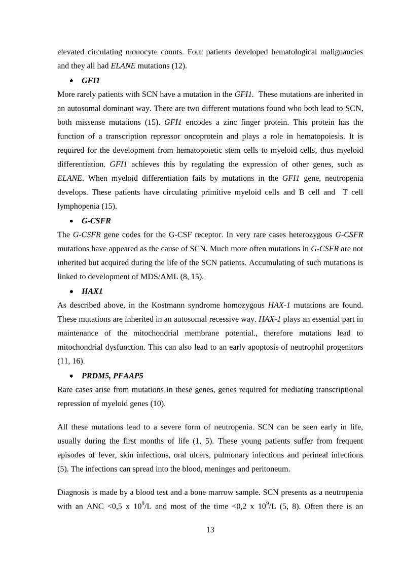
One group of authors developed a model which attempts to explain how mutations in ELANE,
GFI1 and AP3 can cause cyclic and severe congenital neutropenia (15). As described above,
different mutations in ELANE cause cyclic neutropenia and SCN (15), although overlap in
mutations has been reported (8).
They state when the ELANE gene is translated, it can develop into two neutrophil elastase
conformations: a soluble and a transmembrane conformation. The soluble form is a neutrophil
elastase in a granule. This pathway needs the assistance of AP3, which attaches to the
neutrophil elastase and functions as a cargo protein. The transmembrane form does not need
AP3 (15).
(a) Mutations in the ELANE gene in SCN prematurely terminate the gene and
delete the AP3 recognition signal. As a consequence only the
transmembrane conformation of the neutrophil elastase protein appears
(15). Something alike is suggested by another article which states ELANE
mutations appear at the promyelocyte stage, the stage where neutrophil
elastase normally is packaged into granules. The mutant neutrophil elastase
is not packaged normally and its accumulation or activity causes apoptosis
of these promyelocytes (16).
(b) Mutations leading to cyclic neutropenia destroy transmembrane segments,
the cause why only soluble forms develop (15).
(c) GFI1 mutations cause overexpression of the neutrophil elastase protein,
saturation of the AP3 granular transport and an overabundance of
neutrophil elastase in the plasma membrane (15).
18
Figure 1: Proposed model of normal and pathological processing and transport of
neutrophil elastase. (15) Berliner N, Horwitz M, Loughran TP, Jr. Congenital and acquired
neutropenia. Hematology / the Education Program of the American Society of Hematology American
Society of Hematology Education Program. 2004:63-79.
Although there is still a lot to discover about the pathogenesis of both cyclic neutropenia and
SCN, the role of ELANE mutations have been confirmed by several laboratories
independently (15).
6.1.2. Disorders of ribosomal dysfunction
6.1.2.1. Shwachman-Diamond syndrome
This syndrome is a rare multi organ disease, inherited in an autosomal recessive way. The
syndrome is characterized by exocrine pancreatic insufficiency, dwarfism, metaphyseal
chondrodysplasia and marrow failure, with a mild or moderate neutropenia as a consequence.
It is the second most common cause of exocrine pancreatic insufficiency in children, which
leads to the disability to digest fat from the diet. As a result these children have an increased
volume and frequency of stool. Malabsorption, failure to thrive, diarrhea and weight loss are
common problems and can be seen early in life, suggesting the diagnosis (10). Due to the
neutropenia, infections can be seen as well, for example otitis media and pneumonia. Patients
who present with the symptoms above should be tested for Shwachman- Diamond syndrome.
A mutation in the SBDS gene is the cause in the majority of cases, in 90% of the cases
according to one report (10). Most mutations occur during gene conversion, when
recombination takes place between the SBDS gene and the SBDSP pseudogene. However,
point mutations, insertions and deletions can also be seen (8). The normal SBDS protein is
suggested to be involved in RNA processing or maturation. The mutation leads to a defect in
RNA processing essential for haematopoiesis, leading to failure of neutrophil production.

19
Many articles suggest there also is a defect in chemotaxis; in neutrophil motility (10). A mild
to moderate neutropenia is seen in many patients with Shwachman- Diamond syndrome. The
ANCs fall below 1,0 x 10⁹/L in two-thirds of patients (10), but it tends to be constant and this
neutropenia does not lead to severe clinical problems. However other cytopenias (anemia and
tromcocytopenia) or macrocytose can also be seen (10, 11).
G-CSF is used if necessary and pancreatic enzyme replacement will be required (5). Some
authors state G-CSF can increase the risk of developing acute myelogenous leukemia, but this
is not proven.
Due to the marrow failure, patients have the risk of developing myelodysplasia, aplastic
anemia or acute myelogenous leukemia (such as SCN patients). These developments occur at
an average age of 18 years. The incidence of these complications is still unknown. One study
suggests the risk of development is 15% (5, 8). Regularly bone marrow samples and
cytogenetic tests can be useful.
6.1.3. Disorders of metabolism
6.1.3.1. Glycogen-storage disease type 1b(GSD 1b)
Glycogen-storage disease type 1b is a rare metabolic disorder, which affects the glucose-6-
phosphatase metabolism (16). Deficiency of the glucose-6-phosphatase translocase enzyme
inhibits transport of glucose-6-phosphatase into the endoplasmatic reticulum. Therefore
conversion to glucose and phosphatase is not achievable (8). There is no production of
glucose and the liver, spleen and other tissues accumulate glycogen. Thus patients present
with an enlarged liver and spleen, kidney problems and hypoglycemia, in combination with
chronic neutropenia. The presence of an enlarged spleen can be associated with low red blood
cells causing anemia and thrombocytopenia whereas neutropenia is always present. The
neutropenia in these patients is accompanied by a defective function of the neutrophils (10,
16), recent reviews discovered neutrophils are dependent upon glucose for the metabolic burst
and the killing of bacteria (16). Patients respond to treatment with G-CSF not only with an
increase in ANC but also with improvement of the activity of their neutrophils. There is no
increased risk of malignant transformation (8).

20
6.1.4. Disorders of vesicular transport
6.1.4.1. Cohen syndrome
This is a very rare autosomal recessive syndrome, characterized by delay in development,
facial dysmorphism, ophthalmic problems and neutropenia. The Cohen syndrome is caused
by mutations in the COH1 gene or the VPS13B gene. The COH1 protein functions in
vesicular sorting and intracellular protein trafficking (10, 11).
6.1.4.2. Griscelli syndrome
Griscelli syndrome is a disorder inherited in an autosomal recessive way. Patients present
with hypopigmentation of the skin and a silver-gray sheen of the hair with possible pigment
clumps in. They also have a hepatosplenomegaly, neutropenia and immunodeficiency.
Mutations in RAB27a appear to cause this syndrome (10). This gene encodes a protein critical
in the exocytosis of secretory vesicles, which also leads to a decreased NK cell cytotoxicity
(11).
6.1.5. Disorders of immune function
6.1.5.1. Cartilage-hair hypoplasia
Cartilage- hair hypoplasia is an autosomal recessive disorder, with clinical characteristics of
dwarfism due to metaphyseal dysplasia, fine hair, immunodeficiency and neutropenia. The
neutropenia can be accompanied by lymphopenia and macrocytic anemia. Mutations in the
RMRPG gene are responsible, encoding a ribonuclease. The mutant protein leads to defective
T-cell function and a defective humoral immune system (10).
6.1.5.2. Fanconi anemia
Fanconi anemia is a marrow failure syndrome characterized by pancytopenia. It usually arises
in children from 5 to 10 years old, where it is first presented as a thrombocytopenia. The
anemia and neutropenia develop quickly after this. Rare cases present with neutropenia as
first symptom. Patients with Fanconi anemia have mutations in the FANC genes which leads
to defects in DNA repair. This defect leads to extreme chromosomal breakage. The marrow
aspirates show a hypoplasia. Patients also present with a short stature, dysplastic thumbs,
heart and eye abnormalities. Later in life they have a 10% risk of developing myelodysplasia

21
or acute myelogenous leukemia. Fanconi anemia can only be cured by a stem cell
transplantation (5).
6.1.6. Rarer causes
Numerous syndromes are associated with neutropenia and other immunodeficiencies.
Neutropenia has been associated with disorders of immunoglobulin production and with other
bone marrow failure syndromes that are not described above. Some of them are listed in the
table that can be found as appendix 2 at the end of this paper.
6.2. Acquired
Acquired neutropenia is diagnosed much more frequently than congenital neutropenia, but it
still is a relatively rare disorder. Acquired neutropenia is defined as neutropenia that is not
caused by DNA defects or congenital syndromes. This does not mean acquired neutropenia
can not be present at birth, for example immune neutropenia can be present at birth. Besides
immune neutropenia, many other causes can cause neutropenia: infections, drugs, nutritional
deficiencies, …
6.2.1. Post-infectious
The most common cause of neutropenia is a variety of viral infections, for example infections
with Epstein-Barr virus, respiratory syncytial virus, influenza A and B, hepatitis and the
human herpes viruses. These viruses give rise to a transient marrow suppression with low
neutrophil counts at the first days of infection. The neutropenia will stay for 3-8 days.
Bacterial infections can also cause neutropenia (5).
6.2.2. Drug-induced neutropenia
Drug-induced neutropenia is caused by a variety of medications. For instance anti-thyroid
medications, antibiotics, anticonvulsants, chemotherapy and anti-inflammatory agents. They
start an idiosyncratic reaction that results in profound neutropenia. This kind of neutropenia
has a high rate of infections, complications and deaths (5).
6.2.3. Immune neutropenia

22
Immune neutropenia is caused by anti-neutrophil antibodies. These are antibodies directed to
neutrophil-specific antigens, called HNAs. Altogether there are 7 HNAs. They are
glycoproteins, for example part of the Fcgamma III receptor, then they are called HNA-1.
HNA-1 makes up the biggest subgroup of antigens, they are the most common antigens where
antibodies are directed against to (15).
Figure 2: HNA-antigens (15) Berliner N, Horwitz M, Loughran TP, Jr. Congenital and acquired
neutropenia. Hematology / the Education Program of the American Society of Hematology American Society of
Hematology Education Program. 2004:63-79.
The anti-neutrophil antibodies can be detected by several tests, such as the granulocyte
agglutination test (GAT) and the granulocyte immunofluorescence test (GIFT) or the more
advanced enzyme-linked immunoassays (ELISA). The test which uses monoclonal antibody-
specific immobilization of granulocyte antigens (MAIGA) may be even the most specific test
(15). But all these tests are expensive and only available in reference laboratories (11). It is
important to emphasize that screening for anti-neutrophil antibodies is difficult. Test results
may be false positive, which means people without neutropenia have positive test results.
False negative results are even more common, so the diagnosis of autoimmune neutropenia
can be made without a positive test result (15). Likewise test results can be positive at one
moment and negative at another. Exact numbers of sensitivity and specificity of these tests are
not available. All these factors together make it difficult to interpret the results of the tests for
anti-neutrophil antibodies. A positive test result confirms the diagnosis of autoimmune
neutropenia. In patients with a negative test result, tests may need to be repeated several times
(3) and a bone marrow aspirate can be done.

23
6.2.3.1. Neonatal alloimmune neutropenia
Neonatal alloimmune neutropenia results from the transfer of fetal neutrophil antigens into the
maternal circulation. Because these can also be paternal antigens, the mother starts making
antibodies against the shared fetal and paternal antigens (5). These antibodies are most often
directed against HNA-1 antigens. Since IgG antibodies from the mother can pass via the
placenta into the fetal circulation, neutrophils from the fetus will be destructed in utero and
subsequently ex utero. The neutropenia can be mild, moderate or severe. In utero the fetus is
protected from infections, but from birth the fetus is vulnerable to infections (3). Some
authors state infections can be severe to life threatening (3), whereas most authors suggest the
infants only suffer from minor infections (8, 15). The neutropenia will recover, because the
IgG antibodies of the mother disappear. Knowing the half-life of IgG is 5 to 6 weeks, the
recovery of neutropenia takes place after approximately 11 weeks (5, 8, 15).
6.2.3.2. Primary autoimmune neutropenia
Primary autoimmune neutropenia or chronic benign neutropenia of infancy/childhood
typically develops at an average age of 6-12 months. The importance of this neutropenia lies
in the fact that it is much more common in infants than congenital or cyclic neutropenia.
However it is still rare: it has an incidence of 1:100 000 infants (3).
Pathogenesis is also based on the existence of antibodies, mostly directed against HNA-1 and
HNA-2 antigens (5). There are still questions about the origin of these antibodies, but they
probably arise because of a process called ‘molecular mimicry’. Molecular mimicry starts
when the infant is infected with a virus and an epitope on the surface of this virus causes
production of antibodies. Because of sequence similarities of the virus surface and the surface
of a neutrophil, cross-reactivity takes place and auto-antibodies are being formed. These
antibodies destroy neutrophils, which is the reason why bone marrow aspirates show few
mature neutrophils.
The neutropenia can be mild to severe and the infant may develop infections. Moderate
infections can be held under control with antibiotics. Is the clinical course more severe, G-
CSF can be administered to produce sufficient neutrophils to overcome the antibodies during
infections. Fortunately this disease is self-limiting. In 95% of the patients, the neutropenia
spontaneous disappears over an average of 2 years (3, 8, 15).

24
6.3. Idiopathic neutropenia:
Idiopathic neutropenia is a term used when there is no evidence of congenital, cyclic or
immune neutropenia, nor for acquired neutropenia. Idiopathic neutropenia is a diagnosis of
exclusion. This term covers various types of neutropenia that may occur at any point, patients
can present with neutropenia in childhood or adult life. The neutropenia occurs for unknown
reasons, but is probably of heterogeneous cause (3, 5, 8). It is thought to be caused by
ineffective or decreased neutrophil production, but this is not confirmed (3, 5).
Neutrophil counts and clinical problems in these patients vary considerably. Some patients
can still recruit neutrophils when required, thus they do not experience more infections than
normal individuals. Other patients have a more severe neutropenia and suffer from serious
infections, these patients require antibiotics and sometimes even G-CSF. Patients with severe
idiopathic neutropenia respond well to treatment, but long term therapy is often required,
because of the unknown cause that cannot be cured. Overall patients with idiopathic
neutropenia have a good prognosis. The clinical course is usually mild and there is no
evidence for malignant transformation (8).
Patients with mild neutropenia, without infections, are not evaluated extensively because
usually diagnosis cannot be made despite the effort. Patients with a severe neutropenia do
need to be evaluated regularly. Idiopathic neutropenia is a significant challenge for the future.
Guidelines should be made for diagnostic investigations. Research can be done to find the
cause or molecular mechanism of the neutropenia and there can be searched for the best
therapeutic options.
7. Treatment
Decisions about treatment are first of all based on the clinical status of the patient. Second the
neutrophil count can determine treatment. There is a difference between the treatment of a
neutropenic episode with infections and the chronic treatment of neutropenia itself.
7.1. Treatment and prevention of infections:
Not all infections should be treated with antibiotics, only the severe infections with
fever require treatment with antibiotics. Broad-spectrum antibiotics can be used to

25
cover Gram-negative bacteria that can be dangerous, if skin infections are present
antibiotics should cover bacteria of the skin.
Prevention of infections is an important goal. Dental hygiene is necessary. Skin lesions
should be disinfected and taken care of, as for mucosal lesions which can develop to
abscesses. Especially infections or abscesses with Pseudomonas are feared. Prolonged
neutropenia renders the patient vulnerable for fungal infections. Parents and health
staff should stay attentive for these infections.
7.2. Treatment of neutropenia:
7.2.1. Granulocyte colony stimulating factor (G-SCF):
Since the introduction of G-CSF, treatment of neutropenia has improved spectacularly.
However G-CSF is often unnecessary and is only indicated in patients with severe
neutropenia and recurrent severe infections. Patients without severe and recurrent
infections do not require G-CSF (3, 15). The response to G-CSF therapy is rapid and
occurs in most of the patients (15). As a result the ANC increases, patients experience
less infectious episodes, antibiotic use and hospital admission is reduced and mortality
from infections is strongly reduced. In addition the quality of life of these children is
increased (3).
G-CSF induces differentiation of neutrophil granulocytes and reduced apoptosis of
neutrophil progenitors (21).
G-CSF is administered subcutaneously in a usual starting dose of 5 µg/kg/day. The
next 1-2 days an increase in the ANC can be seen and the ANC should be closely
monitored in this period. Adjustment of the dose can be made and the lowest dose to
keep the infections and symptoms under control should be the goal. Most patients
maintain this goal at an alternate day scheme (3). When a steady state is obtained,
blood counts are checked every 2-3 months. On the other hand higher doses can be
given when no effect is seen. When maximum doses have no effect, other therapeutic
options should be regarded and a stem cell transplantation is indicated.
G-CSF can be administered safely because it has few side effects. Bone pain and flu-
like symptoms can be seen in the first days and weeks. Local reactions can be seen at
the site of injection (3).
26
Long-term treatment with G-CSF is not yet fully documented. There are concerns it
effects the bone mineral density and can cause osteoporosis, but there is little proof.
The biggest fear is the evolution to myelodysplasia or acute myelogenous leukemia
(3). As described above, there is nothing proven yet. However, bone marrow aspirates
should be taken yearly for cytogenetic and molecular study to detect somatic
mutations (21).
7.2.2. Haematopoietic stem cell transplant:
A stem cell transplant is the final therapeutic option for patients who are refractory to
G-CSF. It is also indicated in specific causes of neutropenia, such as Fanconi anemia
and dyskeratosis congenita. Stem cell transplant will be necessary in case of bone
marrow failure syndromes as well in those patients with transformation to
myelodysplasia or acute myelogenous leukemia. A stem cell transplant will be given
preferentially before transformation to malignancy took place and before development
of serious infections or fungal infections causing organ damage.
7.2.3 Obsolete treatment options:
Since G-CSF is introduced, other therapeutic options are less used. In the past,
glucocorticosteroids, lithium, intravenous immunoglobulines, immunosuppressive
drugs and splenectomy were broadly used as long term treatment. Nowadays G-CSF is
the first choice of treatment, because G-CSF is much more effective in increasing the
number of neutrophils without many side effects. The treatments above all showed
many long-term side effects (3).
8. Follow-up
Follow up of all patients should be obtained every 3 months. At this consult history of recent
infections should be taken, physical examination should be done and a full blood count and
smear should be checked. A bone marrow aspirate should be taken every year (3).
9. Outlook
Marked progress has been made in the treatment of neutropenia. G-CSF has increased quality
of life in neutropenic patients dramatically. However the correlation between G-CSF and

27
MDS/AML should be closely investigated. Over the last years, progress has been made in
identifying genes causing the different types of neutropenia. This can often explain part of the
pathophysiology and aberrant pathways in neutropenia. New therapeutic options can be based
on this, with a special position for genetic therapy (11). Nevertheless the genetic basis of
congenital neutropenia remains unknown in many children with neutropenia (22).
The group of idiopathic neutropenia patients, remain a significant challenge for the future,
since diagnosis is often difficult.

28
PART B
1. Introduction
Neutropenia is a broad diagnosis with many different causes. In general, children with
neutropenia present with a common image of recurrent infections and low neutrophil counts.
However every patient can present with specific symptoms indicating a specific diagnosis.
These specific findings have been widely described in literature. Information on different
therapeutic options and the outcome of patients with neutropenia has extensively been
described as well. The aim of this study is the collection and analysis of data from pediatric
patients diagnosed and treated for primary neutropenia at the University Hospital Ghent (UZ
Ghent). How do these neutropenic patients of present? How many patients are diagnosed with
congenital, cyclic or autoimmune neutropenia? How many of them present with this typical
image? Can the specific findings described in literature be found in this patient population?
How are these patients treated and what is their outcome? Data has been collected of all
pediatric patients of the UZ Ghent diagnosed with neutropenia over the last 30 years. This
data has been analyzed and tested. Clinical and laboratory findings and findings about
treatment and outcome will be reported and will compared to findings described in literature.
2. Materials and methods
2.1. Patients
Patients admitted to the Department of Pediatric Hematology, Oncology and Stem Cell
Transplant at the University Hospital Ghent who were diagnosed with neutropenia between
1983 and march 2013 were included. Patients with chemotherapy induced febrile neutropenia
as well as patients with a short single period of neutropenia were excluded. Overall there was
a number of 69 patients. Before 1990 the patients were not systematically entered in the
database, from 1990 this registration was done more systematically, but still the number of
patients registered is an underestimation of the real number of patients admitted during this
time period.
2.2. Data collection
Data from patients diagnosed with neutropenia were collected from the medical papers and
electronic files and recorded in an electronic data base. For each patient, age, sex, date of birth

29
and date of admission was registered. Other relevant data were collected: presenting
symptoms, episodes of fever, clinical examination (weight, height, spleen and liver size,
skeletal abnormalities), diagnostic investigations, frequency of infections, duration and
complication of infections, responsible organisms and hospitalization periods. Information
about general history was obtained: information about psychomotor retardation, failure to
thrive, evolution of weight and height. Blood counts were obtained from routine blood
samples, in particular the absolute neutrophil count (ANC). The ANC was noted as cells/µl
and in percentages (%). Other information from the blood tests was collected as well, such as
results of immunoglobuline dosages, presence of anti-neutrophil antibodies and viral
antibodies. If a bone marrow aspirate was performed, the result of the evaluation of this
aspirate was collected. It was noted whether there was a hypo-, hyper- or dysplasia, where the
possible maturation arrest occurred and how many blasts were counted. If chromosomal tests
were performed, these results were included in the database. Information about treatment and
outcome were noted, in particular about G-CSF treatment. ANCs have been collected before
treatment, at the start when G-CSF was administered, during maintenance therapy and after
eventual stop of treatment.
If the cause of the neutropenia was found, the diagnosis was noted. Patients were diagnosed
with congenital neutropenia if they presented with severe neutropenia from birth, the
symptoms are in conformity with a congenital syndrome or the causative syndrome was found
through tests, often genetic tests. If anti-neutrophil antibodies were found, diagnosis of
autoimmune neutropenia was made. If antibody tests were negative, they were repeated or a
bone marrow aspirate was performed to detect characteristics of autoimmune neutropenia. All
diagnoses have been categorized into diagnosis groups. Diagnosis groups included: congenital
neutropenia, cyclic neutropenia, autoimmune neutropenia, idiopathic neutropenia and others.
This last category includes one patient with overgrowth syndrome and one patient with
defective mobilization of neutrophils and three patients for which diagnosis of
myelodysplasia or aplastic anemia was later established. This classification has partly been
based on the classification of the Severe Chronic Neutropenia Internation Registry (SCNIR),
but the autoimmune neutropenia group has been added since this makes up an important
group of patients.
Since this is a retrospective study, the patient files did not provide complete information on all
details described above. For some cases diagnosis was obvious and more investigations were
considered as irrelevant, for other cases, usually diagnosed long ago, some diagnostic

30
investigations did not belong to routine work up at that time. When data was not available,
this was defined as ‘unknown’.
2.3. Statistical Methods
All these data have been stored in an Excell database. Later on statistical tests were performed
by using the Excell program. Mainly descriptive statistics were used, for example the mean
value, frequency, percentage and standard deviation. Results are always from the data
available, when data was unknown, this will be mentioned.
Data about weight and length was edited. By using software from the World Health
Organisation (WHO) site and putting this data in a calculator, we obtained growth percentiles.
3. Results
3.1. Demographics
Of all patients, 37 (53,6%) are male and 32 (46,4%) are female. Thus there is a male
predominance, especially in the idiopathic neutropenia group (56,5%) and cyclic neutropenia
(100,0%) group. In the group with autoimmune neutropenic patients, there are more girls
(56,7%)
The mean age at first presentation, thus at the first contact, is 2,4 years. The mean age in the
congenital neutropenia group is 0,4 years. Patients with cyclic neutropenia present at a mean
age of 3,4 years and those with autoimmune neutropenia at a mean age of 1,6 years. Patients
with unexplained neutropenia, the idiopathic neutropenia patients, present at a mean age of
3,1 years.
Table 1: Sex and mean age at presentation.
Congenital Cyclic Autoimmune Idiopathic MDS & AA Others Total
(n=8) (n=3) (n=30) (n=23) (n=3) (n=2) (n=69)
Sex
(n(%))
Male 4(50,0) 3(100,0) 13(43,3) 13(56,5) 3(100,0) 1(50,0) 37(53,6)
Female 4(50,0) 0(0,0) 17(56,7) 10(43,5) 0(0,0) 1(50,0) 32(46,4)
Age
(years)
Mean 0,4 3,4 1,6 3,1 11,0 1,0 2,4
SD 0,29 2,96 2,19 4,46 3,01 1,20 3,65
MDS & AA = Myelodysplasia and Aplastic Anemia

31
43,5%
33,3%
11,6%
4,3%4,3% 2,9%
autoimmune
idiopathic
congenital
cyclic
myelodysplasia
other
43,5%
33,3%
11,6%
4,3%4,3% 2,9%
autoimmune
idiopathic
congenital
cyclic
myelodysplasia
other
3.2. Diagnosis
Table 2: Diagnoses
Congenital (n(%)) 8(11,6%)
Cyclic (n(%)) 3(4,3%)
Autoimmune (n(%)) 30(43,5%)
Idiopathic (n(%)) 23(33,3%)
MDS & AA (n(%)) 3(4,3%)
Others (n(%)) 2(2,9%)
Total (n(%)) 69(100,0%)
MDS & AA = Myelodysplasia and Aplastic
Anemia
Table 3: Specific congenital diagnoses and specific diagnoses from the subgroup ‘Others’.
Congenital diagnoses n(%) Others n
SCN-Kostmann 2(25,0%) Overgrowth
syndrome
1
Shwachman-Diamond syndrome 2(25,0%) Mobilization defect 1
Griscelli syndrome type 2 1(12,5%)
Cartilage-hair hypoplasia 1(12,5%)
Cohen syndrome 1(12,5%)
Cloves syndrome 1(12,5%)
Table 2 presents the diagnoses of all neutropenic patients. Diagnosis could be made in 46 out
of 69 patients. Congenital neutropenia is decribed in 8 (11,6%) patients, cyclic neutropenia is
described in 3 (4,3%) patients. Diagnosis of autoimmune neutropenia is made in 30 (43,5%)
patients. In 23 (33,3%) of the patients the cause remained unknown, thus making up the
idiopathic group.
Congenital neutropenia covers severe congenital neutropenia (SCN) and all syndromes
associated with neutropenia. The specific diagnoses are shown in Table 3. Two patients were
diagnosed with severe congenital neutropenia, both patients have Kostmann syndrome. A
homozygous HAX-1 mutation is found in one of them. The other patients turn out to have
multisystemic syndromes associated with neutropenia. Shwachman-Diamond syndrome is
described in 2 patients, one patient has Griscelli syndrome type 2 , one patient has Cohen
syndrome and Cartilage-hair hypoplasia is described in another patient. These syndromes are
Figure 3: Distribution of diagnoses

32
described above in Part A. Another patient is diagnosed with Cloves syndrome. This a
recently described syndrome, characterized by vascular malformations and skeletal
abnormalities. Much is still unknown about it, neutropenia can possibly occur (23).
Essentially cyclic neutropenia is also a congenital cause of neutropenia, but it is categorized in
a separate category, since the two categories have different clinical signs.
Myelodysplasia (MDS) and Aplastic anemia (AA) are described in 3 (4,3%) of all patients.
These diseases are associated with bone marrow failure and a deficiency of many blood types:
anemia, thrombocytopenia and neutropenia. MDS and AA develop slowly, pancytopenia can
be seen at first presentation, but some patients only present with neutropenia. These particular
patients are included in this study because neutropenia is the first symptom and development
and diagnosis of MDS or AA occurs later.
3.3. History of infections
Patients with neutropenia often have a history of infections with oral ulcerations, mucosal
lesions upper, respiratory tract infections and skin lesions. A detailed history could be found
in 59 (85,5%) of our patients files.
Table 4: History or signs of infection
History/Signs of infection
(n(%))
59(85,5%)
URTI (n) 35 Omphalitis (n) 2
Otitis media (n) 24 Sepsis (n) 7
LRTI (n) 14 Meningitis (n) 1
Oral infections(n) 13 UTI (n) 1
Skin infections (n) 20 Conjunctivitis (n) 4
URTI= upper respiratory tract infections, LRTI= lower respiratory tract infections, UTI=
urinary tract infections.
Among these 59 patients, 35 (59,3%) patients showed a history of recurrent upper respiratory
tract infections, 24 (40,7%) of otitis media and most often combined (20 patients (33,9%)).
Lower respiratory tract infections were seen in 14 (23,7%) of the patients. A history of oral
ulcerations or oral infections was found in 13 (21,3%) patients.
General skin lesions were found in 20 (33,9%) of the cases, mostly slow healing. Some of
them presented with abcesses, pustules and furuncles of the skin. Patients presented with anal
abcesses (3), with abcesses of the vulva (3) or mycotic infections of the vulva (1). Other

33
patients presented with cellulitis (3), folliculitis (2), lymphangitis of the abdominal wall (1)
erysipelas (1) and flebitis (1).
Four (6,8%) patients showed a history of conjunctivitis, two (3,3%) patients of omphalitis.
One (1,7%) patient had suffered from a urinary tract infection. More severe infections
appeared in some patients: sepsis appeared in 7 (11,9%) patients and meningitis in 1 (1,7%)
patient.
3.4. Additional symptoms
In a minority of patients symptoms not directly related to the neutropenia were present or
recorded. Two patients showed a combination of failure to thrive, steatorrhea and skeletal
abnormalities. These patients were diagnosed with Shwachman-Diamond syndrome. One
patient presented with extreme fatigue, paleness and night sweats. This patient was diagnosed
with myelodysplasia later on. Another patient presented with a history of laryngomalacia, a
retrognathic jaw, gastrointestinal reflux, failure to thrive, myopia, hip dysplasia and
psychomotor retardation, leading to diagnosis of the Cohen syndrome. One patient showed
remarkable clinical signs such as hypertonia, brachycephaly, ocular albinism, defective NK
cells and a dysmorphic facies, but no diagnosis is made yet.
3.5. Physical examination
3.5.1 Weight and length
Table 5: Weight and length of all patients
Congenital Cyclic Autoimmune Idiopathic MDS &AA Others Total
Weight for
length
(percentile)
Mean 26,4 49,1 41,5 33,5 unknown 2,5 35,2
SD 26,61 47,02 32,86 21,03 unknown 3,54 28,7
Weight for
age
(percentile)
Mean 8,9 52,1 48,6 30,3 80,0 17,4 37,4
SD 13,75 40,01 33,43 26,08 0,00 22,84 31,88
Length for
age
(percentile)
Mean 16,8 31,4 56,3 36,1 unknown 50,7 42,7
SD 28,63 23,21 32,25 37,12 unknown 69,51 35,9
MDS & AA = Myelodysplasia and Aplastic Anemia

34
Weight and length were obtained in 60 of all patients, by reports of physical examination.
These reports all date from time of first contact. With these data, percentiles could be
calculated. The mean percentile is reported, plus the standard deviation.
The mean weight for length percentile of all patients is 35,2. The mean weight for length
percentile in patients with congenital neutropenia is 26,4.
Weight and length can also be calculated in function of age. The mean weight for age
percentile is 37,4 and the mean length for age percentile is 42,7.
Table 6: Weight and length of all congenital patients
Cartilage-hair
hypoplasia (n=1)
Shwachman-Diamond
syndrome (n=2)
Others
(n=5)
Total congenital
(n=8)
Weight for
length
(percentile)
Mean 59,1 11,0 26,0 26,4
SD 0,00 14,28 28,86 26,61
Weight for
age
(percentile)
Mean 0,2 2,1 14,6 8,9
SD 0,00 2,90 16,62 13,75
Length for
age
(percentile)
Mean 0,2 1,1 28,7 16,8
SD 0,00 1,56 34,53 28,63
The subgroup congenital neutropenia has been shown more detailed, because some congenital
syndromes are known to cause very low weight and height. These patients are ‘outliers’,
meaning they lower the mean value of all congenital patients, rendering this value less
representative. This is the reason why a separate table has been made (Table 6). The patient
with Cartilage-hair hypoplasia has a weight for age percentile of 0,2 and a length for age
percentile of 0,2. Patients with Shwachman-Diamond syndrome present with a mean weight
for age percentile of 2,1 and a mean length for age percentile of 1,1.
3.5.2. Liver and spleen size
Through physical examination reports, data is obtained about liver and spleen size at the first
presentation, in search for hepatosplenomegaly. The majority of patients had a normal
physical examination. Liver enlargement was described in 14 (20,3%) patients and is reported
as the mean liver size (in cm below costal margin) and correlating range (mean-SD –
mean+SD). Liver enlargement was found in 4 patients with congenital neutropenia, with a

35
mean liver size of 3,9 (2,06-5,74) cm, in 5 patients with autoimmune neutropenia with a mean
liver size of 1,4 (0,85-1,95) cm and in 4 patients with idiopathic neutropenia with a mean liver
size of 2,1 (1,25-2,95) cm. One patient with overgrowth syndrome also presented with an
enlarged liver of 1cm. Overlooking all patients in every subgroup, 16,7% of the autoimmune
neutropenia patients had hepatomegaly, compared to 50% of the patients with congenital
neutropenia.
Spleen enlargement was examined in 7 (10,1%) patients. One patient with congenital
neutropenia presented with a spleen that appeared 2,5 cm below costal margin. An enlarged
spleen was also found in 3 patients with autoimmune neutropenia with a mean spleen size of 1
cm below costal margin, in 2 patients with idiopathic neutropenia with a mean spleen size of 2
cm below costal margin and in 1 patient with myelodysplasia with a spleen size of 2cm below
costal margin.
3.6. Blood counts
Table 7: first ANC
Congenital Cyclic Autoimmune Idiopathic MDS & AA Others Total (n=8) (n=3) (n=30) (n=23) (n=3) (n=2) (n=69)
ANC(/µl)
Mean 531,5 272,7 375,9 560,5 567,0 310,5 456,6
SD 402,89 241,37 500,18 562,33 654,78 381,13 497,73
Median 360,0 359,0 238,0 491,0 567,0 310,5 330,0
Range 134-900 0-459 0-2596 0-2380 104-1030 41-580 0-2596
ANC(%)
Mean 9,3 5,7 5,4 12,4 37,3 4,3 9,2
SD 12,24 6,66 6,91 16,84 24,40 4,67 13,29
Median 4,3 4,0 3,0 7,0 37,3 4,3 4,0
Range 2-36 0-13 0-23,7 0-59,5 20-54,5 1-7,6 0-59,5
MDS & AA = Myelodysplasia and Aplastic Anemia
Table 7 presents neutrophil counts from first presentation. The absolute neutrophil count is
expressed in cells/µl and in percentage of the total amount of white blood cells. The mean
ANC of all patients is 456,6/µl and 9,2%.
Table 8: Distribution of first ANCs in three categories: 0-200/µl, 200-500/µl, > 500/µl.
Congenital Cyclic Autoimmune Idiopathic MDS & AA Others Total
(n=8) (n=3) (n=30) (n=23) (n=3) (n=2) (n=69)
ANC
0-200/µl 25,0% 33,3% 46,4% 31,8% 66,7% 50,0% 38,5%
200-500/µl 37,5% 66,7% 32,1% 22,7% 0,0% 0,0% 29,2% >500/µl 37,5% 0,0% 21,4% 45,5% 33,3% 50,0% 32,3%

36
Table 9: Presence of anemia, trombopenia, pancytopenia and monocytosis.
Cause n
Anemia Hemolytic anemia Autoimmune 4 2 positive Coombs
tests, 2 unknown
Idiopathic 1 Unknown
Idiopathic neutropenia 3
Cyclic neutropenia 1
Shwachman-Diamond syndrome,
Kostmann syndrome, Cartilage-hair
hypoplasia
3
Aplastic anemia 1
Overgrowth syndrome 1
Trombopenia Griscelli syndrome type 2 1
Pancytopenia Myelodysplasia 1
Autoimmune neutropenia (HHV6
infection)
1
Monocytosis Idiopathic neutropenia 2
Kostmann syndrome 2
3.7. Anti-neutrophil antibodies
Anti-neutrophil antibodies test results could be found in 52 (75,4%) of all patients. Positive
test results were found in 14 patients, thus in 14 patients with autoimmune neutropenia. When
only the autoimmune group is considered, anti-neutrophil antibodies could be found in 14 out
of 30 patients, thus in 46,7% of the autoimmune patients.
3.8. Bone marrow aspirates
Bone marrow aspiration was performed in 53 of 69 patients, thus in 76,8% of the patients.
Table 10 : Bone marrow aspiration test performed or not
Congenital Cyclic Autoimmune Idiopathic MDS & AA Others Total
(n=8) (n=3) (n=30) (n=23) (n=3) (n=2) (n=69)
Aspira
tion
Yes (n)
(%)
7
(87,5%)
2
(66,7%)
25
(83,3%)
15
(65,2%)
2
(66,7%)
2
(100,0%
)
53
(76,8%)
No (n) (%)
1 (12,5%)
1 (33,3%)
5 (12,5%)
8 (34,8%)
1 (33,3%)
0 (0,0%)
16 (23,2%)
MDS & AA = Myelodysplasia and Aplastic Anemia

37
Table 11: Results of the bone marrow aspirations.
All patients Autoimmune neutropenia patients
Hypocellular (n (%)) 5 (9,6%) 2 (8,0%)
Normocellular (n (%)) 41 (78,8%) 17 (68,0%)
Hypercellular (n (%)) 6 (11,5%) 6 (24,0%)
Hypocellular bone marrow was found in 5 patients, (temporary) bone marrow aplasia was
described in all of them. These patients include one patient with Shwachman-Diamond
syndrome and one patient with myelodysplasia, two patients with an autoimmune cause of
neutropenia and one patient with idiopathic neutropenia. Hypercellular bone marrow was
found in 6 patients, all patients diagnosed with autoimmune neutropenia.
Bone marrow maturation arrest was found in 8 (15,1%) of the 53 patients with test results.
The two patients with Kostmann syndrome described either an arrest at the promyelocyte
stage or an arrest at the myelocyte stage. Arrest at the stage of the band neutrophils and a bone
marrow without mature neutrophils could be found in one patient with Shwachman-Diamond
syndrome and five patients with idiopathic neutropenia.
3.9. Treatment
3.9.1. G-CSF
Table 12: Patients treated with G-CSF or BMT according to diagnosis.
Congenital Cyclic Autoimmune Idiopathic MDS & AA Others Total
(n=8) (n=3) (n=30) (n=23) (n=3) (n=2) (n=69)
G-CSF
treatment
n 4 2 12 7 1 0 26
% 50,0% 66,7% 40,0% 30,4% 33,3% 0,0% 37,7%
BMT
n 2 0 0 1 1 0 4
% 25,0% 0,0% 0,0% 4,3% 33,3% 0,0% 5,8%
MDS & AA = Myelodysplasia and Aplastic Anemia, BMT= Bone Marrow Transplantation

38
3.9.2. Bone marrow transplantation
Table 13: Patients who received bone marrow transplantation: diagnosis and reason for BMT
Diagnosis Reason for BMT
Patient 1 Kostmann syndrome (SCN) Non-response to G-CSF
Patient 2 Refractory cytopenia of childhood
(Myelodysplasia)
Myelodysplasia
Patient 3 Cartilage-hair hypoplasia Severe immunodeficiency
Patient 4 Idiopathic hemolytic anemia and
neutropenia
Non-response to G-CSF
3.10. Outcome
Table 14: Most recent ANC
Congenital Cyclic Autoimmune Idiopathic MDS
& AA Others Total
(n=8) (n=3) (n=30) (n=23) (n=3) (n=2) (n=69)
ANC(/µl)
Mean 2639,1 1704,5 4380,8 2388,1 1027,0 2340,0 3249,0
SD 1712,39 1038,74 4285,79 2200,14 0,00 777,82 3304,34
Median 2050,0 1704,5 2780,0 1554,0 1027,0 2340,0 2049,5
Range 838-5840 970-2439 633-18273 434-8300 / 1790-2890 434-
18273
ANC(%)
Mean 30,9 21,4 35,8 32,8 60,0 34,7 34,3
SD 16,89 5,74 20,56 17,95 43,77 8,63 19,54
Median 24,6 18,6 36,2 29,5 60,0 34,7 30,1
Range 13,4-56,9 17,6-28 1-67 5-68,8 29-
90,9 28,6-40,8 1-90,9
MDS & AA = Myelodysplasia and Aplastic Anemia
The mean ANC of all patients, most recently measured, is 3249/µl and 34,3%. The mean
ANC in the cyclic neutropenia group is 1704,5/µl and 21,4%. The mean ANC in patients
diagnosed with myelodysplasia is 1027/µl and 60,0%.
Table 15: Deaths
Sex Age of
death
(years)
Cause of death Diagnosis
Patient 1 M 9,8 Unknown Aplastic anemia
Patient 2 M 12,1 Neurological deterioration Myelodysplasia
Patient 3 M 0,6 Progressive cerebral demyelination Congenital(Griscelli syndrome)
Patient 4 V 12,1 Respiratory insufficiency Autoimmune
Patient 5 V 14,1 Metabolic decompensation Idiopathic
39
4. Discussion
The results of this retrospective analysis will be discussed and compared to data found in
literature. The Severe Chronic Neutropenia International Registry (SCNIR) has reported data
about 731 patients enrolled in their study. Hence the large sample size of this study, the
results seem representative and ideal for comparison. In this register however patients with
autoimmune neutropenia were excluded. Data and numbers found in literature studies will
also be compared. Through the results, the diagnostic work-up of the UZ Ghent will become
clear and options for improvement will be discussed.
4.1. Demographics
In the patients with neutropenia of the UZ Ghent, there is a male predominance: 53,6% of all
patients are male. In the SCNIR study only 44,2% of the patients is male and 55,8% are
female patients. Thus these results are different and cannot be explained. In the idiopathic and
cyclic neutropenia group male predominance is even higher, respectively 56,5% and 100,0%.
However the group of cyclic neutropenia consists of only 3 patients, thus might not be
representative. In the group with autoimmune neutropenic patients, there are more girls
(56,7%). This is confirmed by one study that reports a female to male ratio of 6:4 in
autoimmune neutropenia and confirmed by another report with a ratio of 5:3 (24).
The mean age at first presentation, thus at the first contact, is 2,4 years. The big deviation
between the subgroups of diagnoses is remarkable. The mean age in the congenital
neutropenia group is 0,4 years, thus these patients present much younger. Patients with
idiopathic neutropenia, present at a later age, at a mean age of 3,1 years. This can possibly be
explained by the fact that idiopathic neutropenia is less severe and comes later in life to
attention, compared to patients with congenital neutropenia who suffer from severe and
recurrent infections from birth. Autoimmune neutropenic patients have a mean age of 1,4
years at presentation. One study with 72 patients diagnosed with autoimmune neutropenia
reports a mean age of 0,85 years (24). Patients with myelodysplasia present at a mean age of
11,0 years, because myelodysplasia usually develops later in life.
4.2. Diagnosis
Diagnosis is shown in table 2. The congenital neutropenia patients have been described more
specifically in table 3. These results can be compared to a Swedish study that published
results about 32 patients with congenital neutropenia. Kostmann syndrome was reported in
17,3% of their patients and Shwachman-Diamond syndrome in 21,7% (25). This is similar to

40
our findings of 25,0% for each of these diagnoses. However they reported many more patients
with SCN, in particular patients with SCN caused by ELANE mutations. Of all their 23
patients with congenital neutropenia, one patient was diagnosed with Griscelli syndrome type
2 and one patient with Cohen syndrome. This study described one patient of each syndrome
as well, but in a much smaller sample size (8 patients). These numbers are clearly different
but appearance of these congenital causes of neutropenia is so rare that comparison of
incidence and prevalence cannot be made in these small sample sizes. However it can be
noted that the UZ Ghent diagnosed few patients with SCN, especially SCN caused by ELANE
mutations.
Diagnosis is often difficult to make in patients with neutropenia. Classical forms of SCN and
cyclic neutropenia are easy to distinguish, but a continuum of phenotypes makes clinical
diagnosis challenging in some cases. In the UZ Ghent patients, 33,3% is still undiagnosed,
making up the idiopathic group. Below a chart is inserted, showing the distribution of
diagnoses of patients of the SCNIR. This only shows three categories, in contrast to the 6
categories in this paper, in particular patients with autoimmune neutropenia, myelodysplasia,
aplastic anemia, overgrowth syndrome and mobilization defects are left out. Therefore
percentages cannot be compared. If we exclude these patients as well, like in the chart below,
comparison is possible between the distribution of congenital, cyclic and idiopathic
neutropenia in the SCNIR and in the UZ Ghent.
Figures 4 & 5 : Distribution of congenital, cyclic and idiopathic neutropenia, respectively in
the SCNIR and the UZ Ghent.
The SCNIR has more patients diagnosed with congenital and cyclic neutropenia. In only
32,6% of the patients enrolled in the SCNIR the cause of neutropenia is still unknown,
compared to 67,6% patients of the UZ Ghent. This difference can possibly be explained,
47,60%
19,80%
32,60%
Diagnoses SCNIR
congenital
cyclic
idiopathic
23,50%
8,80%67,60%
Diagnoses UZ Ghent
congenital
cyclic
idiopathic

41
some reasons for the higher prevalence of idiopathic patients in the UZ Ghent are written
down below. A critical retrospective look can suggest some reasons why diagnostic
investigations have not always been performed or diagnosis is just hard to find.
1. SCN can be missed because ELANE mutations are not routinely searched for in the
UZ Ghent, especially in patients diagnosed long ago, genetic tests were no routine.
Nowadays ELANE mutation tests are not always performed because tests are not
refunded. Questions arise whether expensive tests should be performed in cases where
it seems unnecessary, for example in cases with a benign course.
2. Diagnosis of cyclic neutropenia is through 3 blood counts per week over a period of 6
weeks, this is practical difficult to achieve and a subtle cyclic pattern can be missed
because of this.
3. Diagnosis of autoimmune neutropenia comes along with some difficulties as well, as
will be described below. Presumably many patients with autoimmune neutropenia can
be found in the idiopathic neutropenia group.
4. In addition some children tend to do well, questioning the need for intensive search for
specific etiology with many investigations.
5. In some cases, G-CSF therapy might be given, improving the condition of these
patients. In these patients further investigations sometimes seem unnecessary.
These can be reasons why the cause of neutropenia is still unknown in some patients admitted
at the UZ Ghent. One can conclude the performance of diagnostic investigations are
determined by the clinical course and the severity of neutropenia in each patient, taking in
account the financial implications. A diagnostic pathway, inserted in appendix 1 can be
followed.
In table 2 one can see that 43,3% patients of the UZ Ghent are diagnosed with autoimmune
neutropenia. Comparison with literature data is not possible, the SCNIR has not published
results about autoimmune neutropenia and other studies focus on a particular subgroup, so
data on distribution of the relative frequencies of each subgroup are not available. However it
is confirmed by literature that autoimmune neutropenia is much more frequent than congenital
and cyclic neutropenia (7). Autoimmune neutropenia is the most frequent diagnosis in this
study, but prevalence can even be higher because diagnosis is not always made. Diagnosis of
autoimmune neutropenia comes along with some difficulties:

42
a) Diagnosis of autoimmune neutropenia is made when anti-neutrophil antibody tests are
positive. But sensitivity of these tests is not as high to detect every patient with
autoimmune neutropenia. Patients with true autoimmune neutropenia often test
negative for anti-neutrophil antibodies, which can also be seen in the patients of the
UZ Ghent: only 46,7% of the autoimmune patients have anti-neutrophil antibodies.
One study states 35% of patients with autoimmune neutropenia have positive
antibodies (24), while another analysis of 116 cases with autoimmune neutropenia
detected antibodies in 60% of all patients (26). One can conclude there are many false
negative test results. Patients should be tested several times, up to 3-5 times (7),
repeating tests leads to a higher sensitivity and higher negative predictive value (24).
b) When antibody test results stay negative, diagnosis of autoimmune neutropenia can be
made through bone marrow evaluation. A normocellular or hypercellular bone marrow
in combination with neutropenia suggests diagnosis of autoimmune neutropenia,
because the bone marrow still produces cells, but these cells are destructed
peripherally. A bone marrow aspiration has not always been done in the idiopathic
neutropenia group, only in 65,2%, thus it is possible there are some undiagnosed cases
of autoimmune neutropenia in this group.
c) The clinical course is not always severe, one study states patients with autoimmune
neutropenia can even be asymptomatic (26). In addition autoimmune neutropenia is a
self-limiting disease. Thus the disease can have a benign course in some patients and
can go unnoticed or further diagnostic investigations seem unnecessary. Accordingly
diagnosis of autoimmune neutropenia is never made and prevalence can actually be
higher.
One can conclude that prevalence of autoimmune neutropenia is uncertain, but is probably
higher than reported and that tests for anti-neutrophil antibodies are complex. The sensitivity
and specificity of these tests are not high, test results are difficult to interpret and there is a
current lack of guidelines. More extensive research can be done in this field. In addition anti-
neutrophil antibody tests are expensive only accessible in reference laboratories. Recently
some investigators started searching for alternative tests (24). Meanwhile tests should be
interpreted thoughtfully and should be repeated a few times. This is no pleading against these
tests, since these tests do help in diagnosing autoimmune neutropenia. These tests allow to
exclude other forms of neutropenia with an increased risk of development to myelodysplasia

43
or acute myelogenous leukemia. Knowing neutropenia in a child is due to a self-limiting
disease, these tests reassure the practitioner and the parents, making these tests of great value.
4.3. Physical examination
4.3.1 Weight and length
Table 5 reports mean weight and height percentiles plus the standard deviation. The standard
deviation is quite big, meaning weight and height data are spread out over a large range of
values. The mean weight for length percentile of all patients is 35,2, thus far beneath the mean
percentile. This is also reflected in the curves in function of age : patients with neutropenia
have very low weight for age percentiles and quite low length for age percentiles. These
results together show that neutropenic patients of the UZ Ghent present with low weight and
height, in particular their weight.
Patients with congenital neutropenia tend to do worse, all numbers are much lower than the
mean value for the whole group of neutropenic patients. Especially the weight is extremely
low in these patients. Patients with cyclic and autoimmune neutropenia in general do not
differ from normal children in weight and height. This correlates with the more benign course
of cyclic and autoimmune neutropenia, compared to the other causes of neutropenia. It seems
weight and height correlate with the severity of the disease. The mean value of the idiopathic
neutropenia subgroup lies between those values from the congenital and autoimmune
subgroups, suggesting some serious cases of neutropenia can be found in the idiopathic
neutropenia group. More research can be done to discover a cause in these patients. When a
patient presents with low weight and height percentiles, there should be searched in particular
for a congenital cause. More tests to discover ELANE mutations should be performed in this
group, as suggested above.
Weight and height percentiles have been reported for the congenital neutropenia patients,
separated in Cartilage-hair hypoplasia, Shwachman-Diamond syndrome and the other
patients. The patient with Cartilage-hair hypoplasia shows very low percentiles, because this
syndrome is associated with dwarfism (10). Both patients with Shwachman-Diamond
syndrome present with low percentiles as well, because they both presented with failure to
thrive. Indeed, Shwachman-Diamond syndrome is characterized by malabsorption, weight
problems and dwarfism as mentioned above (10). These patients are ‘outliers’, meaning they
lower the mean value of all congenital patients. However the mean value of the other
congenital neutropenia patients (without the syndromes described above), is still lower than

44
the mean value of the other subgroups. Thus congenital neutropenia patients do present with
low weight and height.
4.3.2.Liver and spleen size
The results of the liver and spleen sizes show valuable information, however interpretation of
the results is difficult because of possible bias. Measurement of liver and spleen size is not
easy in children and may not always be accurately performed. Besides in infants and young
children the liver and spleen edge may be 1cm below costal margin, thus 1cm can be a normal
value. This renders the results less informative and it is more difficult to determine whether
the enlarged liver or spleen is a normal finding or due to the neutropenia. Enlarged liver is
found in 13 of all patients. Of all patients with autoimmune neutropenia 16,7% had
hepatomegaly, compared to 50% in the patients with congenital neutropenia. The exact liver
sizes has been described as well. The mean liver size in patients with autoimmune neutropenia
and hepatomegaly lies 1,4 cm below costal margin, which is no significant result knowing this
can be a normal value. In the congenital subgroup, the mean liver lies 4cm below costal
margin, which clearly shows liver enlargement. A possible explanation can be that the bone
marrow in congenital neutropenia is truly insufficient and hematopoiesis takes place in the
liver and spleen, leading to hepatomegaly or that the hepatomegaly is part of the congenital
syndrome. The congenital neutropenia patients are more specifically diagnosed with Griscelli
syndrome type 2, Shwachman-Diamond syndrome, Cartilage-hair hypoplasia and Cloves
syndrome. As stated above Griscelli syndrome type 2 can be associated with hepatomegaly
(10). One study reports Cloves syndrome as well is associated with hepatomegaly (23). One
can conclude congenital neutropenia patients can present with hepatosplenomegaly and
physical examination of the abdomen is important. Large liver sizes can direct to a diagnosis
of congenital neutropenia. The 4 patients with idiopathic neutropenia with enlarged livers,
could be tested for congenital neutropenia, in particular with ELANE mutation tests.
Distribution of spleen size shows quite the same results: there are more autoimmune patients
with splenomegaly, but patients with congenital neutropenia and splenomegaly present with
larger spleens. Whether this is a significance difference is difficult to prove, because there are
not enough patients.
4.4. Blood counts
The blood counts show a mean ANC value of 456,6/µl, where neutrophils make up 9,2% of
the white blood cells, meaning patients with neutropenia present at first contact with a severe
neutropenia(<0,5 x 10⁹/L). In particular the cyclic and idiopathic neutropenia patients present

45
with very low ANCs. But the cyclic neutropenia group consist only of three patients and
cannot be representative as described above. Plus ANCs vary considerably in cyclic
neutropenia, the values above are the lowest counts measured. The percentages give few
information, because absolute counts are far more important in predicting the clinical course.
Autoimmune neutropenic patients show the lowest mean value (5,4%) , because only the
neutrophil count is low, but the total of white blood cells is still normal because of an intact
bone marrow. Patients with myelodysplasia show a mean value of 37,3%, because the relative
ratios are still intact, since the total white blood count is low because of bone marrow failure.
One cannot be deceived by this high percentage, there should always be kept in mind absolute
neutrophil counts are much more important.
Table 9 shows the other results of the blood counts. Patients with neutropenia often present
with associated anemia. This anemia is frequently caused by hemolysis. Four patients with
hemolytic anemia belonged to the autoimmune neutropenia subgroup and one patient to the
idiopathic subgroup. This can suggest the undiagnosed patient does have autoimmune
neutropenia, because hemolytic anemia is an autoimmune phenomenon described in literature.
The anemia arises because of breakdown of red blood cells by autoantibodies. These
autoantibodies can be detected by the Coombs test (27). Results of the Coombs test shown in
table 9 , demonstrate the Coombs test is positive in two patients and unknown in two patients.
Autoimmune hemolytic anemia can be secondary to infections, especially Mycoplasma and
EBV (27). One of the patients described above has a history of EBV infection and one patient
clearly developed hemolytic anemia after Mycoplasma infection. Cases like these have been
described in literature (28). Anemia is also seen in patients with congenital syndromes, for
example in one patient with Cartilage-hair hypoplasia. This disease is known to cause
neutropenia with an accompanying anemia (10). One patient with Shwachman-Diamond
syndrome also presents with neutropenia and anemia, this combination does occur, although
not as often (10).
Trombopenia is seen in a patient with Griscelli syndrome type 2, a syndrome known to
present with pancytopenia (10).
Monocytosis can be seen in 4 patients, in particular in 2 patients with Kostmann syndrome or
SCN. Kostmann syndrome and SCN in general are known for the accompanying monocytosis
as mentioned above (8). Two patients with idiopathic neutropenia present as well with an

46
associated monocytosis, there might be a possibility these patients have SCN and tests like
ELANE mutation tests should be performed.
4.5. Bone marrow aspirates
Bone marrow aspiration has been done in 67,8% of all patients. This number is rather low,
knowing bone marrow aspiration is always indicated, unless diagnosis is already made
because of positive anti-neutrophil antibodies. In the idiopathic subgroup 8 out of 23 (34,8%)
patients have not been tested through bone marrow aspiration. Retrospective analysis of these
patients, shows 6 of these presented with a very benign clinical course: two of them only
showed a history of frequent upper respiratory tract infections and 4 of them showed no
history of infections at all. The benign course in these patients probably is the reason why
bone marrow aspiration has not been done, however diagnosis can be missed because of this.
Bone marrow aspiration is a very valuable investigation and should always be performed in
patients with neutropenia in search for diagnosis. Unless diagnosis of autoimmune
neutropenia has been made through positive anti-neutrophil antibodies, bone marrow
aspiration is always indicated, since neutropenia is a serious disorder and rare cases of
myelodysplasia and aplastic anemia present with neutropenia at first. These are diagnoses that
cannot be missed.
Bone marrow aspiration results show information about the cellularity of the bone marrow. It
is remarkable that patients with hypercellular bone marrow are all diagnosed with
autoimmune neutropenia. Knowing these results, a table has been inserted of all bone marrow
results of the autoimmune neutropenia patients where a bone marrow aspiration was
performed. Hypercellular bone marrow was found in 24,0%, normocellular bone marrow in
68,0% and hypocellular in 8,0% of these patients. These findings are also described in
literature: in primary autoimmune neutropenia the bone marrow is typically normocellular or
hypercellular (29). One study with autoimmune neutropenia patients reports 30,0% of their
patients has a reactive bone marrow, 50,0% a normal bone marrow and 20,0% has a
hypocellular bone marrow (24). A possible explanation for the hypercellularity in
autoimmune neutropenia is that anti-neutrophil antibodies destruct mature neutrophils and the
bone marrow tries to compensate for this loss. One study states hypocellularity can be
explained by antibodies as well, by antibodies orientated against the more primitive
hematopoietic cells (24). But hypocellular bone marrow should also raise suspicion about the
quality of the aspiration, few bone marrow cells or blood might have been aspirated. The main
note seems autoimmune neutropenia needs to be suspected when a hypercellular bone marrow
47
result is found. In the 6 patients with autoimmune neutropenia, 2 had already shown positive
anti-neutrophil antibodies but 4 patients tested negative for antibodies. This implies bone
marrow can be helpful in diagnosing autoimmune neutropenia, certainly when anti-neutrophil
antibodies are uninformative.
A maturation arrest could be found in 7 of all tested patients. Two of these patients have
Kostmann syndrome, known for the maturation arrest in the bone marrow at the promyelocyte
stage, a finding confirmed in the patient’s bone marrow. Maturation arrest at the stage of the
band neutrophil was found in four patients with idiopathic neutropenia. These patients should
be more closely investigated with ELANE mutation tests.
4.6. Treatment
4.6.1. G-CSF
Results about treatment show 37,7% of all patients are treated with G-CSF. The percentage
treated with G-CSF is highest in the cyclic neutropenia group (66,7%), followed by the
congenital neutropeniagroup (50,0%). The percentage is lowest in the idiopathic group
(30,4%), but this still stands for a large amount of patients. The SCNIR has publicized
comparable results about treatment with G-CSF in 731 patients. G-CSF was given in 50,6%
of all patients, in 49,1% of the congenital neutropenia patients, in 54,4% of the cyclic patients
and in 50,4% of the patients with idiopathic neutropenia (20). In the UZ Ghent G-CSF
therapy is not as frequently given compared to the patient population of the SCNIR. The
percentages in the congenital subgroup are almost equal and the highest percentages can both
be found in the cyclic neutropenia subgroup. Idiopathic patients enrolled in the SCNIR
receive G-CSF treatment more often than in the UZ Ghent. Possibly there is a wait-and-see
attitude in idiopathic neutropenia patients or the clinical problems are not severe enough to
justify daily subcutaneous injections.
4.6.2. Bone marrow transplantation
Only 4 (5,8%) of all patients received a BMT. However two of these patients belonged to the
congenital neutropenia group, meaning 25,0% of the congenital neutropenia patients received
a BMT. The French Severe Congenital Neutropenia Registry (FSCNR) followed 101 patients
with congenital neutropenia where only 8,9% receives a BMT (30). But numbers cannot be
compared since the study of the UZ Ghent has a small patient group with congenital
neutropenia and therapy of BMT strongly depends upon the diagnosis of the patient. In table
13 the diagnoses of the patients who received a BMT are inserted. Kostmann syndrome is a

48
known indication for BMT because of possible development to malignancy. Cartilage-hair
hypoplasia may develop to hypoplasia and is an indication for BMT as well. Another patient
received a BMT because of myelodysplasia, a myeloid malignancy. If there is a possibility of
malignancy, G-CSF therapy should be used very cautious and BMT is preferred as definite
solution for bone marrow failure. Another patient was transplanted because of idiopathic
hemolytic anemia and neutropenia, because this patient suffered from very severe anemia and
a severe clinical course. Thus indication for BMT depends upon diagnosis, symptoms and
(the possibility of) evolution to malignancy. BMT has to be performed before serious
complications of the neutropenia (fungal infections, organ damage during sepis, ) jeopardize
clinical outcome.
4.7. Outcome
The outcome of patients with neutropenia is described by showing the most recent ANCs. The
patients with cyclic neutropenia show a low mean ANC, but it is uncertain from which
moment in the neutropenic cycle these ANCs derive. Patients with myelodysplasia still
present with a mild neutropenia. However the other subgroups tend to do better, in particular
the autoimmune neutropenia group. This is self-evident since autoimmune neutropenic
patients spontaneously recover from the neutropenia over an average of 2 years (15). The
mean ANC of all patients is 3249/µl thus the mean patient is not neutropenic anymore and is
doing well. Nevertheless only results about ANC are reported above. Other factors such as
history of infections, general status and need of medication determine the outcome of patients
as well. This information was available in most cases but sometimes there was a lack of data
or patients were not followed for a long time, but the majority of patients is doing well.
Unfortunately five patients died. Neurological problems often were the cause of death.
Neurological deterioration or brain damage occurred already at presentation in the patient
with Griscelli syndrome (presumed at diagnosis and confirmed by genetic tests after death)
and doctors and parents decided together to stop reanimation. Two patients who passed away
were diagnosed with bone marrow failure: one with myelodysplasia and one with aplastic
anemia. These numbers are relatively high since this group only consists of 3 patients. Thus
bone marrow failure has no good prognosis.

49
5. Conclusion
This study discusses results about 69 patients with neutropenia admitted to the Department of
Pediatric Hematology, Oncology and Stem Cell Transplant at the University Hospital Ghent.
This is a large patient group for one center, considering the rarity of the disease but the
sample size is proven too little to make significant conclusions. Nevertheless some
noteworthy results were found. Patients present with severe neutropenia (<0,5 x 10⁹/L). The
mean age of presentation is 2,4 years and patients present with low weight and height
percentiles (<p50). Many of them show a history of minor infections such as upper and lower
respiratory tract infections, otitis media and oral ulcerations. However sepsis occurred in 7
patients. G-CSF therapy is given in 37,7% of all patients, only two patients did not response
to G-CSF. A bone marrow transplant is given in 5,8% of the patients, especially in the
congenital neutropenia subgroup.
Patients with autoimmune neutropenia make up the biggest group of patients, with a female
predomination. They present with very low ANCs and some of the patients show an
associated hemolytic anemia. Bone marrow results show a hypercellular bone marrow in
25,0% of the autoimmune patients. All these findings correspond with the autoimmune cause
of neutropenia: anti-neutrophil autoantibodies break down mature neutrophils. The same
autoimmune trigger may cause a hemolytic anemia. Anti-neutrophil antibodies can be
detected by several tests. Antibodies are shown in 46,7% of the autoimmune patients, the
other patients showed negative test results. One can conclude these tests do not have big
enough sensitivity and specificity, false negative results are common. Research for better use
of the current tests is necessary and can be helpful in the future. Recently, there has been
exploration for new tests with higher sensitivity and specificity.
The second biggest group of patients is the undiagnosed, idiopathic neutropenia group. The
fact that diagnosis cannot be found in 33,3% of all patients, underlines the difficulty of
finding the cause of neutropenia. An extensive history and physical examination should be
taken. The current consensus about diagnosis is anti-neutrophil antibodies should be tested
first. When tests are positive, diagnosis of autoimmune neutropenia can be made. If patients
test negative, tests should be repeated up to 3-5 times to increase sensitivity and negative
predictive value. When tests stay negative, a bone marrow is indicated and this can also guide
the physician to a specific diagnosis of neutropenia. Retrospective analysis of the patients
with idiopathic neutropenia, show some of them presented with low weight and height

50
percentiles which implies a more severe clinical course and can possibly direct the physician
to diagnosis of congenital neutropenia. Some of them presented with an enlarged liver or
spleen, which can also be seen in congenital neutropenia. Patients with an accompanying
monocytosis and patients with a bone marrow maturation arrest should be tested for SCN as
well, in particular through ELANE mutation tests. Other patients showed arguments for
autoimmune neutropenia: they presented with higher weight and height percentiles and a
more benign clinical course. An accompanying hemolytic anemia in the blood count and a
hypercellular bone marrow aspirate can guide to autoimmune neutropenia as well. One can
conclude ELANE mutation tests should be performed more frequently and anti-neutrophil
antibody tests should be performed repeatedly until positive test results can make the
diagnosis of autoimmune neutropenia or negative tests indicate the need for a bone marrow
aspirate. In the last 30 years, complete work-up has not been done extensively because long
ago, tests were no routine and nowadays not all tests are refunded. Many patients show a
benign course and sometimes it is estimated that further investigations are unnecessary.
However extensive diagnostic investigations should be performed in cases with low weight
and with a more severe clinical course.
Marked progress is made in therapy, especially through identification of underlying genes,
which can demonstrate pathogenesis of neutropenia. Options for therapy can spring out of
this, with a special position for gene-therapy. G-CSF therapy should be closely evaluated to
determine whether it plays a role in development of MDS/AML. Patient reports and long-term
follow up can be useful in this.

51
REFERENCES 1. Bouma G, Ancliff PJ, Thrasher AJ, Burns SO. Recent advances in the understanding
of genetic defects of neutrophil number and function. British journal of haematology. 2010
Nov;151(4):312-26. PubMed PMID: 20813010. Epub 2010/09/04. eng.
2. Orkin S., Fisher D., Look T., Lux S., Ginsburg D., Nathan D. Hematology of infancy
and childhood. 5th
edition. 1998
3. James RM, Kinsey SE. The investigation and management of chronic neutropenia in
children. Archives of disease in childhood. 2006 Oct;91(10):852-8. PubMed PMID:
16990357. Pubmed Central PMCID: PMC2066017. Epub 2006/09/23. eng.
4. Schwartzberg LS. Neutropenia: etiology and pathogenesis. Clinical cornerstone.
2006;8 Suppl 5:S5-11. PubMed PMID: 17379162. Epub 2007/03/24. eng.
5. Segel GB, Halterman JS. Neutropenia in pediatric practice. Pediatrics in review /
American Academy of Pediatrics. 2008 Jan;29(1):12-23; quiz 4. PubMed PMID: 18166617.
Epub 2008/01/02. eng.
6. Severe Chronic Neutropenia International Registry, Protocol,
http://depts.washington.edu/registry/Protocol.pdf (accessed 19 September, 2003)
7. Fioredda F, Calvillo M, Bonanomi S, Coliva T, Tucci F, Farruggia P, et al. Congenital
and acquired neutropenia consensus guidelines on diagnosis from the Neutropenia Committee
of the Marrow Failure Syndrome Group of the AIEOP (Associazione Italiana Emato-
Oncologia Pediatrica). Pediatric blood & cancer. 2011 Jul 15;57(1):10-7. PubMed PMID:
21448998. Epub 2011/03/31. eng.
8. Ancliff PJ. Congenital neutropenia. Blood reviews. 2003 Dec;17(4):209-16. PubMed
PMID: 14556775. Epub 2003/10/15. eng.
9. Severe Chronic Neutropenia International Registry, Protocol,
http://depts.washington.edu/registry/Protocol.pdf (accessed 19 September, 2003)
10. Boxer LA, Newburger PE. A molecular classification of congenital neutropenia
syndromes. Pediatric blood & cancer. 2007 Oct 15;49(5):609-14. PubMed PMID: 17584878.
Epub 2007/06/23. eng.
11. Boztug K, Welte K, Zeidler C, Klein C. Congenital neutropenia syndromes.
Immunology and allergy clinics of North America. 2008 May;28(2):259-75, vii-viii. PubMed
PMID: 18424332. Epub 2008/04/22. eng.
12. Bellanne-Chantelot C, Clauin S, Leblanc T, Cassinat B, Rodrigues-Lima F, Beaufils S,
et al. Mutations in the ELA2 gene correlate with more severe expression of neutropenia: a
study of 81 patients from the French Neutropenia Register. Blood. 2004 Jun 1;103(11):4119-
25. PubMed PMID: 14962902. Epub 2004/02/14. eng.
13. Dale DC, Person RE, Bolyard AA, Aprikyan AG, Bos C, Bonilla MA, et al. Mutations
in the gene encoding neutrophil elastase in congenital and cyclic neutropenia. Blood. 2000
Oct 1;96(7):2317-22. PubMed PMID: 11001877. Epub 2000/09/26. eng.
14. Ancliff PJ, Gale RE, Liesner R, Hann IM, Linch DC. Mutations in the ELA2 gene
encoding neutrophil elastase are present in most patients with sporadic severe congenital
neutropenia but only in some patients with the familial form of the disease. Blood. 2001 Nov
1;98(9):2645-50. PubMed PMID: 11675333. Epub 2001/10/25. eng.
15. Berliner N, Horwitz M, Loughran TP, Jr. Congenital and acquired neutropenia.
Hematology / the Education Program of the American Society of Hematology American
Society of Hematology Education Program. 2004:63-79. PubMed PMID: 15561677. Epub
2004/11/25. eng.
16. Dale DC. Recent progress in understanding the congenital neutropenias. Current
opinion in hematology. 2010 Jan;17(1):1-2. PubMed PMID: 19881345. Epub 2009/11/03.
eng.

52
17. Germeshausen M, Deerberg S, Peter Y, Reimer C, Kratz CP, Ballmaier M. The
Spectrum of ELANE Mutations and their Implications in Severe Congenital and Cyclic
Neutropenia. Human mutation. 2013 Mar 5. PubMed PMID: 23463630. Epub 2013/03/07.
Eng.
18. Rosenberg PS, Alter BP, Bolyard AA, Bonilla MA, Boxer LA, Cham B, et al. The
incidence of leukemia and mortality from sepsis in patients with severe congenital
neutropenia receiving long-term G-CSF therapy. Blood. 2006 Jun 15;107(12):4628-35.
PubMed PMID: 16497969. Pubmed Central PMCID: PMC1895804. Epub 2006/02/25. eng.
19. Rosenberg PS, Alter BP, Link DC, Stein S, Rodger E, Bolyard AA, et al. Neutrophil
elastase mutations and risk of leukaemia in severe congenital neutropenia. British journal of
haematology. 2008 Jan;140(2):210-3. PubMed PMID: 18028488. Pubmed Central PMCID:
PMC3143022. Epub 2007/11/22. eng.
20. Dale DC, Cottle TE, Fier CJ, Bolyard AA, Bonilla MA, Boxer LA, et al. Severe
chronic neutropenia: treatment and follow-up of patients in the Severe Chronic Neutropenia
International Registry. American journal of hematology. 2003 Feb;72(2):82-93. PubMed
PMID: 12555210. Epub 2003/01/30. eng.
21. Klein C. Congenital neutropenia. Hematology / the Education Program of the
American Society of Hematology American Society of Hematology Education Program.
2009:344-50. PubMed PMID: 20008220. Epub 2009/12/17. eng.
22. Dale DC, Link DC. The many causes of severe congenital neutropenia. The New
England journal of medicine. 2009 Jan 1;360(1):3-5. PubMed PMID: 19118300. Epub
2009/01/02. eng.
23. Klein S, Stroberg A, Ghahremani S, Martinez-Agosto JA. Phenotypic progression of
skeletal anomalies in CLOVES syndrome. American journal of medical genetics Part A. 2012
Jul;158A(7):1690-5. PubMed PMID: 22628280. Epub 2012/05/26. eng.
24. Sella R, Flomenblit L, Goldstein I, Kaplinsky C. Detection of anti-neutrophil
antibodies in autoimmune neutropenia of infancy: a multicenter study. The Israel Medical
Association journal : IMAJ. 2010 Feb;12(2):91-6. PubMed PMID: 20550032. Epub
2010/06/17. eng.
25. Carlsson G, Fasth A, Berglof E, Lagerstedt-Robinson K, Nordenskjold M, Palmblad J,
et al. Incidence of severe congenital neutropenia in Sweden and risk of evolution to
myelodysplastic syndrome/leukaemia. British journal of haematology. 2012 May 25. PubMed
PMID: 22624626. Epub 2012/05/26. Eng.
26. Audrain M, Martin J, Fromont P, Prie N, Thomas C, Muller EJ. Autoimmune
neutropenia in children: analysis of 116 cases. Pediatric allergy and immunology : official
publication of the European Society of Pediatric Allergy and Immunology. 2011
Aug;22(5):494-6. PubMed PMID: 21771084. Epub 2011/07/21. eng.
27. Zeerleder S. Autoimmune haemolytic anaemia - a practical guide to cope with a
diagnostic and therapeutic challenge. The Netherlands journal of medicine. 2011
Apr;69(4):177-84. PubMed PMID: 21527804. Epub 2011/04/30. eng.
28. Khan FY, M Ay. Mycoplasma pneumoniae associated with severe autoimmune
hemolytic anemia: case report and literature review. The Brazilian journal of infectious
diseases : an official publication of the Brazilian Society of Infectious Diseases. 2009
Feb;13(1):77-9. PubMed PMID: 19578637. Epub 2009/07/07. eng.
29. Capsoni F, Sarzi-Puttini P, Zanella A. Primary and secondary autoimmune
neutropenia. Arthritis research & therapy. 2005;7(5):208-14. PubMed PMID: 16207350.
Pubmed Central PMCID: PMC1257445. Epub 2005/10/07. eng.
30. Ferry C, Ouachee M, Leblanc T, Michel G, Notz-Carrere A, Tabrizi R, et al.
Hematopoietic stem cell transplantation in severe congenital neutropenia: experience of the

53
French SCN register. Bone marrow transplantation. 2005 Jan;35(1):45-50. PubMed PMID:
15489867. Epub 2004/10/19. eng.

54
APPENDICES
Appendix 1: Diagnostic pathway
Based on: Severe Chronic Neutropenia International Registry, Protocol,
http://depts.washington.edu/registry/Protocol.pdf (accessed 19 September, 2003)

55
Appendix 2: Congenital causes of neutropenia
Condition Inheritance Pathogenesis Occurrence Associated findings
Disorders of
granulocytopoies
is
Severe congenital
(SCN)
AD and
sporadic
ELANE mutations causing
accelerated apoptosis
GFI1 mutations target
ELANE
Rare (1:200
000)
ANC < 0,5 x 10⁹/L
Leukemia risk of 5% to
10%
T and B cell reduction
Marrow has immature
myeloid cells
AR
(Kostmann
syndrome)
HAX1 mutations
Marrow arrest at the
promyelocyte stage
Rare (1:200
000)
ANC < 0,5 x 10⁹/L
Leukemia risk of 15%
to 20%
Cyclic AD ELANE mutations 0,5 to 1:1
000 000
21-day cycle with fever
and mouth ulcers
Reticular
dysgenesis
AR Stem cell failure and
lymphoid and myeloid
development
Rare Severe combined
immunodeficiency with
neutropenia
Disorders of
ribosomal
dysfunction
Shwachman-
Diamond
syndrome
AR SDS gene conversion,
resulting in failure of
neutrophil production
Defect in RNA processing
Decreased CD34 cells
1:50 000 Pancreatic exocrine
insufficiency, short
stature, metaphyseal
dysplasia, marrow
failure and leukemia
risk(15%)
Dyskeratosis
Congenita
Usually XR
(also AR and
AD)
DKC1 mutations
(TERC/TERT mutations in
AD)
Telomerase defect,
ribosomal dysfunction
Abnormal skin
pigmentation,
leukoplakia, dystrophic
nails
Disorders of
metabolism
Barth syndrome XR TAZ mutation
Cardiolipin defect
Rare Dilated
cardiomyopathy
Skeletal myopathy
Mitochondral
abnormalities
Metabolic
glycogen storage
disease 1b
AR G6PT1 mutations 1 : 100 000 Hypoglycemia,
dyslipidemia, increased
uric acid and lactic
acid, neutropenia in
most patients
Disorders of
vesicular
transport
Griscelli
syndrome Type 2
AR RAB27A mutations
Impaired lytic granule
release
Rare Partial albinism,
neutropenia, infections,
thrombocytopenia with
hemophagocytosis and
T-cell defect
Chediak-Higashi
Syndrome
AR CHS1 ? defect in lysosomal
fission
Abnormal protein trafficking
Rare Decreased NK and T-
cell function
Albinism

56
Condition Inheritance Pathogenesis Occurrence Associated findings
Decreased neutrophil
chemotaxis, degranulation
and killing
Neurological damage
and giant lysosomes
Disorders of
immune function
Cartilage Hair
hypoplasia
AR RMRP mutations
Defect in ribonuclear protein
ribonuclease
Rare Fine hair, short-limbed
dwarfism,
lymphopenia, reduced
CD4 and CD8 cells
Infections, particularly
VZV
Dysgammaglobuli
nemia or Hyper-
IgM
XR (also
AR)
CD40 ligand mutations
? Immune neutropenia, but
anti-neutrophil antibody is
negative
Reduced IgG and IgA,
increased IgM
May have immune
thrombocytopenia and
anemia
Neutropenia only seen
in XR
WHIM Syndrome
and
myelokathexis
AD (also ?
AR)
Imbalance in pro- and anti-
apoptosis
Defects in CXCR4
Warts,
hypogammaglobulinem
ia, infections,
myelokathexis
(WHIM)
Wiscott – Aldrich
Syndrome
XR WASP gene mutations
Results in X-linked
neutropenia
1 to 10: 1
000 000
Impaired lymphoid
development and
maturation of
monocytes
Eczema,
thrombocytopenia,
immune deficiency
Selective IgA
deficiency
Unknown or
multifactoria
l
Unknown Common
(1:600)
Infections of the upper
and lower respiratory
tracts in 1/3
Based on:
Berliner N, Horwitz M, Loughran TP, Jr. Congenital and acquired neutropenia. Hematology /
the Education Program of the American Society of Hematology American Society of
Hematology Education Program. 2004:63-79.
Segel GB, Halterman JS. Neutropenia in pediatric practice. Pediatrics in review / American
Academy of Pediatrics. 2008 Jan;29(1):12-23; quiz 4.

57
Appendix 3: Acquired and idiopathic causes of neutropenia
Condition Pathogenesis Occurence Associated findings
Infection Viral marrow suppression or
viral-induced neutropenia
Bacterial sepsis-endotoxin
suppression
Common
Less
common
EBV/parvovirus/HHV6 and other
viruses
Drug-
induced
Direct marrow suppression
Immune destruction
Common
Less
common
Severe infection
Neonatal
immune
Alloimmune maternal
sensitization
Maternal autoimmune
neutropenia
Rare Antigen difference in newborn
and mother
Maternal neutropenia
Primary
autoimmune
Molecular mimicry Common Monocytosis
Secondary
autoimmune
SLE, Evans syndrome Common
Idiopathic Ineffective or decreased
production
Common Consider familial benign
neutropenia
Often asymptomatic
Based on:
Berliner N, Horwitz M, Loughran TP, Jr. Congenital and acquired neutropenia. Hematology /
the Education Program of the American Society of Hematology American Society of
Hematology Education Program. 2004:63-79.
Segel GB, Halterman JS. Neutropenia in pediatric practice. Pediatrics in review / American
Academy of Pediatrics. 2008 Jan;29(1):12-23; quiz 4.