Incensole Acetate, a Novel anti-inflammatory compound...
38
MOL #38810 1 Incensole Acetate, a Novel anti-inflammatory compound Isolated from Boswellia Resin, Inhibits Nuclear Factor (NF)-kappa B Activation. Arieh Moussaieff, Esther Shohami, Yoel Kashman, Ester Fride, M. Lienhard Schmitz, Florian Renner, Bernd L. Fiebich, Eduardo Munoz, Yinon Ben-Neriah, Raphael Mechoulam. Department of Medicinal Chemistry and Natural Products, Medical Faculty, Hebrew University, Jerusalem, Israel (A.M, R.M.); Department of Pharmacology and Experimental Therapeutics, School of Pharmacy, Hebrew University, Jerusalem, Israel (A.M., E.S.); School of Chemistry, Raymond and Beverly Sackler Faculty of Exact Sciences, Tel-Aviv University, Israel (Y.K.); Departments of Behavioral Sciences and Molecular Biology, The College of Judea and Samaria, Ariel, Israel (E.F.); Institute of Biochemistry, Medical Faculty, Justus-Liebig-University, Giessen, Germany (M.L.S., F.R.); University of Freiburg Medical School, Department of Psychiatry, Germany (B.L.F.); Departamento Biologia Celular, Fisiologia e Inmunologia, Facultad de Medicina, Universidad de Cordoba, Spain (E.M.) The Lautenberg Center for Immunology, Hebrew University-Hadassah Medical School, Jerusalem, Israel (Y.B.). Molecular Pharmacology Fast Forward. Published on September 26, 2007 as doi:10.1124/mol.107.038810 Copyright 2007 by the American Society for Pharmacology and Experimental Therapeutics. This article has not been copyedited and formatted. The final version may differ from this version. Molecular Pharmacology Fast Forward. Published on September 25, 2007 as DOI: 10.1124/mol.107.038810 at ASPET Journals on February 3, 2019 molpharm.aspetjournals.org Downloaded from
Transcript of Incensole Acetate, a Novel anti-inflammatory compound...
MOL #38810
1
Incensole Acetate, a Novel anti-inflammatory compound Isolated from Boswellia
Resin, Inhibits Nuclear Factor (NF)-kappa B Activation.
Arieh Moussaieff, Esther Shohami, Yoel Kashman, Ester Fride, M. Lienhard Schmitz,
Florian Renner, Bernd L. Fiebich, Eduardo Munoz, Yinon Ben-Neriah, Raphael
Mechoulam.
Department of Medicinal Chemistry and Natural Products, Medical Faculty, Hebrew
University, Jerusalem, Israel (A.M, R.M.); Department of Pharmacology and
Experimental Therapeutics, School of Pharmacy, Hebrew University, Jerusalem,
Israel (A.M., E.S.); School of Chemistry, Raymond and Beverly Sackler Faculty of
Exact Sciences, Tel-Aviv University, Israel (Y.K.); Departments of Behavioral
Sciences and Molecular Biology, The College of Judea and Samaria, Ariel, Israel
(E.F.); Institute of Biochemistry, Medical Faculty, Justus-Liebig-University, Giessen,
Germany (M.L.S., F.R.); University of Freiburg Medical School, Department of
Psychiatry, Germany (B.L.F.); Departamento Biologia Celular, Fisiologia e
Inmunologia, Facultad de Medicina, Universidad de Cordoba, Spain (E.M.) The
Lautenberg Center for Immunology, Hebrew University-Hadassah Medical School,
Jerusalem, Israel (Y.B.).
Molecular Pharmacology Fast Forward. Published on September 26, 2007 as doi:10.1124/mol.107.038810
Copyright 2007 by the American Society for Pharmacology and Experimental Therapeutics.
This article has not been copyedited and formatted. The final version may differ from this version.Molecular Pharmacology Fast Forward. Published on September 25, 2007 as DOI: 10.1124/mol.107.038810
at ASPE
T Journals on February 3, 2019
molpharm
.aspetjournals.orgD
ownloaded from

MOL #38810
2
Running title page
a) A running title: Incensole Acetate Inhibits NF-κB Activation
b) Address correspondence to:
Arieh Moussaieff, Department of Medicinal Chemistry and Natural Products, Medical
Faculty, Hebrew University, Jerusalem 91120, Israel; Tel. - 972-2-6758635; fax -
972-2-6758073; e-mail: [email protected].
c) Manuscript information:
Number of text pages: 29 (without Figures)
Number of figures: 8
Number of references: 37
Number of words in the Abstract: 186
Number of words in the Introduction: 578
Number of words in the Discussion: 941
d) ABBREVIATIONS: IA, incensole acetate; IN, incensole; PE, petroleum ether;
NF-κB, nuclear factor kappa B; IKK, IκB kinase; TNFα, tumor necrosis factor-α;
LPS, lipopolysaccharide; PMA, phorbol-12-myristate-13-acetate; KA, Kinase Assays;
EMSA, electrophoretic mobility shift assay; TAK, TGFβ (transforming growth
factor) activated kinase; TAB, TAK-binding protein; JNK, C-Jun N-terminal kinase;
MAPK, mitogen-activated protein kinase; WB, western blotting.
This article has not been copyedited and formatted. The final version may differ from this version.Molecular Pharmacology Fast Forward. Published on September 25, 2007 as DOI: 10.1124/mol.107.038810
at ASPE
T Journals on February 3, 2019
molpharm
.aspetjournals.orgD
ownloaded from

MOL #38810
3
ABSTRACT
Boswellia resin is a major anti-inflammatory agent in herbal medical tradition,
as well as a common food supplement. Its anti-inflammatory activity has been
attributed to boswellic acid and its derivatives. Here, we re-examined the anti-
inflammatory effect of the resin, using IκBα degradation in TNFα-stimulated HeLa
cells as a read-out for a bioassay-guided fractionation. We thus isolated two novel
NF-κB inhibitors from the resin, their structures elucidated as incensole acetate (IA)
and its non-acetylated form, incensole (IN). IA inhibited TAK/TAB-mediated IκB
kinase (IKK) activation loop phosphorylation, resulting in the inhibition of cytokine
and LPS mediated NF-κB activation. It had no effect on IKK activity in vitro, nor did
it suppress IκBα phosphorylation in costimulated T-cells, indicating that the kinase
inhibition is neither direct, nor is it affecting all NF-κB activation pathways. The
inhibitory effect appears specific as IA did not interfere with TNFα-induced
activation of JNK and p38 MAPK. IA treatment had a robust anti-inflammatory effect
in a mouse inflamed paw model. Cembrenoid diterpenoids, and specifically IA and its
derivatives may thus constitute a potential novel group of NF-κB inhibitors,
originating from an ancient anti-inflammatory herbal remedy.
This article has not been copyedited and formatted. The final version may differ from this version.Molecular Pharmacology Fast Forward. Published on September 25, 2007 as DOI: 10.1124/mol.107.038810
at ASPE
T Journals on February 3, 2019
molpharm
.aspetjournals.orgD
ownloaded from

MOL #38810
4
Boswellia species are natives of Eastern Africa, where their resin, commonly
known as "Frankincense" or "Olibanum" is used and exported as incense. It has been
extensively used for many centuries for various medical purposes, especially for the
treatment of inflammatory diseases, in European, Middle Eastern and African medical
traditions. In India, Boswellia resin is widely used in the treatment of inflammatory
conditions, including Crohn’s disease, arthritic diseases and asthma; hence a
considerable amount of work has been done on the anti-inflammatory properties of
Boswellia (for examples, see Gerhardt et al., 2001; Gupta et al., 1998; Altmann et al.,
2003). Numerous previous reports attribute the anti-inflammatory and cytotoxic
properties of Boswellia resin solely to boswellic acid and its derivatives (e.g. Khanna
et al. 2007; Gerhardt et al., 2001; Altmann et al., 2003; Xia et al., 2005).
NF-κB is an inducible transcription factor that plays a central role in the
mammalian innate immune response and chronic inflammation (Karin, 2005; Perkins,
2007). Ubiquitously expressed and involved in the activation of a multitude of genes
in response to various stress stimuli, NF-κB plays a pivotal role in immune and
inflammatory responses (Karin and Ben-Neriah, 2000). This effect is exerted through
the regulation of target genes that encode pro-inflammatory cytokines, adhesion
molecules, chemokines, growth factors and inducible enzymes (Lawrence et al.,
2001). Inappropriate regulation of NF-κB is thus directly involved in a wide range of
human disorders, including arthritis, asthma, inflammatory bowel disease, a variety of
cancers, ataxia telangiectasia and neurodegenerative diseases (Karin and Ben-Neriah,
2000; Ben-Neriah and Schmitz, 2004). Hence, identification of drugs allowing
modulation of the NF-κB transduction pathway is of considerable interest (Bremner
and Heinrich, 2002; Calzado et al., 2007). In non-stimulated cells, NF-κB is normally
sequestered in the cytoplasm and must be translocated into the nucleus for the
This article has not been copyedited and formatted. The final version may differ from this version.Molecular Pharmacology Fast Forward. Published on September 25, 2007 as DOI: 10.1124/mol.107.038810
at ASPE
T Journals on February 3, 2019
molpharm
.aspetjournals.orgD
ownloaded from
MOL #38810
5
exertion of its function. This sub-cellular localization is controlled by a family of
inhibitory proteins, which bind NF-κB, inhibit its DNA-binding and prevent its
nuclear accumulation, namely IκB proteins. Specific extra-cellular stimuli lead to the
rapid phosphorylation, ubiquitination, and ultimately proteolytic degradation of IκB,
which frees NF-κB to translocate to the nucleus, where it regulates gene transcription
(Karin and Ben-Neriah, 2000; Perkins, 2007). Cytokines act through distinct signaling
pathways that converge on the activation of IKK. IκB degradation, following its
phosphorylation by the IKK complex at Ser-32 and Ser-36, is considered to be the
major step in NF-κB regulation (Karin and Ben-Neriah, 2000). Thus, activation of
IKK is a key event in canonical NF-κB activation (Hacker and Karin, 2006). The core
IKK complex consists of the kinases IKKα and IKKβ and the regulatory
IKKγ/NEMO protein. The activation of both IKKs depends on phosphorylation of
serines at their activation loop. This process probably involves
transautophosphorylation of IKKs and phosphorylation by upstream kinases such as
TGFβ (transforming growth factor) activated kinase (TAK)1. TAK1 is recruited to the
IKK complex via the ubiquitin-binding adaptor proteins TAK-binding protein (TAB)2
and TAB3 (Hacker and Karin, 2006).
We revisited the anti-inflammatory properties of Boswellia resin and
examined the mechanism by which the active ingredients of the resin inhibit NF-κB
activition. A bioassay-guided fractionation, testing the inhibition of IκBα
phosphorylation/degradation, led to the identification and isolation of incensole
acetate (IA) and its non-acetylated form, incensole (IN) as inhibitors of NF-κB
activation. Although IA and IN have previously been identified in Boswellia species
(Corsano and Nicoletti, 1967) and are considered to be biomarkers of these species
(Hamm et al., 2005) their biological activities have not been studied so far.
This article has not been copyedited and formatted. The final version may differ from this version.Molecular Pharmacology Fast Forward. Published on September 25, 2007 as DOI: 10.1124/mol.107.038810
at ASPE
T Journals on February 3, 2019
molpharm
.aspetjournals.orgD
ownloaded from

MOL #38810
6
Materials and Methods
Extraction and Isolation of IA. Boswellia carterii resin (20 g., Pamir, Tel-
Aviv, Israel) was extracted with petroleum ether (PE) (3 times with 150 mL). PE
extract was washed with NaOH 5% solution (3 times with 200 mL). The non acid
containing PE fraction was acidified with HCl (1M) and then washed with a saturated
NaCl solution and dried over MgSO4. After evaporation, the residue was
chomatographed on a silica column. Fractions were assayed for their activity on IκBα
degradation as described below. A fraction eluted with 3% diethyl-ether in PE, which
contained IA, showed activity. Pure IA was obtained by chomatography on a semi
preparative HPLC column (Spectra-physics applied bio systems 783 absorbance
detector with a vydac C18 semi-preparative HPLC column – Valco). Acetonitrile and
water were used as mobile phase for HPLC and the gradient consisted of 90-99%
ACN for 30 mins.
Structure Elucidation. A Waters HPLC instrument: pump 600, PDA 996
detector 600 with an analytical C18 Symmetry column (4.6/250 mm) were used to
analyze the purification process.
Electrospray ionization and high resolution mass spectral analyses (Bruker
APEx3 ICRMS) as well as several NMR methods (1H-NMR, 13C-NMR, DEPT,
COSY, HSQC, HMBC, TOCSY and NOESY) were used for the structure elucidation
of the isolated active compounds. NMR spectra were recorded both in CDCl3 and in
C6D6 solutions using a Bruker avance spectrometer 400 MHz and repeated using a
Varian Unity Spectrometer Varian Unity Inova spectrometer 500 MHz.
GC-MS Analysis was performed using a Hewlett-Packard G1800A GCD system with
a HP5971 gas chomatograph with an electron ionization detector. An SPB-5 (30 m x
This article has not been copyedited and formatted. The final version may differ from this version.Molecular Pharmacology Fast Forward. Published on September 25, 2007 as DOI: 10.1124/mol.107.038810
at ASPE
T Journals on February 3, 2019
molpharm
.aspetjournals.orgD
ownloaded from

MOL #38810
7
0.25 mm x 0.25µm film thickness) column was used. The following method was used
for analysis: The column was held at 70ºC for 4 mins, after which, a temperature
gradient was applied from 70ºC to 280ºC, at a rate of 50 degree/min. (Inlet
temperature: 280ºC; Detector temperature: 280ºC; Splitless injection; gas – Helium, 1
mL/min).
Cell Lines. HeLa cells and 293T cells were grown in Dulbecco´s modified
Eagle medium supplemented with 10% foetal calf serum and 1% (v/v)
penicillin/streptomycin (all from Biological Industries, Kibbutz Beit Haemek, Israel)
in a humidified incubator at 37ºC.
The RAW 264.7 macrophage cell line derived from BALB/c mice was obtained from
American Type Culture collection (Rockville, MD, USA). Cells were cultured in
Dulbecco's modified Eagle medium (DMEM) supplemented with 10% foetal calf
serum (Hyclone, Logan, UT), 1% (v/v) penicillin/streptomycin (Beit Haemek, Israel),
nonessential amino acid (Sigma, St. Louis, USA), 1% glutamine (Beit Haemek,
Israel) and 1% pyruvate (Beit Haemek, Israel). Cells were grown in a humidified
incubator at 37ºC.
Jurkat T leukemia cells were grown at 37ºC in RPMI 1640 medium containing 10%
(v/v) heat-inactivated foetal calf serum, 10 mM HEPES, 1% (v/v)
penicillin/streptomycin (all from Life Technologies, Eggenstein, Germany) and 2 mM
glutamine.
The 5.1 Jurkat and HeLa-Tat-Luc cell lines have been previously described (Sancho
et al., 2004). 5.1 cells is a Jurkat derived clone stably transfected with a plasmid
containing the luciferase gene driven by the HIV-1-LTR promoter, responsive to the
NF-κB activator cytokine TNFα. The HeLa-Tat-Luc contains the luciferase gene
This article has not been copyedited and formatted. The final version may differ from this version.Molecular Pharmacology Fast Forward. Published on September 25, 2007 as DOI: 10.1124/mol.107.038810
at ASPE
T Journals on February 3, 2019
molpharm
.aspetjournals.orgD
ownloaded from

MOL #38810
8
driven by the HIV-1 LTR promoter and the Tat gene regulated by the CMV promoter.
Therefore, the HIV-1 LTR is highly activated in this cell line as a consequence of
high levels of intracellular Tat protein and the luciferase activity is in the order of 107
R.L.U./105 cells (considered 100% activation).
A549 cells (105/mL) were transiently co-transfected with the KBF-Luc reporter (0.2
µg/mL) together with empty vectors or over-expressing vectors for IKKα/IKKβ (0.5
µg/ml each), TRAF-2 (1 µg/ml) and TAK1/TAB2 (0.5 µg/ml each). The transfections
were performed using Lipofectamine Plus™ reagent (Life Technologies) according to
the manufacturer’s recommendations for 24 h.
Isolation of Human Monocytes. Human peripheral monocytes from healthy
human donors were prepared following a standardised protocol (Ficoll gradient
preparation, Amersham-Biosciences, Freiburg, Germany) using a completely
endotoxin-free cultivation as previously described (Noble et al., 1968; English and
Andersen, 1974). By using 50 mL tubes, 25 mL Ficoll were loaded with 25 mL blood
of Buffy coats from healthy blood donors. The gradient was established by
centrifugation at 1800 rpm, 20ºC for 40 mins by using slow acceleration and brakes.
Peripheral blood mononuclear cells in the interphase were carefully removed and
resuspended in 50 mL pre-warmed phosphate buffered saline (PBS, Invitrogen,
Karlsruhe, Germany) followed by centrifugation for 10 mins at 1600 rpm and 20ºC.
The supernatant was discarded and the pellet washed in 50 mL PBS and centrifuged
as described above. The pellet was then re-suspended in 50 mL RPMI-1640 low
endotoxin-medium (Invitrogen, Karlsruhe, Germany) supplemented with 10% human
serum (PAA, Coelbe, Germany).
Animals. Female mice – Sabra (Harlan, Israel, 15-20 weeks old) were used
for in vivo anti-inflammatory assessments. Ten mice were housed in each cage. The
This article has not been copyedited and formatted. The final version may differ from this version.Molecular Pharmacology Fast Forward. Published on September 25, 2007 as DOI: 10.1124/mol.107.038810
at ASPE
T Journals on February 3, 2019
molpharm
.aspetjournals.orgD
ownloaded from

MOL #38810
9
animal care and protocols met the guidelines of the U.S. National Institutes of Health,
detailed in the Guide for the Care and Use of Laboratory Animals, and were applied
in conformity with the Institutional Ethics Committees. Temperature in the animal
room was maintained between 20-22°C, the light cycle was 12 h lights on (8:00-
20:00h); 12 h lights off (20:00-8:00h).
IκBα Phosphorylation and Degradation. HeLa cells were pre-incubated
with tested compounds (dissolved in ethanol) for 2 h, and then stimulated for 20 mins
with TNFα (20 ng/mL, Emeryville, CA, USA) or costimulated with phorbol-12-
myristate-13-acetate (PMA; 20 ng/mL) and ionomycin 100 ng/mL for 15 mins. After
removing the slides from plates for immunostaining (see below), proteins were
extracted in NP-40 lysis buffer [50 mM Tris/HCl pH 7.5, 150 mM NaCl, 1 mM
phenylmethylsulfonylfluoride, 10 mM NaF, 0.5 mM sodium vanadate, leupeptin (10
µg/mL), 1% (v/v) NP-40 and 10% (v/v) glycerol] from remaining cells in the plates.
Total protein concentration was determined using the Bradford method. Lysates were
then analyzed either by Western blotting (WB) or by in vitro kinase assays (see
below). Boswellic acid mixture (α and β) was obtained from the laboratory of Dr.
Gerald Culioli (France).
Kinase Assays (KA). The IKK complex was isolated from precleared NP-40
HeLa cell extracts (see above) by immunoprecipitation with 2 µg αIKKγ antibodies
(Santa Cruz, California, USA) and 25 µl protein A/G sepharose. The precipitate was
washed three times in the above NP-40 lysis buffer and two times in kinase buffer (20
mM Hepes/KOH pH 7.4, 25 mM ß-glycerophosphate, 2 mM DTT, 20 mM MgCl2).
The kinase assay was performed using glutathione S-transferase (GST) fusion
proteins as substrates, in a final volume of 20 µl kinase buffer containing 2 µg of
bacterially expressed GST-IκB-α (1-54), 20 µM ATP and 5 µCi γ-32
P-ATP. After
This article has not been copyedited and formatted. The final version may differ from this version.Molecular Pharmacology Fast Forward. Published on September 25, 2007 as DOI: 10.1124/mol.107.038810
at ASPE
T Journals on February 3, 2019
molpharm
.aspetjournals.orgD
ownloaded from

MOL #38810
10
incubation for 20 mins at 30ºC, the reaction was stopped by the addition of 5 x SDS
sample buffer. After separation by SDS-PAGE, the gel was fixed, dried and
autoradiographed. The JNK assays were performed with a similar protocol, with the
difference that the immunoprecipitation was done with αJNK1 and αJNK2 antibodies
(Santa Cruz, California, USA) and that GST-c-Jun (5-89) was used as a substrate
protein.
IKK phosphorylation assay. After binding of an appropriate secondary
antibody coupled to horseradish peroxidase, proteins were visualized by enhanced
chemiluminiscence according to the instructions of the manufacturer (Amersham
Lifescience).
p65 Subunit Immunostaining. HeLa Cells were preincubated with IA and
then stimulated with TNFα, as described in the IκBα degradation assay above. Cells
were then fixed with formaldehyde 1%, permeabilized with 0.25% Triton X-100,
stained with rabbit anti-p65 (Santa Cruz, California, USA) and visualized with anti-
rabbit Rhodamine Red-labeled secondary antibody (Jackson ImmunoResearch,
Baltimore, USA). Cells were also stained with DAPI for nuclei location (data not
shown). The cells were examined under an Axioscope Zeiss microscope with a plan-
Neofluor x 60 lens.
Electrophoretic Mobility Shift Assay (EMSA). Cells were preincubated for
1 h with IA and stimulated for 15 mins as shown. The oligonucleotides were
synthesized at MWG Biotech, Germany, and the single strand oligonucleotides were
annealed according to standard procedure by heating and subsequent cooling down to
50ºC in 10 mM Tris/HCl, pH 7.5 and 100 mM NaCl. Equal amounts of protein
contained in TOTEX buffer [20 mM Hepes/KOH, pH 7.9, 0.35 M NaCl, 20% (v/v)
glycerol, 1% (v/v) Nonidet P-40, 1 mM MgCl2, 0.5 mM EDTA, 0.1 mM EGTA, 1 mM
This article has not been copyedited and formatted. The final version may differ from this version.Molecular Pharmacology Fast Forward. Published on September 25, 2007 as DOI: 10.1124/mol.107.038810
at ASPE
T Journals on February 3, 2019
molpharm
.aspetjournals.orgD
ownloaded from

MOL #38810
11
phenylmethylsulfonyl fluoride] were incubated with a 32P-labelled double stranded
oligonucleotide containing a NF-κB recognition site for 15 mins. Bound and free
oligonucleotides were separated by electrophoresis on a native 0.5 x TBE 4%
polyacrylamide gel. The dried gel was then exposed to an X-ray film.
Luciferase Assays. The various cell lines were preincubated with the
compounds and stimulated as specified in the figure legends. Cells were harvested,
washed with PBS and then lysed in a luciferase lysis buffer (25 mM Tris-phosphate
pH 7.8, 8 mM MgCl2, 1 mM DTT, 1% Triton X-100, and 7% glycerol). Luciferase
activity was measured using an Autolumat LB 953 luminometer (EG&G Berthold,
USA) following the instructions of the luciferase assay kit (Promega, Madison, WI,
USA).
Inflamed Paw Model. Vehicle (isopropanol:Emulphor:saline = 1:1:18) or
vehicle containing IA (50 mg/kg) was administered by intraperitoneal (i.p.) injection
30 mins before applying the inflammatory stimulus, . Emulphor is a commercial
emulsifier. Hind paws were injected with 50 µl of saline (left or right alternatively) or
λ-carrageenin (4%, right or left alternatively), using 26G needles. The resulting
inflammatory swelling was measured by increase in foot volume in a plethysmometer
(Ugo-Basile, Italy) as described before (Calhoun et al., 1987). Paw volume as well as
redness (as a measure of erythema) and licking (as a measure of pain) were assayed
before carrageenin application and every 60 mins until 4 h.
Data Analysis. Dose response data were plotted and analyzed using GraphPad
Prism 4.01 software (San Diego, CA). Differencees were considered statistically
significant if the p value was < 0.05.
Results
This article has not been copyedited and formatted. The final version may differ from this version.Molecular Pharmacology Fast Forward. Published on September 25, 2007 as DOI: 10.1124/mol.107.038810
at ASPE
T Journals on February 3, 2019
molpharm
.aspetjournals.orgD
ownloaded from

MOL #38810
12
Isolation of the Active Components of Boswellia Resin that Inhibit NF-κB
Activation. The PE extract of Boswellia carterii inhibited the TNFα-induced
degradation of IκBα in HeLa cells; solvent partition of the extract into acid and non-
acid fractions resulted in the localization of the active components in the non-acid
fraction. Further fractionation, guided by IκBα degradation assay led to the isolation
of the active compounds.
Structure Elucidation of the Active Compounds. The major active
component was found to exhibit a molecular ion at 349.2745 m/z on high resolution
mass spectrometry (HRMS), indicating an elemental composition of C22
H36
O3. IA
(Fig. 1), which has the same elemental composition, is a known constituent of
Boswellia species. We therefore compared the 13
C NMR spectrum of the active
compound isolated by us with a published 13
C NMR spectrum of IA (Gacs-Baitz et
al., 1978). The spectra were identical (Table 1 in Data Supplement). The 13
C NMR
spectrum of the isolated IA, compared with the known spectrum of IA, together with
proton NMR and several 2D NMR experiments (see Materials and Methods) and the
HRMS analysis, fully elucidated the structure of the active compound. A full NMR
assignment of IA is given as Table 2 in Data Supplement. A second active compound
was isolated from Boswellia carterii resin and its structure was elucidated as IN, the
non-acetylated form of IA. The 13
C NMR spectrum of the isolated IN was compared
with the known spectrum of IN (Gacs-Baitz et al., 1978; see Table 1 in Data
Supplement). Together with proton NMR and several 2D NMR experiments, (see
Materials and Methods) these spectra confirmed the structure of the compound. For
further validation of structure, we hydrolyzed IA (using LiAlH4 as a reducing agent)
and compared the GC-MS and 13C-NMR spectra of the resultant IN to that of isolated
This article has not been copyedited and formatted. The final version may differ from this version.Molecular Pharmacology Fast Forward. Published on September 25, 2007 as DOI: 10.1124/mol.107.038810
at ASPE
T Journals on February 3, 2019
molpharm
.aspetjournals.orgD
ownloaded from

MOL #38810
13
IN. They were found to be identical (see Fig. 1 in Data Supplement for a GC-MS
comparison).
IA and IN, but not Boswellic Acid, Inhibit IκBα Degradation. We
compared the effect of the main component of Boswellia carterii resin that inhibited
IκBα degradation, to that of a mixture of α and β boswellic acids. While the inhibition
of IκBα degradation by IA was statistically significant, boswellic acid did not inhibit
the degradation of IκBα (Fig. 2). We assayed IA and IN at different concentrations for
their effect on IκBα degradation in TNFα-stimulated HeLa cells. Both compounds
inhibited IκBα degradation in a similar dose dependent manner (Fig. 3).
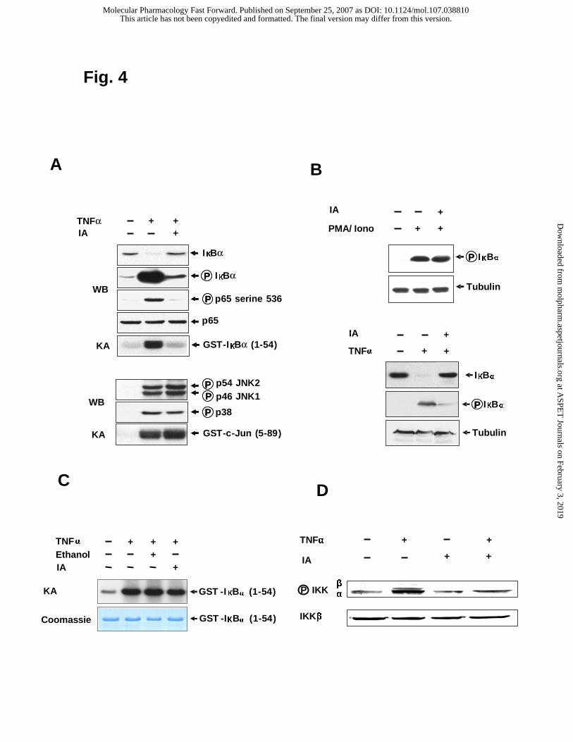
IA Inhibits IκBα and p65 Phosphorylation by Impairment of IKK
Activation. As IKK is also essential for the phosphorylation of p65 at serine 536
(Sizemore et al., 2002; Yang et al., 2003), we further analyzed whether IA affects the
TNFα-induced phosphorylation of p65 in addition to its activity on the degradation of
IκBα. IA inhibited the p65 subunit phosphorylation as well as the phosphorylation of
IκBα and the phosphorylation by IKKs in TNF-stimulated HeLa cells (Fig. 4A, upper
panel). The inhibitory effect of IA on IKK activity is apparently specific, as JNKs and
p38 MAPK, which are also activated after treatment with TNFα (Kang et al., 2004;
Rizzo and carlo-stella, 1996) were unaffected by IA (Fig. 4A, lower panel). In
contrast to IκBα phosphorylation and subsequent degradation in TNF-stimulated
HeLa cells, IA did not inhibit IκBα phosphorylation in human Jurkat T leukemia
cells, costimulated by PMA in combination with ionomycin (Fig. 4B, upper panel).
We examined the effect of IA on TNF-stimulated Jurkat cells and found a robust
inhibition of IκBα phosphorylation and degradation (Fig. 4B, lower panel). The lack
of IA-mediated IKK inhibition in costimulated T-cells raises the possibility that this
compound does not directly target the IKKs (see Mattioli et al., 2004). Accordingly,
This article has not been copyedited and formatted. The final version may differ from this version.Molecular Pharmacology Fast Forward. Published on September 25, 2007 as DOI: 10.1124/mol.107.038810
at ASPE
T Journals on February 3, 2019
molpharm
.aspetjournals.orgD
ownloaded from

MOL #38810
14
in vitro phosphorylation experiments showed full functionality of IKKs in the
presence of IA (Fig. 4C), suggesting that IA targets an upstream event. Collectively,
these data suggest that IA inhibits the NF-κB pathway upstream of IKK. To address
this possibility experimentally, we examined the effect of IA on TNFα-induced
phosphorylation of IKK. These experiments showed inhibition of IKKα/IKKβ
activation loop phosphorylation by IA (Fig. 4D), attributing its effect to an upstream
event.
IA Interfers with TAK/TAB Mediated Phosphorylation of IKKα/β
Activation Loop. To further examine the mechanism of the effect of IA on IKK
activation, we assayed A549 cells, transfected with KBF-luc alone or in combination
with IKKα/IKKβ, TRAF2, or TAK1/TAB2 expression vectors. Treatment with IA
did not interfere with NF-κB activition triggered by TRAF2, while TAK1/TAB2
stimulated NF-κB activition was significantly and dose-depndently inhibited in the
presence of IA (Fig. 5A). In contrast, the effect of IA on NF-κB activation in IKKα/β
overexpressing cells was very mild, and can presumably be attributed to the
interaction of IKKα/β with the endogenous TAK/TAB module. To determine whether
IA can interfere with TAK/TAB induced phosphorylation and thus activation of
IKKα/β, HA-TAK1 and Myc-TAB1 were overexpressed in 293T cells and IKK α/β
phosphorylation was examined by immunoblotting. IA exerted a dose-dependent
reduction of IKKα/β phosphorylation in TAK/TAB overexpressing cells, as displayed
in Fig. 5B. These experiments demonstrate that IA interferes with a critical step
relaying the TAK/TAB module with IKKα/β activation loop phosphorylation.
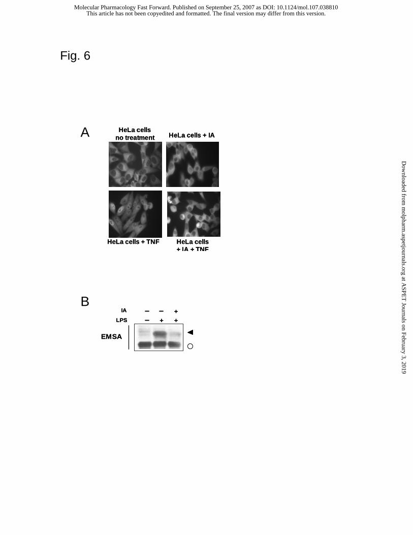
IA Inhibits NF-κB Accumulation in Cell Nuclei and DNA Binding.
Immunostaining of the p65 subunit of NF-κB in TNF-stimulated HeLa cells illustrates
This article has not been copyedited and formatted. The final version may differ from this version.Molecular Pharmacology Fast Forward. Published on September 25, 2007 as DOI: 10.1124/mol.107.038810
at ASPE
T Journals on February 3, 2019
molpharm
.aspetjournals.orgD
ownloaded from

MOL #38810
15
the inhibition of the nuclear accumulation of NF-κB by IA (Fig. 6A).. IA also
inhibited NF-κB DNA-binding in LPS-stimulated human peripheral monocytes, as
examined by EMSA and depicted in Fig. 6B.
IA Inhibits Gene Expression by NF-κB. The HIV-1 promoter contains two
high affinity binding sites for NF-κB and is highly responsive to both the TNFα-
induced NF-κB pathway and the Tat/TAR-dependent pathway. Using stably
transfected cell lines with a plasmid, in which the luciferase gene is driven by the
HIV-1 LTR promoter, we found that IA inhibits TNFα-induced (Fig. 7A), but not Tat-
mediated HIV-1-LTR trans-activation (Fig.7B) in a dose-dependent manner The lack
of interference with Tat-induced transcription rules out potential effects of IA on the
basal transcriptional machinery or other nonspecific effects. In contrast to IA, the
CDK9 inhibitor 5, 6-dichloro-1-β-D-ribofuranosylbenzimidazole riboside (DRB)
effectively inhibited luciferase activity in HeLa-Tat-Luc cells.
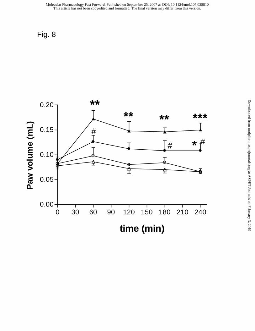
IA suppresses Inflammation in the Mouse Paw Model. Having established
that IA inhibits the NF-κB pathway in vitro, we studied the anti-inflammatory
properties of IA in vivo and found that IA significantly reduced inflammation in the
inflamed paw model in mice (n = 5 per group) during a 4 h period. The decreased
inflamed paw volume in the treated mice reflects a decrease in edema, which is a
component of the inflammatory response. There were highly significant effects of
treatment (F = 11.7, df = 3,64, p < 0.001), time (F = 10.6, df = 4,64, p < 0.0001) and
interaction (F = 3.9, df = 12,64, p < 0.001) as seen in Fig. 8. IA also significantly
reduced other inflammatory parameters: redness (scored by visualization) and pain, as
measured by the paw licking frequency by the mouse (data not shown).
Collectively, these data show that IA inhibits NF-κB activation and exerts
anti-inflammatory properties in the an in vivo model of inflamed paw.
This article has not been copyedited and formatted. The final version may differ from this version.Molecular Pharmacology Fast Forward. Published on September 25, 2007 as DOI: 10.1124/mol.107.038810
at ASPE
T Journals on February 3, 2019
molpharm
.aspetjournals.orgD
ownloaded from

MOL #38810
16
This article has not been copyedited and formatted. The final version may differ from this version.Molecular Pharmacology Fast Forward. Published on September 25, 2007 as DOI: 10.1124/mol.107.038810
at ASPE
T Journals on February 3, 2019
molpharm
.aspetjournals.orgD
ownloaded from
MOL #38810
17
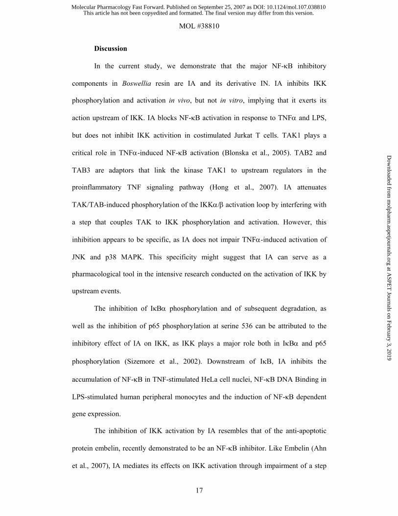
Discussion
In the current study, we demonstrate that the major NF-κB inhibitory
components in Boswellia resin are IA and its derivative IN. IA inhibits IKK
phosphorylation and activation in vivo, but not in vitro, implying that it exerts its
action upstream of IKK. IA blocks NF-κB activation in response to TNFα and LPS,
but does not inhibit IKK activition in costimulated Jurkat T cells. TAK1 plays a
critical role in TNFα-induced NF-κB activation (Blonska et al., 2005). TAB2 and
TAB3 are adaptors that link the kinase TAK1 to upstream regulators in the
proinflammatory TNF signaling pathway (Hong et al., 2007). IA attenuates
TAK/TAB-induced phosphorylation of the IKKα/β activation loop by interfering with
a step that couples TAK to IKK phosphorylation and activation. However, this
inhibition appears to be specific, as IA does not impair TNFα-induced activation of
JNK and p38 MAPK. This specificity might suggest that IA can serve as a
pharmacological tool in the intensive research conducted on the activation of IKK by
upstream events.
The inhibition of IκBα phosphorylation and of subsequent degradation, as
well as the inhibition of p65 phosphorylation at serine 536 can be attributed to the
inhibitory effect of IA on IKK, as IKK plays a major role both in IκBα and p65
phosphorylation (Sizemore et al., 2002). Downstream of IκB, IA inhibits the
accumulation of NF-κB in TNF-stimulated HeLa cell nuclei, NF-κB DNA Binding in
LPS-stimulated human peripheral monocytes and the induction of NF-κB dependent
gene expression.
The inhibition of IKK activation by IA resembles that of the anti-apoptotic
protein embelin, recently demonstrated to be an NF-κB inhibitor. Like Embelin (Ahn
et al., 2007), IA mediates its effects on IKK activation through impairment of a step
This article has not been copyedited and formatted. The final version may differ from this version.Molecular Pharmacology Fast Forward. Published on September 25, 2007 as DOI: 10.1124/mol.107.038810
at ASPE
T Journals on February 3, 2019
molpharm
.aspetjournals.orgD
ownloaded from

MOL #38810
18
connecting TAK to the IKK activation loop phosphorylation. TRAF2 apparently also
recruits the IKK complex directly via the complex of TRADD, TRAF2, TRAF5 and
RIP1 (Hacker and Karin, 2006). Thus, the lack of inhibitory effect by IA on TRAF2
overexpressing cells supports a specific IA intervention at the TAK1-IKK activation
step. The effect of IA on IKK also resembles that of the tetracyclic kaurene diterpenes
as shown by Castrillo et al., as both are signaling rather than direct inhibitors
(Castrillo et al., 2001). However, it appears that IA's activity is more specific to the
NF-κB pathway, as kaurenes also inhibit the phosphorylation of p38, ERK1, and
ERK2 MAPK.
Diterpenoids are natural compounds with a backbone of 20 carbon atoms
biosynthesized from geranylgeranyl pyrophosphate (Hanson, 2005). It is an important
and chemically diverse group of natural products which share some common
biosynthetic steps and are of considerable biological importance (Hanson, 2005).
Interestingly, although members of this group of natural products share no common
chemical moiety, a large arsenal of biologically active compounds has been identified
among them (see Ojo-Amaize et al., 2002; Tempeam et al., 2005; Zhang et al., 2005
for some examples) and several diterpenoids are known as inhibitors of NF-κB
activation (Castrillo et al., 2001; Leung et al., 2005; Yinjun et al., 2005). The
mechanism by which terpenes impair IKK activation has so far been poorly
characterized (Castrillo et al., 2001). IA’s mechanism of action is, however,
completely different from several other anti-inflammtory diterpenoids that inhibit the
NF-κB pathway, for example, oridonin, ponicidin, xindongnin A, and xindongnin B.
These diterpenoids, isolated from Isodon rubescens, directly interfere with the DNA-
binding activity of NF-κB to its response DNA sequence (Leung et al., 2005),
whereas IA inhibits IKK activation. Even with kaurene diterpenoids that inhibit IKK
This article has not been copyedited and formatted. The final version may differ from this version.Molecular Pharmacology Fast Forward. Published on September 25, 2007 as DOI: 10.1124/mol.107.038810
at ASPE
T Journals on February 3, 2019
molpharm
.aspetjournals.orgD
ownloaded from

MOL #38810
19
activation (Castrillo et al., 2001) there are differences, in the mechanism of action,
such as the specificity of action and the effect on p65 phosphorylation. The
observation that some members of a large group of natural products are a source of
NF-κB modulators by a multiplicity of pathways implies that they can serve as a
valuable tool for the examination of the NF-κB pathway, especially upstream of IKK,
where this pathway is still to be unfolded.
Based on our findings, we attribute the main NF-κB inhibitory effect of
Boswellia resin to IA and its derivatives. The resin of Boswellia species, containing
IA derivatives, has been used to treat inflammatory conditions for many centuries in
traditional medicine in Europe, Asia and Africa and is still in such use, besides its
common religious use as incense. One interesting example of its use as an ingredient
of an important anti-inflammatory remedy that has been common in Europe and Asia
for hundreds of years is the Jerusalem Balsam (Moussaieff et al., 2005). Boswellia
extracts are also marketed as food supplements for the treatment of arthritis in the US
and Europe and in view of our current data we propose that these products should be
standardized for IA and its derivatives as well as boswellic acids. Moreover, the
possible synergistic effects of these compounds need to be investigated. We propose
the GC-MS fingerprinting depicted in Data Supplement Fig. 1 as a simple method for
the standardization of Boswellia resin for IA and its derivatives.
IA demonstrates a robust anti-inflammatory effect in a mouse inflamed paw
model. This effect is within the range of drugs such as salicylates injected i.p
(Siqueira-Junior, 2003), However, IA and IN are practically insoluble in water and
poorly soluble in other solvents used for injection to animals or in cell systems.
Hence, the actual active concentrations of these compounds are probably considerably
lower. The identification of the NF-κB inhibitory effect of cembranoid diterpenes, an
This article has not been copyedited and formatted. The final version may differ from this version.Molecular Pharmacology Fast Forward. Published on September 25, 2007 as DOI: 10.1124/mol.107.038810
at ASPE
T Journals on February 3, 2019
molpharm
.aspetjournals.orgD
ownloaded from

MOL #38810
20
important group of common natural products, present in tobacco among other herbs
and marine creatures, may further open these fields to the discovery of novel drugs for
the treatment of diseases that pose unanswered challenges and affect a large segment
of the population.
Acknowledgements
We would like to thank Dr. A. Hatzubai and Dr. M. Davis for their kind advice in
assaying IκBα and p65. We would also like to thank Dr. G. Culioli for providing us
with the boswellic acids mixture. We thank the Miriam and Sheldon Adelson program
in Neural Repair and Rehabilitation for support (to R.M.).
This article has not been copyedited and formatted. The final version may differ from this version.Molecular Pharmacology Fast Forward. Published on September 25, 2007 as DOI: 10.1124/mol.107.038810
at ASPE
T Journals on February 3, 2019
molpharm
.aspetjournals.orgD
ownloaded from

MOL #38810
21
References
Ahn KS, Sethi G and Aggarwal BB (2007) Embelin, an inhibitor of X chromosome-
linked inhibitor-of-apoptosis protein, blocks nuclear factor-kappaB (NF-
kappaB) signaling pathway leading to suppression of NF-kappaB-regulated
antiapoptotic and metastatic gene products. Mol Pharmacol 71:209-219.
Altmann A, Poeckel D, Fischer L, Schubert-Zsilavecz M, Steinhilber D and Werz O
(2004) Coupling of boswellic acid-induced Ca2+ mobilisation and MAPK
activation to lipid metabolism and peroxide formation in human leucocytes. Br
J Pharmacol 141:223-232.
Ben-Neriah Y and Schmitz ML (2004) Of mice and men. EMBO Rep 5:668-673.
Blonska M, Shambharkar PB, Kobayashi M, Zhang D, Sakurai H and Su B, Lin X
(2005) TAK1 is recruited to the tumor necrosis factor-alpha (TNF-alpha)
receptor 1 complex in a receptor-interacting protein (RIP)-dependent manner
and cooperates with MEKK3 leading to NF-kappaB activation. J Biol Chem
280:43056-43063.
Bremner P and Heinrich M (2002) Natural products as targeted modulators of the
nuclear factor-kappaB pathway. J Pharm Pharmacol 54:453-472.
Calhoun W, Chang J and Carlson RP (1987) Effect of selected antiinflammatory
agents and other drugs on zymosan, arachidonic acid, PAF and carrageenan
induced paw edema in the mouse. Agents Actions 21:306-309.
Calzado MA, Bacher S and Schmitz ML (2007) NF-kappaB inhibitors for the
treatment of inflammatory diseases and cancer. Curr Med Chem 14:367-376.
Castrillo A, de las Heras B, Hortelano S, Rodriguez B, Villar A and Bosca L (2001)
Inhibition of the nuclear factor kappa B (NF-kappa B) pathway by tetracyclic
kaurene diterpenes in macrophages. Specific effects on NF-kappa B-inducing
This article has not been copyedited and formatted. The final version may differ from this version.Molecular Pharmacology Fast Forward. Published on September 25, 2007 as DOI: 10.1124/mol.107.038810
at ASPE
T Journals on February 3, 2019
molpharm
.aspetjournals.orgD
ownloaded from

MOL #38810
22
kinase activity and on the coordinate activation of ERK and p38 MAPK. J
Biol Chem 276:15854-15860.
Corsano S and Nicoletti R (1967) The structure of incensole. Tetrahedron 23:1977-
1984.
English D and Andersen BR (1974) Single-step separation of red blood cells.
Granulocytes and mononuclear leukocytes on discontinuous density gradients
of Ficoll-Hypaque. J Immunol Methods 5:249-252.
Gacs-Baitz E, Radics L, Fardella G and Corsano S (1978) Carbon-13 nuclear
magnetic resonance of some fourteen-membered macrocyclic diterpenes. J
Chem Res (M) 1701-1709.
Gerhardt H, Seifert F, Buvari P, Vogelsang H and Repges R (2001) Therapy of active
Crohn disease with Boswellia serrata extract H 15. Z Gastroenterol 39:11-17.
Gupta I, Gupta V, Parihar A, Gupta S, Ludtke R, Safayhi H, and Ammon HP (1998)
Effects of Boswellia serrata gum resin in patients with bronchial asthma:
results of a double-blind, placebo-controlled, 6-week clinical study. Eur J Med
Res 3:511-514.
Hacker H and Karin M (2006) Regulation and function of IKK and IKK-related
kinases. Sci STKE 2006:re13.
Hamm S, Bleton J, Connan J and Tchapla A (2005) A chemical investigation by
headspace SPME and GC-MS of volatile and semi-volatile terpenes in various
olibanum samples. Phytochemistry 66:1499-1514.
Hanson JR (2005) Diterpenoids. Nat Prod Rep 22:594-602.
Hong S, Lim S, Li AG, Lee C, Lee YS, Lee EK, Park SH, Wang XJ and Kim SJ
(2007) Smad7 binds to the adaptors TAB2 and TAB3 to block recruitment of
the kinase TAK1 to the adaptor TRAF2 Nat Immunol 8:504-513.
This article has not been copyedited and formatted. The final version may differ from this version.Molecular Pharmacology Fast Forward. Published on September 25, 2007 as DOI: 10.1124/mol.107.038810
at ASPE
T Journals on February 3, 2019
molpharm
.aspetjournals.orgD
ownloaded from

MOL #38810
23
Kang SW, Chang TS, Lee TH, Kim ES, Yu DY and Rhee SG (2004) Cytosolic
peroxiredoxin attenuates the activation of Jnk and p38 but potentiates that of
Erk in Hela cells stimulated with tumor necrosis factor-alpha. J Biol Chem
279:2535-2543.
Karin M and Ben-Neriah Y (2000) Phosphorylation meets ubiquitination: the control
of NF-[kappa]B activity. Annu Rev Immunol 18:621-663.
Karin M (2005) Inflammation-activated protein kinases as targets for drug
development. Proc Am Thorac Soc 2:386-90.
Khanna D, Sethi G, Ahn KS, Pandey MK, Kunnumakkara AB, Sung B, Aggarwal A
and Aggarwal BB (2007) Natural products as a gold mine for arthritis
treatment. Curr Opin Pharmacol 7:344-51.
Lawrence T, Gilroy DW, Colville-Nash PR and Willoughby DA (2001) Possible new
role for NF-kappaB in the resolution of inflammation. Nat Med 7:1291-1297.
Leung CH, Grill SP, Lam W, Han, QB, Sun HD and Cheng YC (2005) Novel
mechanism of inhibition of nuclear factor-kappa B DNA-binding activity by
diterpenoids isolated from Isodon rubescens. Mol Pharmacol 68:286-297.
Mattioli I, Sebald A, Bucher C, Charles RP, Nakano H, Doi T, Kracht M and Schmitz
ML (2004) Transient and selective NF-kappa B p65 serine 536
phosphorylation induced by T cell costimulation is mediated by I kappa B
kinase beta and controls the kinetics of p65 nuclear import. J Immunol
172:6336-6344.
Moussaieff A, Fride E, Amar Z, Lev E, Steinberg D, Gallily R and Mechoulam R
(2005) The Jerusalem Balsam: from the Franciscan Monastery in the old city
of Jerusalem to Martindale 33. J Ethnopharmacol 101:16-26.
Noble PB, Cutts JH and Carroll KK (1968) Ficoll flotation for the separation of blood
This article has not been copyedited and formatted. The final version may differ from this version.Molecular Pharmacology Fast Forward. Published on September 25, 2007 as DOI: 10.1124/mol.107.038810
at ASPE
T Journals on February 3, 2019
molpharm
.aspetjournals.orgD
ownloaded from

MOL #38810
24
leukocyte types. Blood 31:66-73.
Ojo-Amaize EA, Nchekwube EJ, Cottam HB., Bai R, Verdier-Pinard P, Kakkanaiah,
VN, Varner JA, Leoni L, Okogun JI, Adesomoju AA, Oyemade OA and
Hamel E (2002) Hypoestoxide, a natural nonmutagenic diterpenoid with
antiangiogenic and antitumor activity: possible mechanisms of action. Cancer
Res 62:4007-4014.
Perkins ND (2007) Integrating cell-signalling pathways with NF-kappaB and IKK
function. Nat Rev Mol Cell Biol 8:49-62.
Rizzo M and Carlo-Stella C (1996) Arachidonic acid mediates interleukin-1 and
tumor necrosis factor-alpha-induced activation of the c-jun amino-terminal
kinases in stromal cells. Blood 88:3792-3800.
Sancho R, Medarde M, Sanchez-Palomino S, Madrigal, BM, Alcami J, Munoz E and
San Feliciano A (2004) Anti-HIV activity of some synthetic lignanolides and
intermediates. Bioorg Med Chem Lett 14:4483-4486.
Siqueira-Junior JM, Peters RR, Brum-Fernandes AJ and Ribeiro-do-Valle RM (2003)
Effects of valeryl salicylate, a COX-1 inhibitor, on models of acute
inflammation in mice. Pharmacol Res 48:437-443.
Sizemore N, Lerner N, Dombrowski N, Sakurai H and Stark GR (2002) Distinct roles
of the Ikappa B kinase alpha and beta subunits in liberating nuclear factor
kappa B (NF-kappa B) from Ikappa B and in phosphorylating the p65 subunit
of NF-kappa B. J Biol Chem 277:3863-3869.
Tempeam A, Thasana N, Pavaro C, Chuakul W, Siripong P and Ruchirawat S (2005)
A new cytotoxic daphnane diterpenoid, rediocide G, from Trigonostemon
reidioides. Chem Pharm Bull 53:1321-1323.
Xia L, Chen D, Han R, Fang Q, Waxman S and Jing Y (2005) Boswellic acid acetate
This article has not been copyedited and formatted. The final version may differ from this version.Molecular Pharmacology Fast Forward. Published on September 25, 2007 as DOI: 10.1124/mol.107.038810
at ASPE
T Journals on February 3, 2019
molpharm
.aspetjournals.orgD
ownloaded from

MOL #38810
25
induces apoptosis through caspase-mediated pathways in myeloid leukemia
cells. Mol Cancer Ther 4:381-388.
Yang F, Tang E, Guan K and Wang CY (2003) IKK beta plays an essential role in the
phosphorylation of RelA/p65 on serine 536 induced by lipopolysaccharide. J
Immunol 170:5630-5635.
YinJun L, Jie J and YunGui W (2005) Triptolide inhibits transcription factor NF-
kappaB and induces apoptosis of multiple myeloma cells. Leuk Res 29:99-
105.
Zhang CX, Yan SJ, Zhang GW, Lu WG, Su JY, Zeng LM, Gu LQ, Yang XP and Lian
YJ (2005) Cytotoxic diterpenoids from the soft coral Sinularia microclavata J
Nat Prod 68:1087-1089.
This article has not been copyedited and formatted. The final version may differ from this version.Molecular Pharmacology Fast Forward. Published on September 25, 2007 as DOI: 10.1124/mol.107.038810
at ASPE
T Journals on February 3, 2019
molpharm
.aspetjournals.orgD
ownloaded from
MOL #38810
26
Footnotes
This work was supported by a EC 5th framework consortium grant number QLK3-
CT-2000-00-463 (AINP consortium).
Person to receive reprint request: Prof. Raphael Mechoulam, Department of
Medicinal Chemistry and Natural Products, Medical Faculty, Hebrew University,
Jerusalem 91120, Israel; Tel. - 972-2-6758634; fax - 972-2-6758073; e-mail:
This article has not been copyedited and formatted. The final version may differ from this version.Molecular Pharmacology Fast Forward. Published on September 25, 2007 as DOI: 10.1124/mol.107.038810
at ASPE
T Journals on February 3, 2019
molpharm
.aspetjournals.orgD
ownloaded from
MOL #38810
27
Legends for Figures
Fig. 1. The structures of IA (R = Ac.) and IN (R = H). Structures elucidation was
done according to NMR (see Materials and Methods; Tables 1, 2 in Data Supplement)
and MS data (see Materials and Methods; Fig. 1 in Data Supplement).
Fig. 2. Comparison of isolated IA with boswellic acid on IκBα degradation in TNFα
stimulated HeLa cells. Cells were treated with non-toxic concentrations of IA (220
µM) and boswellic acid (280 µM) and stimulated with TNFα (20 ng/mL for 20 mins).
Equal amounts of protein were separated by SDS-PAGE and further analyzed by
immunoblotting. IκBα / β-actin ratio of untreated cells was considered as 100%. * p <
0.05; ** p < 0.001 by t test (vs. vehicle + TNFα stimulated cells). The error bars
represent SEM
Fig. 3. IA and IN inhibit IκBα degradation in a dose-dependent manner. HeLa cells
were pre-incubated with IA (upper) or IN (lower) at the indicated concentrations for 2
h prior to 20 mins exposure to TNFα (20 ng/mL). A representative experiment is
shown.
Fig. 4. IA inhibits the phosphorylation and subsequent degradation of IκBα by
impairment of IKK phosphorylation and activation. A, HeLa cells were stimulated
with TNFα (20 ng/mL for 20 mins) in the absence or presence of IA (140 µM) as
shown. Subsequently, whole cell extracts were prepared and aliquots thereof analyzed
either for the stability and phosphoryation of the indicated proteins by WB or for IKK
activity by kinase assays (KA). IKKγ/NEMO was immunoprecipitated from cell
This article has not been copyedited and formatted. The final version may differ from this version.Molecular Pharmacology Fast Forward. Published on September 25, 2007 as DOI: 10.1124/mol.107.038810
at ASPE
T Journals on February 3, 2019
molpharm
.aspetjournals.orgD
ownloaded from

MOL #38810
28
lysates and IKK activity was determined by immune complex kinase assays using
recombinant GST-IκB-α (1-54) as substrate. An autoradiogram from a reducing SDS
gel is shown. The lower panel shows the effect of IA on the phosphorylation of p38
and JNK1/2 and JNK in vitro kinase activity. The experiment shown is representative
of three independent experiment sets. B, IA does not impair IκBα phosphorylation in
costimulated T cells. Human Jurkat T leukemia cells were left untreated or incubated
with IA (560 µM). T cells were costimulated by treatment with 20 ng/mL PMA in
combination with ionomycin (100 ng/mL) as shown. After 15 mins, cell extracts were
prepared and analyzed by immunoblotting for the phosphorylation of IκBα. TNF-
stimulated Jurkat cells (lower panel) were left untreated or incubated with IA in the
same manner as costimulated cells. C, IA does not inhibit IKK activity in vitro. HeLa
cells were stimulated with TNFα as shown and the IKK complex was isolated by
immunoprecipitation with αIKKγ/NEMO antibodies. Immune complex kinase assays
using the GST-IκBα substrate protein were performed in the presence of IA (150 µM)
or ethanol as a solvent control. An autoradiogram from a reducing SDS gel (upper) and
a Coomassie staining of the GST-IκBα fusion proteins (lower) are shown. D, IA
inhibits the phosphorylation of IKK activation loop. HeLa cells were stimulated with
TNFα (20 ng/mL for 20 mins) in the presence of IA (140 µM) or ethanol as a solvent
control. Whole cell extracts were prepared and analyzed for the phosphoryation of
IKKα/β by WB.
Fig 5. IA Inhibits TAK/TAB-triggered IKK activation. A,. A549 cells were transiently
transfected with KBF-luc alone or in combination with IKKα/IKKβ, TRAF2 or
TAK1/TAB2 expression vectors. After 24 h of transfection, cells were treated with IA for 6
h and luciferase activity was assayed. In order to allow comparability, activation by
IKKα/IKKβ, TRAF2 or TAK1/TAB2 in untreated cells were given a value of 100% and
This article has not been copyedited and formatted. The final version may differ from this version.Molecular Pharmacology Fast Forward. Published on September 25, 2007 as DOI: 10.1124/mol.107.038810
at ASPE
T Journals on February 3, 2019
molpharm
.aspetjournals.orgD
ownloaded from

MOL #38810
29
the IA inhibitory effect is represented as percentage of activation. Average values from
three independent experiments are shown, error bars show SEM. The data represent the
results of 5 independent experiments. *, p < 0.05; ** p < 0.001 compared to nontreated
cells (Two way ANOVA, followed by a Bonferonni multiple comparison test). B, IA
inhibits the phosphorylation of IKKα/β induced by TAK/ TAB overexpression. 293T cells
were transfected with expression vectors for HA-TAK1 and Myc-TAB1 or with a control
vector. 36 h post transfection, cells were treated for 6 h with increasing concentrations of
IA as shown and lysed. Equal amounts of protein were examined for phosphorylation of
IKKα/β activation loop by immunoblotting. Similar results were obtained when
transfecting cells with TAK1/TAB2 (data not shown).
Fig 6. IA inhibits the accumulation of p65 in cell nuclei of TNFα-stimulated HeLa
cells and the NF-κB DNA-binding of LPS-stimulated human peripheral monocytes.
A, HeLa cells were stimulated with TNFα (20 ng/mL for 20 min) in the presence of
IA (140 µM) or ethanol as a solvent control. Cells were fixed and then stained with
rabbit anti-p65 followed by anti-rabbit Rhodamine Red-labeled secondary antibody
and with DAPI for nuclear location (not shown). The cells were examined under an
Axioscope Zeiss microscope with a plan-Neofluor * 60 lens. Results of one of three
independent experiments are shown. B, Human peripheral monocytes extracts in the
presence or in the absence of IA (100 µM) were tested by EMSA for NF-κB DNA-
binding. The filled arrowhead indicates the location of the DNA-NF-κB complex; the
circle indicates the position of a constitutively DNA-binding protein.
Fig. 7. IA inhibits NF-κB activation in TNFα-stimulated 5.1 Jurkat cells. A, 5.1 Cells
were preincubated with IA for 30 mins at the indicated concentrations and stimulated
with TNFα (2 ng/mL) for 6 h. The luciferase activity was then measured. There was no
effect of solvent on cell viability or on the enzymatic activity of luciferase at the
This article has not been copyedited and formatted. The final version may differ from this version.Molecular Pharmacology Fast Forward. Published on September 25, 2007 as DOI: 10.1124/mol.107.038810
at ASPE
T Journals on February 3, 2019
molpharm
.aspetjournals.orgD
ownloaded from

MOL #38810
30
highest concentration (data not shown). B, HeLa-Tat-Luc cells were incubated with
either DRB (50 µM) or IA at the indicated concentrations for 18 h. Tat-induced LTR-
luciferase activity was measured.
Luciferase activity is given as fold induction or relative light units (RLU), standard
deviations are given. ** p ≤ 0.01 to fold values in the absence of IA by t test.
Fig. 8. IA inhibited inflammation in the inflamed paw model after injection of
carrageenin. IA (50 mg/kg) or vehicle was injected i.p. to Sabra female mice (5 per
group) 30 mins before induction of the inflammatory stimulus. Hind paws were then
injected with 50 µl of saline or λ-carrageenin (4%). Ensuing inflammatory swelling
was measured by increase in foot volume in a plethysmometer. IA also reduced paw
redness (as a measure of erythema) and licking (as a measure of pain) (data not
shown). There were highly significant effects of treatment (F = 11.7, df = 3,64, p <
0.001).
Vehicle + saline, open triangles; vehicle + carrageenin, closed triangles; IA + saline,
open circles; IA + carrageenin, closed circles; *, different from IA + saline, p < 0.05;
**, ***, different from vehicle + saline at p < 0.01, p < 0.001 respectively; #, different
from vehicle + carrageenin, p < 0.05.
This article has not been copyedited and formatted. The final version may differ from this version.Molecular Pharmacology Fast Forward. Published on September 25, 2007 as DOI: 10.1124/mol.107.038810
at ASPE
T Journals on February 3, 2019
molpharm
.aspetjournals.orgD
ownloaded from

R= Ac (IA), H (IN)
O
RO
1
32
4
5
67
10
12
11
13
14
8
915
16
17
18
20
19
Fig. 1
This article has not been copyedited and formatted. The final version may differ from this version.Molecular Pharmacology Fast Forward. Published on September 25, 2007 as DOI: 10.1124/mol.107.038810
at ASPE
T Journals on February 3, 2019
molpharm
.aspetjournals.orgD
ownloaded from

IA Boswellic ac. control
- + - + - +
I КBα
β actin
IA
IA+T
NF
boswell
icac
.
boswell
icac
.+TNF
vehi
cle
vehi
cle+T
NF
0
100
200
***
treatment
IК
B
α
/
β
acti
n(%
)Fig. 2
TNFα
This article has not been copyedited and formatted. The final version may differ from this version.Molecular Pharmacology Fast Forward. Published on September 25, 2007 as DOI: 10.1124/mol.107.038810
at ASPE
T Journals on February 3, 2019
molpharm
.aspetjournals.orgD
ownloaded from

IA (µM)
- + - + - + - +
B
βactin
TNF
60 80 140 Control
I К
TNFα
60 100 160 Control
+ - + - + - +- - +
α
IN (µM)
TNFTNFα
B
βactin
I К α
Fig. 3
This article has not been copyedited and formatted. The final version may differ from this version.Molecular Pharmacology Fast Forward. Published on September 25, 2007 as DOI: 10.1124/mol.107.038810
at ASPE
T Journals on February 3, 2019
molpharm
.aspetjournals.orgD
ownloaded from
A B
Fig. 4
PMA/ Iono +
IA
IКBPP
Tubulin
+
+
P IKK
IKKβ
βα
TNFα + +
IA + +
TNFα +
IA
IКB
IКBPP
Tubulin
+
+
KA
Coomassie
TNFα + +
IA +
+Ethanol +
GST -I КBα (1-54)
GST -IКBα (1-54)
α
α
α
IКBα
IКBα
p65 serine 536
p65
PP
PP
TNFα + +IA +
KA
WB
GST-IКBα (1-54)
p38PP
KA GST-c-Jun (5-89)
p54 JNK2PPPP p46 JNK1
WB
CD
This article has not been copyedited and formatted. The final version may differ from this version.Molecular Pharmacology Fast Forward. Published on September 25, 2007 as DOI: 10.1124/mol.107.038810
at ASPE
T Journals on February 3, 2019
molpharm
.aspetjournals.orgD
ownloaded from

Fig. 5
0 30 70 1400
20
40
60
80
100
120IKKα/βTRAF2TAK1/TAB2
**
**
IA (µM)
% A
ctiv
atio
n
+
-
+
280
+
140
+
70
-
-
HA-TAK1 / Myc-TAB1
IA (µM)
+
-
+
280
+
140
+
70
-
-
HA-TAK1 / Myc-TAB1
IA (µM)
IKKβ
- IKKα/βPP
HA-TAK1
Myc-TAB1
A
B
This article has not been copyedited and formatted. The final version may differ from this version.Molecular Pharmacology Fast Forward. Published on September 25, 2007 as DOI: 10.1124/mol.107.038810
at ASPE
T Journals on February 3, 2019
molpharm
.aspetjournals.orgD
ownloaded from
HeLa cells no treatment HeLa cells + IA
HeLa cells + IA + TNF
HeLa cells + TNF
HeLa cells no treatment HeLa cells + IA
HeLa cells + IA + TNF
HeLa cells + TNF
EMSAEMSA
LPS + +
IA +LPS + +
IA +
A
B
Fig. 6
This article has not been copyedited and formatted. The final version may differ from this version.Molecular Pharmacology Fast Forward. Published on September 25, 2007 as DOI: 10.1124/mol.107.038810
at ASPE
T Journals on February 3, 2019
molpharm
.aspetjournals.orgD
ownloaded from

0
10
20
30
40
TNFα
IA (µM)0 300 153 70 140 280- + + + + ++ +
FO
LD
IND
UC
TIO
N
**** **
0
10
20
30
40
50
R.L
.U(x
106 )
0 300 70 IA (µM)
- -+ - DRB (50 µM)140
-
**
A
B
Fig. 7
This article has not been copyedited and formatted. The final version may differ from this version.Molecular Pharmacology Fast Forward. Published on September 25, 2007 as DOI: 10.1124/mol.107.038810
at ASPE
T Journals on February 3, 2019
molpharm
.aspetjournals.orgD
ownloaded from
0 30 60 90 120 150 180 210 2400.00
0.05
0.10
0.15
0.20 **
*#
***
#
** **
#
time (min)
Paw
vo
lum
e (m
L)
Fig. 8
This article has not been copyedited and formatted. The final version may differ from this version.Molecular Pharmacology Fast Forward. Published on September 25, 2007 as DOI: 10.1124/mol.107.038810
at ASPE
T Journals on February 3, 2019
molpharm
.aspetjournals.orgD
ownloaded from