-Arrestin-Dependent -Opioid Receptor-Activated...
13
-Arrestin-Dependent -Opioid Receptor-Activated Extracellular Signal-Regulated Kinases (ERKs) Translocate to Nucleus in Contrast to G Protein-Dependent ERK Activation Hui Zheng, Horace H. Loh, and Ping-Yee Law Department of Pharmacology, Medical School, University of Minnesota, Minneapolis, Minnesota Received July 16, 2007; accepted October 18, 2007 ABSTRACT The cellular location of extracellular signal-regulated kinases (ERKs) activated by a G protein-coupled receptor was shown to be dependent on the pathway that mediated their activation. In general, fast activation of ERKs (2 min) mediated by G proteins resulted in the nuclear translocation of phosphorylated ERKs, whereas a slower activation of ERKs (10 min) mediated by -arrestins resulted in the cytosolic retention of the phosphor- ylated ERKs. However, we observed distinct differences from this established ERKs cellular itinerary with the -opioid recep- tor-activated ERKs. Agonists such as morphine and metha- done activated ERKs via the protein kinase C-dependent path- way but not the -arrestin-dependent pathway. The activated ERKs did not translocate into the nucleus, but phosphorylated 90-kDa ribosomal S6 kinase and induced the activity of tran- scription factor cAMP response element-binding protein. In contrast, agonists such as etorphine and fentanyl activated ERKs in a -arrestin-dependent manner. The phosphorylated ERKs translocated into the nucleus, resulting in increases in Elk-1 activity and GRK2 and -arrestin2 transcriptions. Thus, the cellular location of phosphorylated ERKs and subsequent activities on gene transcriptions are dictated by the agonist used to activate the receptor and the subsequent signaling pathway involved. G protein-dependent and -arrestin-dependent pathways are two well-established pathways for ERK activation in G q - and G s -coupled GPCRs (DeWire et al., 2007). The involve- ment of the G protein-dependent pathway in ERK activation has been demonstrated by the use of G protein-dependent pathways inhibitors, such as PKC or protein kinase A inhib- itors (Ahn et al., 2004; Gesty-Palmer et al., 2006). -arrestin- dependent ERK activation was illustrated by using siRNA to knockdown -arrestin levels (Ahn et al., 2004). Although these two pathways could be observed at the same time, they are independent of each other, for ERK activation still could be observed if one of the pathways was blocked (DeWire et al., 2007). Although the two pathways have been observed with many GPCRs, the kinetics of ERK activation are different. In cells expressing angiotensin II receptors, G protein-dependent ERK activation usually peaked 2 min after stimulation and returned to basal level at the 10-min time point, whereas the -arrestin-dependent ERK activation peaked 10 min after agonist addition (Ahn et al., 2004). In cells expressing para- thyroid receptors, G protein-dependent ERK activation peaked at 10 min, whereas -arrestin-dependent ERK acti- vation peaked 30 to 60 min after stimulation (Gesty-Palmer et al., 2006). In cells expressing vasopressin receptors, ERK activation mediated by both pathways peaked 2 min after stimulation (Ren et al., 2005). In addition, not only are there agonists that activate ERKs via both the G protein- and -arrestin-dependent pathways (DeWire et al., 2007); there are also agonists that use only one of the two pathways. For example, isoproterenol acti- vates ERKs using both pathways, whereas the 2-adrenergic agonist ICI118551 activates ERKs completely via -arrestin- This research was supported in part by National Institutes of Health grants DA007339, DA016674, DA000564, and DA011806. H.H.L. and P.Y.L. are re- cipients of K05-DA70544 and K05-DA00513, respectively. Article, publication date, and citation information can be found at http://molpharm.aspetjournals.org. doi:10.1124/mol.107.039842. ABBREVIATIONS: ERK, extracellular signal-regulated kinase; 90RSK, 90-kDa ribosomal s6 kinase; CREB, cAMP response element-binding; GPCR, G protein-coupled receptor; HEK, human embryonic kidney; MEF, mouse embryonic fibroblast; MOR, -opioid receptor; PKC, protein kinase C; HA, hemagglutinin; FBS, fetal bovine serum; FACS, fluorescence-activated cell sorting; PBS, phosphate-buffered saline; RT-PCR, reverse transcription-polymerase chain reaction; MEK, mitogen-activated protein kinase kinase; DOR, -opioid receptor; CTOP, D-Phe-Cys-Tyr- D-Trp-Orn-Thr-Pen-Thr-NH 2 ; DAMGO, [D-Ala 2 ,N-Me-Phe 4 ,Gly 5 -ol]-enkephalin; ICI118551, ()-1-[2,3-(dihydro-7-methyl-1H-inden-4-yl)oxy]-3-[(1- methylethyl)amino]-2-butanol; Ro-31-8425, 2-[8-(aminomethyl)-6,7,8,9-tetrahydropyrido[1,2-a]indol-3-yl]-3-(1-methylindol-3-yl)maleimide, HCl; PD98059, 2-amino-3-methoxyflavone; HA-MOR, hemagglutinin-tagged -opioid receptor; TIPP, H-Tyr-Tic[CH 2 NH]-Phe-Phe-OH. 0026-895X/08/7301-178 –190$20.00 MOLECULAR PHARMACOLOGY Vol. 73, No. 1 Copyright © 2008 The American Society for Pharmacology and Experimental Therapeutics 39842/3290530 Mol Pharmacol 73:178–190, 2008 Printed in U.S.A. 178 at ASPET Journals on June 18, 2018 molpharm.aspetjournals.org Downloaded from
Transcript of -Arrestin-Dependent -Opioid Receptor-Activated...
�-Arrestin-Dependent �-Opioid Receptor-ActivatedExtracellular Signal-Regulated Kinases (ERKs) Translocate toNucleus in Contrast to G Protein-Dependent ERK Activation
Hui Zheng, Horace H. Loh, and Ping-Yee LawDepartment of Pharmacology, Medical School, University of Minnesota, Minneapolis, Minnesota
Received July 16, 2007; accepted October 18, 2007
ABSTRACTThe cellular location of extracellular signal-regulated kinases(ERKs) activated by a G protein-coupled receptor was shown tobe dependent on the pathway that mediated their activation. Ingeneral, fast activation of ERKs (2 min) mediated by G proteinsresulted in the nuclear translocation of phosphorylated ERKs,whereas a slower activation of ERKs (10 min) mediated by�-arrestins resulted in the cytosolic retention of the phosphor-ylated ERKs. However, we observed distinct differences fromthis established ERKs cellular itinerary with the �-opioid recep-tor-activated ERKs. Agonists such as morphine and metha-done activated ERKs via the protein kinase C-dependent path-way but not the �-arrestin-dependent pathway. The activated
ERKs did not translocate into the nucleus, but phosphorylated90-kDa ribosomal S6 kinase and induced the activity of tran-scription factor cAMP response element-binding protein. Incontrast, agonists such as etorphine and fentanyl activatedERKs in a �-arrestin-dependent manner. The phosphorylatedERKs translocated into the nucleus, resulting in increases inElk-1 activity and GRK2 and �-arrestin2 transcriptions. Thus,the cellular location of phosphorylated ERKs and subsequentactivities on gene transcriptions are dictated by the agonistused to activate the receptor and the subsequent signalingpathway involved.
G protein-dependent and �-arrestin-dependent pathwaysare two well-established pathways for ERK activation in Gq-and Gs-coupled GPCRs (DeWire et al., 2007). The involve-ment of the G protein-dependent pathway in ERK activationhas been demonstrated by the use of G protein-dependentpathways inhibitors, such as PKC or protein kinase A inhib-itors (Ahn et al., 2004; Gesty-Palmer et al., 2006). �-arrestin-dependent ERK activation was illustrated by using siRNA toknockdown �-arrestin levels (Ahn et al., 2004). Althoughthese two pathways could be observed at the same time, theyare independent of each other, for ERK activation still couldbe observed if one of the pathways was blocked (DeWire etal., 2007).
Although the two pathways have been observed with manyGPCRs, the kinetics of ERK activation are different. In cellsexpressing angiotensin II receptors, G protein-dependentERK activation usually peaked 2 min after stimulation andreturned to basal level at the 10-min time point, whereas the�-arrestin-dependent ERK activation peaked 10 min afteragonist addition (Ahn et al., 2004). In cells expressing para-thyroid receptors, G protein-dependent ERK activationpeaked at 10 min, whereas �-arrestin-dependent ERK acti-vation peaked 30 to 60 min after stimulation (Gesty-Palmeret al., 2006). In cells expressing vasopressin receptors, ERKactivation mediated by both pathways peaked 2 min afterstimulation (Ren et al., 2005).
In addition, not only are there agonists that activate ERKsvia both the G protein- and �-arrestin-dependent pathways(DeWire et al., 2007); there are also agonists that use onlyone of the two pathways. For example, isoproterenol acti-vates ERKs using both pathways, whereas the �2-adrenergicagonist ICI118551 activates ERKs completely via �-arrestin-
This research was supported in part by National Institutes of Health grantsDA007339, DA016674, DA000564, and DA011806. H.H.L. and P.Y.L. are re-cipients of K05-DA70544 and K05-DA00513, respectively.
Article, publication date, and citation information can be found athttp://molpharm.aspetjournals.org.
doi:10.1124/mol.107.039842.
ABBREVIATIONS: ERK, extracellular signal-regulated kinase; 90RSK, 90-kDa ribosomal s6 kinase; CREB, cAMP response element-binding;GPCR, G protein-coupled receptor; HEK, human embryonic kidney; MEF, mouse embryonic fibroblast; MOR, �-opioid receptor; PKC, proteinkinase C; HA, hemagglutinin; FBS, fetal bovine serum; FACS, fluorescence-activated cell sorting; PBS, phosphate-buffered saline; RT-PCR,reverse transcription-polymerase chain reaction; MEK, mitogen-activated protein kinase kinase; DOR, �-opioid receptor; CTOP, D-Phe-Cys-Tyr-D-Trp-Orn-Thr-Pen-Thr-NH2; DAMGO, [D-Ala2,N-Me-Phe4,Gly5-ol]-enkephalin; ICI118551, (�)-1-[2,3-(dihydro-7-methyl-1H-inden-4-yl)oxy]-3-[(1-methylethyl)amino]-2-butanol; Ro-31-8425, 2-[8-(aminomethyl)-6,7,8,9-tetrahydropyrido[1,2-a]indol-3-yl]-3-(1-methylindol-3-yl)maleimide, HCl;PD98059, 2�-amino-3�-methoxyflavone; HA-MOR, hemagglutinin-tagged �-opioid receptor; TIPP�, H-Tyr-Tic�[CH2NH]-Phe-Phe-OH.
0026-895X/08/7301-178–190$20.00MOLECULAR PHARMACOLOGY Vol. 73, No. 1Copyright © 2008 The American Society for Pharmacology and Experimental Therapeutics 39842/3290530Mol Pharmacol 73:178–190, 2008 Printed in U.S.A.
178
at ASPE
T Journals on June 18, 2018
molpharm
.aspetjournals.orgD
ownloaded from

dependent pathway, and CCL19 uses only the G protein-dependent pathway in cells expressing the chemokine recep-tor CCR7 (Azzi et al., 2003; Kohout et al., 2004; Shenoy et al.,2006).
Because the eventual cellular locations of activatedERKs are linked to the pathway mediating the activationand downstream cascades of ERKs involve transcriptionfactors, the pathway for ERK activation becomes critical tooverall cellular responses. In cells expressing angiotensinreceptors, ERKs activated via G protein-dependent path-way were shown to translocate to the nucleus, whereas�-arrestin-activated ERKs remained in the cytosol (Ahn etal., 2004). However, this pathway-dependent cellular loca-tion is not without controversy. Opposite results were re-ported on �-arrestin’s influence on nuclear translocation ofERKs. By overexpressing �-arrestin2 in COS-7 cells withangiotensin II type 1A receptors, Tohgo et al. (2002) re-ported that nuclear translocation of ERKs was inhibited.In contrast, when �-arrestin2 was overexpressed in COS-7cells with �2-adrenergic receptors, nuclear translocation ofERKs was enhanced (Kobayashi et al., 2005). These twoapparently contrasting studies suggest that the eventualcellular location of the activated ERKs should relate to thetype of GPCR.
�-Opioid receptor (MOR), which couples to Gi/o, has beenshown to activate ERKs (Li and Chang, 1996). On the onehand, by using the PKC inhibitor bisindolylmaleimide I,Belcheva et al. (2005) were able to block both DAMGO-and morphine-induced ERK activation in the corticalastrocytes cultures, suggesting the involvement of the Gprotein-dependent pathway in MOR-mediated ERK activa-tion. On the other hand, Ignatova et al. (1999) reportedthat MOR-mediated ERK activation was attenuated by theblockade of receptor internalization (Ignatova et al., 1999).Although subsequent reports have challenged the linkagebetween MOR-mediated ERK activation and receptor in-ternalization (Kramer and Simon, 2000), these studiessuggest the possible involvement of �-arrestin-dependentpathways in MOR-mediated ERK activation. A recent re-port with Chinese hamster ovary cells proved the involve-ment of both �-arrestin- and G protein-dependent activa-tion of ERKs by MOR (Rozenfeld and Devi, 2007).
However, the details of �-arrestin- and G protein-depen-dent ERK activation by MOR have not been resolved. Forexample, the kinetics of ERK activation and the location ofphosphorylated ERKs related to the two pathways are un-clear. In addition, whether all MOR agonists activate ERKssimilarly has not been demonstrated, especially in the situ-ation in which morphine-MOR complex has been shown tohave a low affinity for �-arrestin (Keith et al., 1996). Toaddress these questions, we monitored the activation ofERKs and subsequent cellular location of the activated ki-nases in HEK293 cells expressing high levels of MOR, inhuman neuroblastoma SHSY5Y cells expressing relativelylow levels of endogenous MOR, and in the primary neuronalculture of rat hippocampus, which is one of the regions thatcontain highest levels of MOR (Arvidsson et al., 1995). Inaddition, we examined pathway selectivity for ERK activa-tion with four MOR agonists (morphine, etorphine, metha-done, and fentanyl).
Materials and MethodsCell Culture and Chemicals. HEK293 cells stably expressing
hemagglutinin (HA)-tagged �-opioid receptor were cultured in Ea-gle’s minimal essential medium with Earle’s salt supplement, 10%fetal bovine serum (FBS), and 200 ng/ml G418 sulfate. Wild-type,�-arrestin2�/� and �-arrestin1/2�/� mouse embryonic fibroblast(MEF) cells (generous gifts from Dr. R. Lefkowitz, Duke University,Durham, NC) were cultured in Dulbecco’s modified Eagle’s mediumsupplemented with 10% FBS. Human neuroblastoma SHSY5Y cellswere cultured in 50% Eagle’s minimal essential medium and 50%Ham’s F-12 supplemented with 10% FBS. When ERK activities arebeing monitored, cells are normally cultured in a serum-free mediumovernight before agonist treatment. Effectene (QIAGEN, Valencia,CA) was used to transfect �-arrestin2 and Dynamin-K44E intoHEK293 cells, and the adenovirus system was used for MOR expres-sion in MEF cells. The agonists or inhibitors were added for thedesired concentration and time as described in each figure. PKCinhibitor Ro-31-8425 and mitogen-activated protein kinase inhibitorPD98059 were purchased from LClab (Woburn, MA) and Calbiochem(EMD Biosciences, La Jolla, CA).
Fluorescence Flow Cytometry. The HA-tagged �-opioid recep-tor (HA-MOR) expressed on the plasma membrane was quantified byFACS analysis of the cell surface immunofluorescence. In brief,HEK293 cells stably expressing HA-MOR were transfected withdifferent plasmids using an Effectene transfection reagent from QIA-GEN. After 36 h, the cells were treated with agonists or inhibitors forthe desired time as described in each figure. After rapidly rinsingtwice with PBS at 4°C, the cells were incubated at 4°C for 2 h in PBSwith the anti-HA antibody (1:1000 dilution). Afterward, the cellswere washed twice with PBS at 4°C and then incubated with Alexa488-labeled goat anti-mouse IgG secondary antibody (1:1000) at 4°Cfor 1 additional hour. After washing the cells to remove the excesssecondary antibodies, the cells were fixed with 3.7% formaldehydebefore FACS analysis. Receptor immunofluorescence was measuredby FACScan (BD Biosciences, Palo Alto, CA). Fluorescence intensityof 10,000 cells was collected for each sample. Cell Quest software (BDBiosciences) was used to calculate the mean fluorescence intensity ofthe cell population.
Immunoblotting. Cells from 35-mm dishes were washed withPBS at 4°C twice and 0.1 ml of lysis buffer (50 mM Tris-HCl, pH 7.5,150 mM NaCl, 0.25% sodium deoxycholate, 0.1% Nonidet P-40, 0.1%Triton X-100, 50 mM NaF, 1 mM dithiothreitol, 0.5 mM phenylmeth-ylsulfonyl fluoride, 50 mM sodium pyrophosphate, 10 mM sodiumvanadate, and 1� protease inhibitor cocktail; Roche, Indianapolis,IN) was added. After centrifugation, the supernatant was trans-ferred to a new tube, and SDS-polyacrylamide gel electrophoresissample buffer was added to the supernatant. Approximately 100 �gof protein from each lysate was resolved by SDS-polyacrylamide gelelectrophoresis and transferred to a polyvinylidene difluoride mem-brane for immunoblotting. Primary antibody was added after 1 h of10% milk blocking, and the cells were incubated for 1 h. Afterwashing three times with 0.1% Tween 20, 50 mM Tris-HCl, pH 7.4,and 150 mM NaCl, secondary antibodies conjugated with alkalinephosphate were added, the cells were incubated for 2 h, and then themembrane was washed three times with 0.1% Tween 20, 50 mMTris-HCl, pH 7.4, and 150 mM NaCl. After developing, the fluores-cence intensity of each band was measured with Storm 860. Theintensity of individual bands was determined with the analysis soft-ware ImageQuant (GE Healthcare, Chalfont St. Giles, Buckingham-shire, UK). For ERK activity, the amount of phosphorylated ERKwas monitored by a monoclonal antibody for phosphorylated ERKs(Cell Signaling Technology, Danvers, MA) and was normalized tototal ERKs surveyed with total ERK antibodies (Cell Signaling Tech-nology). For the measurement of PKC activity, the phosphor-(Ser)PKC substrate antibody (Cell Signaling Technology) was used andwas normalized to the immunoreactivity of Rab4. For the measure-ment of 90RSK, the antibody for phosphorylated 90RSK (Cell Sig-
Agonist-Dependent MOR Signaling 179
at ASPE
T Journals on June 18, 2018
molpharm
.aspetjournals.orgD
ownloaded from

naling Technology) was used, and the result was normalized to theimmunoreactivity of Rab4 used as a cytosol marker.
Nuclear Extraction. After washing with PBS at 4°C twice, 100�l of lysis buffer (10 mM HEPES, pH 7.9, 10 mM KCl, 10% 0.1 mMEDTA, 0.1 mM EGTA, 0.6% Nonidet P-40, 10 mM NaF, 1 mMdithiothreitol, 0.5 mM phenylmethylsulfonyl fluoride, 50 mM sodiumpyrophosphate, 10 mM sodium vanadate, and 1�protease inhibitorcocktail; Roche) was added to the cells in 35-mm dishes, and the cellswere incubated on ice for 15 min. After centrifugation, the superna-tant was transferred to a fresh tube and designated as the cytosolicfraction. To fractionate the nuclear proteins further, the nuclearpellet was resuspended in an extraction buffer (20 mM HEPES, pH7.9, 0.4 M NaCl, 1 mM EDTA, 1 mM EGTA, 10 mM NaF, 1 mMdithiothreitol, 0.5 mM phenylmethylsulfonyl fluoride, 50 mM sodiumpyrophosphate, 10 mM sodium vanadate, and 1� protease inhibitorcocktail; Roche) at 4°C. After incubation at 4°C for 15 min withvigorous shaking, the nuclear extract was centrifuged at 14,000 rpmfor 5 min, and the supernatant was removed and designated as thenucleus fraction. The amount of phosphorylated ERK in the nucleuswas determined by immunoblotting and normalized to the immuno-reactivity of Histone 3, which was used as a nuclear marker.
Luciferase Reporter Assays. Elk-1 and CREB activities weremeasured by using the Elk-1 or CREB-driven luciferase reportersystem (Stratagene, La Jolla, CA). In brief, HEK293 cells stablyexpressing HA-MOR were transfected with GAL4-Elk-1 or GAL4-CREB-1, pFR-luc, and pRL-tk-luc. The GAL4-Elk-1 or GAL-CREB-1encodes a fusion protein containing the GAL4 DNA binding domain,the transactivation domain of Elk-1 or CREB. pFR-luc encodes thefirefly luciferase gene under the control of the GAL4 DNA bindingelement, and pRL-tk-luc encodes Renilla reniformis luciferase underthe control of the thymidine kinase promoter. One day after trans-fection, the cells were incubated in serum-free media overnight.Stimulations with agonists were carried out for 12 h. Luciferaseactivities were determined using a dual luciferase assay kit (Pro-mega, Madison, WI). Cells were extracted and assayed sequentiallyfor firefly and R. reniformis luciferase activities. Firefly activitieswere normalized to R. reniformis luciferase activity.
RT-PCR. After culturing in a serum-free medium overnight, thecells were stimulated with agonists for 12 h. Total RNA was ex-tracted using the RNeasy kit from Qiagen, and the level of specificmRNA was measured using the one-step RT-PCR kit from Qiagen.The results were normalized to the mRNA level of actin.
Neuronal Culture. Dissociated neuronal cultures from rats (thehippocampus) at postnatal days 1 and 2 were prepared as describedpreviously (Ghosh and Greenberg, 1995; Liao et al., 2005). Neuronswere plated onto 35-mm Petri dishes at a density of 1 � 106 cells/dish. The age of cultured neurons was counted from the day ofplating (day 1 in vitro), and the cultures at day 21 were used in ourcurrent studies.
ResultsMorphine and Etorphine Induced Similar Maximum
Phosphorylation of ERKs with Similar Kinetics. To de-termine the characteristics of ERK activation, we examinedthe time courses of morphine- and etorphine-induced ERKphosphorylation in HEK293 cells stably expressing MOR.The phosphorylation of ERKs peaked to 2.4 � 0.2-fold (p �0.0065) of the control 10 min after morphine treatment.Then, the stimulatory effect gradually declined, and phos-phorylation returned to the basal level after 1 h of agonistincubation (Fig. 1A). We obtained similar results when etor-phine was used as the agonist to activate MOR. The maxi-mum phosphorylation of ERKs (2.3 � 0.2-fold, p � 0.0008)appeared 10 min after etorphine incubation and returned tothe basal level within 1 h (Fig. 1A). Because the maximalERK phosphorylation after either morphine or etorphinetreatment was observed at 10 min, the concentration-depen-dent studies were carried out 10 min after the initiation ofagonist treatment. The immunoblotting data showed that theEC50 values of the two agonists were significantly differentfrom each other: 3.0 � 2.2 � 10�8 M for morphine and 3.2 �1.7 � 10�10 M for etorphine, but the maximum levels of ERKphosphorylation induced by the two agonists were not signif-icantly different (2.5 � 0.1-fold for morphine and 2.6 � 0.2-fold for etorphine; Fig. 1B).
Etorphine, but Not Morphine, Preferred the �-Arres-tin-Dependent Pathway in ERK Phosphorylation. Be-cause the �-arrestin-dependent pathway is one of the twomajor pathways in the GPCR activation of ERKs, it wasinvestigated in morphine- and etorphine-mediated ERK
Fig. 1. Time- and concentration-dependent activation of ERK. A,HEK293 cells were prepared as de-scribed under Materials and Methods.The ERK phosphorylation was moni-tored by immunoblotting after 1 �Mmorphine or etorphine treatment atvarious time points. B, HEK293 cellswere incubated with different concen-trations of morphine and etorphinefor 10 min. The phosphorylation ofERKs was assayed as described underMaterials and Methods. �, p � 0.05,and experiments were repeated aminimum of three times.
180 Zheng et al.
at ASPE
T Journals on June 18, 2018
molpharm
.aspetjournals.orgD
ownloaded from

phosphorylation. Because MOR was shown to have a higheraffinity for �-arrestin2 than �-arrestin1 (Oakley et al., 2000),we focused on the roles of �-arrestin2 in our current studies.We were surprised to find that morphine- and etorphine-induced ERK phosphorylations were affected differently bythe overexpression of �-arrestin2. With �-arrestin2 overex-pressed, morphine-induced ERK phosphorylation was atten-uated to 1.5 � 0.3-fold compared with 2.3 � 0.1-fold in thecells transfected with the vector control (p � 0.0006). Incontrast, etorphine-induced ERK phosphorylation increasedto 2.8 � 0.2-fold compared with 2.3 � 0.1-fold in the controlcells (p � 0.0026) (Fig. 2A).
The differential effects of �-arrestins on morphine- andetorphine-induced ERK phosphorylation were confirmed inthree types of MEF cells: MEF cells from wild-type mice, MEFcells from �-arrestin2 null mice (�-arrestin2�/�), and MEF cellsfrom �-arrestin1 and -2 null mice (�-arrestin1/2�/�). TheseMEF cells were infected with adenovirus containing the MORgene so as to transiently express MOR. The amount of adeno-virus used to infect the MEF cells was controlled to express asimilar level of MOR in the three cell types. [3H]Diprenorphine
binding assays revealed that the amount of MOR expressedwas 0.5 � 0.1 pmol/mg of protein without any significantdifference in the amount of receptor expressed among thethree cell lines. In the wild-type MEF cells, a 10-min incuba-tion with both morphine and etorphine led to significant ERKphosphorylation, 1.6 � 0.1-fold (p � 0.0421) and 1.7 � 0.1-fold (p � 0.0357), respectively (Fig. 2B). In �-arrestin2�/�
MEF cells, morphine induced significant ERK phosphoryla-tion (1.8 � 0.2-fold, p � 0.0367), but etorphine did not(1.1 � 0.1-fold, p � 0.2342). However, because �-arrestin1was present in the �-arrestin2�/�MEF cells, we could noteliminate the possibility that the morphine-induced ERKphosphorylation was �-arrestin1-dependent. Therefore, weused �-arrestin1/2�/� MEF cells (Fig. 2B). Similar to theobservations with �-arrestin2�/� MEF cells, morphine in-duced ERK phosphorylation in the �-arrestin1/2�/� MEFcells (1.7 � 0.1-fold, p � 0.0215), whereas etorphine did not(1.1 � 0.1-fold, p � 0.347; Fig. 2B). Therefore, etorphine-induced ERK phosphorylation was �-arrestin-dependent,whereas morphine-mediated ERK phosphorylation was �-ar-restin-independent.
Fig. 2. Etorphine activated ERKs via�-arrestin-dependent pathway andindependent of MOR internalizationA, 1 �g of �-arrestin2 or control vectorwas transfected into HEK293 cellscultured in 35-mm dishes. After cul-turing in serum-free medium over-night, 1 �M agonists were added for10 min. ERK phosphorylation thenwas measured. B, MEF cells were in-fected with adenovirus-containingMOR with HA epitope. Twenty-fourhours after virus infection, the cellswere cultured in a serum-free me-dium overnight. Cells then were incu-bated with 1 �M agonists for 10 min,and ERK phosphorylation was ana-lyzed by immunoblotting. C, HEK293cells were transfected with �-arres-tin2, Dynamin-K44E, or control vec-tor for 24 h. After culturing the cellsin a serum-free medium overnight, 1�M agonists were added for 10 min,and ERK phosphorylation was exam-ined. In the sucrose group, 0.4 M su-crose was used to pretreat the cells 10min before agonist incubation. D, tomonitor receptor internalization, theHEK293 cells were treated as men-tioned in C but without the serum-free treatment. The level of receptorinternalization 10 min after agonisttreatment was analyzed by FACS asdescribed under Materials and Meth-ods. �, p � 0.05, and experiments wererepeated a minimum of three times.
Agonist-Dependent MOR Signaling 181
at ASPE
T Journals on June 18, 2018
molpharm
.aspetjournals.orgD
ownloaded from

Because �-arrestins are keys to agonist-induced GPCRinternalization, we assessed the relation between ERK phos-phorylation and �-arrestin-mediated receptor internalizationby blocking the receptor internalization with the dominant-negative mutant of dynamin I, dynamin K44E, or with ahypertonic sucrose medium, 0.4 M sucrose. The possibilitythat a hypertonic medium could lead to ERK phosphorylationwas eliminated before these experiments (data not shown).Consistent with previous reports, both dynamin K44E and0.4 M sucrose blocked the agonist-induced receptor internal-ization (Fig. 2D). Such attenuation in receptor internaliza-tion did not affect the morphine-induced ERK phosphoryla-tion but enhanced etorphine-induced ERK phosphorylationsignificantly (Fig. 2C). Dynamin-K44E and sucrose increasedthe etorphine-induced ERK phosphorylation from 2.3 � 0.1-to 3.0 � 0.1-fold (p � 0.0004) and 3.0 � 0.2-fold (p � 0.0016),respectively. These data and those with MEF cells suggestthat the etorphine-induced ERK phosphorylation required�-arrestin2 independent of the �-arrestin2’s activity in me-diating agonist-induced receptor internalization.
Morphine, but Not Etorphine, Preferred to Use the GProtein-Dependent Pathway to Activate ERKs. Thedecrease of morphine-induced ERK phosphorylation in
HEK293 cells overexpressing �-arrestin2 and the ability ofmorphine to activate ERKs in �-arrestin1/2�/� MEF cellssuggest that morphine-induced ERK phosphorylation is notmediated by the �-arrestin-dependent pathway. As one of thetwo major pathways mediating ERK phosphorylation, G-pro-tein-dependent pathway is the likely candidate pathway inmediating morphine-induced ERK phosphorylation. BecausePKC is one of the normal intermediates within G-protein-dependent pathway (Ahn et al., 2004), and MOR activation ofERKs has been reported to be inhibited by a PKC inhibitor(Belcheva et al., 2005), we used a selective PKC inhibitor,Ro-31-8425. In HEK293 cells pretreated with 1 �M Ro-31-8425, morphine-induced ERK phosphorylation decreased to1.3 � 0.1-fold from the 2.3 � 0.1-fold observed in control cells(p � 0.0003) (Fig. 3A). In contrast, this PKC inhibitor did notsignificantly affect either the kinetics or the magnitude ofetorphine-induced ERK phosphorylation, which were 2.5 �0.1- and 2.4 � 0.1-fold in the absence and presence of thePKC inhibitor, respectively (p � 0.7443) (Fig. 3B).
The role of PKC on morphine-induced ERK phosphoryla-tion was further demonstrated by examining the enzymaticactivity of PKC. PKC activity was assessed by determiningthe amount of phosphorylated PKC substrates. We observed
Fig. 3. Morphine activated ERKs viaG protein-dependent pathway (A andB), 1 �M PKC inhibitor Ro-31-8425,or DMSO (control) was used to pre-treat the cells for 1.5 h, and then 1 �Mmorphine (A) or etorphine (B) wasadded for various times as indicated.The samples were subjected to immu-noblotting for ERK phosphorylation.C, HEK293 cells were treated with1 �M PKC inhibitor Ro-31-8425 orDMSO (control) for 1.5 h followed by a10-min treatment of 1 �M agonists.PKC activities were determined withimmunoblotting using antibody spe-cific for PKC phosphorylated sub-strates. �, p � 0.05, and experimentswere repeated a minimum of threetimes.
182 Zheng et al.
at ASPE
T Journals on June 18, 2018
molpharm
.aspetjournals.orgD
ownloaded from

that etorphine treatment did not increase the amount ofphosphorylated PKC substrates, whereas the amount ofphosphorylated PKC substrates increased significantly aftermorphine treatment (1.5 � 0.5-fold, p � 0.0167) (Fig. 3C).These results suggest that morphine-induced ERK phosphor-ylation requires the activation of PKC and therefore isG-protein-dependent.
Etorphine- and Morphine-Activated ERKs Had Dif-ferent Cellular Locations. With other GPCRs, G protein-dependent and �-arrestin-dependent activation of ERKs re-sulted in different cellular location of the activated enzymes.In general, �-arrestin-activated ERKs were retained in thecytosol, whereas the G protein-activated ERKs translocatedto the nucleus (Ahn et al., 2004). Whether MOR-mediatedERK phosphorylation will follow this cellular translocationscenario remains to be demonstrated.
To investigate the cellular location of phosphorylatedERKs, nucleus fractions were separated after agonist treat-ment as described in Materials and Methods. PhosphorylatedERKs in nucleus fractions isolated from etorphine-treatedcells was 2.2 � 0.3-fold of that in nucleus fractions isolatedfrom control cells (p � 0.0037; Fig. 4A). In contrast, 1 �Mmorphine did not lead to a significant nuclear translocationof phosphorylated ERKs at either 10 (1.1 � 0.2-fold, p �
0.4131) or 20 min (1.1 � 0.2-fold, p � 0.6173) after agonisttreatment (Fig. 4A). Successful nucleus isolation from cytosolfractions could be demonstrated by the absence of �-actin andRab4 immunoreactivities in the isolated nucleus fraction andthe absence of histone 3 immunoreactivities in the isolatedcytosol fractions (Fig. 4A). Concentration-dependent studieswith the two agonists illustrated the same phenomena. Mor-phine did not induce ERK translocation even at 10 �M(p � 0.8679), whereas etorphine-induced ERK translocationcould be observed at 10 nM (1.7 � 0.1, p � 0.0424) (Fig. 4B).Although receptor internalization was not required foretorphine-induced ERK phosphorylation, the involvement ofinternalized MOR in the nuclear translocation of phosphor-ylated ERKs was still possible. Hence, �-arrestin2 and dy-namin-K44E were transiently transfected into HEK293 cells.Both proteins enhanced the amount of phosphorylated ERKsobserved in nucleus fractions from a 1.5 � 0.1-fold increasefrom the basal level in control cells to 1.9 � 0.1-fold increase(p � 0.0296) in cells transfected with �-arrestin and a 1.9 �0.2-fold increase (p � 0.0393) in dynamin K44E-transfectedcells (Fig. 4C). The levels of increase in phosphorylated ERKswithin the nucleus fractions paralleled the increase in ERKphosphorylation when �-arrestin2 and dynamin K44E wereoverexpressed (Fig. 2C). These results suggest that both the
Fig. 4. Differential cellular locationsof activated ERKs. A, 1 �M agonistswere added to the cells for 10 or 20min, and the cytoplasm and nucleusfractions were separated as describedunder Materials and Methods. Immu-noblotting was used to monitor thephosphorylated ERK level, and anti-bodies to �-actin, Rab4, and Histone3were used to examine the success ofseparating nucleus from cytoplasm.m10 and m20 represents phosphory-lated ERKs level 10 and 20 min aftermorphine treatment, respectively; e10and e20 represents phosphorylatedERK levels 10 and 20 min after etor-phine treatment, respectively. B, dif-ferent concentrations of agonists wereadded for 10 min. The level of phos-phorylated ERKs in the nucleus wasmeasured as in A. C, HEK293 cellswere transfected with 1 �g of �-arres-tin2 or Dynamin K44E or vector (con-trol). Then 1 �M agonists were addedfor 10 min. After nucleus extraction,the level of phosphorylated ERKs inthe nucleus was determined by immu-noblotting. �, p � 0.05, and experi-ments were repeated a minimum ofthree times.
Agonist-Dependent MOR Signaling 183
at ASPE
T Journals on June 18, 2018
molpharm
.aspetjournals.orgD
ownloaded from

etorphine-induced ERK phosphorylation and subsequentERK translocation are separate events from agonist-inducedMOR internalization. In addition, because morphine-acti-vated ERKs did not translocate into the nucleus, the amountof phosphorylated ERKs in the nucleus remained similar tothe basal level (Fig. 4C), even when the ERK phosphorylationinduced by morphine was decreased in cells overexpressingthe �-arrestin2 (Fig. 2A).
Etorphine-Activated ERKs Translocated into theNucleus and Resulted in Elk-1 Activation and Tran-scription Increase of GRK2 and �-Arrestin2. Transcrip-tion factor Elk-1 localizes predominantly in the nucleus(Janknecht et al., 1994) and is one of the important sub-strates for the translocated ERKs (Gille et al., 1995; Aplin etal., 2001). To demonstrate the actual nuclear translocation ofERKs after MOR activation, the Elk-1-driven luciferase re-porter system was used (Shoda et al., 2001). A 10% FBStreatment was used as the positive control. Treatment ofHEK293 cells with FBS increased the activity of Elk-1 by2.1 � 0.1 fold (p � 0.0039) from the basal level, which wereserum-free. A 12-h etorphine treatment increased Elk-1 ac-tivity (1.5 � 0.1-fold; p � 0.0004). Under similar conditions,morphine treatment did not result in a significant increase inElk-1 activity (Fig. 5A). Because 12 h were needed to allowthe expression of luciferase proteins, the observed increase inElk-1 activity could have resulted from the prolonged agonisttreatment. This scenario was eliminated by adding the MORantagonist naloxone to the medium 10 min after initiation of
the agonist treatment. Under this paradigm, etorphine re-mained able to induce the increase of Elk-1 activity (1.5 �0.1-fold; p � 0.0045), whereas morphine could not (1.1 �0.1-fold; p � 0.3345) (Fig. 5A). In addition, the increase inElk-1 activity was shown to be a direct result of the ERKactivation. When the HEK293 cells were pretreated withthe selective mitogen-activated protein kinase/ERK kinase(MEK) inhibitor PD98059 (Dudley et al., 1995), the etor-phine-induced increase in Elk-1 activity was blocked com-pletely (Fig. 5B). In addition, transfection of �-arrestin2(1.7 � 0.1-fold, p � 0.0175) or Dynamin K44E (1.7 � 0.1-fold,p � 0.0201) further increased etorphine-induced Elk-1 activ-ity significantly (Fig. 5C), which correlates with the higherincrease in total ERK phosphorylation and the higheramount of phosphorylated ERKs in the nucleus (Figs. 2Cand 4C).
Because Elk-1 initiates transcription of many genes (Cav-igelli et al., 1995; El-Dahr et al., 1998; Stein et al., 1998), andetorphine was reported to induce the GRK2 and �-arrestin2expression in animals (Narita et al., 2006), the observedtranslocation of ERKs probably resulted in the increasedGRK2 and �-arrestin2 transcript levels. Hence, after theHEK293 cells were treated with agonists for 12 h, RT-PCRwas used to determine whether the transcriptions of GRK2and �-arrestin2 were altered. As expected, GRK2 mRNAlevel was observed to increase by 1.9 � 0.1-fold (p � 0.0146),whereas the �-arrestin2 mRNA level was increased to 1.3 �0.1-fold (p � 0.0229) after etorphine treatment (Fig. 5D).
Fig. 5. Etorphine induced an increasein Elk-1 activity and gene transcrip-tion. A, HEK293 cells were trans-fected with the Elk-1 luciferase re-porter system. Then the cells wereincubated with 10% serum or 1 �Magonists for 12 h or 10 min of agonistsfollowed by 12 h of 10 �M naloxonetreatment. Elk-1 luciferase activitywas monitored as described underMaterials and Methods. B, cells weretransfected with the Elk-1 luciferasereporter system and pretreated with a2-h treatment of 40 �M PD98059 orDMSO (control cells). Agonists (1 �M)were then added for 12 h, and Elk-1luciferase activity was monitored asin A. C, HEK293 cells were trans-fected with the Elk-1 luciferase re-porter system plus vector or �-arres-tin2 or Dynamin K44E. Cells werecultured with 1 �M agonists for 12 h,and Elk-1 luciferase activity was mea-sured. D, 1 �M agonists were added tothe medium for 12 h after the 2-h 40�M PD98059 or DMSO pretreatment.The total RNA was extracted, and themRNA levels of GRK2 and �-arres-tin2 were examined as described un-der Materials and Methods. �, p �0.05, and experiments were repeateda minimum of three times.
184 Zheng et al.
at ASPE
T Journals on June 18, 2018
molpharm
.aspetjournals.orgD
ownloaded from
Consistent with the data on nuclear translocation and Elk-1activity, morphine did not result in the transcriptional in-crease of either GRK2 (1.0 � 0.1-fold, p � 0.4257) or �-ar-restin2 (1.0 � 0.1-fold, p � 0.3342). Again, the increasedGRK2 and �-arrestin2 transcriptions were demonstrated tobe consequences of ERK activation for pretreatment ofHEK293 cells with PD98059 attenuated these increases (Fig.5D). These studies clearly indicate that ERKs activated byMOR via the �-arrestin-dependent pathway translocate tothe nucleus.
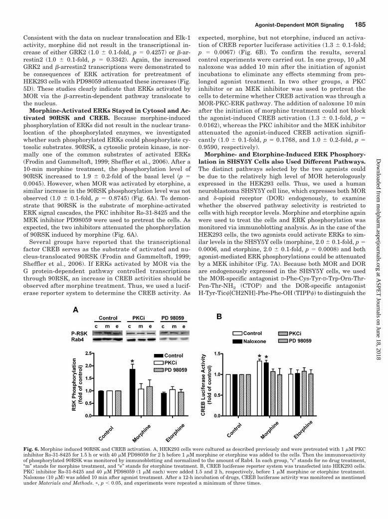
Morphine-Activated ERKs Stayed in Cytosol and Ac-tivated 90RSK and CREB. Because morphine-inducedphosphorylation of ERKs did not result in the nuclear trans-location of the phosphorylated enzymes, we investigatedwhether such phosphorylated ERKs could phosphorylate cy-tosolic substrates. 90RSK, a cytosolic protein kinase, is nor-mally one of the common substrates of activated ERKs(Frodin and Gammeltoft, 1999; Sheffler et al., 2006). After a10-min morphine treatment, the phosphorylation level of90RSK increased to 1.9 � 0.2-fold of the basal level (p �0.0045). However, when MOR was activated by etorphine, asimilar increase in the 90RSK phosphorylation level was notobserved (1.0 � 0.1-fold, p � 0.8745) (Fig. 6A). To demon-strate that 90RSK is the substrate of morphine-activatedERK signal cascades, the PKC inhibitor Ro-31-8425 and theMEK inhibitor PD98059 were used to pretreat the cells. Asexpected, the two inhibitors attenuated the phosphorylationof 90RSK induced by morphine (Fig. 6A).
Several groups have reported that the transcriptionalfactor CREB serves as the substrate of activated and nu-cleus-translocated 90RSK (Frodin and Gammeltoft, 1999;Sheffler et al., 2006). If ERKs activated by MOR via theG protein-dependent pathway controlled transcriptionsthrough 90RSK, an increase in CREB activities should beobserved after morphine treatment. Thus, we used a lucif-erase reporter system to determine the CREB activity. As
expected, morphine, but not etorphine, induced an activa-tion of CREB reporter luciferase activities (1.3 � 0.1-fold;p � 0.0067) (Fig. 6B). To confirm the results, severalcontrol experiments were carried out. In one group, 10 �Mnaloxone was added 10 min after the initiation of agonistincubations to eliminate any effects stemming from pro-longed agonist treatment. In two other groups, a PKCinhibitor or an MEK inhibitor was used to pretreat thecells to determine whether CREB activation was through aMOR-PKC-ERK pathway. The addition of naloxone 10 minafter the initiation of morphine treatment could not blockthe agonist-induced CREB activation (1.3 � 0.1-fold, p �0.0162), whereas the PKC inhibitor and the MEK inhibitorattenuated the agonist-induced CREB activation signifi-cantly (1.0 � 0.1-fold, p � 0.1768, and 1.0 � 0.2-fold, p �0.9590, respectively).
Morphine- and Etorphine-Induced ERK Phosphory-lation in SHSY5Y Cells also Used Different Pathways.The distinct pathways selected by the two agonists couldbe due to the relatively high level of MOR heterologouslyexpressed in the HEK293 cells. Thus, we used a humanneuroblastoma SHSY5Y cell line, which expresses both MORand �-opioid receptor (DOR) endogenously, to examinewhether the observed pathway selectivity is restricted tocells with high receptor levels. Morphine and etorphine againwere used to treat the cells and ERK phosphorylation wasmonitored via immunoblotting analysis. As in the case of theHEK293 cells, the two agonists could activate ERKs to sim-ilar levels in the SHSY5Y cells (morphine, 2.0 � 0.1-fold, p �0.0006, and etorphine, 2.0 � 0.1-fold, p � 0.0008) and bothagonist-mediated ERK phosphorylations could be attenuatedby a MEK inhibitor (Fig. 7A). Because both MOR and DORare endogenously expressed in the SHSY5Y cells, we usedthe MOR-specific antagonist D-Phe-Cys-Tyr-D-Trp-Orn-Thr-Pen-Thr-NH2 (CTOP) and the DOR-specific antagonistH-Tyr-Tic�[CH2NH]-Phe-Phe-OH (TIPP�) to distinguish the
Fig. 6. Morphine induced 90RSK and CREB activation. A, HEK293 cells were cultured as described previously and were pretreated with 1 �M PKCinhibitor Ro-31-8425 for 1.5 h or with 40 �M PD98059 for 2 h before 1 �M morphine or etorphine was added to the cells. Then the immunoreactivityof phosphorylated 90RSK was monitored by immunoblotting and normalized to the amount of Rab4. In each group, “c” stands for no drug treatment,“m” stands for morphine treatment, and “e” stands for etorphine treatment. B, CREB luciferase reporter system was transfected into HEK293 cells.PKC inhibitor Ro-31-8425 and 40 �M PD98059 (1 �M each) were added 1.5 and 2 h, respectively, before 1 �M morphine or etorphine treatment.Naloxone (10 �M) was added 10 min after agonist treatment. After a 12-h incubation of drugs, CREB luciferase activity was monitored as mentionedunder Materials and Methods. �, p � 0.05, and experiments were repeated a minimum of three times.
Agonist-Dependent MOR Signaling 185
at ASPE
T Journals on June 18, 2018
molpharm
.aspetjournals.orgD
ownloaded from

receptor type involved in the ERK phosphorylation. Pretreat-ment with 10 �M CTOP blocked both morphine- and etor-phine-induced ERK phosphorylation, whereas TIPP� did nothave a significant effect on the actions of agonists (Fig. 7B),suggesting that the ERK phosphorylation under this para-digm was mainly mediated by the interaction between ago-nists and MOR.
Similar to the observations with HEK293 cells, there wasagonist selectivity toward pathways for ERK phosphoryla-tion and cellular locations of phosphorylated ERKs in theSHSY5Y cells. Pretreatment of the SHSY5Y cells with thePKC inhibitor Ro-31-8425 resulted in the attenuation of mor-phine-induced but not etorphine-induced ERK phosphoryla-tion (Fig. 7A). In addition, morphine-induced but not etor-phine-induced ERK phosphorylation resulted in an increasein the phosphorylation of 90RSK (1.7 � 0.2-fold, p � 0.0134)(Fig. 7D). The nuclear translocation of etorphine-induced butnot morphine-induced phosphorylated ERKs was demon-strated by the increases in GRK2 (1.7 � 0.1-fold, p � 0.0234)and �-arrestin2 (1.4 � 0.1-fold, p � 0.0156) transcriptions inSHSY5Y cells in the presence of etorphine but not in thepresence of morphine (Fig. 7C). Therefore, even at a lowreceptor level expressed endogenously, MOR agonists selectdistinct signaling pathways in ERK phosphorylation, and the
subsequent cellular locations of the phosphorylated enzymesare also agonist-selective.
Morphine- and Etorphine-Induced ERK Phosphory-lation in Primary Neuronal Culture of Rat Hippocam-pus also Used Different Pathways. To demonstrate theexistence of similar phenomena in MOR-expressing neurons,we monitored ERK phosphorylation in the primary neuronalculture of rat hippocampus. We used the MOR-specific an-tagonist CTOP and the DOR-specific antagonist TIPP� toeliminate the interaction between the two types of receptorsin this primary neuronal culture. CTOP attenuated ERKphosphorylation induced by both morphine and etorphine,whereas TIPP� had little effect on the phosphorylation of thekinases (Fig. 8B). The existence of agonist-selective signalingin the primary culture was demonstrated by using a PKCinhibitor, determining the phosphorylation of 90RSK, andmeasuring the mRNA levels of GRK2 and �-arrestin2. Pre-treating the primary culture with the PKC inhibitor Ro-31-8425 blocked the ERK phosphorylation induced by morphine(1.2 � 0.3-fold, p � 0.5158) but did not affect the etorphine-induced ERK phosphorylation (2.0 � 0.1-fold, p � 0.0084)(Fig. 8A). When the mRNA levels of the GRK2 and �-arres-tin2 were measured after 12-h agonist treatment, only theneurons treated by etorphine had significant increases in
Fig. 7. Agonist-selective ERK phos-phorylation in human neuroblastomaSHSY5Y cells. A, SHSY5Y cells weretreated with 1 �M PKC inhibitor Ro-31-8425 for 1.5 h or with 40 �MPD98059 or DMSO for 2 h. Then ERKphosphorylations were determined af-ter 10 min of 1 �M morphine or 10 nMetorphine treatment. In each group,“c” stands for no drug treatment, “m”stands for morphine treatment, and“e” stands for etorphine treatment. B,SHSY5Y cells were treated with 10�M CTOP or 10 �M TIPP� for 10 min.Then ERK phosphorylations were de-termined after 10-min 1 �M morphineor 10 nM etorphine treatment. In eachgroup, “c” stands for no drug treat-ment, “m” stands for morphine treat-ment, and “e” stands for etorphinetreatment. C, SHSY5Y cells weretreated with 40 �M PD98059 orDMSO for 2 h. Then the total RNAwas extracted after 12 h of 1 �M mor-phine or 10 nM etorphine treatment.The transcription levels of GRK2 and�-arrestin2 were examined as de-scribed under Materials and Methods.D, SHSY5Y cells were treated with 1�M PKC inhibitor Ro-31-8425 orDMSO for 1.5 h. Then 90RSK activi-ties were determined after 10 min of 1�M morphine or 10 nM etorphinetreatment. �, p � 0.05, and experi-ments were repeated a minimum ofthree times.
186 Zheng et al.
at ASPE
T Journals on June 18, 2018
molpharm
.aspetjournals.orgD
ownloaded from

these two transcripts levels (1.7 � 0.1-fold, p � 0.0334, forGRK2, and 1.5 � 0.1-fold, p � 0.0176, for �-arrestin2) (Fig.8C). At the same time, the phosphorylation of 90RSK wasshown to be consistent with our observations with theHEK293 and SHSY5Y cells. In these primary hippocampalneuronal cultures, morphine-induced but not etorphine-induced ERK phosphorylation resulted in an increase in90RSK phosphorylation (1.7 � 0.2-fold, p � 0.0257) (Fig. 8D).Therefore, agonist selectivity for pathways in ERK phosphor-ylation and cellular locations of phosphorylated ERKs existin primary neuronal culture of rat hippocampus, as in thecase of the two cell models that we investigated.
Other Opioid Agonists also Exhibited Pathway Se-lectivity. To obtain broader understanding of agonist-selec-tive pathway-dependent ERK phosphorylation, we tested ad-ditional clinical relevant MOR agonists such as methadoneand fentanyl. The four agonists equally activated ERKs atconcentrations that resulted in maximal receptor occupationbut induced the nuclear translocation of phosphorylatedERKs differentially (Fig. 9A). Similar to etorphine, fentanylincreased the amount of phosphorylated ERKs in the nucleusto 1.5 � 0.1-fold (p � 0.0274) of the basal level, whereasmorphine (1.0 � 0.1-fold, p � 0.4234) and methadone (1.2 �
0.2-fold, p � 0.2154) could not induce significant changes inthe phosphorylated ERK level within the nucleus (Fig. 9B).Likewise, the Elk-1 activity increased significantly after etor-phine (1.8 � 0.2-fold, p � 0.0127) and fentanyl (1.6 � 0.2-fold,p � 0.0285) treatment, but no such increase was observedafter morphine or methadone incubation (Fig. 9C). Morphineand methadone stimulated PKC activity to 1.4 � 0.1-fold(p � 0.0084) and 1.3 � 0.1-fold (p � 0.0156), respectively,whereas etorphine and fentanyl did not (Fig. 9D). Thus, thefour agonists were divided into two groups. Morphine andmethadone induced phosphorylation of ERKs via a PKC-dependent pathway, with the phosphorylated ERKs remain-ing in the cytosol, whereas etorphine and fentanyl used�-arrestin-dependent pathway and resulted in nuclear trans-location of phosphorylated ERKs.
DiscussionThe current studies demonstrate that MOR agonists select
for either the G protein-dependent or the �-arrestin-depen-dent pathway in their activation of ERKs. This selectivitywas observed with a heterologous expression cell system(HEK293 cells) expressing high levels of the receptor, a neu-
Fig. 8. Agonist-selective ERK phos-phorylation in primary neuronal cul-ture of rat hippocampus. The primaryculture of rat hippocampal neuronswas prepared as described under Ma-terials and Methods. A, primary cul-tures of hippocampal neurons weretreated with 1 �M PKC inhibitor Ro-31-8425 for 1.5 h or 40 �M PD98059or DMSO for 2 h. ERK phosphoryla-tions then were determined after 10min of 1 �M morphine or 10 nM etor-phine treatment. In each group, “c”stands for no drug treatment, “m”stands for morphine treatment, and“e” stands for etorphine treatment. B,primary cultures were treated with 10�M CTOP or 10 �M TIPP� for 10 min.Then ERK phosphorylations were de-termined after 10-min 1 �M morphineor 10 nM etorphine treatment. In eachgroup, “c” stands for no drug treat-ment, “m” stands for morphine treat-ment, and “e” stands for etorphinetreatment. C, primary cultures weretreated with 40 �M PD98059 orDMSO for 2 h. The total RNA thenwas extracted after 12 h of 1 �M mor-phine or 10 nM etorphine treatment.The transcription levels of GRK2 and�-arrestin2 were examined as de-scribed under Materials and Methods.D, primary culture were treated with1 �M PKC inhibitor Ro-31-8425 orDMSO for 1.5 h. Then 90RSK activi-ties were determined after 10 min of 1�M morphine or 10 nM etorphinetreatment. �, p � 0.05, and experi-ments were repeated a minimum ofthree times.
Agonist-Dependent MOR Signaling 187
at ASPE
T Journals on June 18, 2018
molpharm
.aspetjournals.orgD
ownloaded from

roblastoma cell line (SHSY5Y cells) expressing low levels ofthe receptor endogenously and a primary culture system ofrat hippocampal neurons. Thus, the agonist-dependent path-way selectivity was not a consequence of receptor reserves orspecific cellular backgrounds and should reflect the in vivoconditions. In our studies, both etorphine- and morphine-induced ERK phosphorylation exhibited similar time coursesand maximal increases. Blockade of the G protein-dependentpathway with a PKC inhibitor or elimination of the �-arres-tin-dependent pathway in the MEF cells resulted in theattenuation of one agonist’s activities without altering eitherthe time course or maximal response of the other agonist.Clearly, the agonists tested in our current studies did notseem to use both the G protein-dependent and �-arrestin-dependent pathways in their activation of ERKs. This is adistinct difference from reported observations that otherGPCRs could use both pathways (Azzi et al., 2003; Kohout etal., 2004; Shenoy et al., 2006). The current studies, however,
could not preclude the possibility that other MOR agonistscould activate both pathways. With the distinct differences inDAMGO’s and morphine’s ability to induce receptor internal-ization and �-arrestin recruitment, and because the PKCinhibitor bisindolylmaleimide I could inhibit both the mor-phine- and DAMGO-induced ERK activation, an agonist suchas DAMGO possibly could activate both pathways (Belchevaet al., 2005). Whether DAMGO could activate ERKs underthe current paradigm and model systems via multiple path-ways remains to be demonstrated. Nevertheless, our obser-vation that morphine uses the G protein-dependent pathwayand etorphine uses the �-arrestin-dependent pathway to ac-tivate ERKs provides a clear demonstration of the agonist-selective signaling theory, which suggests that the efficaciesof a particular agonist on different signaling pathways can bedifferent (Urban et al., 2007).
The nuclear translocation of ERKs is normally regulatedby the phosphorylation status of MEK1 (Whitmarsh and
Fig. 9. ERK phosphorylation and cel-lular location of pERKs induced byopioid agonists. A, HEK293 cells weretreated with morphine (1 �M), etor-phine (1 �M), methadone (10 �M), orfentanyl (1 �M) for 10 min. The cellswere lysed, and the levels of phos-phorylated ERKs were determined byimmunoblotting as described underMaterials and Methods. B, HEK293cells were treated with the four ago-nists, and the nucleus fractions wereisolated to determine the amount ofphosphorylated ERKs in the nucleusby immunoblotting. C, HEK293 cellswere transfected with the Elk-1 lucif-erase report system and incubatedwith 10% serum, morphine (1 �M),etorphine (1 �M), methadone (10 �M),or fentanyl (1 �M) for 12 h. Elk-1 ac-tivity was examined as describedunder Materials and Methods. D,HEK293 cells were incubated withthe same concentration of agonists asin A for 10 min. The PKC activity wasdetermined by measuring the amountof PKC phosphorylated substrates byimmunoblotting. �, p � 0.05, and ex-periments were repeated a minimumof three times.
188 Zheng et al.
at ASPE
T Journals on June 18, 2018
molpharm
.aspetjournals.orgD
ownloaded from
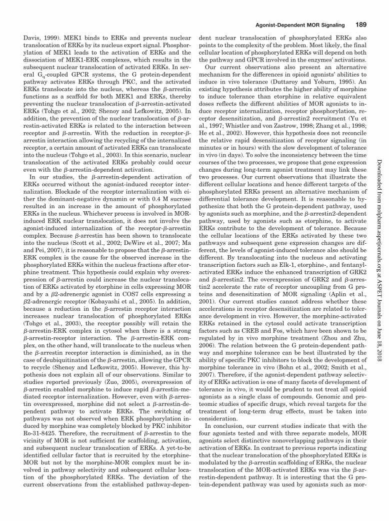
Davis, 1999). MEK1 binds to ERKs and prevents nucleartranslocation of ERKs by its nucleus export signal. Phosphor-ylation of MEK1 leads to the activation of ERKs and thedissociation of MEK1-ERK complexes, which results in thesubsequent nuclear translocation of activated ERKs. In sev-eral Gq-coupled GPCR systems, the G protein-dependentpathway activates ERKs through PKC, and the activatedERKs translocate into the nucleus, whereas the �-arrestinfunctions as a scaffold for both MEK1 and ERKs, therebypreventing the nuclear translocation of �-arrestin-activatedERKs (Tohgo et al., 2002; Shenoy and Lefkowitz, 2005). Inaddition, the prevention of the nuclear translocation of �-ar-restin-activated ERKs is related to the interaction betweenreceptor and �-arrestin. With the reduction in receptor-�-arrestin interaction allowing the recycling of the internalizedreceptor, a certain amount of activated ERKs can translocateinto the nucleus (Tohgo et al., 2003). In this scenario, nucleartranslocation of the activated ERKs probably could occureven with the �-arrestin-dependent activation.
In our studies, the �-arrestin-dependent activation ofERKs occurred without the agonist-induced receptor inter-nalization. Blockade of the receptor internalization with ei-ther the dominant-negative dynamin or with 0.4 M sucroseresulted in an increase in the amount of phosphorylatedERKs in the nucleus. Whichever process is involved in MOR-induced ERK nuclear translocation, it does not involve theagonist-induced internalization of the receptor-�-arrestincomplex. Because �-arrestin has been shown to translocateinto the nucleus (Scott et al., 2002; DeWire et al., 2007; Maand Pei, 2007), it is reasonable to propose that the �-arrestin-ERK complex is the cause for the observed increase in thephosphorylated ERKs within the nucleus fractions after etor-phine treatment. This hypothesis could explain why overex-pression of �-arrestin could increase the nuclear transloca-tion of ERKs activated by etorphine in cells expressing MORand by a �2-adrenergic agonist in COS7 cells expressing a�2-adrenergic receptor (Kobayashi et al., 2005). In addition,because a reduction in the �-arrestin receptor interactionincreases nuclear translocation of phosphorylated ERKs(Tohgo et al., 2003), the receptor possibly will retain the�-arrestin-ERK complex in cytosol when there is a strong�-arrestin-receptor interaction. The �-arrestin-ERK com-plex, on the other hand, will translocate to the nucleus whenthe �-arrestin receptor interaction is diminished, as in thecase of deubiquitination of the �-arrestin, allowing the GPCRto recycle (Shenoy and Lefkowitz, 2005). However, this hy-pothesis does not explain all of our observations. Similar tostudies reported previously (Zuo, 2005), overexpression of�-arrestin enabled morphine to induce rapid �-arrestin-me-diated receptor internalization. However, even with �-arres-tin overexpressed, morphine did not select a �-arrestin-de-pendent pathway to activate ERKs. The switching ofpathways was not observed when ERK phosphorylation in-duced by morphine was completely blocked by PKC inhibitorRo-31-8425. Therefore, the recruitment of �-arrestin to thevicinity of MOR is not sufficient for scaffolding, activation,and subsequent nuclear translocation of ERKs. A yet-to-beidentified cellular factor that is recruited by the etorphine-MOR but not by the morphine-MOR complex must be in-volved in pathway selectivity and subsequent cellular loca-tion of the phosphorylated ERKs. The deviation of thecurrent observations from the established pathway-depen-
dent nuclear translocation of phosphorylated ERKs alsopoints to the complexity of the problem. Most likely, the finalcellular location of phosphorylated ERKs will depend on boththe pathway and GPCR involved in the enzymes’ activations.
Our current observations also present an alternativemechanism for the differences in opioid agonists’ abilities toinduce in vivo tolerance (Duttaroy and Yoburn, 1995). Anexisting hypothesis attributes the higher ability of morphineto induce tolerance than etorphine in relative equivalentdoses reflects the different abilities of MOR agonists to in-duce receptor internalization, receptor phosphorylation, re-ceptor desensitization, and �-arrestin2 recruitment (Yu etal., 1997; Whistler and von Zastrow, 1998; Zhang et al., 1998;He et al., 2002). However, this hypothesis does not reconcilethe relative rapid desensitization of receptor signaling (inminutes or in hours) with the slow development of tolerancein vivo (in days). To solve the inconsistency between the timecourses of the two processes, we propose that gene expressionchanges during long-term agonist treatment may link thesetwo processes. Our current observations that illustrate thedifferent cellular locations and hence different targets of thephosphorylated ERKs present an alternative mechanism ofdifferential tolerance development. It is reasonable to hy-pothesize that both the G protein-dependent pathway, usedby agonists such as morphine, and the �-arrestin2-dependentpathway, used by agonists such as etorphine, to activateERKs contribute to the development of tolerance. Becausethe cellular locations of the ERKs activated by these twopathways and subsequent gene expression changes are dif-ferent, the levels of agonist-induced tolerance also should bedifferent. By translocating into the nucleus and activatingtranscription factors such as Elk-1, etorphine-, and fentanyl-activated ERKs induce the enhanced transcription of GRK2and �-arrestin2. The overexpression of GRK2 and �-arres-tin2 accelerate the rate of receptor uncoupling from G pro-teins and desensitization of MOR signaling (Aplin et al.,2001). Our current studies cannot address whether theseaccelerations in receptor desensitization are related to toler-ance development in vivo. However, the morphine-activatedERKs retained in the cytosol could activate transcriptionfactors such as CREB and Fos, which have been shown to beregulated by in vivo morphine treatment (Zhou and Zhu,2006). The relation between the G protein-dependent path-way and morphine tolerance can be best illustrated by theability of specific PKC inhibitors to block the development ofmorphine tolerance in vivo (Bohn et al., 2002; Smith et al.,2007). Therefore, if the agonist-dependent pathway selectiv-ity of ERKs activation is one of many facets of development oftolerance in vivo, it would be prudent to not treat all opioidagonists as a single class of compounds. Genomic and pro-teomic studies of specific drugs, which reveal targets for thetreatment of long-term drug effects, must be taken intoconsideration.
In conclusion, our current studies indicate that with thefour agonists tested and with three separate models, MORagonists select distinctive nonoverlapping pathways in theiractivation of ERKs. In contrast to previous reports indicatingthat the nuclear translocation of the phosphorylated ERKs ismodulated by the �-arrestin scaffolding of ERKs, the nucleartranslocation of the MOR-activated ERKs was via the �-ar-restin-dependent pathway. It is interesting that the G pro-tein-dependent pathway was used by agonists such as mor-
Agonist-Dependent MOR Signaling 189
at ASPE
T Journals on June 18, 2018
molpharm
.aspetjournals.orgD
ownloaded from

phine, and the phosphorylated ERKs remained in cytosol,contrary to the accepted dogma of ERK activation. Therefore,for such a scenario to happen, we hypothesize that cellularfactor(s) other than �-arrestin recruited by the morphine-MOR complex must be involved in the retention of activatedERKs in the cytosol, and cellular factor(s) other than �-ar-restin recruited by etorphine-MOR complex must be respon-sible for the subsequent nuclear translocation of the acti-vated ERK. The identities of such factors will enable us toelucidate the mechanism of agonist-dependent pathway se-lectivity of GPCR signaling.
Acknowledgments
Dr. Robert Lefkowitz (Duke University) generously provided the�-arrestin and �-arrestin2-V54D constructs, the wild-type, �-arres-tin2�/�, and �-arrestin1/2�/� mouse embryonic fibroblasts cells usedin current studies. Dr. Mark von Zastrow (University of California atSan Francisco) generously provided the Dynamin 1 and DynaminK44E plasmids.
ReferencesAhn S, Shenoy SK, Wei H, and Lefkowitz RJ (2004) Differential kinetic and spatial
patterns of �-arrestin and G protein-mediated ERK activation by the angiotensinII receptor. J Biol Chem 279:35518–35525.
Aplin AE, Stewart SA, Assoian RK, and Juliano RL (2001) Integrin-mediated adhe-sion regulates ERK nuclear translocation and phosphorylation of Elk-1. J Cell Biol153:273–282.
Arvidsson U, Riedl M, Chakrabarti S, Lee JH, Nakano AH, Dado RJ, Loh HH, LawPY, Wessendorf MW, and Elde R (1995) Distribution and targeting of a mu-opioidreceptor (MOR1) in brain and spinal cord. J Neurosci 15:3328–3341.
Azzi M, Charest PG, Angers S, Rousseau G, Kohout T, Bouvier M, and Pineyro G(2003) Beta-arrestin-mediated activation of MAPK by inverse agonists revealsdistinct active conformations for G protein-coupled receptors. Proc Natl Acad SciU S A 100:11406–11411.
Belcheva MM, Clark AL, Haas PD, Serna JS, Hahn JW, Kiss A, and Coscia CJ (2005)� and � opioid receptors activate ERK/MAPK via different protein kinase Cisoforms and secondary messengers in astrocytes. J Biol Chem 280:27662–27669.
Bohn LM, Lefkowitz RJ, and Caron MG (2002) Differential mechanisms of morphineantinociceptive tolerance revealed in (beta)arrestin-2 knock-out mice. J Neurosci22:10494–10500.
Cavigelli M, Dolfi F, Claret FX, and Karin M (1995) Induction of c-fos expressionthrough JNK-mediated TCF/Elk-1 phosphorylation. EMBO J 14:5957–5964.
DeWire SM, Ahn S, Lefkowitz RJ, and Shenoy SK (2007) Beta-arrestins and cellsignaling. Annu Rev Physiol 69:483–510.
Dudley DT, Pang L, Decker SJ, Bridges AJ, and Saltiel AR (1995) A syntheticinhibitor of the mitogen-activated protein kinase cascade. Proc Natl Acad SciU S A 92:7686–7689.
Duttaroy A and Yoburn BC (1995) The effect of intrinsic efficacy on opioid tolerance.Anesthesiology 82:1226–1236.
El-Dahr SS, Dipp S, and Baricos WH (1998) Bradykinin stimulates the ERK–Elk-1–Fos/AP-1 pathway in mesangial cells. Am J Physiol 275:F343–F352.
Frodin M and Gammeltoft S (1999) Role and regulation of 90 kDa ribosomal S6kinase (RSK) in signal transduction. Mol Cell Endocrinol 151:65–77.
Gesty-Palmer D, Chen M, Reiter E, Ahn S, Nelson CD, Wang S, Eckhardt AE, CowanCL, Spurney RF, Luttrell LM, et al. (2006) Distinct �-arrestin- and G protein-dependent pathways for parathyroid hormone receptor-stimulated ERK1/2 acti-vation. J Biol Chem 281:10856–10864.
Ghosh A and Greenberg ME (1995) Distinct roles for bFGF and NT-3 in the regu-lation of cortical neurogenesis. Neuron 15:89–103.
Gille H, Kortenjann M, Thomae O, Moomaw C, Slaughter C, Cobb MH, and Shaw PE(1995) ERK phosphorylation potentiates Elk-1-mediated ternary complex forma-tion and transactivation. EMBO J 14:951–962.
He L, Fong J, von Zastrow M, and Whistler JL (2002) Regulation of opioid receptortrafficking and morphine tolerance by receptor oligomerization. Cell 108:271–282.
Ignatova EG, Belcheva MM, Bohn LM, Neuman MC, and Coscia CJ (1999) Require-ment of receptor internalization for opioid stimulation of mitogen-activated pro-tein kinase: biochemical and immunofluorescence confocal microscopic evidence.J Neurosci 19:56–63.
Janknecht R, Zinck R, Ernst WH, and Nordheim A (1994) Functional dissection ofthe transcription factor Elk-1. Oncogene 9:1273–1278.
Keith DE, Murray SR, Zaki PA, Chu PC, Lissin DV, Kang L, Evans CJ, and vonZastrow M (1996) Morphine activates opioid receptors without causing their rapidinternalization. J Biol Chem 271:19021–19024.
Kobayashi H, Narita Y, Nishida M, and Kurose H (2005) Beta-arrestin2 enhances
beta2-adrenergic receptor-mediated nuclear translocation of ERK. Cell Signal17:1248–1253.
Kohout TA, Nicholas SL, Perry SJ, Reinhart G, Junger S, and Struthers RS (2004)Differential desensitization, receptor phosphorylation, �-arrestin recruitment, andERK1/2 activation by the two endogenous ligands for the CC chemokine receptor7. J Biol Chem 279:23214–23222.
Kramer HK and Simon EJ (2000) mu and delta-opioid receptor agonists inducemitogen-activated protein kinase (MAPK) activation in the absence of receptorinternalization. Neuropharmacology 39:1707–1719.
Li LY and Chang KJ (1996) The stimulatory effect of opioids on mitogen-activatedprotein kinase in Chinese hamster ovary cells transfected to express �-opioidreceptors. Mol Pharmacol 50:599–602.
Liao D, Lin H, Law PY, and Loh HH (2005) Mu-opioid receptors modulate thestability of dendritic spines. Proc Natl Acad Sci U S A 102:1725–1730.
Ma L and Pei G (2007) �-arrestin signaling and regulation of transcription. J Cell Sci120:213–218.
Narita M, Suzuki M, Narita M, Niikura K, Nakamura A, Miyatake M, Yajima Y, andSuzuki T (2006) mu-Opioid receptor internalization-dependent and -independentmechanisms of the development of tolerance to mu-opioid receptor agonists: com-parison between etorphine and morphine. Neuroscience 138:609–619.
Oakley RH, Laporte SA, Holt JA, Caron MG, and Barak LS (2000) Differentialaffinities of visual arrestin, � arrestin1, and � arrestin2 for G protein-coupledreceptors delineate two major classes of receptors. J Biol Chem 275:17201–17210.
Ren XR, Reiter E, Ahn S, Kim J, Chen W, and Lefkowitz RJ (2005) Different Gprotein-coupled receptor kinases govern G protein and beta-arrestin-mediatedsignaling of V2 vasopressin receptor. Proc Natl Acad Sci U S A 102:1448–1453.
Rozenfeld R and Devi LA (2007) Receptor heterodimerization leads to a switch insignaling: beta-arrestin2-mediated ERK activation by mu-delta opioid receptorheterodimers. FASEB J 21:2455–2465.
Scott MG, Le Rouzic E, Perianin A, Pierotti V, Enslen H, Benichou S, Marullo S, andBenmerah A (2002) Differential nucleocytoplasmic shuttling of �-arrestins. Char-acterization of a leucine-rich nuclear export signal in �-arrestin2. J Biol Chem277:37693–37701.
Sheffler DJ, Kroeze WK, Garcia BG, Deutch AY, Hufeisen SJ, Leahy P, Bruning JC,and Roth BL (2006) p90 ribosomal S6 kinase 2 exerts a tonic brake on G protein-coupled receptor signaling. Proc Natl Acad Sci U S A 103:4717–4722.
Shenoy SK and Lefkowitz RJ (2005) Receptor-specific ubiquitination of �-arrestindirects assembly and targeting of seven-transmembrane receptor signalosomes.J Biol Chem 280:15315–15324.
Shenoy SK, Drake MT, Nelson CD, Houtz DA, Xiao K, Madabushi S, Reiter E,Premont RT, Lichtarge O, and Lefkowitz RJ (2006) �-Arrestin-dependent, Gprotein-independent ERK1/2 activation by the �2 adrenergic receptor. J Biol Chem281:1261–1273.
Shoda T, Fukuda K, Uga H, Mima H, and Morikawa H (2001) Activation of mu-opioidreceptor induces expression of c-fos and junB via mitogen-activated protein kinasecascade. Anesthesiology 95:983–989.
Smith FL, Gabra BH, Smith PA, Redwood MC, and Dewey WL (2007) Determinationof the role of conventional, novel and atypical PKC isoforms in the expression ofmorphine tolerance in mice. Pain 127:129–139.
Stein E, Huynh-Do U, Lane AA, Cerretti DP, and Daniel TO (1998) Nck recruitmentto Eph receptor, EphB1/ELK, couples ligand activation to c-Jun kinase. J BiolChem 273:1303–1308.
Tohgo A, Choy EW, Gesty-Palmer D, Pierce KL, Laporte S, Oakley RH, Caron MG,Lefkowitz RJ, and Luttrell LM (2003) The stability of the G protein-coupledreceptor-�-arrestin interaction determines the mechanism and functional conse-quence of ERK activation. J Biol Chem 278:6258–6267.
Tohgo A, Pierce KL, Choy EW, Lefkowitz RJ, and Luttrell LM (2002) beta-Arrestinscaffolding of the ERK cascade enhances cytosolic ERK activity but inhibits ERK-mediated transcription following angiotensin AT1a receptor stimulation. J BiolChem 277:9429–9436.
Urban JD, Clarke WP, von Zastrow M, Nichols DE, Kobilka B, Weinstein H, JavitchJA, Roth BL, Christopoulos A, Sexton PM, et al. (2007) Functional selectivity andclassical concepts of quantitative pharmacology. J Pharmacol Exp Ther 320:1–13.
Whistler JL and von Zastrow M (1998) Morphine-activated opioid receptors eludedesensitization by beta-arrestin. Proc Natl Acad Sci U S A 95:9914–9919.
Whitmarsh AJ and Davis RJ (1999) Signal transduction by MAP kinases: regulationby phosphorylation-dependent switches. Sci STKE 1999:PE1.
Yu Y, Zhang L, Yin X, Sun H, Uhl GR, and Wang JB (1997) � Opioid receptorphosphorylation, desensitization, and ligand efficacy. J Biol Chem 272:28869–28874.
Zhang J, Ferguson SS, Barak LS, Bodduluri SR, Laporte SA, Law PY, and Caron MG(1998) Role for G protein-coupled receptor kinase in agonist-specific regulation ofmu-opioid receptor responsiveness. Proc Natl Acad Sci U S A 95:7157–7162.
Zhou LF and Zhu YP (2006) Changes of CREB in rat hippocampus, prefrontal cortexand nucleus accumbens during three phases of morphine induced conditionedplace preference in rats. J Zhejiang Univ Sci B 7:107–113.
Zuo Z (2005) The role of opioid receptor internalization and beta-arrestins in thedevelopment of opioid tolerance. Anesth Analg 101:728–734.
Address correspondence to: Dr. Hui Zheng, Department of Pharmacology,Medical School, University of Minnesota. 6-120 Jackson Hall, 321 ChurchStreet S.E., Minneapolis, MN 55455-0217. E-mail: [email protected]
190 Zheng et al.
at ASPE
T Journals on June 18, 2018
molpharm
.aspetjournals.orgD
ownloaded from


![Non-opioid & Opioid IV Anesthetics Copy [Compatibility Mode]](https://static.fdocuments.in/doc/165x107/55cf8c8a5503462b138d78d4/non-opioid-opioid-iv-anesthetics-copy-compatibility-mode.jpg)