In vivo Evaluation of Gelatin/Hyaluronic Acid Nanofiber...
7
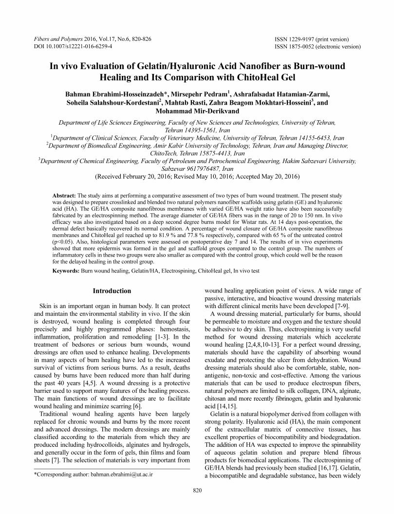
820 ISSN 1229-9197 (print version) ISSN 1875-0052 (electronic version) Fibers and Polymers 2016, Vol.17, No.6, 820-826 In vivo Evaluation of Gelatin/Hyaluronic Acid Nanofiber as Burn-wound Healing and Its Comparison with ChitoHeal Gel Bahman Ebrahimi-Hosseinzadeh*, Mirsepehr Pedram 1 , Ashrafalsadat Hatamian-Zarmi, Soheila Salahshour-Kordestani 2 , Mahtab Rasti, Zahra Beagom Mokhtari-Hosseini 3 , and Mohammad Mir-Derikvand Department of Life Sciences Engineering, Faculty of New Sciences and Technologies, University of Tehran, Tehran 14395-1561, Iran 1 Department of Clinical Sciences, Faculty of Veterinary Medicine, University of Tehran, Tehran 14155-6453, Iran 2 Department of Biomedical Engineering, Amir Kabir University of Technology, Tehran, Iran and Managing Director, ChitoTech, Tehran 15875-4413, Iran 3 Department of Chemical Engineering, Faculty of Petroleum and Petrochemical Engineering, Hakim Sabzevari University, Sabzevar 9617976487, Iran (Received February 20, 2016; Revised May 10, 2016; Accepted May 20, 2016) Abstract: The study aims at performing a comparative assessment of two types of burn wound treatment. The present study was designed to prepare crosslinked and blended two natural polymers nanofiber scaffolds using gelatin (GE) and hyaluronic acid (HA). The GE/HA composite nanofibrous membranes with varied GE/HA weight ratio have also been successfully fabricated by an electrospinning method. The average diameter of GE/HA fibers was in the range of 20 to 150 nm. In vivo efficacy was also investigated based on a deep second degree burns model for Wistar rats. At 14 days post-operation, the dermal defect basically recovered its normal condition. A percentage of wound closure of GE/HA composite nanofibrous membranes and ChitoHeal gel reached up to 81.9 % and 77.8 % respectively, compared with 65 % of the untreated control (p<0.05). Also, histological parameters were assessed on postoperative day 7 and 14. The results of in vivo experiments showed that more epidermis was formed in the gel and scaffold groups compared to the control group. The numbers of inflammatory cells in these two groups were also smaller as compared with the control group, which could well be the reason for the delayed healing in the control group. Keywords: Burn wound healing, Gelatin/HA, Electrospining, ChitoHeal gel, In vivo test Introduction Skin is an important organ in human body. It can protect and maintain the environmental stability in vivo. If the skin is destroyed, wound healing is completed through four precisely and highly programmed phases: hemostasis, inflammation, proliferation and remodeling [1-3]. In the treatment of bedsores or serious burn wounds, wound dressings are often used to enhance healing. Developments in many aspects of burn healing have led to the increased survival of victims from serious burns. As a result, deaths caused by burns have been reduced more than half during the past 40 years [4,5]. A wound dressing is a protective barrier used to support many features of the healing process. The main functions of wound dressings are to facilitate wound healing and minimize scarring [6]. Traditional wound healing agents have been largely replaced for chronic wounds and burns by the more recent and advanced dressings. The modern dressings are mainly classified according to the materials from which they are produced including hydrocolloids, alginates and hydrogels, and generally occur in the form of gels, thin films and foam sheets [7]. The selection of materials is very important from wound healing application point of views. A wide range of passive, interactive, and bioactive wound dressing materials with different clinical merits have been developed [7-9]. A wound dressing material, particularly for burns, should be permeable to moisture and oxygen and the texture should be adhesive to dry skin. Thus, electrospinning is very useful method for wound dressing materials which accelerate wound healing [2,4,8,10-13]. For a perfect wound dressing, materials should have the capability of absorbing wound exudate and protecting the ulcer from dehydration. Wound dressing materials should also be comfortable, stable, non- antigenic, non-toxic and cost-effective. Among the various materials that can be used to produce electrospun fibers, natural polymers are limited to silk collagen, DNA, alginate, chitosan and more recently fibrinogen, gelatin and hyaluronic acid [14,15]. Gelatin is a natural biopolymer derived from collagen with strong polarity. Hyaluronic acid (HA), the main component of the extracellular matrix of connective tissues, has excellent properties of biocompatibility and biodegradation. The addition of HA was expected to improve the spinnability of aqueous gelatin solution and prepare blend fibrous products for biomedical applications. The electrospinning of GE/HA blends had previously been studied [16,17]. Gelatin, a biocompatible and degradable substance, has been widely *Corresponding author: [email protected] DOI 10.1007/s12221-016-6259-4
Transcript of In vivo Evaluation of Gelatin/Hyaluronic Acid Nanofiber...
820
ISSN 1229-9197 (print version)
ISSN 1875-0052 (electronic version)
Fibers and Polymers 2016, Vol.17, No.6, 820-826
In vivo Evaluation of Gelatin/Hyaluronic Acid Nanofiber as Burn-wound
Healing and Its Comparison with ChitoHeal Gel
Bahman Ebrahimi-Hosseinzadeh*, Mirsepehr Pedram1, Ashrafalsadat Hatamian-Zarmi,
Soheila Salahshour-Kordestani2, Mahtab Rasti, Zahra Beagom Mokhtari-Hosseini
3, and
Mohammad Mir-Derikvand
Department of Life Sciences Engineering, Faculty of New Sciences and Technologies, University of Tehran,
Tehran 14395-1561, Iran1Department of Clinical Sciences, Faculty of Veterinary Medicine, University of Tehran, Tehran 14155-6453, Iran
2Department of Biomedical Engineering, Amir Kabir University of Technology, Tehran, Iran and Managing Director,
ChitoTech, Tehran 15875-4413, Iran3Department of Chemical Engineering, Faculty of Petroleum and Petrochemical Engineering, Hakim Sabzevari University,
Sabzevar 9617976487, Iran
(Received February 20, 2016; Revised May 10, 2016; Accepted May 20, 2016)
Abstract: The study aims at performing a comparative assessment of two types of burn wound treatment. The present studywas designed to prepare crosslinked and blended two natural polymers nanofiber scaffolds using gelatin (GE) and hyaluronicacid (HA). The GE/HA composite nanofibrous membranes with varied GE/HA weight ratio have also been successfullyfabricated by an electrospinning method. The average diameter of GE/HA fibers was in the range of 20 to 150 nm. In vivoefficacy was also investigated based on a deep second degree burns model for Wistar rats. At 14 days post-operation, thedermal defect basically recovered its normal condition. A percentage of wound closure of GE/HA composite nanofibrousmembranes and ChitoHeal gel reached up to 81.9 % and 77.8 % respectively, compared with 65 % of the untreated control(p<0.05). Also, histological parameters were assessed on postoperative day 7 and 14. The results of in vivo experimentsshowed that more epidermis was formed in the gel and scaffold groups compared to the control group. The numbers ofinflammatory cells in these two groups were also smaller as compared with the control group, which could well be the reasonfor the delayed healing in the control group.
Keywords: Burn wound healing, Gelatin/HA, Electrospining, ChitoHeal gel, In vivo test
Introduction
Skin is an important organ in human body. It can protect
and maintain the environmental stability in vivo. If the skin
is destroyed, wound healing is completed through four
precisely and highly programmed phases: hemostasis,
inflammation, proliferation and remodeling [1-3]. In the
treatment of bedsores or serious burn wounds, wound
dressings are often used to enhance healing. Developments
in many aspects of burn healing have led to the increased
survival of victims from serious burns. As a result, deaths
caused by burns have been reduced more than half during
the past 40 years [4,5]. A wound dressing is a protective
barrier used to support many features of the healing process.
The main functions of wound dressings are to facilitate
wound healing and minimize scarring [6].
Traditional wound healing agents have been largely
replaced for chronic wounds and burns by the more recent
and advanced dressings. The modern dressings are mainly
classified according to the materials from which they are
produced including hydrocolloids, alginates and hydrogels,
and generally occur in the form of gels, thin films and foam
sheets [7]. The selection of materials is very important from
wound healing application point of views. A wide range of
passive, interactive, and bioactive wound dressing materials
with different clinical merits have been developed [7-9].
A wound dressing material, particularly for burns, should
be permeable to moisture and oxygen and the texture should
be adhesive to dry skin. Thus, electrospinning is very useful
method for wound dressing materials which accelerate
wound healing [2,4,8,10-13]. For a perfect wound dressing,
materials should have the capability of absorbing wound
exudate and protecting the ulcer from dehydration. Wound
dressing materials should also be comfortable, stable, non-
antigenic, non-toxic and cost-effective. Among the various
materials that can be used to produce electrospun fibers,
natural polymers are limited to silk collagen, DNA, alginate,
chitosan and more recently fibrinogen, gelatin and hyaluronic
acid [14,15].
Gelatin is a natural biopolymer derived from collagen with
strong polarity. Hyaluronic acid (HA), the main component
of the extracellular matrix of connective tissues, has
excellent properties of biocompatibility and biodegradation.
The addition of HA was expected to improve the spinnability
of aqueous gelatin solution and prepare blend fibrous
products for biomedical applications. The electrospinning of
GE/HA blends had previously been studied [16,17]. Gelatin,
a biocompatible and degradable substance, has been widely*Corresponding author: [email protected]
DOI 10.1007/s12221-016-6259-4
Gelatin/HA Nanofiber as Burn-wound Healing Fibers and Polymers 2016, Vol.17, No.6 821
used to develop wound dressings of certain types of open
wounds (e.g., traumatic, thermal, or chronic wounds) [18].
In previous studies, gelatin/polyurethane nanofiber containing
silver-sulfadiazine [4] and silk fibroin/gelatin electrospun
nanofibrous dressing loaded with astragaloside IV (AS) [19]
were prepared as burn wound dressings. It is generally
accepted that the incorporation of HA is beneficial in tissue
repair products [20,21]. When HA physiologically degraded
into smaller-sized fragments, it facilitates wound healing by
promoting angiogenesis. Therefore, HA helps cell migration
and cell proliferation and keeps the skin moist [22,23].
Therefore, HA with gelatin may be advantageous to prepare
scaffolds that can mimic the ECM in terms of topography
and chemical composition because of the inherent properties
of gelatin and the unique characteristics of the electrospun
fibers. Moreover, chitosan is an attractive natural polymer
for biomedical use due to its reported desirable bioactive
properties scar reduction and wound healing [15,24]. Chitosan
can be processed into various forms, including films,
hydrogels, nanoparticles, scaffolds, and sponges leading to
the proposed applications for wound dressing [25,26].
In a previous study, a sponge prepared from hyaluronic
acid collagen sponge was used for skin healing in rats. It was
also concluded that this sponge succeeded in creating a
suitable environment for wound healing (a conclusion in
agreement with results of our research). However, it was
shown in previous research that collagen alone was not able
to create a moist environment for wound healing [27]. It
must be mentioned that collagen is a very expensive polymer
and gelatin has an extremely low price compared with
collagen. This is a very important factor in introducing
commercial products.
The main aim of this work was to develop a nanofibrous
wound dressing prepared from gelatin/ HA material by
electrospinning with various blending ratios. GTA vapor
was used to improve the stability of the fiber mats in a moist
environment. The researchers also sought to compare the
wound healing rate in a burn rat model treated with either a
bioactive dressing (gelatin/HA nanofibrous membranes and
a commercial product, ChitoHeal gel) or conservative
treatment (gauze).
Experimental
Materials
Hyaluronic acid (HA, MW=1200000) was provided by
Shandong Freda Biochem Co., Ltd. (Jinan, China). Formic
acid (FA) and N,N-dimethylformamide (DMF) were
supplied by Merck, Germany. Gelatin (bovine skin, type B)
was purchased from Sigma-Aldrich (St Louis, MO, USA).
The commercial product, ChitoHeal gel, was obtained from
ChitoTech Company (Tehran, Iran). All the solvents were
used without further purification.
Preparation of Spinning Solution
20 % (w/v) GE solutions with DMF was prepared at 40oC
under gentle stirring for 20 min. Dissolve certain amounts of
HA powder in DMF under gentle stirring for 15 min, and
then add a specific amount of distilled water into the HA
solution according to the volume ratio of DMF to water (2:3)
and continue to stir the solution for 8 h until the solution
became transparent. The spinning solution of 1.5 w/v% HA
in DMF/water system was prepared. Then, add the GE
solution into the HA solution with specific volumes to
obtain the GE-HA solutions (GE/HA=93/7 and 97/3, weight
ratio) [28].
Electrospinning
The fibers were prepared by the use of electrospinning
technique. A counter electrode was located at 15 cm apart
from the capillary tip. The applied voltage was 20 kV and a
syringe pump was used to feed the polymer solution and the
feeding rate was fixed at 0.1 ml h-1. All experiments were
conducted at room temperature and the fibers were collected
on an aluminum foil.
Chemical Cross-linking of Nanofiber Mats
Electrospun mats were crosslinked to enhance the
mechanical properties and pH stability. After the electrospun
nanofiber was dried in vacuum for 24 h, glutaraldehyde
(GA) vapour cross-linking was carried out by placing the
membrane above the GA solution (25 wt%) in a sealed
desiccator at room temperature for 48 h. Then, the GA cross-
linked GE/HA scaffolds were washed three times with
distilled water and dried in vacuum.
Characterization of the GE/HA Nanofibrous Dressing
Scanning Electron Microscopy (SEM)
The morphology of the electrospun nanofibres was studied
by scanning electron microscopy KYKY SEM (China; EM-
3200) equipped with image analyzer software at an accelerating
voltage of 20 kV. Prior to observation, mat samples were
attached onto the stubs using a double-sided tape and then
coated with gold. The diameter, diameter distribution and
uniformity were measured with image analyzer software
(Sigma Scan Pro 4.0). For each experiment, average fiber
diameter and distribution were determined from about 100
measurements of the random fibers.
FTIR Analysis
The infrared (FT-IR) spectra were recorded on photo
acoustic mode at the frequency range of 5000-4000 cm-1
with 256 consecutive scans at 8 cm-1 resolution on a
Thermo-Nicolet 6700 P FTIR Spectrometer (Germany).
In-vivo Wound Healing
Burn-wound Model
Thermal skin burn was modeled in 24 Wistar male rats
822 Fibers and Polymers 2016, Vol.17, No.6 Bahman Ebrahimi-Hosseinzadeh et al.
weighing 200-250 g. The rats were randomly divided into
three groups each composed of 8 rats for the study to
compare healing of wounds. After hair removal, contact
thermal burn was infected at the middle of the paravertebral
region by applying a copper plate heated to 90oC at a force
of 1.3 N for 10 sec under ether anesthesia, which produced
grade IIb (second-degree burn). After generating burn
wound regions (2×3 cm2), 70 % ethanol was used for
disinfection. Every wound was enclosed with one of antiseptic
gauze (control), ChitoHeal gel (a commercial wound dressing),
and sterilized GE/HA nanofiber wound dressing. Formulations
were repeatedly applied on the burned areas once daily for
14 days. For the control wounds, they were just cleaned with
cotton wetted with normal saline each day. The diameter of
the wounds formed were monitored and measured periodically.
Wound-healing Rate
Images of the wound area were taken with a digital
camera. The wound was traced, and the traced area was
calculated using the Image J (National Institutes of Health)
software program. Wounds were digitally photographed on
1, 7, and 14 days post wounding while maintaining a
constant optical zoom. The percentage of wound closure
was calculated by the initial and final area using metric ruler
during observation as follows:
Wound size reduction (%) = [(A0 − At)/A0] × 100
where A0 is the initial wound area and At represents the open
area of wound at the time of biopsy on day 1, 7, and 14
accordingly [29].
Histopathologic Examination
On day 7 and 14, all animals were scarified under
anesthesia, and wounds were removed from animal for
histopathological evaluation. The removed skin specimens
from each group were collected from wound treatment.
Histological assessment was performed only for formulations
with highest improvement in wound healing to further
compare them. Specimens were fixed in 10 % buffered
formalin, processed, embedded in paraffin, cut into 5 mm
pieces and stained with hematoxyl in and eosin (H&E).
Sections were visualized under a light microscope at 400×
magnification. For histopathological analysis (8 rats per
group), all animals were scarified under anesthesia on day 7
after surgery because the maximal changes occurred during
the first week after wounding [16].
Statistical Analysis
Each experiment was repeated for at least three times and
the data were obtained by calculating the mean of experiments.
Statistical analyses were performed using One-way ANOVA
(SPSS 16) and p<0.05 was considered as p-value indicating
the statistical significance of differences.
Results and Discussion
Electrospinning and Morphology of Fibers
In this study, GE-HA/DMF-water solutions were prepared
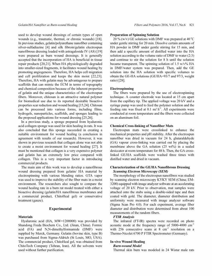
Figure 1. SEM micrographs (magnification=3000× and 30000×, scale bar=5 µm) and histogram distribution of fiber diameter of electrospun
gelatin/hyaluronic acid fibres; (A) 93:7 and (B) 97:3.

Gelatin/HA Nanofiber as Burn-wound Healing Fibers and Polymers 2016, Vol.17, No.6 823
to investigate the electrospinning process of two natural
biopolymer blends. The concentration of HA was fixed at
1.5 w/v% and the volume ratio of DMF to water was 1.5.
The SEM morphologies of the GE/HA electrospun which
contain various concentrations of biopolymer (GE/HA: 93/7
and 97/3) are shown in Figure 1. SEM images at both low
and high magnifications indicated that uniform GE/HA
nanofibrous membranes could be fabricated. Membranes at
different GE/HA compositions and different average diameters
ranging from 20 to 150 nm could also be produced by
electrospinning. Additionally, nanofibrous GE/HA was
successfully generated without the formation of beads when
the proportion of gelatin increased (Figure 1(B)). In the
present procedure, the overall polymer concentration increased
with increasing GE content. Also the higher polymer
concentration led to an increase in the average diameter of
GE/HA nanofibers. Furthermore, gelatin concentration had a
significant effect on the average fiber diameter. The average
fiber diameter increased from 47.4 to 70.6 nm as the gelatin
concentration increased. The reason for the positive
correlation is that the higher viscosity resisted the extension
of the jet.
FTIR Analysis
Chemical analysis was performed to prove the existence
of hyaluronic acid and gelatin in scaffolds and the removal
of solvent from electrospun mats. Intensity of peaks in two
scaffolds is very different due to the difference in the
concentration of polymers in scaffolds. As shown in Figure
2, the peak of 1647.15 cm-1 is related to amide bond in the
net gelatin. Additionally, the NH2 existing in gelatin attacks
the carboxylic functional groups of hyaluronic acid and
enhances this peak. The peak 3100-3400 cm-1 is related to
the absorption of hydroxyl group. The peaks absorbed at
1541.25 cm-1 and 1450.40 cm-1 are related to symmetric and
asymmetric stretching vibrational bonds of carboxyl groups.
Also, the peaks in the range of 1000-1100 cm-1 are assigned
to the absorption of the bond between carbon and oxygen
which is the specific peak of hyaluronic acid. All specific
peaks of hyaluronic acid and gelatin were also identified in
their hybrid which suggested the existence of these two
polymers in the above-mentioned scaffold.
In-vivo Study
Although all mammals have essentially similar skin
structures, there are interspecies differences in their various
body regions. This research was conducted on Wistar strain
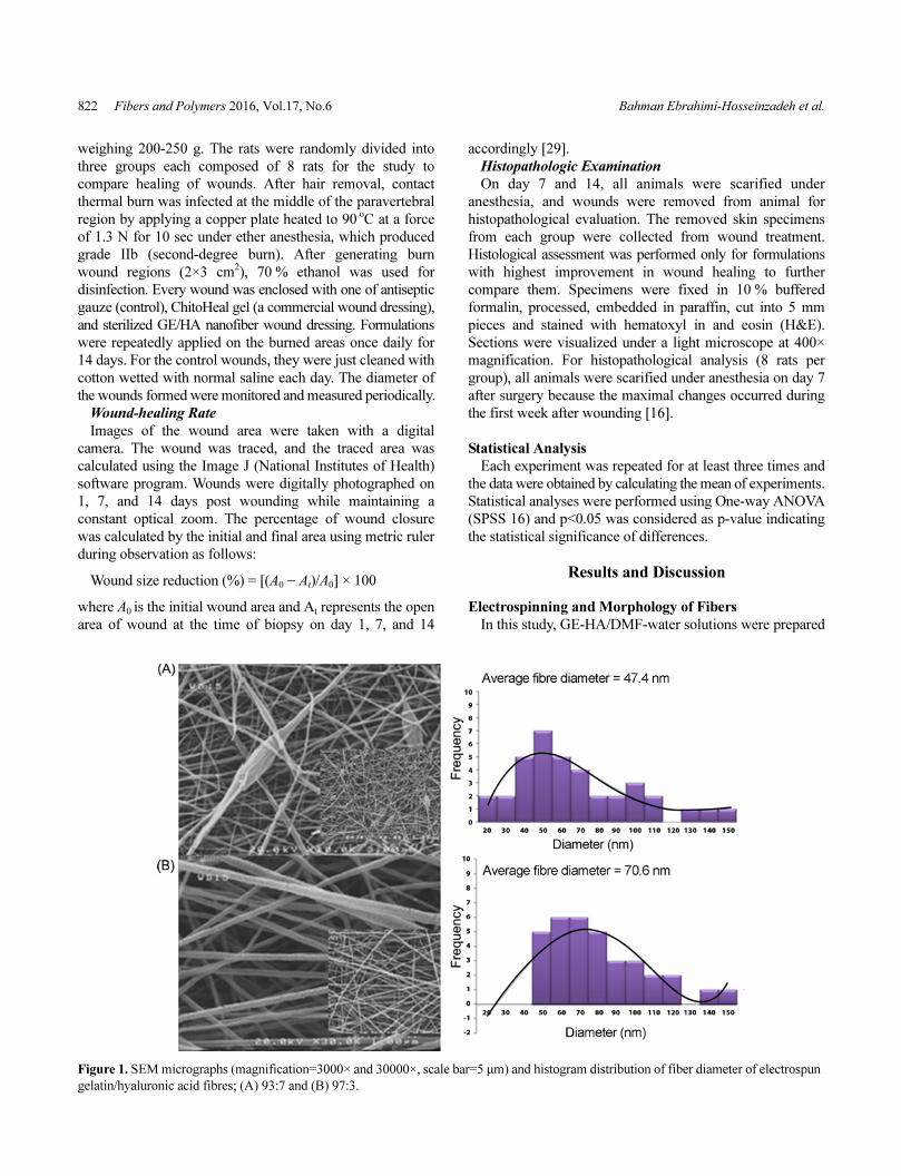
rats. The macroscopic presence of wounds treated with
sterile gauze, ChitoHeal gel (a commercial wound dressing),
and sterilized GE/HA nanofiber wound dressing on several
post-operative days are illustrated in Figure 3. Each wound
was studied for a time period of 1, 7 and 14 days post-
operation. All rats remained alive in the post-operative
period until expiry. They showed no indication of necrosis.
As Figures 4 and 5 show the comparative size decrease of
the wounds was treated with several substances. The results
of morphometric showed that the wound area in the group of
gel and scaffold has been reduced with higher intensity than
that in the control group on the seventh day, while there was
no significant difference between them on the fourteenth
day. The difference in the healed areas in these two groups
was significant. Thus it can be concluded that the gel and the
scaffold have been effective in reducing wound area on the
seventh day. On post-operative day 7, the scaffold of GE/HA
and commercial product considerably reduced the wound
size in comparison with the antiseptic gauze. In the present
study, the trend of wound healing was evaluated on the first,
seventh, and fourteenth days.
However, histological studies showed that the results were
apparent, the epidermis were formed incompletely and there
were excessive inflammatory cells in the wound although
the reduction of wound areas was satisfactory in the control
group on the fourteenth day. The results of previous studies
on the effect of gelatin on wound healing indicated that this
difference was no longer significant on the tenth and
fifteenth days although the healed area in the experimental
group showed a significant increase in comparison with the
control group on the fifth day. This result is consistent with
the findings of the present study [17].
Samples were taken on the 7th and 14th days for
pathological studies. These studies showed that there were
evident differences in many of the wound healing-related
variables. Figure 6 shows samples of the 7th and the 14thFigure 2. FTIR spectra of electrospun gelatin/hyaluronic acid
fibres; (A) 93:7 and (B) 97:3.
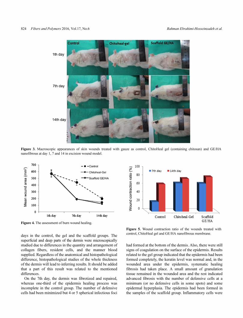
824 Fibers and Polymers 2016, Vol.17, No.6 Bahman Ebrahimi-Hosseinzadeh et al.
days in the control, the gel and the scaffold groups. The
superficial and deep parts of the dermis were microscopically
studied due to differences in the quantity and arrangement of
collagen fibers, resident cells, and the manner blood
supplied. Regardless of the anatomical and histopathological
difference, histopathological studies of the whole thickness
of the dermis will lead to inferring results. It should be added
that a part of this result was related to the mentioned
differences.
On the 7th day, the dermis was fibrotized and repaired,
whereas one-third of the epidermis healing process was
incomplete in the control group. The number of defensive
cells had been minimized but 4 or 5 spherical infectious foci
had formed at the bottom of the dermis. Also, there were still
signs of coagulation on the surface of the epidermis. Results
related to the gel group indicated that the epidermis had been
formed completely, the keratin level was normal and, in the
wounded area under the epidermis, systematic healing
fibrosis had taken place. A small amount of granulation
tissue remained in the wounded area and the rest indicated
advanced fibrosis with the number of defensive cells at a
minimum (or no defensive cells in some spots) and some
epidermal hyperplasia. The epidermis had been formed in
the samples of the scaffold group. Inflammatory cells were
Figure 3. Macroscopic appearances of skin wounds treated with gauze as control, ChitoHeal gel (containing chitosan) and GE/HA
nanofibrous at day 1, 7 and 14 in excision wound model.
Figure 4. The assessment of burn wound healing.
Figure 5. Wound contraction ratio of the wounds treated with
control, ChitoHeal gel and GE/HA nanofibrous membrane.
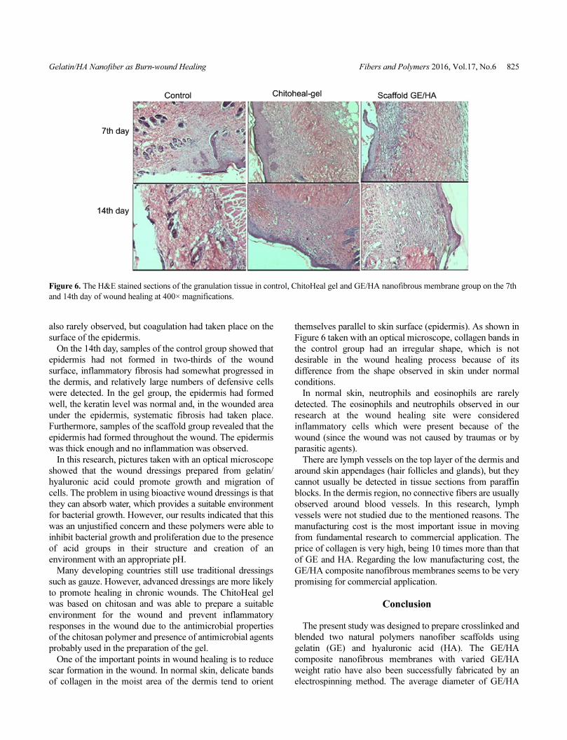
Gelatin/HA Nanofiber as Burn-wound Healing Fibers and Polymers 2016, Vol.17, No.6 825
also rarely observed, but coagulation had taken place on the
surface of the epidermis.
On the 14th day, samples of the control group showed that
epidermis had not formed in two-thirds of the wound
surface, inflammatory fibrosis had somewhat progressed in
the dermis, and relatively large numbers of defensive cells
were detected. In the gel group, the epidermis had formed
well, the keratin level was normal and, in the wounded area
under the epidermis, systematic fibrosis had taken place.
Furthermore, samples of the scaffold group revealed that the
epidermis had formed throughout the wound. The epidermis
was thick enough and no inflammation was observed.
In this research, pictures taken with an optical microscope
showed that the wound dressings prepared from gelatin/
hyaluronic acid could promote growth and migration of
cells. The problem in using bioactive wound dressings is that
they can absorb water, which provides a suitable environment
for bacterial growth. However, our results indicated that this
was an unjustified concern and these polymers were able to
inhibit bacterial growth and proliferation due to the presence
of acid groups in their structure and creation of an
environment with an appropriate pH.
Many developing countries still use traditional dressings
such as gauze. However, advanced dressings are more likely
to promote healing in chronic wounds. The ChitoHeal gel
was based on chitosan and was able to prepare a suitable
environment for the wound and prevent inflammatory
responses in the wound due to the antimicrobial properties
of the chitosan polymer and presence of antimicrobial agents
probably used in the preparation of the gel.
One of the important points in wound healing is to reduce
scar formation in the wound. In normal skin, delicate bands
of collagen in the moist area of the dermis tend to orient
themselves parallel to skin surface (epidermis). As shown in
Figure 6 taken with an optical microscope, collagen bands in
the control group had an irregular shape, which is not
desirable in the wound healing process because of its
difference from the shape observed in skin under normal
conditions.
In normal skin, neutrophils and eosinophils are rarely
detected. The eosinophils and neutrophils observed in our
research at the wound healing site were considered
inflammatory cells which were present because of the
wound (since the wound was not caused by traumas or by
parasitic agents).
There are lymph vessels on the top layer of the dermis and
around skin appendages (hair follicles and glands), but they
cannot usually be detected in tissue sections from paraffin
blocks. In the dermis region, no connective fibers are usually
observed around blood vessels. In this research, lymph
vessels were not studied due to the mentioned reasons. The
manufacturing cost is the most important issue in moving
from fundamental research to commercial application. The
price of collagen is very high, being 10 times more than that
of GE and HA. Regarding the low manufacturing cost, the
GE/HA composite nanofibrous membranes seems to be very
promising for commercial application.
Conclusion
The present study was designed to prepare crosslinked and
blended two natural polymers nanofiber scaffolds using
gelatin (GE) and hyaluronic acid (HA). The GE/HA
composite nanofibrous membranes with varied GE/HA
weight ratio have also been successfully fabricated by an
electrospinning method. The average diameter of GE/HA
Figure 6. The H&E stained sections of the granulation tissue in control, ChitoHeal gel and GE/HA nanofibrous membrane group on the 7th
and 14th day of wound healing at 400× magnifications.
826 Fibers and Polymers 2016, Vol.17, No.6 Bahman Ebrahimi-Hosseinzadeh et al.
fibers was in the range of 20 to 150 nm. In this research,
microscopic study of the wound healing process (two weeks
after the wound was inflicted) showed that more epidermis
was formed in the gel and scaffold groups in comparison
with the control group. The numbers of inflammatory cells
in these two groups were also smaller compared with the
control group, which could well be the reason for the
delayed healing in the control group. In any case, extensive
inflammatory cell infiltration, hyperemia, more severe
edema in the dermis region, and stability of most growth
factors caused infections that were more acute.
References
1. D. Archana, B. K. Singh, J. Dutta, and P. K. Dutta,
Carbohydr. Polym., 95, 530 (2013).
2. N. Liao, A. R. Unnithan, M. K. Joshi, A. P. Tiwari, S. T.
Hong, C. H. Park, and C. S. Kim, Colloid Surf. A-
Physicochem. Eng. Asp., 469, 194 (2015).
3. A. R. Siddiqui and J. M. Bernstein, Clin. Dermatol., 28,
519 (2010).
4. D. N. Heo, D. H. Yang, J. B. Lee, M. S. Bae, J. H. Kim, S.
H. Moon, H. J. Chun, C. H. Kim, H. N. Lim, and I. K.
Kwon, J. Biomed. Nanotechnol., 9, 511 (2013).
5. N. F. Ribeiro, C. H. Heath, J. Kierath, S. Rea, M. Duncan-
Smith, and F. M. Wood, Burns, 36, 9 (2010).
6. X. Liu, T. Lin, J. Fang, G. Yao, H. Zhao, M. Dodson, and
X. Wang, J. Biomed. Mater. Res. Part A, 94, 499 (2010).
7. J. S. Boateng, K. H. Matthews, H. N. Stevens, and G. M.
Eccleston, J. Pharm. Sci., 97, 2892 (2008).
8. P. Zahedi, I. Rezaeian, S. O. Ranaei-Siadat, S. H. Jafari,
and P. A. Supaphol, Polym. Adv. Technol., 21, 77 (2010).
9. S. S. Said, A. K. Aloufy, O. M. El-Halfawy, N. A. Boraei,
and L. K. El-Khordagui, Eur. J. Pharm. Biopharm., 79,
108 (2011).
10. M. S. Khil, D. I. Cha, H. Y. Kim, I. S. Kim, and N. Bhattari,
J. Biomed. Mater. Res. Part A, 67, 675 (2003).
11. L. Yan, S. Si, Y. Chen, T. Yuan, H. Fan, Y. Yao, and Q.
Zhang, Fiber. Polym., 12, 207 (2011).
12. S. Y. Gu, Z. M. Wang, J. Rena, and C. Y. Zhang, Mater.
Sci. Eng. C-Mater. Biol. Appl. Microstruct. Process., 29,
1822 (2009).
13. K. A. Rieger, N. P. Birch, and J. D. Schiffman, J. Mate.
Chem. B, 1, 4531 (2013).
14. J. H. Song, H. E. Kim, and H. W. Kim, J. Mater. Sci.
Mater. Med., 19, 95 (2008).
15. N. Bhardwaj and S. C. Kundu, Biotechnol. Adv., 28, 325
(2010).
16. C. Gong, Q. Wu, Y. Wang, D. Zhang, F. Luo, X. Zhao, Y.
Wei, and Z. Qian, Biomaterials, 34, 6377 (2013).
17. J. Li, A. He, J. Zheng, and C. C. Han, Biomacromolecules,
7, 2243 (2006).
18. L. H. Peng, X. Chen, L. Chen, N. Li, W. Q. Liang, and J.
Q. Gao, Biol. Pharm. Bull., 35, 881 (2012).
19. Y. H. Shan, L. H. Peng, X. Liu, X. Chen, J. Xiong, and J.
Q. Gao, Int. J. Pharm., 479, 291 (2015).
20. W. Y. Chen and G. Abatangelo, Wound Repair. Regen., 7,
79 (1999).
21. E. L. Pardue, S. Ibrahim, and A. Ramamurthi, Organogenesis,
4, 203 (2008).
22. J. A. Brown, J. Wound Care, 13, 48 (2004).
23. I. R. Ellis and S. L. Schor, Exp. Cell Res., 228, 326 (1996).
24. M. Z. Elsabee, H. F. Naguib, and R. E. Morsi, Mater. Sci.
Eng. C-Mater. Biol. Appl. Microstruct. Process., 32, 1711
(2012).
25. R. A. A. Muzzarelli, Carbohyd. Polym., 77, 1 (2009).
26. N. Maeda, J. Miao, T. J. Simmons, J. S. Dordick, and R. J.
Linhardt, Carbohydr. Polym., 102, 950 (2014).
27. B. Balakrishnan, M. Mohanty, P. R. Umashankar, and A.
Jayakrishnan, Biomaterials, 26, 6335 (2005).
28. J. Li, A. He, C. C. Han, D. Fang, B. S. Hsiao, and B. Chu,
Macromol. Rapid Comm., 27, 114 (2006).
29. M. V. Rossum, D. P. Vooijs, X. F. Walboomers, M. J.
Hoekstra, P. H. Spauwen, and J. A. Jansen, J. Mater. Sci.-
Mater. Med., 18, 1449 (2007).