in Bacteriophage T4 Development - CaltechAUTHORSauthors.library.caltech.edu/3104/1/STIjvir80.pdf ·...
15
Vol. 35, No. 3 JOURNAL OF VIROLOGY, Sept. 1980, p. 775-789 0022-538X/80/09-0775/15$02.00/0 Role of the Host Cell in Bacteriophage T4 Development II. Characterization of Host Mutants That Have Pleiotropic Effects on T4 Growth BARBARA L. STITT,t HELEN R. REVEL,4 ILGA LIELAUSIS, AND WILLIAM B. WOOD§* Division of Biology, California Institute of Technology, Pasadena, California 91125 Mutant host-defective Escherichia coli that fail to propagate bacteriophage T4 and have a pleiotropic effect on T4 development have been isolated and characterized. In phage-infected mutant cells, specific early phage proteins are absent or reduced in amount, phage DNA synthesis is depressed by about 50%, specific structural phage proteins, including some tail and collar components, are deficient or missing, and host-cell lysis is delayed and slow. Almost all phage that can overcome the host block carry mutations that map in functionally undefined 'nonessential" regions of the T4 genome, most near gene 39. The mutant host strains are temperature sensitive for growth and show simultaneous reversion of the ts phenotype and the inability to propagate T4+. The host mutations are cotransduced with ilv (83 min) and may lie in the gene for transcription termi- nation factor rho. Roles for the Escherichia coli host cell in the assembly of bacteriophage T4 have been iden- tified in the processes of phage capsid fornation (7, 11, 45), tail fiber assembly (35), and possibly tail assembly (42, 40). We have made an exten- sive search for host-defective (HD) bacterial mutants that affect the assembly of bacterio- phage T4. All mutants that specifically block morphogenesis are found to act during phage capsid formation at the level of T4 gene 31 function (37). One class of HD mutants, desig- nated HDF, shows multiple effects: T4 DNA synthesis is delayed, yet infected cells synthesize significant amounts of phage DNA and even- tually lyse without production of infectious prog- eny, as if the defect were in phage assembly. In this paper we report investigations into the nature of the HDF host defect. We have exam- ined the phenotypes of T4+-infected and unin- fected HDF cells, and we have determined the map positions of the bacterial mutation and the mutations carried by T4 mutant phages that can overcome the host defect. The results support the suggestion (5, 41) that HDF strains, similar strains with mutations at a locus designated tabC (5, 43), and another similar strain desig- nated HD590 (41) carry mutational alterations in the gene for the bacterial transcription ter- mination factor rho. t Present address: Public Health Research Institute of the City of New York, Inc., New York, NY 10016. : Present address: Department of Molecular Biology and Microbiology, Tufts Medical School, Boston, MA 02111. § Present address: Department of Molecular, Cellular and Developmental Biology, University of Colorado, Boulder, CO 80309. (These studies are included in the doctoral dissertation of B.L.S., submitted in partial ful- fillment of the requirements for the Ph.D. de- gree, California Institute of Technology, Pasa- dena, 1978. A preliminary report of some of the studies described here has been published [52].) MATERIALS AND METHODS Materials and methods not reported below have been described previously (37). Media. Tryptone top and bottom agars contained, per liter, 6.5 and 10 g of agar (Difco Laboratories, Detroit, Mich.), respectively, in addition to 10 g of tryptone (Difco) and 5 g of NaCl. Chemicals and enzymes. Threefold recrystallized egg white lysozyme, crystalline vitamin B12, L-homo- cysteine, Tween 40, and DL-a-glycerophosphate were from Sigma Chemical Co., St. Louis, Mo. Oleic acid was from Mallinkrodt; stearate and palmitate were from Serdary Research Laboratories, London, On- tario, Canada. Radioactive compounds. [2-3H(N)]glycerol (200 mCi/mmol) and 2,6-[1,7-'4C]diaminopimelic acid (105 mCi/mmol) were from New England Nuclear Corp., Boston, Mass. ['4C]leucine was used at a specific activ- ity of 312 mCi/mmol. Bacteria and bacteriophages. E. coli K-12 strain SKB178, obtained from A. D. Kaiser, was used for the selection of HD mutants. The properties of SKB178 and the HDF strains derived from it are given in Table 1. B011' thy sup str and HD590 thy sup str hdf are, respectively, an amber suppressor strain of E. coli B and an HDF-type mutant derived from it (42). E. coli Bb, nonpermissive for amber mutants, was used for the preparation of extracts for most in vitro comple- mentation experiments. B/5 and S/6/5 are B strains nonpermissive for amber mutants. CR63 is a K-12 775
Transcript of in Bacteriophage T4 Development - CaltechAUTHORSauthors.library.caltech.edu/3104/1/STIjvir80.pdf ·...

Vol. 35, No. 3JOURNAL OF VIROLOGY, Sept. 1980, p. 775-7890022-538X/80/09-0775/15$02.00/0
Role of the Host Cell in Bacteriophage T4 DevelopmentII. Characterization of Host Mutants That Have Pleiotropic Effects on
T4 GrowthBARBARA L. STITT,t HELEN R. REVEL,4 ILGA LIELAUSIS, AND WILLIAM B. WOOD§*
Division of Biology, California Institute of Technology, Pasadena, California 91125
Mutant host-defective Escherichia coli that fail to propagate bacteriophageT4 and have a pleiotropic effect on T4 development have been isolated andcharacterized. In phage-infected mutant cells, specific early phage proteins are
absent or reduced in amount, phage DNA synthesis is depressed by about 50%,specific structural phage proteins, including some tail and collar components, are
deficient or missing, and host-cell lysis is delayed and slow. Almost all phage thatcan overcome the host block carry mutations that map in functionally undefined'nonessential" regions of the T4 genome, most near gene 39. The mutant hoststrains are temperature sensitive for growth and show simultaneous reversion ofthe ts phenotype and the inability to propagate T4+. The host mutations arecotransduced with ilv (83 min) and may lie in the gene for transcription termi-nation factor rho.
Roles for the Escherichia coli host cell in theassembly of bacteriophage T4 have been iden-tified in the processes of phage capsid fornation(7, 11, 45), tail fiber assembly (35), and possiblytail assembly (42, 40). We have made an exten-sive search for host-defective (HD) bacterialmutants that affect the assembly of bacterio-phage T4. All mutants that specifically blockmorphogenesis are found to act during phagecapsid formation at the level of T4 gene 31function (37). One class of HD mutants, desig-nated HDF, shows multiple effects: T4 DNAsynthesis is delayed, yet infected cells synthesizesignificant amounts of phage DNA and even-tually lyse without production of infectious prog-eny, as if the defect were in phage assembly.
In this paper we report investigations into thenature of the HDF host defect. We have exam-ined the phenotypes of T4+-infected and unin-fected HDF cells, and we have determined themap positions of the bacterial mutation and themutations carried by T4 mutant phages that canovercome the host defect. The results supportthe suggestion (5, 41) that HDF strains, similarstrains with mutations at a locus designatedtabC (5, 43), and another similar strain desig-nated HD590 (41) carry mutational alterationsin the gene for the bacterial transcription ter-mination factor rho.
t Present address: Public Health Research Institute of theCity of New York, Inc., New York, NY 10016.
: Present address: Department of Molecular Biology andMicrobiology, Tufts Medical School, Boston, MA 02111.
§ Present address: Department of Molecular, Cellular andDevelopmental Biology, University of Colorado, Boulder, CO80309.
(These studies are included in the doctoraldissertation of B.L.S., submitted in partial ful-fillment of the requirements for the Ph.D. de-gree, California Institute of Technology, Pasa-dena, 1978. A preliminary report of some of thestudies described here has been published[52].)
MATERIALS AND METHODSMaterials and methods not reported below have
been described previously (37).Media. Tryptone top and bottom agars contained,
per liter, 6.5 and 10 g of agar (Difco Laboratories,Detroit, Mich.), respectively, in addition to 10 g oftryptone (Difco) and 5 g of NaCl.Chemicals and enzymes. Threefold recrystallized
egg white lysozyme, crystalline vitamin B12, L-homo-cysteine, Tween 40, and DL-a-glycerophosphate werefrom Sigma Chemical Co., St. Louis, Mo. Oleic acidwas from Mallinkrodt; stearate and palmitate werefrom Serdary Research Laboratories, London, On-tario, Canada.Radioactive compounds. [2-3H(N)]glycerol (200
mCi/mmol) and 2,6-[1,7-'4C]diaminopimelic acid (105mCi/mmol) were from New England Nuclear Corp.,Boston, Mass. ['4C]leucine was used at a specific activ-ity of 312 mCi/mmol.
Bacteria and bacteriophages. E. coli K-12 strainSKB178, obtained from A. D. Kaiser, was used for theselection of HD mutants. The properties of SKB178and the HDF strains derived from it are given in Table1. B011' thy sup str and HD590 thy sup str hdf are,respectively, an amber suppressor strain of E. coli Band an HDF-type mutant derived from it (42). E. coliBb, nonpermissive for amber mutants, was used forthe preparation of extracts for most in vitro comple-mentation experiments. B/5 and S/6/5 are B strainsnonpermissive for amber mutants. CR63 is a K-12
775

776 STITT ET AL.
TABLE 1. Properties ofHDF strains derived fromSKB178
Bacterium Markers present Comments
SKB178 galE RglI Parent of HDF strainsHDF12.5 galE metG" hdf Temperature sensitive at
Such Rgl I 45°C; at 37°C, 4%"snakes" in H broth, 0%in M9
HDFO.26 galE glp' hdf T6r ts+ derivative of originalRgl I tsHDFO.26; lOOx normal
spontaneous mutationrate
HDF3.41 galE glp' hdf Rgl I Temperature sensitive at42°C; adsorbs T4 poorlyafter growth in M9
HDF3.03 galE glp' hdf Rgl I Temperature sensitive at42°C; hdf allele is leaky;fails to grow phage P1
"The metG mutation was identified by (i) the amino acidrequirement, (ii) the inability to grow in M9 supplementedwith vitamin B12 and homocysteine (2), and (iii) map location(covered by plasmid F103).
h The Suc phenotype indicates lack of growth on sodiumsuccinate as carbon source; the mutation may affect electrontransport (8) and may be in ubiG.
' HDFO.26, HDF3.03, and HDF3.41 each have a mutationin a glp gene, possibly in gipT orglpA. HDFO.26 and HDF3.03do not grow on DL-a-glycerophosphate as the carbon source.HDF3.41 fails to incorporate ['H]glycerol into phospholipids(data not shown).
strain permissive for amber mutants; CR63(A) is non-permissive for T4 rII mutants. CT196 and CT439 arewild nonsuppressing California Institute of Technol-ogy (Caltech) "hospital" strains (50), nonpermissivefor T4 mutants carrying delections in the gene 39-gene 56 region (15) and the tRNA region (50), respec-tively.T4 single and multiple amber mutant strains, listed
in Tables 2 and 3, are from the Caltech collection (nowmaintained in the Department of Molecular, Cellularand Developmental Biology, University of Colorado,Boulder). T4 go mutant phages were selected as de-scribed previously (37). Lysozyme deletion mutants ofT4 (eG19, eG79, eG223, and eG298) and tRNA deletionmutants (psub-A64 and psub-A33) have been describedby Wilson et al. (51). The rII deletion strains used arerEDdf4l (21) and r1589 (1). T4 strains carrying dele-tions in the nonessential region between genes 39 and56 [del(39-56)1, -3, -4, -5, and -12] were provided byT. Homyk (15). The mutation [del(39-56)12] is in aT4D genetic background, whereas the other del(39-56) mutations are in T4B. All del(39-56) strains carrythe rII deletion r1589.
Genetic crosses. Phage crosses were carried out asdescribed previously (37).Measurement of phage DNA synthesis. Bacte-
rial cells were grown in H broth to 108 per ml andconcentrated to 2 x 10⁸ per ml in the same medium.A 10-ml flask containing 2 ml of cells was placed in ashaking water bath at 37°C at t = -2 min. At t = 0min, phage were added at a multiplicity of infection of6 to 8. At t = 3, deoxyadenosine at a final concentrationof 200 ,tg/ml and ["4C]thymidine at a final concentra-tion of 6 ,tg/ml and a specific activity of 5 to 10lCi/,umol were added. At various times 0.1-ml sampleswere pipetted into 2 ml of ice-cold 5% trichloroacetic
acid containing 50 ,ig of unlabeled thymidine per ml.After 30 min at 0°C, the samples were filtered throughWhatman GF/A glass filters; the filters were washedwith 4 volumes of 5% trichloroacetic acid containingthymidine and then with 2 volumes of 95% ethanol;they were then dried and counted in 5 ml of toluene-Liquifluor scintillation fluid.
In vitro complementation tests. The presence ofactive major phage structures (heads, tails, and tailfibers) in T4+-infected HDF strains was assayed as
described in reference 37. The assays for whole base-plates and tail-baseplate gene products were as de-scribed (20), with the following modifications. Cultures(250 ml) of E. coli strain Bb were infected (and super-
infected at t = 10 min in cases where no t mutationwas present) at multiplicity of infection of 7. HDF-infected cells were aerated for 35 min at 37°C andthen chilled for 10 min before harvesting by centrifu-gation. A 20-j,l amount of DNase and 40 to 50,l ofTris-magnesium (TMg) buffer, pH 7.4 (37), were
blended in a Vortex mixer with the infected cell pelletsbefore freezing. The in vitro complementation reactionmixtures consisted of 20 1, of each extract and were
incubated at 30°C for 3 h before assay of plaque-forming phage.
TABLE 2. T4 amber mutants: single
Gene Mutation
3 amNG1315 amN1356 amNl02 amB2517 amB168 amN1329 amE1710 amB25511 amN12812 amN6915 amN13318 amE1819 amE113723 amB1725 amS5226 amS10527 amN12028 amA45229 amB739 amN116 amNG457 amE48051 amS2953 amH2854 amH2160 amE300e amH26 tsC3
TABLE 3. T4 amber mutants: multiple
Strain Defective MutationsgenesX77 34:34:37 amB25:amA455:amN52X143 18:27 amE18:amNI20X379 23:63:rII amB17:amM69:rEDdf4lX381:t 5:6:7:t amB256:amB251:amBl6:amB5
15:t amNI33:amB519:t amE1137:amB548:t amN85:amB5
J. VIROL.
PLEIOTROPIC HOST-DEFECTIVE MUTANTS 777
Tail fiber antigen determination. The endpointserum blocking assay was used as described previously(49), except that twofold dilutions of the sample weremade in the serum and 2,500 tester phage were added.Assay of bacterial protein synthesis. Cells were
grown to 8 x 10⁷ per ml in M9 medium plus all aminoacids, thiamine, lo-5 M FeCl3, and thymine, concen-
trated to 4 x 10⁵ per ml in the same medium minusleucine, and diluted 40-fold into the supplementedmedium containing 2 ,uCi of ['4C]leucine per ml. Sam-ples, 40 pl, were taken every 15 min and added to 2 mlof ice-cold 10% trichloroacetic acid containing 50 ytg ofleucine per ml. After at least 30 min, samples were
filtered through Whatman GF/A glass filters; the fil-ters were washed with 4 volumes of 5% trichloroaceticacid containing cold leucine followed by 2 volumes of95% ethanol, dried, and counted in 5 ml of toluene-Liquifluor scintillation fluid.
Assay of bacterial DNA synthesis. Cells weregrown to 4 x 10" per ml and diluted 40-fold intoprewarmed H broth containing 100 ug of deoxyaden-osine and 1 ItCi of [14C]thymidine per ml. Samples, 50pl, were taken at 15-min intervals into 2 ml of ice-cold10% trichloroacetic acid and prepared for counting as
above.Assay of bacterial cell wall synthesis. Cells
were grown to 4 x 10⁵ per ml and diluted 40-fold intoprewarmed M9 containing 0.05% Casamino Acids,thiamine, 10-5 M FeCl3, and 3,tCi of ['4C]diaminopi-melic acid per ml. Samples (50 IL) were taken at 15-min intervals into 2 ml of ice-cold 10% trichloroaceticacid and prepared for counting as above.Assay of bacterial phospholipid synthesis. The
procedure for assay of bacterial phospholipid synthesisis a modification of methods described by Mindich etal. (31), Goldfine (12), and Radin (36). Cells were
grown to 8 x 10⁷ per ml in M9 plus 0.05% CasaminoAcids, thiamine, 10'- M FeCl3, and 20,tg of glycerolper ml, concentrated to 4 x 10⁵ per ml, and diluted 40-fold into fresh prewarmed medium containing 25 uCiof [3H]glycerol per ml. Samples (20 ,tl) were taken induplicate every 15 min, applied to Whatman GF/Cglass fiber filters, and plunged into 5 ml of ice-cold 10%trichloroacetic acid, which was decanted after 30 minand replaced by 5% trichloroacetic acid for 15 min and
then by two 30-min rinses of cold water. One set ofsamples was then dried and counted; the second was
rinsed three times with 3 ml of 2:1 chloroform-meth-anol before drying and counting. Incorporation of glyc-erol into phospholipid was determined by subtractingthe radioactivity in the chloroform-methanol-ex-tracted samples from that in the unextracted samples.
RESULTS
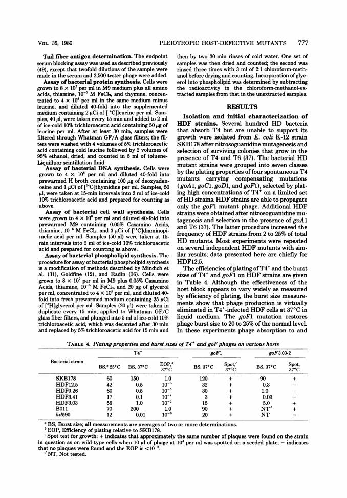
Isolation and initial characterization ofHDF strains. Several hundred HD bacteriathat absorb T4 but are unable to support itsgrowth were isolated from E. coli K-12 strainSKB178 after nitrosoguanidine mutagenesis andselection of surviving colonies that grow in thepresence of T4 and T6 (37). The bacterial HDmutant strains were grouped into seven classesby the plating properties of four spontaneous T4mutants carrying compensating mutations(goAl, goCl, goDl, and goFl), selected by plat-ing high concentrations of T4+ on a limited setofHD strains. HDF strains are able to propagateonly the goFl mutant phage. Additional HDFstrains were obtained after nitrosoguanidine mu-tagenesis and selection in the presence of goAland T6 (37). The latter procedure increased thefrequency of HDF strains from 2 to 25% of totalHD mutants. Most experiments were repeatedon several independent HDF mutants with sim-ilar results; data presented here are chiefly forHDF12.5.The efficiencies of plating ofT4+ and the burst
sizes of T4+ and goFl on HDF strains are givenin Table 4. Although the effectiveness of thehost block appears to vary widely as measuredby efficiency of plating, the burst size measure-ments show that phage production is virtuallyeliminated in T4+-infected HDF cells at 37°C inliquid medium. The goFl mutation restoresphage burst size to 20 to 25% of the normal level.In these experiments phage absorption to and
TABLE 4. Plating properties and burst sizes of T4' and goFphages on various hostsT4+ goFl goF3.03-2
Bacterial strain EOPBS,0 250C BS, 370C 370C BS, 37°C S3p7oC BS, 37°C Spot,
SKB178 60 150 1.0 120 + 90 +HDF12.5 42 0.5 10-6 32 + 0.3 -HDF0.26 60 0.5 10-5 30 + 1.0 -HDF3.41 17 0.1 10-4 3 + 0.03 -HDF3.03 56 1.0 10-2 15 + 5.0 +B011 70 200 1.0 90 + NTd +hd590 12 0.01 10-6 20 + NT -
a BS, Burst size; all measurements are averages of two or more determinations.b EOP, Efficiency of plating relative to SKB178.'Spot test for growth: + indicates that approximately the same number of plaques were found on the strain
in question as on wild-type cells when 10 ,lA of phage at 104 per ml was spotted on a seeded plate; - indicatesthat no plaques were found and the EOP is <10-2.
d NT, Not tested.
VOL. 35, 1980

778 STITT ET AL.
killing of HDF mutant hosts was normal (datanot shown). The temperature dependence of theability of HDF strains to support T4 growth isalso shown in Table 4. At 25°C the burst size ofT4+ in HDF bacteria is almost normal.Upon initial isolation all HDF strains were
temperature sensitive for bacterial growth: bac-terial lawns at 42°C were either very sparse ornonexistent on EHA, LB, or tryptone plates.Cell growth did not stop immediately in liquidculture at 42°C; during a 2-h observation periodthe cell doubling time increased from 30 min at37°C to 50 to 80 min at the restrictive tempera-ture. The original temperature-sensitive strainof HDFO.26 was lost; all experiments withHDFO.26 have been done with a spontaneousts+ derivative of the original.The ability of some other phages to grow on
HDF mutant hosts was determined by spot test.Phages T2 and T6 behave like T4, whereaslambda, T3, T5, T7, and P1 grow on all HDFstrains, with the single exception that P1 fails togrow on HDF3.03.Course of infection in T4+-infected HDF
strains. (i) Phage DNA synthesis is delayedand depressed. DNA synthesis after phageinfection was measured by the continuous incor-poration of ['4C]thymidine into acid-insolublematerial (Fig. 1). The results show that the rateof T4+ DNA synthesis in HDF12.5 is about halfthat in the parental host and that there is a 3- to6-min delay in the initiation of DNA synthesis.In goFl infection of HDF12.5, the rate of DNAsynthesis is increased, but the delay is still pres-ent, suggesting that factors affecting the rate ofDNA synthesis are more crucial to phage pro-duction than those affecting time of initiation.DNA synthesis in SKB178 infected with goFl isnormal.
(ii) Assembly of phage structures is de-fective or absent. Table 5 shows that no activeheads, tails, or free baseplates could be demon-strated in extracts of T4+-infected HDF cells byin vitro complementation tests. The subnormallevel of active tail fibers observed in these ex-periments was verified by serological measure-ments of tail fiber antigens in the same extracts(data not shown). The absence of active majorstructural assembly intermediates was con-firmed by electron microscopy of negativelystained lysates of phage-infected cells. Whereascontrol lysates of T4+-infected wild-type cellsshowed the expected filed heads, tails, and freebaseplates, lysates of T4-infected HDF cellsshowed chiefly empty heads and rare polyheadsand polysheaths, but no tails or baseplate struc-tures (data not shown).The presence of polysheath, the product of
aberrant polymerization of gpl8 that appears
late in normal infection and earlier in the ab-sence of baseplate production (22), suggestedthat in T4+-infected HDF cells some tail proteinsmight be present but unable to assemble nor-mally because of a defect in baseplate morpho-genesis. Further in vitro complementation anal-ysis of all tail gene products revealed that thecore and sheath components gp3, gpl5, gpl8,and gpl9 from T4+-infected HDF cells are active(Table 6). Among the baseplate components,however, only gp9 and gpl2 show significant invitro activity. These results suggest that theabortive baseplate assembly is due to decreasedlevels of many proteins rather than a block at aspecific assembly step. Impaired synthesis orincreased degradation of baseplate proteinscould account for these observations, in view ofthe finding that in vitro baseplate complemen-
0)
x
E0~I.-
000.00
c
E.1_
lq
4
3
2
0 10 20 30 40Minutes after infection
FIG. 1. Incorporation of[14C]thymidine in five T4-infected cell cultures as a measure of phage DNAsynthesis. The arrow indicates the time ofaddition of['4Clthymidine, after which samples were taken atvarious times and trichloroacetic acid-precipitableradioactivity was determined. Symbols: +, SKBI78infected with T4'; x, SKB178 infected with goFI; A,HDF12.5 infected with goFI; 0, HDF12.5 infectedwith T4+; *, SKBl 78 infected with thegene 42 mutantam122 (DNA-negative phenotype). The am122 exper-iment shows that the observed thymidine incorpora-tion is a measure ofphageDNA synthesis. For detailsof the procedure see text.
J. VIROL.

PLEIOTROPIC HOST-DEFECTIVE MUTANTS 779
TABLE 5. In vitro complementation of T4+-infected HDF extracts with defective extracts supplying majorphage structuresa
Plaque-forming phage x 10-'
Complementing extract Head defec- Tail and Baseplate Tail defec- Tail fiberde- T4+ +tive baseplate defective tive fective HDF12.5
T4+ + HDF12.5 9.3 11.6 11.0 -b 62 7.9Tail fiber defective 480 200 - - 3.2Tail defective - - 470 3.6Baseplate defective - - 0.5Tail and baseplate defective 60 0.4Head defective 1.3
a Fifty microliters each of two extracts were mixed, incubated for 3 h at 300C, and then assayed for plaque-forming phage. Results are expressed as plaque-forming phage per milliliter of reaction mixture. Extracts wereprepared as described in Revel et al. (37). The head-defective preparation (tail and tail fiber donor) was anextract made from SKB178 infected with amB17 (gene 23 defective); the tail- and baseplate-defective prepa-ration (head and tail fiber donor) was an extract made from SKB178 infected with X143 (genes 18:27 defective);the baseplate-defective preparation (head and tail fiber donor) was an extract made from Bb infected withX381:t (genes 5:6:7:t defective); the tail-defective preparation (head, baseplate, and tail fiber donor) was anextract made from Bb infected with amE1137:amB5 (genes 19:t-defective); and the tail fiber-defective prepa-ration (donor of particles lacking tail fibers) was an extract made from SKB178 infected with X77 (genes 34:34:37-defective).
b_, Not tested.
TABLE 6. In vitro complementation assays for thepresence of tail proteins in extracts of T4+-infected
HDFaPlaque-forming phage x 10'
Tail structure Mutant Uncom-missing from utane ple-test extract geneplate de- HDF
controls fective
Sheath 18 0.1 220 30
Tube 19 3.8 670 370
Connector 3 32 1000 9115 6.8 600 350
Baseplate plug 29 2.1 86 3.026 1.8 20 2.028 2.6 194 3.051 2.6 10 3.05 0.4 130 2.2
27 1.0 60 2.7
Baseplate wedge 10 0.1 67 1.711 75 65 427 1.0 130 2.18 1.3 52 8.06 8.0 280 10.0
53 0.6 46 1.625 5.4 28 2.6
Baseplate com- 48 2.7 74 7.7pletion proteins 54 0.2 31 0.8
9 5.6 450 31012 3.7 290 80
a Twenty microliters each of two extracts were mixed, in-cubated for 3 h at 30°C, and then assayed for plaque-formingphage. Results are expressed as plaque-forming phage permilliliter of reaction mixture. Extracts were prepared as de-scribed in the text. The baseplate-defective control extractwas made from strain Bb infected with a baseplate structuralgene mutant (gene 5, 7, or 8; different from the mutant genein the test extract). The T4+ + HDF extract was made fromHDF12.5 infected with T4+.
tation reactions are extremely concentration de-pendent (20).
(iii) Patterns of phage-induced proteinsynthesis are altered. Electrophoretic analy-sis of proteins synthesized after phage infectionshows that a few specific early and late proteinsare decreased in amount or absent in T4+-in-fected HDF cells (Fig. 2). Specifically, gp43 andfour early or middle proteins of molecularweights about 58,000, 43,000, 36,000, and 26,000are reduced in T4+-infected mutant hosts. Theidentity of the latter four proteins is unknown,although electrophoretic analysis and in vitrocomplementation tests have shown that the43,000-dalton protein is not gp63 and that the58,000-dalton protein is neither gp39 nor gp3O,but may be gp4l or an unknown protein thatmigrates with it in electrophoresis (data notshown). The unidentified early proteins of43,000and 36,000 daltons are missing at late times inthe T4+-infected HDF cells under both nonper-missive (370C) and permissive (250C) conditions(Fig. 3).At least four viral structural proteins normally
synthesized late in infection, gp34, gp7, gp37,and gpwac are clearly reduced in T4+-infectedHDF cells under nonpermissive conditions.These structural proteins are made at normallevels under conditions permissive for phagegrowth, that is, in goFl infection of HDFO.26 at370C and T4+ infection of HDFO.26 at 250C (Fig.3). Many of the baseplate proteins migrate tocrowded regions of the gel, so that assessment oftheir presence or absence is difficult.
Finally, some proteins are overproduced inthe abortive T4+ infection of HDF cells: bands
VOL. 35, 1980

780 STITT ET AL.
23-63:
2-4' 4-6' 6-8' 8-10' 10-12' 14'on rl
+ F-i- F: + F + F + F + F +
4qw. -' _
4-
w234 8v 6 7 8 9 10 11 ? 3
I23 4 5 6 7 8 ~~9 10 I I 12 13
-347
743_ r! IA-18
-p58,00n 2-2 3--Wo 23*-24*- p43,000--p 36,000-- 32++ B
Wp 26,00042+ 45'IP iIl
FIG. 2. Time ofappearance ofphage proteins in T4+-infected HDFO.26. HDF0.26 was used for mostproteinlabeling experiments in place ofHDF12.5, which behaves anomalously under the conditions used for labeling(see text). Cultures prepared as described in the text were labeled for the indicated periods, chased, andprepared for electrophoresis on a 10%o polyacrylamide gel as described by Revel et al. (37). "+" indicates T4+infection of SKB178; "F" indicates T4+ infection ofHDFO.26. Bands are labeled as the products of T4 geneswhere known, or by a "p" followed by their estimated molecular weight. At early times numerous bacterialbands are visible, particularly in the 2- to 4-min labeling period. Most phage bands were identified by thecomparison ofproteins made in wild-type infections with those made in infections of nonpermissive cells withphage containing known amber mutations. The identifications of gp7, gp43, pg32, and gp22 were made bycomparison withpublished electropherograms (33,48). Molecular weights ofunidentified bands were estimatedfrom aplot ofmigration distances ofknown bands through a 10ogel against the logarithms oftheir molecularweights. Such plots were found to be linear from approximately 80,000 to at least 20,000 daltons. 23:63:rIIindicates infection ofSKB178 with a phage carrying the mutations amB17, amM69, and rEDdf4l.
on electrophoresis gels corresponding to gp32 +rIIB and to gpIPIII are more intense than nor-mal in analyses of samples taken throughoutinfection, and a band just below gp23* appearsmore intense than normnal in samples taken latein infection (Fig. 3). Overproduction of gp32, theDNA binding protein, could be a result of aber-rant DNA synthesis (25).
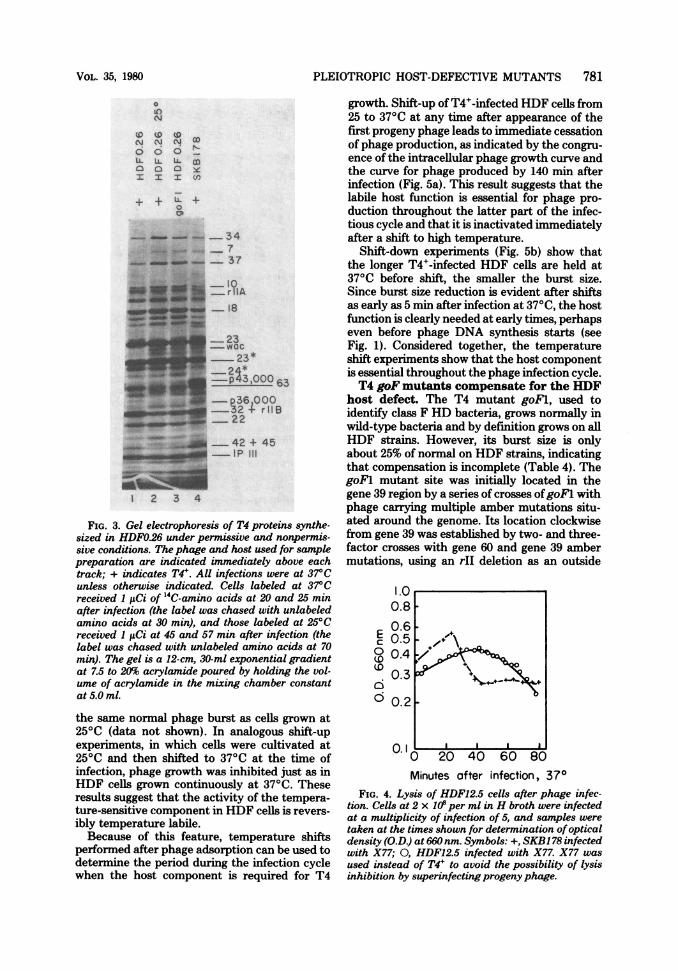
(iv) Cell lysis is delayed. Although T4+-infected HDF strains do not produce activeprogeny phage, the infected cells do undergolysis. However, lysis of T4+-infected HDF12.5 isdelayed about 20 min compared with lysis ofT4+-infected SKB178, and cell distintegrationduring lysis proceeds more slowly (Fig. 4). Whenshaken with chloroform, both T4+-infected HDFcells and wild-type cells lyse promptly at timessubsequent to 12 to 15 min postinfection at 37°C(data not shown). This result suggests that in
T4+-infected HDF cells lysozyme production isclose to normal, but retarded breakdown of thebacterial inner membrane limits access to thecell wall, thus delaying the lysis process. In goFlinfection of HDF strains the delay in lysis isreduced but still evident (data not shown). Thisobservation is consistent with burst size mea-surements, which indicate that the goFl muta-tion does not fully compensate for the host de-fect (see Table 4).
(v) The host component defined by hdfmutations is required throughout T4 infec-tion. T4+ growth in HDF cells is strongly influ-enced by the temperature during phage infection(Table 4) but is independent of the temperatureat which cells are cultivated before infection.Cells that are grown at 37°C, poisoned withKCN, shifted to 25°C, infected with T4+, andthen diluted to relieve KCN inhibition produce
p
J. VIROL.
PLEIOTROPIC HOST-DEFECTIVE MUTANTS 781
0
00z z
C)00
0'
(-
m(I)
-34
-37
~18
0006
-,,, ,, ,r.B
--1,^_ ~ ~ -I_FI
1234
~~ii U -- --3-'0-0-0 63_ _ 3 2;~~~~~~r II
_22
...4'5AR:e _ 42 + 4 5
...... ..
_~~~~Al1 2 3 4
FIG. 3. Gel electrophoresis of T4 proteins synthe-sized in HDFO.26 under permissive and nonpermis-sive conditions. The phage and host used for samplepreparation are indicated immediately above eachtrack; + indicates T4'. All infections were at 37°Cunless otherwise indicated. Cells labeled at 37°Creceived 1 ltCi of '4C-amino acids at 20 and 25 minafter infection (the label was chased with unlabeledamino acids at 30 min), and those labeled at 25°Creceived 1 ,uCi at 45 and 57 min after infection (thelabel was chased with unlabeled amino acids at 70min). The gel is a 12-cm, 30-ml exponential gradientat 7.5 to 20% acrylamide poured by holding the vol-ume of acrylamide in the mixing chamber constantat 5.0 ml.
the same normal phage burst as cells grown at250C (data not shown). In analogous shift-upexperiments, in which cells were cultivated at25°C and then shifted to 370C at the time ofinfection, phage growth was inhibited just as inHDF cells grown continuously at 370C. Theseresults suggest that the activity of the tempera-ture-sensitive component in HDF cells is revers-ibly temperature labile.Because of this feature, temperature shifts
performed after phage adsorption can be used todetermine the period during the infection cyclewhen the host component is required for T4
growth. Shift-up of T4+-infected HDF cells from25 to 37°C at any time after appearance of thefirst progeny phage leads to immediate cessationof phage production, as indicated by the congru-ence of the intracellular phage growth curve andthe curve for phage produced by 140 min afterinfection (Fig. 5a). This result suggests that thelabile host function is essential for phage pro-duction throughout the latter part of the infec-tious cycle and that it is inactivated immediatelyafter a shift to high temperature.
Shift-down experiments (Fig. 5b) show thatthe longer T4+-infected HDF cells are held at370C before shift, the smaller the burst size.Since burst size reduction is evident after shiftsas early as 5 min after infection at 370C, the hostfunction is clearly needed at early times, perhapseven before phage DNA synthesis starts (seeFig. 1). Considered together, the temperatureshift experiments show that the host componentis essential throughout the phage infection cycle.T4 goF mutants compensate for the HDF
host defect. The T4 mutant goFl, used toidentify class F HD bacteria, grows normally inwild-type bacteria and by definition grows on allHDF strains. However, its burst size is onlyabout 25% of normal on HDF strains, indicatingthat compensation is incomplete (Table 4). ThegoFl mutant site was initially located in thegene 39 region by a series of crosses ofgoFl withphage carrying multiple amber mutations situ-ated around the genome. Its location clockwisefrom gene 39 was established by two- and three-factor crosses with gene 60 and gene 39 ambermutations, using an rII deletion as an outside
E0
6D
1.00.80.60.50.40.3
0.2
0.1I
Minutes after infection, 370FIG. 4. Lysis of HDF12.5 cells after phage infec-
tion. Cells at 2 x O10per ml in H broth were infectedat a multiplicity of infection of 5, and samples weretaken at the times shown for determination ofopticaldensity (O.D) at 660 nm. Symbols: +, SKB178 infectedwith X77; 0, HDF12.5 infected with X77. X77 wasused instead of T4' to avoid the possibility of lysisinhibition by superinfectingprogeny phage.
VOL. 35, 1980

782 STITT ET AL.
a)
a)
L
Time of shift (minutes after infection)FIG. 5. Phageproduction in infected-cell cultures subjected to temperature shifts. (a) Infected cellsprepared
as described in the legend to Fig. 4 were incubated with aeration at 27°C. To follow intracellular phagegrowth, samples were diluted at 2-min intervals into chloroform broth andplated on a permissive host. Othersamples were shifted from 27 to 37°C at the indicated times by 200-fold dilution into prewarmedH broth andaerated at the higher temperature. Chloroform was added to these cultures at 140 min after infection, andsamples were plated on CR63 to assay totalphage produced in the temperature-shifted cells. Symbols: +, T4+-infected SKB178, intracellular growth curve; *, T4i-infected SKBI78, temperature shifted; 0, T4+-infectedHDF12.5, intracellular growth curve; O, T4+-infected HDF12.5, temperature shifted. (b) Infected cells wereshifted form 37 to 25°C at the indicated times by 200-fold dilution and treated as in (a). Symbols: +, T4+-infected SKB178; 0, T4+-infected HDF12.5; A, T4+-infected HDF12.5 superinfected with T4+. Points to theright of the dashed line are from samples kept at 37°C for 140 min (i.e., not shifted). The identical resultsobtained for superinfected and nonsuperinfected HDF12.5 show that the decrease in progenyphage is not dueto a decrease in the amount of time available at the permissive temperature before lysis.
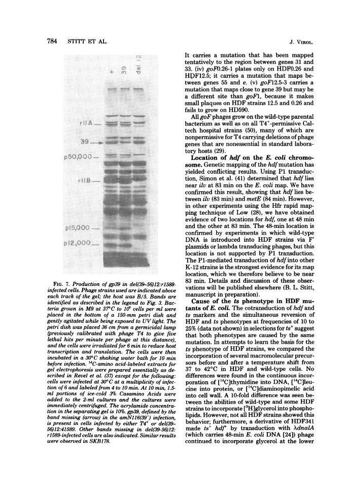
marker. Finally, by crosses ofgoFl with variousphages carrying deletions in the gene 39-56 re-gion, the mutant site was placed between theleft ends of del(39-56)5 and del(39-56)3 (Fig. 6)in a nonessential region of the genome. ThegoFl mutation is not in gene 39, based on thefollowing observations. The deletion mutantdel(39-56)12 fails to yield wild-type recombi-nants when crossed with goFl and thereforemust lack at least part of the gene in which thegoFl mutation lies. However, del(39-56)12forms plaques under conditions that requiregene 39 function and makes a product identifia-ble on sodium dodecyl sulfate-acrylamide gels asgp39 (Fig. 7).
Further experiments with deletion mutantsindicate that the goF phenotype depends uponfunctional alteration of a phage gene productrather than its elimination. The del (39-56)12mutant, which apparently lacks the goFl+ site,grows in wild-type hosts but fails to grow inHDF hosts. In contrast, the multiple mutantsgoF1:del(39-56)3 and goF1:del(39-56)4, whichcarry only slightly smaller deletions than del(39-56)12 (15), do grow in HDF strains.The results of mixed infections of HDF12.5
with wild-type (goF) phage and phage carryingthe goFl mutation are shown in Table 7, exper-iments 1 to 6. The observed burst sizes areintermediate between those for HDF12.5 in-fected with goF+ alone and goFl alone. Similarresults are found in mixed infections with goFland del(39-56)12, which lacks the goF locus(Table 7, experiments 7 to 12). These findingssuggest that thegoFl allele is dominant in mixedinfections, but that there may be a dosage de-pendence of burst size on goFl. The skewedoutput ratio of progeny genotypes obtained inmixedly infected HDF12.5 cells is unexplained.Although 8 of 13 independently selected goF
phage mutants resemble goFl, the following fiveare different. (i) goF3.03-2 grows only onHDF3.03 and carries a mutation that maps nearthe T4 lysozyme gene e (Table 4, Fig. 8). Becausethe lysis function of T4 lysozyme apparently isunaffected in wild-type cells infected withgoF3.03-2 (data not shown), this mutation prob-ably is not in gene e. (ii) goF3.03-8 carries amutation that maps in a similar location. Thismutant fails to grow on HD590, but unlikegoF3.03-2, it plates on all K-12 HDF strains. (iii)goFn.26-6 also compensates for all HDF defects.
J. VIROL.

PLEIOTROPIC HOST-DEFECTIVE MUTANTS 783
0 2 3 4 s 6 7 8 9
rElB rElA 60 39
,o° S Qo $ C< <' C,) dexA; p5QOOO; pseF;T4 chromosome 4, 4 $ @ CP oc sud; mod; dda
I I III
rEDdf41 _ del (39-56)12r1589 4.2 2.7(31 2.7(2l.4(2) del(39-56) 5
_--ng503 del(39-56)341(21- del(39-56)
5.6(2t 7.6 del (39-56) 4' 5.6 9.2 _0"3.4 _ -
6.3
9.1 lo* e>0.44_0 _ 3.7
3.6(2)8.4'6.3
FIG. 6. Location ofgoF1 on the T4genome. The figure shows thepositions ofpoint mutations (short verticallines above the line representing the T4 chromosome) and deletions (heavy bars). The precentages ofrecombination are shown below; arrows indicate the intervals over which recombination was measured.Numbers in parentheses indicate the number of crosses. A notch at the end of a heavy bar indicates that thedeletion continues in that direction. The ends of genes are indicated by vertical lines drawn all the waythrough the line representing the T4 chromosome. The positions ofgenes are indicated by labeled brackets.Amber mutations are indicated by number only, without the prefix am. The scale (top line) is measured inkilobase pairs (kb) from an arbitrary zero point at the boundary between rIIA and rIIB (53). The size of therIIA region, and hence the right endpoint of rEDdf4l (10), is based on the data ofBujard et al. (4) and Kimand Davidson (21). By measuring the distance from del(39-56)1 (previously designated D,) to rII deletionsdefining the right ends of rILA (4,600 base pairs) and rIIB (6,400 base pairs), Bujard et al. (4) estimated thelength of the rIIA region to be 1,800 ± 70 base pairs. The right-hand end ofr1589 has been placed at 0.5 kb onthe basis of data of Homyk and Weil (15), who measured the distance between del(39-56)1 and r1589 to be5,900 base pairs. The left endpoints ofthe del(39-56) mutants are defined by the data ofHomyk and Weil (15).The size ofgene 60 has been estimated from extensive intracistronic mapping (32). The size of gene 39 hasbeen estimated from the polypeptide molecular weight of 64,000 (33). The positions ofplaCTr5x and cef aregiven in reference 15. The crosses were carried out in CR63 at 30°C as described by Revel et al. (37). Totalprogeny was determined by plating on CR63 at 37°C. Scoring of recombinants and determination ofrecombination frequency (%R) was as follows: (i) In crosses between amber mutants in the DNA delay genes39 and 60, wild-type (WT) progeny were determined by plating on S/6/5 at 25°C (32); %R = WT x 200/totalprogeny. When one of the parentalphage also carried a deletion in the rII genes (rEDdf4l), the order of theamber mutations with respect to rII was determined by stabbing wild-type progeny from S/6/5 to a lawn ofCR63(A) to distinguish rIlr from rII. (ii) The intervals between rEDdf4l and ambers in gene 39 weredetermined by crossing rEDdf4l:am double mutants with wild-type phage. am+ phage were determined byplating on S/6/5 at 25°C (= one-half totalprogeny), and individualplaques were stabbed to lawns of CR63(A)to distinguish parental am+:rII+ from recombinant am+:rII phage. %R = am+:rII x 10()/number ofplaquespicked from S/6/5. (iii) The intervals between various del(39-56) deletions and amber mutants in gene 39 weredetermined by crossing the single mutants. am+:del+ recombinants were measured as large plaques on S/6/5 at 250C. %R = am+der x 200/total progeny. (iv) The intervals between goF1 and amber mutants in genes39 and 60 were determined by crossing the single mutants. am+ phage were determined by plating on S/6/5at 250C (= one-half total progeny), and individual plaques were stabbed to lawns of HDF12.5 at 37°C todistinguish parental am+:goF1 from recombinant am+:goF+. %R = am+ goF+ x 100/number of plaquespicked from S/6/5. (v) The intervals between goF1 and the (39-56) deletions were determined by crossing thesingle mutants. Total progeny were determined by plating on CR63, and individual plaques were stabbed tolawns of CT196 (30°C) and HDF12.5 (37°C) to identify the two recombinant type phages. %R = recombinantsx 100/number ofplaques picked from CR63. (vi) The interval between del(39-56)12 and rII deletion r1589 wasdetermined in a cross betweengoF1 and del(39-56)12, which also carries r1589. Totalprogeny were determinedby plating on CR63, and individual plaques were stabbed to lawns of CR63(A) and HDF12.5 to identify rII+:del(39-56)12 and rII:goF1 recombinants. %R = recombinants x 100/number ofplaques picked from CR63.
VOL. 35, 1980
784 STITT ET AL... s ZSO ^ l ( \ ; f S\'tS .::-: A : .- .X : ::: n .:. b.' . :,t t ._) J t:e, ___
_*.' 3.w., .- i C_L
r,.3,,5 ^^ J_-s:gXtee:8>!> e... th6'} }e.1S0. .: m: w:
....,x,5:. ._ _
:*s_ww-.: l | S .K_S *_*_I .w. :e ^l | z S^ j s ;D s B .. XH;se.: . e
M;:Kz::::: w-:...ew,::t;S a P ,2 ....... *},^, ... ,,) , .. ,,,, _.FIG. 7. Production of gp39 in del(39-56)12:rl589-
infected cells. Phage strains used are indicated aboveeach track of the gel; the host was B/5. Bands are
identified as described in the legend to Fig. 3. Bac-teria grown in M9 at 37°C to 108 cells per ml were
placed in the bottom of a 150-mm petri dish andgently agitated while being exposed to UV light. Thepetri dish was placed 36 cm from a germicidal lamp(previously calibrated with phage T4 to give fivelethal hits per minute per phage at this distance),and the cells were irradiated for 6 min to reduce hosttranscription and translation. The cells were thenincubated in a 30°C shaking water bath for 10 minbefore infection. "C-amino acid-labeled extracts forgel electrophoresis were prepared essentially as de-scribed in Revel et al. (37) except for the following:cells were infected at 30°C at a multiplicity of infec-tion of 6 and labeled from 4 to 10 min. At 10 min, 1.5-ml portions of ice-cold 3% Casamino Acids were
added to the 2-ml cultures and the cultures were
immediately centrifuged. The acrylamide concentra-tion in the separating gel is 10%. gp39, defined by theband missing (arrow) in the amNl16(39-) infection,is present in cells infected by either T4+ or del(39-56)12:41589. Other bands missing in del(39-56)12:r1589-infected cells are also indicated. Similar resultswere observed in SKB178.
It carries a mutation that has been mappedtentatively to the region between genes 31 and33. (iv) goFO.26-1 plates only on HDFO.26 andHDF12.5; it carries a mutation that maps be-tween genes 55 and e. (v) goF12.5-3 carries amutation that maps close to gene 39 but may bea different site than goFl, because it makessmall plaques on HDF strains 12.5 and 0.26 andfails to grow on HD590.
All goFphages grow on the wild-type parentalbacterium as well as on all T4+-permissive Cal-tech hospital strains (50), many of which arenonpermissive for T4 carrying deletions ofphagegenes that are nonessential in standard labora-tory hosts (29).Location of hdf on the E. coli chromo-
some. Genetic mapping of the hdfmutation hasyielded conflicting results. Using P1 transduc-tion, Simon et al. (41) determined that hdf liesnear ilv at 83 min on the E. coli map. We haveconfirmed this result, showing that hdf lies be-tween ilv (83 min) and metE (84 min). However,in other experiments using the Hfr rapid map-ping technique of Low (28), we have obtainedevidence of two locations for hdf, one at 48 minand the other at 83 min. The 48-min location isconfirmed by experiments in which wild-typeDNA is introduced into HDF strains via F'plasmids or lambda transducing phages, but thislocation is not supported by P1 transduction.The P1-mediated transduction of hdf into otherK-12 strains is the strongest evidence for its maplocation, which we therefore believe to be near83 min. Details and discussion of these obser-vations will be published elsewhere (B. L. Stitt,manuscript in preparation).Cause of the ts phenotype in HDF mu-
tants of E. coli. The cotransduction of hdf andts markers and the simultaneous reversion ofHDF and ts phenotypes at frequencies of 10 to25% (data not shown) in selections for ts+ suggestthat both phenotypes are caused by the samemutation. In attempts to learn the basis for thets phenotype of HDF strains, we compared theincorporation of several macromolecular precur-sors before and after a temperature shift from37 to 42°C in HDF and wild-type cells. Nodifferences were found in the continuous incor-poration of ['4C]thymidine into DNA, ["4C]leu-cine into protein, or ['4C]diaminopimelic acidinto cell wall. A 10-fold difference was seen be-tween the abilities of wild-type and some HDFstrains to incorporate [3H]glycerol into phospho-lipids. However, not all HDF strains showed thisbehavior; furthermore, a derivative of HDF341made ts+ hdf' by transduction with XdnalA(which carries 48-min E. coli DNA [24]) phagecontinued to incorporate glycerol at the lower
J. VIROL.
PLEIOTROPIC HOST-DEFECTIVE MUTANTS 785
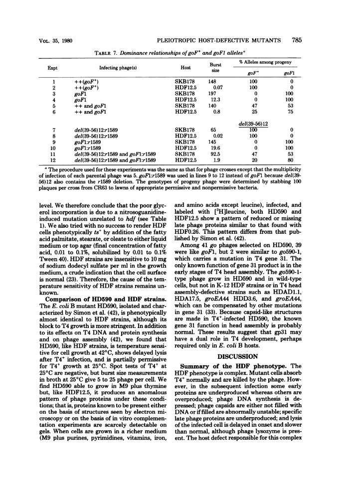
TABLE 7. Dominance relationships ofgoF+ and goFI alleles'
Burst % Alleles among progenyExpt Infecting phage(s) Host size goF+ goFl
1 ++(goF ) SKB178 148 100 02 ++(goF+) HDF12.5 0.07 100 03 goFl SKB178 197 0 1004 goFl HDF12.5 12.3 0 1005 ++ and goFl SKB178 140 47 536 ++ and goFl HDF12.5 0.8 25 75
del(39-56)127 del(39-56)12:rl589 SKB178 65 100 08 del(39-56)12:rl589 HDF12.5 0.02 100 09 goFl:rI589 SKB178 145 0 10010 goFl:r1589 HDF12.5 19.6 0 10011 del(39-56)12:rI589 and goFl:rl589 SKB178 92.5 47 5312 del(39-56)12:rl589 and goFl:rl589 HDF12.5 1.9 20 80
The procedure used for these experiments was the same as that for phage crosses except that the multiplicityof infection of each parental phage was 5. goFl:rl589 was used in lines 9 to 12 instead of goFl because del(39-56)12 also contains the r1589 deletion. The genotypes of progeny phage were determined by stabbing 100plaques per cross from CR63 to lawns of appropriate permissive and nonpermissive bacteria.
level. We therefore conclude that the poor glyc-erol incorporation is due to a nitrosoguanidine-induced mutation unrelated to hdf (see Table1). We also tried with no success to render HDFcells phenotypically ts+ by addition of the fattyacid palmitate, stearate, or oleate to either liquidmedium or top agar (final concentration of fattyacid, 0.01 to 0.1%, solubilized by 0.01 to 0.1%Tween 40). HDF strains are insensitive to 10 mgof sodium dodecyl sulfate per ml in the growthmedium, a crude indication that the cell surfaceis normal (23). Therefore, the cause of the tem-perature sensitivity ofHDF strains remains un-known.Comparison of HD590 and HDF strains.
The E. coli B mutant HD590, isolated and char-acterized by Simon et al. (42), is phenotypicallyalmost identical to HDF strains, although itsblock to T4 growth is more stringent. In additionto its effects on T4 DNA and protein synthesisand on phage assembly (42), we found thatHD590, like HDF strains, is temperature sensi-tive for cell growth at 420C, shows delayed lysisafter T4+ infection, and is partially permissivefor T4+ growth at 250C. Spot tests of T4+ at25°C are negative, but burst size measurementsin broth at 250C give 5 to 25 phage per cell. Wefind HD590 able to grow in M9 plus thyminebut, like HDF12.5, it produces an anomalouspattern of phage proteins under these condi-tions; that is, proteins known to be present eitheron the basis of structures seen by electron mi-croscopy or on the basis of in vitro complemen-tation experiments are scarcely detectable ongels. When cells are grown in a richer medium(M9 plus purines, pyrimidines, vitamins, iron,
and amino acids except leucine), infected, andlabeled with [3H]leucine, both HD590 andHDF12.5 show a pattern of reduced or missinglate phage proteins similar to that found withHDF0.26. This pattern differs from that pub-lished by Simon et al. (42).Among 41 go phages selected on HD590, 39
were like goFl, but 2 were similar to go590-1,which carries a mutation in T4 gene 31. Theonly known function of gene 31 product is in theearly stages of T4 head assembly. The go590-1-type phage grow in HD590 and in wild-typecells, but not in K-12 HDF strains or in T4 headassembly-defective strains such as HDAD1.1,HDA17.5, groEA44 HDD3.6, and groEA44,which can be compensated by other mutationsin gene 31 (33). Because capsid-like structuresare made in T4+-infected HD590, the knowngene 31 function in head assembly is probablynormal. These results suggest that gp3l mayhave a dual role in T4 development, perhapsrequired only in E. coli B hosts.
DISCUSSIONSummary of the HDF phenotype. The
HDF phenotype is complex. Mutant cells absorbT4+ nornally and are killed by the phage. How-ever, in the subsequent infection some earlyproteins are underproduced whereas others areoverproduced; phage DNA synthesis is de-pressed; phage capsids are either not filled withDNA or if filled are abnormally unstable; specificlate phage proteins are underproduced; and lysisof the infected cell is delayed in onset and slowerthan normal, although phage lysozyme is pres-ent. The host defect responsible for this complex
VOL. 35, 1980

786 STI'ITT ET AL.
rI - e
II IV
-_ 57
T4 chromosome
eG298 -eG19eG223eG79 -
psu5A64psu5 A33
0.5 kb
2.22I 0 (3)
5.5 (3)
A-0
8.4 (2)
5.60.7( 4),2.2
0.11'0
31.3
i 5.4
FIG. 8. Location ofgoF3.03-2 on the T4genome. Positions ofpoint mutations and deletions andpercentagesof recombination are indicated as in the legend to Fig. 6. The ends ofgene e, 492 base pairs apart (18), areindicated by vertical dotted lines; the extents andpositions of the deletions were determined by Wilson et al.(51). The line at the lower left indicates the scale of the figure. Crosses were performed in S/6/5 at 30°C(except under iii below) as described by Revel et al. (37). eG deletions and psub strains were crossed to T4D+before use to remove an rIV ('spackle') mutation from the former and a gene 15 (amN133) mutation from thelatter. eG deletions without rIV were recognized by their inability to plate on S/6/5 in the absence of addedlysozyme, and psub deletions without amN133 were recognized by their ability to plate on S/6/5 but not onCT439. The following conditions were used to score recombinants. (i) For crosses between goF3.03-2 and eGdeletion mutants or amH26, progeny were plated (a) on S/6/5 plus lysozyme (500 ,tg/2 ml of top agar perplate) for totalprogeny and (b) on S/6/5 without lysozyme as a source ofplaques for stabbing. Half as manyplaques were found under plating condition (b) as under (a), as expected. To identify wild-type recombinants,plaques were stabbed onto HDF3.03 (nonpernissive) and S/6/5 (permissive). %R = WT x 100/plaques pickedfrom S/6/5. (See legend to Fig. 6 for abbreviations.) (ii) For crosses between goF3.03-2 and psub deletionmutants, progeny were plated on S/6/5 and stabbed onto HDF3.03 at 37C, CT439 at 30°C, and S/6/5. Bothrecombinant classes were scored (on these indicators wild-type growth is zero, +, +, respectively; psub:goFgrowth is +, zero, +, respectively). %R = total recombinants x 100/plaques picked from S/6/5. The tworecombinant classes were found in approximately equal numbers. (iii) The cross tsC3 x amH26 wasperformedin CR63 at 30°C; total progeny was determined on CR63 plus lysozyme at 30°C, and wild-type recombinantswere determined on S/6/5 at 42°C. %R = WT x 200/total progeny. (iv) For tsC3 x eG223, progeny were platedon S/6/5 plus lysozyme at 30°C (totalprogeny) and S/6/5 at 37°C (wild-type recombinants). %oR = WT x 200/total plaques. (v) For crosses of eG deletions with psub-A64, the total progeny was plated on S/6/5 pluslysozyme. Wild-type recombinants were scored by stabbing from S/6/5 (no lysozyme) onto CT439 at 30°C andS/6/5. %R = WT x 100/plaques picked from S/6/5. In all crosses, plaques were stabbed until at least 10 (andusually 20 to 40) recombinants had been identified. Where no recombinants are indicated, 500 (eG79 xgoF3.03-2) or 3,791 (eG19 x goF3.03-2) plaques were tested.
phenotype greatly retards growth of uninfectedcells at 42°C. The active form of the host com-ponent must be present throughout T4 infectionfor production of phage.
Similarity of hdf hd590, and tabC muta-tions. The mutant E. coli B strain HD590 (42)probably carries an hdf mutation, based on theclose similarities of the HD590 and HDF phe-notypes as reviewed in Results. The tabC mu-tations first described by Takahashi (43) also areprobably allelic to hdf mutations. The two havesimilar map locations (5, 43; Stitt, in prepara-tion), they cause similar defects in T4 infection,and strong compensatory T4 mutations (comC-a for tabC; goFl for hdf) arise at very closelylinked sites near T4 gene 39 (43, 44).
The significance of apparent differences be-tween tabC and hdfmutants is difficult to judgebecause of the considerable allele-specific vari-ations within each class. The hdf-0.26 phenotypeis suppressed by lon (41), whereas the tabCcs-110 and C-803 phenotypes are not (5). In goFl-infected hdf strains, certain early proteins stillare underproduced or absent, whereas comC-a-infected tabC mutants show variable restorationof early functions depending on the tabC allele(5). Weakly compensating T4 mutations thatgrow on some of the host mutants are foundnear gene e for some hdf strains and in gene 45for some tabC strains (43). Despite these varia-tions, it seems likely that hdf, hd590, and tabCmutations occur in the same bacterial gene.
m
J. VIROL.

PLEIOTROPIC HOST-DEFECTIVE MUTANTS 787
Are hdfmutations in rho? The pleiotropicnature of HDF phenotype suggests that theprimary host defect may be in the control ofprotein synthesis. Possible defects could be intranscription initiation specificity, terminationspecificity, or translational factors. The follow-ing evidence suggests that hdf mutations lie inrho, the gene for transcription termination factorrho. Both rho (9, 16, 17) and hdf alleles (41;Stitt, in preparation) map at a similar site nearilv (83 min) in P1 transduction experiments. Thehdf-0.26 mutation, like many rho defects, par-tially relieves the polarity ofnonsense mutations(41). Genetic complementation tests with rho-15(9) suggest that a rho defect blocks T4 growthin tabC hosts (5). Finally, lambda transducingphages that carry rho' lysogenize hdf strains,making them T4 sensitive (burst size, 100 at370C), whereas lambda phages carrying rho-15show much weaker complementation (burst size,10) (Stitt, in preparation). These results must beinterpreted with some caution, however, becausehdf, rho-15, and the tabC mutants all were iso-lated after nitrosoguanidine mutagenesis, whichtends to yield clustered multiple mutations (6,14). Thus, it is possible that the complementa-tion results are due to mutations at loci otherthan those of interest. The hdf defects them-selves, however, probably represent single mu-tations in view of their frequency of occurrenceand the frequent simultaneous reversion ofHDFand ts phenotypes.Are the observed hdf effects on T4 infection
consistent with hdfmutations being in rho? An-swering this question could help to clarify thepresently unclear role of transcription termina-tion in control of T4 gene expression.Rho in T4 infection. In wild-type phage-
infected cells, chloramphenicol inhibits the tran-scription of delayed early genes from immediateearly promoters (13, 39), but in strong rho mu-tants like rho-15 this inhibition is not seen (3).In vitro the action of rho has been shown toprevent delayed early transcription (19, 38, 46,47). These results, and other less direct evidencethat a T4 gene product is required for delayedearly gene expression (26, 27), suggest that thephage produces a modulator of rho activity (an-titerminator) early in infection. However, lack ofrho activity also is deleterious; infection ofstrongrho temperature-sensitive mutants at nonper-missive temperature gives burst sizes 10- to 100-fold lower than does infection of wild-type cells(17; our unpublished data). Thus, the evidenceso far suggests that both rho-mediated termi-nation and modulation of rho by an early T4gene product are required for normal transcrip-tion and production of phage progeny.Early events in infection. The foregoing
suggestion leads to the following specific modelas one possible explanation for the effect of hdfmutations on early events in infection. HDFstrains produce an altered rho, which at lowtemperature acts normally but at 370C no longercan be modulated by the proposed early phagegene product that normally serves this function.As a result there is no synthesis of delayed earlytranscripts that depend upon read-through oftermination signals after immediate early pro-moters. The goFl mutations define the gene forthe T4 termination modulator and alter it sothat it can again modulate the altered rho,thereby overcoming the hdf block to infection.This model, similar to that proposed recently byPulitzer and co-workers (5, 34) to explain theproperties of tabC and comC-a mutations, isconsistent with the observed effects of hdf andgoFl mutations. Pulitzer et al. (34), in addition,have reviewed the evidence that delayed earlytranscripts can arise either by read-through fromadjacent immediate early genes or by initiationfrom a middle promoter under the positive con-trol of the T4 mot gene (30), or both, and haveshown strikingly that mot-defective phage in-fecting a tabC host make few if any delayedearly gene products. These results support thedual mechanism for control of delayed earlygene expression and the notion that hdf andtabC mutations alter rho so as to prevent itsearly modulation.Late events in infection. In addition to
aberrant early protein synthesis patterns, wehave described specific deficiencies in late syn-thesis in HDF strains, leading to defects in as-sembly and retardation of the lysis mechanism.Some of these late effects could be secondary,resulting from alterations in DNA synthesis orgene expression earlier in infection. Alterna-tively, the immediate cessation of phage produc-tion after late temperature shift-up of T4+-in-fected HDF cells suggests that Rho function isrequired throughout infection and raises the pos-sibility that expression of some late genes maydepend upon read-through of termination sig-nals by the same modulated rho that is necessaryfor expression of delayed early genes.Identity of the gene defined by goFl mu-
tations. Although goFl and comC-a mutationshave somewhat different effects in compensatingfor their respective host defects and map atnonidentical sites (cf. Fig. 4 and Fig. 1 of refer-ence 44), they are within several hundred nu-cleotides of one another and therefore are likelyto define the same gene, which has been pro-posed above to code for a modulator of rho.Based on map position, the goFl mutationsconceivably could be located in one of the twogenes provisionally defined byplaCTr5x and cef
VOL. 35, 1980

788 STITT ET AL.
mutations, respectively. However, del(39-56)5,which covers goFi, plates on CTr5x(15), imply-ing that the goFl site is in a different gene thanplaCTr5x. Similarly, the cef site is deleted indel(39-56)3 (15; A: Rodriguez-Prieto, Ph.D. the-sis, Vanderbilt University, Nashville, Tenn.,1976) whereas goFi is not (Fig. 6). Therefore,although the evidence is not conclusive, goFland comC-a probably define a new gene.The putative modulator coded by this gene
cannot be essential for normal T4 infection be-cause del(39-56)12, which lacks the goFl site,grows on wild-type hosts, although the burst sizeis only 60% that of nornal (Rodriguez-Prieto,Ph.D. thesis). This result suggests that the goFlgene product is a dispensable enhancer of T4growth. Why the hdfdefect should cause a lowerprogeny yield than do deletions of the goFl siteis not yet apparent. Perhaps goFl function isdispensable only by virtue of the dual rho/motcontrol of most delayed early genes. The furtherwork that will be necessary to understand thiscontrol should be aided by the availability of thehdf and goFl mutants.
ACKNOWLEDGMENTSThese studies were supported by research grants to W.B.W.
from the Public Health Service National Institute of Allergyand Infectious Diseases (AI-09238, AI-14994) and from theAmerican Cancer Society, California Division (Special Grant573). B.L.S. was also supported by a Public Health Servicepredoctoral training grant (GM-00086 from the National In-stitute of General Medical Science) to the California Instituteof Technology, a Caltech Special Institute Fellowship, and theArthur McCallum Fund.
LITERATURE CITED
1. Benzer, S. 1961. On the topography of the genetic finestructure. Proc. Natl. Acad. Sci. U.S.A. 47:403-415.
2. Blumenthal, T. 1972. P1 transduction: formation of het-erogenotes upon cotransduction of bacterial genes witha P2 prophage. Virology 47:76-93.
3. Brody, E., P. Daegelen, and D'Aubenton-Carafa.1978. The role of termination factor rho in the devel-opment of bacteriophage T4. Arch. Intern. Physiol.Biochim. 86:897-898.
4. Bujard, H., A. J. Mazaitio, and E. K. F. Bautz. 1970.The size of the rIl region of bacteriophage T4. Virology42:717-723.
5. Caruso, M., A. Coppo, A. Manzi, and J. F. Pulitzer.1979. Host-virus interactions in the control of T4 pre-replicative transcription. I. tabC (rho) mutants. J. Mol.Biol. 135:959-977.
6. Cerda-Olmedo, E., P. C. Hanawalt, and N. Guerola.1968. Mutagenesis of the replication point by nitroso-guanidine: map and pattern of replication of the Esch-erichia coli chromosome. J. Mol. Biol. 33:705-719.
7. Coppo, A., A. Manzi, J. F. Pulitzer, and H. Takahashi.1973. Abortive bacteriophage T4 head assembly in mu-tants of Escherichia coli. J. Mol. Biol. 76:61-87.
8. Cox, G. B., F. Gibson, and J. Pittard. 1968. Mutantstrains of Escherichia coli K-12 unable to form ubiqui-none. J. Bacteriol. 95:1591-1598.
9. Das, A., D. Court, and S. Adhya. 1976. Isolation andcharacterization of conditional lethal mutants of Esch-
erichia coli defective in transcription termination factorrho. Proc. Natl. Acad. Sci. U.S.A. 73:1959-1963.
10. Edgar, R. S., R. P. Feyman, S. Klein, L. Lielausis, andC. M. Steinberg. 1962. Mapping experiments with rmutants of bacteriophage T4D. Genetics 47:179-186.
11. Georgopoulos, C. P., R. W. Hendrix, A. D. Kaiser,and W. B. Wood. 1972. Role of the host cell in bacte-riophage morphogenesis: effects of a bacterial mutationon T4 head assembly. Nature (London) New Biol. 239:38-41.
12. Goldfine, H. 1969. Filter paper disk assay for lipid syn-thesis. Methods Enzymol. 14:649-651.
13. Grasso, R. J., and J. M. Buchanan. 1969. Synthesis ofearly RNA in bacteriophage T4-infected Escherichiacoli B. Nature (London) 224:882-885.
14. Guerola, N., J. L. Ingraham, and E. Cerda-Olmedo.1971. Induction of closely linked multiple mutations bynitrosoguanidine. Nature (London) New Biol. 230:122-125.
15. Homyk, T., Jr., and J. Weil. 1974. Deletion analysis oftwo nonessential regions of the T4 genome. Virology 61:505-523.
16. Inoko, H., and M. Imai. 1976. Isolation and geneticcharacterization of the nitA mutants of Escherichiacoli affecting the termination factor rho. Mol. Gen.Genet. 143:211-221.
17. Inoko, H., K. Shigesada, and M. Imai. 1977. Isolationand characterization of conditional-lethal rho mutantsof Escherichia coli. Proc. Natl. Acad. Sci. U.S.A. 74:1162-1166.
18. Inouye, M., M. Imada, and A. Tsugita. 1970. The aminoacid sequence of T4 phage lysozyme. IV. Dilute acidhydrolysis and the order of tryptic peptides. J. Biol.Chem. 245:3479-3484.
19. Jayaraman, R. 1972. Transcription of bacteriophage T4DNA by Escherichia coli RNA polymerase in vitro.Identification of some immediate-early and delayed-early genes. J. Mol. Biol. 70:253-263.
20. Kikuchi, Y., and J. King. 1975. Genetic control of bac-teriophage T4 baseplate morphogenesis. I. Sequentialassembly of the major precursor, in vivo and in vitro. J.Mol. Biol. 99:645-672.
21. Kim, J.-S., and N. Davidson. 1974. Electron microscopeheteroduplex study of sequence relations of T2, T4 andT6 bacteriophage DNAs. Virology 57:93-111.
22. King, J. 1968. Assembly of the tail of bacteriophage T4.J. Mol. Biol. 32:231-262.
23. Koplow, J., and H. Goldfine. 1974. Alterations in theouter membrane of the cell envelope of heptose-defi-cient mutants of Escherichia coli. J. Bacteriol. 117:527-543.
24. Kreuzer, K. N., K. McEntee, A. P. Geballe, and N. R.Cozzarelli. 1978. Lambda transducing phages for thenalA gene of Escherichia coli and conditional lethalnalA mutations. Mol. Gen. Genet. 167:129-137.
25. Krisch, H. M., A. Bolle, and R. H. Epstein. 1974.Regulation of the synthesis of bacteriophage T4 gene32 protein. J. Mol. Biol. 88:89-104.
26. LAnder, C. B., and 0. Skold. 1977. Evidence for a diffu-sible T4 bacteriophage protein governing the initiationof delayed early RNA synthesis. J. Virol. 21:7-15.
27. iAnder, C. H., and 0. Skold. 1980. Control of early geneexpression of bacteriophage T4: involvement of the hostrho factor and the mot gene of the bacteriophage. J.Virol. 33:724-732.
28. Low, B. 1973. Rapid mapping of conditional and auxo-trophic mutations in Escherichia coli K-12. J. Bacteriol.113:798-812.
29. Mathews, C. K. 1977. Reproduction of large virulentbacteriophages, p. 179-294. In H. Fraenkel-Conrat andR. R. Wagner (ed.) Comprehensive virology, vol. 7.Plenum Press, New York.
30. Mattson, T., J. Richardson, and D. Goodin. 1974.
J. VIROL.
PLEIOTROPIC HOST-DEFECTIVE MUTANTS 789
Mutant of bacteriophage T4D affecting expression ofmany early genes. Nature (London) 250:48-50.
31. Mindich, L., J. Franzese-Sinclair, and J. Cohen. 1976.The morphogenesis of bacteriophage 06: particlesformed by nonsense mutations. Virology 75:224-231.
32. Mufti, S., and H. Bernstein. 1974. The DNA-delaymutants of bacteriophage T4. J. Virol. 14:860-871.
33. O'Farrell, P. Z., L M. Gold, and W. M. Huang. 1973.The identification of prereplicative bacteriophage T4proteins. J. Biol. Chem. 248:5499-5501.
34. Pulitzer, J. F., A. Coppo, and M. Caruso. 1979. Host-virus interactions in the control of T4 prereplicativetranscription. II. Interaction between tabC (rho) mu-tants and T4 mot mutants. J. Mol. Biol. 135:979-997.
35. Pulitzer, J. F., and M. Yanagida. 1971. Inactive T4progeny virus formation in a temperature-sensitive mu-tant of Escherichia coli K12. Virology 45:539-554.
36. Radin, N. S. 1969. Preparation of lipid extracts, MethodsEnzymol. 14:245-254.
37. Revel, H. R., B. L Stitt, I. Lielausis, and W. B. Wood.1980. Role of the host cell in bacteriophage T4 devel-opment. I. Characterization of host mutants that blockT4 head assembly. J. Virol. 33:366-376.
38. Richardson, J. P. 1970. Rho factor function in T4 RNAtranscription. Cold Spring Harbor Symp. Quant. Biol.35:127-133.
39. Salser, W., A. Bolle, and R. Epstein. 1970. Transcrip-tion during bacteriophage T4 development: a demon-stration that distinct subclasses of the "early" RNAappear at different times and that some are "turnedoff" at late times. J. Mol. Biol. 49:271-295.
40. Simon, L. D. 1969. The infection of Escherichia coli byT2 and T4 bacteriophages as seen in the electron mi-croscope. III. Membrane-associated intracellular bac-teriophages. Virology 38:285-296.
41. Simon, L D., M. Gottesman, K. Tomczak, and S.Gottesman. 1979. Hyperdegradation of proteins inEscherichia coli rho mutants. Proc. Natl. Acad. Sci.U.S.A. 76:1623-1627.
42. Simon, L D., D. Snover, and A. H. Doermann. 1974.Bacterial mutation affecting T4 phage DNA synthesisand tail production. Nature (London) 252:451455.
43. Takahashi, H. 1978. Genetic and physiological charac-terization of Escherichia coli K12 mutants (tabC)which induce the abortive infection of bacteriophageT4. Virology 87:256-265.
44. Takahashi, H., and H. Yoshikawa. 1979. Genetic studyof a new early gene, comC-a, of bacteriophage T4.Virology 95:215-217.
45. Takano, T., and T. Kakefuda. 1972. Involvement of abacterial factor in morphogenesis of bacteriophage cap-sid. Nature (London) New Biol. 239:34-38.
46. Travers, A. A. 1970. Positive control of transcription bya bacteriophage sigma factor. Nature (London) 225:1009-1012.
47. Travers, A. 1970. RNA polymerase and T4 development.Cold Spring Harbor Symp. Quant. Biol. 35:241-251.
48. Vanderslice, R. W., and C. D. Yegian. 1974. The iden-tification of late bacteriophage T4 proteins on sodiumdodecyl sulfate polyacrylamide gels. Virology 60:265-275.
49. Ward, S., R. B. Luftig, J. H. Wilson, H. Eddleman, H.Lyle, and W. B. Wood. 1970. Assembly of bacterio-phage T4 tail fibers. II. Isolation and characterizationof tail fiber precursors. J. Mol. Biol. 54:15-31.
50. Wilson, J. H. 1973. Function of the bacteriophage T4transfer RNAs. J. Mol. Biol. 74:753-757.
51. Wilson, J. H., J. S. Kim, and J. N. Abelson. 1972.Bacteriophage T4 transfer RNA. III. Clustering of thegenes for the T4 transfer RNAs. J. Mol. Biol. 71:547-556.
52. Wood, W. B., R. C. Dickson, R. J. Bishop, and H. R.Revel. 1973. Self-assembly and non-self-assembly inbacteriophage T4 morphogenesis, p. 25-58. In R. Mark-ham (ed.), The generation of subcellular structures, 1stJohn Innes Symposium. North-Holland, Amsterdam.
53. Wood, W. B., and H. R. Revel. 1976. The genome ofbacteriophage T4. Bacteriol. Rev. 40:847-868.
VOL. 35, 1980