Improvement of Sidestream Dark Field Imaging with an Image
7
RESEARCH ARTICLE Open Access Improvement of Sidestream Dark Field Imaging with an Image Acquisition Stabilizer Gianmarco M Balestra 1,2*† , Rick Bezemer 1,3† , E Christiaan Boerma 1,4 , Ze-Yie Yong 5 , Krishan D Sjauw 5 , Annemarie E Engstrom 5 , Matty Koopmans 4 , Can Ince 1,3 Abstract Background: In the present study we developed, evaluated in volunteers, and clinically validated an image acquisition stabilizer (IAS) for Sidestream Dark Field (SDF) imaging. Methods: The IAS is a stainless steel sterilizable ring which fits around the SDF probe tip. The IAS creates adhesion to the imaged tissue by application of negative pressure. The effects of the IAS on the sublingual microcirculatory flow velocities, the force required to induce pressure artifacts (PA), the time to acquire a stable image, and the duration of stable imaging were assessed in healthy volunteers. To demonstrate the clinical applicability of the SDF setup in combination with the IAS, simultaneous bilateral sublingual imaging of the microcirculation were performed during a lung recruitment maneuver (LRM) in mechanically ventilated critically ill patients. One SDF device was operated handheld; the second was fitted with the IAS and held in position by a mechanic arm. Lateral drift, number of losses of image stability and duration of stable imaging of the two methods were compared. Results: Five healthy volunteers were studied. The IAS did not affect microcirculatory flow velocities. A significantly greater force had to applied onto the tissue to induced PA with compared to without IAS (0.25 ± 0.15 N without vs. 0.62 ± 0.05 N with the IAS, p < 0.001). The IAS ensured an increased duration of a stable image sequence (8 ± 2 s without vs. 42 ± 8 s with the IAS, p < 0.001). The time required to obtain a stable image sequence was similar with and without the IAS. In eight mechanically ventilated patients undergoing a LRM the use of the IAS resulted in a significantly reduced image drifting and enabled the acquisition of significantly longer stable image sequences (24 ± 5 s without vs. 67 ± 14 s with the IAS, p = 0.006). Conclusions: The present study has validated the use of an IAS for improvement of SDF imaging by demonstrating that the IAS did not affect microcirculatory perfusion in the microscopic field of view. The IAS improved both axial and lateral SDF image stability and thereby increased the critical force required to induce pressure artifacts. The IAS ensured a significantly increased duration of maintaining a stable image sequence. Background Orthogonal Polarization Spectral (OPS) imaging and its successor Sidestream Dark Field (SDF) imaging are opti- cal techniques allowing microscopic assessment of microcirculatory density and perfusion in clinical set- tings [1,2]. These non-invasive intravital imaging modal- ities have been used in studies for monitoring the severity of shock and efficacy of resuscitation in various patient groups [3-6]. However, as both OPS and SDF imaging technologies are incorporated into hand-held microscopes some operational issues arise in terms of axial and lateral instability of the microscope probes, potentially causing pressure artifacts and image drifting, respectively. Reductions in sublingual microcirculatory density and perfusion have been associated with patient morbidity and mortality [6]. Correcting these microcirculatory parameters has become the focus of new clinical studies aiming at resuscitating the microcirculation rather than the macrocirculation, using vasoactive agents such as nitroglycerin [7,8]. Hence, microcirculatory images are gaining a more prominent role in clinical monitoring and their accurate interpretation is essential and relies * Correspondence: [email protected] † Contributed equally 1 Department of Translational Physiology, Academic Medical Center, University of Amsterdam, Meibergdreef 9, 1105 AZ Amsterdam, The Netherlands Balestra et al. BMC Medical Imaging 2010, 10:15 http://www.biomedcentral.com/1471-2342/10/15 © 2010 Balestra et al; licensee BioMed Central Ltd. This is an Open Access article distributed under the terms of the Creative Commons Attribution License (http://creativecommons.org/licenses/by/2.0), which permits unrestricted use, distribution, and reproduction in any medium, provided the original work is properly cited.
Transcript of Improvement of Sidestream Dark Field Imaging with an Image
RESEARCH ARTICLE Open Access
Improvement of Sidestream Dark Field Imagingwith an Image Acquisition StabilizerGianmarco M Balestra1,2*†, Rick Bezemer1,3†, E Christiaan Boerma1,4, Ze-Yie Yong5, Krishan D Sjauw5,Annemarie E Engstrom5, Matty Koopmans4, Can Ince1,3
Abstract
Background: In the present study we developed, evaluated in volunteers, and clinically validated an imageacquisition stabilizer (IAS) for Sidestream Dark Field (SDF) imaging.
Methods: The IAS is a stainless steel sterilizable ring which fits around the SDF probe tip. The IAS creates adhesionto the imaged tissue by application of negative pressure. The effects of the IAS on the sublingual microcirculatoryflow velocities, the force required to induce pressure artifacts (PA), the time to acquire a stable image, and theduration of stable imaging were assessed in healthy volunteers. To demonstrate the clinical applicability of the SDFsetup in combination with the IAS, simultaneous bilateral sublingual imaging of the microcirculation wereperformed during a lung recruitment maneuver (LRM) in mechanically ventilated critically ill patients. One SDFdevice was operated handheld; the second was fitted with the IAS and held in position by a mechanic arm. Lateraldrift, number of losses of image stability and duration of stable imaging of the two methods were compared.
Results: Five healthy volunteers were studied. The IAS did not affect microcirculatory flow velocities. A significantlygreater force had to applied onto the tissue to induced PA with compared to without IAS (0.25 ± 0.15 N withoutvs. 0.62 ± 0.05 N with the IAS, p < 0.001). The IAS ensured an increased duration of a stable image sequence (8 ±2 s without vs. 42 ± 8 s with the IAS, p < 0.001). The time required to obtain a stable image sequence was similarwith and without the IAS. In eight mechanically ventilated patients undergoing a LRM the use of the IAS resultedin a significantly reduced image drifting and enabled the acquisition of significantly longer stable image sequences(24 ± 5 s without vs. 67 ± 14 s with the IAS, p = 0.006).
Conclusions: The present study has validated the use of an IAS for improvement of SDF imaging bydemonstrating that the IAS did not affect microcirculatory perfusion in the microscopic field of view. The IASimproved both axial and lateral SDF image stability and thereby increased the critical force required to inducepressure artifacts. The IAS ensured a significantly increased duration of maintaining a stable image sequence.
BackgroundOrthogonal Polarization Spectral (OPS) imaging and itssuccessor Sidestream Dark Field (SDF) imaging are opti-cal techniques allowing microscopic assessment ofmicrocirculatory density and perfusion in clinical set-tings [1,2]. These non-invasive intravital imaging modal-ities have been used in studies for monitoring theseverity of shock and efficacy of resuscitation in variouspatient groups [3-6]. However, as both OPS and SDF
imaging technologies are incorporated into hand-heldmicroscopes some operational issues arise in terms ofaxial and lateral instability of the microscope probes,potentially causing pressure artifacts and image drifting,respectively.Reductions in sublingual microcirculatory density and
perfusion have been associated with patient morbidityand mortality [6]. Correcting these microcirculatoryparameters has become the focus of new clinical studiesaiming at resuscitating the microcirculation rather thanthe macrocirculation, using vasoactive agents such asnitroglycerin [7,8]. Hence, microcirculatory images aregaining a more prominent role in clinical monitoringand their accurate interpretation is essential and relies
* Correspondence: [email protected]† Contributed equally1Department of Translational Physiology, Academic Medical Center,University of Amsterdam, Meibergdreef 9, 1105 AZ Amsterdam, TheNetherlands
Balestra et al. BMC Medical Imaging 2010, 10:15http://www.biomedcentral.com/1471-2342/10/15
© 2010 Balestra et al; licensee BioMed Central Ltd. This is an Open Access article distributed under the terms of the Creative CommonsAttribution License (http://creativecommons.org/licenses/by/2.0), which permits unrestricted use, distribution, and reproduction inany medium, provided the original work is properly cited.
heavily on the quality of the images [9,10]. In this light,the current microcirculatory image acquisition guide-lines dictate a minimal recording time of 20 s to allowadequate analysis of microcirculatory density and perfu-sion [11]. Image drifting, due to the difficulty in holdingthe tip of the device in one place however, makes thisparticularly difficult both in sedated and in awakepatients. Furthermore, pressure artifacts caused by thephysical contact and pressure of the microscope probeto the mucosal tissue can alter mucosal capillary bloodflow thereby limit the use of the captured images fordetermination of microcirculatory perfusion.Lindert et al. addressed these technical issues asso-
ciated with hand-held microscopy before by developingan image acquisition stabilizer (IAS) for the OPS ima-ging device [12]. Their IAS consisted of a ring placedaround the tip of the OPS probe through which negativepressure was applied securing the IAS onto the mucosaltissue. The negative pressure appeared not to influenceflow patterns of the microcirculation within the micro-scopic field. However, whether the IAS minimizedimage drift or induction of pressure artifacts was notevaluated. In addition their IAS was not validated interms of clinical applicability and utility, including theease with which the device could be sterilized andcleaned for multiple uses as well as fitting piping ofvacuum sources available at the bed-side.In the present study we developed, evaluated, and vali-
dated an IAS for the SDF device. In a study by Goedhartet al., the SDF imaging device was shown to providemicrocirculatory images of superior quality with respectto the OPS device [13]. In combination with an IAS,this microcirculatory imaging setup should provide highquality microcirculatory images of sufficient durationand stability. The IAS was designed and fabricated toadhere to clinical requirements. The application of theIAS was validated by measuring 1) the effects of applica-tion of peripheral negative pressure on microcirculatoryperfusion, 2) the force required for induction of pressureartifacts with and without the IAS, 3) the time requiredto attain a stable image, and 4) the time that a stableimage could be maintained. Then, to demonstrate theclinical applicability of the SDF setup with the IAS,simultaneous bilateral sublingual SDF measurementswere conducted in critically ill patients undergoing astandard lung recruitment maneuver with one hand-held SDF device and one SDF device mounted in amechanical arm and equipped with the IAS wherebystability of acquired images was evaluated.
MethodsThe study protocol was approved by the local medicalethics committee of the Medical Center of Leeuwarden.Written informed consent was obtained from all studied
subjects respectively their closest relatives. The studywas done in compliance with the principles establishedin the Helsinki Declaration.
Sidestream dark field image acquisition and analysisSublingual microcirculatory density and perfusion weremonitored using an SDF imaging device (MicrovisionMedical BV, Amsterdam, the Netherlands). A detaileddescription of the SDF technology is provided elsewhere[13]. Briefly, in SDF imaging, the tissue is illuminatedwith green light emitting diodes (LEDs) concentricallyplaced around the central microscopy objective to pro-vide SDF illumination. The lens system in the core ofthe objective is optically isolated from the illuminatingouter ring thus preventing the microcirculatory imagefrom contamination by tissue surface reflections. Tofurther improve the imaging of flowing erythrocytes, theSDF device provides pulsed illumination in synchronywith the camera frame rate. This stroboscopic imaging,(partially) prevents smearing of flowing erythrocytes andmotion-induced blurring of capillaries due to the shortillumination intervals [13].The obtained microcirculatory images (one per time
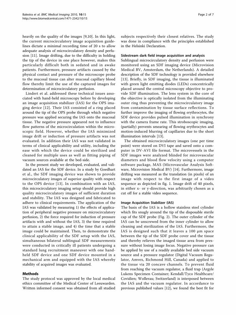
point) were stored on DVI tape and saved onto a com-puter in DV-AVI file format. The microvessels in theSDF images were analyzed blinded for microvasculardiameters and blood flow velocity using a computersoftware package, MAS (Microvascular Analysis Soft-ware, Microvision Medical BV) [14]. Furthermore, imagedrifting was measured as the translation (in pixels) of animage with respect to the first image of a videosequence as depicted in fig. 1. Image drift of 40 pixels,in either x- or y-direction, was arbitrarily chosen as acut off for a stable video sequence.
Image Acquisition Stabilizer (IAS)The basis of the IAS is a hollow stainless steel cylinderwhich fits snugly around the tip of the disposable sterilecap of the SDF probe (Fig. 2). The outer cylinder of theIAS can be unscrewed from the inner cylinder to allowcleaning and sterilization of the IAS. Furthermore, theIAS is designed such that it leaves a 100 μm spacebetween the tip of the SDF probe cover and the tissueand thereby relieves the imaged tissue area from pres-sure without losing image focus. Negative pressure canbe applied by use of a readily available bed side vacuumsource and a pressure regulator (Digital Vacuum Regu-lator, Amvex, Richmond Hill, Canada) and applied tothe tissue via 20 concave channels. To prevent fluidfrom reaching the vacuum regulator, a fluid trap (ArgyleLukens Specimen Container; Kendall/Tyco Healthcare/Covidien; Wollerau; Switzerland) is interposed betweenthe IAS and the vacuum regulator. In accordance toprevious published values [12], we found the best fit for
Balestra et al. BMC Medical Imaging 2010, 10:15http://www.biomedcentral.com/1471-2342/10/15
Page 2 of 7
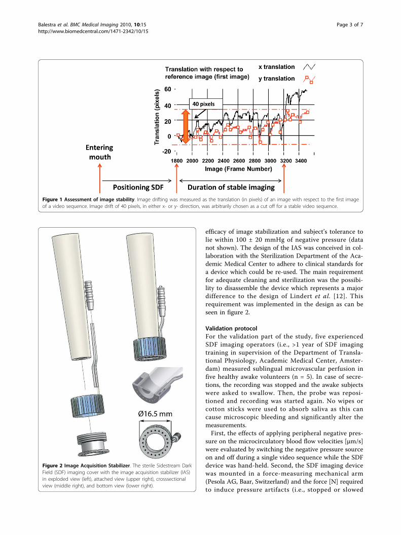
efficacy of image stabilization and subject’s tolerance tolie within 100 ± 20 mmHg of negative pressure (datanot shown). The design of the IAS was conceived in col-laboration with the Sterilization Department of the Aca-demic Medical Center to adhere to clinical standards fora device which could be re-used. The main requirementfor adequate cleaning and sterilization was the possibi-lity to disassemble the device which represents a majordifference to the design of Lindert et al. [12]. Thisrequirement was implemented in the design as can beseen in figure 2.
Validation protocolFor the validation part of the study, five experiencedSDF imaging operators (i.e., >1 year of SDF imagingtraining in supervision of the Department of Transla-tional Physiology, Academic Medical Center, Amster-dam) measured sublingual microvascular perfusion infive healthy awake volunteers (n = 5). In case of secre-tions, the recording was stopped and the awake subjectswere asked to swallow. Then, the probe was reposi-tioned and recording was started again. No wipes orcotton sticks were used to absorb saliva as this cancause microscopic bleeding and significantly alter themeasurements.First, the effects of applying peripheral negative pres-
sure on the microcirculatory blood flow velocities [μm/s]were evaluated by switching the negative pressure sourceon and off during a single video sequence while the SDFdevice was hand-held. Second, the SDF imaging devicewas mounted in a force-measuring mechanical arm(Pesola AG, Baar, Switzerland) and the force [N] requiredto induce pressure artifacts (i.e., stopped or slowed
Figure 1 Assessment of image stability. Image drifting was measured as the translation (in pixels) of an image with respect to the first imageof a video sequence. Image drift of 40 pixels, in either x- or y- direction, was arbitrarily chosen as a cut off for a stable video sequence.
Figure 2 Image Acquisition Stabilizer. The sterile Sidestream DarkField (SDF) imaging cover with the image acquisition stabilizer (IAS)in exploded view (left), attached view (upper right), crosssectionalview (middle right), and bottom view (lower right).
Balestra et al. BMC Medical Imaging 2010, 10:15http://www.biomedcentral.com/1471-2342/10/15
Page 3 of 7
venular flow) was determined with and without the IASby systematically increasing the force applied by the SDFprobe onto the sublingual tissue. Third, the time [s]required for obtaining a stable image sequence and,fourth, the duration [s] of maintaining that stable imagesequence were measured.
Clinical protocolTo demonstrate the clinical applicability of the SDFsetup with the IAS, simultaneous bilateral sublingualSDF measurements were conducted in eight intensivecare patients undergoing a standard lung recruitmentmaneuver with one hand-held SDF device and one SDFdevice mounted in a mechanical arm and equipped withthe IAS. This procedure, with a stepwise increment oftidal volume, was chosen in order to create extensivemovement artifacts in the sublingual imaged areas.When both SDF devices were acquiring stable images,without pressure artifacts, continuous recording of thevideo image was started. The lung recruitment maneu-ver was performed by increasing the inspiratory pressurelevel to a target of 40 cmH2O, followed by graduallyreducing the pressure until the baseline ventilator set-tings were regained. The fraction of inspired oxygen andpositive end-expiratory pressure were maintained at 40%and 12 cmH2O, respectively, throughout the procedure.SDF images were recorded non-stop from 1 min
before till 1 min after the recruitment maneuver and therecorded SDF video sequences were randomized and
analyzed off-line for lateral image drift [μm], driftingvelocity [μm/s], and number of loss of image stability(i.e., image drift of > 40 pixels, Fig. 1), the duration [s]that a stable image could be maintained.
Statistical analysisStatistical analysis was performed using GraphPad Prismversion 5.0 for Windows (GraphPad Software, SanDiego, CA, USA). To test data sets for (non-)parametricdistributions a D’Agostino-Pearson omnibus normalitytest was applied. Comparative analysis between data setswas performed with the unpaired Student’s t-test or theMann-Whitney U test and comparative analysis betweentime points was performed with the paired Student’st-test or the Wilcoxon signed rank test, as appropriate.Differences with a p-value of < 0.05 were consideredstatistically significant. Results are reported as mean ±SEM.
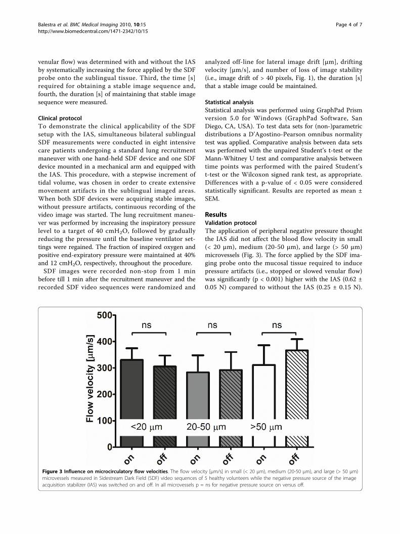
ResultsValidation protocolThe application of peripheral negative pressure thoughtthe IAS did not affect the blood flow velocity in small(< 20 μm), medium (20-50 μm), and large (> 50 μm)microvessels (Fig. 3). The force applied by the SDF ima-ging probe onto the mucosal tissue required to inducepressure artifacts (i.e., stopped or slowed venular flow)was significantly (p < 0.001) higher with the IAS (0.62 ±0.05 N) compared to without the IAS (0.25 ± 0.15 N).
Figure 3 Influence on microcirculatory flow velocities. The flow velocity [μm/s] in small (< 20 μm), medium (20-50 μm), and large (> 50 μm)microvessels measured in Sidestream Dark Field (SDF) video sequences of 5 healthy volunteers while the negative pressure source of the imageacquisition stabilizer (IAS) was switched on and off. In all microvessels p = ns for negative pressure source on versus off.
Balestra et al. BMC Medical Imaging 2010, 10:15http://www.biomedcentral.com/1471-2342/10/15
Page 4 of 7
The time required to obtain a stable SDF imagesequence was similar (p = 0.12) with (99 ± 20 s) andwithout the IAS (150 ± 25 s). The duration of maintain-ing that stable image was approximately five timeslonger with the IAS: 8 ± 2 s without the IAS and 42 ± 8 swith the IAS (p < 0.001).
Clinical protocolIn the eight patients undergoing a lung recruitmentmaneuver (four male and four female), aged 66 ± 5years, the APACHE II, APACHE IV, SOFA scores were19 ± 2, 75 ± 10, and 8 ± 1 points respectively. Fourpatients were diagnosed with abdominal sepsis, one withcoma after cardiac arrest, two had undergone cardiovas-cular surgery, and one head and neck surgery.During the lung recruitment maneuver, inspiratory
pressure level was increased from 11.4 ± 0.8 cmH2O to43 ± 2 cmH2O (p < 0.001). Tidal volume rose accord-ingly from 423 ± 23 ml (baseline) to 1208 ± 90 ml (p <0.001) during the lung recruitment maneuver andreturned to 477 ± 30 ml after the maneuver (p = 0.017vs. baseline).Continuous recording of the SDF video image was
started prior to the lung recruitment maneuver whenboth SDF devices were acquiring stable images, withoutpressure artifacts. During the procedure, image drift in x-and y-direction was 8.9 ± 2.6 and 10.1 ± 2.0 mm respec-tively without the IAS, while the drift was reduced to3.4 ± 0.9 (p = 0.066) and 3.8 ± 1.3 mm (p = 0.018) withthe IAS. Drift velocity in x- and y-direction was 15.5 ±3.9 and 18.4 ± 3.3 μm/s respectively without the IAS,which was reduced to 5.4 ± 1.5 (p = 0.032) and 5.6 ± 1.7μm/s (p = 0.004) with the IAS. Image drift of > 20 pixelswithin one video sequence occurred 50 ± 13 times with-out the IAS and 8 ± 3 times (p < 0.001) with the IAS.The maximum duration of stable imaging during thelung recruitment maneuver was 24 ± 5 s without the IASand 66 ± 14 s (p = 0.006) with the IAS.
DiscussionIn the present study we developed, evaluated, and vali-dated an IAS for the SDF device. The IAS was based oncreating adherence of the SDF probe to the sublingualtissue by applying negative pressure to the periphery ofthe microscopic field of view. The main findings werethat: 1) the IAS did not affect microcirculatory perfusionin the SDF imaging field of view; 2) the IAS preventedpressure artifacts up to a significantly greater forceapplied by the SDF probe onto the tissue; 3) the timerequired to obtain a stable image sequence was similarwith and without the IAS; and 4) the duration of main-taining that stable image sequence was significantlyincreased with the IAS. Ultimately, to demonstrate theclinical applicability of the SDF setup with the IAS,
simultaneous bilateral sublingual SDF measurementswere conducted in intensive care patients undergoing astandard lung recruitment maneuver with one handheldSDF device and one SDF device mounted in a mechani-cal arm and equipped with the IAS. It was shown thatthe IAS significantly reduced image drifting and enabledthe acquisition of significantly longer image sequences.A final and important finding is also that we showed, inproof of concept, that with the IAS it is possible to per-form a measurement without the need for an operatorby mounting the device on a mechanical arm, leavingthe operator free to perform a clinical maneuver.The design of the IAS presented here is based on an
IAS developed by Lindert et al. for OPS imaging, includ-ing the negative pressure level of ≈100 mmHg [12]. Toshow that application of peripheral negative pressuredid not affect microcirculatory perfusion in the SDFimaging field of view Lindert et al. measured blood flowvelocities in venules and arterioles. They found that thevelocities did not change after switching the negativepressure source on. In the present study, for validationpurposes, we investigated the effects on blood flow velo-cities in small, medium, and large microvessels in fivehealthy volunteers and provided evidence that indeedmicrocirculatory perfusion is not affected by applicationof negative pressure though the IAS. These experimentsdemonstrated that the IAS is a valid method for SDFimage stabilization, not affecting microcirculatoryperfusion in the microscopic field of view.It has been well established that pressure artifacts are
easily induced and diminish the reliability of SDF mea-surements of microcirculatory perfusion [6,11]. Thisappreciation known from daily application of SDF ima-ging is confirmed and highlighted by the low force levelrequired to induce pressure artifacts found in the pre-sent study. The SDF imaging device has a mass ofapproximately 360 g. The critical force onto the sublin-gual tissue without the IAS, at which pressure artifactsare induced, was found to amount approximately 1/6 ofthe mass of the SDF device. Hence, physical feedback isimpossible for SDF operators and visual feedback in themicrocirculatory images is necessary to avoid excessivepressure. In fact, most SDF operators use visual feed-back to gauge the pressure exerted by the SDF probe onthe imaged microcirculation as exemplified in a recentpublication [6]. De Backer et al., defined the criticalpressure inducing perfusion artifacts at the point wherevenular flow either stopped or significantly slowed down[11]. Using a similar cut-off in the present study wewere able to show that the larger surface contact areacreated by the presence of the IAS resulted in anapproximately five times greater force required for theinduction of pressure artifacts. This significantlyimproved SDF image acquisition.
Balestra et al. BMC Medical Imaging 2010, 10:15http://www.biomedcentral.com/1471-2342/10/15
Page 5 of 7
Another important advantage of using an IAS for SDFimaging is that it allows acquisition of longer and morestable SDF image sequences. Previous studies reportedthat SDF measurements have low intra- and inter-observervariability [3] and that microcirculatory density and perfu-sion vary highly per site and in time [15]. Hence, studyingthe microcirculation under pathophysiological conditionsrequires multiple measurements per time point in order toeliminate this site- and time-dependency of the obtainedresults. The current microcirculatory image acquisitionguidelines dictate that microcirculatory density and perfu-sion should be measured in 3-5 sites per time point toallow adequate interpretation of the results [11]. Further-more, according to these guidelines, the length of eachSDF image sequence should be > 20 s. This was proven tobe rather difficult without the IAS and fairly easy with theIAS. An alternative for multiple measurements todetermine the microcirculatory state at a certain timepoint, continuous measurements of microcirculatoryperfusion and density during a clinical maneuver orintervention (e.g., nitroglycerin administration) wouldallow direct assessment of their effects on the micro-circulation. The presented IAS would potentiallyenable such studies.Non-invasive intravital imaging modalities, such as
OPS and SDF imaging, have been used in studies formonitoring the severity of shock and efficacy of resusci-tation and alterations in sublingual microcirculatorydensity and perfusion have been associated with patientmorbidity and mortality [3,6,16]. ‘Normalizing’ microcir-culatory density and perfusion has become focus of newclinical studies and microcirculatory images are gaininga more prominent role in clinical monitoring. Adequateinterpretation of microcirculatory images is essentialand relies heavily on the quality of the images, in termsof axial and lateral stability. In the present study weshowed that the IAS improves both axial and lateral sta-bility of the acquired microcirculatory images and signif-icantly reduced pressure artifacts and image drifting.
ConclusionsThe present study has validated the use of an IAS forimprovement of SDF imaging by demonstrating that theapplication of peripheral negative pressure thoughthe IAS does not affect microcirculatory perfusion inthe microscopic field of view. Furthermore, the IAS wasshown to improve both axial and lateral SDF image sta-bility and thereby increased the critical force required toinduce microcirculatory pressure artifacts and increasedthe duration of stable image acquisition.
Key Messages• The application of peripheral negative pressurethough the image acquisition stabilizer (IAS) for
improvement of SDF imaging did not affect micro-circulatory perfusion in the microscopic field ofview.• The IAS improved both axial and lateral SDFimage stability and thereby increased the criticalforce required to induce microcirculatory pressureartifacts.• The IAS increased the duration of stable imageacquisition.
AbbreviationsIAS: Image acquisition stabilizer; SDF: Sidestream Dark Field; PA: pressureartifacts; LRM: lung recruitment maneuver; OPS: Orthogonal PolarizationSpectral; APACHE score: Acute Physiology And Chronic Health Evaluationscore; SOFA score: Sequential Organ Failure Assessment score; DVI: DigitalVisual Interface; DV-AVI: Digital Video-Audio Video Interleave.
AcknowledgementsWe would like to thank Bertus Hendriks of the Department of MedicalInnovations and Technology of the Academic Medical Center of Amsterdamfor the extensive technical support enabling this project.Preliminary results of this study were presented in the 22nd AnnualCongress of the European Society of Intensive Care Medicine and publishedas an abstract [17].
Author details1Department of Translational Physiology, Academic Medical Center,University of Amsterdam, Meibergdreef 9, 1105 AZ Amsterdam, TheNetherlands. 2Department of Internal Medicine and Medical Intensive Care,University Hospital Basel, Petersgraben 4, 4031 Basel, Switzerland.3Department of Intensive Care, Erasmus Medical Center, ‘s-Gravendijkwal 230,3015 CE Rotterdam, The Netherlands. 4Department of Intensive Care, MedicalCenter Leeuwarden, Postbus 888, 8901 BR Leeuwarden, The Netherlands.5Department of Cardiology, Academic Medical Center, University ofAmsterdam, Meibergdreef 9, 1105 AZ Amsterdam, The Netherlands.
Authors’ contributionsGMB designed and performed the validation and clinical measurements,analyzed the data of the validation protocol, and drafted the manuscript. RBdeveloped the IAS, co-designed the validation protocol, and co-drafted themanuscript. ECB co-performed the clinical protocol. ZYY and AEE performedthe validation protocol. MK analyzed the data of the clinical protocol. CIconceived the study and reviewed the manuscript. All authors read andapproved the final version of the manuscript.
Competing interestsCI is inventor of SDF imaging in an Academic Medical Center owned patentand holds shares in Microvision Medical. Furthermore, CI has receivedresearch grants from Hutchinson Technology, Baxter, Eli Lilly, and Novartis.The remaining authors declare no potential conflicts of interest.
Received: 2 June 2010 Accepted: 13 July 2010 Published: 13 July 2010
References1. Groner W, Winkelman JW, Harris AG, Ince C, Bouma GJ, Messmer K,
Nadeau RG: Orthogonal polarization spectral imaging: a new method forstudy of the microcirculation. Nat Med 1999, 5:1209-1212.
2. Ince C: The microcirculation is the motor of sepsis. Crit Care 2005,9(Suppl 4):S13-S19.
3. De Backer D, Creteur J, Preiser JC, Dubois MJ, Vincent JL: Microvascularblood flow is altered in patients with sepsis. Am J Respir Crit Care Med2002, 166:98-104.
4. Boerma EC, van dV, Spronk PE, Ince C: Relationship between sublingualand intestinal microcirculatory perfusion in patients with abdominalsepsis. Crit Care Med 2007, 35:1055-1060.
5. Meinders AJ, Elbers P: Images in clinical medicine. Leukocytosis andsublingual microvascular blood flow. N Engl J Med 2009, 360:e9.
Balestra et al. BMC Medical Imaging 2010, 10:15http://www.biomedcentral.com/1471-2342/10/15
Page 6 of 7
6. Trzeciak S, McCoy JV, Phillip DR, Arnold RC, Rizzuto M, Abate NL, Shapiro NI,Parrillo JE, Hollenberg SM: Early increases in microcirculatory perfusionduring protocol-directed resuscitation are associated with reducedmulti-organ failure at 24 h in patients with sepsis. Intensive Care Med2008, 34:2210-2217.
7. Boerma EC, Koopmans M, Konijn A, Kaiferova K, Bakker AJ, van Roon EN,Buter H, Bruins N, Egbers PH, Gerritsen RT, et al: Effects of nitroglycerin onsublingual microcirculatory blood flow in patients with severe sepsis/septic shock after a strict resuscitation protocol: a double-blindrandomized placebo controlled trial. Crit Care Med 2010, 38:93-100.
8. den Uil CA, Lagrand WK, Spronk PE, van der EM, Jewbali LS, Brugts JJ,Ince C, Simoons ML: Lowdose nitroglycerin improves microcirculation inospitalized patients with acute heart failure. Eur J Heart Fail 2009,11:386-390.
9. Bemelmans RH, Boerma EC, Barendregt J, Ince C, Rommes JH, Spronk PE:Changes in the volume status of haemodialysis patients are reflected insublingual microvascular perfusion. Nephrol Dial Transplant 2009,24:3487-3492.
10. Elbers PW, Ozdemir A, van IM, van Dongen EP, Ince C: Microcirculatoryimaging in cardiac anesthesia: ketanserin reduces blood pressure butnot perfused capillary density. J Cardiothorac Vasc Anesth 2009, 23:95-101.
11. De Backer D, Hollenberg S, Boerma C, Goedhart P, Buchele G, Ospina-Tascon G, Dobbe I, Ince C: How to evaluate the microcirculation: reportof a round table conference. Crit Care 2007, 11:R101.
12. Lindert J, Werner J, Redlin M, Kuppe H, Habazettl H, Pries AR: OPS imagingof human microcirculation: a short technical report. J Vasc Res 2002,39:368-372.
13. Goedhart PT, Khalilzada M, Bezemer R, Merza J, Ince C: Sidestream DarkField (SDF) imaging: a novel stroboscopic LED ring-based imagingmodality for clinical assessment of the microcirculation. Opt Express 2007,15:15101-15114.
14. Dobbe JG, Streekstra GJ, Atasever B, van ZR, Ince C: Measurement offunctional microcirculatory geometry and velocity distributions usingautomated image analysis. Med Biol Eng Comput 2008, 46:659-670.
15. Hubble SM, Kyte HL, Gooding K, Shore AC: Variability in sublingualmicrovessel density and flow measurements in healthy volunteers.Microcirculation 2009, 16:183-191.
16. Sakr Y, Dubois MJ, De BD, Creteur J, Vincent JL: Persistent microcirculatoryalterations are associated with organ failure and death in patients withseptic shock. Crit Care Med 2004, 32:1825-1831.
17. Balestra G, Yong Z-Y, Bezemer R, Sjauw K, Engstrom A, Boerma EC, Ince C:Improving image acquisition for Sidestream Dark Field Imaging of themicrocirculation by a Distancing Immobilizing Device. Intensive Care Med2009, 35:S205.
Pre-publication historyThe pre-publication history for this paper can be accessed here:http://www.biomedcentral.com/1471-2342/10/15/prepub
doi:10.1186/1471-2342-10-15Cite this article as: Balestra et al.: Improvement of Sidestream Dark FieldImaging with an Image Acquisition Stabilizer. BMC Medical Imaging 201010:15.
Submit your next manuscript to BioMed Centraland take full advantage of:
• Convenient online submission
• Thorough peer review
• No space constraints or color figure charges
• Immediate publication on acceptance
• Inclusion in PubMed, CAS, Scopus and Google Scholar
• Research which is freely available for redistribution
Submit your manuscript at www.biomedcentral.com/submit
Balestra et al. BMC Medical Imaging 2010, 10:15http://www.biomedcentral.com/1471-2342/10/15
Page 7 of 7