Impedance and EFP recordings of iCell Cardiomyocytes2 on the CardioExcyte 96
4
Download more Application Notes from www.nanion.de Application Note Channels: hERG, ß-adrenergic receptor Cells: human iPSC Cardiomyocytes Tools: CardioExcyte 96 Cardiomyocytes derived from human induced pluripotent stem cells (hiPSCs) are gaining interest in cardiac safety screening. Given their recapitulation of native behavior, availability, ease of use and standardized production, they are likely to provide a viable alternative to acutely isolated cardiomyocytes to assess the pro-arrhythmic potentials of drug candidates. Although automated patch clamp can provide excellent information about the effects of compounds on cardiac ion channels and possible effects on the cardiac action potential 1 , other outputs such as extracellular field potential (EFP) and impedance, also provide crucial and complementary information about complex physiological parameters such as beat rate, amplitude and duration. The CardioExcyte 96 is a new hybrid screening tool combining impedance (cell contractility) and EFP recordings 2 . These measurements are non-invasive, label-free and have a temporal resolution of 1 ms. The recordings are made from cells within a network thus providing a physiologically relevant environment for measuring drug-induced changes in contractile parameters. This hybrid technology is a high-throughput screening tool which permits the reliable investigation of short- and long-term pharmacological effects. Here we present data recorded on the CardioExcyte 96 using iCell ® Cardiomyocytes 2 from Cellular Dynamics International (CDI). When handled according to the manufacture’s instructions, impedance and EFP signals were stable 4 days after plating and compound effects could be robustly measured and analyzed. Impedance and EFP recordings of iCell ® Cardiomyocytes 2 on the CardioExcyte 96 The electrophysiology team at Nanion Technologies GmbH, Munich. Cardiomyocytes kindly provided by Cellular Dynamics International. Summary Results iCell Cardiomyocytes 2 were plated on the NSP-96 plates on the CardioExcyte 96 and measurements were taken on days 1, 4 and 6 after plating. The cardiomyocyte electrical and contractile activity is stable after 4 DIV, as suggested by the manufacturer and indicated by the impedance signal which changes little between day 4 and day 6 (Figures 1 & 2). Figure 1: Impedance measurements on the CardioExcyte 96 at different timepoints. A Screenshot (4 out of 12 columns) of the CardioExcyte 96 software during measurements in the impedance mode on day 1 (left), 4 (middle) or 6 (right) after plating. B Impedance mean beat on day 1 (left), 4 (middle) and 6 (right) after plating. Impedance mean beat changes little between day 4 and day 6 indicating that the cells can be used for experiments 4 days after plating as suggested by the manufacturer.
-
Upload
ali-obergrussberger -
Category
Health & Medicine
-
view
4.907 -
download
0
Transcript of Impedance and EFP recordings of iCell Cardiomyocytes2 on the CardioExcyte 96
Download more Application Notes from www.nanion.de
Application Note Channels: hERG, ß-adrenergic receptor
Cells: human iPSC Cardiomyocytes
Tools: CardioExcyte 96
Cardiomyocytes derived from human induced pluripotent stem cells (hiPSCs) are gaining interest in cardiac safety screening. Given their recapitulation of native behavior, availability, ease of use and standardized production, they are likely to provide a viable alternative to acutely isolated cardiomyocytes to assess the pro-arrhythmic potentials of drug candidates. Although automated patch clamp can provide excellent information about the effects of compounds on cardiac ion channels and possible effects on the cardiac action potential1, other outputs such as extracellular field potential (EFP) and impedance, also provide crucial and complementary information about complex physiological parameters such as beat rate, amplitude and duration. The CardioExcyte 96 is a new hybrid screening tool combining impedance (cell contractility) and EFP recordings2. These measurements are non-invasive, label-free and have a temporal resolution of 1 ms. The recordings are made from cells within a network thus providing a physiologically relevant environment for measuring drug-induced changes in contractile parameters. This hybrid technology is a high-throughput screening tool which permits the reliable investigation of short- and long-term pharmacological effects.
Here we present data recorded on the CardioExcyte 96 using iCell® Cardiomyocytes2 from Cellular Dynamics International (CDI). When handled according to the manufacture’s instructions, impedance and EFP signals were stable 4 days after plating and compound effects could be robustly measured and analyzed.
Impedance and EFP recordings of iCell® Cardiomyocytes2 on the CardioExcyte 96The electrophysiology team at Nanion Technologies GmbH, Munich.Cardiomyocytes kindly provided by Cellular Dynamics International.
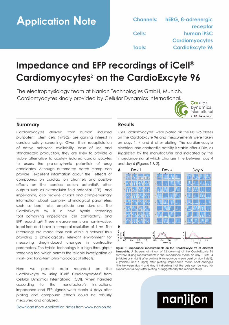
Summary ResultsiCell Cardiomyocytes2 were plated on the NSP-96 plates on the CardioExcyte 96 and measurements were taken on days 1, 4 and 6 after plating. The cardiomyocyte electrical and contractile activity is stable after 4 DIV, as suggested by the manufacturer and indicated by the impedance signal which changes little between day 4 and day 6 (Figures 1 & 2).
Figure 1: Impedance measurements on the CardioExcyte 96 at different timepoints. A Screenshot (4 out of 12 columns) of the CardioExcyte 96 software during measurements in the impedance mode on day 1 (left), 4 (middle) or 6 (right) after plating. B Impedance mean beat on day 1 (left), 4 (middle) and 6 (right) after plating. Impedance mean beat changes little between day 4 and day 6 indicating that the cells can be used for experiments 4 days after plating as suggested by the manufacturer.

Nanion Technologies GmbHGabrielenstr. 980636 Munich, Germany
phone +49 89 218997972fax +49 89 218997960www.nanion.de • [email protected]
Application Note
Figure 2 shows the development of the impedance parameter beat rate over time. After 4 days beat rate had stabilized and the cells could be used for experiments.
This phenomenon was also mirrored in the EFP signal. Figure 3 shows a screenshot of the CardioExcyte 96 software during a recording in the EFP mode on day 1, 4 and 6 after plating. The EFP mean beat displayed in Panel B shows that the EFP signal is stabilized after day 4 - 6.
Figure 3: EFP measurements on the CardioExcyte 96 at different time points. A Screenshot (4 out of 12 columns) of the CardioExcyte 96 software during measurements in the EFP mode on day 1 (left), 4 (middle) or 6 (right) after plating. B EFP mean beat on day 1 (left), 4 (middle) and 6 (right) after plating. EFP mean beat changes little between day 4 and day 6 indicating that the cells can be used for experiments 4 days after plating as suggested by the manufacturer.
Figure 2: Impedance beat rate shown over time course of several days. Medium exchanges were made as indicated by the vertical red lines. Beat rate was stable after 4 days at which point experiments can be started.
Figure 4: EFP amplitude shown over time course of several days. Medium exchanges were made as indicated by the vertical red lines. EFP amplitude was stable after 4 days at which point experiments can be started.
Figure 5 shows the effect of the specific hERG inhibitor, E4031, on the impedance and EFP signals. E4031 has a profound effect on the beat rate in impedance mode and induces early afterdepolarizations (EADs) which can lead to potentially fatal ventricular arrhythmias. This can be clearly recognized using the CardioExcyte 96. The EFP signal is also greatly affected by E4031 in a concentration-dependent manner. In this experiment, impedance measurements were taken at the start of the experiment (control) and then every 5 minutes for 30 s after E4031 application. The time point shown in Figure 5 is 13 mins after the start of E4031 application. EFP measurements were taken at the start of the experiment (control) and then at 30 mins after the start of E4031 application.
60
40
20
0
Bea
tRat
e[B
PM]
6543210 Time [Days]
Medium exchange
403020100
-10-20 E
FP A
mpl
itud
e[µ
V]
6543210 Time [Days]
Medium
exc
hange

Nanion Technologies GmbHGabrielenstr. 980636 Munich, Germany
phone +49 89 218997972fax +49 89 218997960www.nanion.de • [email protected]
Application Note
Figure 6: Effect of the ß-adrenergic receptor agonist, Isoproterenol, on the impedance and EFP signals. A Impedance signal of 12 wells in control conditions (left) and the same 12 wells after 13 mins incubation in isoproterenol at the concentrations indicated. Isoproterenol acts as a heart stimulant, it increases heart rate, an effect which is highlighted in the inset where the trace in the presence of isoproterenol (300 nM; black) and the control trace (red) are shown overlaid. In the presence of even low concentrations of isoproterenol (1 nM), an increase in the beat rate is observed. B Bar graph showing beat rate before (dark blue) and after (light blue) incubation in isoproterenol at the concentrations indicated. At all concentrations, a clear increase in beat rate is observed. C EFP signal of 12 wells in control conditions (left) and the same 12 wells after 30 mins incubation in isoproterenol at the concentrations indicated (right). Isoproterenol shortens the FPD, an effect which is highlighted in the inset where the trace in the presence of isoproterenol (300 nM; black) and the control trace (red) are shown overlaid.
Figure 5: Effect of the specific hERG blocker, E4031, on the impedance and EFP signals. A Impedance signal of 12 wells in control conditions (left) and the same 12 wells after 13 mins incubation in E4031 at the concentrations indicated (right). E4031(300 nM, 3 mins incubation) induces EAD, shown in the inset, which can lead to potentially fatal ventricular arrhythmias. B EFP signal of 12 wells in control conditions (left) and the same 12 wells after 13 mins incubation in E4031 at the concentrations indicated (right). E4031(300 nM, 30 mins incubation) also causes arrhythmic effects in the EFP mode shown in the inset.
Figure 6 shows the effect of the ß-adrenergic agonist, isoproterenol, on the impedance signal. Isoproterenol is used predominantly as a bronchodilator and heart stimulant, particulary after myocardial infarct, cardiac surgical procedures or to treat congestive heart failure3. Isoproterenol acts on the ß-1 adrenergic receptors in the myocardium increasing heart rate and, thereby, cardiac output3. There is a clear increase in beat rate in the impedance mode caused by isoproterenol recorded on the CardioExcyte 96 using the iCell Cardiomyocytes2. In the same way, in EFP mode, field potential duration (FPD) is shortened resulting in an increase in beat rate.

Nanion Technologies GmbHGabrielenstr. 980636 Munich, Germany
phone +49 89 218997972fax +49 89 218997960www.nanion.de • [email protected]
Application Note
References
1. Stoelzle, S., et al., 2011. Front. Pharmacol. doi: 10.3389/fphar.2011.00076.2. Doerr, L., et al., 2014. J. Lab. Autom. pii: 2211068214562832.3.http://pubchem.ncbi.nlm.nih.gov/compound/isoproterenol#section=Drug-Indication
Methods
CardiomyocytesiCell® Cardiomyocytes2 were kindly provided by CDI.
Impedance and EFP measurementsImpedance and EFP measurements were conducted according to Nanion’s standard procedures for the CardioExcyte 96. Cardiomyocytes were seeded at 50,000 viable cells per well on fibronectin-coated Sensor Plates.
Medium was exchanged from iCell Cardiomyocytes Plating to Maintenance Medium 4 hours post thaw, and then every other day until the conclusion of the assay. The cells could be used for pharmacology experiments 4 days after plating but were typically used 7 days after plating. This enabled the formation of a dense monolayer and a suitable network of cell-cell interconnections to ensure a steady propagation of the excitation and synchronized beating. At least 4 hours before drug application the medium was completely removed from the wells and 100 μl fresh medium was added. For the experiments, a 2x concentration of compound was prepared, 50 μl of solution was removed from the well and 50 μl of the 2x compound was added to give the final concentration indicated in the wells, although different methods of addition (e.g. 90 μl addition of fresh medium and then 10x concentration of compound added) could be used. All signals were normalized to a group of control measurements (n=5-11) on the same plate using DMSO (0.01%) as vehicle.
In this experiment, impedance measurements were taken at the start of the experiment (control) and then every 5 minutes for 30 s after isoproterenol application. The time point shown in Figure 6 is 13 mins after the start of isoproterenol application. EFP measurements were taken at the start of the experiment (control) and then at 30 mins after the start of isoproterenol application.
In conclusion, the CardioExcyte 96 can be used to reliably perform EFP and impedance measurements on hiPSCs from CDI (iCell Cardiomyocytes2). The manufacturer claims that the cells can be used for experiments 4 days after plating, and this claim is validated here using the CardioExcyte 96. The impedance and EFP signals are stable after about 4 days of plating. The hERG blocker, E4031, had profound effects on the EFP signal and the beat rate in impedance mode, causing EAD which could be clearly identified on the CardioExcyte 96. The ß-adrenergic ago-nist, isoproterenol, also affected beat rate in both EFP and
impedance modes, causing an increase in the beat rate at low concentrations. Therefore, the CardioExcyte 96 provides a complementary device to automated patch clamp for high throughput cardiac safety and cardiotoxicity studies.
We are confident that the CardioExcyte 96 in combination with hIPSCs will have a great impact on the field of pharmacology, toxicology, and cardiac safety screening in the light of the CIPA (Comprehensive In Vitro Pro-Arrhythmia Assay) proposal. This project aims to define a new, integrated preclinical in vitro/in silico paradigm in which the potential proarrhythmic risk of a new drug will be assessed using hIPSCs.