Identification and Functional Characterization of a High-Affinity Bel-1 ...
RESEARCH ARTICLE Open Access
Identification and functionalcharacterization of three new terpenesynthase genes involved in chemicaldefense and abiotic stresses in SantalumalbumXinhua Zhang1*† , Meiyun Niu1,2†, Jaime A. Teixeira da Silva3, Yueya Zhang1,2, Yunfei Yuan1, Yongxia Jia1,Yangyang Xiao1, Yuan Li1, Lin Fang1, Songjun Zeng1 and Guohua Ma1
Abstract
Background: It is well known that aromatic essential oils extracted from the heartwood of Santalum album L. havewide economic value. However, little is known about the role of terpenoids in response to various adverseenvironmental stresses as other plants do in the form of signals during plant-environment interactions.
Results: In this study, trace amounts of volatiles consisting of α-santalene, epi-β-santalene, β-santalene, α-santalol,β-santalol, (E)-α-bergamotene, (E)-β-farnesene and β-bisabolene were found in the leaves of mature S. album trees.We identified more than 40 candidate terpene synthase (TPS) unigenes by mining publicly-available RNA-seq dataand characterized the enzymes encoded by three cDNAs: one mono-TPS catalyzes the formation of mostly α-terpineol, and two multifunctional sesqui-TPSs, one of which produces (E)-α-bergamotene and sesquisabinene asmajor products and another which catalyzes the formation of (E)-β-farnesene, (E)-nerolidol and (E,E)-farnesol as mainproducts. Metabolite signatures and gene expression studies confirmed that santalol content is closely related withsantalene synthase (SaSSY) transcripts in heartwood, which is key enzyme responsible for santalol biosynthesis.However, the expression of three new SaTPS genes differed significantly from SaSSY in the essential oil-producingheartwood. Increased activities of antioxidant enzymes, superoxide dismutase, catalase, peroxidase and ascorbateperoxidase, were detected in different tissues of S. album plants after applying 1 mM methyl jasmonate (MeJA) and1 mM salicylic acid (SA), or exposure to 4°C, 38°C and high light intensity. MeJA and SA dramatically induced theexpression of SaTPS1 and SaTPS2 in leaves. SaTPS1 to 3 transcripts were differentially activated among differenttissues under adverse temperature and light stresses. In contrast, almost all SaSSY transcripts decreased in responseto these environmental stresses, unlike SaTPS1 to 3.
Conclusions: Multifunctional enzymes were biochemically characterized, including one chloroplastic mono-TPS andtwo cytosolic sesqui-TPSs in sandalwood. Our results suggest the ecological importance of these three new SaTPSgenes in defensive response to biotic attack and abiotic stresses in S. album.
Keywords: Abiotic stress, Methyl jasmonate, Salicylic acid, Santalum album, Sesquiterpene, Terpene synthase
© The Author(s). 2019 Open Access This article is distributed under the terms of the Creative Commons Attribution 4.0International License (http://creativecommons.org/licenses/by/4.0/), which permits unrestricted use, distribution, andreproduction in any medium, provided you give appropriate credit to the original author(s) and the source, provide a link tothe Creative Commons license, and indicate if changes were made. The Creative Commons Public Domain Dedication waiver(http://creativecommons.org/publicdomain/zero/1.0/) applies to the data made available in this article, unless otherwise stated.
* Correspondence: [email protected]†Xinhua Zhang and Meiyun Niu contributed equally to this work.1Key Laboratory of South China Agricultural Plant Molecular Analysis andGenetic Improvement, South China Botanical Garden, Chinese Academy ofSciences, Guangzhou, ChinaFull list of author information is available at the end of the article
Zhang et al. BMC Plant Biology (2019) 19:115 https://doi.org/10.1186/s12870-019-1720-3

BackgroundTerpenoids constitute the largest and most diverse classof chemical compounds present in all living organisms.In particular, flowering plants exhibit an unusually highnumber of terpenoids for a variety of basic functions ingrowth and development, including hormones, compo-nents of electron transfer systems, reagents for proteinmodification, determinants of membrane fluidity, antiox-idants, and others [1]. Different plant lineages alsosynthesize hundreds of distinct and specialized plant ter-penoids. Traditionally, specialized terpenoids are used asnatural flavor and aroma compounds and have a benefi-cial impact on humans as health-promoting compounds.Moreover, the ecological functions of terpenoids havegained increased attention [2].All plant terpenes are made from the two simple
five-carbon building blocks, isopentenyl diphosphate andits isomer, dimethylallyl diphosphate, both of which arederived from the mevalonate pathway in the cytosol orthe methylerythritol phosphate pathway in plastids [3].Condensation of the C5 precursors leads to the forma-tion of prenyl diphosphates, geranyl diphosphate (GPP),farnesyl diphosphate (FPP) and geranylgeranyl diphos-phate (GGPP), which are subsequently converted intomonoterpenes, sesquiterpenes, and diterpenes by ter-pene synthases (TPSs), respectively. Terpenoid biosyn-thesis occurs within specific tissues or at specificdevelopmental stages in plants, such as in resin ductsand glandular trichomes [4].During recent decades, there has been major progress
in the identification and functional characterization ofgenes and enzymes involved in terpenoid biosynthesis[5]. Many mono-TPS and sesqui-TPS genes have beenreported from several plants, including Arabidopsis(Arabidopsis thaliana) [6], grapevine (Vitis vinifera) [7],tomato (Solanum lycopersicum) [8], poplar (Populus tri-chocarpa) [9], spruce (Picea spp.) [10], Artemisia spp.[11] and citrus (Citrus sinensis) [12]. An unusual featureof many TPSs is that they are able to produce terpenoidsof structurally and stereochemically diverse compoundsfrom a single substrate [13]. Based on sequence related-ness, functional assessment and gene architecture, theTPS gene family has been divided into eight subfamilies,designated as TPS-a through TPS-h [14, 15]. Class I con-tains TPS-c (copalyl diphospate synthases), TPS-e(ent-kaurene synthases), TPS-f (other di-TPSs), andTPS-h (lycopod-specific); Class II consists of TPS-d,which is gymnosperm-specific; Class III consists ofangiosperm-specific TPS-a, TPS-b (cyclic mono-TPSsand hemi-TPSs), and TPS-g (acyclic mono-TPSs).Plants produce a number of terpenoid metabolites for
adaptation to adverse environments, including biotic andabiotic stresses [1]. Volatile terpenoids are inducibly emit-ted in response to herbivore or pathogen attacks in plants,
and not only function directly as defensive phytoalexins fordeterring detrimental attackers but also indirectly attractnatural enemies of pathogens and herbivores [16]. Studieshave shown that the plant hormones jasmonic acid (JA)and salicylic acid (SA) play important roles during biotic at-tack as defensive signaling [17, 18]. Therefore, exogenouslyapplied JA and SA can be used as biotic stress mimics forstudying the mechanism underlying the interaction be-tween plants and biotic stresses [19, 20], and for increasingthe production of plant terpenoids in specific organs toproduce specialized terpenoids [21]. For example, in sweetwormwood (Artemisia annua), JA can induce the AaMYC2gene coding for a basic helix-loop-helix-type transcriptionfactor that binds directly to G-box-like motifs within thepromoters of two genes encoding multifunction cyto-chrome P450 monooxygenase (CYP71AV1) and artemisinicaldehyde delta-11 (13) reductase during artemisinin biosyn-thesis [22]. In A. annua, SA enhanced the expression levelof amorpha-4,11-diene synthase and increased artemisininproduction [23]. In agarwood (Aquilaria sinensis), exogen-ously applied methyl jasmonate (MeJA) in cell suspensioncultures, callus and plant stems induced the biosynthesisand accumulation of sesquiterpene compounds, especiallyδ-guaiene [24–26].Moreover, the emission of some biogenic terpenes can
be induced by environmental factors such as temperatureand light. High temperature was first recognized as oneabiotic stress capable of inducing volatile terpenoids inslash pine (Pinus elliottii) [27]. In addition, the elevatedemission of a variety of terpene volatiles, including mono-terpenes and sesquiterpenes, from leaves, flowers andother organs in a range of woody and herbaceous specieswas observed under temperature, light, drought and saltstresses [28–34]. The production of terpenes was also in-duced by ozone, ultraviolet-B rays and γ-rays, which areoxidative stresses [35–37]. A common biochemical mech-anism explaining the emission of volatile terpenoids in-duced by exogenous stresses claims that plants have theability to synthesize terpenoids against oxidative damageresulting in elevated levels of reactive oxygen species(ROS) [1, 38].More than 150 terpenoid compounds were identified
from the essential oil of sandalwood (Santalum album)heartwood (HW) [39]. The major components were α-and β-santalol, but some minor compounds also exist, in-cluding α-santalene, β-santalene, α-bergamotene, andothers. In recent years, key genes responsible for santalolbiosynthesis have been functionally characterized, includ-ing santalene synthase (SaSSY) [40–42], SaCYP736A167and SaCYP76AF39V1 [43, 44]. Another five TPSs, namelySamonoTPS1, SasesquiTPS1, Sasesquisabinene synthases(SaSQS1 and SaSQS2), and Sabisabolene synthase (SaBS),have also been identified [42, 45]. Kulheim et al. (2014)attempted to enhance the essential oil productivity of
Zhang et al. BMC Plant Biology (2019) 19:115 Page 2 of 18

sandalwood by applying 0.01% MeJA to the leaves ofseedling younger than one year [46]. Their findings in-dicate that the expression levels of only two genes cod-ing for hydroxymethylglutaryl CoA reductase (HMCR)and farnesyl diphosphate synthase (FPPS) were slightlyinduced in leaves and stems of seedlings in response toMeJA treatment. Our previous study in S. album indi-cated that six TPS unigenes coding for (E)-β-ocimene/α-farnesene synthase, santalene/bergamotene synthase1 (SS/BS), nerolidol synthase 1-like, myrcene synthase,geranyl linalool synthase, and SamonoTPS1 were differ-entially expressed in response to cold stress (4°C) [47].However, little is known about the contribution of TPSsin response to various adverse environmental stressesin S. album.In this study, transcriptomic data from leaves, stems
and roots which were deposited at the Sequence ReadArchive (SRC) [42, 44, 47, 48] were integrated to identifyexpressed members of the sandalwood TPS gene family.Of these, three SaTPS genes, designated as SaTPS1 to 3,were isolated and functionally characterized. The expres-sion pattern and roles of SaTPS1 to 3 besides SaSSY inresponse to MeJA, SA, and adverse temperature andlight, were comparatively investigated. This study pavesthe way for revealing the regulatory mechanisms under-lying the biosynthesis of terpenoids in biotic and abioticstresses in S. album.
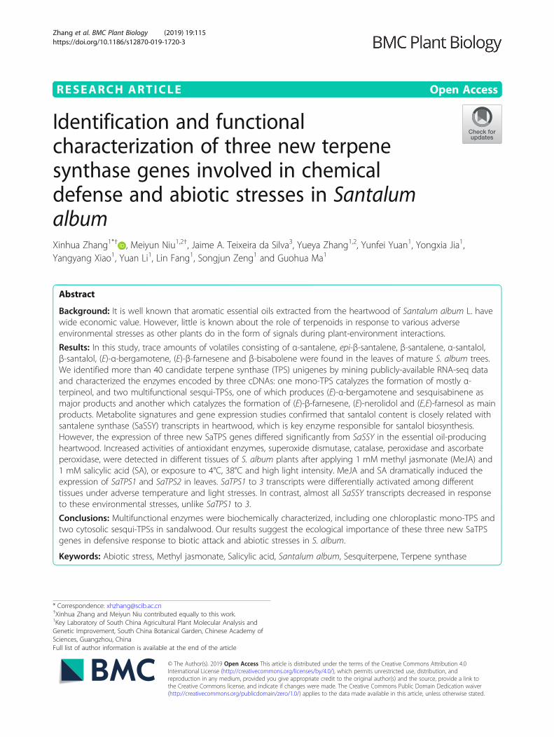
ResultsDifferences in content and composition of volatilesamong four tissuesTo understand the distribution pattern and differencesin components of S. album volatiles, we examined themetabolic compounds from pentane extracts of youngleaves (YL), sapwood (SW) and HW from mature treesand immature wood (IW) from immature trees byGC-MS. There were significant differences in the volatilecontent among the four tissues. As shown in Fig. 1, themost abundant volatile was detected in HW (Fig. 1a-d).Major sesquiterpenols, α- and β-santalol, were detectedin these four tissues, and sesquiterpenes, α- andβ-santalene, and epi-β-santalene were identified in YLand HW, but not in IW and SW (Fig. 1e-i, Additionalfile 1: Table S1). It was curious that trace amounts of α-and β-santalene, epi-β-santalene and α-santalol (peaks 1,3, 5 and 8, respectively) were found in almost the sameproportion in YL (Fig. 1e). Of these, the distribution ofthe first three compounds matched that in HW (Fig. 1h,peaks 1, 3 and 5). Moreover, (E)-α-bergamotene,(E)-β-farnesene, and β-bisabolene were also detectedfrom leaf extracts of S. album. Almost the same distribu-tion of sesquiterpenols as those in SW and HW was ob-served in IH (Fig. 1 f, g and i, peaks 8–15), indicatingthat the accumulation of sandalwood oil occurs at anearly developmental stage of the stems and that more
Retention time (min) Retention time (min)
0.5
1.0
1.5
0.5
1.0
1.5
a
b
0.51.01.52.02.5
IS
IS
IS
Rel
ativ
e ab
unda
nce
(×10
6 )
0.5
1.0
1.5
2.0
2.5
c
12.5 15.0 17.5 20.0 22.5 25.0 27.5
18.0 19.0 20.0 21.0 22.0 23.0 24.0 25.0 26.0 27.0
18.0 19.0 20.0 21.0
(×10
4 )
0.5
1.0
1.5
2.0
13
57
9
1110
13
12 14 15
8
24.5 25.0 25.5 26.0 26.5
0.5
1.0
1.5
2.0
2.5
(×10
6 )
e
13
(×10
4 )(×
104 )
2.0
4.0
6.0
8.0
3
4
1
2
5
6
8
13
8
12159
1411
8
(×10
5 )
0.5
1.0
1.5
2.0
2.5
913
1411
1512
1.02.03.04.05.06.0 f
g
IS
dh i
Fig. 1 Sesquiterpenoid profile from different extracts. Sesquiterpenoids were identified against those in mass spectra reference libraries, NIST2005,NIST2005s, NIST2014, NIST2014s and FFNSC1.3 and a comparison of their retention indices. a, e: YL, young leaves, b, f: IW, immature wood, c, g:SW, sapwood, d, h, i: HW, heartwood. Segments (e, f, g, h and i) of the GC profile (a, b, c, d) are magnified in the right panel, respectively. Peaks:1, α-santalene, 2, (E)-α-bergamotene, 3, epi-β-santalene, 4, (E)-β-farnesene, 5, β-santalene, 6, β-bisabolene, 7, unknown, 8, α-santalol, 9, α-trans-bergamotol, 10, α-santalol isomer, 11, epi-β-santalol, 12, unknown, 13, β-santalol, 14, β-santalol isomer, 15, lanceol. Peak numbers are indicated inorder of elution from a HP-5MS column. IS, internal standard (n-dodecane)
Zhang et al. BMC Plant Biology (2019) 19:115 Page 3 of 18

essential oil is biosynthesized in HW as the sandalwoodtree grows older.
Identification of new terpene synthases in S. albumWe combined transcriptome sets downloaded fromNCBI, including transcripts from leaves, stems and rootsas described in the Methods. De novo assembly yielded164,548 unigenes from the three tissues (Additional file1: Figure S1, Additional file 1: Table S2). Many unigeneswere identified as SaTPS genes, including 38 core ter-pene synthase genes and nine triterpene-specific syn-thase genes (Additional file 1: Table S3). Of these, sixunigene sequences were completely aligned with thoseof S. album submitted to NCBI [40, 42, 45]. Three uni-genes contained full-length ORFs encoding TPSs, desig-nated as SaTPS1 to 3. In order to confirm that theassembled unigenes generated using the Trinity assem-bler were accurate, we designed primers to annealaround the predicted start and stop codons of unigenesto amplify ORFs of the three predicted SaTPS genes(Fig. S2). Subsequent sequencing of these nucleotides
demonstrated that they were exactly as predicted by theunigene assembly.
Sequence and phylogenetic analysis of three SaTPSsThree predicted proteins encoded by SaTPS1 to 3 with arange of 564 to 604 amino acids had predicted pIs of6.01, 5.02 and 5.03, respectively (Additional file 1: TableS4). As shown in Fig. 2, SaTPS1 to 3 contain thearginine-tryptophan motif, R(R) X8W, which is con-served in most mono-TPSs and in some sesqui-TPSsnear the N-terminus. The highly conservedaspartate-rich motif (DDXXD) and NSE/DTE motif,which are crucial for chelating divalent cations, typicallyMg2+, in the C-terminal domain [49], were present inthese three SaTPSs. One of the distinguishing structuralfeatures between mono- and sesqui-TPSs is the presenceof an N-terminal plastid transit peptide sequence. Usingthe signal sequence analysis program ChloroP (http://www.cbs.dtu.dk/services/ChloroP), a putative N-terminalplastid transit peptide (Tp) sequence of 44 amino acidsfor SaTPS1 was predicted, indicating that it is likely a
Fig. 2 Comparison of deduced amino acid sequences of SaTPSs. The deduced amino acid sequences of SaTPS genes were aligned usingDNAMAN 6.0 (Lynnon Biosoft, San Ramon, CA, USA). The Asp-rich domain DDXXD, the RRX8W motif, and the NSE/DTE motif, which are highlyconserved in plant TPSs and required for TPS activity, are indicated. The arrowhead denotes the predicted cleavage site of plastidial transitpeptide of SaTPS1. Completely conserved residues are shaded in black, identical residues in dark grey, and similar residues in light grey. Dashesindicate gaps introduced to maximize sequence alignment
Zhang et al. BMC Plant Biology (2019) 19:115 Page 4 of 18

mono-TPS. However, SaTPS2 and SaTPS3 did not con-tain a plastid Tp sequence, suggesting that they aresesqui-TPSs (Additional file 1: Table S4).A phylogenetic tree based on the deduced amino acid of
sandalwood TPSs and other plant species showed that allSantalum TPSs clustered in the TPS-b clade, with the ex-ception of SasesquiTPS and sesquiTPSs in S. spicatum(SspicsesquiTPS) and S. austrocaledonicum (Saustses-quiTPS) (Fig. 3; Additional file 1: Table S5), among whichthe majority of angiospermous monoterpene synthasesreside [15]. The most similar TPS sequence to SaTPS1 isthat of (−)-α-terpineol synthase from V. vinifera with 63%identity (Additional file 1: Figure S3; Additional file 1:Table S6) (Martin and Bohlmann, 2004). SaTPS2 clusteredmost closely to SaSSY, SspicSSY and SaustSSY, with a
100% bootstrap (Jones et al., 2011), and its amino acid se-quence had highest identity with SaspiSSY (72%). SaTPS3formed a clade with several previously characterized TPSsfrom Santalum species, including SaMonoTPS, SaSQS1,SaSQS2, SaBS, SspicBS and Sspicsesquisabinene [42, 45,50]. Bioinformatics and phylogenetic analyses suggeststhat SaTPS2 and SaTPS3 might be involved in the produc-tion of essential oil in S. album.
Expression patterns of three SaTPS genes differ from SaSSYThe tissue-specific expression of SaTPS1 to 3 genes wasdetermined by qRT-PCR in four key tissues, i.e., YL, IW,SW and HW. The transcript abundance of SaTPS1 andSaTPS2 genes showed a similar expression pattern, withhighest expression in YL (Fig. 4). SaTPS3 showed
Fig. 3 Phylogenetic positioning of SaTPS protein products within representative samples of known plant TPS. The neighbour-joining tree wasdrawn using the MEGA 6 program from an alignment of full-length SaTPSs with other plant TPSs [75]. The seven TPS subfamilies a-g on the right-hand side are delimited based on the taxonomic distribution of the TPS families [15]. Bootstrap values from 1000 replicates were used to assessthe robustness of the trees. Names of organism TPSs (NCBI Protein no.) can be retrieved from Additional file 1: Table S4
Zhang et al. BMC Plant Biology (2019) 19:115 Page 5 of 18

three-fold higher expression in IW than in YL, SW and HW.In contrast, SaSSY was 100- and 250-fold higher in SW andHW, respectively. Additionally, comparisons of the expres-sion levels among these SaTPSs in specific tissues showedthat SaTPS3 showed higher expression than SaTPS1 andSaTPS2 in the four corresponding tissues (Additional file 1:Figure S4). Of note, the level of SaSSY transcript showed a200- and 1200-fold increase relative to SaTPS1 and SaTPS2in SW and HW, respectively. These results suggest thatSaTPS3 might play a role in the accumulation of HW essen-tial oil whereas SaTPS1 and SaTPS2 play a minor role in theformation of terpenoids in S. album HW.
Subcellular localization of SaTPSsTo elucidate the function of SaTPS1 to 3, subcellularlocalization of each SaTPS-YFP (yellow fluorescentprotein) fusion protein provided preliminary evidence.Phylogenetic and bioinformatics-based analyses in anattempt to classify TPSs indicated that SaTPS1-YFP,which has the N-terminal plastid transit peptide se-quence, was localized in chloroplasts, whereasSaTPS2-YFP and SaTPS3-YFP were distributedthroughout the cytosol (Fig. 5). Based on the resultsof subcellular localization, it was concluded thatSaTPS1 is likely involved in monoterpene biosynthesisin plastids, whereas SaTPS2 and SaTPS3 might pro-duce sesquiterpenes in the cytosol.
Functional characterization of the enzymes encoded bySaTPS1 to 3All cDNAs subcloned into pET28a vector with6His-tagged were successfully expressed in E. coliRosetta 2 (DE3) competent cells and recombinant pro-teins were then purified with a Ni-NTA agarose affinitycolumn (Additional file 1 :Figure S5). We found that thiscDNA without the transit peptide sequence could im-prove the expression of SaTPS1 in E. coli. Following anin vitro enzyme activity assay, SaTPS1 was confirmed toencode a mono-TPS enzyme that catalyzed the forma-tion of mostly α-terpineol (45.7%), while sabinene(14.9%), linalool (11.7%) and myrcene (10.8%), as well asa few minor monoterpenes, were also produced withGPP as the substrate and Mg2+, as detected byGC-MS analysis (Fig. 6a, Additional file 1: Table S7).Although the same compounds were produced whenMg2+ was replaced by Mn2+, levels of productformation were 5-fold lower and the three majorcompounds, linalool (35.8%), α-terpineol (25.7%), and ge-raniol (12.9%) were formed (Additional file 1: Figure S6,Additional file 1: Table S7). No terpene product wasobserved in the reaction of SaTPS1 with FPP as a sub-strate and Mg2+ or Mn2+ (Fig. 6b). This enzyme was desig-nated as α-terpineol synthase because it is a majormonoterpene product and due to its preference for themetal ion, Mg2+.
Fig. 4 Expression levels of SaTPS1 to 3 genes in different tissues. YL, young leaves; IW, immature wood; SW, sapwood; HW, heartwood. Significantdifferences were assessed by a student’s t-test: *P < 0.05, **P < 0.01 and ***P < 0.001
Zhang et al. BMC Plant Biology (2019) 19:115 Page 6 of 18

GC-MS analysis of the reaction products catalyzed bySaTPS2 with FPP as a substrate in the presence of Mg2+
identified at least 15 sesquiterpenoids, with (E)-α-berga-motene (24.8%) and sesquisabinene (33.0%) as the twomajor products, β-bisabolene (9.0%), γ-bisabolene iso-mer (7.9%), and minor compounds, 7-epi-sesquithujene,(E)-β-farnesene, α-zingiberene, and others at relativeproportions less than 5% (Fig. 7a; Additional file 1: Table S7).A similar product profile comprising the major compounds,(E)-α-bergamotene (22.4%) and sesquisabinene (35.6%), weredetected when Mg2+ was replaced with Mn2+, but overallyield was relatively lower (Additional file 1: Figure S7,Additional file 1: Table S7). Incubation of SaTPS2 with GPPas a substrate and Mg2+ ion led to the production of ninemonoterpenes (Fig. 7b; Additional file 1: Table S7). The mostabundant compound was linalool (64.9%). Likewise, assayswith Mn2+ instead of Mg2+ showed that almost the samecompounds were observed, but overall yield was 5-fold lower(Additional file 1: Figure S8, Additional file 1: Table S7).
Taken together, these results indicate that SaTPS2 is a multi-functional sesqui-TPS producing major (E)-α-bergamoteneand sesquisabinene in the cytosol.SaTPS3 converts FPP and GPP substrates to sesqui-
and monoterpene products, respectively. When assayedwith FPP and Mg2+, recombinant SaTPS3 catalyzed thesynthesis of seven compounds, including (E)-β-farnesene(20.7%), (E)-nerolidol (29.8%) and (E,E)-farnesol (21.3%)as the three major products, while a high proportion ofγ-bisabolene (13.8%) was also produced (Fig. 8a,Additional file 1: Table S7). In the presence of Mn2+,SaTPS3 recombination enzyme showed no activity, sug-gesting that SaTPS3 prefers Mg2+ in the reaction withFPP. Analysis of the reaction products formed after in-cubation of SaTPS3 with GPP showed that the mainproduct linalool (44.8–53.3%) was formed with Mg2+ orMn2+ in considerable yield (Fig. 8b, Additional file 1:Figure S9, Additional file 1: Table S7). For the threeSaTPSs tested in this study, extracts prepared from E.
Fig. 5 Subcellular localization of SaTPS1 to 3 in Arabidopsis mesophyll protoplasts. Protoplasts were transiently transformed with SaTPS-YFPconstructs or YFP vector using a modified polyethylene glycol method. YFP fluorescence was observed with a laser scanning confocalmicroscope. Yellow fluorescence indicates SaTPS-YFP fusion protein signal. Blue signal indicates chlorophyll (Chl) autofluorescence and red signalindicates m-Cherry fluorescence. The merged images represent a digital combination of Chl autofluorescence, YFP fluorescent and m-Cherryprotein fluorescence images. Fluorescence was excited for YFP at 514 nm, for Chl at 543 nm and for m-Cherry at 587 nm. Scale bar = 10 μm
Zhang et al. BMC Plant Biology (2019) 19:115 Page 7 of 18

a
b
Fig. 6 In vitro enzymatic assays of recombinant SaTPS1. In vitro enzyme assays of recombinant SaTPS1 using GPP (a) and FPP (b) as the substratein the presence of Mg2+. The reaction products were analyzed by GC-MS. The peaks marked with numbers were identified by comparison of theirmass spectra with those in the library data and comparison of their retention index. Peaks: 1, α-thujene, 2, α-pinene, 3, sabinene, 4, β-pinene, 5,myrcene, 6, limonene, 7, cineole, 8, β-ocimene, 9, linalool, 10, α-terpineol, 11, geraniol. Mass spectra for the major product and correspondingauthentic standard are shown on the right side of the figure. m/z, mass-to-charge ratio
a
b
Fig. 7 In vitro enzymatic assays of recombinant SaTPS2. In vitro enzyme assays of recombinant SaTPS2 using FPP (a) or GPP (b) as the substratein the presence of Mg2+. The reaction products were analysed by GC-MS. Peaks marked with numbers were identified by comparison of theirmass spectra with those in the library data and a comparison of their retention index. Peaks in a: 1, 7-epi-sesquithujene, 2, unknown, 3, α-bergamotene isomer, 4, (E)-α-bergamotene, 5, (E)-β-farnesene, 6, sesquisabinene, 7, unknown, 8, α-zingiberene, 9, α-bisabolene, 10, β-bisabolene,11, γ-bisabolene, 12, unknown, 13, γ-bisabolene isomer, 14, β-bisabolol, 15, α-bisabolol. Peaks in b: 1, α-thujene, 2, α-pinene, 3, sabinene, 4, β-pinene, 5, myrcene, 6, limonene, 7, linalool, 8, α-terpineol, 9, geraniol. Mass spectra for the major products and corresponding authentic standardsare shown on the right side of the figure, respectively. m/z, mass-to-charge ratio
Zhang et al. BMC Plant Biology (2019) 19:115 Page 8 of 18

coli transformed with PET28a lacking a cDNA insertand heat-denatured enzyme preparations served as con-trols, and no terpene products were observed. In allcases, expressed proteins incubated with GGPP did notproduce detectable products.The Km value of purified SaTPS1 with GPP was 9.08 ±
1.26 μM and the Kcat/Km value was 0.0171 (Table 1;
Additional file 1 :Figure S10). The SaTPS2 and SaTPS3enzymes not only accept substrate FPP but also acceptthe precursor of monoterpenes, GPP, as analyzed above.Enzyme kinetic properties of SaTPS2 showed that cataly-sis with FPP and GPP resulted in a similar Km and Kcat/Km. The Km value of the recombination SaTPS3 withFPP was 16.29 ± 1.48 μM, which was slightly higher thanwith GPP. The Kcat/Km with FPP was relatively lowerthan with GPP, indicating that the efficiency of thissesqui-TPS catalysis is higher when incubated with GPP.
Elevated antioxidant enzyme activity under multiplestress treatmentsThe phytohormones JA and SA play crucial roles inregulating the defensive signaling network by elevatingthe levels of ROS [51, 52]. Moreover, the mechanism bywhich terpenes alleviate abiotic stress suggests a generalantioxidant mechanism by which harmful ROS can be
a
b
Fig. 8 In vitro enzymatic assays of recombinant SaTPS3. In vitro enzyme assays of recombinant SaTPS3 using FPP (a) or GPP (b) as the substratein the presence of Mg2+. The reaction products were analysed by GC-MS. The peaks marked with numbers were identified by comparison of theirmass spectra with those in library data and a comparison of their retention index. Peaks in a: 1, cedrene, 2, unknown, 3, (E)-β-farnesene, 4,unknown, 5, γ-bisabolene, 6, (E)-nerolidol, 7, (E,E)-farnesol. Peaks in b: 1, myrcene, 2, limonene, 3, unknown, 4, β-ocimene, 5, linalool, 6, α-terpineol,7, geraniol. Mass spectra for the major products and corresponding authentic standards are shown, respectively. m/z, mass-to-charge ratio
Table 1 Enzyme kinetic properties of three SaTPSs
Enzyme Substrate Km (μM) Kcat (s−1) kcat/Km (s−1 μM− 1)
SaTPS1 GPP 9.08 ± 1.26 0.155 ± 0.007 0.0171
SaTPS2 FPP 12.15 ± 1.32 0.200 ± 0.008 0.0164
GPP 12.27 ± 1.32 0.189 ± 0.007 0.0154
SaTPS3 FPP 16.29 ± 1.48 0.171 ± 0.006 0.0105
GPP 14.38 ± 1.20 0.257 ± 0.008 0.0179
Values for FPP and GPP were measured in the presence of 10 mM Mg2+
All values represent mean ± SE, n = 3. Km, Michaelis constant; kcat, turnover
Zhang et al. BMC Plant Biology (2019) 19:115 Page 9 of 18

quenched by reacting with unsaturated isoprene, mono-terpenes, and sesquiterpenes [38]. In our study, the activ-ity of superoxide dismutase (SOD), a key contributor inthe conversion of O− to H2O2 in the presence of elevatedlevels of ROS, increased significantly under all treatments,except for a decrease in leaves exposed to 4°C, 38°C andhigh light intensity (approx. 250 μmol m− 2 s− 1) (Fig. 9a).Catalase (CAT) plays an important role in the antioxidantsystem because it enables plants to eliminate H2O2 byconverting H2O2 into O2 and H2O (Miller et al., 2008).CAT activity was strongly elevated by these two elicitors.When the three tissues were exposed to temperature andlight stresses, CAT activity was upregulated, the only
exception being a slight decreased in leaves in response tohigh light intensity (Fig. 9b).Similarly, peroxidase (POD), which detoxifies H2O2 in
the chloroplasts and cytosol of plant cells, also consti-tutes the main H2O2-scavenging system in cells [53].POD activity increased in all treatments and roots hadtwo-fold higher POD activity than leaves (Fig. 9c). Con-versely, SOD and CAT activities were relatively higher inleaves than in stems and roots, independent of whetherthey were treated or untreated, suggesting differentialendogenous activities of SOD, CAT or POD among dif-ferent tissues. Ascorbate peroxidase (APX) plays a keyrole in protecting plants against oxidative stress by
a b
c d
Fig. 9 Effects of external stresses on activities of antioxidant enzymes in S. album. Six-month-old seedlings were sprayed with 1 mM MeJA or SAfor 24 h or exposed to 4°C, 38°C or high light intensity (approx. 250 μmol m− 2 s− 1) for 12 h. Three measurements were averaged from the resultsof three replicated experiments. a: SOD, superoxide dismutase; b: CAT, catalase; c: POD, peroxidase; d: APX, ascorbate peroxidase. L, leaves; S,stems; R, roots. Significant differences were analyzed by a student’s t-test and indicated as *P < 0.05, **P < 0.01 and ***P < 0.001
Zhang et al. BMC Plant Biology (2019) 19:115 Page 10 of 18

scavenging H2O2 in different cell compartments [54]. Asshown in Fig. 9d, APX activity increased significantly inmost tissues under MeJA, SA, cold, heat and high lightintensity treatments. Thus, the increase in SOD, CAT,POD and APX activities indicate that S. album seedlingsrespond to external stresses to protect cellular mem-branes against oxidative stress, suggesting that these en-zymes play roles in tolerance to elicitors andenvironmental stresses in S. album. In addition, the ef-fect of different adverse stresses on antioxidant enzymeactivities will vary among tissues.
Activation of SaTPS1 to 3 gene expression in response toMeJA and SATo explore the responses of the three SaTPS genesunder elicitors as well as temperature and high light in-tensity treatments, transcript levels for SaTPS1 to 3 andSaSSY were determined by qRT-PCR. SaTPS1 andSaTPS2 exhibited similar expression patterns in responseto the two elicitors. As shown in Fig. 10a and b, exogen-ous MeJA and SA dramatically induced the expressionof SaTPS1 and SaTPS2 genes in leaves at 24 h, withmore than a 170- and 130-fold increase, respectively,when compared with the controls. SaTPS1 and SaTPS2transcripts also increased significantly in stems and rootsafter the application of MeJA. SaTPS3 expression wasactivated in leaves, stems and roots with an approxi-mately 5-, 3-, and 4-fold increase, respectively comparedto the corresponding controls (Fig. 10c). In contrast,SaTPS3 was significantly up-regulated in leaves by SA,but decreased in stems and roots. Moreover, the accu-mulation of SaSSY transcript were similar to that ofSaTPS3 in leaves under MeJA and SA treatments. How-ever, the two exogenous hormones led to a decrease inSaSSY expression in roots compared with the control(Fig. 10d).
Adverse temperature and high light induced differentialaccumulation of SaTPS1, SaTPS2 and SaTPS3SaTPS1 to 3 expression levels were inhibited in leaves at4°C and 38°C, but were significantly activated in rootswhen compared to the controls (Fig. 10e-g), suggestingthat oxidative damage in leaves was more severe than instems and roots. When exposed to high light intensity,SaTPS1 and SaTPS2 transcripts showed higher expres-sion in stems and roots than in leaves, particularlySaTPS1, with a 25-fold increase in stems. The level ofSaTPS3 transcripts was highest in roots under 4°C, 38°Cand high light intensity stresses, with approximately 2-,6-, and 8-fold increases, respectively, compared to thecontrol. Conversely, SaSSY transcript only increased byabout 2-fold in roots at 4°C compared to the control. Inall other cases, the expression level of SaSSY was
down-regulated to some extent in all tissues examined(Fig. 10h).
DiscussionCandidate TPS genes from S. album by RNA-seqNext generation sequencing has emerged as a promisingplatform to discover novel genes, enzymes, and molecu-lar markers from non-model plant species. In recentyears, transcriptomic approaches have been widely usedto mine genes of terpenoid metabolism in Santalum. Re-search has mainly focused on cloning andcharacterization of SaTPSs, including SamonoTPS,SasesquiTPS, SaSSy, SauSSY, SspiSSY, SaSQS1, SaSQS2,SaBS, SauBS, SspiBS, and SspiTPS4 [40, 42, 45, 50]. Cel-edon et al. (2016) [44] found more than 30 SaTPS tran-scripts from a HW-specific transcriptome. In the presentstudy, a set of SaTPSs was identified based on a combin-ation of transcriptome data from leaves, stems and roots,implying that S. album likely has a mid-sized TPS genefamily according to the classification of other angio-sperm TPSs [15]. Currently, the genome assembly dataof S. album is available [55], and we are eager to obtainthe annotated genome so as to assess the exact size ofTPSs in the S. album TPS family. Additionally, someTPSs specific to different tissues were detected(Additional file 1: Table S3), inferring that SaTPSs mightplay different roles in different tissues.
Santalol content is closely related with SaSSY transcriptsin heartwoodThe major components of the total essential oil in theheartwood of S. album are α- and β-santalol, contribut-ing over 80% of relative content [39]. SaSSY is a key en-zyme responsible for the biosynthesis of α- andβ-santalol [40–42]. Our study’s findings are in agreementwith previous studies that showed that sandalwood es-sential oil accumulates in stem HW, followed by SW[44, 45]. However, α- and β-santalene and epi-β-santa-lene were not detected in SW, most likely due to un-detectable or low amounts. To our knowledge, tracelevels of volatiles consisting of α- and β-santalene,epi-β-santalene, α- and β-santalol, and other compoundsin the leaves of S. album were first reported in thisstudy. The distribution pattern of sesquiterpenols similarto those in SW and HW was found in IH (Fig. 1f, g andi). Collectively, these results established a foundation toreveal the mechanism of spatial and temporal distribu-tion of volatiles in S. album. Moreover, high expressionlevels of SaSSY in the HW of S. album confirm that san-talol content is closely related with SaSSY transcript level(Figs. 1 and 4).In contrast, SaTPS1 to 3 genes had relatively high ex-
pression levels in leaves (Fig. 4 and S4a) and traceamounts of (E)-α-bergamotene, (E)-β-farnesene and
Zhang et al. BMC Plant Biology (2019) 19:115 Page 11 of 18

β-bisabolene in the leaves of mature S. album trees werealso detected, suggesting that SaTPS2 and SaTPS3 mightconvert FPP to these sesquiterpenes, which belonged tomajor products when incubated with FPP in in vitro en-zyme assays (Fig. 7a, 8a and Additional file 1: Table S7).
In addition, major α-terpineol catalyzed by SaTPS1 wasnot identified in leaf extracts, inferring that it might beeasily volatilized or that external stimuli might be re-quired for its emission, aspects that still need to beexplored.
a b
c d
e f
g h
Fig. 10 Transcript accumulation of SaTPS1 to 3 under multiple stresses in S. album. (a-d) Six-month-old seedlings were sprayed with 1 mM MeJAor SA for 24 h. (e-h) Six-month-old seedlings were exposed to 4°C, 38°C or high light intensity (approx. 250 μmol m− 2 s− 1) for 12 h. Threemeasurements were averaged from the results of three replicated experiments. L, leaves; S, stems; R, roots. Significant differences were analyzedby a student’s t-test and indicated as *P < 0.05, **P < 0.01 and ***P < 0.001
Zhang et al. BMC Plant Biology (2019) 19:115 Page 12 of 18

SaTPS1 is likely a paralogous gene of SamonoTPS1A study by Jones et al. (2008) [45] documented thatSamonoTPS1 is a monoterpene synthase which pro-duced a mixture of eight compounds, with α-terpineol(38.7%) and limonene (35.3%) as the two main productswhen incubated with GPP. In angiosperms, α-terpineolsynthase from V. vinifera was reported as a monoter-pene synthase, producing 14 compounds, includingmajor monoterpene, α-terpineol (50.1%), 1,8-cineol(11.8%), β-pinene (8.5%), etc. [56]. Gao et al. (2018) [57]reported that FhTPS2 in flowers of Freesia × hybridamainly converted GPP into α-terpineol (78.7%) and afew other monoterpenes, such as 1,8-cineole (6.9%),D-limonene (3.9%), α-pinene (3.2%), etc. In our study,SaTPS1 showed higher similarity at the nucleotide levelwith α-terpineol synthase sequences in V. vinifera thanSamonoTPS1, and both of them have an N-terminalplastid transit peptide sequence and are closely related(Fig. 3; Additional file 1: Figure S3, Additional file 1:Table S6). Our biochemical characterization and subcel-lular localization studies confirm that SaTPS1 is amono-TPS, α-terpineol synthase. Collectively, these re-sults imply that SaTPS1 may be a paralogous gene ofSamonoTPS1.
Functional divergence of TPSs has occurred in SantalumSaSQS1 and SaSQS2 catalyze the cyclization of(E,E)-FPP to a single product, sesquisabinene [42].SspiTPS4 catalyzed the formation of the most abundantsesquisabinene B (58%) in S. spicatum [50]. In this study,although a phylogenetic tree showed that SaTPS2 ismuch closer to SaSSY, SspicSSY and SaustSSY than toSaSQS1, SaSQS2 and SspiTPS4, in vitro enzyme assaysof recombinant SaTPS2 and subcellular localization re-vealed that the mRNA of SaTPS2 encodes a functionalsesqui-TPS producing (E)-α-bergamotene (24.8%) andsesquisabinene (33.0%) as two major products. More-over, both recombinant SaBS and SauBS can react withFPP to produce β-bisabolene as a major product [40,42]. SspiBS produced a mixture of β-bisabolene andα-bisabolol, along with traces of α-bisabolene and farne-sene isomers [40]. SaTPS2 could synthesize a relativelylarge amount of β-bisabolene (9.0%) and γ-bisabolene(1.6%) (Additional file 1: Table S7). Taken together, theseresults suggest that functional divergence of TPS has oc-curred in Santalum species. The diversification of ter-penoid biosynthesis has also been reported in Oryza TPSgenes [58].In angiosperms, only the maize gene TPS1 produces
(E)-β-farnesene (26%), (E)-nerolidol (29%), and (E,E)-far-nesol (45%) as the only three products in vitro [59]. Phylo-genetically, SaTPS3 is paraphyletic group with other TPSsof Santalum spp. in the TPS-b group (Fig. 3). The func-tion of SaTPS3 is similar to TPS1 in maize where it
catalyzes the formation of three major compounds, i.e.(E)-β-farnesene (20.7%), (E)-nerolidol (29.8%) and(E,E)-farnesol (21.3%). However, SaTPS3 additionally syn-thesized γ-bisabolene, cedrene and two unknown sesqui-terpene compounds in relative amounts that exceeded28%, suggesting that SaTPS3 is not a highly conservedTPS in angiosperms. Diversity of function may be an evo-lution of adaptive selection in monocotyledons anddicotyledons.
SaTPS1 to 3 play important roles in chemical defenseThe inductive roles played by JA- and SA-induced bio-synthesis of terpenoids have also been widely demon-strated in recent years, playing similar roles as thoseduring attack induced by fungal elicitation and mechan-ical wounding [60–62]. For example, the transcript levelof TPS1 producing (E)-β-farnesene, (E)-nerolidol and(E,E)-farnesol in vitro in maize cv. B73 was elevated afterherbivory, or after mechanical damage with and withouttreatment with 5 μl of Egyptian cotton leafworm regurgi-tant or 5 μl of volicitin [59]. The volatile (E)-α-bergamo-tene is a well-characterized defensive compound inwounded leaves induced by herbivores [63] and inflowers [64, 65] of Nicotiana attenuata induced byJA-mediated signaling. To date, little is known about theroles of α-terpineol produced by TPSs in plant defense.In S. album, the expression levels of genes coding forHMCR and FPPS were slightly induced in the leaves andstems of seedlings after the foliar application of 0.1%MeJA [46]. In our study, increased enzyme activities inthe ROS antioxidant defense system and the sharp accu-mulation of SaTPS1 and SaTPS2 transcripts by exogen-ously applied MeJA and SA in leaves suggest thatSaTPS1 and SaTPS2 play defensive roles against bioticstresses. Similarly, elevated levels of SaTPS3 transcriptsinduced by MeJA in the three tissues (Fig. 10c) inferredthat SaTPS3 had similarly defensive roles to SaTPS1 andSaTPS2. Moreover, MeJA led to increases of SaTPS1 andSaTPS2 transcripts in stems and roots (Fig. 10a and b).Collectively, this demonstrates that both local and sys-temic defense responses were triggered by exogenouslyapplied MeJA in S. album seedlings. This result is thesame as findings reported by Koo et al. (2009) [66] inArabidopsis and Delaunois et al. (2014) [67] in grape-vine, in which local activation of JA and its biologicallyactive derivatives (i.e., MeJA) as JA signaling moleculesspread systemically throughout the plant and induce JAresponses in distant organs. In general, SA is mainly as-sociated with resistance to biotrophic and hemibio-trophic pathogens, and triggers systemic acquiredresistance [68, 69]. The reason why exogenous SA re-sulted in the downregulation of SaTPS1 to 3 genes intreated roots needs to be further explored. Simultan-eously, activated expression of SaSSY in leaves and stems
Zhang et al. BMC Plant Biology (2019) 19:115 Page 13 of 18

by MeJA and SA offers a cue for further exploring themechanism of santalol biosynthesis in S. album.
SaTPS1 to 3 play roles in response to abiotic stresses indifferent tissuesEnvironmental factors play important roles governingthe emission of biogenic terpenes. The emission of someterpene volatiles is highly dependent on temperature,light and oxidative stresses [32, 38]. In loblolly pine(Pinus taeda), high light intensity and temperature in-duced the emission of (E)-β-farnesene and (E)-α-berga-motene [70]. Our previous work showed that SaSS/BSand SaMonoTPS1 accumulated to high levels in theroots of S. album in response to 4°C [47]. In that study,the full-length of SaSS/BS was rediscovered as SaTPS2in this study. In the present study, the expression levelsof SaTPS2 in leaves and roots after exposure for 12 h to4°C were similar to our previous results, in which a de-crease of SaTPS2 transcripts in leaves and significantup-regulation in roots were observed. Although adversetemperature and light inhibited the expression ofSaTPS1 to 3 genes in leaves, high or low temperaturesignificantly induced an increase in SaTPS1 to 3 tran-scripts in roots. High light intensity induced a remark-able elevation of SaTPS1 and SaTPS3 transcripts instems and roots. There are two possible reasons forthese trends. The first is that leaves are directly exposedto adverse stresses and responded more quickly to thesestresses than stems and roots. The second is that thereare differences in resource allocation between above-and underground tissues during the stress response.Nevertheless, these results imply that SaTPS1 to 3 play arole in protecting sandalwood from abiotic stresses. Incontrast, the decrease of almost all SaSSY transcripts intreated tissues suggests that environmental stressesaffect the expression of SaSSY, unlike SaTPS1 to 3.
ConclusionsAfter mining publicly-available RNA-seq data, three newterpene synthase genes were biochemically character-ized. They were shown to be multifunctional mono- andsesqui-TPSs. The occurrence of functional divergence ofTPS led to the formation of a diversity of terpenoids inSantalum spp. Our work demonstrated that SaTPS1 to 3genes play important roles in chemical defense and inprotection against temperature and light stress. Thisstudy provides a basis for assessing the roles of SaTPSsin direct or indirect defense against pathogen attacksand in regulating environmental stresses. We are cur-rently in the process of overexpressing SaTPS1 to 3 aspart of a program to genetically improve sandalwood forenhanced resistance to pathogens, such as powderymildew, which severely infect S. album leaves [71].Furthermore, greater attention should be paid to the
environmental significance of the volatile terpenoids inS. album essential oil, which is present in the highly val-ued aromatic HW.
Methods
Plant material and stress treatmentYoung leaves (YL) were harvested from 10-year-old ma-ture S. album trees growing in a sandalwood plantationat the South China Botanical Garden. Wood shavingsfrom SW and HW tissues were separately collected bydrilling the stems of the same S. album trees at 30 cmfrom the ground using a Hagloff wood borer. To assesstranscript accumulation of young trees, wood shavingsfrom the stems of immature three-year-old trees (IW,immature wood) about 3 cm in diameter were also ob-tained. Tissue samples from three individuals werepooled as one biological replicate for gene expressionand GC-MS analysis of volatiles, snap frozen in liquidnitrogen and stored at − 80°C until further use.Culture of S. album seedlings followed our previous
methods [47]. The foliage of six-month-old seedlingswas sprayed at a rate of 10 ml per plant with 1 mMMeJA or SA solution which included 0.1% Tween 80 asa surfactant. The induction period was 24 h. Controlplants were treated with a 0.1% Tween 80 solution at thesame application dose. Treated plants were placed separ-ately in different greenhouses in order to avoidcross-contamination under the same growth conditions.Seedlings with uniform growth were acclimated in aphytotron at 28°C/23°C (day/night), 14-h photoperiod,and 100 μmol m− 2 s− 1 photosynthetic photon flux dens-ity for one week. They were then exposed to 4°C and 38°C for 12 h, serving as low temperature or mild heatstress treatments, respectively. Plants grown under nor-mal conditions were transferred to high light intensity(approx. 250 μmol m− 2 s− 1) for 12 h or to normal lightas the control. Leaves, stems and roots were separatelyharvested after the completion of each treatment,quickly frozen in liquid nitrogen, and stored at − 80°Cuntil use. Samples from three individuals were pooled asa biological replicate and repeated three times as threeindependent biological duplicates.
Extraction of volatilesVolatile compounds were extracted following the Cele-don et al. (2016) [44] method with minor changes.Briefly, YL, SW, HW and IW samples were air dried forone week, extracted using 200 mg of ground tissue with5 ml of pentane, spiked with n-dodecane as an internalstandard (0.05 μl/ml), then mixed end-over-end for 48 hat room temperature. Samples were centrifuged at 2000g at 4°C for 20 min and the pentane phase was trans-ferred to a new GC vial. Then 1 μl was injected for GC/
Zhang et al. BMC Plant Biology (2019) 19:115 Page 14 of 18

MS analysis after being reduced to 50 μl with a streamof dry nitrogen. Analyses were performed using threebiological replicates, each of which comprised two tech-nical replicates.
Identification of terpene synthases in S. albumRaw reads were downloaded at BioProject IDs,PRJNA327296 for leaves [47], PRJNA297453 andSRR1725543 for stems [42, 44], and PRJNA243306 forroots [48] from the SRA database at NCBI. De novotranscriptome assembly was conducted using Trinity[72]. Finally, a single set of non-redundant unigeneswere generated from all the unigenes using TGICL [73].The TPS N-terminal domain (PF01397), TPS familymetal binding domain (PF03936), prenyltransferase andsqualene oxidase repeat (PF00432), and prenyltransferaselike (PF13243) were downloaded from the Pfam database(http://pfam.xfam.org/), and used as bait to searchagainst the S. album transcriptome with an E-valuethreshold of 10− 5.
RNA extraction, gene isolation, and qRT-PCRTotal RNA was extracted according to Kolosova et al.(2004) [74] and Jones et al. (2011) [40]. Two microgramsof total RNA were reverse-transcribed using M-MLV re-verse transcriptase according to the manufacturer’s in-structions (Promega, Madison, WI, USA). Specificprimers used to amplify full-length cDNAs are listed inAdditional file 1: Table S8. The DNA molecules wereamplified by PCR using high fidelity Platinum Taq DNApolymerase (Invitrogen), cloned into the pMD18-T vec-tor (Takara Bio Inc., Dalian, China) and sequenced atthe Beijing Genomics Institute (BGI). The gene-specificoligonucleotide primers used for qRT-PCR analysis aredescribed in Additional file 1: Table S8. qRT-PCR wasperformed as described previously [47].
Phylogenetic analysisMultiple alignments were used by DNAMAN8.0 (Lyn-non Biosoft, San Ramon, CA, USA) and a phylogenetictree was constructed by the neighbour-joining (NJ)method using MEGA 6.0 [75].
Subcellular localization of SaTPSsFor YFP fusion constructs, the coding regions of SaTPS1to 3 were subcloned into the pSAT6-EYFP vector usingT4 ligase (Invitrogen) (Additional file 1: Table S8 andS9). All constructs were verified by DNA sequencing atBGI. The fusion constructs and m-Cherry fluorescenceprotein were transformed into Arabidopsis mesophyllprotoplasts as described previously [76]. The trans-formed protoplasts were incubated at 22°C for 16–24 h.YFP fluorescence was observed using a confocal
laser-scanning microscope (Leica TCS SP8 STED 3X,Wetzlar, Germany).
Heterologous expression and purification of recombinantproteinDeletion of plastidial targeting peptide can improve theexpression of functional mono-TPSs [77, 78]. ForSaTPS1, two constructs were made, one with and onewithout the transit peptide. The SaTPS cDNA fragmentsobtained with primers designed with restriction enzymesites at the ends were cloned into the pET-28a vector(Novagen) to generate 6His-SaTPSs (Additional file 1:Table S8 and S9). The constructs were verified by DNAsequencing at BGI. His-SaTPS recombinant proteinswere induced into the E. coli Rossetta 2 (DE3) strain(Novagen) according to the method of Srivastava et al.[42]. Purification of the recombinant proteins was per-formed with Ni-NTA agarose (Qiagen) and the elutedproteins were desalted on a PD-10 desalting column (GEHealthcare) following the Jones et al. method [40]. Theconcentration of proteins was determined using theBradford method [79]. The purified recombinant pro-teins were analyzed using 10% SDS-PAGE.
In vitro enzyme assayEnzymatic reactions were performed in a 2-mlscrew-capped GC glass vial in a final volume of 500 μlcontaining 10 μg recombinant TPS, reaction buffer (25mM HEPES, pH 7.4, 10 mM MgCl2 or MnCl2, 5 mM di-thiothreitol, and 5% (v/v) glycerol), and 50 μM substrate(E,E)-FPP, GPP, or GGPP (Sigma-Aldrich). The reactionmixtures were carefully overlaid with 500 μl of n-hexane(Sigma-Aldrich) and incubated for 2 h at 30°C. The vialwas then vigorously vortexed for 1 min to trap volatileproducts. After centrifuging at 2000 g and 4°C for 30min, the upper hexane layer was transferred to a new2-ml glass vial for GC-MS analysis. As negative controls,heat-inactivated recombinant protein and an empty vec-tor were separately assayed.
GC-MS analysisGC-MS analysis was performed on a GCMS-QP2010 SE(Shimadzu Corporation, Kyoto, Japan) equipped with aHP-5MS column (30 m × 0.25 mm × 0.25 μm). The in-jector temperature was 230°C, splitless mode was usedwith a splitless time of 1 min, and helium was used asthe carrier gas at a flow rate of 1.0 ml/min. The GC tem-peratures were 60°C for 3 min, ramp of 4°C/min to 230°C, and maintained at 230°C for 20 min. Scan ranges:40–220m/z for products of the in vitro enzyme assayand 20–500m/z for compounds of volatiles. The volatilecomponents and enzyme products were identified bycomparison of their mass spectra with those in theNIST2005 (National Institute of Standards and
Zhang et al. BMC Plant Biology (2019) 19:115 Page 15 of 18

Technology, Gaithersburg, MD, USA), NIST2005s,NIST2014, NIST2014s and FFNSC1.3 (Flavors and fra-grances of natural and synthetic compounds version 1.3)library data for GC-MS and comparison of their reten-tion indices (RIs). RIs were determined on the basis ofan n-alkanes (C8-C40) mix standard (Sigma-Aldrich)under the same operation conditions. The identificationsof authentic standards of six main products in the en-zyme assays, including α-terpineol, linalool, geraniol,(E)-β-farnesene, (E,E)-nerolidol and (E,E)-farnesol werefurther verified by NMR (Fig. S11–28). Since no(E)-α-bergamotene and sesquisabinene standards werecommercially available [80], their verifications were re-ferred to those reported by Srivastava et al. (2015) [42].
Steady-state kineticsEstimates of the Vmax and Km of SaTPS1, SaTPS2 andSaTPS3 were determined by using the malachite greenphosphate assay kit (Sigma-Aldrich, No. MAK307) fol-lowing the method of Vardakou et al. (2014) [81]. Onemicrogram of proteins with varying concentrations(0.5 μM to 50 μM) of substrates FPP or GPP and Mg2+
in 500 μl of 25 mM HEPES buffer was reacted at 30°Cfor 10 min. The reaction was terminated by adding 10 μlof 5 M HCl. Released free phosphate was determined asdescribed in the assay kit manual. The data was fit to anonlinear regression of the Michaelis-Menten equationby using GraphPad Prism 7 to obtain Vmax and Km.
Antioxidant enzyme assaysThe activities of antioxidant enzymes, including SOD(EC1.15.1.1), CAT (EC1.11.1.6), POD (EC1.11.1.7) andAPX (EC1.11.1.11) of leaves, stems and roots of un-treated and treated samples were separately tested usingactivity assay kits (product codes BC0175 for SOD,BC0200 for CAT, BC0095 for POD, and BC0220 forAPX; Solarbio, Beijing, China). A total of 100 mg ofplant tissue was crushed using a mortar and pestle incorresponding extraction buffer and the supernatant wascollected after centrifuging at 8000 or 13,000 g for 20min at 4°C. SOD activity was determined on the basis ofthe inhibition of the photochemical reduction of nitroblue tetrazolium at 560 nm [82]. CAT activity wasexpressed as 1 nM of H2O2 degradation/min at 240 nm.One unit of POD activity was defined as the increase byone unit/min at 470 nm. APX activity was expressed as1 μM of ascorbate oxidized/min at 290 nm.
Additional file
Additional file 1: Table S1. Composition of volatiles from foursandalwood tissues. Table S2. Length distribution of assembledtranscripts and unigenes. Table S3. Terpene synthase identified based ontranscriptome data. Table S4. Information of three SaTPSs isolated from
S. album. Table S5. TPS proteins from other plant species used inphylogenetic analysis. Table S6. Predicted chloroplast transit peptides.Table S7. In vitro assay products that each recombination SaTPS and FPPor GPP. Table S8. List of primers used in this study. Table S9 Restrictionenzymes used for YPF plasmid construction and expression vectors.Figure S1. Length distribution of S. album unigenes. Figure S2. Agarosegel electrophoresis of three SaTPS ORFs. Figure S3. Comparison ofdeduced amino acid sequences of SaTPS1 and two other TPSs.Figure S4. Comparison of transcript levels of SaTPSs. Figure S5. SDS-PAGE analysis of recombinant proteins. Figure S6. In vitro enzymaticassays of recombinant SaTPS1 using GPP and Mn2+. Figure S7. In vitroenzyme assays of recombinant SaTPS2 using FPP and Mn2+. Figure S8. Invitro enzyme assays of recombinant SaTPS2 using GPP and Mn2+.Figure S9. In vitro enzymatic assays of recombinant SaTPS3 using GPPand Mn2+. Figure S10. Michaelis-Menten plots for three SaTPSs.Figure S11–28. Characterization of six authentic standards by NMR.(PDF 2014 kb)
AbbreviationsAPX: Ascorbate peroxidase; BS: Bisabolene synthase; CAT: Catalase;CYP: Cytochrome P450; FPP: Farnesyl diphosphate; FPPS: Farnesyldiphosphate synthase; GGPP: Geranylgeranyl diphosphate; GPP: Geranyldiphosphate; HMCR: Hydroxymethylglutaryl CoA reductase; HW: Heartwood;IW: Immature wood; JA: Jasmonic acid; MeJA: Methyl jasmonate;POD: Peroxidase; RI: Retention index; ROS: Reactive oxygen species;SA: Salicylic acid; SOD: Superoxide dismutase; SQS1: sesquisabinene synthase1; SQS2: sesquisabinene synthase 2; SRC: Sequence Read Archive; SS/BS: Santalene/bergamotene synthase 1; SSY: Santalene synthase;SW: Sapwood; Tp: Transit peptide; TPS: Terpene synthase; YFP: Yellowfluorescent protein; YL: Young leaf
AcknowledgementsThe authors would like to thank Dr. Rufang Deng and Ms. Xinlan Xu for theirhelp in using a confocal laser-scanning microscope.
FundingThis study was financially supported by the National Natural ScienceFoundation of China (31870666 and 31470685), Science and TechnologyProgram of Guangzhou (201804010414), Guangdong Special SupportProgram (2017TQ04N303) and the Guangdong Science and Technologyproject (2015B020231008). The funders had no role in the design of thestudy and collection, analysis, and interpretation of data and in writing themanuscript.
Availability of data and materialsSaTPS1 to 3 gene sequences were deposited in Genbank with accessionnumbers MG280891, MG280895 and MG280896. All data analyzed in thisstudy can be found in this published article and its additional files.
Authors’ contributionsXZ, MN, JATS and GM designed the experiments. XZ and MN performedmost of the experiments. YZ, YY, YJ, YX, YL, JATS and LF made substantialcontributions to the materials collection and data analysis. XZ and JATSwrote the manuscript. SZ, JATS and GM revised and edited the manuscript.All authors read and approved the paper.
Ethics approval and consent to participateAll samples from mature S. album trees growing in a sandalwood plantationat the South China Botanical Garden (SCBG), which are available for non-commercial purposes, were collected. S. album seedlings were cultivated inthe SCBG greenhouse. No specific permits were required.
Consent for publicationNot applicable.
Competing interestsThe authors declare that they have no competing interests.
Zhang et al. BMC Plant Biology (2019) 19:115 Page 16 of 18

Publisher’s NoteSpringer Nature remains neutral with regard to jurisdictional claims inpublished maps and institutional affiliations.
Author details1Key Laboratory of South China Agricultural Plant Molecular Analysis andGenetic Improvement, South China Botanical Garden, Chinese Academy ofSciences, Guangzhou, China. 2University of the Chinese Academy of Sciences,Beijing, China. 3P. O. Box 7, cho post office, Ikenobe 3011-2, Kagawa-Ken, Miki761-0799, Japan.
Received: 18 September 2018 Accepted: 14 March 2019
References1. Pichersky E, Raguso RA. Why do plants produce so many terpenoid
compounds? New Phytol; 2016.2. Tholl D. Biosynthesis and biological functions of terpenoids in plants. Adv
Biochem Eng Biot. 2015;148:63–106.3. Tholl D. Terpene synthases and the regulation, diversity and biological roles
of terpene metabolism. Curr Opin Plant Biol. 2006;9(3):297–304.4. Lange BM, Turner GW. Terpenoid biosynthesis in trichomes-current status
and future opportunities. Plant Biotechnol J. 2013;11(1):2–22.5. Abbas F, Ke Y, Yu R, Yue Y, Amanullah S, Jahangir MM, Fan Y. Volatile
terpenoids: multiple functions, biosynthesis, modulation and manipulationby genetic engineering. Planta. 2017;246(5):803–16.
6. Aubourg S, Lecharny A, Bohlmann J. Genomic analysis of the terpenoidsynthase (AtTPS) gene family of Arabidopsis thaliana. Mol Gen Genomics.2002;267(6):730–45.
7. Martin D, Aubourg S, Schouwey M, Daviet L, Schalk M, Toub O, Lund ST,Bohlmann J. Functional annotation, genome organization and phylogeny ofthe grapevine (Vitis vinifera) terpene synthase gene family based ongenome assembly. BMC Plant Biol. 2010;10:226.
8. Falara V, Akhtar TA, Nguyen TT, Spyropoulou EA, Bleeker PM, Schauvinhold I,Matsuba Y, Bonini ME, Schilmiller AL, Last RL, et al. The tomato terpenesynthase gene family. Plant Physiol. 2011;157(2):770–89.
9. Irmisch S, Jiang Y, Chen F, Gershenzon J, Köllner TG. Terpene synthases andtheir contribution to herbivore-induced volatile emission in western balsampoplar (Populus trichocarpa). BMC Plant Biol. 2014;14:270.
10. Warren RL, Keeling CI, Yuen MM, Raymond A, Taylor GA, Vandervalk BP,Mohamadi H, Paulino D, Chiu R, Jackman SD, et al. Improved white spruce(Picea glauca) genome assemblies and annotation of large gene families ofconifer terpenoid and phenolic defense metabolism. Plant J. 2015;83(2):189–212.
11. Muangphrom P, Seki H, Suzuki M, Komori A, Nishiwaki M, Mikawa R,Fukushima EO, Muranaka T. Functional analysis of amorpha-4,11-dienesynthase (ADS) homologs from non-artemisinin-producing Artemisiaspecies: the discovery of novel koidzumiol and (+)-α-bisabolol synthases.Plant Cell Physiol. 2016;57(8):1678–88.
12. Alquezar B, Rodriguez A, de la Pena M, Pena L. Genomic analysis of terpenesynthase family and functional characterization of seven sesquiterpenesynthases from Citrus sinensis. Front Plant Sci. 2017;8:1481.
13. Degenhardt J, Kollner TG, Gershenzon J. Monoterpene and sesquiterpenesynthases and the origin of terpene skeletal diversity in plants.Phytochemistry. 2009;70:1621–37.
14. Bohlmann J, Meyer-G G, Croteau R. Plant terpenoid synthases: molecularbiology and phylogenetic analysis. Pro Natl Acad Sci USA. 1998;95:4126–33.
15. Chen F, Tholl D, Bohlmann J, Pichersky E. The family of terpene synthases inplants: a mid-size family of genes for specialized metabolism that is highlydiversified throughout the kingdom. Plant J. 2011;66(1):212–29.
16. Taniguchi S, Miyoshi S, Tamaoki D, Yamada S, Tanaka K, Uji Y, Tanaka S,Akimitsu K, Gomi K. Isolation of jasmonate-induced sesquiterpene synthaseof rice: product of which has an antifungal activity against Magnaportheoryzae. J Plant Physiol. 2014;171(8):625–32.
17. Zhang W, Zhao F, Jiang L, Chen C, Wu L, Liu Z. Different pathogen defensestrategies in Arabidopsis: more than pathogen recognition. Cells 2018, 7(12).
18. Ku YS, Sintaha M, Cheung MY, Lam HM. Plant hormone signaling crosstalksbetween biotic and abiotic stress responses. Int J Mol Sci. 2018, 19(10).
19. Barah P, Winge P, Kusnierczyk A, Tran DH, Bones AM. Molecular signaturesin Arabidopsis thaliana in response to insect attack and bacterial infection.PLoS One. 2013;8(3):e58987.
20. Martinez-Medina A, Appels FVW, van Wees SCM. Impact of salicylic acid-and jasmonic acid-regulated defences on root colonization by Trichodermaharzianum T-78. Plant Signal Behav. 2017;12(8):e1345404.
21. Lv Z, Zhang L, Tang K. New insights into artemisinin regulation. Plant SignalBehav. 2017;12(10):e1366398.
22. Shen Q, Lu X, Yan T, Fu X, Lv Z, Zhang F, Pan Q, Wang G, Sun X, Tang K. Thejasmonate-responsive AaMYC2 transcription factor positively regulatesartemisinin biosynthesis in Artemisia annua. New Phytol. 2016;210(4):1269–81.
23. Pu GB, Ma DM, Chen JL, Ma LQ, Wang H, Li GF, Ye HC, Liu BY. Salicylic acidactivates artemisinin biosynthesis in Artemisia annua L. Plant Cell Rep. 2009;28(7):1127–35.
24. Okudera Y, Ito M. Production of agarwood fragrant constituents in Aquilariacalli and cell suspension cultures. Plant Physiol. 2009;154:1998–2007.
25. Kumeta Y, Ito M. Characterization of δ-guaiene synthases from cultured cellsof Aquilaria, responsible for the formation of the sesquiterpenes inagarwood. Plant Physiol. 2010;154(4):1998–2007.
26. Xu YH, Liao YC, Lv FF, Zhang Z, Sun PW, Gao ZH, Hu KP, Sui C, Jin Y, Wei JH.Transcription factor AsMYC2 controls the jasmonate-responsive expressionof ASS1 regulating sesquiterpene biosynthesis in Aquilaria sinensis (Lour.)Gilg. Plant Cell Physiol. 2017;58(11):1924–33.
27. Tingey DT, Manning M, Grothaus LC, Burns WF. Influence of light andtemperature on monoterpene emission rates from slash pine. Plant Physiol.1980;65(5):797–801.
28. Loreto F, Ciccioli P, Cecinato A, Brancaleoni E, Frattoni M, Tricoli D. Influenceof environmental factors and air composition on the emission of α-pinenefrom Quercus ilex leaves. Plant Physiol. 1996;110(1):267–75.
29. Schuh G, Heiden A, Hoffmann TH, Kahl J, RockelJ P, Rudolph J, Wildt J. Emissionsof volatile organic compounds from sunflower and beech: dependence ontemperature and light intensity. J Atmos Chem. 1997;27(3):291–318.
30. Loreto F, Delfine S. Emission of isoprene from salts tressed Eucalyptusglobulus leaves. Plant Physiol. 2000;123:1605–167.
31. Vallat A, Gu H, Dorn S. How rainfall, relative humidity and temperatureinfluence volatile emissions from apple trees in situ. Phytochemistry. 2005;66(13):1540–50.
32. Duhl TR, Helmig D, Guenther A. Sesquiterpene emissions from vegetation: areview. Biogeosciences. 2008;5:761–77.
33. Copolovici L, Kannaste A, Pazouki L, Niinemets U. Emissions of green leaf volatilesand terpenoids from Solanum lycopersicum are quantitatively related to theseverity of cold and heat shock treatments. J Plant Physiol. 2012;169(7):664–72.
34. Pazouki L, Kanagendran A, Li S, Kannaste A, Memari HR, Bichele R, NiinemetsU. Mono- and sesquiterpene release from tomato (Solanum lycopersicum)leaves upon mild and severe heat stress and through recovery: from geneexpression to emission responses. Environ Exp Bot. 2016;132:1–15.
35. Vuorinen T, Nerg AM, Holopainen JK. Ozone exposure triggers the emissionof herbivore-induced plant volatiles, but does not disturb tritrophicsignalling. Environ Pollut. 2004;131(2):305–11.
36. Blande JD, Turunen K, Holopainen JK. Pine weevil feeding on Norwayspruce bark has a stronger impact on needle VOC emissions than enhancedultraviolet-B radiation. Environ Pollut. 2009;157(1):174–80.
37. Lee GW, Lee S, Chung MS, Jeong YS, Chung BY. Rice terpene synthase 20(OsTPS20) plays an important role in producing terpene volatiles inresponse to abiotic stresses. Protoplasma. 2015;252(4):997–1007.
38. Holopainen JK, Gershenzon J. Multiple stress factors and the emission ofplant VOCs. Trends Plant Sci. 2010;15(3):176–84.
39. Baldovini N, Delasalle C, Joulain D. Phytochemistry of the heartwood fromfragrant Santalum species: a review. Flavour Frag J. 2011;26(1):7–26.
40. Jones CG, Moniodis J, Zulak KG, Scaffidi A, Plummer JA, Ghisalberti EL, BarbourEL, Bohlmann J. Sandalwood fragrance biosynthesis involves sesquiterpenesynthases of both the terpene synthase (TPS)-a and TPS-b subfamilies,including santalene synthases. J Biol Chem. 2011;286(20):17445–54.
41. Rani A, Ravikumar P, Reddy MD, Kush A. Molecular regulation of santalolbiosynthesis in Santalum album L. Gene. 2013;527(2):642–8.
42. Srivastava PL, Daramwar PP, Krithika R, Pandreka A, Shankar SS, ThulasiramHV. Functional characterization of novel sesquiterpene synthases fromIndian sandalwood, Santalum album. Sci Rep. 2015;5:10095.
43. Diaz-Chavez ML, Moniodis J, Madilao LL, Jancsik S, Keeling CI, Barbour EL,Ghisalberti EL, Plummer JA, Jones CG, Bohlmann J. Biosynthesis ofsandalwood oil: Santalum album CYP76F cytochromes P450 producesantalols and bergamotol. PLoS One. 2013;8(9):e75053.
44. Celedon JM, Chiang A, Yuen MM, Diaz-Chavez ML, Madilao LL,Finnegan PM, Barbour EL, Bohlmann J. Heartwood-specific
Zhang et al. BMC Plant Biology (2019) 19:115 Page 17 of 18

transcriptome and metabolite signatures of tropical sandalwood(Santalum album) reveal the final step of (Z)-santalol fragrancebiosynthesis. Plant J. 2016;86(4):289–99.
45. Jones CG, Keeling CI, Ghisalberti EL, Barbour EL, Plummer JA, Bohlmann J.Isolation of cDNAs and functional characterisation of two multi-productterpene synthase enzymes from sandalwood, Santalum album L. ArchBiochem Biophys. 2008;477(1):121–30.
46. Kulheim C, Jones CG, Plummer JA, Ghisalberti EL, Barbour L, Bohlmann J.Foliar application of methyl jasmonate does not increase terpenoidaccumulation, but weakly elicits terpenoid pathway genes in sandalwood(Santalum album L.) seedlings. Plant Biotechnol. 2014;31(5):585–91.
47. Zhang XH, Teixeira da Silva JA, Niu MY, Li MZ, He CM, Zhao JH, Zeng SJ, DuanJ, Ma GH. Physiological and transcriptomic analyses reveal a responsemechanism to cold stress in Santalum album L. leaves. Sci Rep. 2017;7:42165.
48. Zhang XH, Berkowitz O, Teixeira da Silva JA, Zhang MH, Ma GH, Whelan J,Duan J. RNA-Seq analysis identifies key genes associated with haustorialdevelopment in the root hemiparasite Santalum album. Front Plant Sci.2015;6:661.
49. Marrero PF, Poulter C, Edwards PA. Effects of site-directed mutagenesis ofthe highly conserved aspartate residues in domain II of farnesyldiphosphate synthase activity. J Biol Chem. 1992;267:21873–8.
50. Moniodis J, Jones CG, Barbour EL, Plummer JA, Ghisalberti EL, Bohlmann J.The transcriptome of sesquiterpenoid biosynthesis in heartwood xylem ofWestern Australian sandalwood (Santalum spicatum). Phytochemistry. 2015;113:79–86.
51. Pieterse CM, Van der Does D, Zamioudis C, Leon-Reyes A, Van Wees SC.Hormonal modulation of plant immunity. Annu Rev Cell Dev Biol. 2012;28:489–521.
52. Innes R. The positives and negatives of NPR: a unifying model for salicylicacid signaling in plants. Cell. 2018;173(6):1314–5.
53. Miller G, Suzuki N, Rizhsky L, Hegie A, Koussevitzky S, Mittler R. Doublemutants deficient in cytosolic and thylakoid ascorbate peroxidase reveal acomplex mode of interaction between reactive oxygen species, plantdevelopment, and response to abiotic stresses. Plant Physiol. 2007;144(4):1777–85.
54. Davletova S, Rizhsky L, Liang H, Shengqiang Z, Oliver DJ, Coutu J, Shulaev V,Schlauch K, Mittler R. Cytosolic ascorbate peroxidase 1 is a centralcomponent of the reactive oxygen gene network of Arabidopsis. Plant Cell.2005;17(1):268–81.
55. Mahesh HB, Subba P, Advani J, Shirke MD, Loganathan RM, Chandana SL,Shilpa S, Chatterjee O, Pinto SM, Prasad TSK, et al. Multi-omics drivenassembly and annotation of the sandalwood (Santalum album) genome.Plant Physiol. 2018;176(4):2772–88.
56. Martin DM, Bohlmann J. Identification of Vitis vinifera (−)-alpha-terpineolsynthase by in silico screening of full-length cDNA ESTs and functionalcharacterization of recombinant terpene synthase. Phytochemistry. 2004;65(9):1223–9.
57. Gao F, Liu B, Li M, Gao X, Fang Q, Liu C, Ding H, Wang L, Gao X.Identification and characterization of terpene synthase genes accountingfor volatile terpene emissions in flowers of Freesia x hybrida. J Exp Bot. 2018;69(18):4249–65.
58. Chen H, Li G, Köllner TG, Jia Q, Gershenzon J, Chen F. Positive darwinianselection is a driving force for the diversification of terpenoid biosynthesisin the genus Oryza. BMC Plant Biol. 2014;14:239.
59. Schnee C, Kollner TG, Gershenzon J, Degenhardt J. The maize gene terpenesynthase 1 encodes a sesquiterpene synthase catalyzing the formation of(E)-beta-farnesene, (E)-nerolidol, and (E,E)-farnesol after herbivore damage.Plant Physiol. 2002;130(4):2049–2060.
60. Qian ZG, Zhao ZJ, Xu YF, Qian XH, Zhong JJ. Novel chemically synthesizedsalicylate derivative as an effective elicitor for inducing the biosynthesis ofplant secondary metabolites. Biotechnol Prog. 2006;22:331–3.
61. Krokene P, Nagy NE, Solheim H. Methyl jasmonate and oxalic acidtreatment of Norway spruce: anatomically based defence responses andincreased resistance against fungal infection. Tree Physiol. 2008;28:29–35.
62. Menzel TR, Weldegergis BT, David A, Boland W, Gols R, van Loon JJ,Dicke M. Synergism in the effect of prior jasmonic acid application onherbivore-induced volatile emission by Lima bean plants: transcriptionof a monoterpene synthase gene and volatile emission. J Exp Bot.2014;65(17):4821–31.
63. Paschold A, Halitschke R, Baldwin IT. Co(i)-ordinating defenses: NaCOI1mediates herbivore- induced resistance in Nicotiana attenuata and reveals
the role of herbivore movement in avoiding defenses. Plant J. 2007;51(1):79–91.
64. Li R, Wang M, Wang Y, Schuman MC, Weinhold A, Schafer M, Jimenez-AlemanGH, Barthel A, Baldwin IT. Flower-specific jasmonate signaling regulatesconstitutive floral defenses in wild tobacco. Proc Natl Acad Sci U S A. 2017;114(34):E7205–14.
65. Zhou W, Kugler A, McGale E, Haverkamp A, Knaden M, Guo H, Beran F, YonF, Li R, Lackus N, et al. Tissue-specific emission of (E)-α-bergamotene helpsresolve the dilemma when pollinators are also herbivores. Curr Biol. 2017;27(9):1336–41.
66. Koo AJ, Gao X, Jones AD, Howe GA. A rapid wound signal activates thesystemic synthesis of bioactive jasmonates in Arabidopsis. Plant J. 2009;59(6):974–86.
67. Delaunois B, Farace G, Jeandet P, Clement C, Baillieul F, Dorey S, Cordelier S.Elicitors as alternative strategy to pesticides in grapevine? Currentknowledge on their mode of action from controlled conditions to vineyard.Environ Sci Pollut Res Int. 2014;21(7):4837–46.
68. Anand A, Uppalapati SR, Ryu CM, Allen SN, Kang L, Tang Y, Mysore KS.Salicylic acid and systemic acquired resistance play a role in attenuatingcrown gall disease caused by Agrobacterium tumefaciens. Plant Physiol.2008;146(2):703–15.
69. Grant M, Lamb C. Systemic immunity. Curr Opin Plant Biol. 2006;9(4):414–20.70. Helmig D, Ortega J, Guenther A, Herrick JD, Geron C. Sesquiterpene
emissions from loblolly pine and their potential contribution tobiogenic aerosol formation in the southeastern US. Atmos Environ.2006;40(22):4150–7.
71. Wu C, Wang X. Preliminary research on the identification system foranthracnose and powdery mildew of sandalwood leaf based on imageprocessing. PLoS One. 2017;12(7):e0181537.
72. Grabherr MG, Haas BJ, Yassour M, Levin JZ, Thompson DA, Amit I, AdiconisX, Fan L, Raychowdhury R, Zeng Q, et al. Full-length transcriptome assemblyfrom RNA-Seq data without a reference genome. Nat Biotechnol. 2011;29(7):644–52.
73. Pertea G, Huang X, Liang F, Antonescu V, Sultana R, Karamycheva S, Lee Y,White J, Cheung F, Parvizi B, et al. TIGR gene indices clustering tools (TGICL):a software system for fast clustering of large EST datasets. Bioinformatics.2003;19(5):651–2.
74. Kolosova N, Miller B, Ralph S, Ellis BE, Douglas C, Ritland K, Bohlmann J.Isolation of high-quality RNA from gymnosperm and angiosperm trees.BioTechniques. 2004;36:821–4.
75. Tamura K, Stecher G, Peterson D, Filipski A, Kumar S. MEGA6: molecularevolutionary genetics analysis version 6.0. Mol Biol Evol. 2013;30(12):2725–9.
76. Yoo SD, Cho YH, Sheen J. Arabidopsis mesophyll protoplasts: a versatile cellsystem for transient gene expression analysis. Nat Protoc. 2007;2(7):1565–72.
77. Bohlmann J, Phillips M, Ramachandiran V, Katoh S, Croteau R. cDNA cloning,characterization, and functional expression of four new monoterpenesynthase members of the Tpsd gene family from grand fir (Abies grandis).Arch Biochem Biophys. 1999;368:232–43.
78. Huang XZ, Xiao YT, Kollner TG, Jing WX, Kou JF, Chen JY, Liu DF, Gu SH, WuJX, Zhang YJ, et al. The terpene synthase gene family in Gossypium hirsutumharbors a linalool synthase GhTPS12 implicated in direct defence responsesagainst herbivores. Plant Cell Environ. 2018;41(1):261–74.
79. Bradford MM. A rapid and sensitive method for the quantitation ofmicrogram quantities of protein utilizing the principle of protein-dyebinding. Anal Biochem. 1976;72:248–54.
80. Schuman MC, Palmer-Young EC, Schmidt A, Gershenzon J, Baldwin IT.Ectopic terpene synthase expression enhances sesquiterpene emission inNicotiana attenuata without altering defense or development of transgenicplants or neighbors. Plant Physiol. 2014;166(2):779–97.
81. Vardakou M, Salmon M, Faraldos JA, O'Maille PE. Comparative analysis andvalidation of the malachite green assay for the high throughput biochemicalcharacterization of terpene synthases. MethodsX. 2014;1:187–96.
82. Beauchamp C, Fridovich I. Superoxide dismutase: improved assays and anassay applicable to acrylamide gels. Anal Biochem. 1971;44:276–87.
Zhang et al. BMC Plant Biology (2019) 19:115 Page 18 of 18