Identification and Functional Characterization of a High-Affinity Bel-1 ...
8
JOURNAL OF VIROLOGY, 0022-538X/98/$04.0010 Jan. 1998, p. 504–511 Vol. 72, No. 1 Copyright © 1998, American Society for Microbiology Identification and Functional Characterization of a High-Affinity Bel-1 DNA Binding Site Located in the Human Foamy Virus Internal Promoter YIBIN KANG, 1 WADE S. BLAIR, 1 ² AND BRYAN R. CULLEN 1,2 * Department of Genetics 1 and Howard Hughes Medical Institute, 2 Duke University Medical Center, Durham, North Carolina 27710 Received 18 August 1997/Accepted 22 September 1997 The transcription of genes carried by primate foamy viruses is dependent on two distinct promoter elements. These are the long terminal repeat (LTR) promoter, which regulates expression of the viral structural proteins, and a second internal promoter, located towards the 3* end of the env gene, that directs expression of the viral auxiliary proteins. One of these auxiliary proteins is a potent transcriptional transactivator, termed Bel-1 in human foamy virus (HFV) and Tas or Taf in the related simian foamy viruses, that is critical for foamy virus replication. Previously, it has been demonstrated that the LTR promoter element of HFV contains a DNA binding site for Bel-1 that is critical for transcriptional activation (F. He, W. S. Blair, J. Fukushima, and B. R. Cullen, J. Virol. 70:3902–3908, 1996). Here, we extended this earlier work by using methylation interference analysis to identify and characterize the Bel-1 DNA binding sites located in the HFV LTR and internal promoter elements. Based on these data, we propose a minimal, 25-bp DNA binding site for Bel-1, derived from the HFV internal promoter element, and show that this short DNA sequence mediates efficient Bel-1 binding both in vitro and in vivo. We further demonstrate that, as determined by both in vitro and in vivo assays, the Bel-1 target site located within the HFV internal promoter binds Bel-1 with a significantly higher affinity than the cap-proximal Bel-1 target site located in the LTR promoter. This result may provide a mechanistic explanation for the observation that the internal promoter is activated significantly earlier than the LTR promoter during the foamy virus life cycle. Primate retroviruses belonging to the foamy virus, or spu- mavirus, subfamily encode not only the structural proteins Gag, Pol, and Env but also a potent transcriptional transacti- vator and at least two auxiliary proteins of currently unknown function (7, 11, 26, 29). The transcriptional transactivator, which is termed Bel-1 in the case of human foamy virus (HFV) and Taf or Tas in the case of simian foamy viruses (SFV), has been shown to be essential for foamy virus replication in cul- ture (1, 24). Foamy viruses contain at least two promoter elements that are highly responsive to the Bel-1/Tas protein. The first is the long terminal repeat (LTR) promoter, which may contain as many as three Bel-1/Tas DNA target sites and which is responsible for transcription of genome-length viral transcripts (8, 18, 20, 28, 30, 33). A second, internal promoter element is located towards the 39 end of the viral envelope gene and directs transcription of mRNAs encoding the viral auxiliary proteins, including Bel-1/Tas (5, 22, 25). The internal promoter element is thought to activate expression of these auxiliary proteins early in the viral life cycle and is clearly critical for their efficient expression (21, 23, 25). Therefore, the internal promoter element is required for effective virus rep- lication in culture. Research into the mechanism of action of the HFV Bel-1 protein has identified an acidic transcription activation domain located within the carboxy-terminal ;40 amino acids (aa) of this 300-aa viral regulatory protein and has also defined a DNA targeting domain occupying ;120 aa in the core of Bel-1 (3, 12, 16, 32). While the domain organization of the related SFV type 1 (SFV-1) Tas protein appears to be very similar to that ob- served in Bel-1 (27), Tas and Bel-1 both fail to activate tran- scription directed by promoters containing functional DNA target sites specific for the other protein (5, 12). Although several DNA target sites for Bel-1 have been mu- tationally defined, these have little evident sequence homology (8, 17, 18, 20, 22, 33). Nevertheless, it has been demonstrated that Bel-1 can directly and specifically bind to the major, cap- proximal Bel-1 response element (BRE) located in the viral LTR promoter and also to sequences present in the HFV internal promoter element (15). Similarly, specific Tas binding to the SFV-1 internal promoter, and to a proposed Tas-depen- dent enhancer element located in the SFV-1 gag gene, has also been reported (4, 34). Surprisingly, for both Tas and Bel-1, DNA sequences that are sufficient for DNA binding in vitro were found to be necessary but not sufficient for Tas or Bel-1 function in vivo (15, 34). This observation raises the possibility that other, cellular DNA binding proteins may play a critical role in mediating Bel-1 and Tas function in vivo. Although target sequences for the Bel-1 protein have been loosely defined both based on functional criteria and by in vitro DNA binding (8, 15, 17, 18, 20, 22, 33), the actual DNA target specificity of Bel-1 remains far from clear. In this study, we attempted to shed further light on the DNA binding specificity of Bel-1 by comparing the interactions of Bel-1 with the HFV internal and LTR promoter elements. These experiments dem- onstrate that Bel-1 binds to the BRE present in the internal promoter with significantly higher affinity than to the major LTR promoter BRE. Using modification interference, we have identified individual bases within both the internal and LTR promoters that are critical for Bel-1 DNA binding in vitro. This analysis has permitted the definition of a minimal, 25-bp DNA * Corresponding author. Mailing address: Box 3025, Rm. 426 CARL Bldg., Research Dr., Duke University Medical Center, Durham, NC 27710. Phone: (919) 684-3369. Fax: (919) 681-8979. ² Present address: Department of Virology, Bristol-Myers Squibb Pharmaceuticals, Wallingford, CT 06492. 504 on February 11, 2018 by guest http://jvi.asm.org/ Downloaded from
Transcript of Identification and Functional Characterization of a High-Affinity Bel-1 ...
JOURNAL OF VIROLOGY,0022-538X/98/$04.0010
Jan. 1998, p. 504–511 Vol. 72, No. 1
Copyright © 1998, American Society for Microbiology
Identification and Functional Characterization of a High-AffinityBel-1 DNA Binding Site Located in the Human Foamy
Virus Internal PromoterYIBIN KANG,1 WADE S. BLAIR,1† AND BRYAN R. CULLEN1,2*
Department of Genetics1 and Howard Hughes Medical Institute,2 Duke University Medical Center,Durham, North Carolina 27710
Received 18 August 1997/Accepted 22 September 1997
The transcription of genes carried by primate foamy viruses is dependent on two distinct promoter elements.These are the long terminal repeat (LTR) promoter, which regulates expression of the viral structural proteins,and a second internal promoter, located towards the 3* end of the env gene, that directs expression of the viralauxiliary proteins. One of these auxiliary proteins is a potent transcriptional transactivator, termed Bel-1 inhuman foamy virus (HFV) and Tas or Taf in the related simian foamy viruses, that is critical for foamy virusreplication. Previously, it has been demonstrated that the LTR promoter element of HFV contains a DNAbinding site for Bel-1 that is critical for transcriptional activation (F. He, W. S. Blair, J. Fukushima, and B. R.Cullen, J. Virol. 70:3902–3908, 1996). Here, we extended this earlier work by using methylation interferenceanalysis to identify and characterize the Bel-1 DNA binding sites located in the HFV LTR and internalpromoter elements. Based on these data, we propose a minimal, 25-bp DNA binding site for Bel-1, derived fromthe HFV internal promoter element, and show that this short DNA sequence mediates efficient Bel-1 bindingboth in vitro and in vivo. We further demonstrate that, as determined by both in vitro and in vivo assays, theBel-1 target site located within the HFV internal promoter binds Bel-1 with a significantly higher affinity thanthe cap-proximal Bel-1 target site located in the LTR promoter. This result may provide a mechanisticexplanation for the observation that the internal promoter is activated significantly earlier than the LTRpromoter during the foamy virus life cycle.
Primate retroviruses belonging to the foamy virus, or spu-mavirus, subfamily encode not only the structural proteinsGag, Pol, and Env but also a potent transcriptional transacti-vator and at least two auxiliary proteins of currently unknownfunction (7, 11, 26, 29). The transcriptional transactivator,which is termed Bel-1 in the case of human foamy virus (HFV)and Taf or Tas in the case of simian foamy viruses (SFV), hasbeen shown to be essential for foamy virus replication in cul-ture (1, 24). Foamy viruses contain at least two promoterelements that are highly responsive to the Bel-1/Tas protein.The first is the long terminal repeat (LTR) promoter, whichmay contain as many as three Bel-1/Tas DNA target sites andwhich is responsible for transcription of genome-length viraltranscripts (8, 18, 20, 28, 30, 33). A second, internal promoterelement is located towards the 39 end of the viral envelopegene and directs transcription of mRNAs encoding the viralauxiliary proteins, including Bel-1/Tas (5, 22, 25). The internalpromoter element is thought to activate expression of theseauxiliary proteins early in the viral life cycle and is clearlycritical for their efficient expression (21, 23, 25). Therefore, theinternal promoter element is required for effective virus rep-lication in culture.
Research into the mechanism of action of the HFV Bel-1protein has identified an acidic transcription activation domainlocated within the carboxy-terminal ;40 amino acids (aa) ofthis 300-aa viral regulatory protein and has also defined a DNAtargeting domain occupying ;120 aa in the core of Bel-1 (3, 12,
16, 32). While the domain organization of the related SFV type1 (SFV-1) Tas protein appears to be very similar to that ob-served in Bel-1 (27), Tas and Bel-1 both fail to activate tran-scription directed by promoters containing functional DNAtarget sites specific for the other protein (5, 12).
Although several DNA target sites for Bel-1 have been mu-tationally defined, these have little evident sequence homology(8, 17, 18, 20, 22, 33). Nevertheless, it has been demonstratedthat Bel-1 can directly and specifically bind to the major, cap-proximal Bel-1 response element (BRE) located in the viralLTR promoter and also to sequences present in the HFVinternal promoter element (15). Similarly, specific Tas bindingto the SFV-1 internal promoter, and to a proposed Tas-depen-dent enhancer element located in the SFV-1 gag gene, has alsobeen reported (4, 34). Surprisingly, for both Tas and Bel-1,DNA sequences that are sufficient for DNA binding in vitrowere found to be necessary but not sufficient for Tas or Bel-1function in vivo (15, 34). This observation raises the possibilitythat other, cellular DNA binding proteins may play a criticalrole in mediating Bel-1 and Tas function in vivo.
Although target sequences for the Bel-1 protein have beenloosely defined both based on functional criteria and by in vitroDNA binding (8, 15, 17, 18, 20, 22, 33), the actual DNA targetspecificity of Bel-1 remains far from clear. In this study, weattempted to shed further light on the DNA binding specificityof Bel-1 by comparing the interactions of Bel-1 with the HFVinternal and LTR promoter elements. These experiments dem-onstrate that Bel-1 binds to the BRE present in the internalpromoter with significantly higher affinity than to the majorLTR promoter BRE. Using modification interference, we haveidentified individual bases within both the internal and LTRpromoters that are critical for Bel-1 DNA binding in vitro. Thisanalysis has permitted the definition of a minimal, 25-bp DNA
* Corresponding author. Mailing address: Box 3025, Rm. 426 CARLBldg., Research Dr., Duke University Medical Center, Durham, NC27710. Phone: (919) 684-3369. Fax: (919) 681-8979.
† Present address: Department of Virology, Bristol-Myers SquibbPharmaceuticals, Wallingford, CT 06492.
504
on February 11, 2018 by guest
http://jvi.asm.org/
Dow
nloaded from
sequence that is sufficient to mediate Bel-1 binding both invitro and in vivo.
MATERIALS AND METHODS
Construction of molecular clones. The full-length HFV bel-1 gene was ampli-fied by PCR with primers that introduced flanking XbaI sites and then ligatedinto the XbaI-digested plasmid pYCplacIII (13). pYCplacIII is a single-copy,ARS-CEN based yeast (Saccharomyces cerevisiae) expression plasmid that con-tains ADH1 promoter and terminator sequences. Similarly, Tas coding se-quences from SFV-1 were amplified and cloned between the XbaI and EcoRIsites of pYCplacIII. We have previously described (15) a high-copy-number yeastexpression plasmid for Bel-1, based on the 2mm origin of replication, in whichBel-1 expression is directed by the potent phosphoglycerate kinase promoter.
A yeast indicator plasmid containing the complete cap-proximal BRE presentin the HFV LTR (positions 2112 to 231) linked to the cyc promoter and lacZgene present in the yeast indicator plasmid pJLB has been described previously(10, 15). A similar construct containing the extended internal promoter BRE(2191 to 2105) was generated by PCR with primers that inserted XhoI sites ateach end of this DNA sequence, followed by insertion into the XhoI site presentin pJLB. Wild-type and mutant forms of the HFV internal promoter Bel-1minimal DNA binding site (2164 to 2140, 2169 to 2140, or 2164 to 2135), aswell as the minimal DNA binding site for Tas in the SFV-1 internal promoter(269 to 245), were synthesized as oligonucleotides with flanking XhoI sites,annealed to make them double stranded, and then either cloned into the XhoIsite present in the yeast indicator plasmid pJLB (10) or used directly in in vitrobinding experiments. The internal promoter with a mutation from 2149 to 2144substitutes 59-CTCCCT-39 in place of residues 59-AGAAAG-39. The orientationof inserts was screened by PCR and confirmed by DNA sequencing.
The construction of single-copy, ARS-CEN-based yeast expression plasmidsencoding the GAL4-VP16(413–490) and GAL4-Bel-1(260–290) fusion proteinshas been described (3). An expression plasmid encoding a GAL4-Tas(264–308)fusion protein was constructed in the same manner. Briefly, the ADH1 promoterregion as well as the coding sequences of GAL4-Tas(264–308) was amplifiedfrom plasmid pY-GAL4-Tas(264–308) (3) by PCR with primers that introducedflanking BglII sites. The resultant DNA fragments were then digested with BglIIand inserted into the BamHI site present in the pYCplacIII expression plasmidpolylinker.
Gel retardation analysis. A fusion protein consisting of glutathione S-trans-ferase (GST) linked to residues 1 to 228 of Bel-1 was expressed in the protease-deficient XA90 strain of Escherichia coli by using the pGST/Bel-1(1–228) plasmiddescribed previously (15). The fusion protein was purified to homogeneity byglutathione affinity chromatography followed by chromatography with a Bio-Rex70 anion-exchange column (Bio-Rad).
The LTR probe used for gel retardation and methylation interference analysiswas generated by PCR and extends from an XbaI site introduced at 2108 to aBamHI site introduced at 235 in the HFV LTR. The internal promoter probeextends from a BamHI site introduced at 2191 to an XbaI site introduced at2105 relative to the HFV internal promoter cap site (15). The anti-GST mono-clonal antibody used for supershift experiments was obtained from Santa CruzBiotechnology.
DNA probes were labeled with [g-32P]ATP and T4 polynucleotide kinase, andthe total isotope incorporation was determined by scintillation counting aftercolumn purification. The binding reaction was carried out with ;104 cpm (;0.2ng) of the probe and various amounts of GST–Bel-1(1–228) fusion protein in 40ml of binding buffer as described previously (15). Binding was allowed to proceedfor 30 min at 4°C before the reaction products were resolved on a 5% nativepolyacrylamide gel and visualized by autoradiography. For competition experi-ments, competitor DNAs were incubated with GST–Bel-1 for 10 min prior toaddition of the probe. A 154-bp DNA fragment containing the Mason-Pfizermonkey virus constitutive transport element (9) was amplified by PCR andserved as a nonspecific competitor DNA. Quantitation of results of competitionexperiments was performed with a PhosphorImager and the Image QuaNTprogram (Molecular Dynamics).
Methylation interference analysis. The LTR probe was uniquely end labeledon the coding strand by filling in the recessed 39 end at the introduced BamHIsite with [a-32P]GTP by using avian myeloblastosis virus reverse transcriptase.The internal promoter probe was similarly labeled on the coding strand at anXbaI site with [a-32P]CTP. About 106 cpm of each probe was methylated with 1ml of dimethylsulfide for 1 min at room temperature in 200 ml of methylationbuffer (50 mM sodium cacodylate, pH 8.0; 1 mM EDTA, pH 8.0) (31). Themethylation reaction was stopped by adding 40 ml of stop buffer (1.5 M sodiumacetate, pH 7.0; 1 M 2-mercaptoethanol), and the probe was isolated by ethanolprecipitation. The probe was then incubated with 200 ng of the GST–Bel-1(1–228) fusion protein in 200 ml of binding buffer for 20 min at 4°C, and the unboundand bound DNA fractions were resolved by native polyacrylamide gel electro-phoresis, located by autoradiography, excised, and eluted. The purified DNA wasdissolved in 30 ml of 10 mM sodium phosphate (pH 6.8)–1 mM EDTA andincubated for 10 min at 92°C, and then 3 ml of 1 M NaOH was added to thesolution and the incubation was continued for 30 min at 92°C. The DNA wasprecipitated and dissolved in sequencing buffer. The resultant end-labeled DNAfragments were resolved on an 8% sequencing gel.
Yeast transformation and analysis. The Bel-1 and Tas yeast expression plas-mids and pJLB-derived lacZ indicator plasmids were cotransformed into theyeast strain PSY 316 (2). After 3 days of growth selection in media lacking uraciland leucine, yeast cell extracts were prepared and assayed for b-galactosidase(b-Gal) activity as described previously (3). Yeast plasmids expressing fusionproteins consisting of the GAL4 DNA binding domain and the various viralactivation domains were transformed into the yeast strain Y190, which harborsan integrated lacZ reporter construct (14). After 3 days of growth selection inmedium lacking leucine, b-Gal assays were carried out as described previously(3).
RESULTS
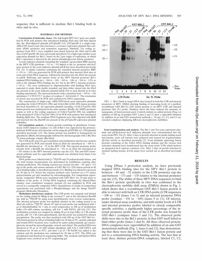
Using DNase I protection analysis, we have previouslymapped DNA binding sites for the HFV Bel-1 protein tobetween 284 and 232 relative to the LTR promoter cap siteand between 2171 and 2135 relative to the internal promotercap site (15). The ability of these HFV DNA sequences to bindthe Bel-1 protein specifically in vitro was confirmed in theelectrophoretic mobility shift assay (EMSA) shown in Fig. 1,which shows that a recombinant GST–Bel-1 fusion protein isable to interact with both an LTR DNA probe (LTR sequences2108 to 235) (lanes 3 to 5) and an internal promoter DNAprobe (residues 2191 to 2105) (lanes 9 to 11). Of interest,under identical assay conditions, and with similar levels of LTRand internal promoter probes labeled to closely comparablespecific activities, a significantly higher percentage of the in-ternal promoter probe than of the LTR probe was bound byGST–Bel-1 (compare lanes 5 and 11). The observed probeshifts were due to the Bel-1 protein, in that GST itself failed tobind either probe (lanes 2 and 8). All three observed protein-DNA complexes were supershifted by addition of an anti-GSTmonoclonal antibody (Fig. 1, lanes 6 and 12), thus demonstrat-ing that these were due to the GST–Bel-1 fusion protein andnot to a contaminating DNA binding activity. The fact that atleast three distinct protein-DNA complexes, labeled C1, C2,
FIG. 1. Bel-1 binds to target DNA sites located in both the LTR and internalpromoters of HFV. EMSA showing binding of increasing levels of a purified,recombinant GST–Bel-1(1–228) fusion protein to an HFV LTR and internalpromoter (Int. Pr.) probe. Numbers above the lanes indicate the amounts, innanograms, of fusion protein used. Controls include no added protein (Neg.) oraddition of 200 ng of purified GST. Lanes 6 and 12 show a supershift obtainedby addition of an anti-GST monoclonal antibody (;50 ng). C1, C2, and C3 areretarded complexes displaying different electrophoretic mobilities.
VOL. 72, 1998 ANALYSIS OF HFV Bel-1 DNA BINDING 505
on February 11, 2018 by guest
http://jvi.asm.org/
Dow
nloaded from

and C3, were observed in this EMSA is of interest, given theprevious report that Bel-1 is able to form multimers in vivo (6).However, it should be noted that the linked GST protein canalso form dimers.
We next sought to confirm that the observed Bel-1–DNAinteractions are specific. As shown in Fig. 2, the ability ofGST–Bel-1 to bind to either an LTR DNA probe (Fig. 2A) oran internal promoter probe (Fig. 2B) was efficiently competedby an excess of either the unlabeled internal promoter probe(lanes 3 and 4) or the LTR probe (lanes 5 and 6) but wasessentially unaffected by the same level of a nonspecific DNAcompetitor (lanes 7 and 8). Therefore, this DNA binding eventis clearly specific and the interactions of Bel-1 with the DNAtarget sequences are likely to be mechanistically similar.
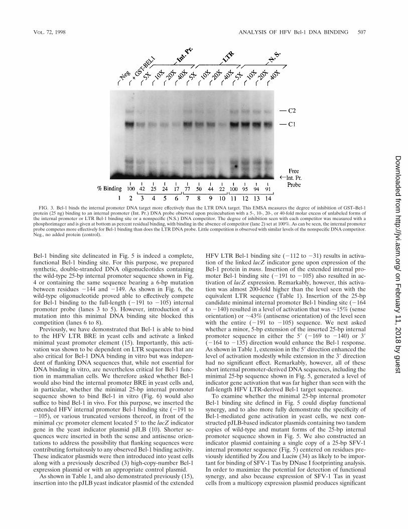
Bel-1 binds the internal promoter more effectively than theLTR promoter. As noted above, the GST–Bel-1 fusion proteinused in these assays bound to a higher percentage of theinternal promoter probe than of the LTR promoter probewhen incubated under the same assay conditions (Fig. 1). Thisobservation suggests that the internal promoter may contain ahigher-affinity binding site for Bel-1 than the one present in theLTR. To address this issue in more detail, we performed aquantitative EMSA with the internal promoter probe and sev-eral different levels of unlabeled internal promoter, LTR, ornonspecific competitor DNAs. As shown in Fig. 3, the internalpromoter-derived competitor DNA (lanes 3 to 6) proved a farmore effective competitor of the internal promoter probe–Bel-1 binding reaction than did the LTR-derived competitor(lanes 7 to 10). Therefore, it is clear that this internal promotertarget sequence is a significantly higher-affinity in vitro DNAbinding site for Bel-1 than is the tested HFV LTR targetsequence.
Modification interference analysis of Bel-1 DNA binding.We next wished to identify specific residues within the internalpromoter and LTR DNA probes required for Bel-1 binding bymodification interference analysis. For this purpose, the 2108to 235 HFV LTR probe and the 2191 to 2105 internal pro-moter probe were each uniquely end labeled and then incu-bated with dimethylsulfide, which specifically methylates G and
A residues (31). The modified probes were then used forEMSA, and the C1 and C2 complexes (Fig. 1) observed withthe internal promoter probe and the C1 complex observed withthe LTR probe were excised, purified, and chemically cleaved.The pattern of DNA modification observed in these shiftedDNA complexes was then compared to the pattern seen in thefree-DNA probes.
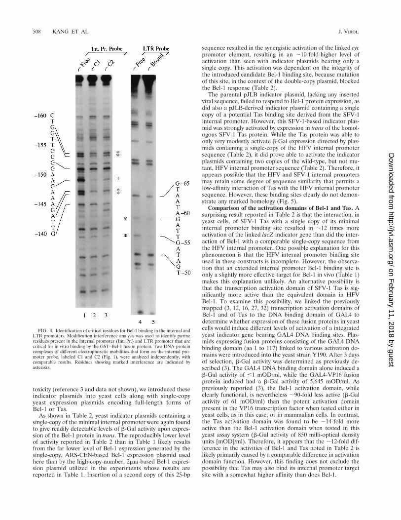
As shown in Fig. 4, this experimental approach identifiedseveral bases whose modification resulted in an inhibition ofinteraction with the GST–Bel-1 fusion protein. In the internalpromoter DNA probe, marked inhibition of binding was ob-served upon modification of residues 2158G, 2157G, 2148G,2144G, and 2143A (Fig. 4). More modest interference withbinding was noted upon modification of residues 2152G,2151G, and 2149A. No additional modification interferencewas observed in the C2 complex compared to the C1 complex(Fig. 4, compare lanes 2 and 3), thus suggesting either that thelower-mobility C2 complex results entirely from a protein-protein interaction or that any second Bel-1 DNA bindingevent is relatively nonspecific. Analysis of LTR probe bindingby Bel-1 by using modification interference showed significantinhibition upon modification of residues 259G and 255G andmodest interference upon modification of residue 265G (Fig.4, lanes 4 and 5).
The analysis presented in Fig. 4 identifies several residuescritical for Bel-1 binding and localizes these between 2158 and2143 in the internal promoter and between 265 and 255 inthe LTR promoter. Therefore, for both the internal promoterand the LTR promoter, the mapped residues are centered inthe Bel-1 binding sites previously mapped to between 2171and 2135 in the internal promoter and to between 284 and232 in the LTR promoter, using DNase I footprinting. Analignment of these two Bel-1 binding sites, shown in Fig. 5,suggests that several purine residues identified as important forBel-1 DNA binding in Fig. 4 are conserved between the high-affinity internal promoter and lower-affinity LTR Bel-1 bindingsites.
Identification of a minimal Bel-1 DNA binding site. We nextwished to determine if the minimal, ;25-bp internal promoter
FIG. 2. DNA binding by HFV Bel-1 is specific. The direct interaction of GST–Bel-1 (25 ng) with an HFV LTR (A) or internal promoter (Int. Pr.) DNA probe (B)was specifically blocked by preincubation with an 80- or 320-fold molar excess of unlabeled forms of these same two DNA probes but was not blocked by preincubationwith a similar excess of a nonspecific (N.S.) DNA competitor.
506 KANG ET AL. J. VIROL.
on February 11, 2018 by guest
http://jvi.asm.org/
Dow
nloaded from
Bel-1 binding site delineated in Fig. 5 is indeed a complete,functional Bel-1 binding site. For this purpose, we preparedsynthetic, double-stranded DNA oligonucleotides containingthe wild-type 25-bp internal promoter sequence shown in Fig.4 or containing the same sequence bearing a 6-bp mutationbetween residues 2144 and 2149. As shown in Fig. 6, thewild-type oligonucleotide proved able to effectively competefor Bel-1 binding to the full-length (2191 to 2105) internalpromoter probe (lanes 3 to 5). However, introduction of amutation into this minimal DNA binding site blocked thiscompetition (lanes 6 to 8).
Previously, we have demonstrated that Bel-1 is able to bindto the HFV LTR BRE in yeast cells and activate a linkedminimal yeast promoter element (15). Importantly, this acti-vation was shown to be dependent on LTR sequences that arealso critical for Bel-1 DNA binding in vitro but was indepen-dent of flanking DNA sequences that, while not essential forDNA binding in vitro, are nevertheless critical for Bel-1 func-tion in mammalian cells. We therefore asked whether Bel-1would also bind the internal promoter BRE in yeast cells and,in particular, whether the minimal 25-bp internal promotersequence shown to bind Bel-1 in vitro (Fig. 6) would alsosuffice to bind Bel-1 in vivo. For this purpose, we inserted theextended HFV internal promoter Bel-1 binding site (2191 to2105), or various truncated versions thereof, in front of theminimal cyc promoter element located 59 to the lacZ indicatorgene in the yeast indicator plasmid pJLB (10). Shorter se-quences were inserted in both the sense and antisense orien-tations to address the possibility that flanking sequences werecontributing fortuitously to any observed Bel-1 binding activity.These indicator plasmids were then introduced into yeast cellsalong with a previously described (3) high-copy-number Bel-1expression plasmid or with an appropriate control plasmid.
As shown in Table 1, and also demonstrated previously (15),insertion into the pJLB yeast indicator plasmid of the extended
HFV LTR Bel-1 binding site (2112 to 231) results in activa-tion of the linked lacZ indicator gene upon expression of theBel-1 protein in trans. Insertion of the extended internal pro-moter Bel-1 binding site (2191 to 2105) also resulted in ac-tivation of lacZ expression. Remarkably, however, this activa-tion was almost 200-fold higher than the level seen with theequivalent LTR sequence (Table 1). Insertion of the 25-bpcandidate minimal internal promoter Bel-1 binding site (2164to 2140) resulted in a level of activation that was ;15% (senseorientation) or ;43% (antisense orientation) of the level seenwith the entire (2191 to 2105) sequence. We next askedwhether a minor, 5-bp extension of the inserted 25-bp internalpromoter sequence in either the 59 (2169 to 2140) or 39(2164 to 2135) direction would enhance the Bel-1 response.As shown in Table 1, extension in the 59 direction enhanced thelevel of activation modestly while extension in the 39 directionhad no significant effect. Remarkably, however, all of theseshort internal promoter-derived DNA sequences, including theminimal 25-bp sequence shown in Fig. 5, generated a level ofindicator gene activation that was far higher than seen with thefull-length HFV LTR-derived Bel-1 target sequence.
To examine whether the minimal 25-bp internal promoterBel-1 binding site defined in Fig. 5 could display functionalsynergy, and to also more fully demonstrate the specificity ofBel-1-mediated gene activation in yeast cells, we next con-structed pJLB-based indicator plasmids containing two tandemcopies of wild-type and mutant forms of the 25-bp internalpromoter sequence shown in Fig. 5. We also constructed anindicator plasmid containing a single copy of a 25-bp SFV-1internal promoter sequence (Fig. 5) centered on residues pre-viously identified by Zou and Luciw (34) as likely to be impor-tant for binding of SFV-1 Tas by DNase I footprinting analysis.In order to maximize the potential for detection of functionalsynergy, and also because expression of SFV-1 Tas in yeastcells from a multicopy expression plasmid produces significant
FIG. 3. Bel-1 binds the internal promoter DNA target more effectively than the LTR DNA target. This EMSA measures the degree of inhibition of GST–Bel-1protein (25 ng) binding to an internal promoter (Int. Pr.) DNA probe observed upon preincubation with a 5-, 10-, 20-, or 40-fold molar excess of unlabeled forms ofthe internal promoter or LTR Bel-1 binding site or a nonspecific (N.S.) DNA competitor. The degree of inhibition seen with each competitor was measured with aphosphorimager and is given at bottom as percent residual binding, with binding in the absence of competitor (lane 2) set at 100%. As can be seen, the internal promoterprobe competes more effectively for Bel-1 binding than does the LTR DNA probe. Little competition is observed with similar levels of the nonspecific DNA competitor.Neg., no added protein (control).
VOL. 72, 1998 ANALYSIS OF HFV Bel-1 DNA BINDING 507
on February 11, 2018 by guest
http://jvi.asm.org/
Dow
nloaded from
toxicity (reference 3 and data not shown), we introduced theseindicator plasmids into yeast cells along with single-copyyeast expression plasmids encoding full-length forms ofBel-1 or Tas.
As shown in Table 2, yeast indicator plasmids containing asingle-copy of the minimal internal promoter were again foundto give readily detectable levels of b-Gal activity upon expres-sion of the Bel-1 protein in trans. The reproducibly lower levelof activity reported in Table 2 than in Table 1 likely resultsfrom the far lower level of Bel-1 expression generated by thesingle-copy, ARS-CEN-based Bel-1 expression plasmid usedhere than by the high-copy-number, 2mm-based Bel-1 expres-sion plasmid utilized in the experiments whose results arereported in Table 1. Insertion of a second copy of this 25-bp
sequence resulted in the synergistic activation of the linked cycpromoter element, resulting in an ;10-fold-higher level ofactivation than seen with indicator plasmids bearing only asingle copy. This activation was dependent on the integrity ofthe introduced candidate Bel-1 binding site, because mutationof this site, in the context of the double-copy plasmid, blockedthe Bel-1 response (Table 2).
The parental pJLB indicator plasmid, lacking any insertedviral sequence, failed to respond to Bel-1 protein expression, asdid also a pJLB-derived indicator plasmid containing a singlecopy of a potential Tas binding site derived from the SFV-1internal promoter. However, this SFV-1-based indicator plas-mid was strongly activated by expression in trans of the homol-ogous SFV-1 Tas protein. While the Tas protein was able toonly very modestly activate b-Gal expression directed by plas-mids containing a single-copy of the HFV internal promotersequence (Table 2), it did prove able to activate the indicatorplasmids containing two copies of the wild-type, but not mu-tant, HFV internal promoter sequence (Table 2). Therefore, itappears possible that the HFV and SFV-1 internal promotersmay retain some degree of sequence similarity that permits alow-affinity interaction of Tas with the HFV internal promotersequence. However, these binding sites clearly do not demon-strate any marked homology (Fig. 5).
Comparison of the activation domains of Bel-1 and Tas. Asurprising result reported in Table 2 is that the interaction, inyeast cells, of SFV-1 Tas with a single copy of its minimalinternal promoter binding site resulted in ;12 times moreactivation of the linked lacZ indicator gene than did the inter-action of Bel-1 with a comparable single-copy sequence fromthe HFV internal promoter. One possible explanation for thisphenomenon is that the HFV internal promoter binding siteused in these constructs is incomplete. However, the observa-tion that an extended internal promoter Bel-1 binding site isonly a slightly more effective target for Bel-1 in vivo (Table 1)makes this explanation unlikely. An alternative possibility isthat the transcription activation domain of SFV-1 Tas is sig-nificantly more active than the equivalent domain in HFVBel-1. To examine this possibility, we linked the previouslymapped (3, 12, 16, 27, 32) transcription activation domains ofBel-1 and of Tas to the DNA binding domain of GAL4 todetermine whether expression of these fusion proteins in yeastcells would induce different levels of activation of a integratedyeast indicator gene bearing GAL4 DNA binding sites. Plas-mids expressing fusion proteins consisting of the GAL4 DNAbinding domain (aa 1 to 117) linked to various activation do-mains were introduced into the yeast strain Y190. After 3 daysof selection, b-Gal activity was determined as previously de-scribed (3). The GAL4 DNA binding domain alone induced ab-Gal activity of #1 mOD/ml, while the GAL4-VP16 fusionprotein induced had a b-Gal activity of 5,645 mOD/ml. Aspreviously reported (3), the Bel-1 activation domain, whileclearly functional, is nevertheless ;90-fold less active (b-Galactivity of 61 mOD/ml) than the potent activation domainpresent in the VP16 transcription factor when tested either inyeast cells, as in this case, or in mammalian cells. In contrast,the Tas activation domain was found to be ;14-fold moreactive than the Bel-1 activation domain when tested in thisyeast assay system (b-Gal activity of 850 milli-optical densityunits [mOD]/ml). Therefore, it appears that the ;12-fold dif-ference in the activities of Bel-1 and Tas noted in Table 2 islikely primarily caused by a comparable difference in activationdomain function. However, this finding does not exclude thepossibility that Tas may also bind its internal promoter targetsite with a somewhat higher affinity than does Bel-1.
FIG. 4. Identification of critical residues for Bel-1 binding in the internal andLTR promoters. Modification interference analysis was used to identify purineresidues present in the internal promoter (Int. Pr.) and LTR promoter that arecritical for in vitro binding by the GST–Bel-1 fusion protein. Two DNA-proteincomplexes of different electrophoretic mobilities that form on the internal pro-moter probe, labeled C1 and C2 (Fig. 1), were analyzed independently, withcomparable results. Residues showing marked interference are indicated byasterisks.
508 KANG ET AL. J. VIROL.
on February 11, 2018 by guest
http://jvi.asm.org/
Dow
nloaded from

DISCUSSION
The expression of genes contained by the primate foamyviruses is regulated by the interplay of two viral promoterelements, located in the LTR and at the 39 end of the env gene,with the viral Bel-1/Tas transcriptional transactivator (29).Early after infection, the internal promoter is activated, result-ing in the synthesis of mRNAs encoding the foamy virus ac-cessory proteins, including Bel-1/Tas (21, 25). Subsequently,the LTR promoter is activated, presumably as a result of Bel-1/Tas expression, and mRNAs encoding the Gag, Pol, and Envstructural proteins begin to accumulate. Therefore, althoughfoamy viruses are not known to encode posttranscriptionalregulatory proteins equivalent to the Rev and Rex proteinsfound in other primate complex retroviruses (7, 19), they nev-ertheless appear to be similar in that they display a temporalregulation of viral gene expression. However, the basis for thistemporal order has been unclear, given that the internal pro-moter does not appear to be significantly more active than theLTR promoter in either the presence or the absence of therelevant Bel-1/Tas protein (5, 22, 25).
In this article, we describe a series of experiments designedto shed further light on the interaction of HFV Bel-1 with boththe LTR and internal promoter elements. An interesting resultto emerge from this analysis is that the Bel-1 DNA binding sitepresent in the HFV internal promoter element is a significantlyhigher-affinity binding site for Bel-1 both in vitro and in vivothan is the cap-proximal Bel-1 binding site located in the HFVLTR promoter element. In particular, when analyzed byEMSA under identical assay conditions, Bel-1 was found toshift a higher percentage of an internal promoter probe than ofa similar LTR promoter probe (Fig. 1). Similarly, internalpromoter sequences also proved able to compete more effec-tively for Bel-1 DNA binding than did LTR promoter se-quences when analyzed in a quantitative EMSA (Fig. 3). Fi-nally, when assayed for in vivo DNA binding in yeast cells by apreviously described assay, Bel-1 was able to activate a linkedlacZ indicator gene ;200-fold more effectively when targetedto the internal promoter Bel-1 DNA binding site than whentargeted to an equivalent LTR-derived Bel-1 binding site (Ta-ble 1). Taken together, these data suggest that activation of thefoamy virus internal promoter may precede activation of theLTR promoter during the foamy virus life cycle because theinternal promoter acts as a more effective Bel-1 protein bind-ing site when Bel-1 levels are limiting, as they are predicted tobe early in infection. We caution, however, that this analysisonly examined binding to the cap-proximal BRE present in theHFV LTR and did not address the affinity of Bel-1 for otherproposed LTR BRE elements (8, 20, 33). Although the cap-proximal BRE is fully sufficient to direct essentially wild-typelevels of LTR-driven transcription in the presence of the Bel-1protein in mammalian cells (18), these more 59 putative BREs
could potentially play an important role in modulating the levelof response of the LTR promoter to Bel-1, particularly giventhe observation (Table 2) that Bel-1 binding sites can displayfunctional synergy.
Using modification interference and EMSA, we were able toidentify several purine residues within both the LTR and in-ternal promoters that are critical for Bel-1 binding in vitro (Fig.4). We were then able to use this information to align these twosites (Fig. 5) and to propose a potential minimal, 25-bp DNAbinding site for Bel-1. This minimal site was then shown to infact function as an effective Bel-1 DNA binding site both invitro (Fig. 6) and in vivo (Tables 1 and 2). The identification ofthis minimal Bel-1 DNA binding site will permit the futuremutational definition of individual bases that form a functionalbinding site and should also allow the identification of residuesthat attenuate Bel-1 binding to the LTR promoter comparedto the internal promoter. It will clearly be of interest to testwhether an enhancement in the affinity of the HFV LTR bind-ing site for Bel-1 results in a disruption of the normal temporalorder of HFV gene expression.
As part of this analysis, we also tested whether a 25-bpsequence derived from the SFV-1 internal promoter, first iden-tified as important for Tas binding by DNase I footprinting(34), was sufficient to function as an effective Tas binding site
FIG. 5. Alignment of possible Bel-1 and Tas minimal binding sites. Candidate minimal binding sites for HFV Bel-1 located in the LTR and internal promoter (Int.Pr.) elements are indicated and aligned. Purine residues that gave rise to detectable inhibition of Bel-1 binding after methylation are indicated. A candidate minimalSFV-1 Tas DNA binding site, based on the DNase I footprinting studies of Zou and Luciw (34), is also given, although the alignment used is largely arbitrary and doesnot show significant conservation of purine residues shown to be critical for Bel-1 binding. DNA sequences used for subsequent analyses are in capital letters. Boxesdefine residues whose mutation blocks Bel-1 binding in vitro and function in vivo (15).
FIG. 6. A 25-bp internal promoter sequence competes for specific Bel-1DNA binding. A double-stranded, synthetic oligonucleotide consisting of the25-bp internal promoter (Int. Pr.) sequence (2164 to 2140) shown in Fig. 5specifically competed Bel-1 binding to the full-length (2191 to 2105) internalpromoter probe (lanes 3 to 5). In contrast, a similar internal promoter-derivedoligonucleotide containing mutations at residues 2149 to 2144 (Fig. 5) failed tocompete (lanes 6 to 8). This competition experiment used 4-, 20-, and 80-foldmolar excesses of each competitor oligonucleotide. WT, wild type; oligo, oligo-nucleotide.
VOL. 72, 1998 ANALYSIS OF HFV Bel-1 DNA BINDING 509
on February 11, 2018 by guest
http://jvi.asm.org/
Dow
nloaded from
in vivo. As shown in Table 2, this short sequence indeed provedfully sufficient to bind Tas efficiently. This observation allowedus to determine whether the inability of Tas to function effec-tively via HFV promoter elements in mammalian cells was dueto an inability of Tas to bind these HFV DNA sequences or,instead, reflected the absence of a critical Tas cofactor bindingsite. As shown in Table 2, Tas was able to interact only poorlywith the minimal internal promoter Bel-1 target site. It istherefore apparent that Tas and Bel-1 have evolved distinctDNA sequence specificities over time. The future definition ofthese differences will clearly be of interest.
A final interesting result is that the activation domain
present in SFV-1 Tas was found to be significantly more activethan the one present in Bel-1. This finding explains the earlierobservation (3) that expression of SFV-1 Tas from a multicopyplasmid in yeast cells is significantly more deleterious for yeastgrowth than is expression of HFV Bel-1, in that it has previ-ously been shown that the level of toxicity observed in yeastupon overexpression of an acidic transcription activation do-main is a function of the activity of the tested domain (2). Thisdifference also appears to explain the finding that the level ofindicator gene expression induced by the binding of Tas to itsminimal binding site in yeast cells is significantly higher thanthe level observed when using Bel-1 and its minimal DNAbinding site (Table 2). The question of whether this differencein activation domain function observed in yeast cells is relevantto the effectiveness of transcriptional activation by Tas andBel-1 in mammalian cells remains to be explored.
ACKNOWLEDGMENTS
This research was supported by the Howard Hughes Medical Insti-tute.
We are grateful to Sharon Goodwin for assistance in preparation ofthe manuscript.
REFERENCES
1. Baunach, G., B. Maurer, H. Hahn, M. Kranz, and A. Rethwilm. 1993.Functional analysis of human foamy virus accessory reading frames. J. Virol.67:5411–5418.
2. Berger, S. L., B. Pina, N. Silverman, G. A. Marcus, J. Agapite, J. L. Regier,S. J. Triezenberg, and L. Guarente. 1992. Genetic isolation of ADA2: apotent transcriptional adaptor required for function of certain acidic activa-tion domains. Cell 70:251–265.
3. Blair, W. S., H. Bogerd, and B. R. Cullen. 1994. Genetic analysis indicatesthat the human foamy virus Bel-1 protein contains a transcription activationdomain of the acidic class. J. Virol. 68:3803–3808.
4. Campbell, M., C. Eng, and P. A. Luciw. 1996. The simian foamy virus type1 transcriptional transactivator (Tas) binds and activates an enhancer ele-ment in the gag gene. J. Virol. 70:6847–6855.
5. Campbell, M., L. Renshaw-Gegg, R. Renne, and P. A. Luciw. 1994. Charac-terization of the internal promoter of simian foamy viruses. J. Virol. 68:4811–4820.
6. Chang, J., K. J. Lee, K. L. Jang, E. K. Lee, G. H. Baek, and Y. C. Sung. 1995.Human foamy virus Bel1 transactivator contains a bipartite nuclear local-ization determinant which is sensitive to protein context and triple multim-erization domains. J. Virol. 69:801–808.
7. Cullen, B. R. 1992. Mechanism of action of regulatory proteins encoded bycomplex retroviruses. Microbiol. Rev. 56:375–394.
8. Erlwein, O., and A. Rethwilm. 1993. Bel-1 transactivator responsive se-quences in the long terminal repeat of human foamy virus. Virology 196:256–268.
9. Ernst, R. K., M. Bray, D. Rekosh, and M. Hammarskjold. 1997. A structuralretroviral RNA element that mediates nucleocytoplasmic export of intron-containing RNA. Mol. Cell. Biol. 17:135–144.
10. Finley, R. L., Jr., S. Chen, J. Ma, P. Byrne, and R. W. West, Jr. 1990.Opposing regulatory functions of positive and negative elements in UASGcontrol transcription of the yeast GAL genes. Mol. Cell. Biol. 10:5663–5670.
11. Flugel, R. M. 1991. Spumaviruses: a group of complex retroviruses. J. Ac-quired Immune Defic. Syndr. 4:739–750.
12. Garrett, E. D., F. He, H. P. Bogerd, and B. R. Cullen. 1993. Transcriptionaltrans activators of human and simian foamy viruses contain a small, highlyconserved activation domain. J. Virol. 67:6824–6827.
13. Gietz, R. D., and A. Sugino. 1988. New yeast-Escherichia coli shuttle vectorsconstructed with in vitro mutagenized yeast genes lacking six-base pair re-striction sites. Gene 74:527–534.
14. Harper, J. W., G. R. Adami, N. Wei, K. Keyomarsi, and S. J. Elledge. 1993.The p21 Cdk-interacting protein Cip1 is a potent inhibitor of G1 cyclin-dependent kinases. Cell 75:805–816.
15. He, F., W. S. Blair, J. Fukushima, and B. R. Cullen. 1996. The human foamyvirus Bel-1 transcription factor is a sequence-specific DNA binding protein.J. Virol. 70:3902–3908.
16. He, F., J. D. Sun, E. D. Garrett, and B. R. Cullen. 1993. Functional organi-zation of the Bel-1 trans activator of human foamy virus. J. Virol. 67:1896–1904.
17. Keller, A., E. D. Garrett, and B. R. Cullen. 1992. The Bel-1 protein of humanfoamy virus activates human immunodeficiency virus type 1 gene expressionvia a novel DNA target site. J. Virol. 66:3946–3949.
18. Keller, A., K. M. Partin, M. Lochelt, H. Bannert, R. M. Flugel, and B. R.
TABLE 1. The HFV internal promoter contains a high-affinityin vivo Bel-1 DNA binding site
Promoter b-Gal activitya
pJLB........................................................................................ #5 (;0)LTR (2112 to 231).............................................................. 450 (0.6)Int. Pr. (2191 to 2105)........................................................ 74,960 (100)
Int. Pr. (2164 to 2140)........................................................ 10,930 (15)Int. Pr. (2140 to 2164)........................................................ 32,530 (43)
Int. Pr. (2169 to 2140)........................................................ 27,420 (37)Int. Pr. (2140 to 2169)........................................................ 41,090 (55)
Int. Pr. (2164 to 2135)........................................................ 28,630 (38)Int. Pr. (2135 to 2164)........................................................ 26,472 (35)
a Sequences derived from the HFV LTR or internal promoter (Int. Pr.) wereinserted 59 of the minimal cyc promoter element present in the lacZ-based yeastindicator plasmid pJLB. Sense or antisense orientation is indicated by the pro-moter coordinates; e.g., the sequence from 2169 to 2140 is sense, while thatfrom 2140 to 2169 is antisense. These reporter plasmids were introduced intoyeast cells together with a multicopy Bel-1 expression plasmid (3) or a similarcontrol plasmid. Induced b-Gal activities were measured after selection forcotransformants and are given in mOD/milliliter. No detectable activity wasobserved with any indicator plasmid in the absence of Bel-1. Activity levels arealso given (in parentheses) relative to the extended internal promoter Bel-1binding site, which is arbitrarily set at 100%. These data are the averages of fourindependent experiments.
TABLE 2. Binding of Bel-1 and Tas to minimal DNAtarget sites in yeast cellsa
Promoterb-Gal activity with plasmid
Control 1Bel-1 1Tas
pJLB #5 #5 #5
HFV Int. Pr. (13 WT1) #5 86 11HFV Int. Pr. (13 WT2) #5 167 17
HFV Int. Pr. (23 WT1) #5 701 120HFV Int. Pr. (23 WT2) #5 1,262 54
HFV Int. Pr. (23 M1) 22 17 48HFV Int. Pr. (23 M2) 22 23 31
SFV-1 Int. Pr. (13 WT1) #5 #5 2,870SFV-1 Int. Pr. (13 WT2) #5 #5 714
a Wild-type (WT) or mutant (M) forms of the minimal 25-bp HFV or SFV-1internal promoter (Int. Pr.) sequence shown in Fig. 5 were inserted in one (13)or two (23) copies into the lacZ-based yeast indicator plasmid pJLB. Orientationis indicated by a plus (sense) or minus (antisense) sign. These reporter plasmidswere introduced into yeast cells together with single-copy plasmids expressingBel-1 or Tas or a similar control plasmid. Induced b-Gal activity was measuredafter selection for cotransformants and is given in mOD/milliliter. These data arerepresentative of three independent experiments.
510 KANG ET AL. J. VIROL.
on February 11, 2018 by guest
http://jvi.asm.org/
Dow
nloaded from

Cullen. 1991. Characterization of the transcriptional trans activator of hu-man foamy retrovirus. J. Virol. 65:2589–2594.
19. Lee, A. H., H. Y. Lee, and Y. C. Sung. 1994. The gene expression of humanfoamy virus does not require a post-transcriptional transactivator. Virology204:409–413.
20. Lee, K. J., A. H. Lee, and Y. C. Sung. 1993. Multiple positive and negativecis-acting elements that mediate transactivation by bel1 in the long terminalrepeat of human foamy virus. J. Virol. 67:2317–2326.
21. Lochelt, M., R. M. Flugel, and M. Aboud. 1994. The human foamy virusinternal promoter directs the expression of the functional Bel 1 transactiva-tor and Bet protein early after infection. J. Virol. 68:638–645.
22. Lochelt, M., W. Muranyi, and R. M. Flugel. 1993. Human foamy virusgenome possesses an internal, Bel-1-dependent and functional promoter.Proc. Natl. Acad. Sci. USA 90:7317–7321.
23. Lochelt, M., S. F. Yu, M. L. Linial, and R. M. Flugel. 1995. The human foamyvirus internal promoter is required for efficient gene expression and infec-tivity. Virology 206:601–610.
24. Lochelt, M., H. Zentgraf, and R. M. Flugel. 1991. Construction of an infec-tious DNA clone of the full-length human spumaretrovirus genome andmutagenesis of the bel 1 gene. Virology 184:43–54.
25. Mergia, A. 1994. Simian foamy virus type 1 contains a second promoterlocated at the 39 end of the env gene. Virology 199:219–222.
26. Mergia, A., and P. A. Luciw. 1991. Replication and regulation of primatefoamy viruses. Virology 184:475–482.
27. Mergia, A., L. W. Renshaw-Gegg, M. W. Stout, R. Renne, and O. Herchen-roeder. 1993. Functional domains of the simian foamy virus type 1 transcrip-tional transactivator (Taf). J. Virol. 67:4598–4604.
28. Mergia, A., K. E. S. Shaw, E. Pratt-Lowe, P. A. Barry, and P. A. Luciw. 1991.Identification of the simian foamy virus transcriptional transactivator gene(taf). J. Virol. 65:2903–2909.
29. Rethwilm, A. 1995. Regulation of foamy virus gene expression. Curr. Top.Microbiol. Immunol. 193:1–24.
30. Rethwilm, A., O. Erlwein, G. Baunach, B. Maurer, and V. Ter Meulen. 1991.The transcriptional transactivator of human foamy virus maps to the bel 1genomic region. Proc. Natl. Acad. Sci. USA 88:941–945.
31. Shaw, P. E., and A. F. Stewart. 1994. Identification of protein-DNA contactswith dimethyl sulfate. Methods Mol. Biol. 30:79–87.
32. Venkatesh, L. K., and G. Chinnadurai. 1993. The carboxy-terminal tran-scription enhancement region of the human spumaretrovirus transactivatorcontains discrete determinants of the activator function. J. Virol. 67:3868–3876.
33. Venkatesh, L. K., P. A. Theodorakis, and G. Chinnadurai. 1991. Distinctcis-acting regions in U3 regulate trans-activation of the human spumaretro-virus long terminal repeat by the viral bel1 gene product. Nucleic Acids Res.19:3661–3666.
34. Zou, J. X., and P. A. Luciw. 1996. The transcriptional transactivator of simianfoamy virus 1 binds to a DNA target element in the viral internal promoter.Proc. Natl. Acad. Sci. USA 93:326–330.
VOL. 72, 1998 ANALYSIS OF HFV Bel-1 DNA BINDING 511
on February 11, 2018 by guest
http://jvi.asm.org/
Dow
nloaded from