HONORS ANATOMY & PHYSIOLOGY Cytoplasm. material between plasma membrane& nucleus site of most...
62
HONORS ANATOMY & PHYSIOLOGY Cytoplasm
-
Upload
beatrice-greer -
Category
Documents
-
view
217 -
download
3
Transcript of HONORS ANATOMY & PHYSIOLOGY Cytoplasm. material between plasma membrane& nucleus site of most...

HONORS ANATOMY & PHYSIOLOGY
Cytoplasm

Cytoplasm
material between plasma membrane& nucleus
site of most cellular activity3 elements1. Cytosol2. Organelles3. Inclusions

Cytosol
viscous, semitransparent fluid in which other elements in cytoplasm are suspended
complex mixture of:waterproteinssaltsSugarsother solutes

Organelles specialized cellular compartments or structures
each has specific function

Organelles
non-membranous:cytoskeletonribosome

Endomembrane System
membranes allow inside to differ from cytoplasm
membranous:mitochondria, Ers, Golgi, lysosomes,
peroxisomesintermembrane system: connections
between ERs, nucleus

structure Function
dbl membraneinner membrane
has enzymes used in e- transport chain
matrix w/in ↑ enzymes for citric acid cycle
aerobic cellular respiration ATP
Mitochondria

Mitochondria

Structure Function
proteins + rRNA2 subunits
large small
translation part of protein synthesis
Ribosomes

Ribosomes

Endoplasmic Reticulum
extensive system of interconnected tubes & parallel membranes enclosing fluid-filled cavities called cisterns
continuous with outer membrane of nuclear envelope
1. Smooth ER2. Rough ER

Structure Function
continuous with RER
enzymes (integral proteins)
metabolize lipidssynthesize cholesterol
& lipoproteinssynthesize steroid
hormonesabsorb, synthesize, &
transport fatsdetoxify drugs, cancer-
causing chemicalsglycogen free glucose
Smooth ER

SER

Structure Function
ribosomes stud surface of membrane
proteins made on these ribosomes thread way into cisterns
proteins then enclosed in vesicles Golgi
synthesize all proteins exported from cell
makes membranes
RER

RER

Structure Function
stacked & flattened membranous sacs
associated with numerous vesicles
modifies,, concentrates, & packages proteins &lipids destined for export
packages enzymes into vesicles lysosomes, peroxisomes
Golgi Apparatus


Structure Function
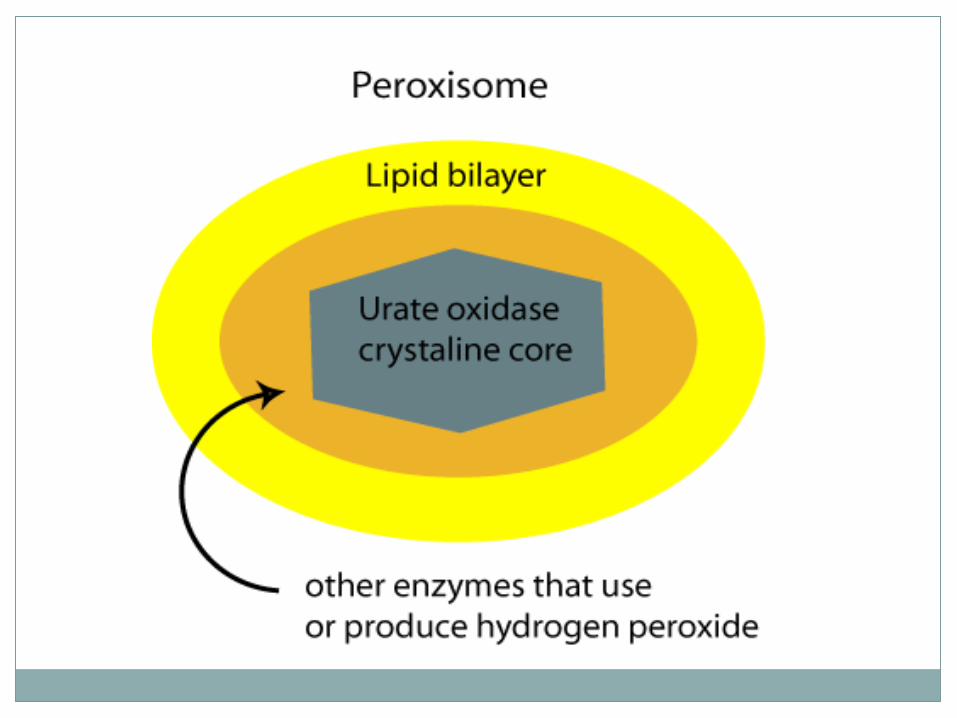
“peroxide bodies”membranous sacs
containing enzymes ex: oxidases,
catalasesmade in ER
detoxify harmful substances (many in liver& kidney)
breakdown & synthesize fatty acids
Peroxisomes

Structure Function
spherical, membranous organelles containing activated digestive enzymes in acid pH
many found in cells that phagocytize
plasma membrane has H+ pumps
enzymes digest all molecules
H+ pumps maintains normal pH in cytosol
degrade worn out organelles
glycogen glucosebreakdown
nonuseful tissues
Lysosome

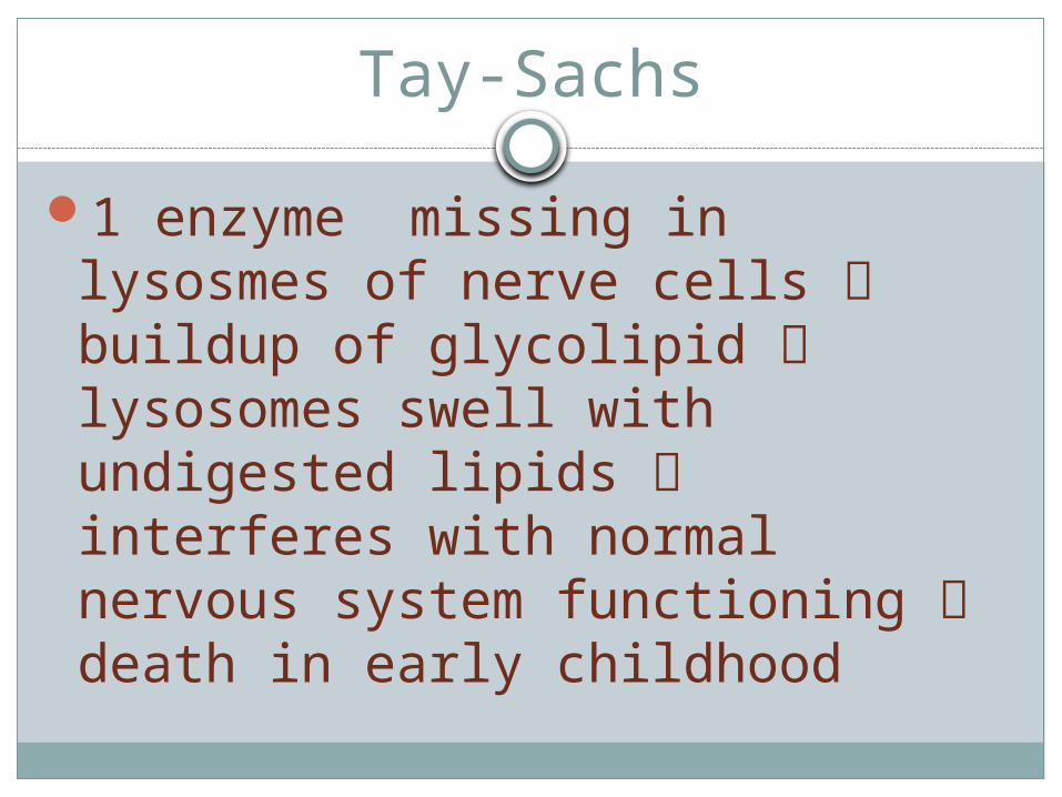
Tay-Sachs
1 enzyme missing in lysosmes of nerve cells buildup of glycolipid lysosomes swell with undigested lipids interferes with normal nervous system functioning death in early childhood

Cytoskeleton
“cell skeleton”network of protein rods thru cytosolsupports cell structureallows cell movementTypes:1. Microfilaments2. Intermediate Filaments3. Microtubules
Structure Function
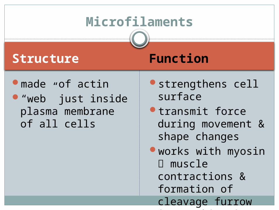
made of actin“web” just inside
plasma membrane of all cells
strengthens cell surface
transmit force during movement & shape changes
works with myosin muscle contractions & formation of cleavage furrow in cytokinesis
Microfilaments


Structure Function
tough, insoluble protein fibers
most stable & permanent of the 3
attach to desmosomes
resist pulling forces exerted on cell
Intermediate Filaments


Structure Function
hollow tubes made of protein tubulin
most radiate from centrosome
very dynamic: self-assemble/dissemble
have associated proteins that help organelles move in cytoplasm
determine overall shape of cell
distribute organelles
https://www.youtube.com/watch?v=4TGDPotbJV4
Microtubules


Structure Function
made of paired centrioles: barrel-shaped organelles oriented @ rt angles to each other
generates & organizes microtubules
organizes mitotic spindle
Centrosomes


Structure Function
whip-like, motile cellular extensions
found in large #s on surface of cell
9 paired microtubules runs length of cilium
as beat in unison mucus/particles swept over surface of cell
Cilia

http://www.zoology.ubc.ca/courses/bio332/Images/Cilia/Cil3/cilium.gif

Structure Function
cell projections formed by centrioles
longer than ciliaonly human cell
with flagella is the sperm
9 + 2 pattern of microtubules
propels entire cell
Flagella
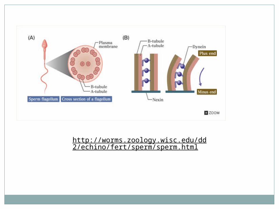
http://worms.zoology.wisc.edu/dd2/echino/fert/sperm/sperm.html

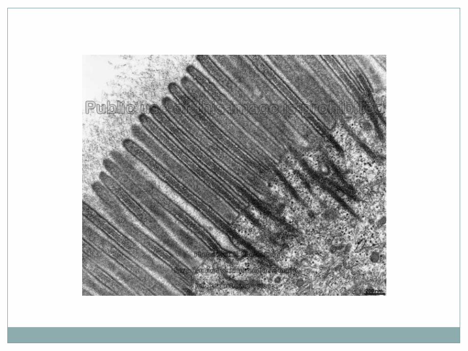
Structure Function
minute projections of plasma membrane on cell surface
found on surface of absorptive cells
have core of microfilaments (actin)
↑ surface area
Microvilli
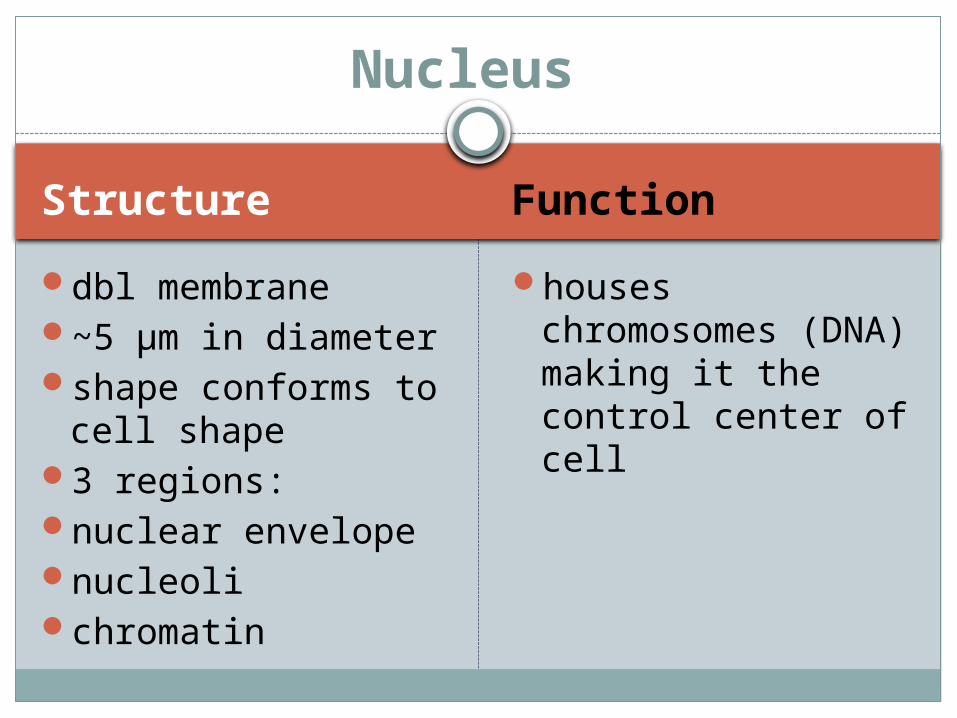
Structure Function
dbl membrane~5 µm in diametershape conforms to
cell shape3 regions:nuclear envelopenucleolichromatin
houses chromosomes (DNA) making it the control center of cell
Nucleus


Structure Function
selectively permeable dbl membrane with nuclear pores
outer membrane continuous with RER
inner membrane lined with rod-shaped proteins that give shape to nucleus
pores: complex of proteins
gives shape to nucleus
pores allow transport route for substance in/out of nucleus
encloses nucleoplasm (jelly-like similar to cytoplasm)
Nuclear Envelope


Structure Function
dark staining spherical bodies w/in nucleus
assemble ribosomal subunits combine rRNA with proteins
Nucleolus


Structure Function bumpy threads
weaving thru nucleoplasm
30% DNA60% histones
(proteins which package and regulate DNA
10% RNA
genomecodes for
proteins
Chromatin


Chromosomes
Structure
DNATelomeres : DNA at
either end of chromosome
Function
Codes for proteinsprotects
chromosome coding portion acting like “bumpers”
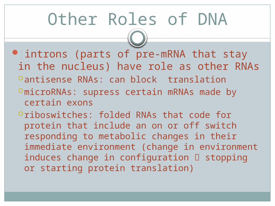
Other Roles of DNA
Iintrons (parts of pre-mRNA that stay in the nucleus) have role as other RNAsantisense RNAs: can block translationmicroRNAs: supress certain mRNAs made by
certain exonsriboswitches: folded RNAs that code for protein
that include an on or off switch responding to metabolic changes in their immediate environment (change in environment induces change in configuration stopping or starting protein translation)
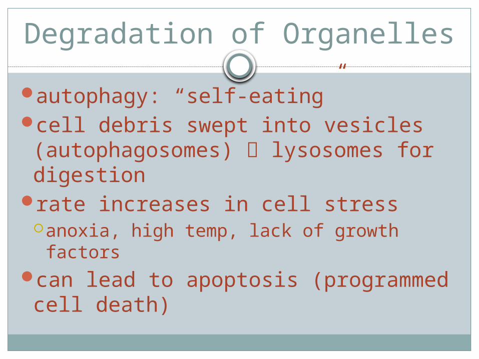
Degradation of Organelles
autophagy: “self-eating”cell debris swept into vesicles (autophagosomes) lysosomes for digestion
rate increases in cell stressanoxia, high temp, lack of growth factors
can lead to apoptosis (programmed cell death)

Ubiquitins
proteins that mark other proteins no longer being used by cell for destruction
once marked hydrolyzed by proteosomes (giant waste disposal complexes) recycle a.a. and release ubiquitins

Extracellular Material (ECM)
any substance contributing to body mass found outside cells
1. Body Fluids2. Cellular Secretions

Body Fluids
1. interstitial fluid2. plasma3. cerebrospinal fluid

Cellular Secretions
gastric juicesbilemucussweatserous fluid

Extracellular Matrix
most abundant ECM = “cell glue”secreted by cells
jellylike substanceproteins + polysaccharides

Apoptosis
programmed cell deathcommon in developing embryo:
especially in nervous systemcarves out digits in developing hands,
feet


Hyperplasia
accelerated division of cells
Atrophy
decrease in size of an organ or body tissueloss of normal stimulation
muscles that lose their nerve supply atrophy & waste away

Cell Aging
still a mysterywear-and tear theorymitochondrial theoryprogressive disorders in immune system
genetic theory: telomere clock