Hisopathological study of skin tumours
5
Indian Journal of Pathology and Oncology 2019;6(4):543–547 Content available at: iponlinejournal.com Indian Journal of Pathology and Oncology Journal homepage: www.innovativepublication.com Original Research Article Hisopathological study of skin tumours Priyanka Pappala 1 , Sudhakar Raksha*, G Vasundara 1 , P Anusha 1 , K V Murali Mohan 1 1 Dept. of Pathology, GIMSR, Visakhapatnam, Andhra Pradesh, India ARTICLE INFO Article history: Received 03-04-2019 Accepted 24-07-2019 Available online 22/11/2019 Keywords: Skin Tumours Histopathology ABSTRACT Introduction: Skin is the largest organ in the body. It is composed of two separate and functionally independent layers comprising variety of cells which gives rise to different tumour types. Aim: The aim of this study is to analyze the pattern of distribution of various histopathological skin tumours in the Department of Pathology at GITAM institute of medical sciences and research, Visakhapatnam. Materials and Methods: The present study is a prospective study of skin tumours from January 2017 to December 2018. A total of 46 cases of skin tumours were included. The slides were stained with Haematoxylin and Eosin. Results: Out of 46 skin tumours, 43 tumours were benign and 3 malignant. The incidence of keratinocytic tumours was highest 28 cases (60.86%) followed by Melanocytic 11 cases (23.91%) and adnexal 7 cases(15.21%). Conclusion: Skin tumours were relatively more common in females than males with wide range of age distribution. Keratinocytic tumours were more common when compared to other skin tumours. Squamous papilloma is the most common benign tumour. © 2019 Published by Innovative Publication. This is an open access article under the CC BY-NC-ND license (https://creativecommons.org/licenses/by/4.0/) 1. Introduction Skin is the largest organ in the body performing multiple functions. More than being a mere protective covering it is a highly sophisticated sensory organ, has endocrine role in synthesising vitamin D. 1 Though ubiquitous and simple, it is a heterogeneous organ. 2 Histologically it is composed of several cell types that function interdependently and cooperatively. 1 The epidermal layer is composed of 90% of keratinocytes and remaining 10% composed by mel anocytes, langerhans cells and merkel cells. Epidermal appendages extend from epidermis to dermis comprising of specialized cells like follicular epithelial cells, sebaceous cells, cells of eccrine and apocrine glands. Different cell types give rise to different types of tumours. 3 Skin tumours are generally divided into surface epidermal tumours and tumours of epidermal appendages. 2 * Corresponding author. E-mail address: [email protected] (S. Raksha). Epidermis has the capacity to develop into wide variety of lesions though commonly show proliferation of ker- atinocytes but produce clinically distinct lesions. Adnexal tumours may be solitary or multiple. 4,5 These tumours clinically present as papules and nodules. So diagnosis may not be conclusive clinically alone, histopathological confirmation becomes a prime requisite for definite diagnosis. At times it is difficult to interpret because of variety and complexity of histologic nature, complex nomenclature, multiple classifications and conflict of opinion regarding histogenesis. 6 Thus clinicopathological correlation is required. Early diagnosis and treatment are necessary for a better cure. This study was done mainly to evaluate various papular and nodular lesions of skin tumours by histopathological findings. https://doi.org/10.18231/j.ijpo.2019.106 2394-6784/© 2019 Innovative Publication, All rights reserved. 543
Transcript of Hisopathological study of skin tumours

Indian Journal of Pathology and Oncology 2019;6(4):543–547
Content available at: iponlinejournal.com
Indian Journal of Pathology and Oncology
Journal homepage: www.innovativepublication.com
Original Research Article
Hisopathological study of skin tumours
Priyanka Pappala1, Sudhakar Raksha*, G Vasundara1, P Anusha1, K V Murali Mohan1
1Dept. of Pathology, GIMSR, Visakhapatnam, Andhra Pradesh, India
A R T I C L E I N F O
Article history:Received 03-04-2019Accepted 24-07-2019Available online 22/11/2019
Keywords:SkinTumoursHistopathology
A B S T R A C T
Introduction: Skin is the largest organ in the body. It is composed of two separate and functionallyindependent layers comprising variety of cells which gives rise to different tumour types.Aim: The aim of this study is to analyze the pattern of distribution of various histopathological skin tumoursin the Department of Pathology at GITAM institute of medical sciences and research, Visakhapatnam.Materials and Methods: The present study is a prospective study of skin tumours from January 2017to December 2018. A total of 46 cases of skin tumours were included. The slides were stained withHaematoxylin and Eosin.Results: Out of 46 skin tumours, 43 tumours were benign and 3 malignant. The incidence of keratinocytictumours was highest 28 cases (60.86%) followed by Melanocytic 11 cases (23.91%) and adnexal 7cases(15.21%).Conclusion: Skin tumours were relatively more common in females than males with wide range of agedistribution. Keratinocytic tumours were more common when compared to other skin tumours. Squamouspapilloma is the most common benign tumour.
© 2019 Published by Innovative Publication. This is an open access article under the CC BY-NC-NDlicense (https://creativecommons.org/licenses/by/4.0/)
1. Introduction
Skin is the largest organ in the body performing multiplefunctions. More than being a mere protective covering itis a highly sophisticated sensory organ, has endocrine rolein synthesising vitamin D.1 Though ubiquitous and simple,it is a heterogeneous organ.2 Histologically it is composedof several cell types that function interdependently andcooperatively.1
The epidermal layer is composed of 90% of keratinocytesand remaining 10% composed by mel anocytes, langerhanscells and merkel cells. Epidermal appendages extend fromepidermis to dermis comprising of specialized cells likefollicular epithelial cells, sebaceous cells, cells of eccrineand apocrine glands. Different cell types give rise todifferent types of tumours.3
Skin tumours are generally divided into surfaceepidermal tumours and tumours of epidermal appendages.2
* Corresponding author.E-mail address: [email protected] (S. Raksha).
Epidermis has the capacity to develop into wide varietyof lesions though commonly show proliferation of ker-atinocytes but produce clinically distinct lesions. Adnexaltumours may be solitary or multiple.4,5
These tumours clinically present as papules and nodules.So diagnosis may not be conclusive clinically alone,histopathological confirmation becomes a prime requisitefor definite diagnosis. At times it is difficult tointerpret because of variety and complexity of histologicnature, complex nomenclature, multiple classificationsand conflict of opinion regarding histogenesis.6 Thusclinicopathological correlation is required. Early diagnosisand treatment are necessary for a better cure.
This study was done mainly to evaluate various papularand nodular lesions of skin tumours by histopathologicalfindings.
https://doi.org/10.18231/j.ijpo.2019.1062394-6784/© 2019 Innovative Publication, All rights reserved. 543
544 Pappala et al. / Indian Journal of Pathology and Oncology 2019;6(4):543–547
2. Materials and Methods
The present study was conducted in the department ofpathology, GITAM Institute of Medical Sciences andresearch, Visakhapatnam. This was a prospective studydone during the period of January 2017 to December2018 i.e. 2 years. Both biopsies and resected specimenswere included. The tissues were fixed in 10% formalinand sections were taken. Then they were processed andembedded in paraffin wax. Thin sections of 3-5 micronswere made and stained with Haematoxylin and Eosin.
Tumors of epidermis along with melanocytic andadnexae were included in the present study. Mesenchymaltumors, haematological, neural and tumour like lesionsincluding cystic lesions were excluded. The tumours arisingfrom mucocutaneous junction were also excluded from ourstudy.
3. Results
A total of 46 cases of skin tumours were studied duringtwo years period. Out of 46 cases studied 18 were male(39.14%) and 28(60.86%) female patients with male tofemale ratio of 2:3 (Table 1).
Table 1: Gender incidence of skin tumours
Male Female Total18 28 4639.14% 60.86% 100%
Tumours of skin are seen in all age groups. The age of thepatients ranged from 11 to 68. However most of the tumorsare seen in the second and third decade of life (Table 2).
On histological examination, total benign tumours were43(93.47%), with only 3 malignant tumours (06.52%). Outof 46 tumours, Epidermal tumours were the most commoncomprising of 28(60.86%) cases followed by Melanocytictumours and tumours of Appendageal origin, comprising of11 (23.9%) and 7(14.2%) cases respectively (Table 3).
Tumours from keratinocytic cell of origin were mostcommon in our study. We had a total of 28 cases of tumoursof which Squamous papilloma is the most common withabout nine cases. It is followed by Achrocordan and Verrucavulgaris. Squamous papillomas were seen in all age groupsranging from 11 years to 61years of age. Most of thesepatients presented as papillary growth over back and twopatients presented over thigh region. Microscopy showedpapillomatosis with elongation of rete ridges, hyperkeratosisand acanthosis.
Acrochordans w ere the common tumours next toSquamous papillomas. It is seen in first three decadesof life and are not confined to single region but seen invarious parts of the body. Histology showed hyperplasiaof epidermis with underlying fibrovascular core and looselyarranged collagen. Verruca vulgaris is seen in second to
fifth decades of life, which were distributed over headand chest region and one over little finger. All thecases showed hyperkeratosis, acanthosis, papillomatosisand parakeratosis overlying the papillomatous projectionalong with koilocytic change in the malpighian layer.Verruca plantaris is seen in sixth and seventh decade of lifewith lesions over the foot region.
Three cases of Seborrheic keratosis were seen in lowerpart of trunk and scalp region. All are acanthotic type withsheets and columns of basaloid cells and squamoid cellswith intervening horn cysts.
We had a single case of pre malignant condition,Bowen’s disease in a 52 year old female patientwho presented as a papular lesion over the abdomen.Microscopy showed hyperproliferative epidermis withmarked dysplasia, hyperchromatic nuclei and increasedmitotic activity. No breach in to the dermis (Figure 1).
Fig. 1: Microscopy of Bowen’s disease showed hyperproliferativeepidermis with marked dysplasia, hyperchromatic nuclei andincreased mitotic activity. No breach in to dermis
Malignant tumours are very less in our study with onlytwo cases of squamous cell carcinomas which are seenin elderly age group of around 60 years. Of these twoone case was seen in a 60 year old female who presentedas a ulceroproliferative growth in the elbow, also showedsecondary deposits in the Lymphnode. Microscopicallyboth cases were well differentiated
3.1. Melanocytic tumours of skin
Intradermal nevus is the most common Melanocytic tumourin our study and is seen in the first three decades of life andthe presentation was hyperpigmented papular lesions. Oftotal eight cases, five cases were seen in the upper trunk andtwo patients presented over the face. One patient presentedas multiple lesions over face and upper back where bothbiopsies were confirmed as Intradermal nevus. Microscopyshowed nevus cells confined to dermis and arranged in nestsand cords.
We had a single case of compound nevus on face in a22 year old male, histology showed nevus cell nests bothin the epidermis and dermis. Spitz nevus was seen in a 15year male on leg which showed large nests of melanocytes

Pappala et al. / Indian Journal of Pathology and Oncology 2019;6(4):543–547 545
Table 2: Age incidence of individual tumours observed in present study
Age 10-20 21-30 31-40 41-50 51-60 61-70 TotalKeratinocyticSquamous papilloma 02 03 01 02 - 01 09Acrochordan 01 01 02 - 02 - 06Verruca vulgaris - 01 01 02 01 - 05Verruca plantaris - - - - 01 01 02Seborrheic keratosis - 01 01 - 01 - 03Bowen’s disease - - - - 01 - 01Squamous cell carcinoma - - - - 01 01 02MelanocyticIntradermal nevus 02 03 02 01 - - 08Compound nevus - 01 - - - - 01Spitz nevus 01 - - - - - 01Malignant melanoma - - - - - 01 01AdnexalEccrine poroma - - - - 01 - 01Nodular hidradenoma - - 01 - - 01 02Chondroid syringoma - - 01 - - 01 02Trichoepithelioma - 01 - - - - 01Pilomatricoma - 01 - - - - 01Total 06 12 09 05 08 06 46
Table 3: Distribution of various skin tumours
Benign Malignant TotalEpidermal 26(56.52%) 02(4.34%) 28(60.86%)Melanocytic 10(21.73%) 01(2.17%) 11(23.91%)Appendageal 07(15.21%) - 07(15.21%)Total 43(93.47%) 03(6.52%) 46(100%)
between the elongated rete ridges microscopically. A singlecase of malignant melanoma was seen in 65 year old femaleover the back. Microscopy showed epithelioid to spindleshaped cells with vesicular nucleus, prominent eosinophilicnucleoli, intra and extracellular melanin pigment (Figure 2).
Fig. 2: Malignant Melanoma (H&E ×10x) showing epithelioid to spindle shaped cells with vesicular nucleus, prominenteosinophilic nucleoli, intra and extracellular melanin pigment
3.2. Adnexal tumours of skin
In the present study there were 7 cases of adnexal tumoursand all were benign tumours. Eccrine tumours were thecommonest in the study (5 cases) followed by tumours offollicular origin (2 cases).
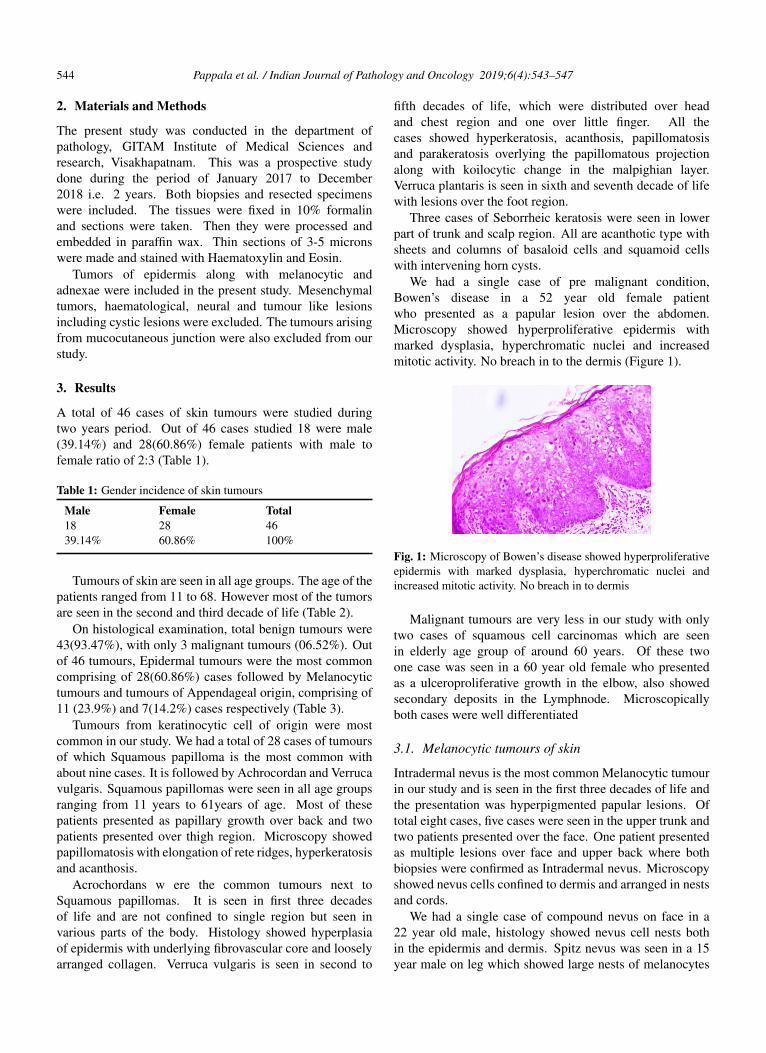
Two cases of Nodular hidradenoma and Chondroidsyringoma were seen in third and sixth decade of life witha single case of Eccrine poroma in a 60 year old female.Microscopy of Nodular hidradenoma showed lobules ofvarying size s and shapes, consisting of polyhedral cellswith round nuclei with slightly basophilic cytoplasm andround cells with clear cytoplasm (Figure 3).
Chondroid syringoma showed cells arranged in solidsheets, cords and ducts intervened by chondromyxoidstroma. Eccrine poroma showed small basaloid cellsextending in to dermis in the form of cords and columns.
We have seen a single case of Trichoepitheliomaand Pilomatricoma each in second decade of life.Trichoepithelioma histology showed multiple islands andnests of uniform small basoloid cells extending fromepidermis to dermis. Pilomatricoma showed sheets of cellswith intervening stroma. The cells at the periphery arebasoloid with ghost like cells at the centre
Following table (Table 4) shows pattern of distribution ofvarious skin tumours in the present study
4. Discussion
The tumours of the skin constitute a small but significantproportion of patients. In the present study of 46 cases of
546 Pappala et al. / Indian Journal of Pathology and Oncology 2019;6(4):543–547
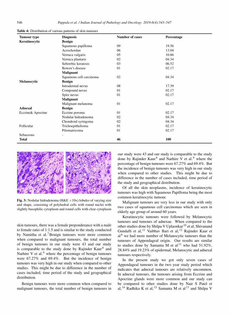
Table 4: Distribution of various patterns of skin tumours
Tumour type Diagnosis Number of cases PercentageKeratinocytic Benign
Squamous papilloma 09 19.56Acrochordan 06 13.04Verruca vulgaris 05 10.86Verruca plantaris 02 04.34Seborrhic keratosis 03 06.52Bowen’s disease 01 02.17MalignantSquamous cell carcinoma 02 04.34
Melanocytic BenignIntradermal nevus 08 17.39Compound nevus 01 02.17Spitz nevus 01 02.17MalignantMalignant melanoma 01 02.17
Adnexal BenignEccrine& Apocrine Eccrine poroma 01 02.17
Nodular hidradenoma 02 04.34Chondroid syringoma 02 04.34
Follicular Trichoepithelioma 01 02.17Pilomatricoma 01 02.17
Sebaceous -Total 46 100
Fig. 3: Nodular hidradenoma (H&E ×10x) lobules of varying sizeand shape, consisting of polyhedral cells with round nuclei withslightly basophilic cytoplasm and round cells with clear cytoplasm
skin tumours, there was a female preponderance with a maleto female ratio of 1:1.5 and is similar to the study conductedby Namitha et al.7Benign tumours were more commonwhen compared to malignant tumours, the total numberof benign tumours in our study were 43 and our studyis comparable to the study done by Rajinder Kaur8 andNarhire V et al.9 where the percentage of benign tumourswere 67.27% and 69.4%. But the incidence of benigntumours was very high in our study when compared to otherstudies. This might be due to difference in the number ofcases included, time period of the study and geographicaldistribution.
Benign tumours were more common when compared tomalignant tumours, the total number of benign tumours in
our study were 43 and our study is comparable to the studydone by Rajinder Kaur8 and Narhire V et al.9 where thepercentage of benign tumours were 67.27% and 69.4%. Butthe incidence of benign tumours was very high in our studywhen compared to other studies. This might be due todifference in the number of cases included, time period ofthe study and geographical distribution.
Of all the skin neoplasms, incidence of keratinocytictumours was high with Squamous Papilloma being the mostcommon keratinocytic tumour.
Malignant tumours are very less in our study with onlytwo cases of squamous cell carcinomas which are seen inelderly age group of around 60 years.
Keratinocytic tumours were followed by Melanocytictumours and tumours of adnexae. When compared to theother studies done by Shilpa V Uplaonkar10 et al, ShivanandGundalli et al,11 Vaibhav Bari et al,12 Rajinder Kaur etal8 we had more number of Melanocytic tumours than thetumours of Appendageal origin. Our results are similarto studies done by Samanta M et al13 who had 51.92%,28.84% and 19.23% of epidermal, Melanocytic and adnexaltumours respectively.
In the present study we got only seven cases ofAppendageal tumours in the two year study period whichindicates that adnexal tumours are relatively uncommon.In adnexal tumours, the tumours arising from Eccrine andApocrine glands were more common and our study canbe compared to other studies done by Nair S Patel etal,14 Radhika K et al,15 Samanta M et al13 and Shilpa V

Pappala et al. / Indian Journal of Pathology and Oncology 2019;6(4):543–547 547
Table 5: Comparision of frequency of various neoplasms of epidermis and epidermal appendages
Tumour type Keratinocytic Melanocytic AdnexalShilpa V et al 10 44.4% 16.6% 38.8%Rajinder Kaur et al 8 75.45% 06.37% 18.18%Shivanand Gundalli et al11 57.14% (76) 16.54% (22) 26.32% (35)Vaibhav Bari et al 12 62.4% (78) 07.2% (09) 12% (15)Samanta et al 13 51.92% (27) 28.84% (15) 19.23% (10)Present study 60.86% (28) 23.91% (11) 15.21% (07)
Table 6: Comparison of frequency of tumours of epidermal appendages
Tumours Nair S 14 Samanta M 13 Shilpa V 10 Present studyHair follicle 12(36.36%) 03(30%) 04(28.5%) 02(28.57%)Sweat gland 19(57.56%) 07(70%) 10(71.42%) 05(71.42%)Sebaceous 02(06.06%) - - -Total 33 10 14 07
Uplaonkar et al.10
Similar to the study done by Shilpa V et al10 andSamanta M et al.,13 we too did not get any tumour withsebaceous gland differentiation, may be due to its rarepresentation.
5. Conclusion
Neoplasms of the skin account for a small percentageamong all the histopathological lesions. In the presentstudy Keratinocytic tumours are the most common skintumours of which Squamous papilloma were commonest.These were followed by Melanocytic and Adnexal tumours.Benign tumours were more common when comparedto malignant tumours. Maximum tumours were foundpredominantl in females. Most of the skin tumours presentas papules and nodules, at times difficult to diagnoseclinically. Hence histopathological examination is must fordefinite diagnosis and classification of various skin tumourswhich is helpful for better treatment of the patient.
6. Source of funding
None.
7. Conflict of interest
None.
References1. Kumar V, Abbas A, Aster J. The skin. Robbins and Cotran.
Pathological basis of disease. Elsevier ; 2014,. 9th ed.2. Skin. Rosai and Ackerman’s Surgical Pathology. Elsevier ; 2011,. p.
128–136.3. Elder DE, Elenitsas R. Benign pigmented lesions and malignant
melanoma. . In Lever’s histopathology of skin. 2005;p. 689–910.Philadelphia: Lippincott Raven.
4. Weedon D. Tumors of the Epidermis.Weedons Skin Pathology.Churchill Livingstone Elsevier. 2010;p. 667–708. 3rd ed.
5. Fletcher C. Tumours of skin. Diagnostic histopathology of tumors.Saunders. 2013;(1):1680–1795. 4th ed.
6. Leboit PE, Burg G, Weedon D, Sarasin A. Pathology and genetics ofskin tumours. In World health organisation classification of tumours.IARC press. Lyon ; 2006,.
7. Chatra N, Bhat R. Clinicopathological correlation of skintumors: A dermatologists perspective. J Cancer Res Therapeutics.2016;12(3):1124–1126.
8. Kaur R, Vanitha K, Kuldeep M, Neelu G, Amritpal S. Histopatho-logical evaluation of Skin Tumours. Indian J Pathol Oncol.2016;3(4):627–631.
9. Narhire V, Swami Y, Baste D. A clinicopathological study of skin andadnexal neoplasms at a rural based tertiary of teaching hospital. AsianPac J Health Sci. 2016;3(2):153–162.
10. Uplaonkar VS, Tengli M, Farheen S, Pratima S. HistopathologicalStudy of Tumours of Epidermis and Epidermal Appendages. Indian JPathol: Res Prac. 2017;6(2):460–466.
11. Gundalli S, Kolekar R, Pai K. Histopathological study of skin tumors.Int J Health Care Sci. 2014;2(2):155–163.
12. Bari V, Murarkar P, Gosavi A, Sulhyan K. Skin Tumours -Histopathological Review of 125 Cases. Indian Medl Gazette. 2014;p.417–428.
13. Samanta M, Mangal N, Bhavani K. Histopathological study of skintumors. Tropical J Pathold Microbiol. 2018;4(2):195–200.
14. A clinico-histopathological study of skin Appendageal tumors. IndianJ Dermatol Venereol Leprol. 2008;74:550–550.
15. Radika K, Phaneendra V, R. A study of biopsy confirmed skinadnexal tumors: experience at a tertiary care hospital. J Clin Sci Res.2013;2:132–138.
Author biography
Priyanka Pappala Assistant Professor
Sudhakar Raksha Associate Professor
G Vasundara Assistant Professor
P Anusha Assistant Professor
K V Murali Mohan Professor and HOD
Cite this article: Pappala P, Raksha S, Vasundara G, P Anusha , MohanKVM. Hisopathological study of skin tumours. Indian J Pathol Oncol2019;6(4):543-547.