
ANGIOGENESIS IN MALIGNANT ORAL-CAVITY TUMOURS … · · 2011-03-11ANGIOGENESIS IN MALIGNANT...
4
Bull Vet Inst Pulawy 53, 463-466, 2009 ANGIOGENESIS IN MALIGNANT ORAL-CAVITY TUMOURS IN DOGS ALEKSANDRA SOBCZYŃSKA-RAK, IZABELA POLKOWSKA, ANNA ŚMIECH 1 , AND ELŻBIETA SOBOLEWSKA 2 Department of Veterinary Surgery, 1 Department of Pathological Anatomy, Faculty of Veterinary Medicine, Lublin University of Life Sciences, 20-612 Lublin, Poland 2 Department of Clinical Pathomorphology, Lublin Medical University, 20-090 Lublin, Poland [email protected] Received for publication May 04, 2009 Abstract The aim of this study was to determine the vascularisation level in malignant tumours of the oral cavity. The studied material comprised 10 samples of neoplastic tissue extracted from dogs with a suspicion of oral cavity carcinoma. The samples were collected from nine male and one female dogs of various breeds, aged between 6 and 16 years. The tumours were located within the areas of mandibular and maxillary gingiva. Tumour specimens were preserved in 10% buffered formalin for 24 h, embedded in paraffin, and then sections stained with haematoxylin and eosin were prepared. Tumour type was diagnosed according to the official WHO classification. In order to render the vascular endothelium visible, immunohistochemical staining was performed with the use of the polyclonal antibody against von Willebrand factor F VIII and DAKO EnVision system. In the quantitative analysis of the blood vessels displaying the FVIII factor expansion, a system of computer-assisted microscopic analysis was utilised. In ten microscopic fields the vessels were counted and a mean value per 1 mm 2 was calculated. The largest numbers of blood vessels were observed in squamous cell carcinomas. In the remaining tumours, the vessel count fluctuated between 78-97/mm 2 (mean 87). The conducted research indicated that angiogenesis in malignant tumours of the oral cavity has potential diagnostic application in determining the malignancy levels of neoplasms. Key words: dog, oral cavity, tumour, angiogenesis. Angiogenesis is a process necessary for the correct development, growth, and maturation of an organism. It involves the creation of new blood vessels based on the already existing vascular network. Physiological angiogenesis is intensified in periods of embryogenesis, ovulation, pregnancy, and healing of wounds (5, 7, 9). Angiogenesis also plays a vital part in the formation of neoplasms as it conditions their growth and metastases (14). The significance of the process in tumour biology has been subject to extensive research in the last 30 years. Folkman (4) was a pioneer in the field, with his hypothesis proposed in 1971 suggesting that angiogenesis is in fact an indispensible condition in the growth of carcinoma outside the in situ stage (pre- invasive). In the early stages, a non-vascularised tumour is only 1-3 mm 3 in volume and comprises approximately 10 6 cells nourished via diffusion (4, 5, 14, 15). Most tumours can survive for months, even years in the pre- vascular stage (14). Invasive hyperplasia is related to the secretion of angiogenic agents by the tumour cells that stimulate endothelium cell proliferation and spatial reorganisation into new capillary tubes. Blood vessels facilitate the oxygenation of tumour cells and provide substances necessary for their proliferation (15). The number of capillary tubes per 1 mm 2 of the tumour can be 10 times the respective value in healthy tissue. The greatest vascular density can be observed on the rim of the neoplasm (5). The aim of this study was to determine the vascularisation level in malignant tumours of the oral cavity of dogs. Material and Methods The studied material comprised 10 samples of neoplastic tissue extracted from dogs with a suspicion of oral cavity carcinoma. The samples were collected from nine male and one female dogs of various breeds aged between 6 and 16 years. The tumours were located within the areas of mandibular and maxillary gingiva (Figs 1, 2). None of the dogs had undergone earlier treatment for the neoplastic disease. They had been fed commercially-available or home-prepared dog feeds. The clinical cancer stage was determined in each of the dogs according to the TNM system. Radiological examinations were performed of the splanchrocranium and thorax bones to rule out the presence of metastases. Tumour specimens were preserved in 10% buffered formalin for 24 h, embedded
Transcript of ANGIOGENESIS IN MALIGNANT ORAL-CAVITY TUMOURS … · · 2011-03-11ANGIOGENESIS IN MALIGNANT...
Bull Vet Inst Pulawy 53, 463-466, 2009
ANGIOGENESIS IN MALIGNANT ORAL-CAVITY TUMOURS IN DOGS
ALEKSANDRA SOBCZYŃSKA-RAK, IZABELA POLKOWSKA,
ANNA ŚMIECH1, AND ELŻBIETA SOBOLEWSKA2
Department of Veterinary Surgery, 1Department of Pathological Anatomy, Faculty of Veterinary Medicine,
Lublin University of Life Sciences, 20-612 Lublin, Poland 2Department of Clinical Pathomorphology, Lublin Medical University, 20-090 Lublin, Poland
Received for publication May 04, 2009
Abstract
The aim of this study was to determine the vascularisation level in malignant tumours of the oral cavity. The studied material comprised 10 samples of neoplastic tissue extracted from dogs with a suspicion of oral cavity carcinoma. The samples were collected from nine male and one female dogs of various breeds, aged between 6 and 16 years. The tumours were located within the areas of mandibular and maxillary gingiva. Tumour specimens were preserved in 10% buffered formalin for 24 h, embedded in paraffin, and then sections stained with haematoxylin and eosin were prepared. Tumour type was diagnosed according to the official WHO classification. In order to render the vascular endothelium visible, immunohistochemical staining was performed with the use of the polyclonal antibody against von Willebrand factor F VIII and DAKO EnVision system. In the quantitative analysis of the blood vessels displaying the FVIII factor expansion, a system of computer-assisted microscopic analysis was utilised. In ten microscopic fields the vessels were counted and a mean value per 1 mm2 was calculated. The largest numbers of blood vessels were observed in squamous cell carcinomas. In the remaining tumours, the vessel count fluctuated between 78-97/mm2 (mean 87). The conducted research indicated that angiogenesis in malignant tumours of the oral cavity has potential diagnostic application in determining the malignancy levels of neoplasms.
Key words: dog, oral cavity, tumour, angiogenesis.
Angiogenesis is a process necessary for the
correct development, growth, and maturation of an organism. It involves the creation of new blood vessels based on the already existing vascular network. Physiological angiogenesis is intensified in periods of embryogenesis, ovulation, pregnancy, and healing of wounds (5, 7, 9). Angiogenesis also plays a vital part in the formation of neoplasms as it conditions their growth and metastases (14). The significance of the process in tumour biology has been subject to extensive research in the last 30 years. Folkman (4) was a pioneer in the field, with his hypothesis proposed in 1971 suggesting that angiogenesis is in fact an indispensible condition in the growth of carcinoma outside the in situ stage (pre-invasive). In the early stages, a non-vascularised tumour is only 1-3 mm3 in volume and comprises approximately 106 cells nourished via diffusion (4, 5, 14, 15). Most tumours can survive for months, even years in the pre-vascular stage (14). Invasive hyperplasia is related to the secretion of angiogenic agents by the tumour cells that stimulate endothelium cell proliferation and spatial reorganisation into new capillary tubes. Blood vessels facilitate the oxygenation of tumour cells and provide substances necessary for their proliferation (15). The number of capillary tubes per 1 mm2 of the tumour can
be 10 times the respective value in healthy tissue. The greatest vascular density can be observed on the rim of the neoplasm (5). The aim of this study was to determine the vascularisation level in malignant tumours of the oral cavity of dogs.
Material and Methods
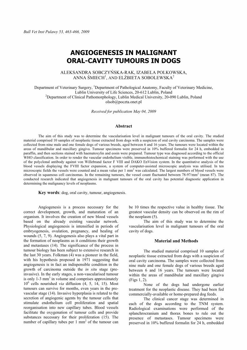
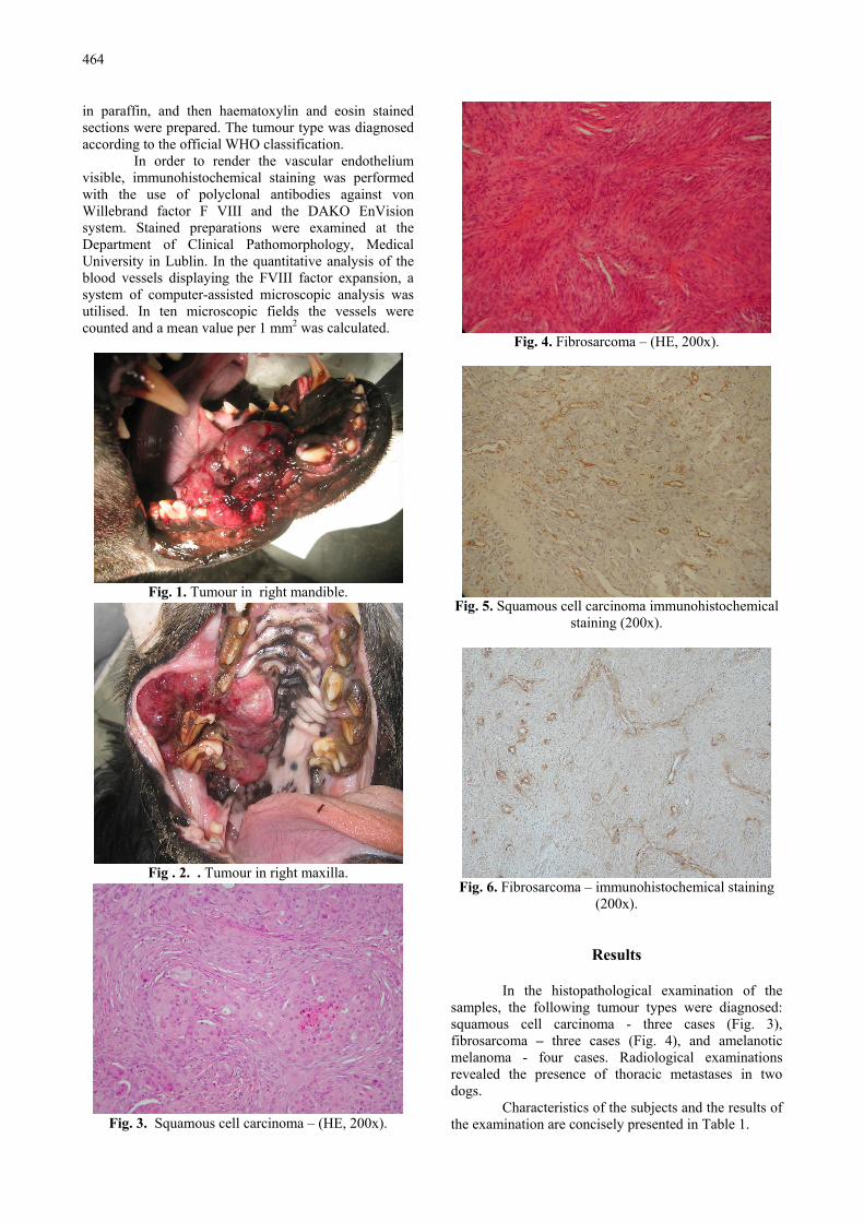
The studied material comprised 10 samples of neoplastic tissue extracted from dogs with a suspicion of oral cavity carcinoma. The samples were collected from nine male and one female dogs of various breeds aged between 6 and 16 years. The tumours were located within the areas of mandibular and maxillary gingiva (Figs 1, 2).
None of the dogs had undergone earlier treatment for the neoplastic disease. They had been fed commercially-available or home-prepared dog feeds. The clinical cancer stage was determined in each of the dogs according to the TNM system. Radiological examinations were performed of the splanchrocranium and thorax bones to rule out the presence of metastases. Tumour specimens were preserved in 10% buffered formalin for 24 h, embedded
464
in paraffin, and then haematoxylin and eosin stained sections were prepared. The tumour type was diagnosed according to the official WHO classification.
In order to render the vascular endothelium visible, immunohistochemical staining was performed with the use of polyclonal antibodies against von Willebrand factor F VIII and the DAKO EnVision system. Stained preparations were examined at the Department of Clinical Pathomorphology, Medical University in Lublin. In the quantitative analysis of the blood vessels displaying the FVIII factor expansion, a system of computer-assisted microscopic analysis was utilised. In ten microscopic fields the vessels were counted and a mean value per 1 mm2 was calculated.
Fig. 1. Tumour in right mandible.
Fig . 2. . Tumour in right maxilla.
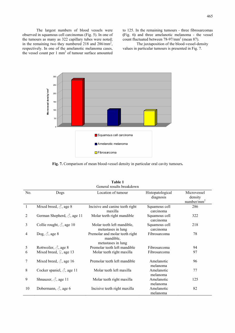
Fig. 3. Squamous cell carcinoma – (HE, 200x).
Fig. 4. Fibrosarcoma – (HE, 200x).
Fig. 5. Squamous cell carcinoma immunohistochemical
staining (200x).
Fig. 6. Fibrosarcoma – immunohistochemical staining
(200x).
Results
In the histopathological examination of the samples, the following tumour types were diagnosed: squamous cell carcinoma - three cases (Fig. 3), fibrosarcoma – three cases (Fig. 4), and amelanotic melanoma - four cases. Radiological examinations revealed the presence of thoracic metastases in two dogs.
Characteristics of the subjects and the results of the examination are concisely presented in Table 1.
465
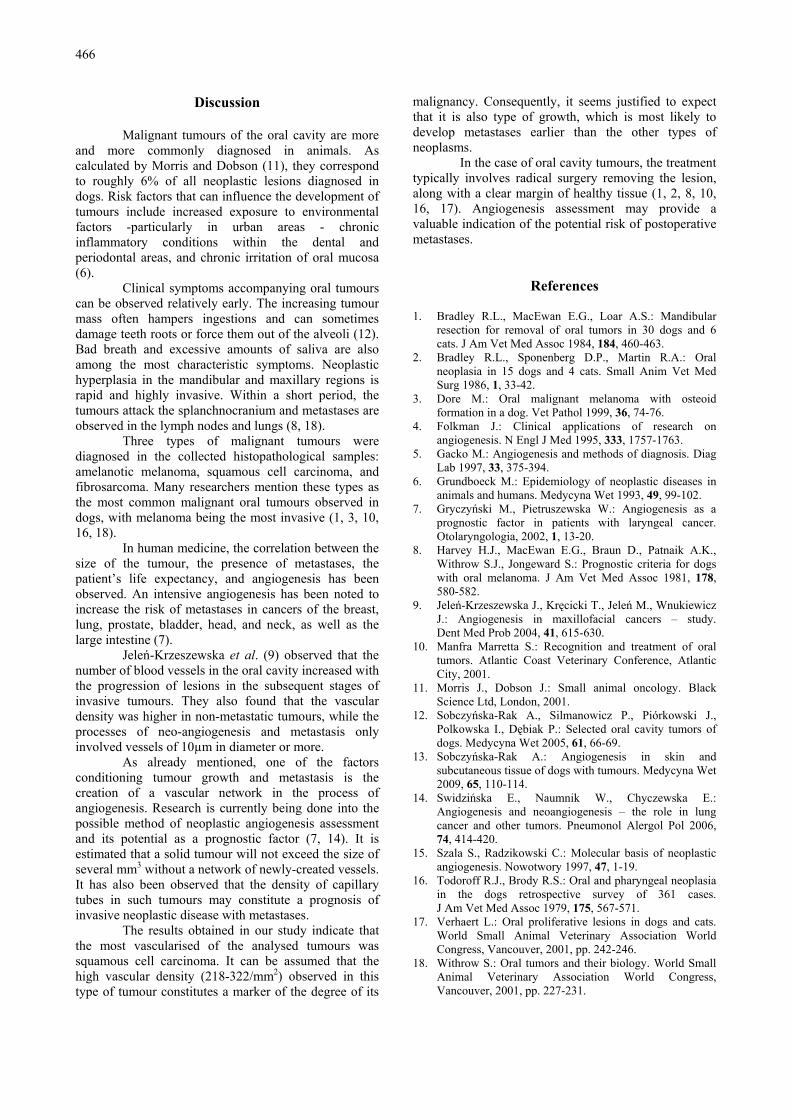
The largest numbers of blood vessels were observed in squamous cell carcinomas (Fig. 5). In one of the tumours as many as 322 capillary tubes were noted; in the remaining two they numbered 218 and 286/mm2, respectively. In one of the amelanotic melanoma cases, the vessel count per 1 mm2 of tumour surface amounted
to 125. In the remaining tumours - three fibrosarcomas (Fig. 6) and three amelanotic melanoma - the vessel count fluctuated between 78-97/mm2 (mean 87).
The juxtaposition of the blood-vessel-density values in particular tumours is presented in Fig. 7.
0
50
100
150
200
250
300
Mic
rove
ssel
den
sity
/ mm
2
Squamous cell carcinoma
Amelanotic melanoma
Fibrosarcoma
Fig. 7. Comparison of mean blood-vessel density in particular oral cavity tumours.
Table 1 General results breakdown
No. Dogs Location of tumour Histopatological diagnosis
Microvessel density
number/mm2
1 Mixed breed, ♂, age 8
Incisive and canine teeth right maxilla
Squamous cell carcinoma
286
2 German Shepherd, ♂, age 11
Molar teeth right mandible Squamous cell carcinoma
322
3 Collie rought, ♂, age 10
Molar teeth left mandible, metastases in lung
Squamous cell carcinoma
218
4 Dog, ♂, age 8
Premolar and molar teeth right mandible,
metastases in lung
Fibrosarcoma 78
5 Rottweiler, ♂, age 8 Premolar teeth left mandible Fibrosarcoma 94 6 Mixed breed, ♀, age 13
Molar teeth right maxilla Fibrosarcoma 97
7 Mixed breed, ♂, age 16
Premolar teeth left mandible Amelanotic melanoma
96
8 Cocker spaniel, ♂, age 11
Molar teeth left maxilla Amelanotic melanoma
77
9 Shnauzer, ♂, age 11
Molar teeth right maxilla Amelanotic melanoma
125
10 Dobermann, ♂, age 6
Incisive teeth right maxilla Amelanotic melanoma
82
466
Discussion
Malignant tumours of the oral cavity are more and more commonly diagnosed in animals. As calculated by Morris and Dobson (11), they correspond to roughly 6% of all neoplastic lesions diagnosed in dogs. Risk factors that can influence the development of tumours include increased exposure to environmental factors -particularly in urban areas - chronic inflammatory conditions within the dental and periodontal areas, and chronic irritation of oral mucosa (6).
Clinical symptoms accompanying oral tumours can be observed relatively early. The increasing tumour mass often hampers ingestions and can sometimes damage teeth roots or force them out of the alveoli (12). Bad breath and excessive amounts of saliva are also among the most characteristic symptoms. Neoplastic hyperplasia in the mandibular and maxillary regions is rapid and highly invasive. Within a short period, the tumours attack the splanchnocranium and metastases are observed in the lymph nodes and lungs (8, 18).
Three types of malignant tumours were diagnosed in the collected histopathological samples: amelanotic melanoma, squamous cell carcinoma, and fibrosarcoma. Many researchers mention these types as the most common malignant oral tumours observed in dogs, with melanoma being the most invasive (1, 3, 10, 16, 18).
In human medicine, the correlation between the size of the tumour, the presence of metastases, the patient’s life expectancy, and angiogenesis has been observed. An intensive angiogenesis has been noted to increase the risk of metastases in cancers of the breast, lung, prostate, bladder, head, and neck, as well as the large intestine (7).
Jeleń-Krzeszewska et al. (9) observed that the number of blood vessels in the oral cavity increased with the progression of lesions in the subsequent stages of invasive tumours. They also found that the vascular density was higher in non-metastatic tumours, while the processes of neo-angiogenesis and metastasis only involved vessels of 10µm in diameter or more.
As already mentioned, one of the factors conditioning tumour growth and metastasis is the creation of a vascular network in the process of angiogenesis. Research is currently being done into the possible method of neoplastic angiogenesis assessment and its potential as a prognostic factor (7, 14). It is estimated that a solid tumour will not exceed the size of several mm3 without a network of newly-created vessels. It has also been observed that the density of capillary tubes in such tumours may constitute a prognosis of invasive neoplastic disease with metastases.
The results obtained in our study indicate that the most vascularised of the analysed tumours was squamous cell carcinoma. It can be assumed that the high vascular density (218-322/mm2) observed in this type of tumour constitutes a marker of the degree of its
malignancy. Consequently, it seems justified to expect that it is also type of growth, which is most likely to develop metastases earlier than the other types of neoplasms.
In the case of oral cavity tumours, the treatment typically involves radical surgery removing the lesion, along with a clear margin of healthy tissue (1, 2, 8, 10, 16, 17). Angiogenesis assessment may provide a valuable indication of the potential risk of postoperative metastases.
References 1. Bradley R.L., MacEwan E.G., Loar A.S.: Mandibular
resection for removal of oral tumors in 30 dogs and 6 cats. J Am Vet Med Assoc 1984, 184, 460-463.
2. Bradley R.L., Sponenberg D.P., Martin R.A.: Oral neoplasia in 15 dogs and 4 cats. Small Anim Vet Med Surg 1986, 1, 33-42.
3. Dore M.: Oral malignant melanoma with osteoid formation in a dog. Vet Pathol 1999, 36, 74-76.
4. Folkman J.: Clinical applications of research on angiogenesis. N Engl J Med 1995, 333, 1757-1763.
5. Gacko M.: Angiogenesis and methods of diagnosis. Diag Lab 1997, 33, 375-394.
6. Grundboeck M.: Epidemiology of neoplastic diseases in animals and humans. Medycyna Wet 1993, 49, 99-102.
7. Gryczyński M., Pietruszewska W.: Angiogenesis as a prognostic factor in patients with laryngeal cancer. Otolaryngologia, 2002, 1, 13-20.
8. Harvey H.J., MacEwan E.G., Braun D., Patnaik A.K., Withrow S.J., Jongeward S.: Prognostic criteria for dogs with oral melanoma. J Am Vet Med Assoc 1981, 178, 580-582.
9. Jeleń-Krzeszewska J., Kręcicki T., Jeleń M., Wnukiewicz J.: Angiogenesis in maxillofacial cancers – study. Dent Med Prob 2004, 41, 615-630.
10. Manfra Marretta S.: Recognition and treatment of oral tumors. Atlantic Coast Veterinary Conference, Atlantic City, 2001.
11. Morris J., Dobson J.: Small animal oncology. Black Science Ltd, London, 2001.
12. Sobczyńska-Rak A., Silmanowicz P., Piórkowski J., Polkowska I., Dębiak P.: Selected oral cavity tumors of dogs. Medycyna Wet 2005, 61, 66-69.
13. Sobczyńska-Rak A.: Angiogenesis in skin and subcutaneous tissue of dogs with tumours. Medycyna Wet 2009, 65, 110-114.
14. Swidzińska E., Naumnik W., Chyczewska E.: Angiogenesis and neoangiogenesis – the role in lung cancer and other tumors. Pneumonol Alergol Pol 2006, 74, 414-420.
15. Szala S., Radzikowski C.: Molecular basis of neoplastic angiogenesis. Nowotwory 1997, 47, 1-19.
16. Todoroff R.J., Brody R.S.: Oral and pharyngeal neoplasia in the dogs retrospective survey of 361 cases. J Am Vet Med Assoc 1979, 175, 567-571.
17. Verhaert L.: Oral proliferative lesions in dogs and cats. World Small Animal Veterinary Association World Congress, Vancouver, 2001, pp. 242-246.
18. Withrow S.: Oral tumors and their biology. World Small Animal Veterinary Association World Congress, Vancouver, 2001, pp. 227-231.


![Malignant tumours of temporomandibular joint · 2020. 10. 6. · Temporomandibular joint (TMJ) disorders are very common and can be easily diagnosed [1, 2]. However, malignant tumours](https://static.fdocuments.in/doc/165x107/609cf658aa942f17d538f23e/malignant-tumours-of-temporomandibular-joint-2020-10-6-temporomandibular-joint.jpg)