High prevalence of Foxp3 and IL17 in MMR-proficient ... · MSI (MSI-H) are considered to ......
8
High prevalence of Foxp3 and IL17 in MMR-proficient colorectal carcinomas S Le Gouvello, 1,2,3 S Bastuji-Garin, 2,4 N Aloulou, 5 H Mansour, 1 M-T Chaumette, 2,6 F Berrehar, 1 A Seikour, 6 A Charachon, 2,5 M Karoui, 2,7 K Leroy, 2,6 J-P Farcet, 1,2 I Sobhani 2,5 See Commentary, p 725 c An additional table is published online only at http:// gut.bmj.com/content/vol57/ issue6 1 Department of Biological Immunology, AP-HP, Ho ˆpital Henri Mondor, Cre ´teil, France; 2 University Paris XII, Cre ´teil, France; 3 INSERM U841, Cre ´teil, France; 4 Department of Public Health, AP-HP, Ho ˆpital Henri Mondor, Cre ´teil, France; 5 Department of Gastroenterology, AP-HP, Ho ˆpital Henri Mondor, Cre ´teil, France; 6 Department of Pathology, AP-HP, Ho ˆpital Henri Mondor, Cre ´teil, France; 7 Department of Surgery, AP-HP, Ho ˆpital Henri Mondor, Cre ´teil, France Correspondence to: Profesor I Sobhani, Department of Gastroenterology, Ho ˆpital Henri Mondor, 51 av. du Mare ´chal de Lattre de Tassigny, 94010 Cre ´teil, France; iradj. [email protected] Revised 16 August 2007 Accepted 28 August 2007 Published Online First 19 October 2007 ABSTRACT Background and aims: Colorectal cancer (CRC) harbours different types of DNA alterations, including microsatellite instability (MSI). Cancers with high levels of MSI (MSI-H) are considered to have a good prognosis, probably related to lymphocyte infiltration within tumours. The aim of the present study was to characterise the intratumoural expression of markers associated with the antitumour immune response in mismatch repair (MMR)- proficient (MSS) colon cancers. Methods: Ninety human colon cancers (T) and autologous normal colon mucosa (NT) were quantified for the expression of 15 markers of the immune response with quantitiative reverse transcription-PCR (qRT-PCR). mRNA expression levels were correlated with MMR status. Immunohistochemistry (IHC) was performed using both interleukin 17 (IL17) and CD3 antibodies. Results: Expression of cytotoxic markers (FasL, granzyme B and perforin), inflammatory cytokines (IL1b, IL6, IL8, IL17 and transforming growth factor b (TGFb)) and a marker of regulatory T cells (forkhead box P3 (Foxp3)) was significantly higher in tumours than in autologous normal tissues. Adjusting for MMR status, higher tumoural expression of both granzyme B and perforin was associated with the MSI-H phenotype, and the perforin T/NT ratio was higher in MSI-H tissues than in MSS tissues. Higher tumoural expression of Foxp3, IL17, IL1b, IL6 and TGFb was associated with the MSS phenotype, and the IL17 T/NT ratio was higher in MSS tissues than in MSI-H tissues as assessed by both qRT-PCR and IHC. Conclusions: Immune gene expression profiling in CRC displayed different patterns according to MMR status. Higher Foxp3, IL6, TGFb and IL17 expression is a particular determinant in MMR-proficient CRC. These may be potential biomarkers for a new prognostic ‘‘test set’’ in sporadic CRCs. Colorectal cancer (CRC) is the second leading cause of cancer mortality in the Western world, despite recent advances in surgery, radiotherapy and chemotherapy. 1 Tumour progression of CRCs is governed by either genetic or epigenetic changes intrinsic to cancer cells, and by environmental factors. Approximately 15–20% of sporadic CRCs and nearly all large bowel malignancies in the hereditary non-polyposis colorectal cancer (HNPCC) syndrome are characterised by wide- spread microsatellite instability. 2–4 Microsatellite instability is a genome-wide instability in repeti- tive DNA sequences observed at the nucleotide level, and it is caused by the inactivation or loss of expression of the mismatch repair (MMR) genes: hMSH2, hMLH1, hPMS1, hPMS2, hMSH6/GTBP or MSH3, as a result of either mutations or epigenetic silencing. 5 Besides MMR-deficient (MSI-H) cancer, the majority of sporadic CRSs are MMR proficient (MSS) tumours, with chro- mosomal instability in some cases. 67 Most studies revealed longer disease-free and overall survival for patients with MSI-H CRC than for patients with MSS CRC, 8–12 even in the case of deep local invasion of the primary tumour. 13 These studies suggested a protective role for T lympho- cytes, which clearly infiltrated more extensively in MSI-H than in MSS CRCs. 14 Pronounced lympho- cytic infiltration of tumours has long been associated with an improved prognosis. 15 16 MSI in combination with a high content of intraepithe- lial cytotoxic lymphocytes was related to improved overall survival in a group of exclusively right-sided CRCs, 17 18 suggesting that a combination of both the tumour’s local lymphocytosis and MMR characterization may be useful for a more accurate prognostic assessment. Contradictory results come from mouse models of CRC, and these results are consistent with the association between either chronic inflammation 19 20 or an increased number of inflammatory cells in tumours 21 and tumour progression. To determine the putative impact of microsa- tellite status on the antitumoural immune response, we analysed histological and molecular features of tumours in patients with sporadic CRCs. We also developed a quantitative PCR assay for 15 inflammatory and T lymphocyte markers in tumours with reference to the MMR status. PATIENTS AND METHODS Study population This retrospective analysis included a group of 45 patients from a consecutive cohort survey study in Henri Mondor Hospital. To be eligible, patients had to have undergone surgery of primary rectal or colon tumours, for which both frozen and paraffin- embedded tumoural and normal tissues were available in the tissue collection bank. None had known hereditary cancer, ulcerative colitis or Crohn’s disease. Consecutive patients were routi- nely informed about biological research on tissues that did not include hereditary genetic character- isation. Their tissue materials could be used for the current study except if ‘‘formal opposition’’ was mentioned to doctors. The ethical committee approved the procedure. Colorectal cancer 772 Gut 2008;57:772–779. doi:10.1136/gut.2007.123794 on 7 July 2019 by guest. Protected by copyright. http://gut.bmj.com/ Gut: first published as 10.1136/gut.2007.123794 on 26 October 2007. Downloaded from
Transcript of High prevalence of Foxp3 and IL17 in MMR-proficient ... · MSI (MSI-H) are considered to ......
High prevalence of Foxp3 and IL17 in MMR-proficientcolorectal carcinomas
S Le Gouvello,1,2,3 S Bastuji-Garin,2,4 N Aloulou,5 H Mansour,1 M-T Chaumette,2,6
F Berrehar,1 A Seikour,6 A Charachon,2,5 M Karoui,2,7 K Leroy,2,6 J-P Farcet,1,2
I Sobhani2,5
See Commentary, p 725
c An additional table ispublished online only at http://gut.bmj.com/content/vol57/issue6
1 Department of BiologicalImmunology, AP-HP, HopitalHenri Mondor, Creteil, France;2 University Paris XII, Creteil,France; 3 INSERM U841, Creteil,France; 4 Department of PublicHealth, AP-HP, Hopital HenriMondor, Creteil, France;5 Department ofGastroenterology, AP-HP,Hopital Henri Mondor, Creteil,France; 6 Department ofPathology, AP-HP, Hopital HenriMondor, Creteil, France;7 Department of Surgery, AP-HP,Hopital Henri Mondor, Creteil,France
Correspondence to:Profesor I Sobhani, Departmentof Gastroenterology, HopitalHenri Mondor, 51 av. duMarechal de Lattre de Tassigny,94010 Creteil, France; [email protected]
Revised 16 August 2007Accepted 28 August 2007Published Online First19 October 2007
ABSTRACTBackground and aims: Colorectal cancer (CRC)harbours different types of DNA alterations, includingmicrosatellite instability (MSI). Cancers with high levels ofMSI (MSI-H) are considered to have a good prognosis,probably related to lymphocyte infiltration within tumours.The aim of the present study was to characterise theintratumoural expression of markers associated with theantitumour immune response in mismatch repair (MMR)-proficient (MSS) colon cancers.Methods: Ninety human colon cancers (T) andautologous normal colon mucosa (NT) were quantified forthe expression of 15 markers of the immune responsewith quantitiative reverse transcription-PCR (qRT-PCR).mRNA expression levels were correlated with MMRstatus. Immunohistochemistry (IHC) was performed usingboth interleukin 17 (IL17) and CD3 antibodies.Results: Expression of cytotoxic markers (FasL, granzymeB and perforin), inflammatory cytokines (IL1b, IL6, IL8,IL17 and transforming growth factor b (TGFb)) and amarker of regulatory T cells (forkhead box P3 (Foxp3))was significantly higher in tumours than in autologousnormal tissues. Adjusting for MMR status, highertumoural expression of both granzyme B and perforin wasassociated with the MSI-H phenotype, and the perforinT/NT ratio was higher in MSI-H tissues than in MSStissues. Higher tumoural expression of Foxp3, IL17, IL1b,IL6 and TGFb was associated with the MSS phenotype,and the IL17 T/NT ratio was higher in MSS tissues than inMSI-H tissues as assessed by both qRT-PCR and IHC.Conclusions: Immune gene expression profiling in CRCdisplayed different patterns according to MMR status.Higher Foxp3, IL6, TGFb and IL17 expression is aparticular determinant in MMR-proficient CRC. These maybe potential biomarkers for a new prognostic ‘‘test set’’ insporadic CRCs.
Colorectal cancer (CRC) is the second leadingcause of cancer mortality in the Western world,despite recent advances in surgery, radiotherapyand chemotherapy.1 Tumour progression of CRCsis governed by either genetic or epigenetic changesintrinsic to cancer cells, and by environmentalfactors. Approximately 15–20% of sporadic CRCsand nearly all large bowel malignancies in thehereditary non-polyposis colorectal cancer(HNPCC) syndrome are characterised by wide-spread microsatellite instability.2–4 Microsatelliteinstability is a genome-wide instability in repeti-tive DNA sequences observed at the nucleotidelevel, and it is caused by the inactivation or loss ofexpression of the mismatch repair (MMR) genes:hMSH2, hMLH1, hPMS1, hPMS2, hMSH6/GTBP
or MSH3, as a result of either mutations orepigenetic silencing.5 Besides MMR-deficient(MSI-H) cancer, the majority of sporadic CRSsare MMR proficient (MSS) tumours, with chro-mosomal instability in some cases.6 7
Most studies revealed longer disease-free andoverall survival for patients with MSI-H CRC thanfor patients with MSS CRC,8–12 even in the case ofdeep local invasion of the primary tumour.13 Thesestudies suggested a protective role for T lympho-cytes, which clearly infiltrated more extensively inMSI-H than in MSS CRCs.14 Pronounced lympho-cytic infiltration of tumours has long beenassociated with an improved prognosis.15 16 MSIin combination with a high content of intraepithe-lial cytotoxic lymphocytes was related to improvedoverall survival in a group of exclusively right-sidedCRCs,17 18 suggesting that a combination of boththe tumour’s local lymphocytosis and MMRcharacterization may be useful for a more accurateprognostic assessment. Contradictory results comefrom mouse models of CRC, and these results areconsistent with the association between eitherchronic inflammation19 20 or an increased numberof inflammatory cells in tumours21 and tumourprogression.
To determine the putative impact of microsa-tellite status on the antitumoural immuneresponse, we analysed histological and molecularfeatures of tumours in patients with sporadicCRCs. We also developed a quantitative PCR assayfor 15 inflammatory and T lymphocyte markers intumours with reference to the MMR status.
PATIENTS AND METHODS
Study populationThis retrospective analysis included a group of 45patients from a consecutive cohort survey study inHenri Mondor Hospital. To be eligible, patientshad to have undergone surgery of primary rectal orcolon tumours, for which both frozen and paraffin-embedded tumoural and normal tissues wereavailable in the tissue collection bank. None hadknown hereditary cancer, ulcerative colitis orCrohn’s disease. Consecutive patients were routi-nely informed about biological research on tissuesthat did not include hereditary genetic character-isation. Their tissue materials could be used for thecurrent study except if ‘‘formal opposition’’ wasmentioned to doctors. The ethical committeeapproved the procedure.
Colorectal cancer
772 Gut 2008;57:772–779. doi:10.1136/gut.2007.123794
on 7 July 2019 by guest. Protected by copyright.
http://gut.bmj.com
/G
ut: first published as 10.1136/gut.2007.123794 on 26 October 2007. D
ownloaded from

Histology and pathologyTissues were immediately examined after surgery, and normaland tumoural representative samples were frozen, fixed informalin, and archived in a tissue collection bank. Diagnosesand tumour descriptions were done on H&E-stained tissuesaccording to the pTNM classification. Histopathology wasfurther examined for lymphocyte infiltration and for a mucouscomponent.
Analysis of MMR status by immunohistochemistryRepresentative samples from adenocarcinoma and normalmucosa adjacent to the tumour were selected in each case andparaffin sections were sent to an automated immunostainer.Tissue sections were incubated in citrate buffer with thefollowing antibodies: G168-728 antibody (recognises hMLH1antigen; the antibody was diluted 1:40; Pharmingen, San DiegoCalifornia, USA), FE11 antibody (recognises hMSH2 antigen;the antibody was diluted 1:25; Calbiochem, Cambridge,Massachusetts, USA), 44 antibody (recognises hMSH6; theantibody was diluted 1:100; Zymed Laboratories Inc., South SanFrancisco, California, USA), all in a boiling water bath. Anavidin–biotin complex (ABC Vectastain Kit, VectorLaboratories, Burligame, California, USA) was used to revealthe antigen. Positive controls included slides from tumours withnormal expression of hMLH1, hMSH2 and hMSH6; negativecontrols included slides with no primary antibodies. Twoobservers with no prior knowledge of the PCR results assessedall cases independently, and cases with discrepancies werefurther evaluated until agreement was reached between theobservers.
Analysis of IL17 expression by immunohistochemistryRepresentative samples (n.5) from tumoural and autologousnormal mucosa were selected for each case, and paraffin-embedded sections (4 mm) were treated by boiling in citratebuffer (pH 6) in a microwave (800 W, 5 min 63). Endogenousperoxidase activity was blocked by incubation in 3% hydrogenperoxide for 20 min followed by a wash in phosphate-bufferedsaline (PBS) for 5 min. Non-specific antibody-binding sites werecoated by the addition of a horse serum (1:20 in PBS, for25 min). Serum was then removed and antihuman IL17 goatantibody (R&D Systems, Lille, France) diluted in the PBS (1:40)was added to the tissue sections (room temperature for 1 h).Immunohistochemical staining was undertaken using aVectastain Universal Elite kit according to the manufacturer’sinstructions (Vector Laboratories). The chromagen, SigmafastDAB (39,39-diaminobenzidine) (Sigma-Aldrich, St Louis,Missouri, USA), was incubated with the tissue sections in thedark (room temperature for 4 min). The sections were thencounterstained with haematoxylin.
For double staining IL17/CD3, the goat antihuman IL17antibody (diluted 1:40) was applied first and incubated for 2 h.Immunohistochemical staining was undertaken using aVectastain AP kit according to the manufacturer’s instructions(Vector Laboratories) and visualization was done withNaphthol/Fast Red (Sigma-Aldrich). Subsequently, a rabbitantihuman CD3 (diluted 1:50 in PBS; Dako, France) wasincubated for 1 h. Immunohistochemical staining was under-taken using the ImmPRESS system (Vector Laboratories) andvisualization was done with DAB substrate.
Analysis of microsatellite stability by PCRFormalin-fixed paraffin-embedded (FFPE) sections fromtumours were stained with H&E, and were macrodissected ifnecessary to select a tissue fragment comprising .40% tumourcells. Normal and tumour autologous DNA was extracted froma 50 mm FFPE or frozen tissue section with QiAmp DNA minikits (Qiagen, Courtaboeuf, France), according to the manufac-turer’s instructions. The genetic instability (MSI-H) status ofthe tumours was established using pentaplex PCR for markersas described by Suraweera et al.22 Microsatellites were co-amplified in a single 20 ml pentaplex fluorescent PCR, contain-ing 0.1 mM Bat-25, 0.25 mM Bat-26 and NR-22, 0.5 mM NR-24and NR-21 primers, 0.15 mM dNTP, 1.5 mM MgCl2, 16GeneAmp PCR Buffer II (Applied Biosystems, Courtaboeuf,France), 50 ng of genomic DNA and 0.5 U of AmpliTaq GoldDNA polymerase (Applied Biosystems). The PCR program wasdenaturation (95uC, 10 min), then 35 amplification cycles(denaturation (95uC, 30 s), annealing (55uC, 45 s) and extension(72uC, 30 s)), and a final elongation step (72uC, 7 min).Separation and detection of the fluorescent PCR products wereperformed on an ABI PRISM 3100 Genetic Analyzer (AppliedBiosystems), and the data were analysed with GeneScanAnalysis Software (Applied Biosystems). The MSI-H phenotypewas defined as the detection of length alterations of at leastthree markers in tumoural versus normal DNAs from the sameindividual.22 We found that 31/45 tumoural tissues had the MSSphenotype, and 14/45 had the MSI-H phenotype.
Quantitative reverse transcription-PCRTotal RNA was prepared from 90 colorectal tumoural orautologous non-tumoural specimens, using TrizolH reagent(Gibco-BRL, Life Technologies, Cergy-Pontoise, France) follow-ing the manufacturer’s protocol. To avoid misinterpretation ofthe variability in marker gene expression due to heterogeneity ofthe cellular distribution and/or leucocyte densities in tissuesamples, 10 sections of 50 mm for each sample were verifiedbefore extracting total RNA. Microdissection was used ifnecessary to select tumour sections with tumoural cells .60%of total. We normalised the ratio of mRNA copy number of thegene of interest/CD3 mRNA for each sample. First-strandcDNA was synthesized in reverse transcription samples, eachcontaining: 2 mg of total RNA isolated from colorectal tissue, 16U/ml M-MLV reverse transcriptase (Gibco-BRL), 4 mMoligo(dT)12–18 (Amersham-Pharmacia Biotech, Saclay, France)and 0.8 mM mixed dNTPs (Amersham-Pharmacia Biotech).Quantitative reverse transcription-PCR (RT-PCR) was per-formed in a LightCycler 2.0 System (Roche Diagnostics,Meylan, France) using a SYBR Green PCR kit or aHybridization Probes PCR Kit from Roche Diagnostics, aspreviously described.23 24 The sequences of primers and probesare indicated in the Supplementary data. Normalisation wasachieved by quantification of the expression of the controlhousekeeping gene (HKG) b2mglobulin, which was chosen asthe control among three HKGs tested because of its stableexpression in tumoural and non-tumoural specimens (data notshown). All PCR conditions were adjusted in order to obtainequivalent optimal amplification efficiency between the differ-ent assays. Chemokine receptor 1 (CXCR1), CD3, IL8, IL17,granzyme A (GzA), perforin, and FoxP3 mRNA expression wasquantified by the relative quantification of real-time PCRaccording to Gibson et al,25 using the SYBR Green PCR Kit,and using peripheral blood mononuclear cells stimulated withphorbol myristate acetate and ionomycin for 1 h as thecalibrator sample. CD3, IL1b, tumour necrosis factor a (TNFa),
Colorectal cancer
Gut 2008;57:772–779. doi:10.1136/gut.2007.123794 773
on 7 July 2019 by guest. Protected by copyright.
http://gut.bmj.com
/G
ut: first published as 10.1136/gut.2007.123794 on 26 October 2007. D
ownloaded from

IL6, interferon c (IFNc), IL4, TGFb, IL10, GzB and FasL mRNAexpression was quantified by absolute quantification of real-time PCR using the Hybridization Probes PCR Kit from RocheDiagnostics. The copy number for all target genes and for theHKG was obtained by plotting sample Ct values against thestandard curve obtained by analysing the corresponding‘‘quantitative DNA standard’’26 dilution series using theLightCycler software 4.0, and the magnitude of target genemRNA expression is calculated as the copy number of the targetgene per 106 copies of b2mglobulin. All PCR experiments weredone in duplicate.
Statistical analysisResults of the quantification of CD3, IL8, CXCR1, IL1b, IL6,TGFb and TNFa expression are presented as the median andinterquartile range (IQR) of the ratio of the immune markergene to HKG mRNA expression. IL4, IL10, IL17, Foxp3, FasL,perforin, GzA and GzB are mainly expressed in CD3 mRNA-expressing leucocytes. Therefore, for normalisation betweenspecimens, we used the ratio of mRNA copy number of eachmarker gene/CD3.
Non-parametric tests were used because of the non-normaldistributions of mRNA levels. Overall differences in immunemarker expression between the tumoural (T) and autologousnon-tumoural (NT) specimens were analysed by using thepaired non-parametric Wilcoxon signed-rank test. For signifi-cant variations, gene expression levels between tumoural andnon-tumoural specimens were also compared in each pheno-typic subgroup (MSI-H and MSS). Differences between MSI-Hand MSS phenotypes were evaluated by comparing the T/NTratio of mRNA expression using the two-sample Wilcoxonrank-sum test. All tests were two-tailed. An adjustment formultiple testing was done using the Bonferroni correction, andp values (0.003 (0.05/15 = 0.0033) were considered statisticallysignificant. Data were analysed using Stata Statisticalsoftware (StataCorp 2003, Release 8.0, College Station, Texas,USA)
RESULTS
Characterisation of patients with CRCsPatients with the MSI-H phenotype were not significantlydifferent from those with the MSS phenotype in terms of age,diagnosis, therapy or surveillance procedures (table 1). The rateof tumour relapse within a 36-month follow-up was notdifferent depending on MMR status. Tumours with the MSI-H phenotype were right-sided with significantly differenthistopathological features: they had more lymphocyte infiltra-tion and a greater mucoid component, and less local or systemictumour cell extension.
The MSI-H phenotype correlates with the presence of cytotoxicmarkersThe CD3 mRNA copy number in tumoural (T) tissues was notsignificantly different from that in autologous (NT) tissuefragments (0.18 (0.13 to 0.28) vs 0. 17 (0.11 to 0.26)) when allsamples were considered (irrespective of MMR status). TheCD3 T/NT ratio did not differ between MSI-H and MSS tissues.The expression ratios of the cytotoxic parameters, FasL/CD3,perforin/CD3 and GzB/CD3, were significantly higher intumour tissues than in autologous NT specimens (table 2).Adjusting on MMR status, only the perforin T/NT ratio washigher in MSI-H tissues than in MSS tissues. The expression ofGzB/CD3 was significantly higher in tumour (as compared withNT) specimens only if they had an MSS phenotype. The GzA/CD3 ratio did not differ between tumoural and autologous NTspecimens.
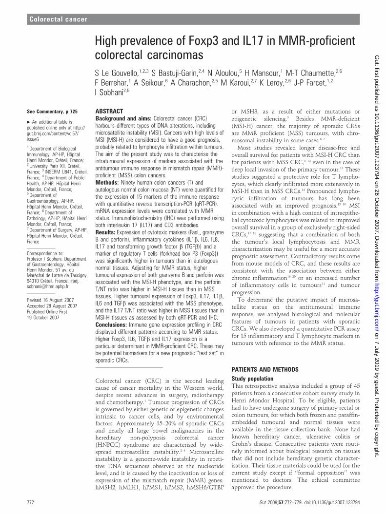
MSS phenotype and higher Foxp3 and IL17 expression in tumourinfiltratesOverall analysis showed that the expression ratios IL17/CD3and Foxp3/CD3 (table 2), and IL8, IL1b, IL6 and TGFbexpression (table 3) were significantly higher in tumour tissuesthan in autologous NT specimens. However, adjusting forMMR status, the higher expression of Foxp3, IL17, IL1b, IL6 andTGFb was observed only in MSS phenotype tumours (tables 2and 3). The IL17 T/NT ratio was higher in MSS tissues than in
Table 1 Characteristics of the study population
Parameters
All cases MSS MSI-H
p Value*
(n = 45) (n = 31) (n = 14)
n (%) n (%) n (%)
Gender (F) 21 (47) 11 (35) 10 (71) 0.05
Mean age at surgery (SD) 69.7 (1.5) 68.8 (1.8) 71.8 (2.8) 0.40
Right-sided tumour 19 (42) 9 (29) 10 (71) 0.01
No or poor differentiation 27 (60) 20 (70) 7 (50) 0.21
Mucous component 4 (1) 0 (0) 4 (40) 0.01
Lymphocyte infiltration{ 14 (31) 1 (3) 13 (93) 0.0001
Tumour staging
pT 1–2 4 (9) 2 (6) 2 (14)
pT 3 27 (60) 16 (52) 11 (79)
pT 4 14 (31) 13 (42) 1 (7) 0.05
N0 26 (58) 15 (48) 11 (79)
N1–2 19 (42) 16 (52) 3 (21) 0.10
Synchronous metastasis 22 (49) 19 (61) 3 (21) 0.02
Vascular emboli 25 (56) 20 (65) 5 (36) 0.03
Perinervous infiltration 7 (16) 5 (16) 2 (14) 1
Relapse within 36 months of follow-up 10 (22) 8 (26) 2 (14) 0.47{
*p Value of Fisher exact test or Mann–Whitney test as appropriate. {Assessed by histopathology evaluation (yes = + to ++;no = 0 to +/2). {p Value of log-rank test for equality of survivor functions.F, female, MSS, microsatellite stability; MSI-H, high-frequency microsatellite instability.
Colorectal cancer
774 Gut 2008;57:772–779. doi:10.1136/gut.2007.123794
on 7 July 2019 by guest. Protected by copyright.
http://gut.bmj.com
/G
ut: first published as 10.1136/gut.2007.123794 on 26 October 2007. D
ownloaded from

MSI-H tissues. Moreover, quantification by qRT-PCR of IL22expression, another IL17-producing T cell- (TH-17 cell) secretedcytokine, showed IL22 overexpression in MSS tumour tissuescompared with MSI-H tumour tissues (p = 0.042 for the ratiodifference between MSI T/NT and MSS T/NT). The IL10/CD3,IL4/CD3 ratios (table 2), and CXCR-1 and TNF expression(table 3) did not differ between the tumoural and autologousnon-tumoural specimens. Expression of IFNc was not detectedin any specimen.
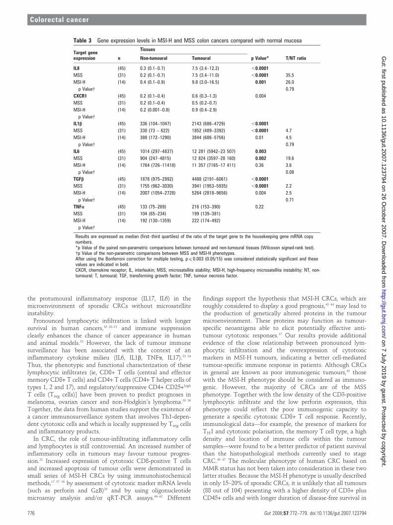
ImmunohistochemistryIL17-immunoreactive cells were rarely detected in normaltissues in the lamina propria (fig 1A). However, their numberwas higher in tumour sections than in autologous normaltissues. IL17 cell infiltration was higher in MSS tumours than inMSI tumours (fig 1B). Only a few CD3+ cells in tumours weredouble-stained with IL17 antibody (fig 1c).
DISCUSSIONElucidation of the nature of the intratumoural inflammatoryreaction is relevant not only to understand better the
pathobiology of CRCs but also to assess the possible influenceof the host immune response on patient outcome. Our studyexamined the differences in the pattern of inflammatory andimmune response markers expressed in the tumour microenvir-onment of a series of 45 tumours from patients with sporadicCRC. About one-third of the patients had MSI-H colorectalcancer, although no-one displayed HNPCC syndrome and/orthe Bethesda criteria.27 MSI-H phenotype tumours, as assessedby immunohistochemistry, showed more pronounced lympho-cytic infiltration than MSS phenotype tumours. The MSI-Hphenotype was associated with a higher expression level ofcytotoxic cell markers, especially perforin. Conversely, theMMR-proficient (MSS) phenotype was associated with higherexpression levels of Foxp3, a marker of regulatory T cells, and ofinflammatory (IL1b, IL6, IL8, IL17) or suppressive (TGFb)cytokines, with IL17 expressed at particularly high levels. Nodifference was observed between MSI-H and MSS tumoursregarding CD4+ Th1 and Th2 cell subset infiltration, as assessedby the mRNA levels of the relevant cytokines (IFNc, TNFa andIL4). Thus, we show for the first time a paradoxical geneexpression signature combining markers associated with inhibi-tion of the antitumour immune response (TGFb, Foxp3) and
Table 2 Gene expression levels/CD3 in colon cancers compared with normal mucosa
Target geneexpression n
Tissues
p Value* T/NT ratioNon-tumoural Tumoural
Fas ligand (45) 1390 (463–2055) 2294 (964–3842) ,0.001
MSS (31) 1500 (274–2040) 1731 (708–3376) 0.01 1.7
MSI-H (14) 1275 (750–2154) 3305 (2031–7821) 0.02 2.6
p Value{ 0.68
Perforin (45) 13.2 (8.1–17.9) 19.3 (13.2–41.5) 0.002
MSS (31) 13.5 (8.8–20.9) 17.1 (11.3–35.1) 0.07 1.2
MSI-H (14) 8.9 (5.4–18.5) 38.5 (18.5–69.0) 0.009 3.2
p Value{ 0.003
Granzyme A (45) 1.6 (1.0–2.6) 1.6 (1.0–2.0)1 0.72
MSS (31) 1.6 (1.2–2.5) 1.5 (.9–2.0)
MSI-H (14) 1.6 (0.9–2.6) 2.1 (1.6–3.5)
p Value{Granzyme B (45) 5555 (2992–11 501) 19 065 (14 440–30 586) ,0.0001
MSS (31) 5545 (2586–7640) 16 289 (9396–23 507) ,0.0001 3.4
MSI-H (14) 7915 (4167–14 431) 24758 (17 223–61 150) 0.006 4.1
p Value{ 0.71
IL4 (45) 99 (48–421) 238 (120–450) 0.06
MSS (31) 81 (46–421) 238 (120–450)
MSI-H (14) 124 (50–515) 203 (91–471)
p Value{IL10 (45) 227 (88–529) 284 (199–527) 0.06
MSS (31) 250 (76–646) 284 (203–509) 1.7
MSI-H (14) 154 (106–529) 317 (143–654) 1.2
p Value{ 0.68
IL17 (45) 0.6 (0.2–1.8) 4.5 (1.8–11.5) ,0.0001
MSS (31) 0.5 (0.2–1.8) 7.2 (2.3–18.8) ,0.0001 13.7
MSI-H (14) 1.4 (0.6–2.0) 2.4 (0.4–4.4) 0.30 1.4
p Value{ 0.002
Foxp3 (45) 2.7 (1.7–4.0) 15.6 (11.2–20.1) ,0.001
MSS (31) 2.6 (1.7–3.9) 16.0 (13.7–21.0) ,0.0001 6.8
MSI-H (14) 3.1 (1.7–6.2) 10.5 (3.9–19.7) 0.07 3.2
p Value{ 0.04
Results are expressed as median (first–third quartiles) of the ratio of target gene to CD3 mRNA copy numbers.*p Value of the paired non-parametric comparisons between tumoural and non-tumoural tissues (Wilcoxon signed-rank test).{p Value of the non-parametric comparisons between MSS and MSI-H phenotypes. After using the Bonferroni correction formultiple testing, p(0.003 was considered statistically significant and these values are indicated in bold.Foxp3, forkhead box P3; IL, interleukin; MSS, microsatellite stability; MSI-H, high-frequency microsatellite instability; NT, non-tumoural; T, tumoural.
Colorectal cancer
Gut 2008;57:772–779. doi:10.1136/gut.2007.123794 775
on 7 July 2019 by guest. Protected by copyright.
http://gut.bmj.com
/G
ut: first published as 10.1136/gut.2007.123794 on 26 October 2007. D
ownloaded from
the protumoural inflammatory response (IL17, IL6) in themicroenvironment of sporadic CRCs without microsatelliteinstability.
Pronounced lymphocytic infiltration is linked with longersurvival in human cancers,16 28–31 and immune suppressionclearly enhances the chance of cancer appearance in humanand animal models.32 However, the lack of tumour immunesurveillance has been associated with the context of aninflammatory cytokine milieu (IL6, IL1b, TNFa, IL17).33 34
Thus, the phenotypic and functional characterization of theselymphocytic infiltrates (ie, CD8+ T cells (central and effectormemory CD8+ T cells) and CD4+ T cells (CD4+ T helper cells oftypes 1, 2 and 17), and regulatory/suppressive CD4+ CD25+high
T cells (Treg cells)] have been proven to predict prognoses inmelanoma, ovarian cancer and non-Hodgkin’s lymphoma.35 36
Together, the data from human studies support the existence ofa cancer immunosurveillance system that involves Th1-depen-dent cytotoxic cells and which is locally suppressed by Treg cellsand inflammatory products.
In CRC, the role of tumour-infiltrating inflammatory cellsand lymphocytes is still controversial. An increased number ofinflammatory cells in tumours may favour tumour progres-sion.21 Increased expression of cytotoxic CD8-positive T cellsand increased apoptosis of tumour cells were demonstrated insmall series of MSI-H CRCs by using immunohistochemicalmethods,17 37 38 by assessment of cytotoxic marker mRNA levels(such as perforin and GzB)39 and by using oligonucleotidemicroarray analysis and/or qRT-PCR assays.40–42 Different
findings support the hypothesis that MSI-H CRCs, which areroughly considered to display a good prognosis,43 44 may lead tothe production of genetically altered proteins in the tumourmicroenvironment. These proteins may function as tumour-specific neoantigens able to elicit potentially effective anti-tumour cytotoxic responses.17 Our results provide additionalevidence of the close relationship between pronounced lym-phocytic infiltration and the overexpression of cytotoxicmarkers in MSI-H tumours, indicating a better cell-mediatedtumour-specific immune response in patients. Although CRCsin general are known as poor immunogenic tumours,45 thosewith the MSI-H phenotype should be considered as immuno-genic. However, the majority of CRCs are of the MSSphenotype. Together with the low density of the CD3-positivelymphocytic infiltrate and the low perforin expression, thisphenotype could reflect the poor immunogenic capacity togenerate a specific cytotoxic CD8+ T cell response. Recently,immunological data—for example, the presence of markers forTH1 and cytotoxic polarisation, the memory T cell type, a highdensity and location of immune cells within the tumoursamples—were found to be a better predictor of patient survivalthan the histopathological methods currently used to stageCRC.46 47 The molecular phenotype of human CRC based onMMR status has not been taken into consideration in these twolatter studies. Because the MSI-H phenotype is usually describedin only 15–20% of sporadic CRCs, it is unlikely that all tumours(88 out of 104) presenting with a higher density of CD3+ plusCD45+ cells and with longer duration of disease-free survival in
Table 3 Gene expression levels in MSI-H and MSS colon cancers compared with normal mucosa
Target geneexpression n
Tissues
p Value* T/NT ratioNon-tumoural Tumoural
IL8 (45) 0.3 (0.1–0.7) 7.5 (3.4–12.2) ,0.0001
MSS (31) 0.2 (0.1–0.7) 7.5 (3.4–11.0) ,0.0001 35.5
MSI-H (14) 0.4 (0.1–0.9) 9.8 (3.0–16.5) 0.001 26.0
p Value{ 0.79
CXCR1 (45) 0.2 (0.1–0.4) 0.6 (0.3–1.3) 0.004
MSS (31) 0.2 (0.1–0.4) 0.5 (0.2–0.7)
MSI-H (14) 0.2 (0.001–0.8) 0.9 (0.4–2.9)
p Value{IL1b (45) 336 (104–1047) 2143 (686–4729) ,0.0001
MSS (31) 330 (73 – 622) 1852 (489–3392) ,0.0001 4.7
MSI-H (14) 388 (172–1290) 3844 (686–5756) 0.01 4.5
p Value{ 0.79
IL6 (45) 1014 (297–4837) 12 281 (5942–23 507) 0.003
MSS (31) 904 (247–4815) 12 824 (3597–28 160) 0.002 19.6
MSI-H (14) 1764 (726–11418) 11 357 (7165–17 411) 0.36 3.6
p Value{ 0.08
TGFb (45) 1878 (975–2992) 4488 (2191–6061) ,0.0001
MSS (31) 1755 (962–3030) 3941 (1953–5935) ,0.0001 2.2
MSI-H (14) 2007 (1054–2728) 5264 (2818–9656) 0.004 2.5
p Value{ 0.71
TNFa (45) 133 (75–269) 216 (153–390) 0.22
MSS (31) 104 (65–234) 199 (139–381)
MSI-H (14) 192 (130–1359) 222 (174–492)
p Value{
Results are expressed as median (first–third quartiles) of the ratio of the target gene to the housekeeping gene mRNA copynumbers.*p Value of the paired non-parametric comparisons between tumoural and non-tumoural tissues (Wilcoxon signed-rank test).{p Value of the non-parametric comparisons between MSS and MSI-H phenotypes.After using the Bonferroni correction for multiple testing, p(0.003 (0.05/15) was considered statistically significant and thesevalues are indicated in bold.CXCR, chemokine receptor; IL, interleukin; MSS, microsatellite stability; MSI-H, high-frequency microsatellite instability; NT, non-tumoural; T, tumoural; TGF, transforming growth factor; TNF, tumour necrosis factor.
Colorectal cancer
776 Gut 2008;57:772–779. doi:10.1136/gut.2007.123794
on 7 July 2019 by guest. Protected by copyright.
http://gut.bmj.com
/G
ut: first published as 10.1136/gut.2007.123794 on 26 October 2007. D
ownloaded from

related patients, in this series,46 47 might be of the MSI-Hphenotype. Thus, MSI-H CRC may still be considered as asingle prognostic marker in CRC.
We report for the first time the coexistence of higherexpression of markers associated with the antitumour immuneresponse (Foxp3 and TGFb) and of the two inflammatorycytokines IL6 and IL17, in MMR-proficient CRC. The higherexpression of Foxp3 in the current study could be the hallmarkof either naturally occurring, thymically produced nTreg cells orextrathymically generated Tr1/Th3 cell infiltrates. The extra-thymic T cells are distinct from thymically produced nTreg cells,because they generally do not need contact-dependent mechan-isms and may react to soluble cytokines (typically IL10 orTGFb; for a review, see Fehervari et al.48) Although CD25+CD4+ T cells could be generated, in vitro, from peripheral naıveCD4+ T cells of Foxp3 reporter mice,49 there is no evidence thatthese events could occur in physiological conditions and/or intumour-induced immune responses in vivo. However, theseunknowns do not undermine the pluripotential role of TGFbin the maintenance of Foxp3 expression, regulatory functionand homeostasis in peripheral CD4(+)CD25(+) regulatory Tcells,50 and well established Foxp3-dependent suppressiveabilities of extrathymically generated Tr1/Th3 cells.51
Moreover, the immunosuppressive capability of TGFb is
alternatively supported by its ability to prevent the maturationof dendritic cells (DCs) by maintaining a low expression ofcostimulatory molecules52 53 and by generating DCs thatpromote Treg-dependent tolerance.54 Alternatively, tumour cellsproduce soluble factors such as IL6 that cause a specific DCsubset to secrete bioactive TGFb, which in turn acts as acostimulator of the expansion of Foxp3-expressing Treg cells.55
Thus, in our study, the higher expression of TGFb, whatever thenature of the TGFb-secreting cells (eg, Treg cells tumour cells orfibroblasts), could participate to the induction of suppressivemechanisms inducing tumour tolerance in the MSS tumourtissues.
Of potential interest for further functional investigation isanalysis of the coexpression of cytokines—for example, IL17A-expressing cells, in the context of IL6 and TGFb expression inMSS tumour tissues. Although the differentiation of TH-17 cellswas described to be determined by exposure to TGFb andIL6,56 57 we cannot definitively conclude whether the changes incytokine gene expression in MSS tumours are caused bytumour-infiltrating lymphocytes or innate immune cells, ratherthan by tumour-infiltrating fibroblasts. Using double stainingprocedures on immunocytochemistry, we could show that theminority of IL17-immunostained cells were also of the CD3phenotype. This is consistent with data from the literature
Figure 1 Interleukin 17 (IL17)-immunoreactive cells in colonic tissues.IL17-producing cells are mainly located inthe lamina propria in the normal (Nl)colonic mucosa (A, left with highermagnification 660 in the insert at thebottom) as compared with the control(right) with the main antibody omittedduring the immunohistochemistryreaction. IL17-immunostained cellsinfiltrated tumours with the MSS(macrosatellite-stable) phenotype (B, left)in a proportion higher than in those withthe MSI (macrosatelitte-instable)phenotype (B, right). CD3 immunostainingin a colon tumour section (C, left) ascompared with a double staining reactionusing CD3 and IL17 (brown and red,respectively) in the same colon cancer (C,right with magnification on opticmicroscopy 620 and higher magnification660 of some double-stained cells in theinsert at the bottom. Mag, magnification.
Colorectal cancer
Gut 2008;57:772–779. doi:10.1136/gut.2007.123794 777
on 7 July 2019 by guest. Protected by copyright.
http://gut.bmj.com
/G
ut: first published as 10.1136/gut.2007.123794 on 26 October 2007. D
ownloaded from

indicating that ,15% of CD4+ conventional T cells are IL17(TH-17) when about 60% of the IL17A-producing cells are cdTcells, and 25% are natural killer T-like cells. TH-17 cellscoexpress IL17 and IL22.58 In an attempt to discriminate furtherbetween the three types of IL17-secreting T cells, we quantifiedIL22 expression in the same set of MSS and MSI-H tissuesamples and autologous controls, and found higher IL22expression levels in MSS tumours than in MSI-H tumourtissues. Although indirect, this result supports the hypothesis ofa higher infiltration of MSS tumours by TH-17 cells, potentiallylinked to the simultaneous higher expression of TGFb and IL6 inthe microenvironment of MSS tumours. Our results are inagreement with those of different studies showing that theenhanced tumour growth elicited by IL17 was associated bothwith its proangiogenic effect59 and with the increased expressionof IL6 at the tumour site.60 Our data are also consistent with aprevious report conducted in a murine tumour model. It showedthat IL17 may exert protumour or antitumour effects, depend-ing on the immunogenicity of the tumour and the presence ofspecific cytolytic T lymphocytes.61 Although further investiga-tions are needed to clarify the cellular sources of TGFb, IL6 andIL1735 62 we think it is worth considering that the poorlyimmunogenic MSS tumour might generate regulatory T cellsinstead of antitumour effector cytotoxic cells, leading toexacerbation, and not to control, of the disease. We wouldsuggest that the coexistence of higher Foxp3, IL6, TGFb andIL17 expression represents a potential biomarker for the futuredevelopment of a new prognostic ‘‘test set’’ for sporadic coloncancer.
Acknowledgements: We would like to thank Professor Philipe Gaulard, Mrs NadineMartin Garcia, and Mr and Mrs Feriel Bouabbas for their management and technicalassistance. We also thank the ‘‘Ligue Nationale Contre le Cancer LNCC’’ and ACD(Association Charles Debray) for their financial support.
Funding: Supported by grants from DRCD Assistance Publique-Hopitaux de Paris,Ligue Contre le Cancer, ACD.
Competing interests: None.
Ethics approval: The ethical committee approved the procedure.
Patient consent: Patients were routinely informed about biological research ontissues that did not include hereditary genetic characterisation and their tissuematerials was used for the current study except if ‘‘formal opposition’’ was mentionedto doctors.
REFERENCES1. Boyle P, Leon ME. Epidemiology of colorectal cancer. Br Med Bull 2002;64:1–25.2. Liu B, Nicolaides NC, Markowitz S, et al. Mismatch repair gene defects in sporadic
colorectal cancers with microsatellite instability. Nat Genet 1995;9:48–55.3. Herman JG, Umar A, Polyak K, et al. Incidence and functional consequences of
hMLH1 promoter hypermethylation in colorectal carcinoma. Proc Natl Acad Sci USA1998;95:6870–5.
4. Bocker T, Ruschoff J, Fishel R. Molecular diagnostics of cancer predisposition:hereditary non-polyposis colorectal carcinoma and mismatch repair defects. BiochimBiophys Acta 1999;1423:O1–O10.
5. Jascur T, Boland CR. Structure and function of the components of the human DNAmismatch repair system. Int J Cancer 2006;119:2030–5.
6. Georgiades IB, Curtis LJ, Morris RM, et al. Heterogeneity studies identify a subsetof sporadic colorectal cancers without evidence for chromosomal or microsatelliteinstability. Oncogene 1999;18:7933–40.
7. Tang R, Changchien CR, Wu MC, et al. Colorectal cancer without high microsatelliteinstability and chromosomal instability—an alternative genetic pathway to humancolorectal cancer. Carcinogenesis 2004;25:841–6.
8. Lothe RA, Peltomaki P, Meling GI, et al. Genomic instability in colorectal cancer:relationship to clinicopathological variables and family history. Cancer Res1993;53:5849–52.
9. Watson P, Lin KM, Rodriguez-Bigas MA, et al. Colorectal carcinoma survival amonghereditary nonpolyposis colorectal carcinoma family members. Cancer 1998;83:259–66.
10. Wright CM, Dent OF, Barker M, et al. Prognostic significance of extensivemicrosatellite instability in sporadic clinicopathological stage C colorectal cancer.Br J Surg 2000;87:1197–202.
11. Gryfe R, Kim H, Hsieh ET, et al. Tumor microsatellite instability and clinical outcomein young patients with colorectal cancer. N Engl J Med 2000;342:69–77.
12. Samowitz WS, Curtin K, Ma KN, et al. Microsatellite instability in sporadic coloncancer is associated with an improved prognosis at the population level. CancerEpidemiol Biomarkers Prev 2001;10:917–23.
13. Buckowitz A, Knaebel HP, Benner A, et al. Microsatellite instability in colorectalcancer is associated with local lymphocyte infiltration and low frequency of distantmetastases. Br J Cancer 2005;92:1746–53.
14. House AK, Watt AG. Survival and the immune response in patients with carcinomaof the colorectum. Gut 1979;20:868–74.
15. Watt AG, House AK. Colonic carcinoma: a quantitative assessment of lymphocyteinfiltration at the periphery of colonic tumors related to prognosis. Cancer1978;41:279–82.
16. Jass JR. Lymphocytic infiltration and survival in rectal cancer. J Clin Pathol1986;39:585–9.
17. Dolcetti R, Viel A, Doglioni C, et al. High prevalence of activated intraepithelialcytotoxic T lymphocytes and increased neoplastic cell apoptosis in colorectalcarcinomas with microsatellite instability. Am J Pathol 1999;154:1805–13.
18. Guidoboni M, Gafa R, Viel A, et al. Microsatellite instability and high content ofactivated cytotoxic lymphocytes identify colon cancer patients with a favorableprognosis. Am J Pathol 2001;159:297–304.
19. Coussens LM, Werb Z. Inflammation and cancer. Nature 2002;420:860–7.20. Clevers H. At the crossroads of inflammation and cancer. Cell 2004;118:671–4.21. Balkwill F, Coussens LM. Cancer: an inflammatory link. Nature 2004;431:405–6.22. Suraweera N, Duval A, Reperant M, et al. Evaluation of tumor microsatellite
instability using five quasimonomorphic mononucleotide repeats and pentaplex PCR.Gastroenterology 2002;123:1804–11.
23. Mesel-Lemoine M, Cherai M, Le Gouvello S, et al. Initial depletion of regulatory Tcells: the missing solution to preserve the immune functions of T lymphocytesdesigned for cell therapy. Blood 2006;107:381–8.
24. Desvaux D, Schwarzinger M, Pastural M, et al. Molecular diagnosis of renal-allograftrejection: correlation with histopathologic evaluation and antirejection-therapyresistance. Transplantation 2004;78:647–53.
25. Gibson UE, Heid CA, Williams PM. A novel method for real time quantitative RT-PCR.Genome Res 1996;6:995–1001.
26. Sarkar G, Bolander ME. The ‘‘looped oligo’’ method for generating referencemolecules for quantitative PCR. Biotechniques 1994;17:864–6.
27. Umar A, Boland CR, Terdiman JP, et al. Revised Bethesda Guidelines for hereditarynonpolyposis colorectal cancer (Lynch syndrome) and microsatellite instability. J NatlCancer Inst 2004;96:261–8.
28. Clemente CG, Mihm MC Jr, Bufalino R, et al. Prognostic value of tumor infiltratinglymphocytes in the vertical growth phase of primary cutaneous melanoma. Cancer1996;77:1303–10.
29. Naito Y, Saito K, Shiiba K, et al. CD8+ T cells infiltrated within cancer cell nests as aprognostic factor in human colorectal cancer. Cancer Res 1998;58:3491–4.
30. Schumacher K, Haensch W, Roefzaad C, et al. Prognostic significance of activatedCD8(+) T cell infiltrations within esophageal carcinomas. Cancer Res 2001;61:3932–6.
31. Nakano O, Sato M, Naito Y, et al. Proliferative activity of intratumoral CD8(+) T-lymphocytes as a prognostic factor in human renal cell carcinoma: clinicopathologicdemonstration of antitumor immunity. Cancer Res 2001;61:5132–6.
32. Yang L, Carbone DP. Tumor-host immune interactions and dendritic cell dysfunction.Adv Cancer Res 2004;92:13–27.
33. Weiner HL. The mucosal milieu creates tolerogenic dendritic cells and T(R)1 andT(H)3 regulatory cells. Nat Immunol 2001;2:671–2.
34. Tato CM, O’Shea JJ. Immunology: what does it mean to be just 17? Nature2006;441:166–8.
35. Zou W. Immunosuppressive networks in the tumour environment and theirtherapeutic relevance. Nat Rev Cancer 2005;5:263–74.
36. Zou W. Regulatory T cells, tumour immunity and immunotherapy. Nat Rev Immunol2006;6:295–307.
37. Michael-Robinson JM, Biemer-Huttmann A, Purdie DM, et al. Tumour infiltratinglymphocytes and apoptosis are independent features in colorectal cancer stratifiedaccording to microsatellite instability status. Gut 2001;48:360–6.
38. Quinn E, Hawkins N, Yip YL, et al. CD103+ intraepithelial lymphocytes—a uniquepopulation in microsatellite unstable sporadic colorectal cancer. Eur J Cancer2003;39:469–75.
39. Barry M, Bleackley RC. Cytotoxic T lymphocytes: all roads lead to death. Nat RevImmunol 2002;2:401–9.
40. Phillips SM, Banerjea A, Feakins R, et al. Tumour-infiltrating lymphocytes incolorectal cancer with microsatellite instability are activated and cytotoxic. Br J Surg2004;91:469–75.
41. Banerjea A, Ahmed S, Hands RE, et al. Colorectal cancers with microsatelliteinstability display mRNA expression signatures characteristic of increasedimmunogenicity. Mol Cancer 2004;3:21.
42. di Pietro M, Sabates BJ, Menigatti M, et al. Defective DNA mismatch repairdetermines a characteristic transcriptional profile in proximal colon cancers.Gastroenterology 2005;129:1047–59.
43. Kee F, Collins BJ, Patterson CC. Prognosis in familial non-polyposis colorectal cancer.Gut 1991;32:513–6.
44. Sankila R, Aaltonen LA, Jarvinen HJ, et al. Better survival rates in patients withMLH1-associated hereditary colorectal cancer. Gastroenterology 1996;110:682–7.
45. Banerjea A, Bustin SA, Dorudi S. The immunogenicity of colorectal cancers withhigh-degree microsatellite instability. World J Surg Oncol 2005;3:26.
Colorectal cancer
778 Gut 2008;57:772–779. doi:10.1136/gut.2007.123794
on 7 July 2019 by guest. Protected by copyright.
http://gut.bmj.com
/G
ut: first published as 10.1136/gut.2007.123794 on 26 October 2007. D
ownloaded from

46. Pages F, Berger A, Camus M, et al. Effector memory T cells, early metastasis, andsurvival in colorectal cancer. N Engl J Med 2005;353:2654–66.
47. Galon J, Costes A, Sanchez-Cabo F, et al. Type, density, and location of immunecells within human colorectal tumors predict clinical outcome. Science2006;313:1960–4.
48. Fehervari Z, Sakaguchi S. Development and function of CD25+CD4+ regulatory Tcells. Curr Opin Immunol 2004;16:203–8.
49. Wan YY, Flavell RA. Identifying Foxp3-expressing suppressor T cells with abicistronic reporter. Proc Natl Acad Sci USA 2005;102:5126–31.
50. Marie JC, Letterio JJ, Gavin M, et al. TGF-beta1 maintains suppressor function andFoxp3 expression in CD4+CD25+ regulatory T cells. J Exp Med 2005;201:1061–7.
51. Fontenot JD, Rasmussen JP, Williams LM, et al. Regulatory T cell lineagespecification by the forkhead transcription factor foxp3. Immunity 2005;22:329–41.
52. Geissmann F, Revy P, Regnault A, et al. TGF-beta 1 prevents the noncognatematuration of human dendritic Langerhans cells. J Immunol 1999;162:4567–75.
53. Roncarolo MG, Levings MK, Traversari C. Differentiation of T regulatory cells byimmature dendritic cells. J Exp Med 2001;193:F5–9.
54. Alard P, Clark SL, Kosiewicz MM. Mechanisms of tolerance induced by TGF beta-treated APC: CD4 regulatory T cells prevent the induction of the immune responsepossibly through a mechanism involving TGF beta. Eur J Immunol 2004;34:1021–30.
55. Liu Y, Bi X, Xu S, et al. Tumor-infiltrating dendritic cell subsets of progressive orregressive tumors induce suppressive or protective immune responses. Cancer Res2005;65:4955–62.
56. Veldhoen M, Hocking RJ, Atkins CJ, et al. TGFbeta in the context of an inflammatorycytokine milieu supports de novo differentiation of IL-17-producing T cells. Immunity2006;24:179–89.
57. Bettelli E, Carrier Y, Gao W, et al. Reciprocal developmental pathways for thegeneration of pathogenic effector TH17 and regulatory T cells. Nature 2006;441:235–8.
58. Liang SC, Tan XY, Luxenberg DP, et al. Interleukin (IL)-22 and IL-17 are coexpressedby Th17 cells and cooperatively enhance expression of antimicrobial peptides. J ExpMed 2006;203:2271–9.
59. Numasaki M, Fukushi J, Ono M, et al. Interleukin-17 promotes angiogenesis andtumor growth. Blood 2003;101:2620–7.
60. Tartour E, Fossiez F, Joyeux I, et al. Interleukin 17, a T-cell-derived cytokine,promotes tumorigenicity of human cervical tumors in nude mice. Cancer Res1999;59:3698–704.
61. Benchetrit F, Ciree A, Vives V, et al. Interleukin-17 inhibits tumor cell growth bymeans of a T-cell-dependent mechanism. Blood 2002;99:2114–21.
62. Dong C. Diversification of T-helper-cell lineages: finding the family root of IL-17-producing cells. Nat Rev Immunol 2006;6:329–33.
ANSWERFrom the question on page 755Cullen1 reported an umbilical discolouration in a patient with aruptured extrauterine pregnancy, a sign later found to be alsoassociated with acute pancreatitis. Grey Turner described a ‘‘dirty-green’’ discolouration in the loin, also associated with acutepancreatitis.2 In acute pancreatitis, one or both signs have beenreported in 3% of all cases and have been associated with severedisease in almost 90% of these, with a total mortality rate of 39%.3–5
Computed tomography (CT) examinations may showdensities around the umbilicus (multislice CT – area of Cullensign marked; fig 1) as well as skin thickening and densities onboth flanks (area of Grey Turner sign marked; fig 2).
Unfortunately, the presence of both signs in our patient wasindicative of a poor prognosis. She developed pulmonary and
renal failure and finally died due to septic complications.Autopsy showed a near total necrosis of the pancreas.
Gut 2008;57:779. doi:10.1136/gut.2007.122580a
REFERENCES1. Cullen TS. A new sign in ruptured extrauterine pregnancy. Am J Obstet Gynecol
1918;78:457.2. Grey Turner G. Local discoloration of the abdominal wall as a sign of acute
pancreatitis. Br J Surg 1920;7:394–5.3. Meyers MA, Feldberg MAM, Oliphant M. Grey Turner’s sign and Cullen’s sign in acute
pancreatitis. Gastrointest Radiol 1989;14:31–7.4. Bem J, Bradley III EL. Subcutaneous manifestations of severe acute pancreatitis.
Pancreas 1998;16:551–5.5. Dickson AP, Imrie CW. The incidence and prognosis of body wall ecchymosis in acute
pancreatitis. Surg Gynecol Obstet 1984;159:343–7.
Figure 1 The ecchymosis in the periumbilical region impress asdensities around the umbilicus in the area of Cullen’s sign. Informedconsent was obtained for publication of this figure.
Figure 2 Skin thickening and densities on both flanks as the expressionof the body ecchymosis in this area (Grey Turner’s sign). Informedconsent was obtained for publication of this figure.
Editor’s quiz : GI snapshot
Colorectal cancer
Gut June 2008 Vol 57 No 6 779
on 7 July 2019 by guest. Protected by copyright.
http://gut.bmj.com
/G
ut: first published as 10.1136/gut.2007.123794 on 26 October 2007. D
ownloaded from