High-Pressure Liquid Chromatography of Sialic Acids on a...
13
ANALYTICAL BIOCHEMISTRY l&3,20-32 (1990) High-Pressure Liquid Chromatography of Sialic Acids on a Pellicular Resin Anion-Exchange Column with Pulsed Amperometric Detection: A Comparison with Six Other Systems’ Adriana E. Manzi,2 Sandra Diaz, and Ajit Varki3 Division of Hematology-Oncology, Department of Medicine, The Cancer Biology Program, UCSD Cancer Center, and the San Diego Veterans Medical Center, University of California at San Diego, La Jolla, California 92093 Received December 28,1989 A wide variety of different sialic acids have been re- ported in nature. Following their release and purifica- tion, detection and quantitation of these molecules is now possible by a number of techniques. We and others have previously reported high-pressure liquid chroma- tography separation of sialic acids with several differ- ent columns, elution methods, and detection techniques. We report here a new method for the separation of sialic acids at neutral pH on a Carbopac PA-1 anion-ex- change column of pellicular resin, with pulsed ampero- metric detection following postcolumn addition of al- kali. The major advantages of this system are the separation of a variety of sialic acids, sensitive detec- tion (into the picomole range), and the relative ease of use for preparative purposes. Using a set of defined si- alit acid standards, this method is compared and con- trasted with six other HPLC methods previously de- scribed by us and by others. The advantages and disadvantages of each system are also addressed. In the final analysis, no single method is adequate to com- pletely separate and quantitate all of the known sialic acids. However, used in appropriate combinations, these methods allow exploration of the biology of sialic acids in a manner heretofore not possible. o 1ssoAcademic Press, Inc. i This research was supported by ROl-GM32373 from the USPHS (to A.V.). A.M. was partially supported by a postdoctoral fellowship from the Consejo National de Investigaciones Cientificas y Tecnicas, Argentina. A.V. is the recipient of a faculty research award from the American Cancer Society (FRA-295). ’ Current address: Departamento de Quimica Organica, Facultad de Ciencias Exactas y Naturales, Universidad de Buenos Aires, Ciudad Universitaria, Pabellon 2,142s Buenos Aires, Argentina. 3 To whom correspondence should be addressed at UCSD, Q-063, La Jolla, CA 92093. 20 The sialic acids are a family of g-carbon carboxylated 2-keto sugars usually found as the terminal residues of eucaryotic oligosaccharides. More than 25 different kinds of modified sialic acids have now been reported in nature. The most commonly occurring sialic acid is N- acetylneuraminic acid (&acetamido-3,5-dideoxy-D-glyc- ero-D-galactononulosonic acid, or Neu5Ac,4 which is also presumed to be the biosynthetic precursor of all the other molecules (1,2). One modification is the substitu- tion of one of the hydrogens on the N-acetyl group by a hydroxyl group, giving rise to N-glycolylneuraminic acid (Neu5Gc). Another is the replacement of the amino group at the 5 position with a hydroxyl group, giving rise to 2-keto-3-deoxynonulosonic acid (KDN) (3). Most of the other sialic acids arise from O-substitution of one or more of the hydroxyl groups of Neu5Ac, Neu5Gc, or KDN with acetyl, methyl, lactyl, or sulfate groups. Un- saturated and dehydro forms of sialic acids have also been reported in nature (see Fig. 1, reviewed in Refs. (1,2,4). These modifications show tissue-specific and de- velopmentally regulated distribution (5-9) and are known to affect a wide spectrum of biological phenom- ena (10-17). In order to further explore the biology of sialic acid modifications it is necessary to release and purify these compounds from biological sources in an in- tact state (18), and to identify and quantitate the various sialic acids present in small quantities in complex bio- logical mixtures. Characterization and quantitation of purified sialic acids can be achieved by thin-layer chromatography 4 Abbreviations used: The various sialic acids are designated accord- ing to Schauer and others (1) using combinations of Neu (neuraminic), AC (acetyl), Me (methyl), and Gc (glycolyl), e.g., Neu5,7Ac2 is 7-O- acetyl-N-acetylneuraminic acid (see Table 1 for a complete list). Other abbreviations used: BSM, bovine submaxillary mucin; DMB, 1,2&- 0003-2697/90 $3.00 Copyright 0 1990 by Academic Press, Inc. All rights of reproduction in any form reserved.
Transcript of High-Pressure Liquid Chromatography of Sialic Acids on a...

ANALYTICAL BIOCHEMISTRY l&3,20-32 (1990)
High-Pressure Liquid Chromatography of Sialic Acids on a Pellicular Resin Anion-Exchange Column with Pulsed Amperometric Detection: A Comparison with Six Other Systems’
Adriana E. Manzi,2 Sandra Diaz, and Ajit Varki3 Division of Hematology-Oncology, Department of Medicine, The Cancer Biology Program, UCSD Cancer Center, and the San Diego Veterans Medical Center, University of California at San Diego, La Jolla, California 92093
Received December 28,1989
A wide variety of different sialic acids have been re- ported in nature. Following their release and purifica- tion, detection and quantitation of these molecules is now possible by a number of techniques. We and others have previously reported high-pressure liquid chroma- tography separation of sialic acids with several differ- ent columns, elution methods, and detection techniques. We report here a new method for the separation of sialic acids at neutral pH on a Carbopac PA-1 anion-ex- change column of pellicular resin, with pulsed ampero- metric detection following postcolumn addition of al- kali. The major advantages of this system are the separation of a variety of sialic acids, sensitive detec- tion (into the picomole range), and the relative ease of use for preparative purposes. Using a set of defined si- alit acid standards, this method is compared and con- trasted with six other HPLC methods previously de- scribed by us and by others. The advantages and disadvantages of each system are also addressed. In the final analysis, no single method is adequate to com- pletely separate and quantitate all of the known sialic acids. However, used in appropriate combinations, these methods allow exploration of the biology of sialic acids in a manner heretofore not possible. o 1ssoAcademic
Press, Inc.
i This research was supported by ROl-GM32373 from the USPHS (to A.V.). A.M. was partially supported by a postdoctoral fellowship from the Consejo National de Investigaciones Cientificas y Tecnicas, Argentina. A.V. is the recipient of a faculty research award from the American Cancer Society (FRA-295).
’ Current address: Departamento de Quimica Organica, Facultad de Ciencias Exactas y Naturales, Universidad de Buenos Aires, Ciudad Universitaria, Pabellon 2,142s Buenos Aires, Argentina.
3 To whom correspondence should be addressed at UCSD, Q-063, La Jolla, CA 92093.
20
The sialic acids are a family of g-carbon carboxylated 2-keto sugars usually found as the terminal residues of eucaryotic oligosaccharides. More than 25 different kinds of modified sialic acids have now been reported in nature. The most commonly occurring sialic acid is N- acetylneuraminic acid (&acetamido-3,5-dideoxy-D-glyc- ero-D-galactononulosonic acid, or Neu5Ac,4 which is also presumed to be the biosynthetic precursor of all the other molecules (1,2). One modification is the substitu- tion of one of the hydrogens on the N-acetyl group by a hydroxyl group, giving rise to N-glycolylneuraminic acid (Neu5Gc). Another is the replacement of the amino group at the 5 position with a hydroxyl group, giving rise to 2-keto-3-deoxynonulosonic acid (KDN) (3). Most of the other sialic acids arise from O-substitution of one or more of the hydroxyl groups of Neu5Ac, Neu5Gc, or KDN with acetyl, methyl, lactyl, or sulfate groups. Un- saturated and dehydro forms of sialic acids have also been reported in nature (see Fig. 1, reviewed in Refs. (1,2,4). These modifications show tissue-specific and de- velopmentally regulated distribution (5-9) and are known to affect a wide spectrum of biological phenom- ena (10-17). In order to further explore the biology of sialic acid modifications it is necessary to release and purify these compounds from biological sources in an in- tact state (18), and to identify and quantitate the various sialic acids present in small quantities in complex bio- logical mixtures.
Characterization and quantitation of purified sialic acids can be achieved by thin-layer chromatography
4 Abbreviations used: The various sialic acids are designated accord- ing to Schauer and others (1) using combinations of Neu (neuraminic), AC (acetyl), Me (methyl), and Gc (glycolyl), e.g., Neu5,7Ac2 is 7-O- acetyl-N-acetylneuraminic acid (see Table 1 for a complete list). Other abbreviations used: BSM, bovine submaxillary mucin; DMB, 1,2&-
0003-2697/90 $3.00 Copyright 0 1990 by Academic Press, Inc.
All rights of reproduction in any form reserved.

(19), gas-liquid chromatography (19), calorimetry (2O), or calorimetry combined with HPLC (21). Structural analysis has been performed by gas-liquid chromatogra- phy-mass spectrometry (GLC-MS) (22,23) as well as by NMR spectroscopy (19,24), provided the necessary amount of material can be obtained. GLC and GLC-MS require derivatization of the purified sample prior to analysis (19,22) and NMR spectroscopy requires highly purified samples and is not suitable for the analysis of complex mixtures (24). We have recently also demon- strated quantitation and identification of sialic acids in partially purified, complex biological mixtures by fast atom bombardment-mass spectrometry (FAB-MS).5
‘;& = H, ACETYL(4/7/8/9), LACTYL(9) METHYL(I), SULFATE(8), PHOSPHATE(S), ANHYDA0(4,8 or 2,7)
‘ii, = N-ACETYL, N-GLYCOLYL, AMINO, HYDROXYL
;{ 9 = Ga1(3/4/6), GalNAc(6). GlcNAc(4/6) or Sialic Acid (8/g) Absent in 2,3 dehydro and 2,7 anhydro
In general, HPLC has the advantage of allowing rapid and direct quantitation of underivatized samples, char- acterization of a given sample (number of components), and tentative identification (by comparison with the elu- tion times of known standards and/or coinjection with them). In some cases it is also possible to perform pre- parative chromatography under similar conditions. Different HPLC systems for the characterization of si- alit acids have been reported (25-32). Most previously described approaches suffer from lack of specificity in the detection of sialic acids (absorbance at 200-210 nm), and the overlapping elution positions of several of the compounds. More specific detection requires prior deriv- atization (2833).
FIG. 1. The sialic acids. The nine-carbon backbone common to all known sialic acids is shown in a stylized chair conformation. Substitu- ents, linkages, and anhydro forms reported to date are indicated.
MATERIALS AND METHODS
Sialic Acid Standards (see also Table 1)
Recently, high-performance anion-exchange chroma- tography with pellicular resins has been shown to sepa- rate a variety of positional isomers of neutral monosac- charides, oligosaccharides, and glycopeptides (34,35) as well as sialylated and phosphorylated oligosaccharides (36,3’7). Elution at high pH allows separation of the oli- gosaccharides as their oxyanions. This is coupled to pulsed amperometric detection (PAD), an electrochemi- cal method that makes use of the ionization of hydroxyl groups at high pH (34,38), and is characterized by high sensitivity and relative specificity for compounds with hydroxyl groups. PAD enables detection of lo-100 pmol of each carbohydrate in a mixture, although relative de- tector responses could be different for each component. We have explored the use of high-performance anion- exchange chromatography on pellicular resins with pulsed amperometric detection for the analysis of a vari- ety of sialic acids. To protect labile 0-acetyl esters, sepa- ration was achieved with a pH gradient of acetate ion below neutral pH, and postcolumn addition of alkali was used to prepare the eluate for the PAD detector. In this study, we directly compare this new method with six other previously described HPLC systems and point out advantages and disadvantages of each.
[4-14C]Neu5Ac and [9-3H]Neu5Ac were prepared from labeled [l-‘“Cl and [6-3H]ManNAc (American Radiola- belled Chemicals), as previously described (39); [O-ace- tyl-3H]Neu5,7Ac, and Neu5,9Acz, were prepared from rat liver Golgi vesicles labeled with [acetyZ-3H]acetyl-co- enzyme A (ICN) as described elsewhere (32); [acetyl- 3H]- and [acetyZ-‘4C]Neu5Ac were synthesized by treat- ment of neuraminic acid ,&methyl glycoside (NeuBPMe) with a 1:l (v/v) mixture of [2-3H]- or [1-14C]acetic anhy- dride (in toluene) and pyridine, for 24 h at RT. The re- sulting 3H- or 14C-labeled N-acetylneuraminic acid p- methyl glycosides (carrying some 0-acetyl esters) were converted into N-acetylneuraminic acid by acid hydroly- sis with 0.1 N HCl for 1 h at 80°C (which also removed 0-acetyl esters), and purified by HPLC using System I. The neuraminic acid methyl ester, methyl-p-glycoside (Neul,2Me,) was prepared by treating Neu2PMe in methanol with diazomethane in ether until a persistent faint yellow color was obtained, followed by evaporation with nitrogen. N-Acetylneuraminic acid (>99% purity) was from Kantoishi Pharmaceutical (MECT) Co., To- kyo, Japan, and N-glycolylneuraminic acid (Neu5Gc) from Sigma. 9-Acetamido-Neu5Ac was kindly provided by Professor Rienhard Brossmer, University of Heidel- berg. 2,3-Dehydro, 2,6-anhydro-Neu5Ac (Neu2en5Ac) was from Boehringer-Mannheim. Sialic acids from bo- vine submaxillary mucin, equine serum, or the total lipid extract of the starfish Pisaster brevispinus were released, purified, and fractionated as previously described (32). Precautions to prevent loss and migration of 0-acetyl esters were as previously described (18,32).
amino-4,5-methylenedioxybenzene; ES, equine serum; KDN, P-keto- 3-deoxynonulosonic acid; PAD, pulsed amperometric detection; TBA, Z-thioburbituric acid; RT, room temperature.
Chemicals
’ Manzi, A., et al. (1990) J. Biol. Chem., in press.
1,2 - Diamino - 4,5 - methylenedioxybenzene dihydro- chloride (DMB) and 3-aminopropyl-triethoxysilane
HIGH-PRESSURE LIQUID CHROMATOGRAPHY OF SIALIC ACIDS 21
22 MANZI, DIAZ, AND VARKI
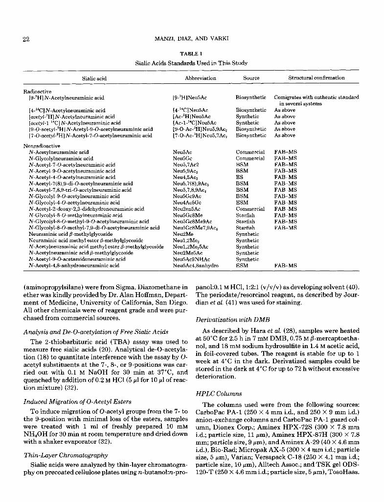
TABLE 1
Sialic Acids Standards Used in This Study
Sialic acid Abbreviation Source Structural confirmation
Radioactive [9-3H] N-Acetylneuraminic acid [9-3H]Neu5Ac Biosynthetic Comigrates with authentic standard
in several systems
[4-i%] N-Acetylneuraminic acid [4-‘%]Neu5Ac Biosynthetic As above
[acetyk3H] N-Acetylneuraminic acid [Ac-sH]Neu5Ac Synthetic As above
[ace&l-l-i%] N-Acetylneuraminic acid [AC-l-“C]Neu5Ac Synthetic As above
[9-O-acetyl-3H] N-Acetyl-S-0-acetylneuraminic acid [9-0-Ac-3H]Neu5,9Ac2 Biosynthetic As above [7-O-acetyl-3H]N-Acetyl-7-O-acetylneuraminic acid [7-0-Ac-3H]Neu5,7Ac2 Biosynthetic As above
Nonradioactive N-Acetylneuraminic acid Neu5Ac Commercial FAB-MS N-Glycolylneuraminic acid Neu5Gc Commercial FAB-MS N-Acetyl-7-0-acetylneuraminic acid Neu5,7Ac2 BSM FAB-MS N-Acetyl-S-0-acetylneuraminic acid Neu5,9Ac2 BSM FAB-MS N-Acetyl-4-0-acetylneuraminic acid Neu4,5Ac2 ES FAB-MS N-Acetyl-7(8),9-di-0-acetylneuraminic acid Neu5,7(8),9Acs BSM FAB-MS N-Acetyl-7&S-tri-0-acetylneuraminic acid Neu5,7,8,9Aq BSM FAB-MS N-Glycolyl-S-0-acetylneuraminic acid Neu5GcSAc BSM FAB-MS N-Glycolyl-4-0-acetylneuraminic acid Neu4AcBGc ESM FAB-MS N-Acetyl-2-deoxy-2,3didehydroneuraminic acid NeuZen5Ac Commercial FAB-MS N-Glycolyl-8-O-methylneuraminic acid Neu5Gc8Me Starfish FAB-MS N-Glycolyl-8-0-methyl-S-0-acetylneuraminic acid Neu5GcSMeSAc Starfish FAB-MS N-Glycolyl-8-0-methyl-7,9-di-0-acetylneuraminic acid NeuSGcSMe7,9Ac, Starfish FAB-MS Neuraminic acid P-methylglycoside Neu2Me Synthetic Neuraminic acid methyl ester @methylglycoside Neul,2Me, Synthetic N-Acetylneuraminic acid methyl ester &methylglycoside Neul,2Me,5Ac Synthetic N-Acetylneuraminic acid f3-methylglycoside NeuPMe5Ac Synthetic N-Acetyl-S-0-acetamidoneuraminic acid Neu5AcSNHAc Synthetic N-Acetyl-4,8-anhydroneuraminic acid Neu5Ac4,Sanhydro ESM FAB-MS
(aminopropylsilane) were from Sigma. Diazomethane in ether was kindly provided by Dr. Alan Hoffman, Depart- ment of Medicine, University of California, San Diego. All other chemicals were of reagent grade and were pur- chased from commercial sources.
Analysis and De-0-acetylation of Free Sialic Acids
The 2-thiobarbituric acid (TBA) assay was used to measure free sialic acids (20). Analytical de-o-acetyla- tion (18) to quantitate interference with the assay by O- acetyl substituents at the 7-, 8, or g-positions was car- ried out with 0.1 M NaOH for 30 min at 37”C, and quenched by addition of 0.2 M HCl(5 ~1 for 10 ~1 of reac- tion mixture) (32).
Induced Migration of 0-Acetyl Esters
To induce migration of 0-acetyl groups from the 7- to the g-position with minimal loss of the esters, samples were treated with 1 ml of freshly prepared 10 mM NH40H for 30 min at room temperature and dried down with a shaker evaporator (32).
Thin-Layer Chromatography
Sialic acids were analyzed by thin-layer chromatogra- phy on precoated cellulose plates using n-butanol:n-pro-
pano1:O.l M HCl, 1:2:1 (v/v/v) as developing solvent (40). The periodate/resorcinol reagent, as described by Jour- dian et al. (41) was used for staining.
Derivatization with DMB
As described by Hara et al. (28), samples were heated at 50°C for 2.5 h in 7 mM DMB, 0.75 M P-mercaptoetha- nol, and 18 mM sodium hydrosulfite in 1.4 M acetic acid, in foil-covered tubes. The reagent is stable for up to 1 week at 4°C in the dark. Derivatized samples could be stored in the dark at 4°C for up to 72 h without excessive deterioration.
HPLC Columns
The columns used were from the following sources: CarboPac PA-1 (250 X 4 mm i.d., and 250 X 9 mm i.d.) anion-exchange columns and CarboPac PA-1 guard col- umn, Dionex Corp.; Aminex HPX-72s (300 X 7.8 mm i.d.; particle size, 11 pm), Aminex HPX-87H (300 X 7.8 mm; particle size, 9 pm), and Aminex A-29 (40 X 4.6 mm i.d.), Bio-Rad, Micropak AX-5 (300 X 4 mm i.d.; particle size, 5 pm), Varian; Versapack C-18 (250 X 4.1 mm i.d.; particle size, 10 pm), Alltech Assoc.; and TSK gel ODS- 120-T (250 X 4.6 mm i.d.; particle size, 5 pm), TosoHaas.

HIGH-PRESSURE LIQUID CHROMATOGRAPHY OF SIALIC ACIDS 23
HPLC Pumps
A QIC reciprocating single-piston pump (Dionex Corp.) was used for the Carbopac column. All other anal- yses used Spectra-Physics 8700 and 8700XR pumps.
Detectors
The detectors used were: SP-8440 uv-visible detector, Spectra Physics; FD-300 dual monochromator fluores- cence detector (fitted with a 24 ~1 flow-cell), Spectra Vi- sion, Inc.; Ionchrom pulsed amperometric detector, Dio- nex Corp.; and Flow One Beta radioactive flow detector, Radiomatic Instruments.
HPLC Solvents, Buffers, and E&ion Procedures
Deionized water was passed through a five-stage Milli- Q Plus system, Millipore Corp. All solvents were HPLC grade, and all the buffers used for HPLC were prepared with Milli-Q purified water and filtered using a 0.45pm Nylon 66 membrane, Alltech Associates, Inc. Certified 50% (19.6 M) sodium hydroxide solution, with less than 0.05% sodium carbonate (Fisher Scientific) was used to prepare the 300 mM solution for postcolumn addition of base, prior to detection by the PAD.
System I-Ion-Exchange Chromatography on Pellicular Resin with PAD Detection
Carbopac PA-l columns were eluted at RT in the iso- cratic mode for 5 min with 5 mM sodium acetate solution, followed by a 30-min linear gradient to 50% 5 mM so-
dium acetate, 50% 5 mM acetic acid. The column was then washed with 100% 5 mM acetic acid for 10 min, and with 100% 5 mM sodium acetate for a further 15 min. The flow was 1 ml/min for the analytical column (4 X 250 mm) and 5 ml/min for the semipreparative col- umn (9 X 250 mm). Typical back pressure was 950-1400 psi. For analytical runs, the column effluent was mixed with 0.3-0.4 ml/min of 300 mM NaOH pumped under helium pressure, and monitored with the PAD using the following settings: E, = 0.05; E2 = 0.6; E3 = -0.8; T1 = 2; T2 = 2; T3 = 5; response time = 1; range = 100 or 300 nA. In preparative runs, 0.25-min fractions were collected and checked by the TBA assay. Fractions were pooled accordingly and desalted with a Dowex 50 (H+) column, washed with water. Pooled fractions were recovered by lyophilization, and analyzed by TLC on cellulose plates.
System II-Amine Adsorption/Ion Suppression Chromatography
The Micropak AX-5 column was eluted in the iso- cratic mode with acetonitrile:water:0.25 M NaH2P04 (72:18:10), at 1 ml/min and RT (25). The proportions of the solvents depended upon the separation desired (see
Results). The effluent was monitored for uv absorption at 200-205 nm, or for radioactivity according to the sam- ple. To detect radioactivity with maximum efficiency, it was necessary to use a high ratio of cocktail to effluent for the flow detector, or to evaporate the solvent from each collected fraction to be counted, and then add water and scintillation cocktail (1:lO).
System III-Reverse-Phase Chromatography of Derivatized Sialic Acids
Sialic acids derivatized with DMB were applied to a TSK-ODS 120T column eluted isocratically with 0.9 ml/ min of acetonitrile:methanol:water (9:7:84, v/v/v) at RT (28). Fluorescence of the eluate was monitored at an ex- citation wavelength of 373 nm and an emission wave- length of 448 nm; the high voltage was set at 900, the range at 500, and the response time at 0.5 s.
System IV-Anion-Exchange Chromatography on Aminex HPX- 72s
The column was eluted isocratically with 0.1 M sodium
sulfate solution (31,32). Depending on the particular col- umn, the flow rate and working temperature were 1.0 ml/ min at RT or 0.8 ml/min at 40°C (due to differences in the physical properties of the resin from batch to batch). The effluent was monitored for uv absorbance (200 nm) or for radioactivity. For preparative chromatography, fractions were collected beyond the uv detector directly into tubes on ice. Pooled fractions were diluted 20-fold in ice-cold distilled water and passed directly over a Do- wex 3-X4A column (formate form) equilibrated in 10 mM sodium formate, pH 5.5, maintained at 4°C. Sialic acids were then eluted with 10 column vol of 1 M formic acid, and recovered by lyophilization.
System V-Anion-Exchange Chromatography on Aminex A-29
Elution was in the isocratic mode at RT at 0.5 ml/ min flow, using 0.5-0.75 mM sodium sulfate (27,30). The exact concentration of sulfate depended upon the condi- tion of the individual column. The effluent was moni- tored for uv absorbance (200 nm) or for radioactivity.
System VI-Strong Cation-Exchange Chromatography
The Aminex HPX-87H column was eluted isocrati- tally with 0.006 M sulfuric acid at RT and 0.5 ml/min flow (29). The column effluent was monitored by means of a flow detector or by uv at 205 nm.
System VII-Reverse-Phase/Ion-Pair Chromatography
The eluting buffer was 0.4% tetrabutylammonium for- mate, pH 4.5, at 1.0 ml/min in the isocratic mode, at RT (25). The system was used only for radioactive samples,

24 MANZI, DIAZ, AND VARKI
and the radioactivity was continuously monitored by a flow detector, or by collecting fractions. For preparative runs, pooled fractions were passed directly over a 1 ml Dowex 50 (H+ form) column in water, washed through with 4 ml of water, and taken to dryness to remove the formic acid generated.
RESULTS AND DISCUSSION
System I-CarboPac PA-l Strong Anion-Exchange Pellicular Resin Column with Pulsed Amperometric Detection
Principles. This type of anion-exchange chromatog- raphy on a polymeric, nonporous, pellicular resin was re- cently introduced for the analysis of neutral saccharides, which are either partially or completely ionized at high pH (12-14) (Ref. (38)). Separation of neutral carbohy- drates is achieved by adjusting the eluant pH (Refs. (34,35)), and anionic oligosaccharides by sodium ace- tate/sodium hydroxide gradients (36,37). Such systems have separated polysaccharides and monosaccharides and have resolved structural isomers of oligosaccharides from biological samples. The only previous report of si- alit acid fractionation is the separation of N-acetylneur- aminic acid (retention time 4.0 min) and N-glycolyl- neuraminic acid (retention time 10 min) in the isocratic mode using 100 mM sodium hydroxide/l50 mM sodium acetate as the eluent (Dionex handbook).
Pulsed amperometric detection utilizes a repeating se- quence of three applied potentials, applied for specific durations (see Dionex Handbook for detailed princi- ples). The total current which results is the sum of (a) the carbohydrate oxidation current, (b) the current due to the charging of the electrode surface, and (c) the cur- rent caused by the oxidation of the gold electrode. Therefore, the current is measured only during the ap- plication of El (after a delay of several tens to hundreds of milliseconds), thus improving the signal-to-noise ra- tio. PAD has been shown to be linear over more than four- and one-half orders of magnitude (38). Approxi- mately 1% of the sample is oxidized, and molecules with similar size and structure are reported to have similar response factors (38). On the other hand, substituents on hydroxyl groups are expected to affect the sensitivity of detection. The detector works only at very high pH. Thus, when sialic acids are separated at neutral pH, it is necessary to add alkali to the effluent (300 mM sodium hydroxide at 0.3-0.4 ml/min) before it enters the PAD cell.
Elution characteristics. Since sialic acids have a pK, of approximately 2.0 they are bound to the resin at neu- tral pH, and it is necessary to use an anion to elute them. Sodium hydroxide was avoided because 0-acetyl esters could be saponified during chromatography. Samples were loaded in water to ensure binding and submitted
i0 i0 a0
TIME (min)
FIG. 2. Elution profile of standard sialic acids on a CarboPac PA-1 column with pulsed amperometric detection. A mixture of Neu5Ac, NeuB,OAc,, Neu5,7(8),9Ac3, Neu4,5Ac,, and Neu5Gc (5 nmol of each standard) was studied. Neu5Ac and Neu5,9Ac, overlap closely, giving one broad peak.
to a pH gradient elution with dilute sodium acetate and acetic acid (see Materials and Methods). This permitted separation of different 0-acetylated sialic acid species, whose binding to the resin is relatively weaker with higher numbers of substituents. Pure sialic standards and a variety of known mixtures from biological sources were analyzed by this method. Figure 2 shows an exam- ple with a mixture of known sialic acid standards. While some of the mono- and di-0-acetylated species can be separated from their parent molecule Neu5Ac, the rela- tive detector responses vary significantly with the pres- ence of different numbers of free hydroxyl groups (see below). Figure 3 shows the analysis of a mixture of sialic acids from equine serum (ES). After base hydrolysis, the two 0-acetylated sialic acids disappeared as expected (Fig. 3B). In addition to the increase in the parent com- pounds, a broad peak appears corresponding to 4&an- hydro compounds, a phenomenon peculiar to base hy- drolysis of 4-0-acetylated sialic acids (2). With loss of two hydroxyl groups, the anhydro sialic acids also give a poorer detector response.
Detection limits. The reported limit of sensitivity of the PAD detector for carbohydrates is about 50 pmol (34). We found that accurate detection of N-acetylneur- aminic acid can be done with as little as 200 pmol, using the above elution conditions (with the range setting of 100 nA, attenuation of the integrator at 1024, and a sig- nal-to-noise ratio of 2).
Relative detector responses. As expected, the detec- tor response of N-glycolylneuraminic acid, bearing one more hydroxyl group than N-acetylneuraminic acid, is 15% higher. In contrast, the response of the detector for

HIGH-PRESSURE LIQUID CHROMATOGRAPHY OF SIALIC ACIDS 25
Nsu4RAnhydroSAc
ative to Neu5Ac. This column can also be used for the analysis of labeled sialic acids, detected by means of an on-line flow detector, or by collecting and counting frac- tions (see Fig. 4 for an example of the latter). In this case, postcolumn addition of alkali is obviously not necessary.
Use for preparative purposes. For preparative runs the larger semipreparative column was used, and a maxi- mum of 2 pmol of total sialic acids could be injected with- out overloading the column. PAD could not be used since the postcolumn sodium hydroxide would hydrolyze the 0-acetyl esters. We therefore first checked the profile of the mixture to be separated using the analytical column, and the PAD. The semipreparative column was then checked and calibrated with labeled standards (a mix- ture of [4-l*C]Neu5Ac, [3H-O-acetyZ]Neu5,7Ac,, and Neu5,9Acz) using the flow detector to monitor the profile (as in Fig. 4). Finally, the preparative chromatography was done by collecting fractions, and monitoring the profile with the TBA assay for sialic acids (with analyti- cal de-0-acetylation). Different areas were pooled,
TABLE 2
FIG. 3. Elution profile of a mixture of sialic acids from equine serum on a CarboPac PA-l column with pulsed amperometric detection. Samples were injected onto the column untreated (A) or after base hydrolysis (B). The elution positions are as indicated.
Relative Elution Times of Sialic Acids in the Different Systems
R Neus~e in system
Neu5,9Ac, is only 46%, and for Neu5,7(8),9Ac3 45% that of the response for Neu5Ac. Some of the relative detec- tor response factors (PV-s/pmol) calculated for sialic acid standards from at least five different runs in differ- ent range and attenuation settings are: Neu5Ac, 30,500; Neu5,9Ac,, 14,500; Neu4,5Ac,, 14,100; Neu5Gc, 35,400; Neu5,7(8),9Ac,, 13,700; Neu5,7,8,9Ac4, 19,600; and Neu2en5Ac, 13,500. The detection limits are higher for substituted and anhydro sialic acids, because there are a lower number of ionizable hydroxyl groups. However, because of the possibility of partial de-0-acetylation be- tween the point of addition of postcolumn alkali and the PAD detector, the detector response factors may vary somewhat in different laboratories.
Relative elution times. Under these conditions we were able to separate standard mixtures of Neu5Ac and Neu5Gc, their mono-, di-, and tri-0-acetylated deriva- tives, and Neu2en5Ac, and its mono-0-acetyl ester. However, some overlap occurred between various com- pounds (e.g., Neu5Ac and Neu5,9Ac&. Mixtures of puri- fied sialic acids obtained from BSM and from the total lipid extract of the starfish Pisaster brevispinus whose compositions were previously determined by FAB-MS5) were also tested. In the latter case, the Neu5Gc8Me was separated from its mono- and di-0-acetyl esters (not shown). Table 2 shows retention times of all the ana- lyzed sialic acids (including some synthetic analogs) rel-
Sialic acid I II III IV v VI VII
Neu5Ac 1.00 1.00 1.00 1.00 1.00 1.00 1.00
Neu5Gc 1.17 1.50 0.84 1.20 1.33 0.93 1.00
Neu5,7Ac2 0.74 0.36 1.06 0.95 0.87 0.97 1.71
Neu5,gAc.r 0.95 0.35 1.57 1.30 1.47 0.97 1.71
Neu4,5Acz 0.76 0.39 1.68 1.22 1.41
Neu5,7(8),9Ac, 0.74 0.23 1.90 1.17 1.76
Neu5,7,8,9Ac, 0.62 ’ 1.98 1.62 2.05
Neu5GcSAc 1.06 0.49 0.88 1.57 1.70
Neu4Ac5Gc 0.86 0.54 1.53 1.59 1.69
NeuBen5Ac 2.21 0.66 b 1.54 1.68
Neu5GcSMe 0.82 0.75 1.03 0.94 0.89
Neu5GcSMeSAc 0.67 0.36 1.70 1.21
Neu5Gc8Me7,9Acz 0.59 0.29 2.20 1.47
NeuPMe 0.17 0.13 b 0.23
Neul,SMe, 0.40 0.85 b 0.46
NeufMeSAc 0.73 1.02 b 0.34 0.73
Neu5AcSNHAc 0.85 0.68 0.96
Neu5Ac4,8anhydro 2.15 0.58 1.39 2.04 Acetic acid 0.11 0.08 1.70
Note. Values in roman type are derived from our studies. Values in italic are from others reported in the literature. Typical RNeuSAc values for the various systems were as follows: System I, 18-24 min; System 11(72:18:6), 21-24 min; System III, 12-13 min; System IV, 15-17 min; System V, 5-6 min; System VI, 11-12 min; System VII, 12-13 min. Note that relative elution times can vary somewhat with specific col- umns and temperatures, particularly with the isocratic systems. How- ever, the order of elution of the various compounds relative to each other and to Neu5Ac is always maintained.
’ Does not react with DMB. b Elutes close to salt peak.

26 MANZI, DIAZ, AND VARKI
I Neu5,9Ac2
1 I I Neu5Ac I I
4 t
10 20
TIME (min)
of sialic acids). Also, the relative detector responses of the sialic acids in the PAD depend upon the number of free hydroxyl groups. The possibility of variable de-O- acetylation at the point of addition of postcolumn alkali (prior to entry into the PAD detector) further compli- cates the determination of accurate detector responses.
FIG. 4. Elution profile of labeled sialic acids on the CarboPac PA-l column. A mixture of [O-acetyZ-3H]Neu5,7Ac, and [O-acetyl- aH]Neu5,9Acz with an internal standard of [i4C]Neu5Ac was injected on the analytical CarboPac PA-1 column, and 0.5-min fractions were collected and counted.
Column maintenance. If the cleaning procedure with 100% of 5 mM acetic acid is not used in between runs the capacity of the column is significantly diminished after about 60 injections of 10 nmol each. When complete re- generation of the resin becomes necessary, the following procedure is useful: (i) run 3 column vols of 1 M HCl, (ii) wash well with water, (iii) run 3 column vols of 1 M
NaOH, and (iv) wash well with water. After this proce- dure the retention times return to their normal values. However, with some columns the decreased resolution is not completely recovered.
passed over a Dowex 50 (H+) column (1 ml in water) to eliminate the acetate, lyophilized, and monitored by TLC on cellulose plates. One example of such prepara- tive chromatography is shown in Fig. 5, for the mixture of sialic acids obtained from BSM. The recovery of total sialic acids was 87% in this case. As shown by the TLC (see Fig. 5B) there are four general areas in the profile: (i) di- (and probably tri-) 0-acetylated sialic acids area, (ii) mono-0-acetylated Neu5Ac area, (iii) Neu5Ac plus mono-0-acetylated Neu5Gc area, and (iv) Neu5Gc area. Although several of the pools obtained contain more than one component, all showed substantial enrichment in one class of molecules. Thus, it is possible to do sev- eral preparative runs with 2 pmol of the starting mix- ture, pool desired areas from each run, and rerun the combined pools for further purification.
Comparison of the Dionex System with other HPLC Systems
The set of sialic acid standards described above were also analyzed using six other previously described HPLC systems for sialic acids. In several cases modifications in described procedures were also devised to improve anal-
TIME (min)
Advantages. A major advantage of this system is the preparative separation of a variety of substituted sialic acids without exposure to conditions that cause loss or migration of 0-acetyl esters. The acetate can be easily removed by Dowex 50 (H+ form) followed by lyophiliza- tion, again without effects upon the 0-acetyl esters. To- tal recovery of sialic acids is also quite good (ca. 90%). Another advantage is analytical separation with detec- tion at a high sensitivity (minimum of 200 pmol for Neu5Ac). Finally, this is one of the systems capable of separating the 7- and 9-mono-0-acetyl isomers of Neu5Ac and Neu5Gc.
,,I' ,' ,, ,,,' ,/,'
,/' ,,* ,I ,' ,' ., ,a ,I ,, /' *' ,I ,I ,' ,, ,,'
,/ //. <.. ,“ *. ,,
,, ,,,' ,,~~',,1~,,/,,1~ ,,' ,,' ,/',,/ <_.
,:' ,,' ,,' ,I' ,,.' ,,' ,*' ,,' ,*' ,s' ,,' Neu5,(7/9)9Ac,
B
-J-=-l- Neu5,9Ac,
I- II Neu5Gc9Ac
-II 111 Neu5,7Ac,
Limitations and disadvantages. For preparative pur- poses, it is not possible to use the PAD to monitor the profile since the 0-acetylated sialic acids are destroyed by the high concentration of sodium hydroxide. Thus, it is necessary to use a more laborious procedure (TBA reaction followed by TLC analysis) to monitor the prep- aration. The capacity of the preparative column cur- rently marketed is somewhat limited (maximum 2 pmol indicated accurately.
FIG. 5. Elution profile of sialic acids from bovine submaxillary mu- tin on a semipreparative CarboPac PA-l column. Sialic acids from bovine submaxillary mucin (2 pmol) were chromatographed and frac- tions (0.25 min) collected. (A) The elution profile determined by the TBA reaction with and without prior de-0-acetylation. (B) The TLC profile of the pooled fractions on cellulose plates, using equal amounts (10 nmol) of sialic acids from each pool. Standards of Neu5Ac (ex- treme left lane) and Neu5Gc (extreme right lane) were also included. The plates were developed with resorcinol reagent. Because of poor photographic reproduction, the plates were photocopied and scanned using an Apple scanner. The location of the individual bands is thus

HIGH-PRESSURE LIQUID CHROMATOGRAPHY OF SIALIC ACIDS 27
ysis. The results for each system are presented below along with an outline of basic principles, advantages, and disadvantages in each case.
System II-Amine Adsorption/Ion Suppression Chromatography: Micropak AX-5 Column with UV Detection
This approach is based upon hydrogen bonding be- tween the hydroxyl groups of saccharides and the amine functions of the stationary phase (42,43). In order to fractionate anionic molecules, an ion pair is added to the mobile phase to suppress the ionic effects, while retain- ing hydrogen bonding (44). We have previously reported use of this type of chromatography with a Micropak AX- 5 column, for the analysis of some sialic acids (25). In this study, we report further experience with this sys- tem, and the retention times of several additional sialic acids. We obtained good separations and sharp peaks in the isocratic mode with a mixture of acetonitrile:water: 0.25 M monobasic sodium phosphate at 1 ml/min. The relative ratio of the solvents can be varied depending on the kind of sialic acid mixture to be separated. It is nec- essary to maintain a minimum concentration of 10% phosphate and a maximum working percentage of aceto- nitrile (72%) above which the phosphate starts precipi- tating. A 64:26:10 (v/v/v) ratio of acetonitrile:water:0.25 M sodium phosphate is useful for the separation of Neu5Ac and Neu5Gc. However, for 0-acetylated sialic acids it is necessary to increase the percentage of aceto- nitrile to increase their retention times (72:18:10, v/v/ v). Chromatography is monitored by absorption at 200 nm, which requires extreme purity of the samples and reagents. Residual amounts of purification reagents such as acetic or formic acids produce an absorption peak close to the void volume. Typical profiles on this column for some radioactive sialic acids have previously been published (25). Table 2 shows the relative retention times for several sialic acids previously reported by us and those for some sialic acids and sialic acid analogs not previously reported. The lower limit of confident de- tection was 2 nmol, and the molar responses are similar for all the sialic acids.
The same analytical column can be used for prepara- tive purposes (up to 400 nmol of sialic acids with a good resolution of the peaks). The eluant was monitored by uv and the peaks were collected directly on ice, diluted lo-fold with cold water, and purified by ion-exchange chromatography on Dowex 3-X4A (formate) at 4°C. The column was washed with 10 vol of 10 mM formic acid and eluted with 7 vol of 1 M formic acid, and sialic acids were recovered by lyophilization. We obtained -65% recov- ery of sialic acids, but some loss and migration of O-ace- tyl groups occurred.
The various advantages and disadvantages of this sys- tem are summarized in Table 3. These columns also have
many other uses in the study of oligosaccharides (42,45,46), making them economically attractive. When a given analysis takes more than one day, it is recom- mended that a slow flow of the working buffer be main- tained overnight. When the column loses resolution and retention is reduced, it can be cleaned by running 0.5 M
phosphoric acid for at least 1 h and washing extensively with water before changing to the working buffer. More complete regeneration of the column can be achieved by injecting 3-aminopropyltriethoxysilane (3 X 1 ml) while running the column in 100% acetonitrile, washing ex- tensively with the same solvent, and then with water and 0.5 M phosphate before use. This treatment restores the amino groups on the resin. The performance of the col- umn is fully recovered after this treatment; however, the retention times may not be identical.
Other similar systems. Other amine-adsorption:ion- suppression based methods have been reported to be useful for the separation of sialic acids and sialic acid analogs, including Spherisorb-NH, columns (47,48). We have also explored use of a Varian Micropak-SP oligonu- cleotide column (300 X 4 mm), that can be run basically under the same conditions discussed for the Micropak AX-5, with similar results (not shown). However, this size of oligonucleotide column is no longer available.
System III-Reverse-Phase Chromatography of Derivatized Sialic Acids:TSK-ODS 120T with Fluorometric Detection
Hara et al. (33) have developed an HPLC separation of fluorescent derivatives of Neu5Ac and Neu5Gc, ob- tained by reaction with DMB in sulfuric acid and sepa- rated by reverse-phase chromatography on a Radial-Pak Cl8 cartridge with a mixture of methanol-acetonitrile- water. More recently, these authors formed the fluores- cent derivatives in acetic acid at lower temperatures, completely avoiding migration and de-0-acetylation (28). An extensive list of other DMB-reacting sub- stances that could be present in biological samples was also examined, all of which gave very weak responses and/or very long retention times under the recom- mended conditions (28). We have confirmed the value of this technique and extended it to the analysis of several additional sialic acid molecules (see Table 2). Figure 6 shows the elution profile of a complex mixture of sialic acids from BSM, with or without prior treatment with alkali. It can be seen that after induced migration with a very mild base, the peaks corresponding to the 7-0- acetylated molecules disappear because of migration of the esters to the g-position (18,32,49). After more exten- sive base hydrolysis, there is almost complete elimina- tion of the 0-acetylated molecules, with an increase in the amount of the parent compounds, Neu5Ac and Neu5Gc.
The previously reported limits of detection were in the range of 100-200 fmol at a signal-to-noise ratio of 3 (28).

28 MANZI, DIAZ, AND VARKI
TABLE 3
Comparison of Some Features of HPLC Systems for Sialic Acids
System
Feature I II III” IV V VI VII
1. Sensitivity 2. Specificity of detection 3. Separation by number of hydroxyls or
substituents 4. Separation of isomers 5. Preparative use 6. Avoids ester migration during purification 7. Avoids ester loss during purification
Good Good
Good Good Best Good Good
Poor Poor
Good Poor Fair Poor Fair
Best Best
Good Good NA Good
N/A
Poor Poor
Good Good Good Poor Fair
Poor Poor
Good Good Poor NT NT
Poor Poor
Poor No NT NT NT
NA* NA*
Good No Good* NT Good
Note. NA, not applicable, NT, not tested. ’ Derivatized samples only. * Radioactive samples only.
In our hands, confident detection and quantitation was possible with 2.5 pmol of any sialic acid. The original study reported no significant differences in detector re- sponses between the various sialic acids (28). However,
10
TIME (mi;)
DMBVATIVES OF:
1 = NeuSGc
2 q NeuSGc7Ac
3 = Neu5Ac
4 = Neu5,7Ac2
5 = NeuSGcSAc
6.?
7 = Neu5,9As
a-7
9 I Neu5,(7/Q)9Ac3
MIGRATION
>E-0-ACETYLATION
FIG. 6. Elution profile of DMB-derivatized sialic acids on a TSK- ODS 120T column with fluorometric detection. A mixture of purified sialic acids from bovine submaxillary mucin (16.6 nmol) was deriva- tired with DMB after no treatment (A), induced migration of 0-acetyl groups (B), or de-0-acetylation with base (C). The position of elution of known DMB-derivatives of sialic acids is indicated.
we have some observed differences in the area of the peaks corresponding to the derivatives of different sialic acids, and found that it is probably due to the different degree of derivatization that each individual molecule reaches. In the prior study, the fluorescent peak profiles suggested that complete derivatization of the sialic acids might be occurring. However, analysis of labeled sialic acids by this method showed that the derivatization re- action does not go to completion. Thus, as shown in Fig. 7, different radioactive sialic acids gave different degrees of incorporation of radioactivity into the corresponding chromophore peak. A substantial portion of the radioac- tivity ran in other areas of the chromatogram, not corre- sponding to any fluorescence (note that the sample ana- lyzed was identical to that shown in Fig. 4, and did not
Neu5AeDMB
1’0 2‘0
Time (min)
1
1 t
7
d 0
z
x
s 1.
FIG. 7. Elution profile of DMB-derivatized radioactive sialic acids on a TSK-ODS 120T column. A mixture of [O-acetyl-3H]Neu5,7Ac2 and Neu5,9Ac, with an internal standard of [‘%]Neu5Ac (same sam- ple as in Fig. 4) was derivatized with DMB and run on the TSK-ODS 120T column. Fractions (0.5 min) were collected and counted. The positions of elution of known fluorometric standards are indicated. The unknown radioactive peaks are indicated by a question mark.

HIGH-PRESSURE LIQUID CHROMATOGRAPHY OF SIALIC ACIDS 29
contain any intrinsic labeled contaminants). Attempts to improve derivatization by increasing the reaction time to 4.5 h, or the concentration of reagent to 21 mM,
did not result in an increase of the derivative peak, but of the third radioactive peak, indicating breakdown of the product increased (data not shown). However, as long as appropriate standards are run in parallel, the in- complete derivatization does not pose a practical prob- lem in quantitation of unlabeled sialic acids.
The advantages and disadvantages of this system are summarized in Table 3. Preparative use of this column is not possible, because the samples are irreversibly de- rivatized. However, derivatized samples can be pooled, lyophilized, and rechromatographed after base treat- ment, to confirm 0-acetylation (28). The original au- thors reported that the column could be used for more than 1000 injections (10 ~1 each) if it was washed with methanol:water (l:l, v/v) at 1.0 ml/min for 30 min at the end of each day (28). However, in our hands, the pressure goes over the manufacturers recommended limit; we therefore wash at 0.5 ml/min. We also found it impor- tant to keep the amount of DMB reagent in each injec- tion as low as possible to maintain column performance. Therefore, when very small amounts of material that need to be injected completely into the column are ana- lyzed, we suggest a reduction in the total volume of the derivatization reaction.
System IV-Anion-Exchange Chromatography on an Aminex HPX 72-S Column with uv Detection
This column is normally used to separate weak or- ganic bases or acids by a combination of ion-exclusion, ion-exchange, and partition chromatography on an 8% crosslinked quaternized polystyrene divinylbenzene CO-
polymer in the sulfate form. When moderately concen- trated sulfate solutions were used as the mobile phase, we found that this column separated sialic acids on the basis of the negative charge and the total number of hy- droxyl groups (32). However, the presence of an extra hydroxyl group in the N-acetyl group (as in Neu5Gc) in- creased retention time relative to Neu5Ac (31,32). We have previously reported elution of sialic acids in the iso- cratic mode with 100 mM sodium sulfate, at room tem- perature and a flow rate of 1.0 ml/min (32). However, the manufacturer has recently reported a change in the physical characteristics of the resin, causing a need to change operating parameters. It is now necessary to in- crease the temperature (at least up to 40°C; the maxi- mum temperature allowed by the resin is 70°C) and to reduce the flow rate (at least down to 0.8 ml/min) to achieve the same separation and to avoid compaction of the resin. About 2 nmol of each sialic acid can be confi- dently detected. As commented before for other HPLC methods, the relative detector responses can be assumed to be the same for the different sialic acid derivatives.
We previously reported the use of an Aminex HPX-72s column to separate Neu5,7Acz and Neu5,9Acz (32) and recently the utilization of the system for the analysis and preparative purification of complex mixtures of sialic acids from biological sources.’ We report here also the retention times of a variety of other sialic acids and sialic acid analogs (see Table 2).
For preparative separations, the load limit is about 500 nmol of total sialic acids. Peaks are collected on ice directly from the effluent, immediately diluted with wa- ter (20-fold) and submitted to anion-exchange chroma- tography on Dowex 3-X4A (formate form) at 4°C. Sialic acids are eluted with 1 M formic acid and recovered by lyophilization. This procedure causes a low but variable loss of 0-acetylated species because of migration and de- 0-acetylation during post-HPLC purification. We have recently employed this method to completely separate pairs of sialic acids obtained by cellulose chromatogra- phy of total sialic acids from bovine submaxillary mucin.2
The major advantages and disadvantages of this method are summarized in Table 3. The retention time of acetate is quite long in this system (approx 30 min), eluting beyond all the sialic acids. This can allow the characterization of the sample and the quantitation of the percentage of 0-acetyl esters present in the starting material, following base treatment. A major current lim- itation of this method is the variation in availability of a resin of the required quality to resist the high pressures involved in the analysis. While the previously available columns showed stable performance for over 250 injec- tions, the physical properties of the packing material are no longer as consistent. It has become necessary to work at a lower flow rate and higher temperatures in order to avoid high back pressure. The higher pressure limit is set at 1000 psi because whenever the working back pressure exceeds this value, the subsequent increase in back pres- sure becomes exponential. When the limit is reached, it is possible to further increase the temperature (up to the limit of 70°C) or reduce the flow rate, and to recheck the retention times and resolution of standards in the new conditions. In order to extend the lifetime of the column it is also necessary to use a guard column. It is advisable to start heating the column with solvent flowing through it, but not to initially operate the column at a flow rate greater than 0.2 ml/min at ambient temperature. During the shutdown procedure the heater is first turned off and the flow rate is reduced to 0.2 ml/min and continued un- til the column reaches ambient temperature. The col- umn can be cleaned with 5 or 20% acetonitrile in 0.1 M
(NH&SO4 and regenerated with 0.5 M (NH&SO,.
System V-Anion-Exchange Chromatography on Aminex A-28 or A-29 with uv Detection
Bio-Rad Aminex ion-exchange resins for HPLC are finely sized spherical beads of polystyrene divinylben-

30 MANZI, DIAZ, AND VARKI
zene copolymer with attached functional groups. Shukla and Schauer reported anion-exchange HPLC of sialic acids carried out on quaternary amine resin types, with 8% crosslinkage, and a particle size of 11 -+ 2 pm (Ami- nex A-28) or 5-8 pm (Aminex A-29) (27,30,50,51). These very short columns (40 X 4.6 mm) are eluted in the iso- cratic mode with dilute sodium sulfate. Schauer and co- workers usually use 0.75 mM sodium sulfate at a flow rate of 0.5 ml/min and ambient temperature. In our hands, the resolution is improved when 0.50 mM sodium sulfate is used as the eluant at that flow rate. This difference might depend on the particular column that is used, be- cause of the extremely small column volume. Each run takes approximately 10 min and can be monitored by on- line reading of the absorbance at 200 nm, or by monitor- ing of radioactivity. In our hands, the detection of 2 nmol of Neu5Ac is possible. However, Schauer’s group re- ported lower limits of detection (27,50). The relative de- tector response can be considered the same for all com- pounds in the family that have an N-acyl group. Table 2 contains all the reported retention times for sialic acids and sialic acid analogs including our data and the data of Schauer.
The general advantages and disadvantages of this sys- tem are summarized in Table 3. The overlapping of peaks in complex mixtures is too high and the capacity of the column too low to permit preparative chromatog- raphy. A major advantage of this system is the short time required for the analysis, making it specially suitable for the study of enzyme reactions, where one aliquot of the enzyme assay mixture can be directly injected into the column. This usually gives information about both substrates and reaction products in one chromato- graphic run. Several such examples have been reported (27,30,52). The fast elution of substituted sialic acids also helps to avoid migration of acetyl groups and/or de- 0-acetylation. However, because of the short time of the run, elution times are very close and it is easy to miss the presence of a minor component. Therefore, it is not recommended for the analysis of complex mixtures from biological sources.
The column can be used for the analysis of total en- zyme reaction mixtures if small volumes are injected, but when crude enzymatic preparations are used it is advis- able to previously purify the sialic acids. The column can be cleaned by running it backward for 30 min in 5% ace- tonitrile in the working buffer at 0.1 ml/min; the per- centage of acetonitrile can be increased up to 30% if nec- essary. More complete regeneration is done with 0.2 M
ammonium or sodium sulfate in water at 0.1 ml/min, at room temperature, overnight. However, in our hands the life of the column is relatively limited.
System VI-Strong Cation-Exchange Chromatography: Bio-Rad HPX-87H Column with uu Detection The Aminex HPX-87H column is packed with cation-
exchange resin in the hydrogen form. It was previously
used for the analysis and quantitation of Neu5Ac in se- rum of melanoma and breast carcinoma patients and was able to separate Neu5Ac and Neu5Gc (29). A mobile phase of 0.006 N sulfuric acid was previously used at a flow rate of 0.65 ml/min and 42°C for the separation of Neu5Ac, and Neu5Gc, with the uv detector set at 206 nm (29). In order to maintain the back pressure of the column within the permitted limits, we used a flow rate of 0.5 ml/min, at room temperature. The maximum re- ported sensitivity for Neu5Ac in this system is 0.8 nmol/ ml (total injection volume = 50 ~1) with a signal-to-noise ratio >3 (29). Previously reported elution times are in- cluded in Table 2, together with some of our recent data in this system. Analysis of sialic acids on this column is limited only to NeuSAc and Neu5Gc, since substituted sialic acids coelute with their parent molecules and may be destroyed by sulfuric acid. No major advantage of this system has been found (see Table 3). It provides a faster comparative analysis of sialic acids levels in serum hy- drolysates than the TBA-HPLC assay (21) (because of the lack of the derivatization step), but is not better in this regard than the Aminex A-28 or A-29 system. The use for preparative purposes has many obvious limita- tions. Similar precautions to those for the HPX 72-S column are necessary to control the back pressure and to prevent compaction.
System VII-Reverse-Phase/Ion-Pair Chromatography: Cl8 Column with Radioactive Detection
Reverse-phase, ion-pair, column partition chromatog- raphy neutralizes the negatively charged carboxyl groups of the sialic acids with a positively charged lipo- philic counterion (e.g., tetrabutylammonium) dissolved in the mobile phase, to form an ion pair that is able to partition into the lipophilic stationary phase of the col- umn. The elution of the ion pair is produced by increas- ing the solvent strength (e.g., methanol concentration) of the mobile phase. We previously reported the use of a PBondapak Cl8 column (Waters) with TBA formate as a counterion for the analysis of free sialic acids (25). This system cannot be monitored by uv because of the high absorbance of the buffer, limiting analysis to radio- active samples. The relative elution times of the labeled sialic acids that have been analyzed by this method are included in Table 2. The system cannot separate Neu5Ac and Neu5Gc, but allows separation according to the number of 0-acetyl groups, due to the increase in hydrophobicity.
Although the use of this system for preparative purposes has not been explored extensively, it is possible to easily eliminate the tetrabutylammonium formate by passing the eluant through a Dowex 50 (Hf form) col- umn. At the present time, we see no obvious advantage to this method over other previously described methods. However, because it can only be used for radioactive si-

HIGH-PRESSURE LIQUID CHROMATOGRAPHY OF SIALIC ACIDS 31
alit acids, we have not been able to examine a large num- ber of standards. As with many other reverse-phase sep- arations, it shows significant variability in elution time if the column is not thoroughly cleaned with organic sol- vents such as methanol and well-equilibrated with the ion-pair containing solution.
CONCLUSIONS
In this study, we have reported a new HPLC method for the separation and analysis of sialic acids: anion-ex- change chromatography on a pellicular resin column with pulsed amperometric detection. The high sensitiv- ity of this type of detection gives the system the lowest detection limit available for the HPLC analysis of un- derivatized sialic acids (minimum of 200 pmol). The CarboPac PA-1 column was especially useful for prepar- ative purposes, allowing the separation of a variety of substituted sialic acids without loss or migration of O- acetyl esters and with excellent recovery. Using a variety of natural and synthetic sialic acids, we have compared this method with all of the previously described HPLC methods for the analysis of these compounds. In four of these systems, we have made a direct comparison with the present method, using the same set of standards, and have reported new information to build upon the exist- ing literature. Two other systems of lesser utility that we have experience with are mentioned primarily for com- pleteness. We have mentioned the advantages and dis- advantages of each system, and made general sugges- tions for their use (see Table 3 for a summary). We have also described the basic principles of each method and the precautions to be taken in their use. It should be noted that less than half of all the possible combinations of sialic acid modifications have been currently reported in the literature. Thus, it is possible that the use of these techniques will result in the discovery of new sialic acids, with different combinations of currently known substi- tutions, or new types of substitutions.
In general, the effect of substitutions on retention times is different for different systems. For example, O- acetyl esters retard elution in the HPX 72-S system, whereas free hydroxyl groups cause retardation in the AX-5 system. Thus, it is possible to roughly predict where a particular type of sialic acid might elute in the various systems. However, variations between isomers occur, making exact predictions impossible. It is also im- portant to note that in all isocratic separations, differ- ences in the exact retention times can be expected between different columns or different laboratory condi- tions. However, the order of elution of the different sialic acids is very consistent. In the final analysis, no single system is ideal for all aspects of the preparation and analysis of the various sialic acids. However, taken to- gether, the various methods allow the detailed study of the wide variety of sialic acids that are found in nature.
Rosenberg, A., and Schengrund, C. (1976) in Biological Roles of Sialic Acid (Rosenberg, A., and Schengrund, C., Eds.), pp. 295- 359, Plenum, New York/London.
Schauer, R. (1982) Sialic Acids: Chemistry, Metabolism and Func- tion, Cell Biology Monographs, Vol. 10, Springer-Verlag, New York.
Nadano, D., Iwasaki, M., Endo, S., Kitajima, K., Inoue, S., and Inoue, Y. (1986) J. Biol. Chem. 261,11,550-11,557.
Schauer, R. (1982) Adu. Carbohydr. Chem. Biochem. 40,131-234.
Muchmore, E., Varki, N., Fukuda, M., and Varki, A. (1987) FA- SEBJ. 1,229-235.
REFERENCES
1.
2.
3.
4.
5.
6.
7.
8.
9.
10.
11.
12.
Constantine-Paton, M., Blum, A. S., Mendez-Otero, R., and Barn- stable, C. J. (1986) Nature (London) 324,459-462.
Sparrow, J. R., and Barnstable, C. J. (1988) J. Neurosci. 21,398- 409.
Levine, J. M., Beasley, L., and Stallcup, W. B. (1986) Deu. Brain Res. 27,211-222.
Schlosshauer, B., Blum, A. S., Mendez-Otero, R., Barnstable, C. J., and Constantine-Paton, M. (1988) J. Neurosci. 8,580-592.
Varki, A., and Kornfeld, S. (1980) J. Exp. Med. 152,532-544.
Higa, H. H., Rogers, G. N., and Paulson, J. C. (1985) Virology 144,279-282.
13.
14.
Rogers, G. N., Herrler, G., Paulson, J. C., and Klenk, H. D. (1986) J Biol. Chem. 261,5947-5951. Muchmore, E., and Varki, A. (1987) Science 236,1293-1295.
Cheresh, D. A., Reisfeld, R. A., and Varki, A. (1984) Science 225, 844-846.
15
16.
17.
18.
19.
20.
21.
22.
23.
24.
25.
26.
27.
28.
29.
30.
Higashi, H., Hirabayashi, Y., Fukui, Y., Naiki, M., Matsumoto, M., Ueda, S., and Kato, S. (1985) Cancer Res. 45,3796-3802.
Orskov, F., Orskov, I., Sutton, A., Schneerson, R., Lin, W., Egan, W., Hoff, G. E., and Robbins, J. B. (1979) J. Exp. Med. 149,669- 685.
Corfield, A. P., Sander Wewer, M., Veh, R. W., Wember, M., and Schauer, R. (1986) Biol. Chem. Hoppe Seyler 367,433-439.
Varki, A., and Diaz, S. (1984) Anal. Biochem. 137,236-247.
Schauer, R. (1987) in Methods in Enzymology (Ginsburg, V., Ed.), Vol. 138, pp. 132-161, Academic Press, San Diego.
Warren, L. (1959) J. Biol. C&m. 234‘1971-1975.
Powell, L. D., and Hart, G. W. (1986) Anal. Biochem. 157, 179-
185.
Casals Stenzel, J., Buscher, H. P., and Schauer, R. (1975) Anal. Biochem. 65,507-524.
Reuter, G., Pfeil, R., Stoll, S., Schauer, R., Kamerling, J. P., Ver- sluis, C., and Vliegenthart, J. F. (1983) Eur. J. Biochem. 134,139- 143.
Haverkamp, J., van Halbeek, H., Dorland, L., Vliegenthart, J. F., Pfeil, R., and Schauer, R. (1982) Eur. J. Biochem. 122,305-311. Diaz, S., and Varki, A. (1985) Anal. Biochem. 150,32-46. Shukla, A. K., Scholz, N., Reimerdes, E. H., and Schauer, R. (1982) Anal. Biochem. 123,78-82.
Shukla, A. K., and Schauer, R. (1986) Anal. Biochem. 158, 158- 164.
Hara, S., Yamaguchi, M., Takemori, Y., Furuhata, K., Ogura, H., and Nakamura, M. (1989) Anal. Biochem. 179.162-166. Silver, H. K. B., Karim, K. A., Gray, M. J., and Salinas, F. A. (1981) J. Chromatogr. 224,381-388.
Schauer, R., Schroder, C., and Shukla, A. K. (1984) Adu. Exp. Med.Biol. 174,75-86.

32 MANZI, DIAZ, AND VARKI
31. Holzhauser, R., Faillard, H., Klose, W., Huber, W., Stickl, H., and Landthaler, M. (1988) K2in. Wochenschr. 66,540-544.
32. Diaz, S., Higa, H. H., Hayes, B. K., and Varki, A. (1989) J. Biol. Chem. 264,19,416-19,426.
33. Hara, S., Yamaguchi, M., Takemori, Y., Nakamura, M., and Oh- kura, Y. (1986) J. Chromatogr. 377,111-119.
34. Hardy, M. R., Townsend, R. R., and Lee, Y. C. (1988) Anal. Bio- them. 170,54-62.
35. Hardy, M. R., and Townsend, R. R. (1989) Carbohydr. Res. 188, l-7.
36. Townsend, R. R., Hardy, M. R., Hindsgaul, O., and Lee, Y. C. (1988) Anal. Biochem. 174,459-470.
37. Townsend, R. R., Hardy, M. R., Cumming, D. A., Carver, J. P., and Bendiak, B. (1989) An&. Biochem. 182,1-8.
38. Hardy, M. R., and Townsend, R. R. (1988) Proc. Natl. Acad. Sci. U.S.A. 85,3289-3293.
39. Warren, L., and Glick, M. C. (1966) in Methods in Enzymology (Eastabrook, R. W., and Pullman, M. E., Eds.), Vol. 8, pp. 131- 133, Academic Press, San Diego.
40. Schauer, R. (1978) in Methods in Enzymology (Ginsburg, V., Ed.), Vol. 50, pp. 64-89, Academic Press, San Diego.
41. Jourdian, G. W., Dean, L., and Roseman, S. (1971) J. Biol. Chem. 246,430-435.
42. Mellis, S. J., and Baenziger, J. U. (1981) Anal. Biochem. 114,276- 280.
43. Bergh, M. L., Koppen, P., and Van den Eijnden, D. H. (1981) Car- bohydr. Res. 94,225-229.
44. Mellis, S. J., andBaenziger, J. U. (1983) Anal. Biochem. 134,442- 449.
45. Mellis, S. J., and Baenziger, J. U. (1983) J. Biol. Chem. 258, 11,546-11,556.
46. Baenziger, J. U., and Natowicz, M. (1981) A&. Biochem. 112, 357-361.
47. Gross, H. J., Bunsch, A., Paulson, J. C., andBrossmer, R. (1987) Eur. J. Biochem. 168,595-602.
48. Higa, H. H., and Paulson, J. C. (1985) J. Biol. Chem. 260,8838- 8849.
49. Kamerling, J. P., Schauer, R., Shukla, A. K., Stoll, S., van Hal- beek, H., and Vliegenthart, J. F. (1987) Eur. J. Biochem. 162, 601-607.
50. Shukla, A. K., Schauer, R., Unger, F. M., Zahringer, U., Rietschel, E. T., and Brade, H. (1985) Curbohydr. Res. 140,1-8.
51. Shukla, A. K., Schauer, R., Schade, U., Moll, H., and Rietschel, E. T. (1985) J. Chromatogr. 337,231-238.
52. Herrler, G., Rott, R., Klenk, H. D., Muller, H. P., Shukla, A. K., and Schauer, R. (1985) EMBO J. 4,1503-1506.


![Review Structure, function and metabolism of sialic acids · 2017. 8. 23. · acids in some protozoa, viruses and bacteria [1, 2, 9–11]. Thus, several strains of Escherichia coli](https://static.fdocuments.in/doc/165x107/603a67dedc73e72b9149b168/review-structure-function-and-metabolism-of-sialic-acids-2017-8-23-acids-in.jpg)


![Determination of sialic acids in the nervous system of silkworm … · mammals, the central nervous system has the highest concentration of sialic acids [56]. The majority is pres-ent](https://static.fdocuments.in/doc/165x107/5edd48dfad6a402d66685251/determination-of-sialic-acids-in-the-nervous-system-of-silkworm-mammals-the-central.jpg)