Hecker / Steveling / Peuker / Kastner / Liebchen Color ... · PDF fileHecker / Steveling /...
36
Hecker / Steveling / Peuker / Kastner / Liebchen Color Atlas of Acupuncture Reading excerpt Color Atlas of Acupuncture of Hecker / Steveling / Peuker / Kastner / Liebchen Publisher: MVS Medizinverlage Stuttgart http://www.narayana-verlag.com/b11868 In the Narayana webshop you can find all english books on homeopathy, alternative medicine and a healthy life. Copying excerpts is not permitted. Narayana Verlag GmbH, Blumenplatz 2, D-79400 Kandern, Germany Tel. +49 7626 9749 700 Email [email protected] http://www.narayana-verlag.com
Transcript of Hecker / Steveling / Peuker / Kastner / Liebchen Color ... · PDF fileHecker / Steveling /...

Hecker / Steveling / Peuker / Kastner / LiebchenColor Atlas of Acupuncture
Reading excerptColor Atlas of Acupuncture
of Hecker / Steveling / Peuker / Kastner / LiebchenPublisher: MVS Medizinverlage Stuttgart
http://www.narayana-verlag.com/b11868
In the Narayana webshop you can find all english books on homeopathy, alternative medicineand a healthy life.
Copying excerpts is not permitted.Narayana Verlag GmbH, Blumenplatz 2, D-79400 Kandern, GermanyTel. +49 7626 9749 700Email [email protected]://www.narayana-verlag.com

254
Description of the Muscle
Origin: Lateral epicondyle of humerus,annular and collateral ligaments ofradius, fascia of lower arm.
Insertion: Dorsal aponeurosis; proximalto the middle finger joints, the aponeuro-sis divides into the ulnar and radial ten-dinous portions that reunite distally tothe joint in an aponeurosis and insert atthe base of the terminal phalanges.
Innervation: Deep branch of radial nerve(C6 to C8).
Action: Extends finger joints, extendswrist, and supports ulnar abduction.
Trigger Points of the Extensor Muscle ofFingers
Preliminary Remarks
Trigger points are found here predomi-nantly in the muscle bellies of the exten-sor muscles of the ring and middle fin-gers. Activation of trigger points usuallytakes place through chronic strain. As-sociated trigger points are often also pre-sent in the finger muscles and the exten-sor muscle of wrist.
Examination of Trigger Points
Typical local twitch responses can betriggered in the middle of the musclebelly in the region of the trigger points.
Therapy of Trigger Points
Targeted intramuscular stimulation withsubsequent passive stretching of themuscle is effective within a short time.Conventional needling and therapeuticlocal anesthesia may also be considered.
� Extensor Muscle of Fingers
aus: Hecker u. a., Color Atlas of Acupuncture (ISBN 9783131252227) © 2008 Georg Thieme Verlag

255
1
2
Trigger Points and Areas of PainProjection
� Extensor Muscle of Fingers, TriggerPoint 1
The trigger point of the middle finger ex-tensor is found close to the elbow in theregion of the muscle belly. Typical painprojection runs along the muscle into themiddle finger; now and then pain is alsolocalized over the proximal flexion creaseof the wrist.
� Extensor Muscle of Fingers, TriggerPoint 2
The trigger point of the ring finger exten-sor lies distal and ulnar to trigger point 1.Its area of pain projection reaches intothe ring finger and up toward the radio-humeral joint.
Trigger Points of the Extensor Muscle of Fingers
aus: Hecker u. a., Color Atlas of Acupuncture (ISBN 9783131252227) © 2008 Georg Thieme Verlag
256
LI-11
LI-10
LI-9
LI-8
Important Acupuncture Points and TheirLocalizations
LI-8
Location: 4 Cun distal to acupoint LI-11.
LI-9
Location: 3 Cun distal to acupoint LI-11.
LI-10
Location: 2 Cun distal to acupoint LI-11.
LI-11
Location: Lateral to the radial end of theflexion crease of elbow when the lowerarm is flexed at a right angle, in a depres-sion between the end of the crease andthe lateral epicondyle in the region of thelong radial extensor of the wrist.
� Extensor Muscle of Fingers
aus: Hecker u. a., Color Atlas of Acupuncture (ISBN 9783131252227) © 2008 Georg Thieme Verlag

257
TB-9
TB-8
TB-6TB-5
TB-4
TB-4
Location: Slightly ulnar to the center ofthe dorsal flexion crease of the wrist (thejoint space between radius, ulna, andproximal wrist bone series), ulnar to thetendon of the extensor muscle of fingers,radial to the tendon of the extensormuscle of little finger.
TB-5
Location: 2 Cun proximal to acupointTB-4, between the radius and ulna, on aline connecting acupoint TB-4 and the tipof the olecranon process of ulna.
TB-6
Location: 3 Cun proximal to acupointTB-4, between the radius and ulna, on theline connecting acupoint TB-4 and the tipof the olecranon.
TB-8
Location: 4 Cun proximal to acupointTB-4, between the radius and ulna.
TB-9
Location: 7 Cun proximal to acupointTB-4 on the line connecting acupointTB-4 and the tip of the olecranon. Hence,on the connecting line described, thepoint lies 1 Cun proximal to the middlebetween acupoint TB-4 and the flexioncrease of elbow.
Acupuncture Points of the Extensor Muscle of Fingers
aus: Hecker u. a., Color Atlas of Acupuncture (ISBN 9783131252227) © 2008 Georg Thieme Verlag

258
Description of the Muscle
Origin: Humeral head: medial epicon-dyle of humerus; ulnar head: coronoidprocess of ulna.
Insertion: Lateral surface of radius andpronator tuberosity.
Innervation: Median nerve (C6 and C7).
Action: Pronates the forearm and con-tributes to flexion of the elbow joint.
Trigger Points of the Pronator TeresMuscle
Preliminary Remarks
Trigger points are usually found in theproximal part of the muscle belly. Theiractivation is caused by repetitive prona-tion of the forearm, either through exces-sive workload or through chronic stressfrom sports (e. g., occasional tennis playerwith poor serving technique).The median nerve passes underneath thepronator teres muscle, and sometimes itruns through the muscle. Compression ofthe nerve may lead to a characteristic en-trapment neuropathy that may finally re-semble carpal tunnel syndrome.
Examination of Trigger Points
The muscle is easy to examine by deeppalpation in the cubital fossa. Palpationtriggers the characteristic radiation ofpain.
Therapy of Trigger Points
There is a risk of damaging the mediannerve. Before dry-needling or injectingthe trigger points, the course of the me-dian nerve must be accurately identified.Manual treatment by acupressure isanother option.
� Pronator Teres Muscle
aus: Hecker u. a., Color Atlas of Acupuncture (ISBN 9783131252227) © 2008 Georg Thieme Verlag

259
PC-3 HT-3
Trigger Points and Areas of PainProjection
� Pronator Treres Muscle, Trigger Point
The main trigger point is found in themuscle belly in the cubital fossa near theorigin of the muscle. The pain radiatesfrom the proximal anteroradial part ofthe forearm to the wrist, where it reachesthe proximal palmar part of the thumb.
Important Acupuncture Points and theirLocalizations
PC-3
Location: On the ulnar side of the tendonof the biceps brachii muscle, in the elbowcrease.
HT-3
Location: Between the ulnar end of theelbow crease and the medial epicondyleof the humerus when the elbow is flexed.
Trigger and Acupuncture Points of the Pronator Teres Muscle
aus: Hecker u. a., Color Atlas of Acupuncture (ISBN 9783131252227) © 2008 Georg Thieme Verlag

260
Description of the Muscle
Origin: Humero-ulnar head: medial epi-condyle of humerus and coronoid processof ulna.Radial head: anterior surface of radius.
Insertion: Four tendons insert on thelateral bony ridges of the middle pha-langes of fingers II to V.
Innervation: Median nerve (C7 to T1).
Action: Flexes the metacarpophalangealjoints II to V and the proximal inter-phalangeal joints II to V.
Miscellaneous: The tendons of the deepflexor muscle of the fingers pass betweenthe parts of the tendon insertion on theend phalanges.
Trigger Points of the Superficial FlexorMuscle of the Fingers
Preliminary Remarks
The flexors of the fingers, like the tensorsof the fingers, are superficial muscles. Toavoid damage to the nerves, deep nee-dling should never be performed. Triggerpoint activation is caused by chronicstrain due to manual work. In particular,monotonous grasping movements acti-vate these trigger points.
Examination of Trigger Points
It requires only slight pressure to palpatethe trigger points in the middle of themuscle belly. This is done by gently pal-pating through the ulnar and radial flexormuscles of the wrist as well as the palmarmuscle. Accurate identification is con-firmed by increased sensation of painwhen palpating the trigger points whilesimultaneously checking muscle func-tion.
� Superficial Flexor Muscle of the Fingers
aus: Hecker u. a., Color Atlas of Acupuncture (ISBN 9783131252227) © 2008 Georg Thieme Verlag

261
Therapy of Trigger Points
Damage to the ramifications of the me-dian nerve and to the ulnar artery andvein should be avoided by taking greatcare during dry-needling or injection.The trigger points are easy to inactivate.Subsequent stretching of the flexors bydorsal extension of the fingers is essentialfor preventing relapses, and patientsshould be advised to do this on their own.
Trigger Points and Areas of PainProjection
In the radial portion of the flexor muscles,the pain radiates into the palmar side ofthe middle finger; in the ulnar portion itradiates into the ring finger or little fin-ger, sometimes with further projectioninto the palm.
Trigger Points of the Superficial Flexor Muscle of the Fingers
aus: Hecker u. a., Color Atlas of Acupuncture (ISBN 9783131252227) © 2008 Georg Thieme Verlag
262
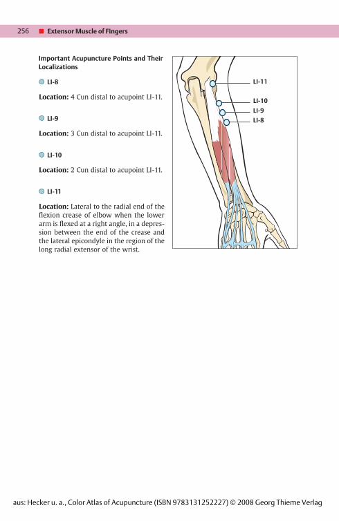
HT-3
PC-6
HT-4
HT-7
PC-7HT-5
LU-7
PC-3LU-5
Important Acupuncture Points and theirLocalizations
LU-5
Location: Radial to the biceps tendons inthe elbow crease.
LU-7
Location: On the radial side of the fore-arm, in a V-shaped groove proximal tothe styloid process of the radius, 1.5 Cunproximal to the crease of the wrist. Theacupoint is located where the proximalportion of the styloid process of theradius merges into the shaft of the radius.
PC-3
Location: On the ulnar side of the tendonof the biceps brachii muscle, in the elbowcrease.
PC-6
Location: 2 Cun proximal to the palmarflexion crease of the wrist that lies proxi-mal to the pisiform bone, between thetendons of the palmaris longus muscleand the radial flexor muscle of the wrist.As described for the location of acupointHT-7, choose the wrist crease that lies be-tween the radius and ulna on the one sideand the proximal row of carpal bones onthe other. As the proximal row of carpalbones is marked by the pisiform bone,the crease in question is located proximalto the pisiform bone.
PC-7
Location: In the middle of the palmarflexion crease of the wrist that lies proxi-mal to the pisiform bone, between thetendons of the palmaris longus muscleand the radial flexor muscle of the wrist.
� Superficial Flexor Muscle of the Fingers
aus: Hecker u. a., Color Atlas of Acupuncture (ISBN 9783131252227) © 2008 Georg Thieme Verlag

263
HT-3
Location: Between the ulnar end of theelbow crease and the medial epicondyleof the humerus when the elbow is flexed.
HT-4
Location: 1.5 Cun proximal to acupointHT-7, radial to the tendon of the ulnarflexor muscle of the wrist.
HT-5
Location: 1 Cun proximal to acupointHT-7, radial to the tendon of the ulnarflexor muscle of the wrist.
HT-7
Location: At the palmar flexion crease ofthe wrist, radial to the tendon of the ulnarflexor muscle of the wrist.
Acupuncture Points of the Superficial Flexor Muscle of the Fingers
aus: Hecker u. a., Color Atlas of Acupuncture (ISBN 9783131252227) © 2008 Georg Thieme Verlag

264
5th Rib
12th Rib
5th Rib
12th Rib
Description of the Muscle
Origin: Inferior borders and outer sur-faces of the 5th to 12th ribs.
Insertion: Pubic tubercle, pubic crest,outer margin of iliac crest, inguinal liga-ment, and linea alba.
Innervation: Intercostal nerves (T5 toT11), subcostal nerve (T12), iliohypogas-tric nerve (T12 to L1), and ilioinguinalnerve (L1).
Action: Unilateral contraction rotates thethorax against the pelvis to the con-tralateral side. Bilateral contraction flexesthe vertebral column. Furthermore, itacts as auxiliary muscle for abdominalcompression and forced expiration.
Trigger Points of the External ObliqueMuscle of the Abdomen
Preliminary Remarks
Trigger points frequently develop in con-nection with an acute abdomen (board-like abdomen). Trigger points are also ob-served with diseases of the inner organs,such as dysmenorrhea, diarrhea, spasmof the urinary bladder, and testicularpain. They may occur primarily and thencause secondary abdominal symptoms.More often, however, it is the other wayaround: the presence of visceral afferentstimuli leads to trigger point formation inthe abdominal muscles. Acute lumbago isalso frequently associated with triggerpoints in the oblique abdominal muscles.
Examination of Trigger Points
With the patient sitting, taut bands andtrigger points in this muscle are provokedby rotating movements.
Therapy of Trigger Points
Dry-needling is possible without anyproblem, and trigger point infiltration isalso an option. Injection or acupunctureof the trigger points is performed withthe patient in a supine position. Punctureof the peritoneum must be avoided.However, damage to the inner organsrarely occurs.
� External Oblique Muscle of the Abdomen
aus: Hecker u. a., Color Atlas of Acupuncture (ISBN 9783131252227) © 2008 Georg Thieme Verlag
265
1
2
1
2
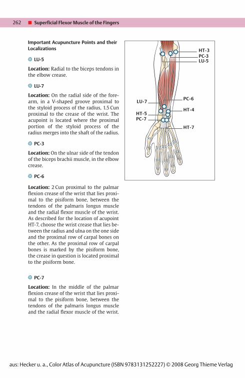
Trigger Points and Areas of PainProjection
� External Oblique Muscle of Abdomen,Trigger Point 1
It lies on the anterior border of the costalarch toward the epigastrium. The charac-teristic radiation of pain into the epi-gastrium mimics symptoms of anginapectoris or epigastric complaints.
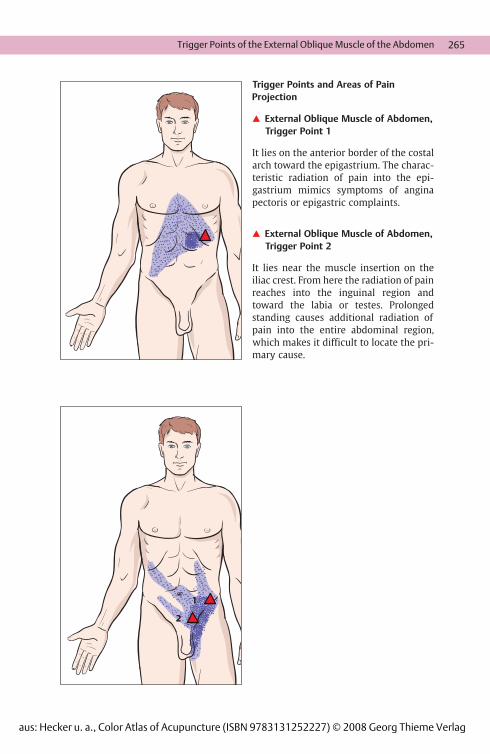
� External Oblique Muscle of Abdomen,Trigger Point 2
It lies near the muscle insertion on theiliac crest. From here the radiation of painreaches into the inguinal region andtoward the labia or testes. Prolongedstanding causes additional radiation ofpain into the entire abdominal region,which makes it difficult to locate the pri-mary cause.
Trigger Points of the External Oblique Muscle of the Abdomen
aus: Hecker u. a., Color Atlas of Acupuncture (ISBN 9783131252227) © 2008 Georg Thieme Verlag

266
CV-6
CV-4
CV-2
CV-3
CV-12
CV-15CV-14
LR-14
LR-13
GB-25
ST-25
SP-15
GG b 3 0
5th Rib
GG b 3 0
12th Rib
Important Acupuncture Points and theirLocalizations
CV-2
Location: At the superior border of thepubic symphysis, on the anterior midline.
CV-3
Location: 1 Cun superior to the middle ofthe superior border of the pubic symphy-sis.
CV-4
Location: 2 Cun superior to the middle ofthe superior border of the pubic symphy-sis (for correct orientation, see acupointCV-3).
CV-6
Location: 1.5 Cun inferior to the umbili-cus (for correct orientation, see acupointCV-3).
CV-12
Location: Midway between the base ofthe xiphoid process and the umbilicus.
CV-14
Location: 1 Cun inferior to the tip of thexiphoid process (acupoint CV-15).
CV-15
Location: Just below the tip of the xi-phoid process on the anterior midline.
CV-17
Location: On the anterior midline at thelevel of the nipples in the fourth ICS.
� External Oblique Muscle of the Abdomen
aus: Hecker u. a., Color Atlas of Acupuncture (ISBN 9783131252227) © 2008 Georg Thieme Verlag
267
LR-14
Location: In the sixth ICS, below thenipple on the mammillary line.
ST-25
Location: 2 Cun lateral to the umbilicus.
SP-15
Location: 4 Cun lateral to the umbilicus.
Acupuncture Points of the External Oblique Muscle of the Abdomen
aus: Hecker u. a., Color Atlas of Acupuncture (ISBN 9783131252227) © 2008 Georg Thieme Verlag

268
Description of the Muscle
Origin: Iliac fossa to terminal line of pel-vis, anterior inferior iliac spine, andlacuna of muscles to anterior surface ofthe hip joint capsule.
Insertion: Lesser trochanter of femur
Innervation: Femoral nerve (T12 toL3/L4).
Action: Together with the greater psoasmuscle it forms the strongest flexormuscle of the hip joint (iliopsoas muscle).With the pelvic and lumbar regions fixed,it flexes the thigh. With the femur immo-bilized, it rotates the ipsilateral pelvislaterally.
Trigger Points of the Iliac Muscle
Preliminary Remarks
Muscle shortenings are very commonwith coxarthrosis. The muscle has ageneral tendency to contract and developtrigger points. This tendency is often pro-moted by visceral afferent stimuli occur-ring in response to irritation of the cecumbordering directly on the fascia of theiliac muscle. The trigger points frequentlyappear in association with trigger pointsof other muscles (e. g., quadratus lum-borum, rectus abdominis, and rectusfemoris muscles; tensor muscle of fascialata). Treatment of these secondary trig-ger points is therefore recommended.
Examination of Trigger Points
With the relaxed patient in supine posi-tion, the muscle is directly palpated be-tween the cecum and the inside of theiliac bone. However, adhesions in the re-gion often make this difficult. In this case,manual mobilization of the cecum is usu-ally necessary. One trigger point is foundin the more anterior part of the muscle.Another trigger point is found at the levelof the hip joint.
Therapy of Trigger Points
Acupuncture of the trigger points in theiliac muscle may be attempted if thececum can be moved far enough in me-dial direction. It is important to treat thecause of the visceral lesion as well. Re-lapses are avoided by physiotherapeuticstretching techniques involving exten-sion of the ipsilateral hip joint with maxi-mum flexion of the contralateral hip jointat the same time, and stretching of therectus femoris muscle, which is usuallyalso contracted.
� Iliac Muscle
aus: Hecker u. a., Color Atlas of Acupuncture (ISBN 9783131252227) © 2008 Georg Thieme Verlag

269
21
32
1
2
Trigger Points and Areas of PainProjection
� Iliac Muscle, Trigger Points 1 and 2
Trigger point 1 lies in the ventral portionof the iliopsoas muscle and preverte-brally at the level of vertebra L3. Triggerpoint 2 lies directly above the hip joint.Their areas of pain projection are founddirectly paravertebrally in the lumbar re-gion with radiation into the sacroiliacjoint and the upper medial gluteal area.Another area of pain projection appearsover the rectus femoris muscle withradiation toward the anterior inferioriliac spine.
Trigger Points of the Iliac Muscle
aus: Hecker u. a., Color Atlas of Acupuncture (ISBN 9783131252227) © 2008 Georg Thieme Verlag

270
Description of the Muscle
Origin: Lateral surfaces of vertebrae T12to L4, and intervertebral disks and costalprocesses of the lumbar vertebrae.
Insertion: Lesser trochanter of femur.
Innervation: Femoral nerve (T12 toL3/L4).
Action: Together with the iliac muscle, itforms the strongest flexor muscle of thehip joint (iliopsoas muscle). With thefemur fixed, it flexes the lumbar spine,posteriorly rotates the ipsilateral half ofpelvis, and laterally flexes the lumbarspine.
Miscellaneous: Between the two por-tions of the psoas muscle lies the lumbarplexus.
Trigger Points of the Psoas Muscle
Preliminary Remarks
The psoas muscle is subdivided into thesmaller psoas muscle and the greaterpsoas muscle. Trigger points arefrequently found in the region of thegreater psoas muscle. They are associatedwith repetitive strain injuries and poorposture of the lumbar spine, and alsowith coxarthrosis. Here, too, there may bevisceral afferent stimuli; they originatefrom the kidney directly overlying thepsoas muscle or from the sigmoid colontraversing on the left. An anterior iliac le-sion is therefore frequently found on theright (anterior rotation of the pelvic half),or a posterior iliac lesion is found on theleft (posterior rotation of the pelvic half).This results in a functional difference inleg length caused by shortening of theleft leg, or lengthening of the right leg,due to distal displacement (on the right)or proximal displacement (on the left) ofthe rotational center of the hip joint. It istherefore recommended to treat not onlythe trigger points but definitely also the
causes of the underlying distortion of thepelvis.
Examination of Trigger Points
The greater psoas muscle can only be ex-amined in a relaxed patient and by usingdeep palpation. It is often very sensitiveto pressure. Jump signs are absent.
Therapy of Trigger Points
Trigger points in the region of the psoasmuscle are usually not accessible to dry-needling or injection, and if so, then onlywith difficulty. Other stretching methodsare therefore recommended, such as my-ofascial release.
Important Acupuncture Points and TheirLocalizations
Because of the relatively protected deepposition of the psoas muscle it is difficultto access via acupuncture.
� Greater Psoas Muscle
aus: Hecker u. a., Color Atlas of Acupuncture (ISBN 9783131252227) © 2008 Georg Thieme Verlag

271Trigger Points of the Psoas Muscle
aus: Hecker u. a., Color Atlas of Acupuncture (ISBN 9783131252227) © 2008 Georg Thieme Verlag
272
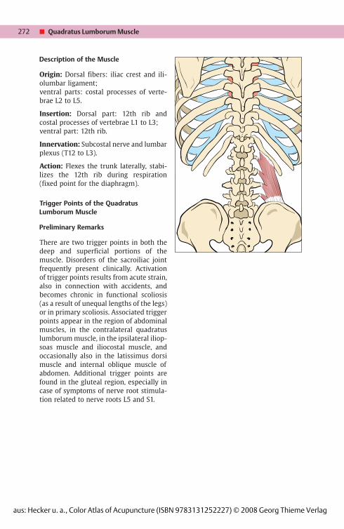
Description of the Muscle
Origin: Dorsal fibers: iliac crest and ili-olumbar ligament;ventral parts: costal processes of verte-brae L2 to L5.
Insertion: Dorsal part: 12th rib andcostal processes of vertebrae L1 to L3;ventral part: 12th rib.
Innervation: Subcostal nerve and lumbarplexus (T12 to L3).
Action: Flexes the trunk laterally, stabi-lizes the 12th rib during respiration(fixed point for the diaphragm).
Trigger Points of the QuadratusLumborum Muscle
Preliminary Remarks
There are two trigger points in both thedeep and superficial portions of themuscle. Disorders of the sacroiliac jointfrequently present clinically. Activationof trigger points results from acute strain,also in connection with accidents, andbecomes chronic in functional scoliosis(as a result of unequal lengths of the legs)or in primary scoliosis. Associated triggerpoints appear in the region of abdominalmuscles, in the contralateral quadratuslumborum muscle, in the ipsilateral iliop-soas muscle and iliocostal muscle, andoccasionally also in the latissimus dorsimuscle and internal oblique muscle ofabdomen. Additional trigger points arefound in the gluteal region, especially incase of symptoms of nerve root stimula-tion related to nerve roots L5 and S1.
� Quadratus Lumborum Muscle
aus: Hecker u. a., Color Atlas of Acupuncture (ISBN 9783131252227) © 2008 Georg Thieme Verlag

273
1
2
1
23
4
3
4
Examination of Trigger Points
First of all, the following orthopediccauses should be clarified: functional orstructural scoliosis, scoliotic pelvis, ob-lique position of pelvis, and displacementof pelvis. Palpation of trigger points isperformed while the patient is lying re-laxed on his/her side. Local twitch re-sponses are rarely observed; usuallythere is distinct hardening of the muscle.
Therapy of Trigger Points
Direct needling is only possible withacupuncture needles of at least 60 mm inlength. Therapeutic local anesthesia is apossible alternative. However, dry-nee-dling can usually be successfully per-formed as well; in lateral position, theneedle is aimed in the direction of thetransverse processes. As follow-up treat-ment, stretching the muscles is carriedout in the dorsal position with the hipjoint flexed approximately 80° usingpostisometric relaxation by adduction ofthe hip joint. This also stretches the en-tire gluteal region.
Trigger Points of the Quadratus Lumborum Muscle
aus: Hecker u. a., Color Atlas of Acupuncture (ISBN 9783131252227) © 2008 Georg Thieme Verlag

274
1
2
1
2
Trigger Points and Areas of PainProjection
� Quadratus Lumborum Muscle,Trigger Points 1 and 2
The superficial trigger point 1 lies ap-proximately 2 Cun below the lateral endof the muscle border and 2 Cun below the12th rib; it shows an area of pain projec-tion at the level of the lateral and dorsalproximal gluteal regions with radiationtoward the groin and the sacroiliac joint.Trigger point 2 is found at the level of L4,just above the insertion of the quadratuslumborum muscle at the dorsolateraliliac crest. Its pain projection lies at thelevel of the greater trochanter and radi-ates in ventral and dorsal directions.
� Quadratus Lumborum Muscle
aus: Hecker u. a., Color Atlas of Acupuncture (ISBN 9783131252227) © 2008 Georg Thieme Verlag
275
BL-52
BL-51
BL-23
1
3
4
3
4
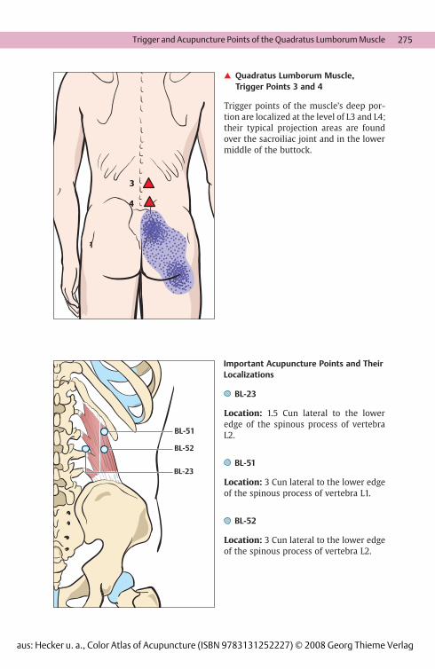
� Quadratus Lumborum Muscle,Trigger Points 3 and 4
Trigger points of the muscle’s deep por-tion are localized at the level of L3 and L4;their typical projection areas are foundover the sacroiliac joint and in the lowermiddle of the buttock.
Important Acupuncture Points and TheirLocalizations
BL-23
Location: 1.5 Cun lateral to the loweredge of the spinous process of vertebraL2.
BL-51
Location: 3 Cun lateral to the lower edgeof the spinous process of vertebra L1.
BL-52
Location: 3 Cun lateral to the lower edgeof the spinous process of vertebra L2.
Trigger and Acupuncture Points of the Quadratus Lumborum Muscle
aus: Hecker u. a., Color Atlas of Acupuncture (ISBN 9783131252227) © 2008 Georg Thieme Verlag

276
Description of the Muscle
Origin: Dorsal aspect of the ilium, thora-columbar fascia, lateral edge of thesacrum and coccyx, sacrotuberal liga-ment.
Insertion: Gluteal tuberosity of thefemur, iliotibial tract of the fascia lata,lateral intermuscular septum.
Innervation: inferior gluteal nerve (L4 toS1).
Action: Extends the thigh in the hip joint;upper fibers: abduction,lower fibers: adduction, rotate the thighlaterally.
Trigger Points of the Gluteus MaximusMuscle
Preliminary Remarks
The muscle has three trigger points. Trig-ger points in this region often appear incombination with those of the gluteusminimus muscle and the sciaticocruralmuscle. Trigger points of the deep dorsalextensor muscles are also found to be as-sociated. Activation often results fromacute events associated with increasedstrain of the gluteus maximus muscle.Such trigger points are thereforefrequently found in athletes.
Examination of Trigger Points
The trigger points lie superficially andcan easily be palpated. Local twitch re-sponses are rarely observed. Especially inthe case of trigger points 1 and 2, directpressure sensibility of the sciatic nerve inthe sense of Valleix’s points should bedifferentiated.
Therapy of Trigger Points
Inactivation of trigger points is achievedwithout any problem by acupuncture,dry-needling, and therapeutic local anes-thesia. Targeted stretching exercisesusing postisometric relaxation completethe treatment.
� Gluteus Maximus Muscle
aus: Hecker u. a., Color Atlas of Acupuncture (ISBN 9783131252227) © 2008 Georg Thieme Verlag

277
11
22
Trigger Points and Areas of PainProjection
� Gluteus Maximus Muscle, TriggerPoint 1
Trigger point 1 lies on the extension of avertical line through the posterior iliacspine at the level of the proximal end ofthe gluteal fold; it has its main projectionarea along the medial and caudal marginsof the muscle.
� Gluteus Maximus Muscle, TriggerPoint 2
Trigger point 2 is found at the level of thecaudal margin of the muscle approxi-mately 4 to 5 cm above the gluteal crease.The projection areas are localized in thisregion, the entire gluteal region includingthe region over the caudal sacrum, andabove the greater trochanter.
Trigger Points of the Gluteus Maximus Muscle
aus: Hecker u. a., Color Atlas of Acupuncture (ISBN 9783131252227) © 2008 Georg Thieme Verlag
278
33
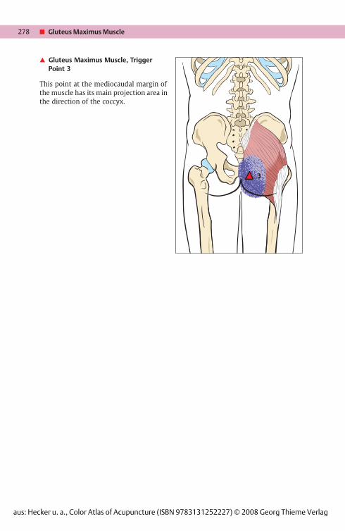
� Gluteus Maximus Muscle, TriggerPoint 3
This point at the mediocaudal margin ofthe muscle has its main projection area inthe direction of the coccyx.
� Gluteus Maximus Muscle
aus: Hecker u. a., Color Atlas of Acupuncture (ISBN 9783131252227) © 2008 Georg Thieme Verlag

279
BL-27 30
BL-27
B 36
BL-28BL-29BL-30 BL-54
BL-53
GB-30
BL-28BL-29BL-30
BL-53
BL-54
BL-36
GB-30GB-30
Important Acupuncture Points and TheirLocalizations
BL-27
Location: At the level of the first sacralforamen, 1.5 Cun lateral to the dorsal me-dian line in a depression between thesacrum and upper region of the posteriorsuperior iliac spine.
BL-28
Location: At the level of the 2nd sacralforamen, 1.5 Cun lateral to the dorsal me-dian line.
BL-29
Location: At the level of the 3rd sacralforamen, 1.5 Cun lateral to the dorsal me-dian line.
BL-30
Location: At the level of the 4th sacralforamen, 1.5 Cun lateral to the dorsal me-dian line.
BL-36
Location: In the middle of the glutealfold.
BL-53
Location: At the level of the 2nd sacralforamen, 1.5 Cun lateral to acupointBL-28.
BL-54
Location: At the level of the 4th sacralforamen, 3 Cun lateral to the sacral hia-tus.
GB-30
Location: On the lateral side of the hipjoint, on the line connecting the greatertrochanter and the sacral hiatus, betweenthe outer and middle third.
Trigger and Acupuncture Points of the Gluteus Maximus Muscle
aus: Hecker u. a., Color Atlas of Acupuncture (ISBN 9783131252227) © 2008 Georg Thieme Verlag

280
11 2 31 2 3
Description of the Muscle
Origin: Ala of the ilium between theanterior and posterior gluteal lines.
Insertion: Greater trochanter of femur.
Innervation: Superior gluteal nerve (L4to S1).
Action: Abducts the leg in the hip joint.Stabilizes the pelvis on the side of thesupporting leg, contributes to medial ro-tation of the unsupporting leg.
Trigger Points of the Gluteus MediusMuscle
Trigger points are found along the entiremuscle. They develop especially becauseof strain caused by sports or work, butalso after accidents. Dysfunction of thesacroiliac joint is frequently observed.
Examination of Trigger Points
When the hip joint is flexed by 90° andadducted, direct palpation usually pro-vokes the trigger points. The same posi-tion is used for stretching the shortenedmuscle groups during follow-up treat-ment.Targeted intramuscular stimulation withsubsequent passive follow-up treatmentby stretching the muscle is very effectivehere. Manual therapy, including adjust-ment of the affected sacroiliac joint,should be performed concurrently. Alter-natively, conventional needling or ther-apeutic local anesthesia may be used.
� Gluteus Medius Muscle
aus: Hecker u. a., Color Atlas of Acupuncture (ISBN 9783131252227) © 2008 Georg Thieme Verlag

281
222
111
Trigger Points and Areas of PainProjection
� Gluteus Medius Muscle,Trigger Point 1
It lies in the posterior portion of the glu-teus medius muscle near the posteriorsuperior iliac spine and leads to radiationof pain around the sacroiliac joint.
� Gluteus Medius Muscle,Trigger Point 2
It lies in the middle of the gluteus mediusmuscle and leads to radiation of pain inthe gluteal region and into the greatertrochanter.
Trigger Points of the Gluteus Medius Muscle
aus: Hecker u. a., Color Atlas of Acupuncture (ISBN 9783131252227) © 2008 Georg Thieme Verlag
282
333
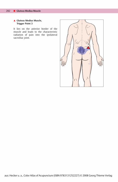
� Gluteus Medius Muscle,Trigger Point 3
It lies on the anterior border of themuscle and leads to the characteristicradiation of pain into the ipsilateralsacroiliac joint.
� Gluteus Medius Muscle
aus: Hecker u. a., Color Atlas of Acupuncture (ISBN 9783131252227) © 2008 Georg Thieme Verlag

283
EX-B-6
EX-B-7
BL-53
BL-54
GB-30
Important Acupuncture Points and theirLocalizations
EX-B-6
Location: Below the inferior border ofthe spinous process of vertebra L4 and3 Cun lateral to the posterior midline.
EX-B-7
Location: 3.5 Cun lateral to the inferiorborder of the spinous process of vertebraL4.
BL-53
Location: At the level of the second sacralforamen, 1.5 Cun lateral to acupointBL-28.
BL-54
Location: 3 Cun lateral to the sacral hia-tus at the level of the fourth sacral fora-men.
GB-30
Location: On the lateral side of the hip,one-third of the distance between thegreater trochanter and the sacral hiatus.In China, this acupoint is always needledwith the patient in a lateral position. Thehip and knee of the side to be treated areflexed, while the leg beneath is straight.This position prevents the sciatic nervefrom being injured.
Trigger and Acupuncture Points of the Gluteus Minimus Muscle
aus: Hecker u. a., Color Atlas of Acupuncture (ISBN 9783131252227) © 2008 Georg Thieme Verlag

284
Description of the Muscle
Origin: Ala of the ilium between theanterior and posterior lines.
Insertion: Greater trochanter of femur.
Innervation: Superior gluteal nerve (L4to S1).
Action: When fully contracted, themuscle abducts the thigh. With only theanterior portion of the muscle con-tracted, it rotates the unsupporting legmedially; with only the posterior portioncontracted, it rotates the unsupportingleg laterally and extends it slightly. Con-traction on the side of the supporting legcauses stabilization of the pelvis.
Trigger Points of the Gluteus MinimusMuscle
Preliminary Remarks
These trigger points develop quitefrequently in combination with those ofthe gluteus medius. The causes are simi-lar.
Examination of Trigger Points
The gluteus minimus muscle can only bepalpated when the gluteus mediusmuscle is relaxed; the origin of the glu-teus medius is further proximal and su-perficial. With the patient in the lateralposition, palpation is performed with thehip joint flexed by 90° and abducted.
Therapy of Trigger Points
As with the gluteus medius muscle, directmethods such as intramuscular stimula-tion by dry-needling are very effective,when followed by passive stretching withthe hip joint flexed and abducted by 90°.Therapeutic local anesthesia or conven-tional needling are also an option. Patientsshould be advised to do the stretching ofthe muscle on their own.
� Gluteus Minimus Muscle
aus: Hecker u. a., Color Atlas of Acupuncture (ISBN 9783131252227) © 2008 Georg Thieme Verlag
285
BL-53
BL-54
GB-30
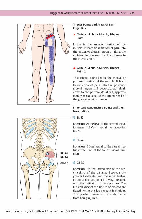
Trigger Points and Areas of PainProjection
� Gluteus Minimus Muscle, TriggerPoint 1
It lies in the anterior portion of themuscle. It leads to radiation of pain intothe posterior gluteal region or along theiliotibial tract across the knee down tothe lateral ankle.
� Gluteus Minimus Muscle, TriggerPoint 2
This trigger point lies in the medial orposterior portion of the muscle. It leadsto radiation of pain into the posteriorgluteal region and posterolateral thighdown to the posterolateral calf, approxi-mately at the level of the lateral head ofthe gastrocnemius muscle.
Important Acupuncture Points and theirLocalizations
BL-53
Location: At the level of the second sacralforamen, 1.5 Cun lateral to acupointBL-28.
BL-54
Location: 3 Cun lateral to the sacral hia-tus at the level of the fourth sacral fora-men.
GB-30
Location: On the lateral side of the hip,one-third of the distance between thegreater trochanter and the sacral hiatus.In China, this acupoint is always needledwith the patient in a lateral position. Thehip and knee of the side to be treated areflexed, while the leg beneath is straight.This position prevents the sciatic nervefrom being injured.
Trigger and Acupuncture Points of the Gluteus Minimus Muscle
aus: Hecker u. a., Color Atlas of Acupuncture (ISBN 9783131252227) © 2008 Georg Thieme Verlag

286
Description of the Muscle
Origin: Anterior surface of the sacrum.
Insertion: Tip of greater trochanter offemur.
Innervation: Sacral plexus (L5 to S2).
Action: Abducts the thigh and rotates itlaterally.
Miscellaneous: In case of early divisionof the sciatic nerve, the common fibularnerve passes across the piriformis muscleand can be constricted here (piriformissyndrome).
Trigger Points of the Piriformis Muscle
Preliminary Remarks
The two trigger points of the piriformismuscle are often associated with chronicpain in the region of the loin, pelvis, andhip. They are activated by chronic dis-orders of the lumbosacral transition butonly rarely as a reaction to acute strain. Incases where the muscle is shortened, en-trapment of the sciatic nerve (especiallyof the peroneal portion) takes place in ap-proximately 10 % of cases due to the aber-rant course of the muscle; this should beconsidered in the differential diagnosis.Active associated trigger points of the in-ferior and superior gemellus muscles andof the obturator internus muscle appearregularly, as do those of the gluteus me-dius and gluteus maximus muscles.
Examination of Trigger Points
Activation of trigger points is achieved byadducting the hip joint when flexed at90° and simultaneously counter-rotatingthe remaining part of the spinal column.With the patient lying on his/herstomach, the piriformis muscle can begrasped by careful deep palpation be-tween dorsal trochanter and sacrum.
Therapy of Trigger Points
Inactivation is possible by conventionalacupuncture and dry-needling, and alsoby therapeutic local anesthesia. Passivestretching supported by postisometric re-laxation decisively contributes to thesuccess of treatment.
� Piriformis Muscle
aus: Hecker u. a., Color Atlas of Acupuncture (ISBN 9783131252227) © 2008 Georg Thieme Verlag

287
112 2
12
BB L-54 BL-54
Gb 30GB-30
Trigger Points and Areas of PainProjection
� Piriformis Muscle, Trigger Points 1and 2
Trigger point 1 lies close to the insertionand has its main area of pain projectiondorsal to the greater trochanter. By con-trast, trigger point 2 lies close to theorigin and has its projection area at thecaudal pole of the sacroiliac joint. Bothpoints share a common area of radiationover and beyond the buttocks into thedorsal thigh.
Important Acupuncture Points and TheirLocalizations
BL-54
Location: 3 Cun lateral to the sacral hia-tus at the level of the 4th sacral foramen.
GB-30
Location: Lateral side of the hip on theline connecting the greater trochanterand the sacral hiatus, between the outerand the middle third.
Trigger and Acupuncture Points of the Piriformis Muscle
aus: Hecker u. a., Color Atlas of Acupuncture (ISBN 9783131252227) © 2008 Georg Thieme Verlag

Hecker / Steveling / Peuker / Kastner / Liebchen
Color Atlas of AcupunctureBody Points, Ear Points, Trigger Points
300 pages, pbpublication 2008
More books on homeopathy, alternative medicine and ahealthy life www.narayana-verlag.com