Guideline for the treatment of women undergoing ... · Is it possible to use misoprostol combined...
160
Guideline for the treatment of women undergoing termination of pregnancy Dutch Association of Abortion Specialists (NGvA) www.ngva.net 1
Transcript of Guideline for the treatment of women undergoing ... · Is it possible to use misoprostol combined...

Guideline for the treatment of women
undergoing termination of pregnancy
Dutch Association of Abortion Specialists (NGvA)www.ngva.net
1

Guideline for the treatment of women undergoing
termination of pregnancy
Initiative Dutch Association of Abortion Specialists (NgvA)
Supported byDutch Order of Medical Specialists (OMS)
Funding The development of this guideline was financially supported by a grant from the Dutch Ministry of Health, Welfare and Sport to the Dutch Association of Abortion Specialists, for the purpose of stimulating the quality of abortion care in The Netherlands.
Colophon'Guideline for the treatment of women undergoing termination of pregnancy' translated by Joop Hoekstra © 2011 Original title: 'Richtlijn behandeling van vrouwen die een zwangerschapsafbreking ondergaan' © 2011Nederlands Genootschap van Abortusartsen (Dutch Association of Abortion Specialists)Mercatorlaan 1200NL-3528 BL UtrechtThe NetherlandsTel. +31(0)30-2823823Email: [email protected] Website: http://www.ngva.net/
All rights reserved The text in this publication may not be multiplied, stored in a digital data base, or made public in any form or format, whether electronically, or mechanically (by photocopying or in any other manner), unless the prior permission of the editor was granted. Permission for the use of (part of) this text may be obtained from the editor by mail or email exclusively. For mail and email addresses: see above.
2

Recommendations: an overview
Below is a summary list of all recommendations from the monodisciplinary guideline
‘Treatment of women undergoing termination of pregnancy’.
What is the most efficient and effective method in first trimester termination of
pregnancy?
Where a surgical method is preferred, the study group considers vacuum aspiration
as the first choice, in view of the low percentage of complications.
Are there any differences in success rate, patient contentment or the number of
complications between first trimester surgical termination of pregnancy and medical
termination of pregnancy?
A woman with an amenorrhoea length under 63 days must be informed about the
treatment options: surgical or medical.
A woman must be informed about the differences between both forms of treatment; it
must be made clear to her that medical termination of pregnancy takes more time
and has a higher risk of complications, such as continuing pregnancy, incomplete
abortion and bleeding, compared to surgical termination of pregnancy.
What evidence can be inferred from the (inter)national literature for the application
of priming by prostaglandins and their administration route in first trimester
termination of pregnancy?
Routine priming of the cervix with prostaglandins (e.g. misoprostol) has no added
value in first trimester surgical termination of pregnancy.
What is the most efficient and effective medical treatment in first trimester
termination of pregnancy?
Women with amenorrhoea under 63 days must be informed about the treatment
options open to them: surgical or medical.Women must be informed about the differences between both treatment courses and
be told that medical termination of pregnancy takes longer and has a higher risk of
complications, such as continuing pregnancy, incomplete abortion, and haemorrhage,
compared to surgical termination of pregnancy
The preferred medical intervention is by 200 mg mifepristone orally followed up by a
minimum of 800 mcg misoprostol vaginally
3
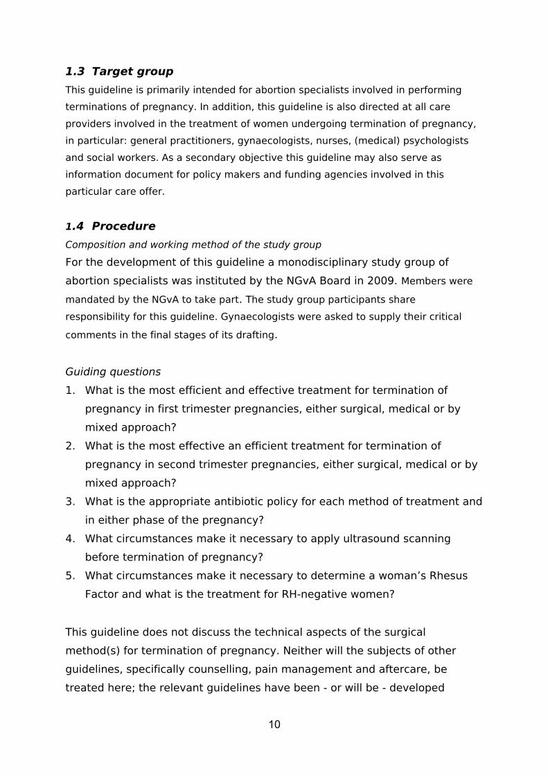
An interval of eight to seventy-two hours between the administration of mifepristone
and misoprostol should preferably be maintained.
For this indication, the recommended dosages of mifepristone and misoprostol, have
not been registered; neither has the administration protocol. It is therefore a legal
requirement to inform the woman about this, preferably in writing.
The importance of follow-up checks must be emphasized – also in writing – because
even after severe haemorrhages it cannot be excluded that the pregnancy is still
intact. Follow-up must take place after one to four weeks, depending on the stage of
the pregnancy at the start of the intervention.
In the case of a continuing pregnancy or incomplete expulsion of the amniotic sac
after medical treatment, suction curettage is offered as a rule. A second medical
termination procedure may be considered at follow-up after one week.
What is the optimal method for termination of pregnancy in the second trimester of
pregnancy?
For women in their second trimester of pregnancy, D&E is recommended for use in
abortion clinics.
In hospitals, women should be offered a choice between medical termination of
pregnancy and D&E; for the latter option, the woman should be referred to an
abortion clinic.
The study group recommends the institution of a national working group to develop a
registration system for late complications of second trimester termination of
pregnancy.
What evidence can be derived from the (inter)national literature with respect to the
application of priming with prostaglandins and their course of administration in first
trimester termination of pregnancy?
Cervical priming by misoprostol in the first trimester is recommended.
Is it possible to use misoprostol combined with D&E after a sectio caesarea?
It is recommended to apply D&E, with misoprostol priming if necessary, for second
trimester termination of pregnancy in the case of women with one low transversal C-
section in their anamnesis.
4

Which genital bacterial infections are common in women undergoing termination of
pregnancy and how should they be treated?
As Chlamydia trachomatis is most common among women between 15 and 25, it is
recommended to treat them prophylactically for this condition after a surgical
termination of pregnancy.
What antibiotic policy should be preferred in the case of surgical termination of
pregnancy?
After surgical termination of pregnancy, it is recommended to administer 1.000 mg
azithromycin as a prophylaxis to the patient on the same day.
When there is a suspicion of bacterial vaginosis four times 500 mg metronidazol is to
be prescribed.
What antibiotic policy should be preferred in the case of medical termination of
pregnancy?
The study group takes the position that antibiotic prophylaxis in medical termination
of pregnancy is not necessary.
When should ultrasound scanning be applied in termination of pregnancy?
What is the function of ultrasound scanning in establishing the diagnosis of pregnancy? In termination of pregnancy ultrasound must be employed to establish the diagnosis
of pregnancy, multi-foetal pregnancy and abnormal pregnancy.
What is the role of ultrasound in determining pregnancy length?Pregnancy length must always be determined by ultrasound and be documented.
Ultrasound will also show if there is a vital intra-uterine pregnancy inside a normal
uterus.
What is the role of ultrasound during surgical termination of pregnancy?Pre-operative abdominal ultrasound scanning must be used in the case of complex
interventions (like deviant positioning, congenital abnormalities, pathology) and in
surgical abortion in the second trimester.
The use of ultrasound scanning during first and second trimester interventions is
appropriate
Macroscopic inspection of the curettement must be carried out.
What is the role of ultrasound scanning after surgical termination of pregnancy?There are good reasons to carry out ultrasonography at the conclusion of the
5

treatment, to exclude the presence of retained tissue.
It is advisable to conduct ultrasound scanning when new or lingering complaints are
observed during the period of time after the termination of pregnancy, such as
severe abdominal pain, loss of too much blood or continuing haemorrhage.
If a woman has no complaints after a termination of pregnancy, ultrasound scanning
has little added value. Generally, no abnormalities are found.
If the pregnancy test is still positive after four weeks, it is necessary, however, to
conduct ultrasonography.
What is the role of ultrasound after medical termination of pregnancyIt is advisable to carry out a sonography after a medical termination of pregnancy, in
the case of continuing abdominal pains, excessive blood loss or prolonged bleeding,
or continuing symptoms of pregnancy.
It is advisable to carry out sonography at one to four weeks after a medical
termination of pregnancy, depending on the stage of the pregnancy.
When must rhesus factor determination take place and what is the treatment in the
case of Rh-?
Up to 7/0 weeks (49 days) of amenorrhoea there is no need to administer anti-d
immunoglobulin.
Starting at 7/0 weeks (49 days) of amenorrhoea Rh(D) negative women undergoing
termination of pregnancy must be protected with an adequate dose of anti-d, of 250
IE in the first and 375 IE in the second trimester, with the exception of already
isosensitized women.
6

Composition of the study group
• P. Hanewald MD (chair), abortion specialist, NGvA
• Ms.M. Denteneer MD, abortion specialist, NGvA
• Ms.Talens MD, abortion specialist, NGvA
• J.R. Soedirman MD, (formerly) NGvA
• L. Querido MD, abortion specialist, NGvA (until July 2010)
• W. Shadmanfar MD, abortion specialist, NGvA
• Ms. M. van Kleffens MD, abortion specialist, NGvA
• Ms. E. Bonsen MD, abortion specialist, NGvA
Supported on the part of OMS by
• Ms. J.W. Hagemeijer MSc, senior consultant, OMS, Utrecht
• Ms. Wessels MSc, information specialist, OMS, Utrecht
7
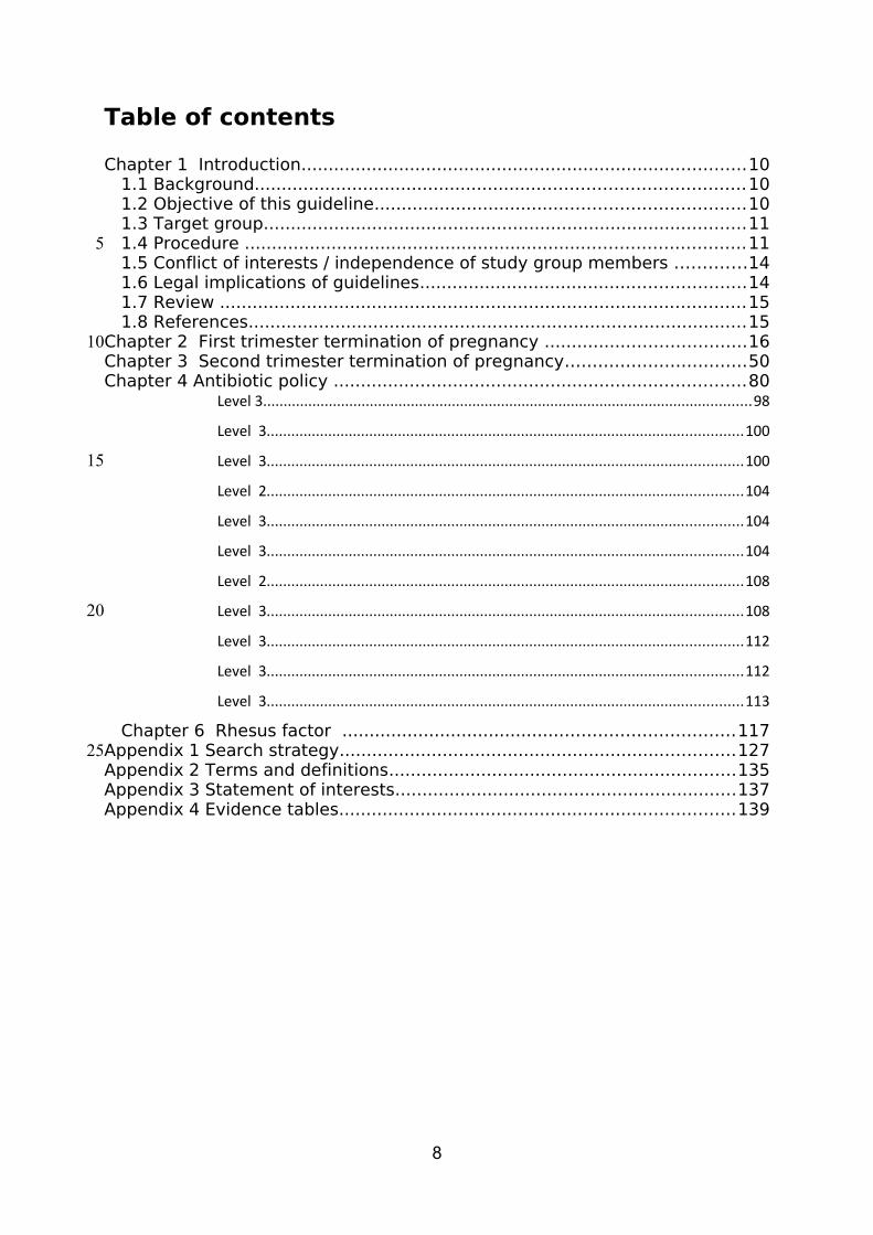
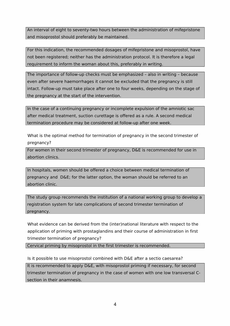
Table of contents
Chapter 1 Introduction..................................................................................101.1 Background..........................................................................................101.2 Objective of this guideline....................................................................101.3 Target group.........................................................................................111.4 Procedure ............................................................................................111.5 Conflict of interests / independence of study group members .............141.6 Legal implications of guidelines............................................................141.7 Review .................................................................................................151.8 References............................................................................................15
Chapter 2 First trimester termination of pregnancy .....................................16Chapter 3 Second trimester termination of pregnancy.................................50Chapter 4 Antibiotic policy ............................................................................80
Level 3.......................................................................................................................98
Level 3....................................................................................................................100
Level 3....................................................................................................................100
Level 2....................................................................................................................104
Level 3....................................................................................................................104
Level 3....................................................................................................................104
Level 2....................................................................................................................108
Level 3....................................................................................................................108
Level 3....................................................................................................................112
Level 3....................................................................................................................112
Level 3....................................................................................................................113
Chapter 6 Rhesus factor ........................................................................117Appendix 1 Search strategy.........................................................................127Appendix 2 Terms and definitions................................................................135Appendix 3 Statement of interests...............................................................137Appendix 4 Evidence tables.........................................................................139
8
5
10
15
20
25

Chapter 1 Introduction
1.1 Background
Approximately 33,000 abortions take place in The Netherlands each year . In our
country, termination of pregnancy is regulated by law. The Termination of Pregnancy
Act (Waz (1984)) is the most important legal rule, specifying the framework in which
the intervention may be carried out.
The Netherlands can be ranked among the countries with the lowest abortion figures
worldwide (Netherlands Health Inspectorate, 2009).
The law was evaluated in 2005 (Visser et al., 2005). It was concluded that the Waz is
generally well respected, a view shared by the inspectorate; this also applies to the
heart of the legal rule, the various aspects of the decision making process. The
overall balance between a woman’s right to assistance and the protection of unborn
life that the legislators had in mind is borne out in practice. The quality of the care
delivered is good, but may be further improved; recommendations were made for
improving the quality in referrals, counselling and decision making (Visser et al,
2005; p. 183-186). On the basis of this evaluation, the government saw no reason to
adapt the law and endorsed most recommendations (Parliamentary Papers II
2005/06, 30 371, nr. 2).
The Dutch Association of Abortion Specialists (NGvA) received funding from the
Ministry of Health, Wellness and Sport for a period of four years starting in 2008, to
stimulate the quality of abortion care, by developing and realizing, i.e., an integrated
quality policy. An important part of the project plan under funding is the development
of a number of evidence-based guidelines. Second in this series is the current
guideline “Treatment of women undergoing termination of pregnancy”.
1.2 Objective of this guideline
The object of the guideline is to improve the quality of abortion care by optimizing
the treatment of women undergoing termination of pregnancy. Thus, the guideline is
designed to function as a directive for a uniform care offer in the treatment of women
undergoing a termination of pregnancy in The Netherlands.
9
1.3 Target group
This guideline is primarily intended for abortion specialists involved in performing
terminations of pregnancy. In addition, this guideline is also directed at all care
providers involved in the treatment of women undergoing termination of pregnancy,
in particular: general practitioners, gynaecologists, nurses, (medical) psychologists
and social workers. As a secondary objective this guideline may also serve as
information document for policy makers and funding agencies involved in this
particular care offer.
1.4 Procedure
Composition and working method of the study group
For the development of this guideline a monodisciplinary study group of
abortion specialists was instituted by the NGvA Board in 2009. Members were
mandated by the NGvA to take part. The study group participants share
responsibility for this guideline. Gynaecologists were asked to supply their critical
comments in the final stages of its drafting.
Guiding questions
1. What is the most efficient and effective treatment for termination of
pregnancy in first trimester pregnancies, either surgical, medical or by
mixed approach?
2. What is the most effective an efficient treatment for termination of
pregnancy in second trimester pregnancies, either surgical, medical or by
mixed approach?
3. What is the appropriate antibiotic policy for each method of treatment and
in either phase of the pregnancy?
4. What circumstances make it necessary to apply ultrasound scanning
before termination of pregnancy?
5. What circumstances make it necessary to determine a woman’s Rhesus
Factor and what is the treatment for RH-negative women?
This guideline does not discuss the technical aspects of the surgical
method(s) for termination of pregnancy. Neither will the subjects of other
guidelines, specifically counselling, pain management and aftercare, be
treated here; the relevant guidelines have been - or will be - developed
10

separately. When the current guideline reached the final stage, it became
clear that (too) little attention was paid to late(r) complications. It is
suggested that this issue be addressed by a joint study group of
gynaecologists and abortion experts in the near future.
Method of guideline development
This guideline was drafted following the “Appraisal of Guidelines for Research &
Evaluation” (AGREE) (www.agreecollaboration.org). This instrument is a widely (and
internationally) accepted instrument for judging the quality of guidelines.
The study group worked on the creation of the draft guideline for two years. Desktop
searches were undertaken and group members assessed the content and quality of
the literature. As a next step the study group members would write a section for the
guideline, in which the relevant literature had been incorporated. If no relevant
scientific literature was available the text was written on the basis of the group
members’ own expertise. During meetings the texts were elucidated and discussed.
Next, texts were edited by the editing committee and then finalized in a plenary
session. The Department of Professional Quality Support of the Dutch Order of
Medical Specialists supported and made recommendations to the study group.
Strategy for literature search and assessment
First, a quick scan was carried out of existing guidelines in Dutch or English and of
the databases of the National Guideline Clearinghouse (http://www.guideline.gov/),
NICE (http://www.nice.org.uk/), SIGN (http://www.sign.ac.uk/) and of the Medical
Reference Organisation for quality of care CBO
(http://www.cbo.nl/thema/Richtlijnen/). In addition, the SUM Search search engine
(http://sumsearch.uthscsa.edu/) was used to trace guidelines in Dutch or English.
Finally, systematic reviews in de Cochrane Library were identified. Next, for each of
the initial questions the Medline electronic databases (OVID) (1967-2008) were
browsed on the basis of specific browsing terms for scientific studies published in
Dutch or English. Furthermore, an additional search “by hand” was undertaken for
studies in Dutch or English taking the literature lists of the articles identified as a
starting point. As a first step (systematic reviews or meta-analyses of) randomized
controlled studies (RCTs) were focused on. If more than one systematic review was
available, the most recent was chosen. Where RCTs were not available the search
was widened to include prospective controlled studies, comparative studies,
prospective non-comparative and retrospective studies.
11
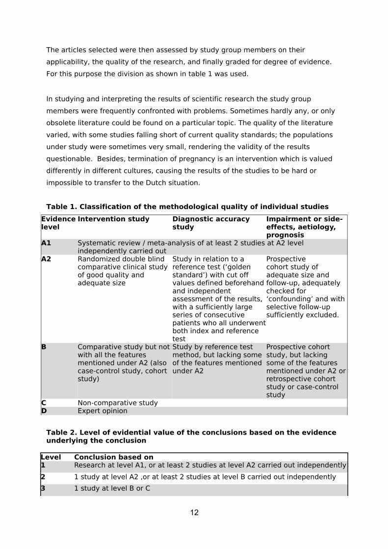
The articles selected were then assessed by study group members on their
applicability, the quality of the research, and finally graded for degree of evidence.
For this purpose the division as shown in table 1 was used.
In studying and interpreting the results of scientific research the study group
members were frequently confronted with problems. Sometimes hardly any, or only
obsolete literature could be found on a particular topic. The quality of the literature
varied, with some studies falling short of current quality standards; the populations
under study were sometimes very small, rendering the validity of the results
questionable. Besides, termination of pregnancy is an intervention which is valued
differently in different cultures, causing the results of the studies to be hard or
impossible to transfer to the Dutch situation.
Table 1. Classification of the methodological quality of individual studies
Evidence level
Intervention study Diagnostic accuracy study
Impairment or side-effects, aetiology, prognosis
A1 Systematic review / meta-analysis of at least 2 studies at A2 level independently carried out
A2 Randomized double blind comparative clinical study of good quality and adequate size
Study in relation to a reference test (‘golden standard’) with cut off values defined beforehand and independent assessment of the results, with a sufficiently large series of consecutive patients who all underwent both index and reference test
Prospectivecohort study of adequate size and follow-up, adequately checked for ‘confounding’ and with selective follow-up sufficiently excluded.
B Comparative study but not with all the features mentioned under A2 (also case-control study, cohort study)
Study by reference test method, but lacking some of the features mentioned under A2
Prospective cohort study, but lacking some of the features mentioned under A2 or retrospective cohort study or case-control study
C Non-comparative studyD Expert opinion
Table 2. Level of evidential value of the conclusions based on the evidence underlying the conclusion
Level Conclusion based on 1 Research at level A1, or at least 2 studies at level A2 carried out independently
2 1 study at level A2 ,or at least 2 studies at level B carried out independently
3 1 study at level B or C
12
4 Expert opinion
Drafting of recommendations
The ‘recommendations’ were phrased in reply to each guiding question and were
based both on data from scientific studies and on the study group’s most important
deliberations. In their deliberations the validity of the literature selected and its
applicability to the Dutch situation were discussed. The patients’ preference,
availability of the facilities and organisational aspects also played a role.
Implementation and evaluation
In the various stages of guideline development the implementation of the guideline
and the practicability of the recommendations were taken into account. Explicit
attention was given to factors which may promote or impede the introduction of this
guideline in practice.
This guideline is only available in digital format and was distributed among all
relevant professional groups and all abortion clinics and hospitals. The document can
also be downloaded from the website of the Dutch Association of Abortion Specialists
(www.ngva.nl). This guideline is intended to be used for the development or
evaluation of local protocols. The guideline is not intended as a list of points for
attention or checklist in decision making.
1.5 Conflict of interests / independence of study group members
Declarations by the members of the study group about potential (economic) conflicts
of interest are open to inspection and can be viewed at the Department of
Professional Quality Support of the Dutch Order of Medical Specialists; an overview
has been added in appendix 3. No conflict of interest was reported.
1.6 Legal implications of guidelines
Guidelines contain recommendations based on a maximum of scientific evidence.
Recommendations are part of the ambition to provide good or ‘optimal’ quality care.
As these recommendations are based on ‘general proof of optimal care’ and the
study group’s relevant insights brought up by its members, professionals may
deviate from this guideline in individual cases where appropriate. If the patient’s
situation demands, such a deviation may even be absolutely necessary. There must
be good arguments for such a step, however; it must be well-documented, and
discussed with the patient where relevant.
13

1.7 Review
No later than 2017 the Board of NGvA will determine if this guideline is still up to
date. If necessary a new study group will be installed to see to its revision. The
validity of the guideline will terminate earlier if new developments make it prudent to
have it revised.
As holder of this guideline the NGvA is the first responsible for its up-to-date status.
The users of this guideline share the responsibility for it and shall inform the first
responsible about relevant developments within their professional fields.
1.8 References
Inspectie voor de Gezondheidszorg. (2009). Jaarrapportage 2008 van de Wet afbreking zwangerschap
Den Haag: Inspectie voor de Gezondheidszorg. (Netherlands Health Inspectorate, 2008 annual report.)
M. Visser et al (2005). Evaluatie Wet afbreking zwangerschap. (Waz Evaluation.)
14

Chapter 2 First trimester termination of
pregnancy
Guiding question
What is the most efficient and effective treatment in first trimester pregnancies:
surgical, medical or by mixed approach?
2.1 Introduction
The NGvA members drew a sharp demarcation line between first an second
trimester, because they felt a need to define the necessary competence of abortion
doctors – and so their training – according to clearly distinguishable areas. Ultrasound
scans render foetal size an objectifiable parameter; in the NGvA general meeting of
September 2009 the line between first and second trimester was drawn at a BPD
(biparietal diameter) < 23 mm (12 weeks + 6 days amenorrhoea = first trimester)
and ≥23 mm (13 weeks am. = second trimester). In the guideline ‘Counselling of
women considering termination of pregnancy’ this criterion was subsequently
redefined: pregnancies up to 91 days are now considered first trimester pregnancies.
Abortion techniques in both pregnancy trimesters can be either surgical or medical in
nature, but this distinction is clearly too simplistic in practice. Because studies do not
strictly differentiate between purely surgical approaches and surgical approaches
after initial medical priming, conclusions from a scientific perspective are hard to
draw. The same is true with regard to the absence of the degree of dilatation. It is to
be expected that mixed approaches will be developed more and more.
On 21 September 2005 the Cochrane, Medline, Embase en Popline literature D-bases
were screened for literature allowing the answering of the guiding question above. By
applying filters for systematic reviews and for randomized controlled trials, efforts
were made to detect and collect literature for first trimester termination of pregnancy
(‘first trimester’, ‘early abortion’). In particular, a search was made for literature on
surgical methods (‘curettage’, ‘vacuum curettage’, ‘curettage and dilatation’,
‘vacuum extracti*’, ‘surgical methods’) and medical interventions (‘abortive agent’,
‘mifepristone’, ‘misoprostol’, ‘prostaglandin’, ‘methotrexate’) under ‘induced
abortion’ and ‘cervical ripening’. A language filter was applied, allowing only the
selection of articles in English, Dutch, French and German. As the Cochrane Database
of Systematic Reviews turned up three reviews discussing the literature up to 2000
(at least), it was decided to restrict further desk research to the period between 1999
and 2005. By this strategy 195 articles were uncovered in total. On 20 December
15

2009 a renewed literature search with respect to surgical termination of pregnancy
was undertaken in the same D-bases, this time for the period from 2005 through
2009. This search did not yield new literature or perspectives regarding the surgical
termination of pregnancy. However, several studies were published which attempt to
compare surgical and medical termination of pregnancy.
16
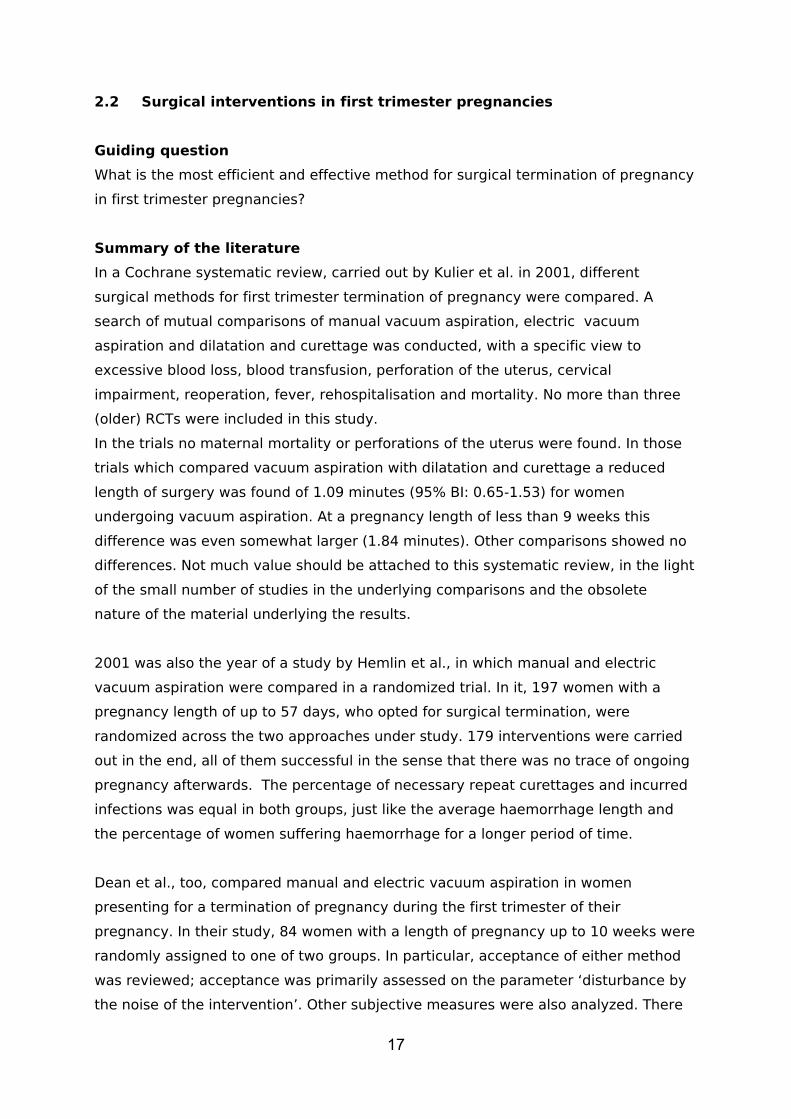
2.2 Surgical interventions in first trimester pregnancies
Guiding question
What is the most efficient and effective method for surgical termination of pregnancy
in first trimester pregnancies?
Summary of the literature
In a Cochrane systematic review, carried out by Kulier et al. in 2001, different
surgical methods for first trimester termination of pregnancy were compared. A
search of mutual comparisons of manual vacuum aspiration, electric vacuum
aspiration and dilatation and curettage was conducted, with a specific view to
excessive blood loss, blood transfusion, perforation of the uterus, cervical
impairment, reoperation, fever, rehospitalisation and mortality. No more than three
(older) RCTs were included in this study.
In the trials no maternal mortality or perforations of the uterus were found. In those
trials which compared vacuum aspiration with dilatation and curettage a reduced
length of surgery was found of 1.09 minutes (95% BI: 0.65-1.53) for women
undergoing vacuum aspiration. At a pregnancy length of less than 9 weeks this
difference was even somewhat larger (1.84 minutes). Other comparisons showed no
differences. Not much value should be attached to this systematic review, in the light
of the small number of studies in the underlying comparisons and the obsolete
nature of the material underlying the results.
2001 was also the year of a study by Hemlin et al., in which manual and electric
vacuum aspiration were compared in a randomized trial. In it, 197 women with a
pregnancy length of up to 57 days, who opted for surgical termination, were
randomized across the two approaches under study. 179 interventions were carried
out in the end, all of them successful in the sense that there was no trace of ongoing
pregnancy afterwards. The percentage of necessary repeat curettages and incurred
infections was equal in both groups, just like the average haemorrhage length and
the percentage of women suffering haemorrhage for a longer period of time.
Dean et al., too, compared manual and electric vacuum aspiration in women
presenting for a termination of pregnancy during the first trimester of their
pregnancy. In their study, 84 women with a length of pregnancy up to 10 weeks were
randomly assigned to one of two groups. In particular, acceptance of either method
was reviewed; acceptance was primarily assessed on the parameter ‘disturbance by
the noise of the intervention’. Other subjective measures were also analyzed. There
17

appeared to be no significant differences in intervention length, blood loss,
complications, pain relief and recovery time. Neither could a difference be
established between the pain experienced by the patient and the doctor’s estimation
of that pain. Women who underwent electric vacuum aspiration more frequently
reported their observation of the accompanying noise; 19% found this noise slightly
disturbing. This finding was higher than for the group who underwent manual
vacuum aspiration (p = 0.03).
Conclusions
Level 2
Vacuum aspiration and dilatation and curettage do not apparently differ
from each other in terms of complications and side-effects. Surgery length
is a little shorter for vacuum aspiration.
A2 Kulier 2001
Level 2
It seems plausible that manual and electric vacuum aspiration are both
safe methods for termination of pregnancy. Randomized trials did not
reveal differences in effectiveness, side-effects or complications.
B Hemlin, Dean 2003
Discussion and evaluation
Suction curettage is a safe method for pregnancy termination in the first trimester;
this method shows good results and few complications. Success and complication
scores vary between 0 and 5% across studies. Scores will also depend on the
abortion doctor’s degree of experience.
In the – mostly non-Dutch – literature many surgical terminations of pregnancy were
performed under general anaesthesia. This is different for the Dutch situation,
although daily practice seems to show an increase in the patients’ wish for sedation
or general anaesthesia. The pain experienced may be reduced, but the costs are
higher; these concern for instance the purchase of surveillance appliances, the
training of professional staff and the involvement of an anaesthesiologist.
The option to receive sedation (or not) is at the patient’s discretion and must be
respected and facilitated.
Recommendations
18
Where a surgical method is preferred, the study group considers vacuum aspiration
as the first choice, in view of the low percentage of complications.
References
Dean G, Cardenas L, Darney P, Goldberg A. Acceptability of manual versus electric aspiration for first trimester abortion: a randomized trial. Contraception 2003; 67:201-06.
Hemlin J, Moller B. Manual vacuum aspiration, a safe and effective alternative in early pregnancy termination. Acta Obstet.Gynecol.Scand. 2001; 80:563-67.
Kulier R, Gulmezoglu AM, Hofmeyr GJ, Cheng LN, Campana A. Medical methods for first trimester abortion. The Cochrane Database of Systematic Reviews 2004, Issue 2.Art.No.: CD002855.pub3.DOI: 10.1002/14651858.CD002855.pub3. 2004.
Kulier R, Fekih A, Hofmeyr GJ, Campana A. Surgical methods for first trimester termination of pregnancy. The Cochrane Database of Systematic Reviews 2001, Issue 4.Art. No.: CD002900.DOI: 10.1002/14651858.CD002900. 2001.
19

2.3 Surgical versus medical termination of pregnancy in the first
trimester
Guiding question
Are there any differences in success rate, patient contentment or number of
complications between surgical and medical termination of pregnancy in the first
trimester?
Summary of the literature
In 2010, a Cochrane review by Say et al. was published. This review covers seven
studies comparing medical methods versus vacuum aspiration. Four different
interventions, prostaglandins only, mifeprostone only, mifegyne plus misoprostol,
and methotrexate plus misoprostol are compared with vacuum aspiration. The most
important outcomes concern effectiveness, side-effects and patient comfort. This
review shows that women undergoing medical termination of pregnancy generally
suffer more blood loss than women undergoing vacuum aspiration. As regards
experienced pain comparisons are difficult to make, as women undergoing surgical
termination of pregnancy very often received one or other form of – general –
anaesthesia. In The Netherlands, local anaesthesia is still applied in many cases. The
authors conclude – admittedly on the basis of limited evidence – that, in the first
trimester, vacuum aspiration is more effective than medical termination with
prostaglandins alone, and that vacuum aspiration proceeds faster, with less bleeding
and less pain.
In 2004 an RCT by Rorby et al. was published. This study included 1.033 women with
a pregnancy length under 63 days. Part of these women were randomly assigned to a
surgical or medical approach, the other part were free to choose. Surgical
termination entailed vacuum aspiration under general anaesthesia, the medical
intervention entailed 600 mg. of mifeproston and 1 mg. of gemeprost. These regimes
thus differ somewhat from the Dutch situation. At two and eight weeks after the
intervention the women filled out questionnaires with respect to contentment, side-
effects and expectations. In both questionnaires, after two and eight weeks, the
same patterns were found; women who had opted for surgical termination were
satisfied to highly satisfied at 92%, versus 94% in the randomized surgical group.
Satisfaction was lower for the medical group; of the women who had opted for
medical termination, 82% were satisfied to highly satisfied, while in the randomized
group this was true for 68%. Satisfaction in the medical group was inversely related
to gestation length, intensity of the pain experience and the prevalence of nausea
20
and vomiting. In the case of surgical termination these factors had no influence. The
authors conclude that satisfaction is high in both groups, but higher after surgical
termination of pregnancy. They also conclude that contentment figures are higher if
women are allowed to choose which method they wish to undergo for the termination
of their pregnancy.
The study 'Randomised preference trial of medical versus surgical termination of
pregnancy less than 14 weeks gestation’ by Robson et al appeared in Health
Technology Assessment in 2009. (TOPS). The surgical intervention included women
with a pregnancy length of 6 to 14 weeks. Two hours before the intervention each
woman was administered 400 micrograms of misoprostol, the intervention was
carried out by suction curettage under general anaesthesia. The medical intervention
was carried out under 14 weeks of pregnancy. Each woman took 200 milligrams of
mifeproston orally and presented at the clinic 36 to 48 hours later. Under 9 weeks of
pregnancy the regime was 800 micrograms of misoprostol vaginally and sometimes
400 micrograms of misoprostol 4 hours later if there had been no onset of the
termination. In pregnancies over 9 weeks 800 micrograms of misoprostol vaginally
was again the starting dose, followed by 400 micrograms (vaginally) every 3 hours,
up to a maximum of 4 doses. If no effect was observed, 200 micrograms of
mifeproston were administered vaginally, followed by a mg. of gemeprost, every 3
hours, with a maximum of 5 doses. If no onset of the termination was observed, the
surgical approach was applied. The outcome measure in this study was whether
women would again choose to undergo the same method in a possible next
termination of pregnancy. 1,877 women were included in total. The medical
approach was more negatively appreciated than vacuum aspiration. More pain was
reported. There was no difference across the two groups as regards anxiety or
feelings of depression after three months. With the medical approach, there were
more emergency requests: 4.2% versus 0.7% of the women presented at the
emergency department. Despite the latter effect, the authors calculated that the
surgical method is more expensive than the medical termination, also due to the
general anaesthesia and hospitalisation involved.
In 2004 and 2005, the American Food and Drug Administration (FDA) reported on
septic shock as a potential complication after using mifepristone and misoprostol.
Five women died as a result of septic shock, caused by Clostridium sordellii. This
happened within a week after medical termination of pregnancy.
21

The authors of the articles reviewed indicate that the pathophysiology is not fully
clear; there was no clear-cut explanation for these deaths. Further research is
required.
Besides, the authors point out the extremely small risk of incurring this complication.
In Europe, around 1.5 million women have made a choice for medical termination
over the past 15 years. The complication mentioned was described no more than
once in Europe (Fischer et al.. 2005; Greene et al. 2005; Fiala et al. 2005). In Portugal
one Chlostridium sordellii infection with fatal consequences was reported in 2009
(21st European Congress of Clinical Microbiology and Infectious Diseases, ECCMID,
Milan, 07.10.2011).
Conclusions
Level 1
Compared to surgical termination of pregnancy, medical termination in
the first trimester causes higher blood loss, while the intervention also
lasts longer.
A1 Say et al. 2010
Level 3
It seems plausible that, compared to vacuum aspiration, medical
termination of pregnancy more frequently causes complications.
B Robson et al. 2009
Level 4
It is unlikely that there is a relation between medical termination of
pregnancy and toxic shock syndrome.
D Fischer et al.. 2005; Greene et al. 2005; Fiala et al. 2005
Discussion and evaluation
Various complications may occur in first trimester termination of pregnancy. When a
surgical intervention is performed, particularly vacuum aspiration, these may
comprise perforation, bleeding, infection or an incomplete abortion, or complications
of anaesthesia.
During and after medical termination severe and long-term blood loss may occur,
besides failed or incomplete abortion, or an infection. Complication figures for first
trimester termination of pregnancy in The Netherlands have unfortunately not been
sublabeled for surgical or medical termination. Inquiries at the National Health
Inspectorate and a study of the LAR data showed a complication score for The
Netherlands of 4 in 1,000 first trimester terminations. This amounts to 0.4% of all
22

first trimester abortions carried out in this country. The literature does not further
elucidate on these figures, but success rates can be derived. The 0.4 % reconstructed
for The Netherlands is considerably lower than the figures for failed abortions found
in the international literature. For instance, the Cochrane review speaks of success
rates of between 71% (with a regime of prostaglandins only) and 97.5% (a regime of
mifegyne with misoprostol) and a success rate of between 94% and 100% for
surgical termination of pregnancy.
The differences in complication figures are relatively large and explanations cannot
be readily provided. Does underreporting play a role? Are professionals in The
Netherlands more experienced, since abortion treatment is a medical specialism here
and the abortion doctor a recognized specialist? This is not clear and raises the
question, to what extent the studies found can be used for the Dutch situation. Apart
from the complication figures, the regimes under the medical approach are different
as well, and also the maximum length of gestation for which medical termination of
pregnancy is (still) carried out and the fact that very many surgical methods are
applied under general anaesthesia. These shortcomings have made the drafting of
(well-founded) conclusions and recommendations a slightly dubious affair.
Recommendations
A woman with amenorrhoea under 63 days must be informed about the treatment
options open to her: surgical or medical
A woman must be informed about the differences between both forms of treatment; it
must be made clear to her that medical termination of pregnancy takes more time
and has a higher risk of complications, such as ongoing pregnancy, incomplete
abortion and bleeding, compared to surgical termination of pregnancy
References
Fiala e.a. Review of medical abortion using mifepristone in combination with a prostaglandin analogue. Contraception
74; 66-86 2005Fischer e.a. Fatal toxic shock syndrome associated with Clostridium sordellii after medical abortion. New England
journal of medicine, Dec. 2005Greene, Fatal infections associated with Mifepristone-induced abortion. New England journal of medicine, Dec.
2005Niinimaki e.a. 'Immediate complications after medical abortion compared with surgical termination of pregnancy'.
2009Rorby e.a. 'Medical versus surgical abortion; comparing satisfaction and potential confounded in a partly randomized
study'. 2004Robson e.a. 'Randomised preference trial of medical versus surgical termination of pregnancy less than 14 weeks
23

gestation.' (TOPS), 2009Say e.a. Cochrane review: Medical versus surgical methods for first trimester termination of pregnancy. 2010Loeber. Motivation and satisfaction with early medical vs. surgical abortion in the Netherlands. Reproductive health matters, 2010;18(35):145-153.
24

2.4 Cervix priming in the first trimester
Guiding question
What evidence may be inferred from the (inter)national literature with respect to the
application of priming with prostaglandins and their administration route in first
trimester termination of pregnancy?
Introduction
Prostaglandins have been applied for a long time to prepare the uterus mouth (cervix
uteri) for dilatation. Prostaglandins facilitate dilatation. Blood loss during and after
the intervention is reduced. Cervical priming is regularly used in surgical termination
of pregnancy. In abortion clinics, misoprostol is the prostaglandin almost exclusively
applied to prime the cervix. Below, aspects of effectiveness, side-effects and safety
will be discussed.
Summary of the literature
In first trimester termination of pregnancy prostaglandins make dilatation of the
cervix easier. Various prostaglandin analogues have been developed (Goldberg e.a.,
2001; Goldberg e.a., 2003; Shannon e.a., 2004; Sivalal e.a., 2004). Misoprostol is a
prostaglandin E1 analogue (PGE1), which came on the market in 1988. As regards
first trimester terminations, ample research was conducted into misoprostol as a
cervix primer.
Misoprostol (as a cervix primer) has no detectable effect on the complication rates
for the first trimester (Goldberg e.a., 2003). The side-effects of misoprostol are less
than those of earlier prostaglandins (Ngai e.a., 2003). Misoprostol has the advantage
over gemeprost that it is cheap and stable at room temperature (Ngai e.a., 2003;
Sivalal e.a., 2004).
Misoprostol can prompt a spontaneous abortion in the first trimester. The addition of
mifepristone will accelerate this process (Goldberg e.a., 2001; Ngai e.a., 2003).
Mifepristone – a competitive progestogen antagonist - is more expensive as a cervix
primer than misoprostol. According to the Royal College of Obstetricians and
Gynaecologists (RCOG), mifepristone must be administered 36-48 hours before the
intervention.
The optimal time lapse between cervix priming with the help of misoprostol and
treatment is estimated at 2 to 4 hours; most articles mention a (minimum) time lapse
of 3 hours (Goldberg e.a., 2001; Goldberg e.a., 2003). All of these articles are
concerned with cervix priming in the first trimester.
25
The best route of administration for misoprostol appears to be the vaginal
administration of 400 mcg, but buccal or sublingual administration is a good
alternative (Goldberg e.a., 2001; Goldberg e.a., 2003). The further the gravidity has
progressed, the higher the sensitivity to uterotonics. For this reason decreasing
doses of misoprostol are required to achieve similar effects (Goldberg e.a., 2001).
The scientific basis outlined above is mainly founded on five review articles of good
quality. The most recent review article dates from 2004. In 2010, a renewed search
turned up two articles, neither of which yielded any new insights. The RCOG guideline
recommends priming in nulliparae, women under 18 and women with a pregnancy
length of 10 weeks or more.
A claim found again and again holds that cervix priming in the first trimester will
reduce the number of complications. This claim is based, partially, on two articles
from 1983 and 1984, respectively, which are often cited. These and other older
articles were left out here because, at the time, there was no use of misoprostol.
Goldberg et al. (2003) conclude there is no difference in the number of complications
with or without cervix priming in the first trimester. In either case, the number of
complications is very low
The evidence tables in the RCOG guideline show a complication rate of about 1 per
1,000, which underlines Goldberg’s conclusion.
Conclusion
Level 3
It seems safe to assume that misoprostol as a cervix primer in the
first trimester does not show a clear reduction in the number of
complications, partly because complications in the first trimester are
rare.
B Goldberg, 2003
Level 4
It is likely that misoprostol in the first trimester contributes to the
ease of dilatation in nulliparae, women under 18 and in women with a
pregnancy length of more than 10 weeks.
D RCOG, 2004
Level 4
It is likely that 400 mcg of misoprostol taken as a cervix primer at two
to four hours before first trimester vacuum aspiration may be
administered by either vaginal, buccal or sublingual route.
26

D RCOG, 2004
Discussion and evaluation
In other countries, cervix priming often precedes first trimester surgical treatment. In
The Netherlands, suction curettage (in the first trimester) is carried out with a
relatively thinner suction tube, making cervix priming (mostly) unnecessary.
The time of stay in the clinic will be considerably longer when priming is applied; this
requires additional investments on the part of both patient and clinic.
As mentioned above, priming does not influence the number of complications or their
nature; however, side-effects and medication-related risks may occur.
Recommendation
Routine priming of the cervix with prostaglandins (e.g. misoprostol) has no added
value in first trimester surgical termination of pregnancy.
References
Ashok PW, Flett GM, Templeton A. Mifepristone versus vaginally administered misoprostol for cervical priming before first-trimester termination of pregnancy: a randomized, controlled study. Am.J.Obstet.Gynecol 2000; 183 (4): 998-1002.
Goldberg AB, Greenberg MB, Darney PD. Misoprostol and Pregnancy. NEJM 2001 jan 4; 344 (1): 38-47.
Goldberg AB, Carusi DA, Meckstroth KR. Misoprostol in Gynecology. Current Women’s Health Reports 2003; 3: 475-483.
Ngai SW, Tang OS, Ho PC. Prostaglandins for induction of second-trimester termination and intrauterine death. Best Practice & Research Clinical Obstetrics & Gynaecology 2003; 17 (5): 765-775.
Shannon CS, Winikoff B. Misoprostol, an emerging technology for women’s health, report of a seminar. Population Council 2001, updated to 2004: 1-66.
Sharma S, Refaey H el, Stafford M, Purkayastha S, Parry M, Axby M. Oral versus vaginal misoprostol administered one hour before surgical termination of pregnancy: a randomised controlled trial. BJOG 2005 April; 112: 456-460.
Sivalal S, Sivasampu S. Health technology assessment report, misoprostol in pregnancy. Health technology assessment unit, medical development division, ministry of health Malaysia 2004: 3-29.
Vimala N, Mittal S, Kumar S. Sublingual misoprostol before first trimester abortion: a comparative study using two dose regimens. Indian J Med Sci 2004; 58 (2): 54-61.
Royal College of Obstetricians and Gynaecologists (RCOG) Guideline: the Care of Women Requesting
Induced Abortion, Chapter 7, 2004
27

28

2.5 Medical termination of pregnancy
Guiding question
What is the most efficient and effective medical treatment in first trimester medical
termination of pregnancy?
IntroductionBelow, the guiding question with respect to the medical treatment for first trimester
termination of pregnancy is discussed. There will be a focus on the use of
mifepristone in combination with misoprostol.
There is a multitude of publications on the results of studies into medical termination
of pregnancy. In view of the poor results, the various monotherapies may be left
aside. Most successful are combinations of methotrexate with prostaglandin
analogues and mifepristone with prostaglandin analogues.
In The Netherlands, methotrexate is not applied in medical termination of pregnancy,
although it is in the treatment of extra-uterine pregnancies.
Since the registration of mifepristone in 1999, medical treatment has conquered a
position for itself as a first trimester intervention, besides curettage. Advantages
mentioned by Dutch gynaecologists (compared to surgical treatment): smaller risk of
impairment of cervix and uterus, no anaesthesia, no intravenous incursion, fewer
systemic complications like heart failure, ease of the intervention (Brouns, Burger, &
van Wijngaarden, 2010). Since its registration, further studies have taken place, to
optimize the combination treatment of mifepristone, particularly with misoprostol, in
terms of their efficacy and unwanted side-effects. Besides, the legal product
information (Summary of Product Characteristics) has changed since the
introduction.
Mifepristone
The suppression of progesterone during early pregnancy causes uterus contractions
and expulsion of the embryo through a mechanism involving prostaglandin (Csapo,
Pulkkinen, & Kaihola, 1973). Mifepristone has the following effects on uterus and
cervix:
- Mifepristone provokes contractility in the uterus by restraining the effects of
progesterone and increasing the sensitivity of the myometrium to
prostaglandins (Garfield, Blennerhassett, & Miller, 1988) (Swahn & Bygdeman,
1988a);
- Mifepristone leads to necrosis of the deciduas by impacting the capillary
endothelial cells, which causes rejection of the trophoblast, accompanied by
bleeding, decrease in HCG levels and an increased release of prostaglandin
29
(Johannisson, Oberholzer, Swahn, & Bygdeman, 1989) (Schindler et al., 1985)
(Herrmann et al., 1982);
- Mifepristone ripens the cervix, which stimulates expulsion.
Studies in human subjects gave the impression that the contractility of the uterus
does not increase earlier than 24-36 hours after administration of mifepristone and is
preceded by increased sensitivity of the myometrium to prostaglandins (Swahn &
Bygdeman, 1988b). Uterus sensitivity to exogenously administered prostaglandin
rose to five times its normal level. However, the effect of vaginally administered
misoprostol becomes noticeable within 15 minutes, while the effects of the
mechanisms induced by mifepristone listed above may possibly not be complete
(Creinin et al., 2007).
When taken orally, mifepristone is easily resorbed and shows peak serum levels
within two hours, depending on the dosage, both in pregnant and in non-pregnant
women (Shi et al., 1993). At dosages between 100-800 mg of mifepristone the serum
peak levels are comparable (2,0-2,5 µg/ml). These non-linear pharmacokinetic
findings are probably associated with the saturation of a specific transport protein:
alpha-1-acid glycoprotein, starting at 100 mg mifepristone. It would seem likely,
therefore, that a dosage of 100 mg is already effective, while it is extremely unlikely
that a dosage above 600 mg would show better effects (Lahteenmaki et al., 1987)
(Heikinheimo et al., 1987) (Heikinheimo, 1989) (Heikinheimo, Tevilin, Shoupe,
Croxatto, & Lahteenmaki, 1986).
Misoprostol
Misoprostol is a synthetic prostaglandin E1 analogue, with both a mucosa protecting
and a gastric acid reducing capacity. The mucosa protecting effect is based on the
fact that misoprostol stimulates the production of mucus and hydrogen carbonate,
next to other possible mechanisms such as the maintaining or strengthening of
perfusion levels in the gastric mucosa. Resorption is quick and nearly complete. The
active substance is misoprostolic acid, which is formed almost at once. Misoprostol is
not registered as a support agent to combine with mifepristone in termination of
pregnancy, but it is indicated to prevent the formation of ulcers of the stomach or
bowel provoked by NSAIDs.
Gemeprost(SmPC:http://www.medicines.org.uk/emc/medicine/3968/SPC/Gemeprost/
#PRODUCTINFO)
Gemeprost, too, is a prostaglandin E1 analogue, which is administered in a dosage of
1 mg by means of a pessary inserted in the vagina, to effect:
30
- softening and dilatation of the cervix opening prior to surgical intervention in
the first trimester;
- induction of an abortion in the second trimester.
Summary of the literatureThe first relatively large-scale study on the combination of mifepristone and
misoprostol was performed on 873 women with an amenorrhoea length of up to 49
days (Peyron et al., 1993). Peyron et al. described two successive studies in which all
women used 600 mg mifepristone orally, followed 48 hours later by a single oral dose
of misoprostol (400 µg). In the second study the women were allowed an extra 200
µg of misoprostol orally if the expulsion of the foetus had not taken place within four
hours. In the group as a whole, abortion occurred in 4% of the women through
mifepristone alone. In the first group, complete termination of pregnancy occurred in
96.9% of the 488 women (95% CI: 94.1-97.7%); in the second group of 385 women in
98.7% (95% CI: 96.8-99.5%). Statistically, these differences are non-significant. The
abortion percentages during the first four hours after misoprostol were 61% and
69%, respectively. Nausea, vomiting and diarrhoea were found in 40%, 15%, and
10% of the women, respectively.
In a multicenter trial by Aubény en Peyron (Aubeny et al., 1995) involving 1,108
women with an amenorrhoea length up to 63 days, the same
mifepristone/misoprostol schedule was followed up with an oral dose of 200 µg
misoprostol if abortion did not start within three hours after the first dose; the
highest success percentages were found at a shorter amenorrhoea length: 97.6% up
to 42 days am.; 94.8% at 42-49 days am.; 93.4% at 50-56 days of amenorrhoea and
86.8% at 57-63 am. Continuation of the pregnancy was found to be related to an
increased amenorrhoea length: in 0.8%, 1.4%, 1.6% and 5.1%, respectively. On the
whole, 61.6% of the women had no expulsion of the foetus within three hours after
the first dose of misoprostol and were given a second dose of 200 µg.
A Cochrane systematic review on medical interventions (Kulier, Gulmezoglu,
Hofmeyr, Cheng, & Campana, 2004) appeared in 2004. It included randomized
clinical trials, in which different medical interventions were compared to each other
or a placebo in women in the first trimester of their pregnancy. Although 39
randomized clinical trials were included in this well-delivered systematic review, it
must be observed that most analyses were based on only a few studies, sometimes
even no more than one. This entails that many results were based on a limited
31
number of patients, so that differences found between mono or combined use of
medication often show wide reliability intervals. From one of the studies included in
this meta-analysis it appeared that administration of 200 mg mifepristone, followed
up by 800 μg misoprostol on day 1 gave a slightly raised risk of incomplete abortions
compared to the administration of misoprostol on day 3 (RR: 1.94; 95% RI: 1.05-3.58)
(Schaff et al., 2000a).
The review also showed that the combination of mifepristone and misoprostol (800
μg) mentioned above lead to higher success rates under vaginal administration than
under oral administration of misoprostol (RR: 4.41; 95% RI: 2.32-8.38) (Schaff,
Fielding, & Westhoff, 2001a). In the same analysis vaginal administration gave a
slightly lower risk of nausea and diarrhoea, but a slightly higher risk of vomiting at
the same time.
The other results from this systematic review are statistically non-significant.
Von Hertzen et al. (von Hertzen et al., 2003a) and Honkanen et al. (Honkanen et al.,
2004) described various aspects of a WHO study, in which three misoprostol regimes
were compared in 2,219 women with a pregnancy length of up to nine weeks,
calculated from the last menstruation period. In this population, Von Hertzen et al.
analysed first of all the effectiveness of the intervention, while Honkanen et al.
focused on the side-effects and women’s perceptions, for which three study arms
were compared:
- O/O: oral mifepristone (200 mg) on day 1, oral misoprostol (0.8 mg) and a vaginal
placebo on day 3, oral misoprostol (0.4 mg) twice daily on day 4 through 10;
- V/O: oral mifepristone (200 mg) on day 1, vaginal misoprostol (0.8 mg) and an oral
placebo on day 3, oral misoprostol (0.4 mg) twice daily on days 4-10;
- V: oral mifepristone (200 mg) on day 1, vaginal misoprostol (0.8 mg) and an oral
placebo on day 3, oral placebos twice daily on days 4-10.
For the women in the O/O group the percentage rate for a complete abortion was
92.3% (95% RI: 90.1-94.1); in the V/O group this was 93.5% (95% RI: 92.9-96.2) and
in the V group 93.5% (95% RI: 91.5-95.2). The differences between these groups
were statistically non-significant. After stratification for length of pregnancy there
were no significant differences in abortion success rate. In the O/O group more
women than in the other group indicated that they suffered from diarrhoea (6.8% vs.
1.8% and 1.1%, p < 0.0001). Women in the O/O group reported low abdominal pain
directly after the administration of misoprostol, while women in the V/O group and V
group reported this after 3 hours (p varying from < 0.0001 to 0.0027, directly after
administration versus after 3 hours).
32

In a randomized study, Creinin et al. (Creinin et al., 2004a) compared administration
of misoprostol 6 to 8 hours after administration of mifepristone with administration of
misoprostol 24 hours after administration of mifepristone. In this prospective
multicenter trial 1,080 women with a pregnancy up to 63 day after the last
menstruation were included. In terms of effectiveness of the medication there was no
significant difference between both groups (95.8% vs. 98.1%). In the interval
between the administration of mifepristone and misoprostol more side-effects of the
medication were observed in the 24 hour group of women than in the other group.
Besides, women in this group suffered more from nausea and vomiting after
administration of misoprostol than the group of women with a shorter interval
between mifepristone and misoprostol.
In a double blind placebo controlled randomized clinical trial Liao et al. (Liao et al.,
2004) compared mifepristone 75 mg caplets with mifepristone 150 mg tablets,
followed up with misoprostol in both arms of the trial. Apart from the effectiveness of
these different forms of administration, the side effects and the prevalence of
bleeding were studied in 480 woman participants with a pregnancy length up to 49
days after the last menstruation period. Complete abortion was achieved in 95.4% in
the group using the tablets and 96.3% in the group receiving mifepristone in capsule
form. These percentages do not differ significantly from each other. The duration of
the haemorrhages experienced by the women and their intensity were similar to their
experience during normal menstruation; the side-effects were comparable for both
groups under study.
In a multicenter randomized clinical trial, Schaff et al. (Schaff, Fielding, & Westhoff,
2001b) compared the effectiveness of oral versus vaginal administration of
misoprostol after administration of mifepristone 200 mg. All women with a pregnancy
length up to nine weeks were given mifepristone 200 mg, followed up after two days
by one of three regimes: 400 μg misoprostol orally, 800 μg misoprostol orally or 800
μg misoprostol vaginally. The complete abortion percentage was highest in the group
undergoing vaginal administration of misoprostol (p < 0.001), even after additional
vaginal administration of misoprostol in all groups (p < 0.,001). Oral misoprostol 400
μg showed the smallest effect; the success rate was lower in more advanced
pregnancies. After vaginal administration more women suffered cramps compared to
oral administration, but fewer had diarrhoea (p < 0.001). In both groups with oral
administration more women found the pain acceptable (p = 0.017).
33
Tang et al. (Tang, Chan, Ng, Lee, & Ho, 2003a) studied the difference in effectiveness
between sublingual and vaginal administration of misoprostol 800 μg, 48 hours after
administration of mifepristone 200 mg. In this double blind placebo controlled
randomized clinical trial 224 women with a pregnancy up to nine weeks were
included. Complete abortion was achieved in 98.2% of the women in the sublingual
group and in 93.8% in the vaginal group. This difference was non-significant,
statistically. But the women in the sublingual group did report nausea, vomiting,
diarrhoea, shivering and fever more often (p < 0.05).
Mifepristone at lower doses
On the basis of the pharmacokinetic profile described above, it was supposed that
mifepristone at lower doses could have a similar degree of effectiveness. In 1992,
Thong en Baird (Thong & Baird, 1992) were the first to describe the results of
mifepristone 200 mg followed up 48 hours later by 600 µg of misoprostol taken orally
by 100 women with an amenorrhoea length of up to 56 days. The effectiveness found
was at 92%, while in 79% of the women expulsion of the foetus occurred within 4
hours after taking misoprostol.
The success rate at an amenorrhoea length of up to 57 days is 93.8% after
administration of mifepristone 200 mg and 94.3% at 600 mg (WHO 1993); these
figures are 92.4% and 91.7% at an amenorrhoea length of 57-63 days (WHO 2001),
in combination with gemeprost in both studies. Following this, most major studies
investigated the use of 200 mg of mifepristone.
After their previous review of 2004, Kulier et al. recently updated their review (Kulier
R., Kapp N., Gülmezoglu A.M., Hofmeyr G.J., Cheng L., & Campana A. The Cochrane
Library, 2010, Issue 8). They cite Von Hertzen et al. (2009), who compared 200 and
100 mg of mifeprestone followed up by 800 µg misoprostol administered vaginally,
24 or 48 hours later, in women with an amenorrhoea length of 63 days or less.
Effectiveness and side-effects were similar in all four groups.
Alternative administration routes of misoprostol
In their meta-analysis, Kulier et al. (2010) had fewer women with a complete abortion
after oral administration of 800 µg of misoprostol than after vaginal administration of
misoprostol at the same dose (statistically significant; based on two trials among
1,407 women in total: RR 3.05; 95% RI 1.98-4.70). Nausea and diarrhoea less
frequently occur after vaginal administration of misoprostol, compared to oral
administration. In other comparisons (buccal versus vaginal, buccal versus oral, sub-
lingual versus vaginal, sublingual versus oral) buccal and sublingual administration
34

proved equally effective as vaginal administration, but the first two cause more side-
effects to occur and, more particularly, an unpleasant taste.
35
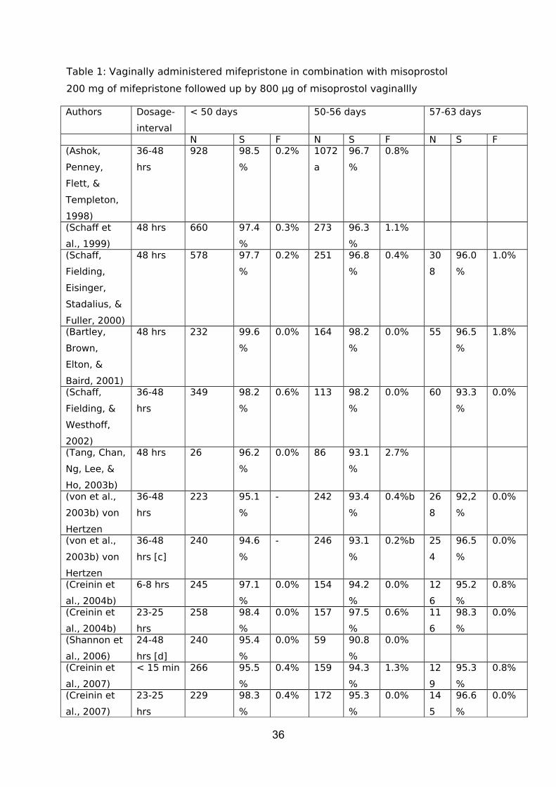
Table 1: Vaginally administered mifepristone in combination with misoprostol
200 mg of mifepristone followed up by 800 µg of misoprostol vaginallly
Authors Dosage-
interval
< 50 days 50-56 days 57-63 days
N S F N S F N S F(Ashok,
Penney,
Flett, &
Templeton,
1998)
36-48
hrs
928 98.5
%
0.2% 1072
a
96.7
%
0.8%
(Schaff et
al., 1999)
48 hrs 660 97.4
%
0.3% 273 96.3
%
1.1%
(Schaff,
Fielding,
Eisinger,
Stadalius, &
Fuller, 2000)
48 hrs 578 97.7
%
0.2% 251 96.8
%
0.4% 30
8
96.0
%
1.0%
(Bartley,
Brown,
Elton, &
Baird, 2001)
48 hrs 232 99.6
%
0.0% 164 98.2
%
0.0% 55 96.5
%
1.8%
(Schaff,
Fielding, &
Westhoff,
2002)
36-48
hrs
349 98.2
%
0.6% 113 98.2
%
0.0% 60 93.3
%
0.0%
(Tang, Chan,
Ng, Lee, &
Ho, 2003b)
48 hrs 26 96.2
%
0.0% 86 93.1
%
2.7%
(von et al.,
2003b) von
Hertzen
36-48
hrs
223 95.1
%
- 242 93.4
%
0.4%b 26
8
92,2
%
0.0%
(von et al.,
2003b) von
Hertzen
36-48
hrs [c]
240 94.6
%
- 246 93.1
%
0.2%b 25
4
96.5
%
0.0%
(Creinin et
al., 2004b)
6-8 hrs 245 97.1
%
0.0% 154 94.2
%
0.0% 12
6
95.2
%
0.8%
(Creinin et
al., 2004b)
23-25
hrs
258 98.4
%
0.0% 157 97.5
%
0.6% 11
6
98.3
%
0.0%
(Shannon et
al., 2006)
24-48
hrs [d]
240 95.4
%
0.0% 59 90.8
%
0.0%
(Creinin et
al., 2007)
< 15 min 266 95.5
%
0.4% 159 94.3
%
1.3% 12
9
95.3
%
0.8%
(Creinin et
al., 2007)
23-25
hrs
229 98.3
%
0.4% 172 95.3
%
0.0% 14
5
96.6
%
0.0%
36

Dosage-interval = interval between mifepristone and misoprostolN = number of womenS = complete expulsionF = continuing pregnanciesa = 50-63 days of amenorrhoeab = continuing pregnancies, combined group up to 56 days of amenorrhoeac = after vaginal misoprostol during 1 week: misoprostol 400 µg orally 2ddd = after limited blood loss the option of self-administration of a second dose of misoprostol after 24 hrs
37
Administering misoprostol in the buccal pouch (between cheek and gum) appears to
be equally effective as vaginal administration. Middleton et al. (2005) randomized
442 women with an amenorrhoea length of 56 days across groups receiving 200 mg
of mifepristone followed up 1-2 days later by 800 µg of misoprostol in the buccal
pouch (allowed to melt for 30 minutes, after which the residue could be swallowed
down), and vaginally, respectively. Complete abortion was achieved in 95% and 93%
of the women, respectively (non-significant, statistically). Side-effects were similar,
with the exception of diarrhoea, which occurred significantly more frequently in the
group who received misoprostol via the buccal pouch. A second study confirmed this
degree of effectiveness at longer lengths of amenorrhoea, up to 63 days (Winikoff et
al., 2008).
Misoprostol was also given sublingually in combination with mifepristone. In a
randomized study among 340 women with an amenorrhoea length up to 13 weeks
200 mg of mifepristone was followed up either with 600 µg of misoprostol
sublingually (n=171) or with 800 µg of misoprostol vaginally (n=169), 36-48u later,
with an additional 400 µg of misoprostol via the same channel if needed. Most
women (62%) had been pregnant longer than 63 days of amenorrhoea. The
effectiveness was similar in both groups (97% versus 98%, respectively). However,
after sublingual administration subjects reported diarrhoea, shivering and an
unpleasant taste significantly more frequently (Hamoda, Ashok, Flett, & Templeton,
2005).
Shorter time interval between mifepristone and misoprostol
Creinin et al. (2001) compared the effectiveness of oral misoprostol in 86 randomized
women if administered 24 or 48 hours after mifepristone, respectively. Complete
abortion was achieved in 50% and 91% of the women, respectively (RR = 0.55, 95%
CI: 0.42-0.73).
Greater effectiveness is reached if misoprostol is administered non-orally.
Misoprostol 800 µg was self-administered vaginally, at 24, 48 or 72 hours after 200
mg of mifepristone (Schaff et al., 2000b). At follow-up after one week the misoprostol
dose was repeated if necessary, whenever expulsion could not be confirmed by
ultrasound. In this multicenter trial a total group of 2,295 women with an
amenorrhoea length up to 56 days were randomized. Complete abortion was
observed in 98%, 98%, and 96% of the women (95% CI 97-99, 97-99, 95-97
respectively). Up to 63 days of amenorrhoea, the same clinicians also observed a
high effectiveness using misoprostol 800 µg vaginally at 24 hours after mifepristone
(Schaff, Fielding, & Westhoff, 2001c).
38
Creinin et al. (2004b) investigated the use of vaginal misoprostol inserted at 6-8
hours (n=540) or 23-25 hours (n=540) after mifepristone. In this multicenter study,
1,080 women with an amenorrhoea length of 63 days at most were randomized. The
achieved complete abortion percentages were 96% and 98%, respectively. The
percentages of continuing pregnancies were 0.4% and 0.1%, respectively.
Conspicuous was that the number of side-effects was larger in the group with
misoprostol after 23-25 hours.
Guest et al. (2007) undertook a similar randomized study in 450 women with an
amenorrhoea length up to 63 days, in which misoprostol was given 6 hours (n=225)
or 36-48 hours after mifepristone. Complete abortion was observed in 89% and 96%
of the women, respectively (RR=0.92; 95% CI: 0.84-0.98). The results in this British
trial differ from those of the American study above. Guest et al. saw more incomplete
expulsions (4% vs. 2%); also, more women had to undergo suction curettage later
due to a retained amniotic sac (4% vs. 0.6%). The difference may well be associated
with the fact that British women were inpatients, which made it easier to move on to
suction curettage.
The American clinical study group of Creinin et al. (2007) also looked at the vaginal
administration of misoprostol within fifteen minutes after mifepristone (n=567),
compared to vaginal misoprostol at 23-25 hours after mifepristone (n=561) in 1,128
women with an amenorrhoea length of up to 63 days. In this multicenter randomized
trial the number of complete abortions was equal, at 95% and 97%, respectively.
Continuing pregnancies were seen in 0.7% and 0.2% of the women, respectively.
They also investigated the simultaneous administration of mifepristone and
misoprostol in the buccal pouch, but this proved insufficiently effective as a
procedure (Lohr, Reeves, Hayes, Harwood, & Creinin, 2007).
In the meta-analysis by Kulier et al. (2010) the number of trials is too small to pass
judgement on misoprostol given in one dose compared to misoprostol in two doses
with a short interval (some hours) in between, or about misoprostol administered as
a single dose compared to misoprostol administered over a number of successive
days.
Home treatment
During their first visit, women indicated that they worried and felt uncertain about
the effectiveness of the medical treatment (Fielding, Edmunds, & Schaff, 2002), their
feelings of guilt and ambivalence, and the wish to avoid surgical intervention. During
home treatment their uneasiness decreased, while the sense of control they
experienced over the treatment was felt to be agreeable. A number of women kept
39

worrying about their health in the longer term. In 22 home-treated New York women,
in-depth interviews were also held (Elul, Pearlman, Sorhaindo, Simonds, & Westhoff,
2000). Almost all women were satisfied about treatment at home. They described the
intervention as “natural”, “taking place in private", and "non-invasive". Side-effects
were tolerable, especially with a trusted person at hand. There is no need to confine
home treatment to an amenorrhoea length of 49 days; it can also take place at an
amenorrhoea length up to 63 days, at least in Sweden (Kopp, Fiala, Stephansson, &
Gemzell-Danielsson, 2010). In the United Kingdom there was less preference for
home treatment at an amenorrhoea length > 49 days (Lohr, Wade, Riley, Fitzgibbon,
& Furedi, 2010).
On the preconditions for home treatment, such as access to a clinic, presence of a
hospital nearby, command of the local language or a widely spoken foreign language,
or the need for follow-up, no literature was found.
Pain relief
Ibuprofen was clearly more effective as a pain relief agent than
paracetamol and the effectiveness of the combination treatment
with mifeprison/misoprostol was apparently not inhibited (Livshits et
al., 2009), not even when NSAIDs (Non-Steroidal Anti-Inflammatory
Drugs) were administered as a prophylactic agent (Fiala et al.,
2005). No publications were found about the use of diclofenac
combined with misoprostol (trade name: Arthrotec).
40
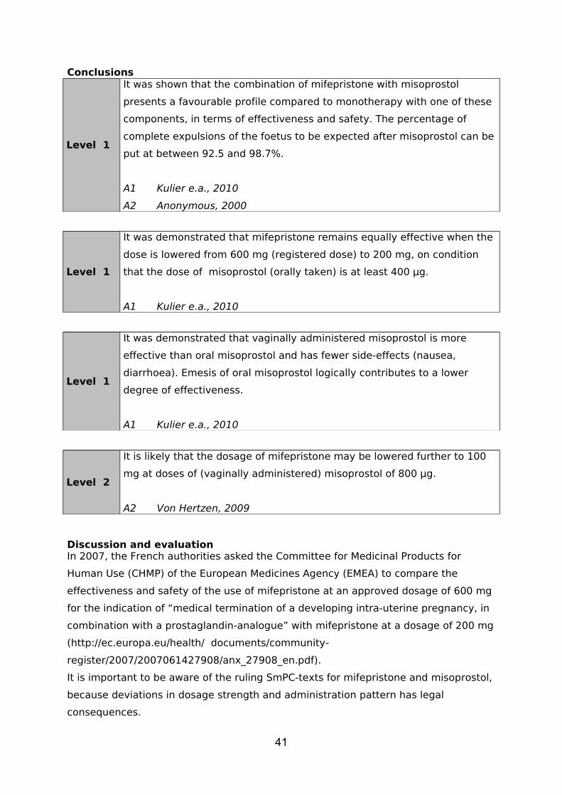
Conclusions
Level 1
It was shown that the combination of mifepristone with misoprostol
presents a favourable profile compared to monotherapy with one of these
components, in terms of effectiveness and safety. The percentage of
complete expulsions of the foetus to be expected after misoprostol can be
put at between 92.5 and 98.7%.
A1 Kulier e.a., 2010
A2 Anonymous, 2000
Level 1
It was demonstrated that mifepristone remains equally effective when the
dose is lowered from 600 mg (registered dose) to 200 mg, on condition
that the dose of misoprostol (orally taken) is at least 400 µg.
A1 Kulier e.a., 2010
Level 1
It was demonstrated that vaginally administered misoprostol is more
effective than oral misoprostol and has fewer side-effects (nausea,
diarrhoea). Emesis of oral misoprostol logically contributes to a lower
degree of effectiveness.
A1 Kulier e.a., 2010
Level 2
It is likely that the dosage of mifepristone may be lowered further to 100
mg at doses of (vaginally administered) misoprostol of 800 µg.
A2 Von Hertzen, 2009
Discussion and evaluationIn 2007, the French authorities asked the Committee for Medicinal Products for
Human Use (CHMP) of the European Medicines Agency (EMEA) to compare the
effectiveness and safety of the use of mifepristone at an approved dosage of 600 mg
for the indication of “medical termination of a developing intra-uterine pregnancy, in
combination with a prostaglandin-analogue” with mifepristone at a dosage of 200 mg
(http://ec.europa.eu/health/ documents/community-
register/2007/2007061427908/anx_27908_en.pdf).
It is important to be aware of the ruling SmPC-texts for mifepristone and misoprostol,
because deviations in dosage strength and administration pattern has legal
consequences.
41

If the prescribing physician wishes to prescribe an agent in an “unlicensed” or “off
label” format, (s)he must accept the responsibility for this choice. This responsibility
can be defined as the explicit obligation to provide information and obtain the
patient’s consent (so-called informed consent). When (serious) side-effects occur
when medication is prescribed beyond the registered SmPC, the primary
responsibility rests with the prescribing physician.
Among Dutch gynaecologists there is a wide variety in the use of mifepristone and
misoprostol (Brouns et al., 2010): mifepristone as a monotherapy in two dosages
(200 or 600 mg); misoprostol as a monotherapy (200, 400, 600, 800 µg); as a
combination therapy misoprostol is given on one, two or three consecutive days, or
2-5 times on the same day.
Misoprostol in therapeutic dosages does not cause hypotension. In the author’s
experience fainting (in the absence of pain) is primarily seen when a hot shower is
taken (vasodilatation). A first case of vasospasm in a coronary artery after (vaginally
administered) misoprostol to a 32-year old smoking and overweight woman was
recently described in Barcelona (Illa et al., 2010). There are no indications of
misoprostol causing disturbances in the metabolism of carbohydrates.
Only when sufficient communication is possible with the woman (both in terms of
language and comprehension) may medical treatment be carried out.
After taking misoprostol, the woman may not be allowed to stay on her own. In home
treatment with misoprostol the woman must be able to call for medical assistance.
The woman (and possibly her partner or other person sharing her household) must
be given clear instructions, to whom they should turn if the woman suffers too much
pain or blood loss (or shows any other medical problem).
There is a single publication according to which previous pregnancies, in particular
when carried to term, negatively influence the effectiveness of medical termination
of pregnancy (Odeh et al., 2010). This is not an experience shared by the study group
members, rather to the contrary. Side-effects, too, appear to be less frequent or
severe in the case of earlier deliveries.
The most frequent side-effects of the combination mifepristone/misoprostol are:
- highly frequent: bowel cramps (the first hours after administration of
misoprostol); nausea, vomiting, diarrhoea (attributed to misoprostol),
shivering and fever.
- frequent: bowel cramps, slight or modest; infection after abortion
(suspected or confirmed infections were reported by < 5% of the women);
severe haemorrhage (necessitating curettage in 1.4% of the cases).
42
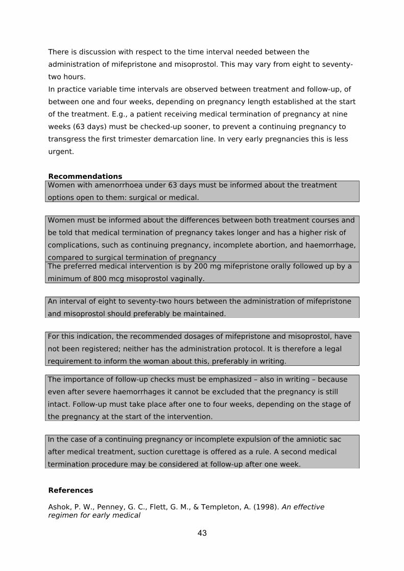
There is discussion with respect to the time interval needed between the
administration of mifepristone and misoprostol. This may vary from eight to seventy-
two hours.
In practice variable time intervals are observed between treatment and follow-up, of
between one and four weeks, depending on pregnancy length established at the start
of the treatment. E.g., a patient receiving medical termination of pregnancy at nine
weeks (63 days) must be checked-up sooner, to prevent a continuing pregnancy to
transgress the first trimester demarcation line. In very early pregnancies this is less
urgent.
RecommendationsWomen with amenorrhoea under 63 days must be informed about the treatment
options open to them: surgical or medical.
Women must be informed about the differences between both treatment courses and
be told that medical termination of pregnancy takes longer and has a higher risk of
complications, such as continuing pregnancy, incomplete abortion, and haemorrhage,
compared to surgical termination of pregnancyThe preferred medical intervention is by 200 mg mifepristone orally followed up by a
minimum of 800 mcg misoprostol vaginally.
An interval of eight to seventy-two hours between the administration of mifepristone
and misoprostol should preferably be maintained.
For this indication, the recommended dosages of mifepristone and misoprostol, have
not been registered; neither has the administration protocol. It is therefore a legal
requirement to inform the woman about this, preferably in writing.
The importance of follow-up checks must be emphasized – also in writing – because
even after severe haemorrhages it cannot be excluded that the pregnancy is still
intact. Follow-up must take place after one to four weeks, depending on the stage of
the pregnancy at the start of the intervention.
In the case of a continuing pregnancy or incomplete expulsion of the amniotic sac
after medical treatment, suction curettage is offered as a rule. A second medical
termination procedure may be considered at follow-up after one week.
References
Ashok, P. W., Penney, G. C., Flett, G. M., & Templeton, A. (1998). An effective regimen for early medical
43
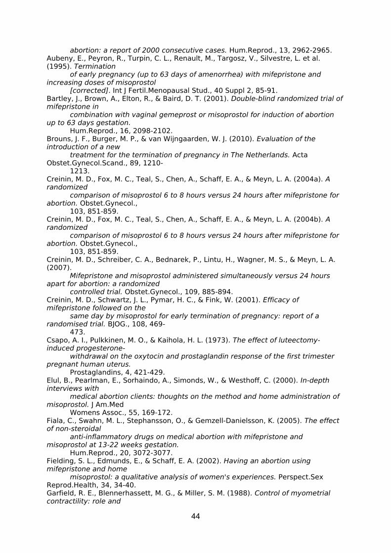
abortion: a report of 2000 consecutive cases. Hum.Reprod., 13, 2962-2965.Aubeny, E., Peyron, R., Turpin, C. L., Renault, M., Targosz, V., Silvestre, L. et al. (1995). Termination
of early pregnancy (up to 63 days of amenorrhea) with mifepristone and increasing doses of misoprostol
[corrected]. Int J Fertil.Menopausal Stud., 40 Suppl 2, 85-91.Bartley, J., Brown, A., Elton, R., & Baird, D. T. (2001). Double-blind randomized trial of mifepristone in
combination with vaginal gemeprost or misoprostol for induction of abortion up to 63 days gestation.
Hum.Reprod., 16, 2098-2102.Brouns, J. F., Burger, M. P., & van Wijngaarden, W. J. (2010). Evaluation of the introduction of a new
treatment for the termination of pregnancy in The Netherlands. Acta Obstet.Gynecol.Scand., 89, 1210-
1213.Creinin, M. D., Fox, M. C., Teal, S., Chen, A., Schaff, E. A., & Meyn, L. A. (2004a). A randomized
comparison of misoprostol 6 to 8 hours versus 24 hours after mifepristone for abortion. Obstet.Gynecol.,
103, 851-859.Creinin, M. D., Fox, M. C., Teal, S., Chen, A., Schaff, E. A., & Meyn, L. A. (2004b). A randomized
comparison of misoprostol 6 to 8 hours versus 24 hours after mifepristone for abortion. Obstet.Gynecol.,
103, 851-859.Creinin, M. D., Schreiber, C. A., Bednarek, P., Lintu, H., Wagner, M. S., & Meyn, L. A. (2007).
Mifepristone and misoprostol administered simultaneously versus 24 hours apart for abortion: a randomized
controlled trial. Obstet.Gynecol., 109, 885-894.Creinin, M. D., Schwartz, J. L., Pymar, H. C., & Fink, W. (2001). Efficacy of mifepristone followed on the
same day by misoprostol for early termination of pregnancy: report of a randomised trial. BJOG., 108, 469-
473.Csapo, A. I., Pulkkinen, M. O., & Kaihola, H. L. (1973). The effect of luteectomy-induced progesterone-
withdrawal on the oxytocin and prostaglandin response of the first trimester pregnant human uterus.
Prostaglandins, 4, 421-429.Elul, B., Pearlman, E., Sorhaindo, A., Simonds, W., & Westhoff, C. (2000). In-depth interviews with
medical abortion clients: thoughts on the method and home administration of misoprostol. J Am.Med
Womens Assoc., 55, 169-172.Fiala, C., Swahn, M. L., Stephansson, O., & Gemzell-Danielsson, K. (2005). The effect of non-steroidal
anti-inflammatory drugs on medical abortion with mifepristone and misoprostol at 13-22 weeks gestation.
Hum.Reprod., 20, 3072-3077.Fielding, S. L., Edmunds, E., & Schaff, E. A. (2002). Having an abortion using mifepristone and home
misoprostol: a qualitative analysis of women's experiences. Perspect.Sex Reprod.Health, 34, 34-40.Garfield, R. E., Blennerhassett, M. G., & Miller, S. M. (1988). Control of myometrial contractility: role and
44

regulation of gap junctions. Oxf Rev.Reprod.Biol., 10, 436-490.Guest, J., Chien, P. F., Thomson, M. A., & Kosseim, M. L. (2007). Randomised controlled trial comparing
the efficacy of same-day administration of mifepristone and misoprostol for termination of pregnancy with the
standard 36 to 48 hour protocol. BJOG., 114, 207-215.Hamoda, H., Ashok, P. W., Flett, G. M., & Templeton, A. (2005). A randomised controlled trial of
mifepristone in combination with misoprostol administered sublingually or vaginally for medical abortion up to
13 weeks of gestation. BJOG., 112, 1102-1108.Heikinheimo, O. (1989). Pharmacokinetics of the antiprogesterone RU 486 in women during multiple dose
administration. J Steroid Biochem., 32, 21-25.Heikinheimo, O., Lahteenmaki, P. L., Koivunen, E., Shoupe, D., Croxatto, H., Luukkainen, T. et al.
(1987). Metabolism and serum binding of RU 486 in women after various single doses. Hum.Reprod., 2,
379-385.Heikinheimo, O., Tevilin, M., Shoupe, D., Croxatto, H., & Lahteenmaki, P. (1986). Quantitation of RU
486 in human plasma by HPLC and RIA after column chromatography. Contraception, 34, 613-624.Herrmann, W., Wyss, R., Riondel, A., Philibert, D., Teutsch, G., Sakiz, E. et al. (1982). [The effects of an
antiprogesterone steroid in women: interruption of the menstrual cycle and of early pregnancy]. C.R.Seances
Acad.Sci.III, 294, 933-938.Honkanen, H., Piaggio, G., Hertzen, H., Bartfai, G., Erdenetungalag, R., Gemzell-Danielsson, K. et
al. (2004). WHO multinational study of three misoprostol regimens after mifepristone for early medical
abortion. BJOG., 111, 715-725.Illa, M., Bennasar, M., Eixarch, E., Berge, R., Font, C., & Palacio, M. (2010). Acute coronary artery
vasospasm associated with misoprostol for termination of pregnancy. Fetal Diagn.Ther., 27, 174-177.Johannisson, E., Oberholzer, M., Swahn, M. L., & Bygdeman, M. (1989). Vascular changes in the human
endometrium following the administration of the progesterone antagonist RU 486. Contraception, 39, 103-
117.Kopp, K. H., Fiala, C., Stephansson, O., & Gemzell-Danielsson, K. (2010). Home self-administration of
vaginal misoprostol for medical abortion at 50-63 days compared with gestation of below 50 days.
Hum.Reprod., 25, 1153-1157.Kulier, R., Gulmezoglu, A. M., Hofmeyr, G. J., Cheng, L. N., & Campana, A. (2004). Medical methods
for first trimester abortion. Cochrane.Database.Syst.Rev., CD002855.Kulier R., Kapp N., Gülmezoglu A.M., Hofmeyr G.J., Cheng L., & Campana A. The Cochrane Library, 2010, Issue 8Lahteenmaki, P., Heikinheimo, O., Croxatto, H., Spitz, I., Shoupe, D., Birgerson, L. et al. (1987).
Pharmacokinetics and metabolism of RU 486. J Steroid Biochem., 27, 859-863.Liao, A. H., Han, X. J., Wu, S. Y., Xiao, D. Z., Xiong, C. L., & Wu, X. R. (2004). Randomized, double-
45

blind, controlled trial of mifepristone in capsule versus tablet form followed by misoprostol for early medical
abortion. Eur J Obstet.Gynecol.Reprod.Biol., 116, 211-216.Livshits, A., Machtinger, R., David, L. B., Spira, M., Moshe-Zahav, A., & Seidman, D. S. (2009).
Ibuprofen and paracetamol for pain relief during medical abortion: a double-blind randomized controlled study.
Fertil.Steril., 91, 1877-1880.Lohr, P. A., Reeves, M. F., Hayes, J. L., Harwood, B., & Creinin, M. D. (2007). Oral mifepristone and
buccal misoprostol administered simultaneously for abortion: a pilot study. Contraception, 76, 215-220.Lohr, P. A., Wade, J., Riley, L., Fitzgibbon, A., & Furedi, A. (2010). Women's opinions on the home
management of early medical abortion in the UK. J Fam.Plann.Reprod.Health Care, 36, 21-25.Middleton, T., Schaff, E., Fielding, S. L., Scahill, M., Shannon, C., Westheimer, E. et al. (2005).
Randomized trial of mifepristone and buccal or vaginal misoprostol for abortion through 56 days of last
menstrual period. Contraception, 72, 328-332.Odeh, M., Tendler, R., Sosnovsky, V., Kais, M., Ophir, E., & Bornstein, J. (2010). The effect of parity and
gravidity on the outcome of medical termination of pregnancy. Isr.Med Assoc.J, 12, 606-608.Peyron, R., Aubeny, E., Targosz, V., Silvestre, L., Renault, M., Elkik, F. et al. (1993). Early termination
of pregnancy with mifepristone (RU 486) and the orally active prostaglandin misoprostol. N.Engl.J Med,
328, 1509-1513.Schaff, E. A., Eisinger, S. H., Stadalius, L. S., Franks, P., Gore, B. Z., & Poppema, S. (1999). Low-dose
mifepristone 200 mg and vaginal misoprostol for abortion. Contraception, 59, 1-6.Schaff, E. A., Fielding, S. L., Eisinger, S. H., Stadalius, L. S., & Fuller, L. (2000). Low-dose mifepristone
followed by vaginal misoprostol at 48 hours for abortion up to 63 days. Contraception, 61, 41-46.Schaff, E. A., Fielding, S. L., & Westhoff, C. (2001a). Randomized trial of oral versus vaginal misoprostol at
one day after mifepristone for early medical abortion. Contraception, 64, 81-85.Schaff, E. A., Fielding, S. L., & Westhoff, C. (2001b). Randomized trial of oral versus vaginal misoprostol at
one day after mifepristone for early medical abortion. Contraception, 64, 81-85.Schaff, E. A., Fielding, S. L., & Westhoff, C. (2001c). Randomized trial of oral versus vaginal misoprostol at
one day after mifepristone for early medical abortion. Contraception, 64, 81-85.Schaff, E. A., Fielding, S. L., & Westhoff, C. (2002). Randomized trial of oral versus vaginal misoprostol 2
days after mifepristone 200 mg for abortion up to 63 days of pregnancy. Contraception, 66, 247-250.Schaff, E. A., Fielding, S. L., Westhoff, C., Ellertson, C., Eisinger, S. H., Stadalius, L. S. et al. (2000a).
46
Vaginal misoprostol administered 1, 2, or 3 days after mifepristone for early medical abortion: A randomized
trial. JAMA, 284, 1948-1953.Schaff, E. A., Fielding, S. L., Westhoff, C., Ellertson, C., Eisinger, S. H., Stadalius, L. S. et al. (2000b).
Vaginal misoprostol administered 1, 2, or 3 days after mifepristone for early medical abortion: A randomized
trial. JAMA, 284, 1948-1953.Schindler, A. M., Zanon, P., Obradovic, D., Wyss, R., Graff, P., & Herrmann, W. L. (1985). Early
ultrastructural changes in RU-486-exposed decidua. Gynecol.Obstet.Invest, 20, 62-67.Shannon, C., Wiebe, E., Jacot, F., Guilbert, E., Dunn, S., Sheldon, W. R. et al. (2006). Regimens of
misoprostol with mifepristone for early medical abortion: a randomised trial. BJOG., 113, 621-628.Shi, Y. E., Ye, Z. H., He, C. H., Zhang, G. Q., Xu, J. Q., Van Look, P. F. et al. (1993). Pharmacokinetic
study of RU 486 and its metabolites after oral administration of single doses to pregnant and non-pregnant
women. Contraception, 48, 133-149.Spitz, I. M., Bardin, C. W., Benton, L., & Robbins, A. (1998). Early pregnancy termination with mifepristone
and misoprostol in the United States. N.Engl.J Med, 338, 1241-1247.Swahn, M. L. & Bygdeman, M. (1988a). The effect of the antiprogestin RU 486 on uterine contractility and
sensitivity to prostaglandin and oxytocin. Br.J Obstet.Gynaecol., 95, 126-134.Swahn, M. L. & Bygdeman, M. (1988b). The effect of the antiprogestin RU 486 on uterine contractility and
sensitivity to prostaglandin and oxytocin. Br.J Obstet.Gynaecol., 95, 126-134.Tang, O. S., Chan, C. C., Ng, E. H., Lee, S. W., & Ho, P. C. (2003a). A prospective, randomized, placebo-
controlled trial on the use of mifepristone with sublingual or vaginal misoprostol for medical abortions of less
than 9 weeks gestation. Hum.Reprod., 18, 2315-2318.Tang, O. S., Chan, C. C., Ng, E. H., Lee, S. W., & Ho, P. C. (2003b). A prospective, randomized, placebo-
controlled trial on the use of mifepristone with sublingual or vaginal misoprostol for medical abortions of less
than 9 weeks gestation. Hum.Reprod., 18, 2315-2318.Thong, K. J. & Baird, D. T. (1992). Induction of abortion with mifepristone and misoprostol in early pregnancy.
Br.J Obstet.Gynaecol., 99, 1004-1007.von, H. H., Honkanen, H., Piaggio, G., Bartfai, G., Erdenetungalag, R., Gemzell-Danielsson, K. et al.
(2003a). WHO multinational study of three misoprostol regimens after mifepristone for early medical
abortion. I: Efficacy. BJOG., 110, 808-818.von, H. H., Honkanen, H., Piaggio, G., Bartfai, G., Erdenetungalag, R., Gemzell-Danielsson, K. et al.
(2003b). WHO multinational study of three misoprostol regimens after mifepristone for early medical
abortion. I: Efficacy. BJOG., 110, 808-818.von, H. H., Piaggio, G., Wojdyla, D., Marions, L., My Huong, N. T., Tang, O. S. et al. (2009). Two
mifepristone doses and two intervals of misoprostol administration for termination of early pregnancy: a
47

randomised factorial controlled equivalence trial. BJOG., 116, 381-389.World Health Organisation. Comparison of two doses of mifepristone in combination with misoprostol for early
medical abortion: a randomised trial. World Health Organisation Task Force on Post-ovulatory
Methods of Fertility Regulation(2000). BJOG., 107, 524-530.Winikoff, B., Dzuba, I. G., Creinin, M. D., Crowden, W. A., Goldberg, A. B., Gonzales, J. et al.
(2008). Two distinct oral routes of misoprostol in mifepristone medical abortion: a randomized controlled
trial. Obstet.Gynecol., 112, 1303-1310
48
Chapter 3 Second trimester termination of
pregnancy
Guiding question
What is the most efficient and effective treatment in second trimester pregnancies:
surgical, medical or by mixed approach?
3.1 General introduction
What is the optimal approach to termination of pregnancy in the second trimester?
What are advantages and disadvantages of surgical interventions (dilatation and
evacuation), with or without priming, and medical interventions (including mixed
approaches)? What can be said with respect to effectiveness, efficiency, safety and
complications?
The demarcation line between first and second trimester was defined at the NGvA’s
members’ meeting of September 2009. The second trimester starts at an
amenorrhoea length of 13 (weeks) + 0 (days), corresponding to a biparietal diameter
(BPD) of >22.9 mm. In the guideline ‘Counselling of women considering termination
of pregnancy’ the first trimester was defined as ending at 91 days’ amenorrhoea. The
upper boundary was established by law at the viability line, which is currently at 24
weeks’ amenorrhoea. This figure depends on what views are held with respect to
viability, which are, in part, related to medico-technical developments in the field of
neonatology. Within this context, each clinic determines its own restrictions,
depending on the competencies of the abortion specialists in the particular clinic and
the clinic’s license conditions.
In Dutch abortion care second trimester interventions generally employ dilatation
and evacuation (D&E). In the literature consulted the following aspects were studied:
length of treatment, time invested by client and professional, safety and
complications of the method(s) used. A complementary search was undertaken in
September 2011 with a focus on specific terms (Asherman’s syndrome, DIC, low birth
weight and prematurity). The search did not yield specific results on Asherman or DIC
(Diffuse Intravascular Coagulation). In the eyes of the study group, it would appear
useful to set up a registration system to collect data about late complications of D&E
in the second trimester.
49
3.2 Surgical termination of pregnancy
Guiding question
What is the most efficient and effective surgical treatment (or mixed approach) in
second trimester termination of pregnancy?
Introduction
Surgical – or instrumental - termination of pregnancy is defined as any approach to
termination of pregnancy using instruments, either dilatators, (suction) curettes or
evacuation forceps (tongs). Up to about 17 weeks this is generally done by means of
mechanical dilatation, followed by evacuation, without cervix priming.
In more advanced pregnancies, different regimes are followed in the abortion clinics
which carry out this intervention, viz. no cervical priming, priming with misoprostol
400 microgram, orally or vaginally, or the two-step method in pregnancies above 17
weeks in nulliparae and above 19 weeks in multiparae (in the first step, the
membranes are ruptured and the umbilical cord is severed; in the second step
evacuation takes place).
Summary of the literature
No relevant literature was found before 2005. In 2010, six articles were found on
early and late complications of D&E. One was of such limited quality that it was not
included (Pauli), the other five are described below.
Lohr (2008) wrote a review in which she discusses current interventions in the
second trimester, including their safety, pros and cons, acceptability and associated
complications. Her article also reviews methods which make surgical termination of
pregnancy in the second trimester safer and more efficient, like cervical priming and
treatment under ultrasound scanning. Although the question remains whether
surgical or medical termination in the second trimester should be preferred, she
claims that the basis for deciding which method is to be used is multifactorial.
Specialized training and the experience of performing sufficient numbers of
interventions are necessary to carry out a D&E safely. Choice of procedure is also
affected by the presence or absence of necessary equipment; the availability of an
operating theatre; the policy with respect to facilities; acceptance by staff and
preference of patients. When D&E is offered, women do not always wish to be
randomized for a medical intervention (Grimes, 2004).
Advantages for both caretaker and patient are the precise planning allowed by the
procedure, the short treatment time and the avoidance of hospitalization.
50

Second trimester surgical termination is in the developing stage. Variation in
methods for cervical priming, instrumentation and complementary interventions like
foeticide are carried out to achieve optimal safety and efficiency in uterus
evacuation.
Cervical priming may take place through mechanical dilatation, slowly expanding
osmotic dilatators, or medically, with misoprostol or mifegyne. Laminaria and Dilapan
are preferred in pregnancies of longer duration, to reduce the risk of cervix
impairment or perforation compared to mechanical dilatation alone. The use of
Dilapan or laminaria primarily depends on the professional’s preference; osmotic
dilatators are not available in all circumstances; besides, they require admission and
an overnight stay.
Misoprostol has been studied as an alternative; it is cheap, almost always available,
and suitable for self-administration via a variety of routes: oral, vaginal, buccal and
sublingual. However, the ideal dose, route and limitation in terms of gestation stage
have not been established yet. Buccal misoprostol in doses of 400-800 mcg would
seem to serve as adequate cervical priming up to 18 weeks of gestation. Women also
appear to prefer one day treatments. Further study is needed to establish the
possible benefits of combination regimes, especially in the case of later stage
pregnancies.
Excessive blood loss is the most common complication in second trimester surgical
termination of pregnancy; its risk increases with pregnancy length. Excessive
haemorrhage may be caused by impairment of the cervix or uterus, an incomplete
procedure or failure of the uterus to contract. Moreover, the risk of DIC (Diffuse
Intravascular Coagulation) increases during the second trimester.
Uterus perforation is a potentially dangerous complication in second trimester
surgical termination of pregnancy, with an incidence of 0.2-0.4%. Routine use of
ultrasound during the intervention proved to reduce the risk of perforation (Darney,
1989); cf. chapter 5 for a further discussion of the role of ultrasound (or sonography)
during treatment.
Recent studies with respect to D&E and complications in future pregnancies did not
reveal any relationship. Premature partus appears less likely when laminaria were
used, compared to mechanical dilatation, possibly as a consequence of the smaller
impact on the cervix. According to Lohr there are no data about the effects of
misoprostol, which is generally followed up with mechanical dilatation, and the
outcome of succeeding pregnancies. Currently, however, data are available, cf.
Winner and Nucatola on this point (below).
Lohr concludes that D&E is a safe, cost-effective method for termination of
51

pregnancy for women who are in a position to choose for this method in the second
trimester of their pregnancy.
In a 2006 study, Jackson tried to establish if the anamnesis of women undergoing
mid-trimester D&E (at 12-24 weeks of gestation) shows an increased risk of
complications during a successive pregnancy, compared with a control group who
had not undergone this intervention. This concerns a retrospective cohort study into
women with D&E during the second trimester, which included dilatation by laminaria
japonica. Of the 317 women who underwent D&E, 85 had a vital successive
pregnancy. These women delivered a little earlier than the 170 control group women
(at 38.9 versus 39.5 weeks, p=0.001), but there was no significant difference
between the two groups with respect to birth weight, spontaneous premature partus,
abnormal placentation and total number of complications.
This study is limited in a number of respects. To be able to indicate a difference in
premature partus < 37 weeks the groups under study would have to number 169 and
338 women, respectively. Absence of a difference with respect to less frequent
pregnancy complications may be based on a type II error.
Neither can this study pronounce itself on the influence of D&E on other reproductive
outcomes such as future fertility, ectopic pregnancy, neonatal findings or a
succeeding spontaneous first or second trimester abortion.
As it stands, this study predicts an acute complication risk of 0.3%, which is within
the limits otherwise known from the literature (0.23%- 0.69%) (Grimes, 1977, Cates,
1982).
In a retrospective cohort study, Turok compared outcome and cost of second
trimester termination of pregnancy using D&E between specialized abortion clinics,
hospitals in which the procedure was carried out by an assistant medical practitioner,
or induced abortion in hospitals otherwise. This study concerned women in hospitals
with a pregnancy length of 13-24 weeks and in clinics with a pregnancy length of 13-
21 weeks.
Major complications occurred in 11% of the women undergoing D&E in the hospital;
in 10% in the case of hospital induction and 1% of the women undergoing D&E in a
specialized clinic. The cost level, too, was very different; $1,105 for treatment in the
specialized clinic, $4,425 for a D&E in the hospital and $5,029 for a hospital
induction.
The conclusion is that second trimester termination of pregnancy by means of D&E
in selected patients in a specialized clinic may be safer and cheaper than D&E or
induction in the hospital; especially the skill and expertise of the professional
conducting the termination of pregnancy play a major role.
52

The objective of the study by Nucatola was to determine a number of serious
complications during second trimester abortions by means of D&E after cervical
priming with misoprostol, at a gestation of 12-16 weeks, in 6,620 interventions, using
retrospective data analysis. The author claims that cervix priming reduces the length
of the intervention, causes less pain and blood loss and therefore belongs to the
standard procedures for second trimester termination of pregnancy. In this study a
perforation risk of 0.45/1000 is shown, compared to figures in the literature of 0.8-
7/1000 for abortions with D&E. Misoprostol as a cervix primer was not shown to lead
to an increased risk of perforation.
Winer´s study (9) is a case control study, to ascertain the effect of misoprostol on the
risk of late spontaneous abortions and premature partus in successive pregnancies.
245 women with a partus at 16-36 weeks and 490 controls who delivered at 37
weeks or beyond were included in this study. The anamnesis of forty cases (16.3%)
and 56 controls (11.5%) showed cervix priming with misoprostol alone, or in
combination with mifegyne, had taken place in the context of vacuum curettage,
evacuation or medical termination of pregnancy.
With an OR of 1.33 (CI 0.81-2.17) it may be concluded that the results confirm that
induced abortion by means of misoprostol is safe for later pregnancies.
Conclusion
Level 3
D&E is a safe and cheap method for termination of pregnancy in the
second trimester.
C Lohr 2008; Turok 2008
53
3.3 Cervix priming in surgical termination of pregnancy
Guiding question
What evidence can be derived from the (inter)national literature for the application of
prostaglandins and the course of administration in second trimester termination of
pregnancy?
Introduction
Prostaglandins have been used for a long time to prepare the mouth of the uterus
(cervix uteri) for dilatation. Prostaglandins make dilatation easier. Blood loss during
and after the intervention is restricted. Priming is often applied in surgical
interventions. In the abortion clinics misoprostol is the prostaglandin almost
exclusively used to prime the cervix. Effectiveness, side-effects and safety are
elements discussed further below.
Summary of the literature
Various prostaglandin analogues have been developed. Misoprostol is a
prostaglandin E1 analogue (PGE1)., which came on the market in 1988. Before 2005,
few studies were found: Todd’s study (Todd et al., 2002) is non-randomized and only
covers 110 women. After 2005, four articles are found which investigate priming with
misoprostol in D&E.
In a double blind randomized study involving 83 women Goldberg et al. (2005)
compare the effect of laminaria one day prior to surgery and misoprostol 400
microgram vaginally at 3-4 hours pre-op. They measured spontaneous dilatation with
either method, of 14 mm with laminaria and 11 mm with misoprostol before suction
curettage, optionally followed up by an instrument, at a pregnancy length of 13-16
weeks. With nulliparae the intervention proceeds more quickly and smoothly with
laminaria, in multiparae there is no difference. Women preferred misoprostol, as it
requires only one visit to the clinic.
Carbonell et al. (2007) studied the effect of mifepristone administered two days prior
to D&E. They randomized 900 women with a pregnancy length between 12 and 20
weeks across four groups in a prospective non-blinded trial. 450 women were given
mifepristone 200 mg 2 days before D&E, the others were not. On the day of
treatment, half the women in both groups received 600 microgram misoprostol
vaginally and the other 225 received 600 microgram misoprostol sublingually. In the
case of a rigid cervix Dilapan was added in all groups. One of the aspects studied was
the degree of dilatation achieved without effort and the duration of the intervention.
54
In the mifepristone group 12.5 mm of dilatation was found; this was 8.5 mm without
mifepristone. No relationship was found with respect to the misoprostol route. The
time needed before the intervention was shorter in the mifepristone group: 1.7
versus 2.1 hours, respectively (p<0.001). The duration of the intervention itself was
shorter in the mifepristone group in combination with sublingual misoprostol: 11.9 vs.
13 minutes (p=0.007); in the vaginal misoprostol group the difference was also
observed, but was non-significant: 12.3 vs. 13 minutes, p=0.031. In the mifepristone
group cervix rigidity decreased from 65% to 15%, with less frequent application of
Dilapan as a result.
Carbonell et al. arrive at the following conclusions. There are some problems
attached to the use of mifepristone two days before the intervention, like increased
costs (200 mg mifepristone comes at about €25,-); the fact that the woman needs to
visit the clinic twice; the possibility of expulsion of the foetus before the actual
intervention and the possibility of teratogenicity in women who do not return for a
D&E. No indications of the latter problem arising have been found as yet, however. In
contrast, they also observe advantages in the use of mifepristone two days before a
D&E. One is the shorter waiting time after administration of misoprostol on the day of
the intervention; further, the clear reduction in the use of Dilapan, which in itself
saves on costs (one dose of Dilapan costs €20,-) and less risk of cervical lacerations.
The biggest shortcoming in this study is the use of Dilapan, which was not applied at
random, in contrast with other studies.
Edelman et al. (2006) gave 400 microgram misoprostol or a placebo 60 to 90
minutes before a D&E after laminaria one day earlier in women with a pregnancy
length of 13 up to 15+6 weeks and from 16 to 20+6 weeks and observed the
maximum dilatation achieved and the effort needed for dilatation. They found no
significant differences. But if they only considered pregnancies from 19+0 onward,
they did find a significant difference. However, the study had not been properly set
up to fully warrant this latter finding.
Patel et al. (2006) undertook a descriptive retrospective study into the safety and
effectiveness of buccal misoprostol in various dosages, sometimes combined with
laminaria, in 19 different member centres of the Planned Parenthood Federation of
America. These centres use the buccal misoprostol protocols in a number of
variations, combined with laminaria or not, at one, two or three days prior to
treatment. Of 2,218 women with an amenorrhoea between 12 and 23+6 weeks they
looked at the (serious) adverse events registered, the ease of dilatation and side-
effects. They found 19.39/1000 adverse events, of which 4.51/1000 were classified
as serious, defined as: death; life-threatening medication problems; hospitalization or
prolonged hospital stay; lasting or important disability or a disease. In 75.3% the
55
ease of dilatation was found to be adequate; they remarked that misoprostol at lower
dosages is more adequate, both with and without laminaria. Side-effects increase
with the dosage strength of the misoprostol. The authors conclude that there is no
reason for worries with respect to the use of buccal misoprostol in D&E in the second
trimester of the pregnancy.
The literature found does not suggest any adaptation of the usual dose of 400 mcg
misoprostol as priming in the second trimester; there are no grounds for preferring a
specific route, be it vaginal, sublingual, buccal or oral.
The further the pregnancy has progressed, the greater the sensitivity to uterotonics.
As a result, gradually decreasing levels of misoprostol are needed to achieve the
same effect (Goldberg e.a., 2001). For the induction of a termination of pregnancy
800 μg (every 12 hours) is needed in the first trimester (Darney&Sweet, 1989). In the
third trimester this is only 50 μg (every 12 hours). The dosage for induction in the
second trimester probably lies between 50 μg and 800 μg, showing decreasing
necessary dosages for misoprostol as the number of weeks increases
(Darney&Sweet, 1989).
In principle, Misoprostol shows the same dosage dependent side-effects (Goldberg
e.a., 2001; Shannon e.a., 2004; Sivalal e.a., 2004) as other prostaglandins do, with
gastro-intestinal side-effects, pain and fever (more than 38° Celsius rectally) as the
most common (Goldberg e.a., 2001; Shannon e.a., 2004; Sivalal e.a., 2004; Goldberg
e.a., 2003; Ngai e.a., 2003; Vimala e.a., 2004).
Prostaglandins have been associated with coronary failures (Goldberg e.a., 2001;
Ngai e.a., 2003) and bronchospasms (Goldberg e.a., 2001), but these have never
been reported for misoprostol. (Cf. also first trimester medical termination of
pregnancy in the previous chapter).
Misoprostol may cause preoperative blood loss, but this is mostly modest (Sivalal
e.a., 2004); however, serious blood loss may occur (Shannon e.a., 2004; Sharma e.a.,
2005). The higher the dosage (Goldberg e.a., 2001; Goldberg e.a., 2003) and the
longer the priming interval (Vimala e.a., 2004), the greater the risks.
Conclusion
Level 3
It is likely that misoprostol 400 μg vaginally, three to four hours prior
to the intervention, enables sufficient dilatation of the cervix D&E.
A2 Goldberg et al., 2005, Edelman et al. 2006
C Todd et al., 2002
D RCOG guideline, 2004; SFP guidelines, 2007 en 2008
56
3.4 The use of misoprostol after sectio caesarea
Guiding question
Can misoprostol be used for a D&E after a sectio caesarea (C-section)?
Introduction
In practice it is suspected that the use of misoprostol in connection with a D&E after
an earlier C-section increases the risk of a uterus rupture. This prompted a specific
search for data on this subject.
Summary of the literature
Two systematic reviews appeared in 2009 with respect to the use of the
prostaglandin misoprostol in the case of a second trimester termination of pregnancy
after an earlier C-section, one by V. Berghella e.a. and one by V. Goyal.
From the Berghella review (2009) of case studies and cohort studies in women with a
pregnancy length of 16-28 weeks, in whom misoprostol was used as initial priming
agent, the chances of a uterus rupture were established in the case of one earlier low
transversal C-section 0.43% (95% CI 0.08-1.67%). When cases from case reports are
added, this figure rises to 1.1%. Out of the five cases of uterus rupture found, three
took place at 26 weeks; two of these were given oxytocin in addition to misoprostol.
The other two cases, which took place under 26 weeks, received an overall dose of
misoprostol equal to or greater than 1000 microgram. No uterus rupture was found in
46 cases with two earlier low transversal C-sections. From case reports two cases
were uncovered. In either case there were additional risk factors involved, like
chorioamnionitis in one of them; the other occurred during curettage. Of the seven
cases of three prior C-sections none had a uterus rupture. Of the two cases of earlier
classic C-sections one woman suffered a rupture (a fact from a personal retrospective
data analysis by Berghella). The data are thus too limited to warrant judgment on the
safety of misoprostol in second trimester termination of pregnancy in women with
one or more low transversal C-section or after prior classic C-section. The conclusion
is that misoprostol appears to be a safe abortion-inducing agent in women with one
earlier low transversal C-section in their anamnesis, in the context of a second
trimester termination of pregnancy; additional use of oxytocin, however, must be
kept at a minimum.
In his systematic review, Goyal (2009) calculates risk levels of 0.28% (95% CI 0.08-
1.00%) for uterus rupture in women with a prior C-section versus 0.04% (95% CI
0.01-0.20%) in women without prior C-section in their anamnesis. The study is
concerned with the use of misoprostol in termination of pregnancy in the second
57

trimester. Goyal reviewed studies in which misoprostol alone was applied in women
with one low transversal C-section and found a risk of 0 (95% CI 0-1.48%) among 256
women. When combined with another induction method use of misoprostol showed a
risk of 0.43% (95% CI 0.12-1.55%) in women with a C-section in their anamnesis and
0.06% (95% CI 0.01-0.31%) in women with no C-section. In the two cases in which
uterus rupture occurred, there had been induction by means of mifepristone, while
misoprostol had been added to other agents. The review concluded that women with
one prior low transversal C-section undergoing induction in the context of a second
trimester termination of pregnancy with misoprostol only run no additional risk of a
uterus rupture; when mifepristone is used, the risk amounts to 1.15% (95% CI 0.45-
2.92%). In view of the small numbers the risk of a uterus rupture cannot be
determined with sufficient statistical accuracy from these findings with respect to the
use of mifepristone.
Besides these systematic reviews and the articles reviewed in them, two recent
publications were uncovered, one about misoprostol, administered vaginally in a
second trimester termination of pregnancy, and one about the safety of D&E in the
case of a scarred uterus.
A study by Naguib (2010) concerns a prospective observation in women with a C-
section in their anamnesis. They received induction with vaginally administered
misoprostol 4x200 microgram every 4 hours in the context of a second trimester
termination of pregnancy, at a gestation stage of 16 to 20 weeks. There were no
cases of uterus rupture. As the study included no more than 50 women with a C-
section in their anamnesis the authors conclude that inducing abortion in the second
trimester by means of misoprostol at low dosages appears safe, but that larger-scale
studies are needed to validate the results.
In a study by Ben Ami (2009) the safety of a D&E is determined in cases of scarred
uterus in second trimester termination of pregnancy at 17-24 weeks of gestation,
with prior use of laminaria japonica. They come to the conclusion that there are
probably no raised levels of peri-operative risk, but that larger studies involving more
subjects are needed to confirm these results.
Conclusions
Level 3
It may be assumed that misoprostol is a safe abortion-inducing agent
in the second trimester in women with a history of one low
transversal C-section.
C Berghella 2009; Goyal 2009
58
Level 3
It is possible that mifepristone in women with one low transversal C-
section in their anamnesis forms an additional risk in medical second
trimester termination of pregnancy, as opposed to D&E.
C Berghella 2009
Level 3
There are indications that additional oxytocin besides misoprostol,
used for the induction of a D&E in the context of a second trimester
termination of pregnancy in women with one low transversal C-
section in their anamnesis raises the risk of a rupture.
C Berghella 2009
59
3.5 Mixed approaches
Guiding question
Is the two step method useful for termination of pregnancy in the second trimester of
pregnancy?
Introduction
The term mixed approach refers to the two step method already described (cf. the
Introduction under Surgical Termination of Pregnancy). In a specific search in March
2010 two references from Sweden were found, which are not accessible to us. RCOG
mentions the two step method in its description of options for the second trimester.
The search also lead to an article in which a study was described into foeticide prior
to treatment with KCl (Elimian 1999).
Summary of the literature
In a retrospective status study, Elimian (1999) investigated 68 cases, to determine
the effect of a cardiac puncture with KCl on the course of treatment with PGE2, in
terms of the time lapse before expulsion and the necessary dose of PGE2. Twenty-
two women received this priming treatment. The average time before expulsion is
reported to be shorter when spontaneous intra-uterine foetal death (IUFD) occurs
compared to intact pregnancies. Although there was a slight statistical deviation in
the group with cardiac puncture with respect to gestation stage (21.7+/- 1.7 weeks
versus 20.3 +/- 1.4 weeks) the average dose of PGE2 needed and the time interval
from the start of the treatment with PGE2 until expulsion proved significantly shorter
in the group with cardiac puncture.
Conclusion
Level 3
It may be assumed that foeticide requires lower levels of
prostaglandin and reduces the time interval before spontaneous
expulsion.
B Elimian 1999
60
3.6 Medical termination of pregnancy
Guiding question
What is the most efficient and effective medical course of treatment (including mixed
approaches) for termination of pregnancy in the second trimester of the pregnancy?
Introduction
Medical termination of pregnancy in the second trimester consists of administering
mifepristone on day one followed up by misoprostol according to a variety of regimes
at admission two days later. In The Netherlands, this course of treatment is only
practised in hospitals.
For the standard procedure in Dutch hospitals we refer to NVOG’s1 guideline
“Termination of pregnancy up to 24 weeks” (2005).
Summary of the literature
In a double blind randomized controlled trial, Dickinson et al. (2002) studied the
effectiveness and side-effects of three different dosages of vaginally applied
misoprostol to effect termination of pregnancy between 14 and 30 weeks (Shannon
e.a., 2004). In this trial, 150 women were randomly assigned to one of three groups,
each with a different dosage. It is unclear from this study if part of the abortions were
carried out on social indication. This may have been the case in 22 women at most:
for these women, the indication for a termination of pregnancy was stated as
‘maternal reasons’, the others were cases of foetal death or foetal abnormality. This
makes it difficult to interpret the results of this study for their adoption in the present
guideline. In the group with the lowest dosage the time interval before abortion was
longer than in the other groups (18.2 hours vs. 15.1 and 13.2 hours, p = 0.035);
besides, the percentage reaching abortion within 24 hours was smaller (p = 0.013)
and the percentage after 48 hours was larger (p = 0.021). There was no difference as
regards side-effects between the three groups.
In a study by Wong et al. two different regimes of vaginally administered misoprostol
were compared in women with a pregnancy length of 14 to 20 weeks. In the high
dosage group (400 μg every three hours, with a maximum of five times in 24 hours)
the time interval before abortion as calculated from the first dose was shorter than in
the low dosage group (400 μg every six hours, with a maximum of three times per 24
hours): the time intervals were 24.4 versus 43.4 hours on average (p < 0.01). The
success rate for termination of pregnancy within 48 hours was also higher in this
1
NVOG: Dutch Society of Obstetricians and Gynaecologists
615

group (90.5% versus 75.7%, p < 0.02). The side-effects noted in either group were
more or less similar, with the exception of a higher incidence of fever in the high
dosage group.
In a single blind randomized trial, Ngai et al. (2000) compared the differences in
effects and side-effects of orally administered misoprostol 400 μg with vaginally
administered misoprostol 200 μg. The administration of misoprostol was placebo-
controlled and occurred after all 142 women in this study, who had been pregnant
between 14 and 20 weeks, had received mifepristone. The women received a little
over four doses of misoprostol on average, irrespective of the course of
administration: oral or vaginal. However, in view of the differences in dosage
between both courses, it may be concluded that a double dose of misoprostol is
required when administered orally compared to the vaginal route of administration.
Side-effects like nausea, dizziness, vomiting and fatigue were similar across both
groups. The women who had been administered misoprostol orally suffered more
from diarrhoea than women who had been given the vaginal application of this
agent. Women who did not have an abortion within 24 hours did not have a
preference for one of either method. Women who did succeed in reaching an abortion
within 24 hours largely preferred oral administration, because they found it more
suitable or less painful, or for reasons of privacy.
In the literature search carried out in 2010 various articles were found about second
trimester medical termination of pregnancy, a number of which will be summarized
below.
In a prospective randomized study among 54 women with a pregnancy length
between14 and 26 weeks, Nor Azlin et al. (2006) compared misoprostol 200 mcg
vaginally every twelve hours (four doses max.) with Gemeprost 3 mg vaginally every
three hours (five doses max.). They looked at time intervals before abortion, side-
effects, experienced pain and costs. There was no significant difference between the
number of women in both groups delivering within 48 hours. The women in the
misoprostol group did have an earlier delivery (19.15 hours+14.70 versus 28.26
hours+48.52), but this difference was not significant either. Side effects and pain
experience were equal in both groups, except that the Gemeprost group showed
significantly more fever. In both groups subsequent curettage was required: 29.6% in
the misoprostol group and 37% in the Gemeprost group. Treatment with misoprostol
is much cheaper than with Gemeprost: $ 0.60-2.30 versus $ 48-122.
In a study by Bhattcharyya et al. (2006) 138 women (at 14 to 20 weeks) were
randomly assigned to different regimes of misoprostol: group A received 400 mcg
62
misoprostol vaginally every three hours and group B a starting dose of 600 mcg,
followed up by 200 mcg every three hours; in both groups a twelve hour
administration pause was respected after twelve hours. The time interval between
induction and abortion was similar for either group (about twelve hours), but women
in group B with three or more previous children delivered more quickly. This group
also needed significantly less misoprostol and showed significantly less fever. Only
few women did not deliver within 48 hours in this study; it was not recorded whether
an intervention was started when the placenta had not been born two hours after the
foetus.
Copying first trimester trials Chai et al. (2009) set up a randomized prospective trial
in 2009, to look at the option of administering mifepristone and misoprostol
simultaneously, compared with the standard administration of misoprostol after 36-
38 hours. 141 women were randomized: the time interval before abortion was
shorter for the standard treatment: 4.9 hours vs. 10 hours (p<0.0001). After 24
hours, 100% of the subjects in the standard group had delivered and 91.5% in the
intervention group; p=0.028. Cold shivers and fever occurred significantly less
frequently for the standard treatment; this is probably due to the much lower dosage
of misoprostol.
The benefit of simultaneously starting mifepristone and misoprostol is a reduction of
the woman’s visits to the clinic by one. However, from research it had become clear
that this advantage does not outweigh the disadvantages, namely that the woman
delivers at home and needs to use more misoprostol before successful abortion;
besides, after simultaneous administration the time interval before abortion is much
longer, while 9.5% of the women had not delivered after 24 hours.
In a double blind randomized trail, Oi Shan Tang et al. (2005) compared oral and
sublingual administration of 400 microgram misoprostol every three hours at a
pregnancy length of 12-20 weeks. The sample size of 120 was based on the
hypothesis that, in the sublingual group, 20% more women would deliver within 24
hours. However, this was not demonstrated: 91.%% vs. 85% is not significant. As the
time interval until abortion does prove to be significantly shorter, they conclude that
sublingual is superior to oral. In view of the set-up it is questionable if this conclusion
is valid.
In two trials, Yilmaz et al. (2005, 2007) tried to ascertain if the wide variation in
absorption of vaginal misoprostol could be improved by moistening the tablets with a
physical salt solution or acetic acid, respectively. They compared dry tablets with
63

tablets moistened with a physiological salt solution in 2007 and did not find any
differences. In 2005 they compared physiological salt with acetic acid and found that
acetic acid would speed up the effect of misoprostol. However, Yilmaz does mention
that contradictory findings were obtained in earlier trials. The acetic acid study was
small and its sample size based on a small pilot.
In a randomized non-blinded trial, Bhattacharjee et al. (2008) compared the
administration of sublingual versus vaginal misoprostol in nearly 300 women. They
found no differences with respect to complete abortions after 24 and 48 hours, nor in
the time interval until the delivery of the foetus, nor in side-effects. However, in both
groups women preferred sublingual administration.
Karsidag et al. (2009) found the same outcome in a group of 49 women.
The only reference found for The Netherlands is Jansen et al. (2008). They compared
mifepristone and misoprostol with Dilapan and Sulproston in 16 women. They
concluded that there is no difference in effectiveness, as their hypothesis that they
would achieve a time reduction of 50% - of 36 to 18 hours – until delivery was not
proven to hold true and the Sulproston group already delivered after 20 hours.
However, the Sulproston group had already been hospitalized for 24 hours at the
start of the treatment with Sulproston. They decided to alter their protocol to
mifepristone and misoprostol.
Von Hertzen et al. (2009) conducted a double blind randomized multicenter trial in
seven countries and compared vaginal versus sublingual administration of
misoprostol. They concluded that vaginal administration in nulliparae is more
effective (lower failure rate after 24 hrs.). Apart from that they only found more fever
after vaginal administration.
Hamoda et al. (2005) compared the sublingual and vaginal routes, but the trial’s
strength is too small for valid conclusions about effectiveness; their calculation of the
sample size was inadequate. Women found their own route acceptable. Women in
the sublingual group suffered more from an unpleasantly tasting mouth, in the
vaginal group there was more flushing and lethargy. The sublingual group received
more pain medication.
In a randomized trial among 162 women, Caliskan et al. (2009) found that 100 mcg
sublingually in twelve doses every two hours is equally effective as 200 mcg
sublingually with the same administration schedule.
Because the median treatment time after admission varies from 12 to 18 hours,
(Dickinson et al., 2002; Tang et al., 2005; Yilmaz et al., 2007; Bhattacharjee et al.,
64
2008; Karsidag et al., 2009; Caliskan et al., 2009) and the practical consequences of
this fact, this type of treatment is not possible in the setting of Dutch abortion clinics.
It is a drawback of medical treatment that a surgical intervention turns out to be
necessary after all in 10-30% of the cases, due to placenta retention and
haemorrhages (Dickinson et al., 2002; Nor Azlin et al., 2006; Yilmaz et al., 2005;
2007).
It is noticeable that part of the studies discussed were done in the case of intra-
uterine foetal death or foetal abnormalities (Yilmaz et al., 2005; 2007; Karsidag et al.,
2009; Jansen et al., 2008).
Conclusions
Level 2
It may be assumed that a high loading dose (600 mcg) of misoprostol
alone, followed up by more frequent administration of further doses
improves the effectiveness of medical termination of pregnancy in
the second trimester.
A2 Dickinson, 2002
B Wong, 2000; Bhattacharyya, 2006
Level 2
It may be assumed that simultaneous administration of mifepristone
and misoprostol is less effective than administration of mifepristone
at 36-48 hours before misoprostol
B Chai, 2009
Level 3
In nulliparae, vaginal administration of misoprostol is more effective
than sublingual administration, but this is not true for other women.
B von Hertzen, 2009
Level 3
There are indications that women prefer sublingual administration of
misoprostol.
B Bhattacharjee, 2008
65

3.7 Surgical versus medical termination of pregnancy
Guiding question
What is the optimal method for termination of pregnancy in second trimester
pregnancies?
Introduction
To be able to answer the question above a search was made for studies comparing
surgical. medical and mixed courses of treatment. Little literature appeared to exist.
Summary of the literature
An attempt to compare medical (mifepristone followed up by misoprostol) and
surgical termination of pregnancy (after insertion of laminaria instead of cervix
priming with misoprostol) in the second trimester was undertaken by Grimes et al.
(2004). The outcome appeared to favour surgical termination of pregnancy. Women
who were treated with the combined medication suffered significantly more pain than
women who underwent dilatation and evacuation (p = 0.03; assessment of pain on a
five point scale). As regards other outcome measures, no differences were found. The
number of women willing to participate was small (n=18), as women preferred
surgical treatment and did not wish to be randomized. The trial was therefore
prematurely stopped.
In 2010, two attempts were made to compose systematic reviews of the literature on
this subject. Grossmann et al. searched Pubmed for English language RCTs2 and
cohort studies, using the following search terms: second trimester abortion, mid-
trimester abortion, dilatation and evacuation, mifepristone and misoprostol. Because
they found little, they extended their search to include cohort studies and successive
case studies with more than 400 patient subjects. They particularly looked at
complications like uterus perforation or rupture; haemorrhage necessitating
transfusion; incomplete abortion necessitating surgical evacuation; cervical lesions;
infection and other complications reported. They only found one RCT, viz. the one by
Grimes et al. (2004) mentioned above, a retrospective cohort study from which an
OR of 0.1 (CI 0.0-0.3) for adverse events was calculated for D&E vs. medical
treatment.
In five articles, over 400 cases of D&E were studied. In addition, they found a case
series with mifepristone-misoprostol.
2 RCT: Randomized controlled trial
66
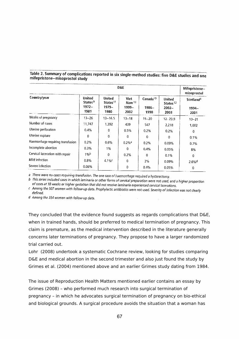
They concluded that the evidence found suggests as regards complications that D&E,
when in trained hands, should be preferred to medical termination of pregnancy. This
claim is premature, as the medical intervention described in the literature generally
concerns later terminations of pregnancy. They propose to have a larger randomized
trial carried out.
Lohr (2008) undertook a systematic Cochrane review, looking for studies comparing
D&E and medical abortion in the second trimester and also just found the study by
Grimes et al. (2004) mentioned above and an earlier Grimes study dating from 1984.
The issue of Reproduction Health Matters mentioned earlier contains an essay by
Grimes (2008) – who performed much research into surgical termination of
pregnancy – in which he advocates surgical termination of pregnancy on bio-ethical
and biological grounds. A surgical procedure avoids the situation that a woman has
67
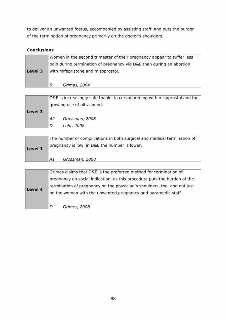
to deliver an unwanted foetus, accompanied by assisting staff, and puts the burden
of the termination of pregnancy primarily on the doctor’s shoulders.
Conclusions
Level 3
Women in the second trimester of their pregnancy appear to suffer less
pain during termination of pregnancy via D&E than during an abortion
with mifepristone and misoprostol.
B Grimes, 2004
Level 3
D&E is increasingly safe thanks to cervix priming with misoprostol and the
growing use of ultrasound.
A2 Grossman, 2008
D Lohr, 2008
Level 1
The number of complications in both surgical and medical termination of
pregnancy is low; in D&E the number is lower.
A1 Grossman, 2008
Level 4
Grimes claims that D&E is the preferred method for termination of
pregnancy on social indication, as this procedure puts the burden of the
termination of pregnancy on the physician’s shoulders, too, and not just
on the woman with the unwanted pregnancy and paramedic staff.
D Grimes, 2008
68
3.8 Discussion and evaluation
In Dutch abortion clinics second trimester termination of pregnancy is carried out by
D&E, involving cervix priming in a variable number of cases. In Dutch hospitals, in
the majority of cases, second trimester termination of pregnancy is carried out on
social indication in the event of foetal abnormalities. Medical termination is especially
applied in pregnancies beyond the 18th week. If the woman prefers termination via
D&E she is referred to one of the abortion clinics.
In The Netherlands, but also in the United States, Canada, England and Wales, the
majority of elective second trimester terminations of pregnancy is performed with
D&E. This is unlike the Northern European countries, where many medical
interventions take place.
In abortion clinics medical termination of pregnancy is not offered because all
interventions are polyclinic and must be carried out within 24 hours at the most.
On the basis of the literature found it cannot be concluded whether medical
termination or D&E is the best course of treatment in the second trimester.
However, the study group finds that D&E is safer, more efficient and effective,
providing the abortion specialist has ample skills.
Women usually prefer D&E, because it is commonly an outpatient intervention.
It is recommended that hospitals and abortion clinics in each region come to an
understanding about reciprocal referrals.
The safety of termination of pregnancy is partly determined by the method used, but
also, and especially so, by pregnancy length. The table shows that postponing
termination of pregnancy renders the intervention less safe (cf. table 2). In the case
of second trimester D&E it is therefore necessary that the intervention is carried out
by trained medical professionals in specialized second trimester clinics (Turok, 2008).
69
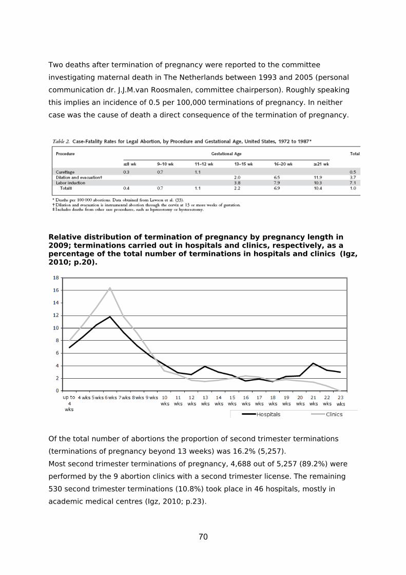
Two deaths after termination of pregnancy were reported to the committee
investigating maternal death in The Netherlands between 1993 and 2005 (personal
communication dr. J.J.M.van Roosmalen, committee chairperson). Roughly speaking
this implies an incidence of 0.5 per 100,000 terminations of pregnancy. In neither
case was the cause of death a direct consequence of the termination of pregnancy.
Relative distribution of termination of pregnancy by pregnancy length in 2009; terminations carried out in hospitals and clinics, respectively, as a percentage of the total number of terminations in hospitals and clinics (Igz, 2010; p.20).
Of the total number of abortions the proportion of second trimester terminations
(terminations of pregnancy beyond 13 weeks) was 16.2% (5,257).
Most second trimester terminations of pregnancy, 4,688 out of 5,257 (89.2%) were
performed by the 9 abortion clinics with a second trimester license. The remaining
530 second trimester terminations (10.8%) took place in 46 hospitals, mostly in
academic medical centres (Igz, 2010; p.23).
70

The share of hospitals in second trimester abortions has tripled since 2000, from
3.4% to 10.8% (Igz, 2010; p. 42). Especially in 2007-2008 there was a relative
increase in second trimester terminations in hospitals, particularly at a duration of 20
to 24 weeks. In 2006 7.4 percent of all abortions in hospitals were terminations
between 20 and 24 weeks (140); in 2007 this was 11.5% (227) and in 2008 13.4%
(276) (Igz, 2010; p. 37). In 2009 288 terminations between 20 and 24 weeks were
carried out in hospitals: a small increase compared to 2008 (4%). The proportion of
this group compared to the total of terminations in hospitals decreased slightly, to
13.2% (Igz, 2010; p. 23-24).
71
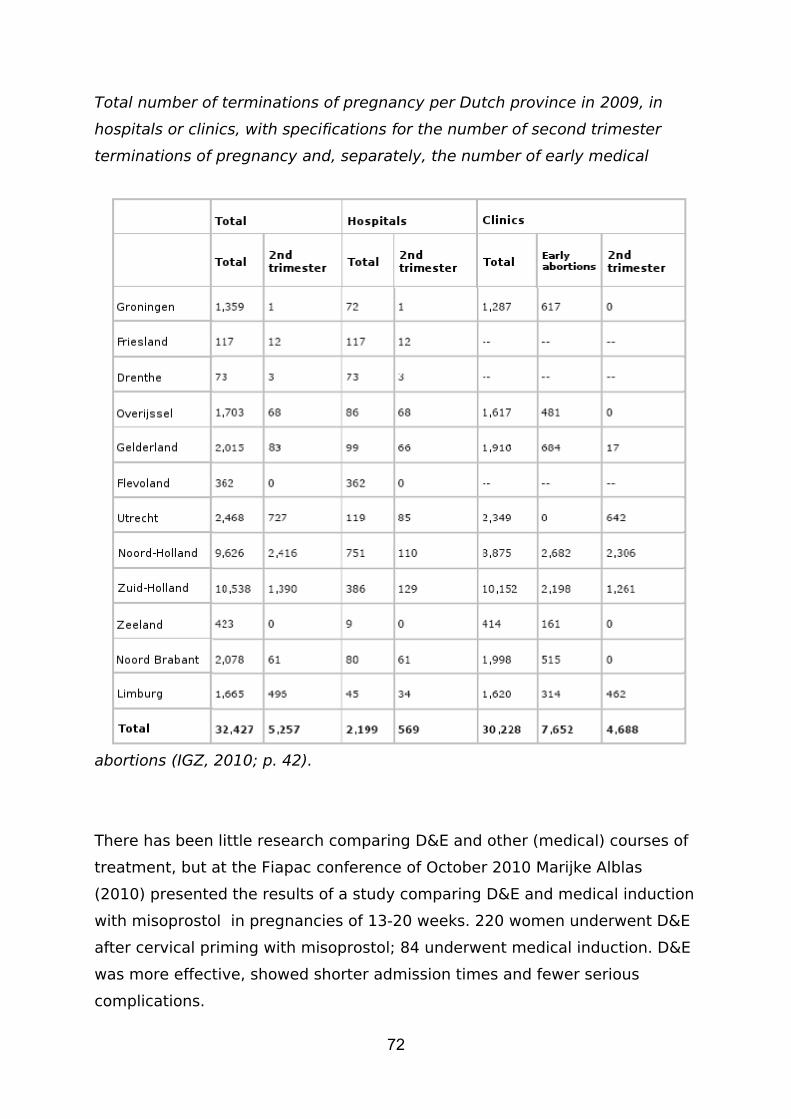
Total number of terminations of pregnancy per Dutch province in 2009, in
hospitals or clinics, with specifications for the number of second trimester
terminations of pregnancy and, separately, the number of early medical
abortions (IGZ, 2010; p. 42).
There has been little research comparing D&E and other (medical) courses of
treatment, but at the Fiapac conference of October 2010 Marijke Alblas
(2010) presented the results of a study comparing D&E and medical induction
with misoprostol in pregnancies of 13-20 weeks. 220 women underwent D&E
after cervical priming with misoprostol; 84 underwent medical induction. D&E
was more effective, showed shorter admission times and fewer serious
complications.
72

In The Netherlands, too, it should be possible to undertake such a study,
comparing hospitals and abortion clinics.
73

Recommendations
For women in their second trimester of pregnancy, D&E is recommended for
use in abortion clinics.
In hospitals, women should be offered a choice between medical termination
of pregnancy and D&E; for the latter option, the woman should be referred to
an abortion clinic.
The study group recommends the institution of a national working group to
develop a registration system for late complications of second trimester
termination of pregnancy.
The use of misoprostol as cervical priming in the second trimester is
recommended.
It is recommended to apply D&E, with misoprostol priming if necessary, for
second trimester termination of pregnancy in the case of women with one low
transversal C-section in their anamnesis.
74
3.9 References
Igz (2010). Jaarrapportage 2009 van de Wet afbreking zwangerschap Den Haag: Inspectie voor de Gezondheidszorg www.igz.nl.
Lohr PA, Surgical Abortion in the Second Trimester, Reproductive Health Matters, Volume 16, Number 31, supplement 2008,151-61
Grimes DA, Smith MS, Wtham AD. Mifepristone and Misoprostol versus dilatation and evacuation for midtrimester abortion: a pilot randomised controleed trial, BJOG, 2004; 111:148-53
Darney P, Sweet RL, Routine ultrasonogragraphy for second trimester abortion reduces incidence of uterine perforation, J Ultrasound Med 1989, 8:71-5
JacksonJE, Grobman WA, Haney E, Casele H. Mid-trimester dilation and evacuation with laminaria does not increase the risk for severe subsequent pregnancy complicationsd. International Journal of Gynaecology an Obstetrics, 2007, 96(1):12-5
Grimes DA, SchulzKF, Cates W, Jr., Tyler CW, Jr. Mid-trimester abortion by dilatation an evacuation: a safe and practical alternative. N.Engl.J.Med. 1977;296:1141-45
Cates W. Fr., Schulz KF, Grimes DA, Horowitz AJ, Lyon FA, Kravitz FH et al. Dilatation and evacuation procedures and second-trimester abortions. The role of physician skill and hospital setting. JAMA 1982;248:559-63
Turok DK, Gurtcheff SE, Esplin MS, Shah M, Simonsen SE, Trauscht-Van HJ, et al. Second trimester termination of pregnancy: a review by site and procedure type. Contraception 2008;77(3):155-61
Nucatola D, Roth N, Saulsberry V, Gatter M. Serious adverse events associated with the use of Misoprostol alone for cervical preparation prior to early second trimester surgical abortion (12-16 weeks). Contraception 2008;78(3):245-8
Winer N, Resche-Rigon M, Morin C, Ville Y, RozenbergP. Is induced abortion with misoprostol a risk factor for late abortion or preterm delivery in subsequent pregnancies? European Journal of Obstetrics, Gynecology & Reproductive Biology 2009;145(1):53-6
Todd CS, Soler M, Castleman L, Rogers MK, Blumenthal PD. Buccal Misoprostol as cervical preparation for second trimester pregnancy termination. Contraception. 2002;65:415-8
Goldberg AB, Drey EA, Withaker AK, Mi-Suk Kang, Meckstroth KR and Darney PD. Misoprostol compared with laminaria before early second-trimester surgical abortion: a randomized trial. Obestetrics and Gynecology, 2005;106(2):234-41
Carbonell JL, Garcia Gallego F, Perez Llorente M, Bermudez SB, Sanchez Sala, E, Varela Gonzale L, Sanches Texido C. Vaginal vs. sublingual Misoprostol with mifepristone for cervical primin in second-trimester abortion by dilation and evacuation: a randomized clinical trial. Contraception. 2007;75:230-7
Edelman AB, Buckmaster JG, Goetsch MF, Nichols MD, Jensen JT. Cervical preparation using laminaria with adjunctive buccal misoprostol before second trimester dilation and evacuation procedure: A randomized clinical trial. American Journal of Obstetrics and Gynecology. 2006;194:425-30
Patel A, Talmont E, Morfesis J, Pelta M, Gatter M, Momtaz MR, Piotrowski H, Cullins V, the Planned Parenthood Federation of America Buccal
75

Misoprostol Waiver Group. Adequacy and safety of buccal Misoprostol for vervical preparation prior to termination of second-trimest pregnancy. Contraception;2006:73:420-30
Goldberg AB, Greenberg MB, Darney PD. Misoprostol and Pregnancy. NEJM. 2001;344(1):38-47
Shannon CS, WinikoffB. Misoprotol an emerging technology for women’s health, reprot of a seminar, Population Caouncil 2001, updated 2004:1-66
Sivalal S, Sivasampu S. Health technology assessment report, Misoprostol in pregnanacy. Health technology assessment unit, medical development division, ministry of health Malaysia 2004: 4-29
Goldberg AB, Carusi DA, Meckstroth KR, Misporstol in pregnancy. Current Women’s Helath Reports 2003;3:475-83
Ngai SW, Tang OS, Ho PC, Prostaglandins for induction of second trimester termination and intrauterine death. Best Practice & Research Clinical Obstetrics an Gynaecology. 2003;17(5):765-75
Vimala N, Mittal S, Kumar, S. Sublingual Misoprostol before first trimester abortion: a comparative study using two dose regimens. Indian J Med Sci. 2004;58(2)54-61
Sharma S, Refaey H el, Stafford M, Purkayastha S, Parry M, Axby M. Oral versus vaginal misoprotol administration administered one hour before surgical termination of pregnancy: a randomised controleed trial. BJOG. 2005;112:456-60
Berghella V, Airoldi J, O’Neill AM, Einhorn K, Hoffman M. Misoprostol for second trimester pregnancy termination in women with prior ceasarean: a systematic review. BJOG.2009; online 1151-57
Goyal V. Uterine Rupture in Second-Trimester Misoprostol-Induced Abortion After Cesarean Delivery, a systematic review. Obstetrics and Gynecology. 2009;113(5)1117-23
Naguib AH, Morsi Hm, Borg TF, Fayed ST, Hemeda HM. Vaginal Misoprostol for second-trimester pregnancy termination after one previous caesarean delivery. International Journal of Gynaecology and Obstetrics. 2010;108(1)48-51
Ben-Ami I, Schneider D, Svirsky R, Smorgick N, Pansky M, Halperin R. Safety of late second-trimester pregnancy termination by laminaria dilatation and evacuation in patients with previous multiple caesarean sections. AJOG. 2009;201:154.e1-5
Elimia A, Verma U, Tejani N. Effect of causing fetal cardiac asystole on second-trimester abortion. Obstetrics and gynecology. 1999;94(1)139-41
Dickinson JE, Evans SF. The optimization of intravaginal Misoprostol dosing schedules in second-trimester pregnancy termination. American Journal of Obstetrics and Gynecology. 2002;186-470-4
Wong Ks, Ngai CSW,Yeo ELK, Tang LCH, Ho Pc. A comparison of two regimens of intravaginal Misoprostol for termination of second trimester pregnancy: A randomized comparative trial. Human Reproduction. 200;15:709-12
Ngai SW, Tang OS, Ho PC. Randomized comparison of vaginal (220 microgr every 3 h) and oral (400 microgr every 3h) Misoprostol when combined with mifepristone in termination of second trimester pregnanact.Human Reproduction. 2000;15:2205-8
76
Nor Azlin MI, Abdullah HSNA, Zainul Rashid MR, Jamil MA. Misoprostol (Alone) in second trimester terminations of pregnancy: As effective as Gemeprost? Journal of Obstetrics and Gynaecology. 2006;26(6)546-9
Bhattacharyya SK, Mukheri J, Kamilya GS, Ray S, Hazra A. Two regimens of vaginal misporstol in second trimester termination of pregnancy – a prospective randomised trial. Acta Obstetrica et Gyecologica. 2006;85:1458-62
Chai J, Tang OS, Hong QQ, Chen QF, Cheng LN, NG E, Ho PC. A randomized trial to compare dosing intervals of Misoprostol following mifepristone administration in second trimester medical abortion. Human Reproduction. 2009;24(2):320-4
Tang OS, Chan CCW, Kan ASY, Ho PC. A prospective randomized comparison of sublingual and oral Misoprostol when combined with mifepristone for medical abortion at 12-20 weeks gestation. Human Reproduction. 2005;20(11):3062-6
Yilmaz B, Kelekci S, Ertas IE, Kahyaoglu S, Ozel M, Sut N, Danisman N. Misoprostol moistened with acetic acid or saline for second trimester pregnancy termination: a randomized prospective double blind trial. Human Reproduction. 2005;20(11):3067-71
Yilmaz B, Kelekci S, Ertas IE, Ozel M, Sut N, Mollamahmutoglu L, Danisman N. Randomized comparison of second trimester pregnancy termination utilizing saline moistened or dry Misoprostol. Arch Gynecol Obestet. 2007;276:511-6
Bhattacharjee N, Sha SP, Ghoshroy SC, Bhowmik S, Barui G. A randomised comparative study on sublingual versus vaginal administration of Misoprostol fro termination of pregnancy between 13 to 20 weeks. Australian and New Zealand Journal of Obstetrics and Gynaecology. 2008;48:165-71
Karsidag AYK, Buyukbayrak EE, Kars B, Dansuk R. Vaginal versus sublingual Misoprostol for second-trimester pregnancy termination and effect on Doppler measurements. International Journal of Gynecology and Obstetrics. 2009;106:250-3
Jansen NE, Pasker-deJong P, Zondervan HA. Mifepristone and Misoprostol versus Dilapan and sulprostone for second trimester temination of pregnancy. The Journal of Maternal-Fetal end Neonatal Medicine. 2008;21(11)847-51
von Hertzen H, Piaggio G, Wojdyla D, Nguyen TMH, Marions L, Okoev G, Khomassuridze A, Kereszturi A, Mittal A, Nair R, Daver R, Pretnar-Darovec A, Dickson K, Nguyen DH, Nguyen HB, Hoang TDT, Pereogoudov A and for the WHO Research Group on Post-ovulatory Methods of Fertitlity regulation. Comparison of vaginal and sublingual Misoprostol for second trimester abortion: randomised controlled equivalence trial. Human Reproduction. 2009;24(1)106-12
Hamoda H, Ashok PW, Flett GMM, Templeton A. A randomized trial of mifepristone in combination with Misoprostol administered sublingually or vaginally for medical abortion at 13-20 weeks gestation. Human Reproduction. 2005;20(8)2348-54
Caliskan E, Doger, E, Cakiroglu Y, Corakei A, Yucesoy I. Sublingual Misoprostol 100 microgram versus 200 microgram for second trimester abortion a randomised trial. The European Journal of Contraception and Reproductive Health Care. 2009;14(1)55-60
77
Grossman D, Blanchard K, Blumenthal P. Complications after Second Trimester Surgical and Medical Abortion. Reproductive Health Matters. 2008; 16(31 supplement)173-82
Lohr PA Hayes JL, Gemzell-Danielsson K. Surgical versus medical methods for second trimester induced abortion. Cochrane Database of Systematic Reviews. 2008(1)
Grimes DA. The coice of second trimester abortion method: evolution, evidence and ethics. Reproductive Helath Matter. 2008;16(31 supplement)183-88
RCOG. The care of women requesting induced abortion (hoofdstuk 7) 2004SFP. Cervical preparation for second-trimester surgical abortion prior to 20
weeks of gestation. Contraception. 2007;76:486-495SFP. Cervical preparation for surgical abortion from 20-24 weeks gestation.
Contraception. 2008;77:308-14Alblas M, Blanchard K, Constant D, Grossman D, Harries JJ, Lince N,. The
efficacy, safety and acceptability of medical and surgical second trimester of pregnanacy in Cape Town, South Africa. Fiapac Congress 2010
78
Chapter 4 Antibiotic policy
Guiding question
What antibiotic policy should be preferred in surgical or medical termination
of pregnancy?
4.1 Introduction
Before there were specialized abortion clinics and well-trained abortion
specialists, infections after a termination of pregnancy occurred fairly
frequently. Possible consequences were infertility, chronic abdominal
complaints, extra-uterine gravidity, and neonatal problems. The following
guiding question will be answered: what antibiotic policy should be preferred
after surgical or medical termination of pregnancy?
Although no data at the national level about infections after a termination of
pregnancy are available for The Netherlands, the incidence of the number of
infections does not raise suspicions (Inspectorate’s Report, 2009). A possible
explanation could be that most clinics in The Netherlands prescribe
prophylactic antibiotics.
A literature search was undertaken in Medline (up to 2010), with primarily
studies from the US as a result. Unfortunately, little literature was found on
complications due to infections in The Netherlands. The guidelines of the
following authorities were consulted: The Dutch Society of Obstetrics and
Gynaecology (NVOG), the Dutch Society of Dermatology and Venereology
(NVDV), the National Society of Cardiology (Hart Stichting) with a view to
endocarditic prophylaxis, the standards of the Dutch Association of General
Practitioners (NHG), the Dutch Working Party on Antibiotic Policy (SWAB), and
Soaids Netherlands.
Antibiotic prophylaxis is defined as follows: the administration of antibiotics
for a short period of time in the context of surgical intervention, for the
prevention of post-operative infections in the area of invasion (Van Kasteren,
2000).
79

Prophylaxis policy after termination of pregnancy can be based on Mayhall’s
surgical wound classification. Termination of pregnancy in the second or first
trimester after pelvic inflammatory disease (= PID) is classified as a ‘clean-
contaminated’ wound. Consequently, the advantages of prophylaxis outweigh
the possible disadvantages (Mayhall, 1993). Van Kasteren (2000) defines the
following criteria for the prescription of antibiotics: they must be directed at
the expected causative microorganisms; they must be administered at the
correct time and administration should not last longer than necessary; the
spectrum must be as narrow as possible; they must be as safe and
inexpensive as possible; it must be possible to administer them via the
desired route.
80
4.2 Prevalence of genital bacterial infections
Guiding question
Which genital bacterial infections are common in women undergoing termination of
pregnancy and how should they be treated?
Summary of the literature
Recently a screening for genital bacterial infections was carried out among
youngsters between 16 and 29 years of age, in Amsterdam, Rotterdam and Southern
Limburg. Over 630,000 invitations were sent out in the course of 15 months. In three
rounds this target group was tested; 3,735 infections were traced. In the first round,
4.2% of the participants tested positive for Chlamydia trachomatis (CT); in the
second round this figure was 4.0%, in the third round 3.5%. According to regions the
figures are: Rotterdam 5,1%-4.9%-4.5% (round 1, 2, 3), Amsterdam: 3.6%-3.4%-2.9%
and Limburg: 5.1%-4.7%-3.8% (van Bergen et al., 2010).
The prevalence in abortion patients of Chlamydia trachomatis, serotypes D through
K, rates probably around 10% (data from abortion clinics in The Hague, Arnhem and
Amsterdam). In The Hague, in 1998 and 1999, 1,009 and 972 women were screened,
respectively; in Amsterdam there was a screening in 1992, in Arnhem probably
before 1995.
In selective screening in Arnhem GP practices a prevalence for CT of 10% was been
found for many years, but the total number of applications has been rising
(unpublished data). The prevalence for CT among the Dutch population as a whole
appears to be following a continuously ascending line, partly to be explained by
increased willingness to test for sexually transmitted infections (STI)3 (Mayhall, 1993;
Epidemiologisch Bulletin, 2003; RIVM, 2009)4.
3 In Dutch: SOA
4 Epidemiological Bulletin; for RIVM, cf. next footnote
815
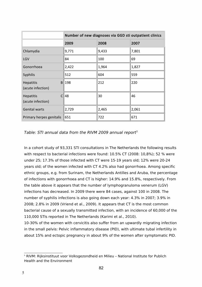
Number of new diagnoses via GGD sti outpatient clinics
2009 2008 2007
Chlamydia 9,771 9,433 7,801
LGV 84 100 69
Gonorrhoea 2,422 1,964 1,827
Syphilis 512 604 559
Hepatitis B(acute infection)
198 212 220
Hepatitis C(acute infection)
48 30 46
Genital warts 2,729 2,465 2,061
Primary herpes genitalis 651 722 671
Table: STI annual data from the RIVM 2009 annual report5
In a cohort study of 93,331 STI consultations in The Netherlands the following results
with respect to bacterial infections were found: 10.5% CT (2008: 10,8%); 52 % were
under 25; 17.3% of those infected with CT were 15-19 years old; 12% were 20-24
years old; of the women infected with CT 4.2% also had gonorrhoea. Among specific
ethnic groups, e.g. from Surinam, the Netherlands Antilles and Aruba, the percentage
of infections with gonorrhoea and CT is higher: 14.9% and 15.8%, respectively. From
the table above it appears that the number of lymphogranuloma venerum (LGV)
infections has decreased. In 2009 there were 84 cases, against 100 in 2008. The
number of syphilis infections is also going down each year: 4.3% in 2007; 3.9% in
2008; 2.8% in 2009 (Vriend et al., 2009). It appears that CT is the most common
bacterial cause of a sexually transmitted infection, with an incidence of 60,000 of the
110,000 STIs reported in The Netherlands (Karimi et al., 2010).
10-30% of the women with cervicitis also suffer from an upwardly migrating infection
in the small pelvis: Pelvic inflammatory disease (PID), with ultimate tubal infertility in
about 15% and ectopic pregnancy in about 9% of the women after symptomatic PID.
5 RIVM: Rijksinstituut voor Volksgezondheid en Milieu – National Institute for Publich Health and the Environment
825
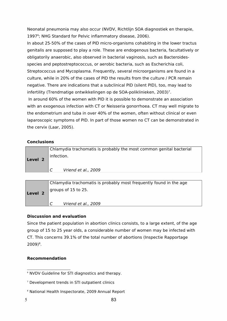
Neonatal pneumonia may also occur (NVDV, Richtlijn SOA diagnostiek en therapie,
19976; NHG Standard for Pelvic inflammatory disease, 2006).
In about 25-50% of the cases of PID micro-organisms cohabiting in the lower tractus
genitalis are supposed to play a role. These are endogenous bacteria, facultatively or
obligatorily anaerobic, also observed in bacterial vaginosis, such as Bacteroides-
species and peptostreptococcus, or aerobic bacteria, such as Escherichia coli,
Streptococcus and Mycoplasma. Frequently, several microorganisms are found in a
culture, while in 20% of the cases of PID the results from the culture / PCR remain
negative. There are indications that a subclinical PID (silent PID), too, may lead to
infertility (Trendmatige ontwikkelingen op de SOA-poliklinieken, 2003)7.
In around 60% of the women with PID it is possible to demonstrate an association
with an exogenous infection with CT or Neisseria gonorrhoea. CT may well migrate to
the endometrium and tuba in over 40% of the women, often without clinical or even
laparoscopic symptoms of PID. In part of those women no CT can be demonstrated in
the cervix (Laar, 2005).
Conclusions
Level 2
Chlamydia trachomatis is probably the most common genital bacterial
infection.
C Vriend et al., 2009
Level 2
Chlamydia trachomatis is probably most frequently found in the age
groups of 15 to 25.
C Vriend et al., 2009
Discussion and evaluation
Since the patient population in abortion clinics consists, to a large extent, of the age
group of 15 to 25 year olds, a considerable number of women may be infected with
CT. This concerns 39.1% of the total number of abortions (Inspectie Rapportage
2009)8.
Recommendation
6 NVDV Guideline for STI diagnostics and therapy.
7 Development trends in STI outpatient clinics
8 National Health Inspectorate, 2009 Annual Report
835

As Chlamydia trachomatis is most common among women between 15 and 25, it is
recommended to treat them prophylactically for this condition after a surgical
termination of pregnancy.
84

4.3 Antibiotic policy in the case of surgical termination of pregnancy
Guiding question
What antibiotic policy should be preferred in the case of surgical termination of
pregnancy?
Summary of the literature
Sawaya (1996) writes that, in American retrospective studies, the risk of
infection is estimated at 0.5%. From prospective trials an incidence of
infection was shown of between 5 and 20%. To prevent infection many care
professionals routinely administer an antibiotic. Others only do so in the case
of women with a raised risk profile (earlier STI/PID, multiple partner contacts).
Sawaya reviews twelve randomized trials. The risk of infection in the group of
women with antibiotic prophylaxis was 1- 11%. In the placebo group the risk
was 5-23%. The NVOG9 guideline on surgical termination of pregnancy (2005)
underlines the necessity of antibiotic prophylaxis in every surgical termination
of pregnancy. This would diminish the risk of postoperative infection by about
50%. Sawaya’s meta-analysis demonstrates a 50% reduction of the chances
of postoperative infection when prophylactic antibiotics are used in surgical
termination of pregnancy.
Stray-Pedersen (1991) tested 1,193 women <35 years (53%< 25 years) for
CT and other infections, before first trimester termination of pregnancy. 139
women (11.7%) proved to be CT infected and 0.8% suffered from Neisseria
gonorrhoea; a group B streptococcal infection was found in 2.9% of these
women. CT and gonorrhoea were mostly found in teenagers. 26 women
(2.2%) developed PID; 13 women (0.9%) developed endometritis. Of the CT-
positive women 95 had received pre-operative or peri-operative treatment;
two of them developed PID (2.1%). Of the 44 women who had not been
treated ten ended up with PID (22.7%). Of the group of 108 women who
tested positive on Mycoplasma hominis and were not treated, (22.1%) eight
(8.1%) developed PID. Of the group of ten women who had received
treatment, not one developed PID.
9 NVOG: Royal Society of Obstetricians and Gynaecologists of The Netherlands
85
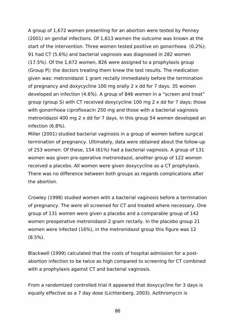
A group of 1,672 women presenting for an abortion were tested by Penney
(2001) on genital infections. Of 1,613 women the outcome was known at the
start of the intervention. Three women tested positive on gonorrhoea (0.2%);
91 had CT (5.6%) and bacterial vaginosis was diagnosed in 282 women
(17.5%). Of the 1,672 women, 826 were assigned to a prophylaxis group
(Group P); the doctors treating them knew the test results. The medication
given was: metronidazol 1 gram rectally immediately before the termination
of pregnancy and doxycycline 100 mg orally 2 x dd for 7 days. 35 women
developed an infection (4.6%). A group of 846 women in a “screen and treat”
group (group S) with CT received doxycycline 100 mg 2 x dd for 7 days; those
with gonorrhoea ciprofloxacin 250 mg and those with a bacterial vaginosis
metronidazol 400 mg 2 x dd for 7 days. In this group 54 women developed an
infection (6.8%).
Miller (2001) studied bacterial vaginosis in a group of women before surgical
termination of pregnancy. Ultimately, data were obtained about the follow-up
of 253 women. Of these, 154 (61%) had a bacterial vaginosis. A group of 131
women was given pre-operative metronidazol, another group of 122 women
received a placebo. All women were given doxycycline as a CT prophylaxis.
There was no difference between both groups as regards complications after
the abortion.
Crowley (1998) studied women with a bacterial vaginosis before a termination
of pregnancy. The were all screened for CT and treated where necessary. One
group of 131 women were given a placebo and a comparable group of 142
women preoperative metronidazol 2 gram rectally. In the placebo group 21
women were infected (16%), in the metronidazol group this figure was 12
(8.5%).
Blackwell (1999) calculated that the costs of hospital admission for a post-
abortion infection to be twice as high compared to screening for CT combined
with a prophylaxis against CT and bacterial vaginosis.
From a randomized controlled trial it appeared that doxycycline for 3 days is
equally effective as a 7 day dose (Lichtenberg, 2003). Azithromycin is
86
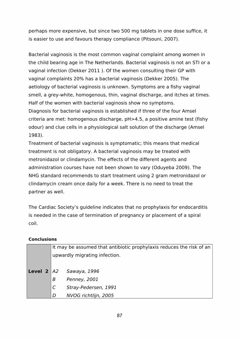
perhaps more expensive, but since two 500 mg tablets in one dose suffice, it
is easier to use and favours therapy compliance (Pitsouni, 2007).
Bacterial vaginosis is the most common vaginal complaint among women in
the child bearing age in The Netherlands. Bacterial vaginosis is not an STI or a
vaginal infection (Dekker 2011 ). Of the women consulting their GP with
vaginal complaints 20% has a bacterial vaginosis (Dekker 2005). The
aetiology of bacterial vaginosis is unknown. Symptoms are a fishy vaginal
smell, a grey-white, homogenous, thin, vaginal discharge, and itches at times.
Half of the women with bacterial vaginosis show no symptoms.
Diagnosis for bacterial vaginosis is established if three of the four Amsel
criteria are met: homogenous discharge, pH>4.5, a positive amine test (fishy
odour) and clue cells in a physiological salt solution of the discharge (Amsel
1983).
Treatment of bacterial vaginosis is symptomatic; this means that medical
treatment is not obligatory. A bacterial vaginosis may be treated with
metronidazol or clindamycin. The effects of the different agents and
administration courses have not been shown to vary (Oduyeba 2009). The
NHG standard recommends to start treatment using 2 gram metronidazol or
clindamycin cream once daily for a week. There is no need to treat the
partner as well.
The Cardiac Society’s guideline indicates that no prophylaxis for endocarditis
is needed in the case of termination of pregnancy or placement of a spiral
coil.
Conclusions
Level 2
It may be assumed that antibiotic prophylaxis reduces the risk of an
upwardly migrating infection.
A2 Sawaya, 1996
B Penney, 2001
C Stray-Pedersen, 1991
D NVOG richtlijn, 2005
87
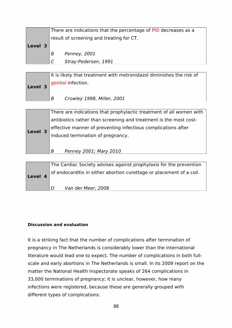
Level 3
There are indications that the percentage of PID decreases as a
result of screening and treating for CT.
B Penney, 2001
C Stray-Pedersen, 1991
Level 3
It is likely that treatment with metronidazol diminishes the risk of
genital infection.
B Crowley 1998, Miller, 2001
Level 3
There are indications that prophylactic treatment of all women with
antibiotics rather than screening and treatment is the most cost-
effective manner of preventing infectious complications after
induced termination of pregnancy.
B Penney 2001; Mary 2010
Level 4
The Cardiac Society advises against prophylaxis for the prevention
of endocarditis in either abortion curettage or placement of a coil.
D Van der Meer, 2008
Discussion and evaluation
It is a striking fact that the number of complications after termination of
pregnancy in The Netherlands is considerably lower than the international
literature would lead one to expect. The number of complications in both full-
scale and early abortions in The Netherlands is small. In its 2009 report on the
matter the National Health Inspectorate speaks of 264 complications in
33,000 terminations of pregnancy; it is unclear, however, how many
infections were registered, because these are generally grouped with
different types of complications.
88
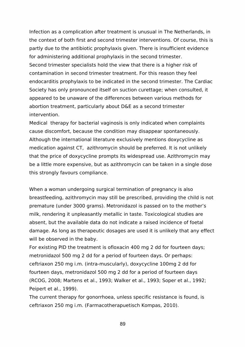
Infection as a complication after treatment is unusual in The Netherlands, in
the context of both first and second trimester interventions. Of course, this is
partly due to the antibiotic prophylaxis given. There is insufficient evidence
for administering additional prophylaxis in the second trimester.
Second trimester specialists hold the view that there is a higher risk of
contamination in second trimester treatment. For this reason they feel
endocarditis prophylaxis to be indicated in the second trimester. The Cardiac
Society has only pronounced itself on suction curettage; when consulted, it
appeared to be unaware of the differences between various methods for
abortion treatment, particularly about D&E as a second trimester
intervention.
Medical therapy for bacterial vaginosis is only indicated when complaints
cause discomfort, because the condition may disappear spontaneously.
Although the international literature exclusively mentions doxycycline as
medication against CT, azithromycin should be preferred. It is not unlikely
that the price of doxycycline prompts its widespread use. Azithromycin may
be a little more expensive, but as azithromycin can be taken in a single dose
this strongly favours compliance.
When a woman undergoing surgical termination of pregnancy is also
breastfeeding, azithromycin may still be prescribed, providing the child is not
premature (under 3000 grams). Metronidazol is passed on to the mother’s
milk, rendering it unpleasantly metallic in taste. Toxicological studies are
absent, but the available data do not indicate a raised incidence of foetal
damage. As long as therapeutic dosages are used it is unlikely that any effect
will be observed in the baby.
For existing PID the treatment is ofloxacin 400 mg 2 dd for fourteen days;
metronidazol 500 mg 2 dd for a period of fourteen days. Or perhaps:
ceftriaxon 250 mg i.m. (intra-muscularly), doxycycline 100mg 2 dd for
fourteen days, metronidazol 500 mg 2 dd for a period of fourteen days
(RCOG, 2008; Martens et al., 1993; Walker et al., 1993; Soper et al., 1992;
Peipert et al., 1999).
The current therapy for gonorrhoea, unless specific resistance is found, is
ceftriaxon 250 mg i.m. (Farmacotherapuetisch Kompas, 2010).
89

In cases of allergy to azithromycin, doxycycline can be an alternative: 100
mg 2 dd for a period of seven days. In the case of allergy to both azithromycin
and doxycycline, amoxicillin 500 mg three times daily for seven days is
prescribed. All cures must be fully complied with without interruption
(Farmacotherapuetisch Kompas, 2010). The use of Azithromycin, in
combination with vaso-constrictive agents such as methergine and
syntocinon, may lead to arterial spasms and ischemic reactions.
Recommendations
When there is a suspicion of bacterial vaginosis four times 500 mg metronidazol is
prescribed.
90
After surgical termination of pregnancy, it is recommended to administer 1.000 mg
azithromycin as a prophylaxis to the patient on the same day.

4.4 Antibiotic policy in medical termination of pregnancy
Guiding question
What antibiotic policy should be preferred in medical termination of pregnancy?
Summary of the literature
In 2005, four US women died, and one Canadian woman in 2001, following a medical
termination of pregnancy with mifepristone and misoprostol vaginally due to a septic
shock caused by Clostridium sordellii. A similar case occurred in Portugal in 2009
(21ste European Congress of Clinical Microbiology and Infectious Diseases, 2011). The
infection was accompanied by leucocytosis, no fever and low blood pressure. Another
woman died of a ruptured ectopic pregnancy. Mortality figures after abortion range
from 0.1 per 100,000 up to 8 week of pregnancy, tot 8.9 per 100,000 at 21 weeks of
gestation or more (Greene 2005).
Planned Parenthood analysed 72,195 women who had undergone medical
termination of pregnancy with mifepristone and misoprostol vaginally. 67 serious
infections were reported (0.09%). There was one case of death due to Clostridium
perfringens. 78,794 women took misoprostol buccally; 20 women developed an
infection (0.025%). In 35,837 women who took misoprostol buccally and received
prophylactic doxycycline two infections occurred (0.005%). The pregnancy length in
this group was 56 days. In a group under the same regime and a pregnancy length of
63 days the percentage of infections was also 0.005% (Planned Parenthood, 2005).
A retrospective study showed that the number of serious infections after medical
termination of pregnancy decreased by 93% after vaginal administration of
misoprostol was replaced by buccal administration, in combination with routine use
of antibiotics. In view of the small number of complications, vaginal administration of
misoprostol is also admissible (Fjerstad, 2009). In The Netherlands antibiotic
prophylaxis after medical termination of pregnancy is not common.
Conclusions
Level 3
In the USA, in pregnancies under 8 weeks, the mortality rate after
medical termination of pregnancy equals that of surgical
termination of pregnancy.
C Greene, 2005
Level 3It may be assumed that antibiotic prophylaxis is not necessary in
medical termination of pregnancy.
91

C Planned Parenthood 2005
Discussion and evaluation
Some critical remarks are in order with respect to the Planned Parenthood
study. A randomized comparative trial between vaginal and buccal
misoprostol would have been better. The number of infections is extremely
small. To prevent an infection developing 1,000 women are unnecessarily
given an antibiotic. Screening and treatment of CT in combination with
medical termination of pregnancy may lead to a reduction of vaginal infection
in young women. CT screening is usually impossible because of the time
investment needed, the test is 99.5% certain. In the target group undergoing
termination of pregnancy a higher percentage of CT infections may be
expected.
Recommendation
The study group takes the position that antibiotic prophylaxis in medical
termination of pregnancy is not necessary.
92

4.5 References
Van Kasteren, et al. Optimaliseren van het antibiotica beleid in Nederlad,V. SWAB-richtlijnen voor perioperatieve antibiotische prophylaxis; Ned. Tijdschrift voor Geneeskunde Oktober 2000; 144(43)
Mayhall C.G. Surgical infections including burns. Wenzel RP, editor, prevention and control of nosocomial infections. Baltimore 1993
van Bergen et al. Chlamydia Screening 2008-2010: resultaten, conclusies en Recommendations. JEAM December 2010
Trendmatige ontwikkelingen op de SOA-poliklinieken. Epidemiologisch Bulletin, Tijdschrift voor Volksgezondheid en Onderzoek in Den Haag 2003; 38 (1): 2-7.
H.J. Vriend F.D.H. Koedijk, I.V.F van den Broek,M.G.van Veen, E.L.M.op de Coul, A.I. van Sighem, R.A. Verheij, M.A.B.van der Sande RIVM (Rijksinstituut voor Volksgezondheid en milieu) Jaarrapportage 2009.
Soa jaarcijfers GGD-soa poliklinieken 2008 Richtlijn SOA Diagnostiek en therapie. Nederlandse Vereniging voor Dermatologie en
Venereologie 1997 van de.Uitgegeven mede onder auspiciën van de Nederlandse Vereniging voor Obstetrie en Gynaecologie en Nederlandse Vereniging voor Medische Microbiologen samenwerking met de Stichting SOA-bestrijding Tweede editie
Karimi, A. et al. Chlamydia trachomatis-infecties,Epidemiology, diagnostics and treatment of urogenital Chlamydia trachomatis infections. Tijdschrift voor Infectieziekten; v o l . 5 n r . 6 – 2010
NHG-Standaard 'Pelvic inflammatory disease' (Gebu 2006 (mrt); 40: 38-39.Laar M. Soa nemen opnieuw toe, voorlopige cijfers 2004. Soa Aids Magazine (Online)
2005, 2 blz. 28Jaarrapportage 2009 wet afbreking zwangerschap; inspectie gezondheidszorg; 11-02-
2010; p21, 48Sawaya. USA Sawaya obstet Gynaecol 1996 87.884-90Stray-Pedersen Larsson PG, Platz-Christensen JJ, Dalaker K, Eriksson K, Fåhraeus L,
Irminger K, Jerve F, Stray-Pedersen B, Wölner-Hanssen P. 1991Penney GC, Grimshaw JM, Ramsay CR, Walker AE, MacLennan G, Stearns SC,
McKenzie L, Glasier A.BJOG. 2004 Jul;111(7):726-33.PMID: 15198764Miller BJOG 2004.111.982-8Crowley oktober 1996- december 1998 Crowley T, Low N, Turner A, Harvey I,
Bidgood K, Horner P.Blackwell AL, Emery SJ, Thomas PD, Wareham K.Int J STD AIDS. 1999 Aug;10(8):508-
13.PMID: 10471099Miller L, Thomas K, Hughes PH, et al. Randomised treatment trial of bacterial
vaginosis to prevent post-abortion complication. BJOG 2004; 111: 982-8. 37) Mary N, Mahmood TA. Best Pract Res Clin Obstet Gynaecol. 2010 Aug;24(4):539-49.
Epub 2010 Jun 12. Review.van Bergen et al,Chlamydia Screening 2008-2010: resultaten, conclusies en
Recommendations ; JEAM. December 2010Lichtenberg ES, Shott S. A randomized clinical trial of prophylaxis for vacuum
abortion: 3 versus 7 days of doxycycline. Ostet Gynecol 2003; 101: 726-31. 36Pitsouni E, Iavazzo C, Athanasiou S, Falagas ME.Single-dose azithromycin versus
erythromycin or amoxicillinfor Chlamydia trachomatis infection during pregnancy: a meta-analysis of randomised controlled trials.Int J Antimicrob Agents. 2007;30:213-21)
RCOG richtlijnen Management of acute pelvic inflammatory disease. Green-top Guideline No. 32; November 2008
Martens MG, Gordon S, Yarborough DR, Faro S, Binder D, Berkeley A. Multicenter randomized trial of ofloxacin versus cefoxitin and doxycycline in outpatient treatment of pelvic inflammatory disease. Ambulatory PID Research Group. Southern Med J 1993;86:604–10.
93

Walker CK, Kahn JG, Washington AE, Peterson HB, Sweet RL. Pelvic inflammatory disease: metaanalysis of antimicrobial regimen efficacy. J Infect Dis 1993;168:969–78.
Soper DE, Brockwell NJ, Dalton HP. Microbial etiology of urban emergency department acute salpingitis: treatment with ofloxacin. Am J Obstet Gynecol 1992;167:653–60.
Peipert JF, Sweet RL, Walker CK, Kahn J, Rielly-Gauvin K. Evaluation of ofloxacin in the treatment of laparoscopically documented acute pelvic inflammatory disease (salpingitis). Infect Dis Obstet Gynecol 1999;7:138–44.
Richtlijnen farmcotheapeutisch kompas; Tractus genitalis femininus / Middelen bij vaginale aandoeningen ) jaartal 2010; 6; 767.
Grimes DA, Schulz KF. Prophylactic antibiotics for intrauterine device insertion: a metaanalysis of the randomized controlled trials. Contraception 1999;60:57-63.
prophylactic antibiotics for intrauterine device insertion (Review) 15 Copyright © 2010 De Cochrane Collaboration. Geplaatst door John Wiley & Sons, Ltd ;BIJDRAGEN VAN AUTEURS Grimes D en K Schulz ;L Lopez herzien van de zoekresultaten in 2007 en 2009
Richtlijnen endocarditis prophylaxis; van der Meer Hartstichting Michael F. Greene, M.D. 12-2005 new england journal of medicine 353;22hristin-
Maitre S, Bouchard P, Spitz IM.Medical termination of pregnancy. N Engl;J Med 2000;342:946-56.
Clostridium sordellii toxic shock syndromeafter medical abortion with mifepristone and intravaginal misoprostol — United States and Canada, 2001–2005. MMWR Morb Mortal;Wkly Rep 2005;54:724.
Mary Fjerstad;2009/Fjerstad N ebg J Med;2009 361(2).145-51Planned Parenthood 2005. http://www.lifeissues.org/pp/report_05-06.pdf.Dekker JH.Afdeling huisartsgeneeskunde UMCG.Vaginale
aandoeningen:candidiasis,bacteriele vaginose en trichomoniasis;juli 2011 Seksoa.
Dekker JH,Boeke AJP,Gercama AJ,Kardolus GJ,Boukes FS.NHG-standaard Fluor vaginalis.Huiarts en Wetenschap. 2005;48:459-466
Amsel R,Totten PA,Spiegel CA,Chen KC.Eschenbach D,Holmes KK. Nonspecific vaginitis.Diagnostic criteria and microbiological and epidemiologic associations.Am J Med.1983; 74:14-22
Oduyebo OO,Anorlu RI,Ogunsola FT.The effects of antimicrobial therapy on bacreriel vaginosis in non-pregnant women. Cochrane Database Syst. Rev. 2009 ;3 : CD006055
94

Chapter 5 Use of ultrasound
Guiding questionWhen should ultrasound scanning be applied in termination of pregnancy?
5.1 IntroductionUntil the introduction of ultrasonographic techniques a diagnosis of pregnancy and
the establishment of its duration were simply based on anamnesis and a bimanual
internal examination, with the possible addition of a pregnancy test.
In the eighties, ultrasound equipment was primarily used to establish pregnancy
length. Thanks to the strongly improved technology of the equipment, it is now
possible to diagnose abnormalities like myoma, missed abortion, molar pregnancy
and ectopic pregnancy before treatment. Because of the application of sonography
during the intervention, the instruments used can often be clearly visualized, possibly
reducing the risk of perforations in particular, false routes and incomplete abortions.
Ultrasound scanning is also used sometimes at follow-up.
The Royal College of Obstetricians and Gynaecologists (RCOG) holds the view that
ultrasonography is not an essential requirement in termination of pregnancy, but that
the option must be available to make ultrasound scans in certain circumstances
(McGalliard en Gaudoin, 2004). Here we will try to make evidence-based
pronouncements on the basis of existing literature about the role of ultrasound
scanning in induced abortions.
In our search in Pubmed, three systematic reviews were found, none of which were
suitable because they did not deal with termination of pregnancy. Of the 124 other
Pubmed articles, the majority was not appropriate for our query. In the end our
search resulted in one RCT and 25 observational and comparative studies about the
application of ultrasonography in relation to abortion. In February 2010 two more
articles followed.
To be able to answer the question a number of sub-questions was defined. These will
be answered below.
95

5.2 Establishing a diagnosis
Guiding questionWhat is the function of ultrasound scanning in establishing the diagnosis of pregnancy?
Summary of the literature Using pre-operative abdominal ultrasound in 120 women, Fakih et al. (1986) found
five instances of missed abortion, one multiple pregnancy, and one ectopic
pregnancy. According to the authors, ultrasound scanning is essential to establishing
the diagnosis of abnormal pregnancy (missed abortion, twins, ectopic pregnancy,
extra-uterine gravidity).
In a prospective trial involving 217 women, Fiala et al. (2003) compared the value of
ultrasonography with an hCG indication before and after medical termination of
pregnancy. Using ultrasound the authors found no gestational sac in 6 women
(2.7%), a gestational sac but no embryonic sac in 44 women (20.3%), both a
gestational and an embryonic sac, but no foetus or a CRL<2 mm in 105 women
(48.4%) and a CRL>2 mm in 62 women (28.6%). The authors claim that ultrasound is
not a reliable procedure for establishing pregnancy if no embryonic sac is visible yet,
which was true for 50 of the cases in their population(23%). On the basis of a serum
hCG test it was always possible to demonstrate pregnancy but impossible to
determine its length.
From the retrospective study by Jarallah and Geisson (1985), in which abdominal
ultrasound was applied in 753 women, it appeared that 19 (2.5%) turned out not to
be pregnant; 52 women (6.9%) had a non-vital pregnancy and 11 women had a
multifoetal pregnancy. Incidental findings, like ovary cysts, myoma, IUDs etc., were
reported for 27 (3.6%) women.
In a prospective study involving 283 women, McGalliard and Gaudoin (2003) found
that, after ultrasound scanning and a pregnancy test, four women (1%) appeared
non-pregnant. There were 259 continuing single pregnancies (92%), 3 continuing
multiple pregnancies (1%) and 17 missed abortions (6%).
In their retrospective study involving vaginal ultrasound scanning in 140 women,
Sinha et al. (2004) found that 15 women showed a missed abortion. Apart from
these, two multi-foetal pregnancies, one ectopic pregnancy, one mola pregnancy and
one case of uterus bicornis were found.
96
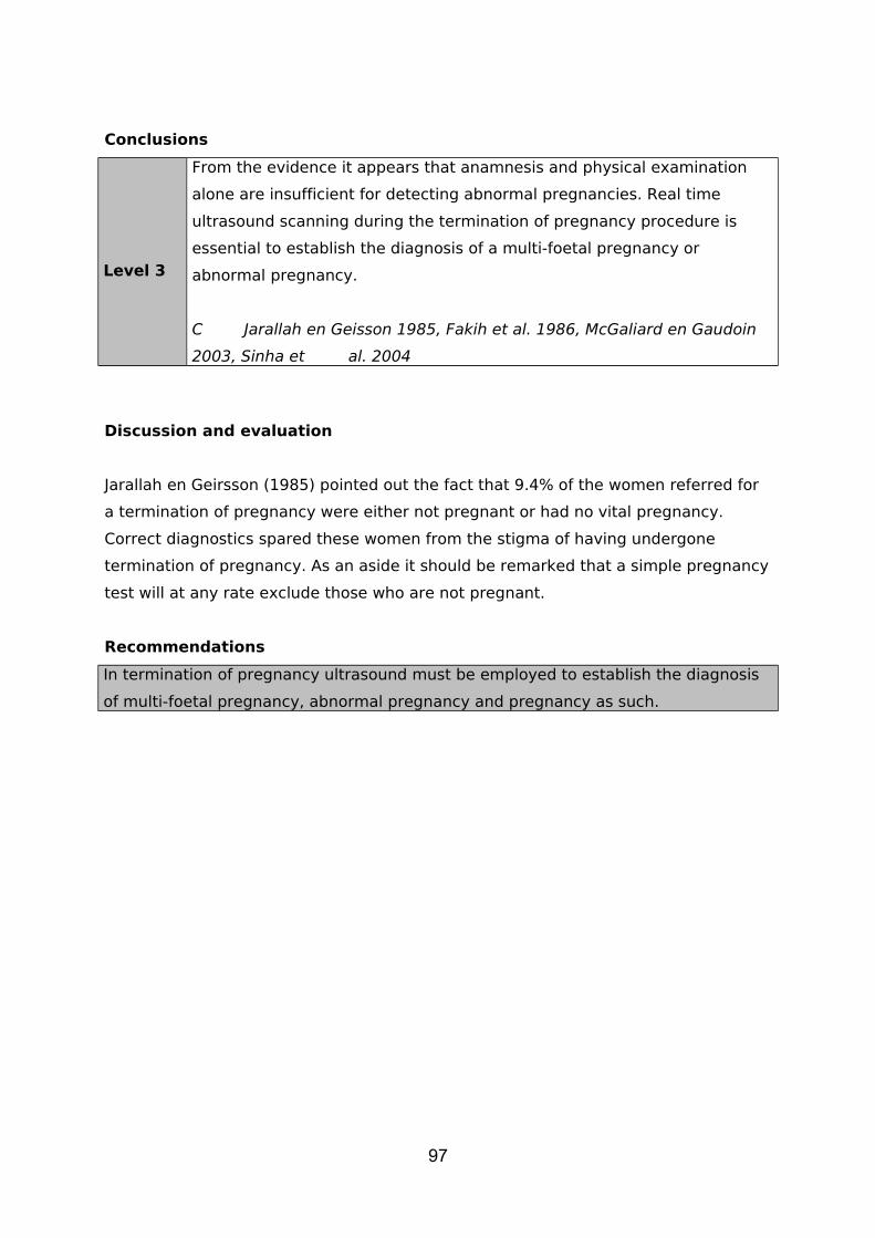
Conclusions
Level 3
From the evidence it appears that anamnesis and physical examination
alone are insufficient for detecting abnormal pregnancies. Real time
ultrasound scanning during the termination of pregnancy procedure is
essential to establish the diagnosis of a multi-foetal pregnancy or
abnormal pregnancy.
C Jarallah en Geisson 1985, Fakih et al. 1986, McGaliard en Gaudoin
2003, Sinha et al. 2004
Discussion and evaluation
Jarallah en Geirsson (1985) pointed out the fact that 9.4% of the women referred for
a termination of pregnancy were either not pregnant or had no vital pregnancy.
Correct diagnostics spared these women from the stigma of having undergone
termination of pregnancy. As an aside it should be remarked that a simple pregnancy
test will at any rate exclude those who are not pregnant.
Recommendations
In termination of pregnancy ultrasound must be employed to establish the diagnosis
of multi-foetal pregnancy, abnormal pregnancy and pregnancy as such.
97
5.3 Determining pregnancy length
Guiding questionWhat is the role of ultrasound in determining pregnancy length?
Summary of the literatureBlanchard et al. (2007) conducted a prospective comparative study into the value of
self-estimated LMP (time elapsed since the last menstrual period), clinical findings
(over or under 8 weeks since LMP) and ultrasound results in 673 women. The
women’s estimate (LMP) appeared on average to be 19 days below the outcome of
the ultrasound, while the findings of doctors (LMP and internal examination) were still
one to two days below the ultrasound outcome. In a prospective study of 120 women
considering a first trimester termination of pregnancy Fakih et al. (1986) compared
pregnancy length as determined by pre-operative ultrasound with the stated length
of amenorrhoea and the findings of an internal examination. When the findings of the
abdominal ultrasound were matched with those of the internal examination there
was a difference of less than one week in 81% of the cases; in 13% the difference
was one to two weeks; in 6% there was a difference of more than two weeks. Of the
women with a difference of more than 2 weeks between ultrasound and internal
exam two were very young primigravidae, both hard to examine, two missed
abortions, one grand multipara, one uterus myomatosus and one case of obesity. The
authors conclude that there is a good overall correlation between both methods. The
correlation between ultrasound scans and stated length of amenorrhoea was not so
good: in 15% a discrepancy of more than two weeks was found.
In the US the extra cost involved in having an ultrasound made before and after
medical termination of pregnancy puts up a barrier to the further acceptance of this
method. In a prospective multicenter trial, involving 15 centres and including 1,016
women who underwent medical termination of pregnancy at a pregnancy length of
63 days of amenorrhoea, Fielding et al. (2002) therefore compared the outcomes of
the anamnesis and internal exam with ultrasound findings. On the basis of an internal
examination, experienced clinicians dated the length of pregnancy correctly in 87%
of the cases (< 63 days); they underestimated the length of pregnancy in only 1%. In
the population in this trial the length of pregnancy was estimated >63 days in 9%,
where this was actually only true for 3%. The authors concluded that medical
termination of pregnancy may be performed safely without ultrasounds.
Goldstein et al. (1988) conducted a prospective study in 250 women considering a
first trimester termination of pregnancy. All women had a stated length of
amenorrhoea of twelve weeks or less and a positive pregnancy test. After abdominal
98

ultrasounds were made, in four women the pregnancy turned out to have progressed
beyond twelve weeks.
Jarallah and Geirsson (1985) did a retrospective study of 753 women undergoing
termination of pregnancy. Abdominal ultrasounds were made for all women. Only
73% of the women were able to indicate a date from which the length of
amenorrhoea could be inferred. In 62% of these women the amenorrhoea length
turned out to coincide with the ultrasound findings; 22% was at a later stage,
whereas 16% had progressed less far than they had calculated.
McGalliard and Gaudoin (2003) conducted a prospective trial including 283 women.
All women underwent vaginal ultrasound scanning, plus an abdominal scanning if,
after abdominal palpation, the uterus appeared much larger than the calculated
length of amenorrhoea would lead one to expect. First trimester terminations of
pregnancy were performed by surgery and second trimester terminations by
medication. Of the 262 women with a normal pregnancy 25 could not remember the
date of the last menstrual period. Of those who did remember there was a deviation
of a week or more in 90 (38%) compared to the ultrasound (in 35 women the
pregnancy was at a later stage than predicted by calculated amenorrhoea length, in
55 it was earlier).
In a retrospective study of 140 women who had undergone a medical termination of
pregnancy at an amenorrhoea length of under 63 days, Sinha et al. (2004) found a
discrepancy between the stated date of their last menstrual period and the findings
of a vaginal ultrasound. The authors conclude that vaginal ultrasound scanning
should be an essential element in the screening of all women considering medical
termination of pregnancy.
Conclusions
Level 3
It seems likely that, in early pregnancies, there is a clear correlation
between the findings from the stated date of the last menstrual period
together with an internal exam by experienced clinicians and an
ultrasound.
B Fielding et al. 2002
C Fakih et al. 1986; Blanchard et al. 2007
Level 3It seems likely that there is no clear correlation between stated length of
amenorrhoea and the outcome of an ultrasound alone.
99

C Jarallah en Geisson 1985; Fakih et al. 1986; Goldstein et al. 1988;
McGalliard en Gaudoin 2003; Sinha et al. 2004; Blanchard et al.
2007
Discussion and evaluationDetermining pregnancy length before termination of pregnancy is important to select
the appropriate method. The risk of a continuing pregnancy after a medical
termination of pregnancy with mifepristone and misoprostol increases at a pregnancy
length of more than 49 days of amenorrhoea. The technique to be used in first
trimester abortion differs from the one used in second trimester termination of
pregnancy. Besides, the highest possible degree of precision in determining
pregnancy length can be of importance to the woman when she chooses to have
termination of pregnancy (unacceptable progenitor, pregnancy too far advanced,
etc.).
The relevance of subjective pregnancy symptoms for determining pregnancy length
is small (Robinson en Barber, 1977). Only 15% of all women have a regular menstrual
cycle of 28 days, which is the basic assumption underlying the LMP approach. It is
not surprising, therefore, that there is no clear correlation between the length of
amenorrhoea and the findings of ultrasound screening. That these findings, together
with an internal exam, do yield a clear correlation with ultrasound data can
undoubtedly be attributed to the experience of the doctors involved and the absence
or non-availability of an ultrasound scanner. Although the study by Fakih et al. (1986)
took place 25 years after the introduction of ultrasound scanners, their use in
termination of pregnancy was still exceptional. Similar remarks can be made about
the study by Fielding et al. (2002), however recent. In the US agencies providing
medical termination of pregnancy often do not have ultrasound equipment at their
disposal and must therefore be skilled at palpation. Dutch abortion clinics do have
ultrasound scanners at their disposal and use them to determine pregnancy length.
Internal exams are increasingly left out. It may therefore be expected that, if the
study by Fakih et al. were to be repeated in The Netherlands today, the discrepancy
between ultrasound findings and internal examination would be much bigger.
Fielding et al. (2002) concluded that medical termination of pregnancy could safely
be executed without ultrasound screening.. The study group does not agree with this
conclusion and feels that, in medical termination of pregnancy in the Dutch situation,
ultrasound scanning must always take place, as scanners are available everywhere
and scanning contributes to the choice of treatment method.
100

The tables used in the establishment of pregnancy length by ultrasound are based on
known conception date (IVF); thus, ultrasound screening is the golden standard for
determining pregnancy length. In a review, Paul et al. (2000) claim that vaginal
ultrasound scanning will detect an early pregnancy about a week earlier than
abdominal scanning and is more exact (margin of error of ± 3 days, versus ± 1
week).
On 4 June 2010 the NVOG approved the guideline ‘dating of pregnancies, version
1.0’. This guideline establishes that ultrasound images will be the standard from now
on in determining pregnancy length. For the measurement tables included in it the
study by Verburg et al. (2008) was used. For every known measure, such as CRL,
BPD, HC, femur length and TCD this article specifies the median, and the 5th and 95th
percentiles. For instance, for a CRL of 20mm the median is 9+0 weeks/days of
amenorrhoea, with 8+2 as its 5th percentile and 9+5 as its 95th percentile. For further
information we refer to this guideline and the article.
RecommendationPregnancy length must always be determined by ultrasound and be documented.
Ultrasound will also show if there is a vital intra-uterine pregnancy inside a normal
uterus.
101
5.4 Surgical termination of pregnancy
Guiding questionWhat is the role of ultrasound during surgical termination of pregnancy?
Summary of the literatureIn a randomized trial Acharya et al. (2004) compared 115 women who had
undergone abdominal ultrasound screening during suction curettage in the first
trimester with 115 women who had not. 1 mg gemeprost was given preoperatively at
an LMP>11 weeks, dilatation was performed up to an opening width corresponding
with the length of pregnancy and finally there was sharp post-curettage treatment. In
the group without ultrasound the complication rate was 15.9%, in the group with
ultrasound 3.7%. There were no statistical differences between both groups as
regards complications during the intervention, but there were with respect to
immediate post-operative complications. Infection occurred in 7.5% of the cases in
the group without ultrasound and in 1.9% of the cases in the group with ultrasound.
For post-curettage treatment in connection with retained pregnancy products these
figures were 4.7% and 0%, respectively. In relation to the infections the authors
remarked that these probably resulted from selective prescription of antibiotics. In
addition, the use of ultrasound had a significantly positive effect on the time needed
for the intervention, because sharp post-curettage treatment could be skipped, and
convalescence time and blood loss improved (also due to skipping sharp post-
curettage).
In a retrospective study, Darney and Sweet (1989) compared 353 women undergoing
a second trimester termination of pregnancy (D&E) without a pre-operative
ultrasound check with 457 women who received a pre-operative abdominal
ultrasound check. For one quarter of the women in the first group and one-third in
the second a pregnancy length of 20 weeks of amenorrhoea or more was
established. Despite this difference the risk of perforation was 1.4 versus 0.2%.
Fakih et al. (1986), mentioned earlier, performed abdominal ultrasounds in 120
women immediately after the abortion. In six cases there were suspicions of
incomplete abortions, confirmed by further curettage later. The use of ultrasound
directly after the intervention prevents the continuation of incomplete abortions.
Helm et al. (1986) carried out a prospective trial into the value of ultrasound
screening directly following the intervention in 50 women undergoing surgical
termination of pregnancy in the first or second trimester. Of the 39 first trimester
pregnancies 15 (33%) were not empty on ultrasound; this proved unjustified in two
102

cases. Of the seven second trimester pregnancies the cavum was not fully cleared in
four instances (two instances of a foetal caput). The authors conclude that ultrasound
scanning during and directly after second trimester termination of pregnancy with
D&E makes identification of retained foetal tissue and its removal easier.
Conclusions
Level 2
It is likely that the use of abdominal ultrasound scanning during and
directly after first and second trimester termination of pregnancy (suction
curettage) will simplify the identification of retained pregnancy products
and will render post-curettage superfluous.
A2 Acharya et al. 2004
C Fakih et al. 1986
Level 3
It is likely that the use of abdominal ultrasounds during second trimester
termination of pregnancy (D&E) will make the identification of retained
foetal tissue and its removal easier.
C Helm et al. 1986
Level 3
It is likely that the use of abdominal ultrasounds during second trimester
termination of pregnancy (D&E) reduces the risk of perforation.
B Darney en Sweet 1989
Discussion and evaluationThe incidence of traces of chorion villi or trophoblasts after a first trimester
termination of pregnancy is estimated at less than 5% (Grimes en Gates, 1979) and
at 0.3-0.9% after a second trimester intervention with D&E (Grimes et al. 1977).
These figures are exclusively based on the apparent need to explore the uterus
cavity once again in the second instance. It cannot be excluded that retained tissue
often does not give rise to complications as it is expelled spontaneously through the
vagina. In the study by Helm et al. (1986) the quality of the medical proceedings may
be questioned in a number of respects. Overlooking the foetal caput is generally a
103
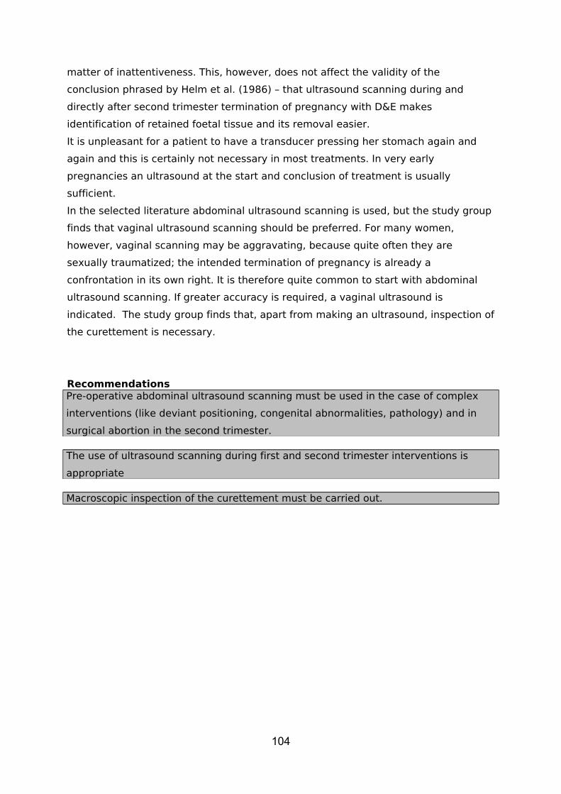
matter of inattentiveness. This, however, does not affect the validity of the
conclusion phrased by Helm et al. (1986) – that ultrasound scanning during and
directly after second trimester termination of pregnancy with D&E makes
identification of retained foetal tissue and its removal easier.
It is unpleasant for a patient to have a transducer pressing her stomach again and
again and this is certainly not necessary in most treatments. In very early
pregnancies an ultrasound at the start and conclusion of treatment is usually
sufficient.
In the selected literature abdominal ultrasound scanning is used, but the study group
finds that vaginal ultrasound scanning should be preferred. For many women,
however, vaginal scanning may be aggravating, because quite often they are
sexually traumatized; the intended termination of pregnancy is already a
confrontation in its own right. It is therefore quite common to start with abdominal
ultrasound scanning. If greater accuracy is required, a vaginal ultrasound is
indicated. The study group finds that, apart from making an ultrasound, inspection of
the curettement is necessary.
RecommendationsPre-operative abdominal ultrasound scanning must be used in the case of complex
interventions (like deviant positioning, congenital abnormalities, pathology) and in
surgical abortion in the second trimester.
The use of ultrasound scanning during first and second trimester interventions is
appropriate
Macroscopic inspection of the curettement must be carried out.
104

5.5 Ultrasound after surgical termination of pregnancy
Guiding questionWhat is the role of ultrasound scanning after surgical termination of pregnancy?
Summary of the literatureBar-Hava et al. (2001) conducted a prospective study involving 74 women analyzing
the “normal” ultrasound scan image after termination of pregnancy. Of these
women, 57 had undergone termination of pregnancy at 8-12 weeks of amenorrhoea;
10 had received treatment after an incomplete spontaneous abortion (8-12 weeks of
amenorrhoea) and 7 after a missed abortion (at 6-12 weeks of amenorrhoea). Suction
curettage was applied in 60 cases, D&E in 14. Vaginal ultrasounds were made after
24 hours in 26 women, after 48 hours in 10 women, on day three or four in 18
women and on day five or six in 20 women. The ultrasound findings were categorized
in three groups: A thin, regular midline < 7 mm (N=17), B a thick midline of 7-19 mm
(N=37) and C a midline > 20 mm or irregular Ultrasoundgenicity (N=20). The midline
in women who were screened within 48 hours was thicker and more irregular than in
women who were screened later. No correlation was found with clinical parameters
or method of treatment and the findings were similar for women with or without
complaints. The most striking outcome was that the uterus cavity was rarely totally
empty in the first days after first trimester termination of pregnancy and that modest
to large quantities of intra-uterine material of varying Ultrasoundgenicity was found
in 57 women (77%). They concluded that the presence of thick heterogeneous
material is an acceptable finding, to be expected after a first trimester termination of
pregnancy, and that this does not imply that a second curettage must follow
straightaway.
In a study discussed before, Fakih et al. (1986) performed abdominal ultrasound
scans on 120 women directly after the abortion. In six cases the images raised
suspicions of incomplete abortions, which was confirmed by further curettage. The
use of ultrasound directly following the intervention will prevent a continuing
incomplete abortion.
In a study of 21 women undergoing first trimester suction curettage, Haddad et al.
(1992) found an elliptical sac-like structure in 19 of the 21 women, 4-6 hours after
surgery (Ultrasoundgenic edges and hypo-Ultrasoundgenic contents), the so-called
pseudo-embryonic sac; the other two women showed a scan image with clearly an
empty uterus cavity. After three days this structure was still found in eight women,
albeit clearly smaller, and on day five the uterus cavity was empty in all women.
105

Helm et al. (1986) conducted a prospective study including 50 women into the value
of abdominal ultrasound scanning directly after abortion. Of the 39 first trimester
pregnancies, 15 (33%) were non-empty on ultrasound; in two cases this proved
incorrect.
A prospective study was carried out by Marenco et al. (1988) in 144 women
undergoing surgical termination of pregnancy in the first trimester, for the purpose of
gaining a better insight into ultrasonographic images directly after termination of
pregnancy, to avoid unnecessary re-curettage. Vaginal scanning was applied, 8-36
hours after termination of pregnancy. In 84 women there was a linear-shaped
endometrium, in the other sixty this was incomplete or the image was non-
homogeneous. Incomplete abortion was only observed in two of these sixty women.
A pseudo-embryonic sac was observed eight times. The authors relativize the
importance of an ultrasound 8-36 hours after abortion.
Maslovitz et al. (2004) conducted a comparative retrospective study into 69 women
who had undergone re-curettage on suspicion of retained traces after first or second
trimester surgical termination of pregnancy; repeat curettage took place within 14
days (range 6-43 days) on average. In 59% of the women no trophoblastic tissue was
found, histopathologically.
In a prospective study, McEwing et al. (2007) described the ultrasound outcomes of
38 women without complaints after first trimester termination of pregnancy and
compared these with the images of 105 women with complaints indicative of
retained tissue. Determined were: thickness of the endometrium; irregularities of the
cavity; the Ultrasoundgenicity of the contents of the uterus cavity and Doppler colour
flow. They concluded that there is a poor relation between ultrasound findings and
symptoms and between ultrasound findings and symptoms with histological findings.
Mikkelsen en Fielding (1994) carried out a prospective study in 117 women who
underwent a first trimester suction curettage, to establish the value of abdominal
ultrasound imaging directly after treatment. Repeat curettage was undertaken in five
women within four weeks because of pain and blood loss. Retained tissue was only
found in three women. They observed that ultrasound imaging subsequent to
treatment does not lead to a reduction in the number of cases of tissue retention and
should therefore not be recommended.
106

In the prospective trial by Stone and Elder (1974) abdominal ultrasonography was
conducted within 24 hours in 105 women undergoing first trimester suction
curettage; retained placenta tissue was found in 33 women. The authors conclude
that there is a clear relationship between immediate complications and placenta
traces on the ultrasound. Of greater interest is their observation that a uterus cavity
which appeared empty on ultrasound appeared not to be empty at all times, as this
was the case in two women.
A retrospective study by Tribalat et al. (1987) in 75 women who underwent first
trimester suction curettage points out the danger of unnecessary repeat curettage on
the basis of the abdominal ultrasound images at 24 hours after the intervention.
Linear endometrium was only found in 14 cases, in one case there was a continuing
pregnancy, a small residue was found twenty times; in forty cases the images
showed a sac-like structure with hypo-Ultrasoundgenic contents (pseudo-gestational
sac).
Conclusions
Level 2
It may be assumed that the presence of a pseudo-gestational sac, 4-6
hours after surgery, or the presence of thick heterogeneous material
during the first days after treatment is an acceptable finding, to be
expected after first trimester termination of pregnancy, especially in the
first two days after the intervention. This does not imply that an
immediate repeat curettage must be carried out.
B Tribalat et al. 1987, Bar-Hava et al. 2001, McEwing et al. 2007
C Marenco et al. 1988, Haddad et al. 1992
Level 3
It appears likely that the use of abdominal ultrasound imaging
immediately upon or shortly after treatment may lead to unnecessary re-
curettage or to the conclusion that the uterus cavity is empty.
C Stone en Elder 1974, Helm et al. 1986, Tribalat et al. 1987,
107
Marenco et al. 1988, Mikkelsen en Fielding 1994, Maslovitz et al.
2004B,
Discussion and evaluation
Contrary to the findings and conclusions above, the study group holds the view that
there are good reasons to carry out ultrasonography at the conclusion of the
treatment. By ultrasound it is possible to check if the cavity is empty at the end of
the treatment. However, the specialist in charge must also use the suction curette to
feel if treatment is complete, and inspect the curettement.
When complaints of serious pain, too much blood loss, or continuing symptoms of
pregnancy occur in the first weeks after the termination of pregnancy, ultrasound
may reveal complications.
RecommendationsThere are good reasons to carry out ultrasonography at the conclusion of the
treatment, to exclude the presence of retained tissue.
It is advisable to conduct ultrasound scanning when new or lingering complaints are
observed during the period of time after the termination of pregnancy, such as
severe abdominal pain, loss of too much blood or continuing haemorrhage.
If a woman has no complaints after a termination of pregnancy, ultrasound scanning
has little added value. Generally, no abnormalities are found.
If the pregnancy test is still positive after four weeks, it is necessary, however, to
conduct ultrasonography.
108
5.6 Ultrasound after medical termination of pregnancy
Guiding questionWhat is the role of ultrasound imaging after medical termination of pregnancy?
Summary of the literatureIn a retrospective study by Acharya et al. (2004) of 645 women who underwent
medical termination of pregnancy complications arose in 66 women (10.2%). On the
ultrasound the uterus was not empty in 98 (15.2%) of the women when they were
checked 2-3 weeks later. Of these women, 43 underwent curettage. The authors
conclude routine ultrasound imaging at check-up to be an efficient method of
identifying continuing pregnancies and for a diagnosis of completed abortion. Vaginal
ultrasounds after medical termination of pregnancy at an LMP< 63 days is only an
aid with respect to the decision to carry out additional curettage: the clinical
presentation after termination of pregnancy is decisive.
Creinin (1996) concludes that a decrease of the serum-ßhCG of less than 50% at 24
hours after administration of misoprostol makes it unlikely that a complete abortion
was achieved.
In a prospective trial including 165 women undergoing medical termination of
pregnancy Edelman et al. (2004) found that ultrasound, after a minimum interval of
four hours after taking misoprostol, held additional value. An empty cavity (50 cases
in total) predicted that no additional curettage was necessary in 96% of the women
with an amenorrhoea of <49 days; this figure was 98% at an amenorrhoea <63 days.
In this population curettage had to be performed in 31 cases.
In a retrospective cohort study in 437 women undergoing medical termination of
pregnancy at an amenorrhoea up to 63 days the endometrial thickness was
measured after 7-10 days, using vaginal ultrasound (Cowett et al. 2004). The
average thickness was 4.1 ± 1.8 mm (spread: 67-13.4 mm). In view of the spread it
is impossible to make pronouncements about treatment success by measurement of
endometrial thickness. The decision to start additional treatment must be based on
the clinical presentation, unless there is still an embryonic sac.
In their previously mentioned prospective trial involving 217 women undergoing
medical termination of pregnancy Fiala et al. (2003) repeated vaginal sonography
and the serum-ßhCG test after 6-18 days. Verification by ultrasound was only
possible in those cases in which an initial ultrasound had determined the presence of
intrauterine gravidity (yolk sac or crown-rump length). This was true for 167 (77%) of
109
the women: in 17 of them, (10.2%) complete abortion could not be demonstrated on
day 7-12 by sonography alone, because of a non-homogeneous picture. The hCG
levels went down to an average of 3% compared with the values before termination
of pregnancy (spread 1-44%). The authors conclude the prognostic value in terms of
successful expulsion to be 0.995 at a decrease to below 20% or less of the initial
values. When the decrease is less than 80%, the negatively prognostic value will be
0.5 and further evaluation needs to take place. In view of the fact that the hCG tests
took place after 6-18 days, these figures must be interpreted with care.
In 1,016 women undergoing medical termination of pregnancy Fielding et al. (2002)
found a remaining yolk sac in 24 cases at follow-up. In seven cases the experienced
clinicians saw no indication for sonography after examination and so would have
missed these continuing pregnancies. The authors conclude that if clinical
practitioners determine hCG levels in order to identify ectopic or continuing
pregnancies medical termination of pregnancy can be carried out safely without
ultrasound.
Machtinger et al. (2005) carried out a prospective trial involving 191 women
undergoing medical termination of pregnancy at an amenorrhoea of < 49 days.
Vaginal sonography was carried out in 170 women after 10-14 days; in 31 women in
this group there were suspicions of retained traces of tissue. Of these 31, four
received curettage. Four women wished to step out of the trial; in 23 the vaginal
sonography was repeated after menstruation. In 9 out of these 23 there were
suspected retained traces of pregnancy products, confirmed by hysteroscopy in 7
cases.
Menashe et al. (1995) conducted a study in 20 women undergoing medical
termination of pregnancy with mifepristone alone. The success rate was 70%. Vaginal
ultrasound correlated well with the clinical picture, in particular as regards the arrest
of embryonic cardiac activity. This contrasted with serum-ßhCG testing in the first
seven days; the test results had no prognostic value as regards a successful
termination of pregnancy.
The review by Paul et al. (2000) discusses the value of serum ßhCG determination
and vaginal ultrasound imaging after medical first trimester termination of
pregnancy. Elimination of ßhCG is characterized by a rapid initial downtrend, followed
by a gradual downtrend by around 50% per two days. A retrospective analysis of
women participating in four medical abortion trials shows that the average
110
downtrend in serum-ßhCG values in women with a complete abortion rated 66 ± 8%
24 hours after administration of misoprostol; in all others the decrease rated 25 ±
19%. The authors judge that if there is clinical suspicion of a failed termination of
pregnancy, vaginal sonography is more effective than repeated determinations of
serum-ßhCG.
In a prospective trial including 871 women undergoing medical termination of
pregnancy with an amenorrhoea of < 63 days (Rorby et al. 2004) measured serum-
ßhCG on day 8 and 15 and established endometrium thickness on day 15, using
vaginal sonography. In this trial, the prognostic value of vaginal ultrasound imaging
and repeated serum testing (ßhCG) was calculated. The authors conclude neither
determinator to be useful as a clinical test, to predict late failures.
Schaff et al. (1995) studied 100 women with an LMP < 56 days who underwent
medical termination of pregnancy and found that the average interval before the
serum-ßhCG had disappeared was 33 days (spread of 4-90 days).
Tsuchudin et al. (2004) conducted a retrospective study of 225 women after a
medical termination of pregnancy (amenorrhoea < 49 days). In 40% of these women
suspicions of retained tissue persisted after ultrasonography at 2 weeks; this
included 50 of the 123 women with a confirmed complete abortion. This percentage
went down after a longer waiting interval. Additional misoprostol did not influence
this outcome. A policy of waiting and seeing would seem justified when retained
products are found in sonography, or an endometrium thickness of < 16 mm.
Conclusions
Level 3
Vaginal sonography after medical termination of pregnancy is primarily
intended to verify if the gestational sac has disappeared.
B Fiala et al. 2003
C Paul et al. 2000
Level 3
It may be assumed that sonography after medical termination of
pregnancy leaves a picture which is often difficult to interpret. The
endometrium is often thick; sometimes there is no homogeneity in the
picture, resembling an incomplete abortion. Ultrasound does not always
make it possible to pronounce an expert judgement about success or
failure within two weeks after a medical termination of pregnancy.
111

C Cowett et al. 2004, Rorby et al. 2004, Tsuchudin et al. 2004,
Machtinger et al. 2005
Level 3
It is plausible to found the decision to start up additional treatment after a
medical termination of pregnancy on the clinical picture, unless
sonography reveals the presence of a gestational sac. When the uterus is
not empty after sonography, a policy of waiting and seeing would seem
justified.
B Fiala et al. 2003
C Paul et al. 2000, Acharya et al. 2004, Cowett et al. 2004
Discussion and evaluationThere is no consensus about what is best practice in verifying the outcome of a
medical termination of pregnancy. Some clinical professionals use ultrasound before
and after treatment, with rather large differences with respect to the time lapse after
treatment, other trust serum-ßhCG levels or use a less sensitive pregnancy test (500
IE/ml) at follow-up.
Elimination of ßhCG is characterized by a rapid initial downtrend – which in medical
termination of pregnancy actually does not start until after the administration of
misoprostol – followed by a gradual downtrend of around 50% per two days. An
increase of the serum-ßhCG level compared to the initial value indicates a continuing
pregnancy. In other respects, the available data are conflicting. Creinin (1996) claims
that a decrease of less than 50% 24 hours after administration of misoprostol renders
a complete abortion unlikely. Menashe et al. (1995) determined serum-ßhCG levels in
the first seven days and Rorby et al. (2004) on days 8 and 15; both find that this
indicator has no prognostic value for a successful termination of pregnancy. Fiala et
al. (2003) determined serum-ßhCG on day 6 and 18. They claim – with the proviso of
the time of determination – that a decrease down to 20% or less of the initial value
implies a prognostic value for successful expulsion of 0.995, while a decrease of less
than 20% of the initial value implies that an additional evaluation must take place.
The importance of serum-ßhCG determination and its timing will thus remain
hovering in mid-air, for the time being.
Determining serum-ßhCG prior to surgical termination of pregnancy or medical
termination of pregnancy does not yield essential information for the intended
treatment. This particular determination does not yield any information about the
length of amenorrhoea, either. A rising serum-ßhCG level indicates the need for a
surgical intervention, but a continuing pregnancy may also be revealed by
112
ultrasound. Apart from this, there is also the importance of determining the presence
of an ectopic pregnancy, which is not discussed in this framework.
At a week after starting the medical termination of pregnancy it is possible to check if
the pregnancy was in fact expelled. This does not imply that treatment nay be
terminated. There is often evidence of retained tissue; when the woman keeps
reporting too much loss of blood or symptoms of a continuing pregnancy, she must
report back for an ultrasound check.
The study group feels that if no pregnancy can be demonstrated by ultrasound, it is
best not to start a medical termination of pregnancy, but to wait for another week. It
may also be a case of extra-uterine gravidity. If treatment is started up, it is
necessary to obtain a β-hCG-determination on days 1 and 4, to ascertain if the
pregnancy has come to en end.
RecommendationsIt is advisable to carry out a sonography after a medical termination of pregnancy, in
the case of continuing abdominal pains, excessive blood loss or prolonged bleeding,
or continuing symptoms of pregnancy.
It is advisable to carry out sonography at one to four weeks after a medical
termination of pregnancy, depending on the stage of the pregnancy.
113
5.7 ReferencesAcharya G, Morgan H, Paramanantham L, Fernando R A randomized controlled trial
comparing surgical termination of pregnancy with and without continous guidance. Eur J Obstet Gynecol Reprod Biol 2004; 114:69-74
Acharya G, Haugen M, Brathen A, Nilsen I, Maltau JM. Role of routine ultrasonography in monitoring the outcome of medical abortion in a clinical setting. Acta Obstet Gynecol Scand 2004; 83: 390-4
Bar-Hava I, Aschkenazi S, Orvieto R, Perri T, Shalev J, Dicker D, Ben-Rafael Z, Dekel A. Spectrum of normal intrauterine cavity sonographic findings after first-trimester abortion. J Ultrasound Med 2001; 20:1277-81
Blanchard K, Cooper D, Dickson K et al. A comparison of women´s, provider´s and ultrasound assessments of pregnancy duration among termination of pregnancy clients in South Africa. Int J Gynecol Obstet 2007; 114:569-75
Cowett AA, Cohen LS, Lichtenberg S, Stika CS. Ultrasound evaluation of the endometrium after medical termination of pregnancy. Obstet Gynecol 2004; 103:871-5
Creinin MD. Change in serum B-human chorionic gonadotrophin after abortion with methotrexate and misoprostol. Am J Obstet Gynecol 1996; 174:776-8
Darney P, Sweet RL. Routine ultrasonography for second trimester abortion reduces incidence of uterine perforation. J Ultrasound Med 1989: 8:71-5
Edelman A, Thomas L, Jensen J. Transvaginal ultrasound and the success of medical abortion. Int J Gynecol Obstet 2004; 85:62-3
Fakih MH, Barnea ER, Yarkoni S, DeCherney AH. The value of real time ultrasonography in first trimester termination. Contraception 1986; 33:533-8
Fiala C, Safar P, Bygdeman M, Gemzell-Danielsson K. Veryfying the effectiveness of medical abortion; ultrasound versus hCG testing. Eur J Obstet Gynecol Reprod Biol 2003; 190-5
Fielding SL, Schaff EA, Nam B. Clinicians’ perception of sonogram for mifepristone abortion up to 63 days. Contraception 2002; 66:27-31
Goldstein SR, Snyder JR, Watson C, Danon M. Combined sonographic-pathologic surveillance in elective first-trimester termination of pregnancy. Obstet Gynecol 1988; 71:747-50
Grimes DA, Cates W. Complications from legally induced abortion: a review. Obstetrical Gynecological Survey 1979; 34:177-190
Grimes DA, Schulz KF, Gates W, Tyler CW. Mid-trimester abortion by dilatation and evacuation. N Engl J Med 1977; 296: 1141-5
Haddad S, Ruach M, Ohel G. Sonographic demonstration of endometrial fluid collection following termination of pregnancy. Obstet Gynecol 1992; 5:703-4
Helm CW, Maxwell DJ, Lilford RJ. A prospective study of the use of ultrasound in vaginal legal abortion before 20 weeks of pregnancy. J Obstet Gynaecol 1986; 7:143-5
Hornstein MD, Osathanondh R, Birnholz JC, Kapnic SJ, Jones TB, Safon LE, Cayea PD, Doubilet PM. Ultrasound guidance for selected dilatation and evacuation procedures. J Reprod Med 1986; 31:947-50
Jarallah Y, Geirsson RT. Is routine ultrasound before termination of pregnancy worthwhile? Scott Med J 1985; 30: 232-3
Machtinger R, Seidman DS, Goldenberg M, Stockheim D, Schiff E, Shulman A. Transvaginal ultrasound and operative hysteroscopy in women undergoing medical termination of pregnancy as a part of routine follow-up. Fertil Steril 2005; 84: 1536-8
Marenco M, Sarramon MF, Berrebi A, Bergis A, Kobuch WE. Le controle Ultrasoundgraphique en post interruption volontaire de grossese immediate. J Gynaecol Obstet Biol Reprod 1988; 17 :51-5
Markovitch O, Tepper R, Klein Z et al. Sonographic appearance of pregnancy. J Clin Ultrasound 2006; 34:278-82
114

Maslovitz S, Almog B, Mimouni GS, Jaffa A, Lessing JB, Many A. Accuracy of diagnosis of retained products of cenception after dilatation and evacuation. J Ultrasound Med 2004; 23: 749-56
McEwing RL, Anderson NG, Meates JB et al. Sonographic appearances of the endometrium after termination of pregnancy in asymptomatic versus symptomatic women. J Ultrasound Med 2009; 28:579-86
McGalliard C, Gaudoin M. Routine ultrasound for pregnancy termination requests increases women’s choice and reduces inappropriate treatments. Br J Obstet Gynaecol 2004; 111:79-82
Menashe Y, Bider D, Shalev J, Pariente C, Serr DM. Clinical, hormonal and sonographic predictors of successful RU-486 induced abortions. Gynecol Obstet Invest 1995; 40:108-12
Mikkelsen AL, Fielding C. The value of peroperative ultrasound examination in first trimester legally induced abortion. Clin Exp Obst Gyn 1994: 3:150-1
NVOG, Datering van de zwangerschap, versie 1.0, 04-10-2010, www.nvog.nlPaul N, Schaff E, Nichols M. The roles of clinical assessment, human gonadotropin
assays, and ultrasonography in medical abortion practice. Am J Obstet Gynecol 2000; 183: s34-s43
Robinson ET, Barber, JH. Early diganosis of pregnancy in general practice. J R Coll. Gen Pract 1977; 27:335-8
Rorbye C, Norgaard M, Nilas L. Prediction of late failure after medical abortion from serial beta-hCG measurements and ultrasonography. Human Reprod 2004; 19:85-9
Schaff EA, Eisinger SH, Franks P, Kim SS. Combined methotrexate and misoprosotol for early induced abortion. Arch Fam Med 1995; 4:774-9
Sinha P, Pradhan A, Chowdhury V. Value of routine transvaginal ultrasound scan in women requesting early termination of pregnancy. Obstet Gynecol 2004; 24:426-28
Stone M, Elder MG. Evaluation of sonar in the prediction of complications after vaginal termination of pregnancy. Am J Obstet Gynecol 1974; 120:890-4
Tribalat S, Balladur A, Lewin D. Images Ultrasoundgraphiques le lendemain d’ une interruption volontaire de grossesse. J Gynecol Obstet Biol Reprod 1987 ; 16 :100-1001
Tschudin S, Zanotelli D, Bitzer j, Holzgreve W. Erfahrungen mit dem medikamentosen Schwangerschaftsabbruch, insbesondere nachkontrolle, nachbehandlung und nachfolgende Kontrazeption. Geburtsh Frauenheil 2004; 64:1299-1304
Verburg B, Steegers E, de Ridder M, Snijders R, Smith E, Hofman A, Moll H, Jaddoe V en Witteman J, New charts for ultrasound dating of pregnancy and assesment of fetal growth: longitudinal data from a population based cohort study, Ultrasound Obstet Gynaecol 2008; 31: 388-396
115
Chapter 6 Rhesus factor
Guiding questionWhen must rhesus factor determination take place and what is the treatment in the
case of Rh-?
6.1 IntroductionIn the case of blood group antagonism a pregnant woman may become immunized
by blood group antigens of the foetus. Isoimmunisation is started against antigens
which the foetus inherits from its father and which are absent in the mother. In
rhesus antagonism a rhesus negative woman produces antibodies during her
pregnancy against her rhesus positive child. In most cases this concerns rhesus D.
Around 85% of the population is rhesus-D positive, the other part rhesus-D negative.
Out of 4,972 cases of pregnancy isoimmunisation,Van Went (1963) only found 70
(1.4%) with antibodies other than anti-Rh(D), while in 46 of those 70 anti-Rh(D) was
also present; so, isoimmunisation by an agent other than anti-Rh(D) only exclusively
occurred in 24 cases (0.48%). As early as 1948, Levine suspected that the production
of antigens is induced by Rh(D) positive foetal erythrocytes which end up in the
circulation of the mother via transplacental channels. This phenomenon was termed
foetomaternal transfusion (FMT). Around week 12 of embryonic development
syncytium cells develop into the endothelial lining of the arteries; at that point the
mother’s blood starts to penetrate the lacunae system. In this way placentary
circulation comes into place. The pumping activity of the embryonic heart and thus
its blood circulation starts at about four weeks. Rh(D) antigen was already
demonstrated in a foetus of 38 days (Bergstrom c.s. 1967). It is open to question if
the antigen is already immunogenic at that point, however. Theoretically a
transplacental haemorrhage in a rhesus negative woman brings along the risk of
rhesus isoimmunisation at this point of embryonic development. Rhesus subgroup
isoimmunisations (against subgroups C, c, E and e) and also isoimmunisations
against other blood group antigens (Kell, Duffy, Lewis e.d.) do not proceed differently
from Rh (D) isoimmunisation, but their frequency is much lower.
During a first FMT of a Rh(D) negative mother and a Rh(D) positive foetus the onset
of antigen production is probably very slow: antigens are usually not found until
between the 3rd and 6th month after delivery. Once primary immunisation has set in
(primary response) the body will respond to minute quantities of Rh(D) positive blood
with a rapid increase in antigen production (the so-called booster reaction). In a next
pregnancy the antibody titer will rise relatively early and may turn both mother and
child gravely ill. This is the reason why the rhesus factor is determined in every
116
pregnant mother. A search was conducted in Medline and Embase with respect to
termination of pregnancy, using controlled key terms and free text terms (starting
1966 in Medline and 1980 in Embase up to December 2009). Explode “Abortion-
Induced”/all subheadings was used as a controlled key term. This topic was also
searched using free text terms like pregnancy termination and induced abortion. The
theme of rhesus antagonism was added via the controlled key terms “Rh-
Isoimmunisation”/all subheadings or Immunoglobulin-administration-and –dosage.
The outcome was restricted to English, Dutch, German and French language sources.
Five articles were selected on the basis of this literature search. The authors also had
articles in their possession which they had collected on this subject over the years. A
total set of 15 articles was available which met the criteria, on which evidence tables
could be based showing the risk of foetomaternal transfusion in termination of
pregnancy expressed as a percentage of positive KB tests and the risk of rhesus
isosensitization in termination of pregnancy in relation to method, amenorrhoea and
prophylaxis.
6.2 Summary of the literatureFactors which influence the frequency of rhesus isoimmunisation
The first precondition for isoimmunisation is rhesus antagonism (after all, the foetus
may be Rh(D) negative). Among Caucasians, around 15% is Rh(D) negative, in the
African population around 7.5% and among Asians about 3.5%. Isoimmunisation
turns out to be more restricted than what the figures would lead one to expect.
The Rh(D) antigens are exclusively found in erythrocytes; rhesus isoimmunisation in
the course of pregnancy can therefore only be caused by FMT. The FMT must,
however, be of sufficient volume. Mollison (1971) estimates the minimum quantity of
Rh(D) positive blood to cause isoimmunisation of a Rh(D) negative receiver to be 0.1-
0.25 ml. The chances of isoimmunisation appear to be rising with the volume of FMT
(Borst-Eilers, 1972). The maximum isoimmunisation percentage is reached after
haemorrhages of 1 ml or more and lies at 60-65%. Thus, not every FMT is followed by
isoimmunisation: the antigen must have immunizing power and the mother must be
capable of forming antibodies, which is only the case in 2/3 of all Rh(D) negative
women. Apart from that, immunological hypo-reactivity during the pregnancy and
ABO-incompatibility play a role (Querido 1977).
Demonstrating foetomaternal transfusion
By using the Kleihauer-Betke (KB)-test (Kleihauer, 1957) it can be demonstrated
which erythrocytes in the maternal blood primarily hold HbF. However, HbF
erythrocytes need not always be of foetal origin; besides, not all foetal erythrocytes
contain HbF, either exclusively or mainly (Querido 1977). The percentage of HbA and
117
HbF is assessed with HPCL after lysis. In adults the proportion is 95/5; in a foetus this
is 5/95 up to 10/90, depending on its age. Implied is the proportion inside each cell.
The KB test can distinguish a foetal cell from an adult cell with a great degree of
certainty. Apart from this, when interpreting the KB test it is important to take into
account the presence or absence of natural or acquired antibodies against foetal
erythrocytes in the mother’s blood, in view of the survival time of foetal erythrocytes.
The technical execution of the test has not been standardized, nor has the method of
counting, rendering comparisons between the results of different authors difficult to
make. A raised alpha foetoprotein level in the maternal serum appears to be a more
sensitive and reliable indicator than the KB test (Lachman 1977). The KB test is not
sensitive enough to demonstrate FMT of <= 0.1 ml. Flow cytometrics is a novel and
more sensitive method.
Foetomaternal transfusion in termination of pregnancy
In a prospective trial, Jorgensen (1969) studied 227 women with respect to the
incidence of FMT after a termination of pregnancy. Before the intervention, all
women tested negative on the KB test. After spontaneous abortion, no FMT higher
than 0.1 ml was found; after suction curettage in one case only (1.2%); after
curettage also in a single case (6.7%); after hypertonic saline four cases (5.9%) and
after rupture of the membranes (punctio ovi) three cases (25%). Leong (1979)
studied 75 women undergoing suction curettage at a pregnancy length up to 8 weeks
of amenorrhoea on the presence of foetal erythrocytes, using the KB test 15 minutes
before surgery and 30 minutes after surgery. Before surgery, the KB test was positive
in two cases (2.6%) and in twelve cases after surgery (15.5%). At six weeks of
amenorrhoea there was no positive KB test, neither before nor after surgery.
Murray (1970) studied 483 women on the incidence of FMT before and directly after
spontaneous and surgical abortion, and again after a time interval. Before abortion,
the KB test was positive in 54 cases (11.6%). Of the 243 cases in which the
pregnancy was ended by means of suction curettage, it was ended before the 14th
week of amenorrhoea, in all but six cases. In the suction curettage group, 39 women
tested positive on the KB test before the intervention and 39 after the intervention. In
the group of women with a spontaneous abortion, 21 women tested positive on the
KB test before the abortion and 22 after the abortion. On the basis of these findings
the authors conclude that it makes no difference whether the abortion was
spontaneous or induced.
Queenan (1971) studied 606 women on the presence of FMT after termination of
pregnancy: 404 women underwent termination of pregnancy by suction curettage
(384 women up to 13 weeks and 20 women at between 13 and 16 weeks of
118

amenorrhoea) and 202 women (13 up to 23 weeks of amenorrhoea) by hypertonic
saline solution. None of these women had any foetal cells in their blood before
treatment. The FMT was monitored with the KB test 24 hours after the termination of
pregnancy; FMT was defined as the presence of two or more foetal cells per 50,000
maternal cells (which corresponds with an FMT of 0.2 ml). In the entire suction
curettage group FMT could be demonstrated in 7.2%; in the suction curettage group
up to 13 weeks this figure was 6.0%. In the group receiving the hypertonic saline
solution FMT could be demonstrated in 20.2%.
Borst-Eilers (1972) studied the frequency of FMT in 72 women before and after
termination of pregnancy. In twelve cases no assessment was possible. Of the other
60, 50 underwent suction curettage, 8 curettage and 2 sectio parva. In just three
cases, the number of HbF erythrocytes was considerably higher after the intervention
than before (5 versus 1; 11 versus 2 and 10 versus 3). An FMT of over 0.1 ml thus
only occurred in two cases.
Isoimmunisation and prevention in termination of pregnancy
Jorgensen (1969) conducted a prospective trial in 30 Rh(D) negative women whose
pregnancy was terminated. The group consisted of 8 primigravidae and 22
multigravidae. In one primigravida (13%) and two multigravidae (9%) antibodies
were demonstrated 6-12 months later (overall percentage: 10%). No antibodies had
been found in any of the women before termination of pregnancy. The primigravida
was curettaged at 15 weeks of amenorrhoea; after the intervention, the KB test was
positive (FMT >1.5 ml). The two multigravidae were curettaged at 10 and 14 weeks
of amenorrhoea.
Murray c.s. (1970) carried out a prospective trial involving 23 Rh(D) negative
multiparae after termination of pregnancy. In three instances isoimmunisation was
found. The KB test after the abortion was negative.
In the study by Queenan (1971) discussed above, 66 women were Rh(D) negative: 15
were given anti-D immunoglobulin after termination of pregnancy; 3 had already
become isosensitized. Of the remaining 48 women nobody was isosensitized before
the intervention. Follow-up of 25 women after four months showed that one
multipara had become isosensitized (had developed antibodies); she had undergone
suction curettage at nine weeks of amenorrhoea and tested negative on a KB test
after her termination of pregnancy.
Murray (1971) conducted a comparative prospective study including 96 Rh(D)
negative women (48 primigravidae and 48 multigravidae). The KB test was
administered both before and after termination of pregnancy. After 6-9 months she
found antigens in 3 primigravidae and 6 multigravidae. Of these nine cases showing
119
antibodies five had undergone suction curettage (four out of a group of 27
multiparae and one in a group of 27 primiparae), three had undergone saline
treatment and one a hysterotomy abortion.
177 Rh(D) negative women undergoing termination of pregnancy were studied by
Murray (1972). A KB test was administered to 146 women before and after the
termination of pregnancy. 96 women were monitored for isosensitization: 44 had an
amenorrhoea length of <12 weeks, 40 > 12 weeks while in 12 women their
pregnancy length was uncertain. Of the entire group, seven (6.2%) tested positive on
the KB test with an FMT of > 0.1 ml. However, only two of the patients (2%) appeared
isosensitized at a pregnancy length of seven and ten weeks, respectively. Both
women tested negative on the KB test after surgery.
Visscher (1972) carried out a prospective comparative trial in 57 Rh(D) negative
women with Rh(D) positive partners after spontaneous abortion. Their pregnancy
length was 8-24 weeks of amenorrhoea; after an incomplete abortion curettage was
carried out. In the double blind part of the study, 19 women received 300 μg anti-d
while 29 did not. In a follow-up study of nine women none were given anti(Rh)D
immunoglobulin. At check-up after three and six months no antibodies were found.
The authors find that Rh-immunoprophylaxis is unnecessary after a spontaneous
abortion <12 weeks of amenorrhoea. An important argument in this context is that it
is assumed that foetal necrosis and placenta degradation occur 4-5 weeks before the
spontaneous abortion sets in.
Goldman (1972) conducted a study of 170 Rh(D) negative women after a
spontaneous or an induced termination of pregnancy. 48 women in all (29 in their
first and 19 women in their second trimester) were given 200 μg anti-D
immunoglobulin, while 22 did not. In the group thus treated no isosensitization
occurred. Isosensitization did occur in two non-protected women after spontaneous
abortion; this was also the case for three women who underwent surgical termination
of pregnancy. According to the authors, the risk of FMT is greater after surgical
termination of pregnancy compared to spontaneous abortion.
Keith (1977) researched 315 Rh(D) negative women undergoing a first trimester
termination of pregnancy. Seventeen women received 300 μg anti-D immunoglobulin
and 298 women 50 μg. No isosensitization could be found in any of the women at six
months. In the first trimester 50 μg immunoglobulin is sufficient to prevent
isoimmunisation.
Bennebroek Gravenhorst (1986) c.s. monitored serum AFP levels in 103 women
directly before and after termination of pregnancy, at an amenorrhoea length
between 12 and 19 weeks. FMT increased with pregnancy length; in 6-8% of the
120

women the quantities ranged between 1 and 3.5 ml. Administration of 75 μg of anti-D
immunoglobulin proved enough to prevent isosensitization.
Hensleigh (1977) studied 187 rhesus negative women with a pregnancy length of 5-
14 weeks of amenorrhoea. FMT occurred in 27%, based on the KB test. There was a
subdivision in three groups. Each group received a different dose of anti-D
immunoglobulin: 499 μg, 155 μg and 73 μg, respectively. Isosensitization was found
in none of the patients.
Urquhart (1990) compared 20 women undergoing medical termination of pregnancy
with 20 women undergoing vacuum aspiration. Age and pregnancy length were the
same in both groups. Serum-AFP was monitored before mifepristone was taken, 48
hours later before taking misoprostol and again at 4 hours after taking misoprostol. In
the case of vacuum aspiration serum-AFP was monitored before the administration of
anaesthesia and immediately after surgery. In the medical termination of pregnancy
group not a single patient presented an increase in serum-AFP between
administration of mifepristone and administration of misoprostol. After taking
misoprostol the AFP level rose to 25% above the starting value in three patients after
four hours (15%). In the vacuum aspiration group an increase of 25% was found in
twelve patients (60%). Thus, FMT occurs more frequently after vacuum aspiration
than after medical termination of pregnancy. This does not imply that isosensitization
will develop more frequently, too. According to the authors, all Rh(D) negative
women must be given anti-d immunoglobulin; in medical termination of pregnancy at
the moment when misoprostol is taken.
Jabara carried out a literature study of the years between 1964 and 2003, on rhesus
antagonism and termination of pregnancy. They concluded that the evidence
supporting the administration of anti-d immunoglobulin in the first trimester is very
thin, but that there is theoretical evidence for the necessity to do so nonetheless.
Mollison (1971) estimated that at least 0.1-0.25 ml of foetal blood is needed to effect
isosensitization. It may be assumed that 1000 IE (200µg) of anti-D will neutralize
positive full blood (= 10 ml Erythrocyte concentrate). 20 ug anti-D immunoglobulin
will suppress immunogenic problems caused by 1 ml of blood; 50 ug anti-D
immunoglobulin will therefore offer protection against 2.5 ml of blood, a quantity
bigger than the entire blood volume in a foetus in the first trimester. This quantity
was already established by the WHO in 1970. Research by Kiss c.s. (1974), the
Working Party (1975) and Keith (1977) also proves that a dosage of 50 ug anti-D
immunoglobulin is adequate in the first trimester. For the second trimester the
dosage should be between 50 and 200 ug (WHO 1970, Bennebroek Gravenhorst
1981, Bennebroek Gravenhorst, 1986). The latter quantity is adequate protection
after delivery (i.e. it will protect against FMT of 20 ml).
121

Conclusions
Level 3
The KB test appears to be reliable in demonstrating HbF. This test is a
measure of foetomaternal transfusion. FMT is a precondition to rhesus
isosensitization. When interpreting the findings derived from the literature
both false-positive and false-negative outcomes of the KB test must be
taken into account.
C Kleihauer 1957, Querido 1977
Level 3
It appears likely that practically no foetal cells pass through the placenta
barrier during the first trimester of pregnancy, not even after a
spontaneous abortion. The frequency of FMT (and so the risk of
isosensitization) rises with pregnancy length; theoretically, FMT may be
found from 52 days of amenorrhoea onward. Not every instance of FMT is
followed by isosensitization.
B Queenan 1971
C Borst-Eilers 1972, Bennebroek Gravenhorst 1986
Level 3
It appears likely that Rh isosensitization may also occur without prior
demonstration of FMT as measured by the KB test (false-negative KB
outcome). It takes a few weeks before a primary response can be
demonstrated.
B Murray 1970, Queenan 1971
C Murray 1972
Level 2
It appears likely that the risk of sensitization is negligible in the case of
spontaneous abortions at under eight weeks of amenorrhoea. After eight
weeks, the risk is 3-4%; in the second trimester it is 6-8%.
A2 Visscher 1972
Level 3
Foetomaternal transfusion more frequently occurs in induced termination
of pregnancy, compared to spontaneous abortions.
B Jorgenson 1969, Goldman 1972
122

Level 2
In the case of an induced termination of pregnancy it is likely that
immunoglobulin at a dose of 50 ug (250 IE) is adequate in the first
trimester; a dose of 75 ug (375 IE) is adequate in the second trimester
A2 Hensleigh 1977
B Keith 1977
C Kiss 1974, Working Party 1975, Bennebroek Gravenhorst 1981,
Bennebroek Gravenhorst 1986
6.3 Discussion and evaluationMedical termination of pregnancy is no spontaneous abortion. Lacking are the most
important characteristics of spontaneous abortion: foetal necrosis and placenta
degeneration some weeks before expulsion. It is advisable to follow the same policy
in medical termination of pregnancy as in surgical termination of pregnancy. The
anti-D immunoglobulin may be administered on the same day as the mifepristone.
Critical remarks are warranted with respect to the study by Leong et al. (1979).
Firstly, LMP and gestation are mixed up. Next, the authors claim that they measured
foetal erythrocytes rather than HbF. The numbers recorded by the authors do not
match the percentages they published.
According to Urquhart (1990) the optimal administration of anti-D immunoglobulin is
during the intake of misoprostol, but as anti-D immunoglobulin is effective up to 21
days after administration, it may well be given at the moment of taking mifepristone.
Anti-D immunoglobulin must always be given within 48 hours after termination of the
pregnancy. Administration of anti-D immunoglobulin beyond 48 hours after
exposition is probably less effective, but still proven to be effective up to 13 days
after exposition and is recommended up to one month after exposition (Bowman
1985, Samsonen Mollison, 1975).
Dutch abortion practice shows that we may refrain from administering anti-D under
specific circumstances (e.g. absolutely no wish to have a child). It is an absolute
prerequisite that the possible consequences are discussed with the woman.
As anti-D-vaccine is expensive and is made of human blood, it is advisable to put
doses of 50 µg (250 IE) and 75 µg (375 IE) immunoglobulin on the market.
In the NVOG guideline the following recommendation was adopted: Rhesus negative
women must be given anti-D in the case of a termination of pregnancy, with the
exception of a medical termination of pregnancy before week 10. This
recommendation and the doses specified above have been taken over from the 1998
IGZ10 report. Doses of 50 µg (250 IE) were not available at that time.
10 National Health Inspectorate of The Netherlands
123
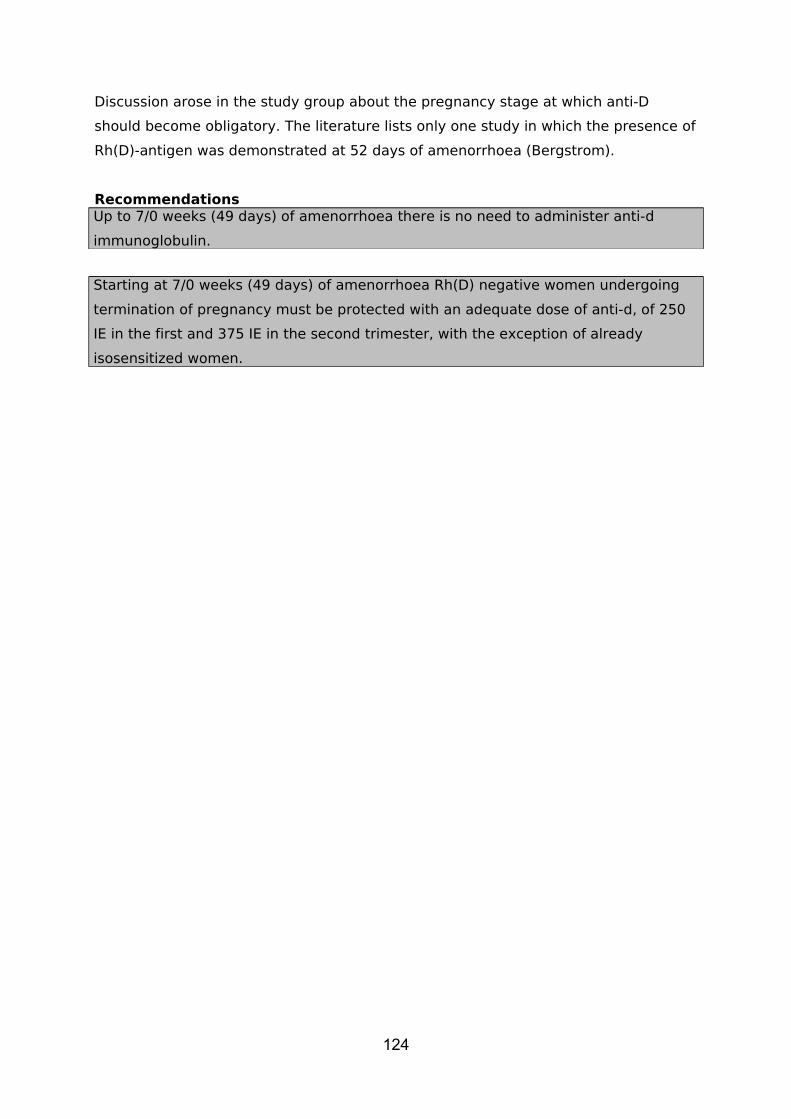
Discussion arose in the study group about the pregnancy stage at which anti-D
should become obligatory. The literature lists only one study in which the presence of
Rh(D)-antigen was demonstrated at 52 days of amenorrhoea (Bergstrom).
RecommendationsUp to 7/0 weeks (49 days) of amenorrhoea there is no need to administer anti-d
immunoglobulin.
Starting at 7/0 weeks (49 days) of amenorrhoea Rh(D) negative women undergoing
termination of pregnancy must be protected with an adequate dose of anti-d, of 250
IE in the first and 375 IE in the second trimester, with the exception of already
isosensitized women.
124
6.4 References
Bennebroek Gravenhorst J. De preventie van Rh(D) immunisatie mbv anti Rhesus(D) immunoglobuline. GHI Bulletin. Leidschendam. 1981
Bennebroek Gravenhorst J,Beintema-Dubbeldam A,van Lith D.A.F.,Beekhuizen W. Elevation of maternal a-fetoprotein serum levels in relation to fetomaternal haemorrhage after second trimester pregnancy termination by aspirotomy. Br. J. Obstet. Gynaecol. 93, 1302-4, 1986
Bergstrom, H., Nilsson, L.A., Nilsson, L., en L. Ryttinger. Demonstration of Rh antigens in a 38 day-old fetus. Amer. J. Obstet. Gynec. 99, 130, 1967
Borst-Eilers, E. Rhesusimmunisatie: ontstaan en preventie. Acad. Proefschrift, Utrecht, 1972
Bowman JM. Controversies in RH profylaxis.Who needs RH.immunoglobulin and when should it given? Am.J. Obstet. Gynecol 1985: 151:289-94
Goldman, J.A.,Eckerling B. Prevention of RH Immunization after abortion with anti-Rh(D)-immunoglobulin. Obstet.Gynecol. 40, 366-70, 1972
Hensleigh, P.A.,Leslie W., Dixon E,Hall E,Kitay D.Z.,Jackson J.E. Reduced dose of Rh(D) immunoglobulin following induced first- trimester abortion. Am. J. Obstet. Gynecol. 129, 413-6, 1977
Jabara, S. en K.T. Barnhart. Is Rh immune globulin needed in early first-trimester abortion. Am.J. Obstet. Gynecol. 129, 623-7, 2003
Jorgensen, J. Rhesus-antibody development after abortion. Lancet II, 1253, 1969Keith, L.,Bozorgi N. Small dose anti-Rh therapy after first trimester abortion.
Int.J.Gynaecol.Obstet. 15(3) 235-7, 1977Kiss, D en B. Szoke. Praevention der Rh-Isoimmunisation in Verbindung mit Spontan-
abortus und Schwangerschaftsunterbrechungen. Zentralbl. Gynaekol. 96, 398, 1974
Kleihauer, E., Braun, H. en K. Betke. Demonstration vom fetalem Haemoglobin in den Erythrocyten eines Blutausstrichs. Klin. Wschr. 35, 687, 1957
Lachman, E., Hingley, S.M., Bates, C. e.a.. Detection and measurement of fetomaternal haemorrhage: serum alpha-fetoprotein and the Kleihauer technique. BMJ 1, 1377, 1977
Leong, M., Duby, S. en R.A.H. Kinch. Fetal-maternal transfusion following early abortion. Obstet. Gynecol. 54, 424-6, 1979
Levine, P. The mechanism of tranplacental iso-immunization. Blood 3, 404, 1948Mollison, P.L. The role of Rh antibodies in causing haemolytic disease of the newborn
and in preventing it. J. Clin. Path. 24, 479, 1971Murray, S., Barron, S.L. en R.A. McNay. Transplacental haemorrhage after abortion.
Lancet I, 631, 1970Murray, S. en S.L. Barron. Rhesus iso immunization after abortion. BMJ 3, 90, 1971Murray, S. en S.L. Barron. Transplacental haemorrhage in induced abortion. Lancet I,
1972, 815-7Queenan, J.T, Shah, S, Kubarych, S,F. en B. Holland. Role of induced abortion in
rhesus immunisation. Lancet I, 815-7, 1971Querido, L. Abortus en rhesusprophylaxis. Stimezo, Dan Haag, 1977Samson, D. en P.L. Mollison. Effect on primary Rh immunisation of delayed
administration of anti-Rh. Immunology 1975 :28:394-397Urquhart, D.R.,Templeton A. Reduced risk of isoimmunisation in medical abortion.
Lancet, 914, 1990Visscher R.D. en H.C. Visscher. Do Rh-gevative women with an early spontaneous
abortion need Rh immune prophylaxis. Am.J.Obste.Gynecol. 15, 158-65, 1972Van Went, J.J. Rhesusimmunisatie. Acad. Proefschrift, Amsterdam, 1963WHO Report 486: Prevention of Rh sensitisation. Geneva, 1970Working Party on the use of anti-D immunoglobulin. Controlled trial of various anti-D
dosages in suppression of Rh sensitisation following pregnancy. Brit. Med. J. 2, 75, 1975
125
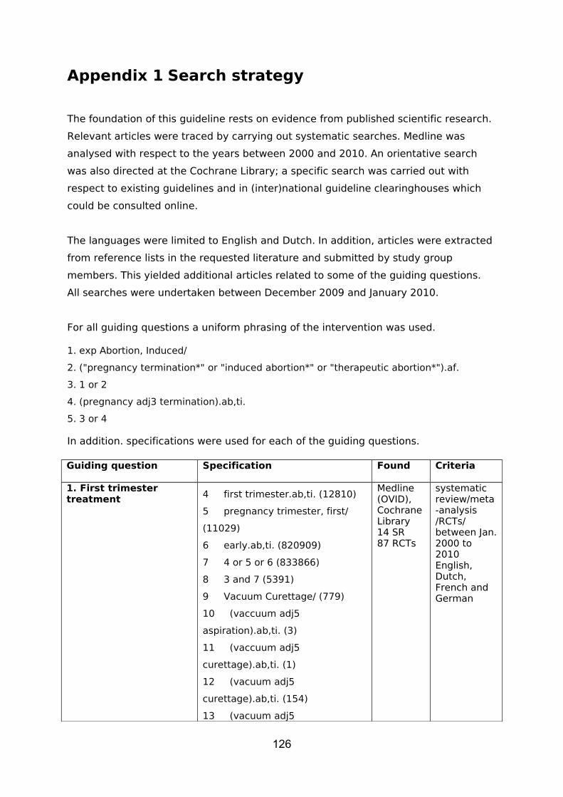
Appendix 1 Search strategy
The foundation of this guideline rests on evidence from published scientific research.
Relevant articles were traced by carrying out systematic searches. Medline was
analysed with respect to the years between 2000 and 2010. An orientative search
was also directed at the Cochrane Library; a specific search was carried out with
respect to existing guidelines and in (inter)national guideline clearinghouses which
could be consulted online.
The languages were limited to English and Dutch. In addition, articles were extracted
from reference lists in the requested literature and submitted by study group
members. This yielded additional articles related to some of the guiding questions.
All searches were undertaken between December 2009 and January 2010.
For all guiding questions a uniform phrasing of the intervention was used.
1. exp Abortion, Induced/
2. ("pregnancy termination*" or "induced abortion*" or "therapeutic abortion*").af.
3. 1 or 2
4. (pregnancy adj3 termination).ab,ti.
5. 3 or 4
In addition. specifications were used for each of the guiding questions. Guiding question Specification Found Criteria
1. First trimester treatment 4 first trimester.ab,ti. (12810)
5 pregnancy trimester, first/
(11029)
6 early.ab,ti. (820909)
7 4 or 5 or 6 (833866)
8 3 and 7 (5391)
9 Vacuum Curettage/ (779)
10 (vaccuum adj5
aspiration).ab,ti. (3)
11 (vaccuum adj5
curettage).ab,ti. (1)
12 (vacuum adj5
curettage).ab,ti. (154)
13 (vacuum adj5
Medline (OVID), Cochrane Library14 SR87 RCTs
systematic review/meta-analysis /RCTs/between Jan. 2000 to 2010English, Dutch, French and German
126
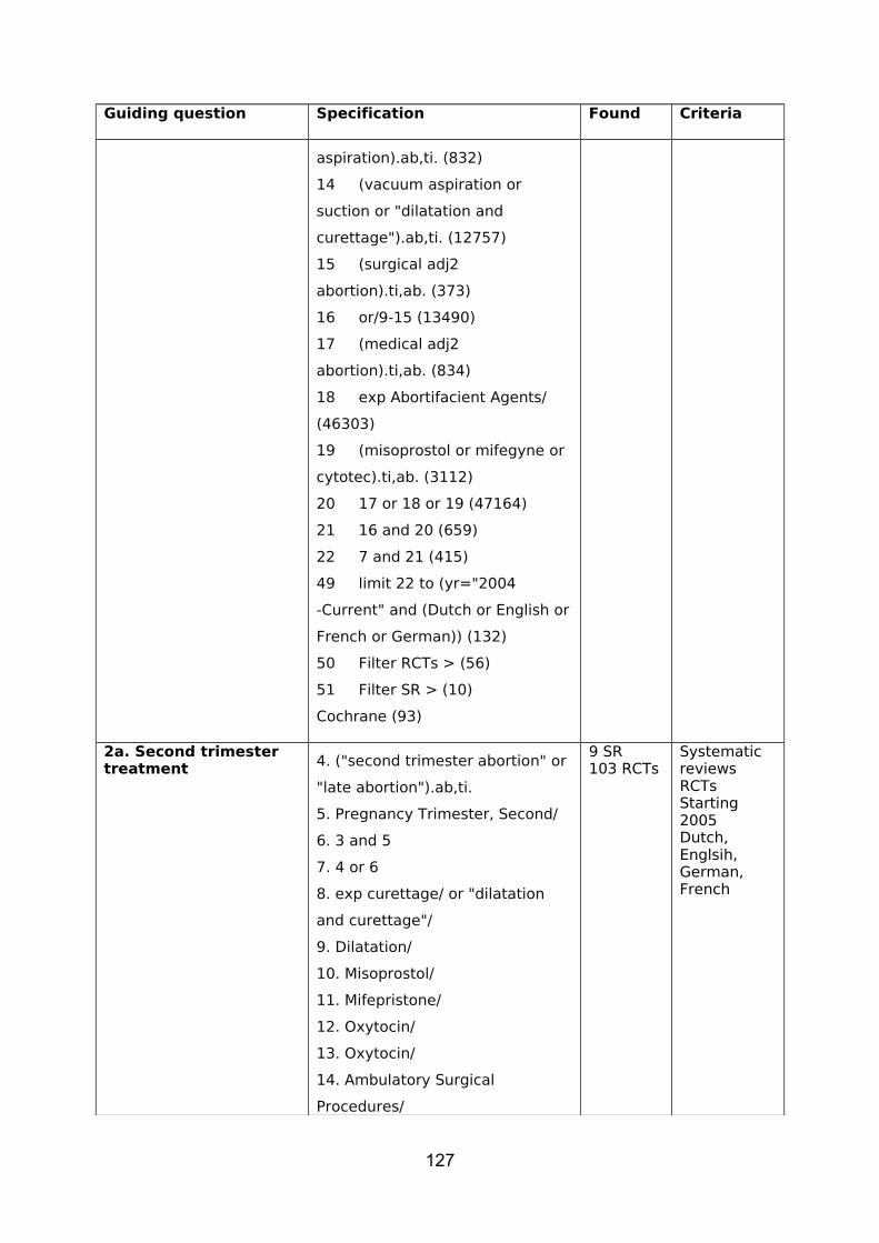
Guiding question Specification Found Criteria
aspiration).ab,ti. (832)
14 (vacuum aspiration or
suction or "dilatation and
curettage").ab,ti. (12757)
15 (surgical adj2
abortion).ti,ab. (373)
16 or/9-15 (13490)
17 (medical adj2
abortion).ti,ab. (834)
18 exp Abortifacient Agents/
(46303)
19 (misoprostol or mifegyne or
cytotec).ti,ab. (3112)
20 17 or 18 or 19 (47164)
21 16 and 20 (659)
22 7 and 21 (415)
49 limit 22 to (yr="2004
-Current" and (Dutch or English or
French or German)) (132)
50 Filter RCTs > (56)
51 Filter SR > (10)
Cochrane (93)
2a. Second trimester treatment 4. ("second trimester abortion" or
"late abortion").ab,ti.
5. Pregnancy Trimester, Second/
6. 3 and 5
7. 4 or 6
8. exp curettage/ or "dilatation
and curettage"/
9. Dilatation/
10. Misoprostol/
11. Mifepristone/
12. Oxytocin/
13. Oxytocin/
14. Ambulatory Surgical
Procedures/
9 SR103 RCTs
Systematic reviewsRCTsStarting 2005Dutch, Englsih, German, French
127

Guiding question Specification Found Criteria
15. (surgery or sugical).ab,ti.
16. misoprostol.ab,ti.
17. currettage.ab,ti.
18. curettage.ab,ti.
19. cervical dilatation.ab,ti.
20. cervical priming.ab,ti.
21. or/8-20
22. 7 and 2123. limit 22 to (yr="2005 -Current" and (dutch or english or french or german))
2b Complications of second trimester treatment
77 ((Early or late) adj complication*).ti,ab. (11097)78 complications.fs. (1348289)79 77 or 78 (1356966)81 adverse effects.fs. (1115542)82 79 or 81 (2333206)
3 SR21 RCT 44 various
2c Cervical priming1. exp Abortion, Induced/
2. ("pregnancy termination*" or
"induced abortion*").af.
3. 1 or 2
4. ("second trimester abortion" or
"late abortion").ab,ti.
5. Pregnancy Trimester, Second/
6. 3 and 5
7. 4 or 6
20. cervical priming.ab,ti.
25. vaginal touch.mp.
26. exp Palpation/
27. 7 and 26
28. digital vaginal
examination*.ab,ti.
29. vaginal examination*.ab,ti.
30. physical examination*.ab,ti. or
exp Physical Examination/
31. 25 or 29 or 30
32. 7 and 31
33. 20 and 32
34. 3 and 31
17 various
128
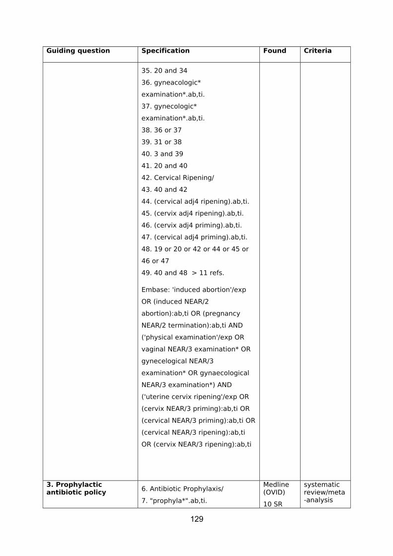
Guiding question Specification Found Criteria
35. 20 and 34
36. gyneacologic*
examination*.ab,ti.
37. gynecologic*
examination*.ab,ti.
38. 36 or 37
39. 31 or 38
40. 3 and 39
41. 20 and 40
42. Cervical Ripening/
43. 40 and 42
44. (cervical adj4 ripening).ab,ti.
45. (cervix adj4 ripening).ab,ti.
46. (cervix adj4 priming).ab,ti.
47. (cervical adj4 priming).ab,ti.
48. 19 or 20 or 42 or 44 or 45 or
46 or 47
49. 40 and 48 > 11 refs.
Embase: 'induced abortion'/exp
OR (induced NEAR/2
abortion):ab,ti OR (pregnancy
NEAR/2 termination):ab,ti AND
('physical examination'/exp OR
vaginal NEAR/3 examination* OR
gynecelogical NEAR/3
examination* OR gynaecological
NEAR/3 examination*) AND
('uterine cervix ripening'/exp OR
(cervix NEAR/3 priming):ab,ti OR
(cervical NEAR/3 priming):ab,ti OR
(cervical NEAR/3 ripening):ab,ti
OR (cervix NEAR/3 ripening):ab,ti
3. Prophylactic antibiotic policy 6. Antibiotic Prophylaxis/
7. "prophyla*".ab,ti.
Medline (OVID)
10 SR
systematic review/meta-analysis
129

Guiding question Specification Found Criteria
8. Anti-Bacterial Agents/ or
Azithromycin/ or
Azithromycin.ab,ti. or
Metronidazole/ or
Metronidazole.ab,ti.
7 or 8
9 and 6
9. exp Anti-Bacterial Agents/ or Azithromycin/ or Azithromycin.ab,ti. or Metronidazole/ or Metronidazole.ab,ti. or exp Bacterial Infections/pc [Prevention & Control] (500733)
28 RCT52 other trials
/RCTs/ other comparative studies
from 2000 to 2010English, Dutch, German, French
4. Ultrasound7. exp *Ultrasonography/
8. (ultrasound or sonograph* or
Ultrasound).ti.
9. 7 or 8
10. 5 and 9
11. abortion, therapeutic/ or
pregnancy reduction, multifetal/
12. 10 not 11
13. Ultrasonography, Prenatal/
14. 10 not 13
15. 12 or 14
16. limit 15 to (dutch or english)
17. Search filter systematic
reviews
45. 16 and 44 > 2 References
(Probably not relevant)
46. Search filter RCTS
69. 16 and 68 > 7 References
70. exp epidemiologic studies/
71. 16 and 70
72. limit 16 to (clinical trial, all or
clinical trial or comparative study
or consensus development
2 SR7 RCTs29 various trials
130
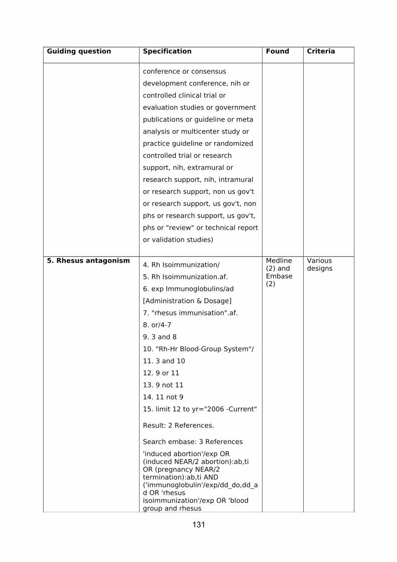
Guiding question Specification Found Criteria
conference or consensus
development conference, nih or
controlled clinical trial or
evaluation studies or government
publications or guideline or meta
analysis or multicenter study or
practice guideline or randomized
controlled trial or research
support, nih, extramural or
research support, nih, intramural
or research support, non us gov't
or research support, us gov't, non
phs or research support, us gov't,
phs or "review" or technical report
or validation studies)
5. Rhesus antagonism 4. Rh Isoimmunization/
5. Rh Isoimmunization.af.
6. exp Immunoglobulins/ad
[Administration & Dosage]
7. "rhesus immunisation".af.
8. or/4-7
9. 3 and 8
10. "Rh-Hr Blood-Group System"/
11. 3 and 10
12. 9 or 11
13. 9 not 11
14. 11 not 9
15. limit 12 to yr="2006 -Current"
Result: 2 References.
Search embase: 3 References
'induced abortion'/exp OR (induced NEAR/2 abortion):ab,ti OR (pregnancy NEAR/2 termination):ab,ti AND ('immunoglobulin'/exp/dd_do,dd_ad OR 'rhesus isoimmunization'/exp OR 'blood group and rhesus
Medline (2) and Embase (2)
Various designs
131
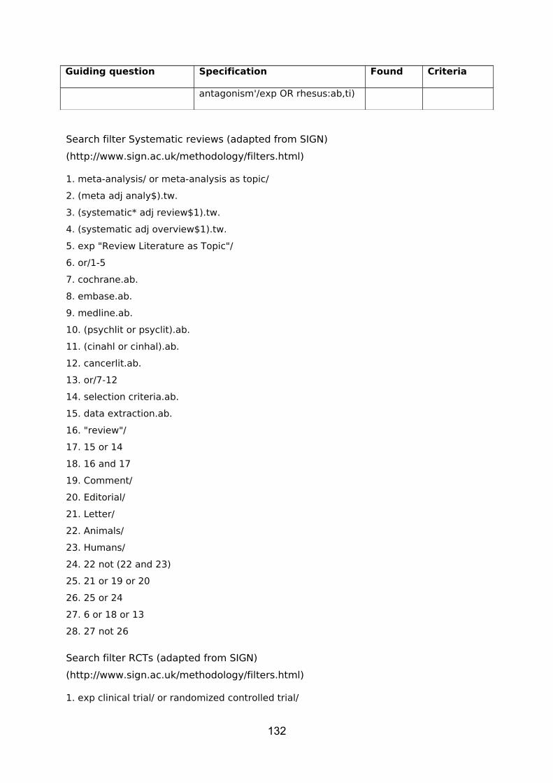
Guiding question Specification Found Criteria
antagonism'/exp OR rhesus:ab,ti)
Search filter Systematic reviews (adapted from SIGN)
(http://www.sign.ac.uk/methodology/filters.html)
1. meta-analysis/ or meta-analysis as topic/
2. (meta adj analy$).tw.
3. (systematic* adj review$1).tw.
4. (systematic adj overview$1).tw.
5. exp "Review Literature as Topic"/
6. or/1-5
7. cochrane.ab.
8. embase.ab.
9. medline.ab.
10. (psychlit or psyclit).ab.
11. (cinahl or cinhal).ab.
12. cancerlit.ab.
13. or/7-12
14. selection criteria.ab.
15. data extraction.ab.
16. "review"/
17. 15 or 14
18. 16 and 17
19. Comment/
20. Editorial/
21. Letter/
22. Animals/
23. Humans/
24. 22 not (22 and 23)
25. 21 or 19 or 20
26. 25 or 24
27. 6 or 18 or 13
28. 27 not 26
Search filter RCTs (adapted from SIGN)
(http://www.sign.ac.uk/methodology/filters.html)
1. exp clinical trial/ or randomized controlled trial/
132

2. exp clinical trials as topic/ or randomized controlled trials as topic/
3. Random Allocation/
4. Double-Blind Method/
5. Single-Blind Method/
6. (clinical trial, phase i or clinical trial, phase ii or clinical trial, phase iii or clinical trial,
phase iv or controlled clinical trial or randomized controlled trial or multicenter study or
clinical trial).pt.
7. or/1-6
8. clinic$ trial$1.tw.
9. (clinic$ adj trial$1).tw.
10. ((singl$ or doubl$ or treb$ or tripl$) adj (blind$3 or mask$3)).tw.
11. Placebos/
12. placebo$.tw.
13. randomly allocated.tw.
14. (allocated adj2 random$).tw.
15. or/8-14
16. 7 or 15
17. Letter/
18. Animals/
19. Humans/
20. 18 not (18 and 19)
21. 16 not 20
133

Appendix 2 Terms and definitions
Abortus provocatus
(Induced) termination of pregnancy, induced abortion
Abortion figure
The number of induced abortions per 1,000 women in the child bearing age (15-44
years).
Abortion ratio
The number of induced abortions per 1,000 live born children in a particular year.
Amenorrhoea length
The period of time starting from the first day of the last menstrual period. This time
indicator is often used to specify pregnancy length. The manner in which this is done
is described under ‘pregnancy length’.
Dutch national abortion register (Lar)
In the Dutch National Abortion Register (LAR)abortion clinics collect characteristics of
and data from abortion patients on a voluntary basis. 14 of the 17 abortion clinics
collaborate in the LAR; hospitals do not contribute to the LAR. Although no full
coverage is achieved for this reason, the LAR is a valuable additional source of data
to the inspectorate’s registration, as relations between individual data can be
obtained.
Early abortion
Early termination of pregnancy, up to 16 days’ absence of menstruation (amenorrhea
length up to 44 days). Early abortion is usually medical.
First trimester abortion
Termination of pregnancy up to 91 days of amenorrhoea.
Pregnancy length
The nominal length of pregnancy, calculated from the first day of the last
menstruation (in a regular four weeks’ cycle and assuming there are no
indications justifying a discussion of terms) or possibly calculated starting 14 days
preceding ovulation.
134

Second trimester abortion
Termination of pregnancy beyond 91 days of amenorrhoea uyp to the (legal) viability
line.
Spontaneous abortion
Non-induced termination of pregnancy, miscarriage
135

Appendix 3 Statement of interests
Statement about a possible conflict of interests and embargo with respect to the guideline ‘Counselling of women considering termination of pregnancy’, developed at the initiative of the Dutch Association of Abortion Specialists, with the support of the Department of Support in Professional Quality of the Dutch Order of Medical Specialists
24 August 2008
Re: Guideline ‘Teatment of women undergoing termination of pregnancy’
Dear Sir / Madam, In connection with your contribution to the development of the guideline ‘Treatment of women undergoing termination of pregnancy’ we kindly request you to fill out the statement on the annexed page (reproduced below).
In the domain of science it has been recognized over the past few years that conflicts of interests cannot always be avoided. The Dutch Order of Medical Specialists thus takes steps to assure full transparency in this matter.
You are therefore requested to declare on the annexed form if you (have) maintained a (financially supported) connection with commercial enterprises, organizations or institutions within the past five years, connected with the subject of the guideline ‘Counselling of women considering termination of pregnancy’.The details of your statement will be open to anyone requesting this information at the Department for Support in Professional Quality’s secretariat.
Embargo
The texts of the draft version(s) of this guideline will be under embargo during the full course of its development. This implies that it is not permitted to pass on passages from the draft guideline, or the full draft text, to other parties without the written consent of the commissioner of this guideline. This proviso also includes annexes and addenda, such as evidence-based tables.
The undersigned declares by his/her signature to agree to the above......................................................................................................................................(name)................................................................. .....................................................................(place, date) (signature)
136

Statement about conflicts of interests
Did you have interests over the past five years which may interfere with the study group’s decision making with respect to the interpretation of scientific evidence and the drafting of recommendations, or do you foresee such interests during the course of development of the guideline? Yes / No
If affirmative, can you describe from what activities these interests derive and which organizations or enterprises are concerned? Examples of activities may be found in consultations, advisory roles, (re)training, courses and the support of scientific research.1. ........................................................................................................................................................................2. ........................................................................................................................................................................3. ........................................................................................................................................................................
If you have more than three entries, please use separate page.
The undersigned declares to have truthfully submitted the above information and to be prepared to communicate all mutations of the above to the study group’s chairman and secretary.
Re: guideline ‘Treatment of women undergoing termination of pregnancy’Name: ................................................................................................................................................................Participant on behalf of: .................................................................................................................................................................................................... .....................................................................(place, date) (signature)
137

Appendix 4 Evidence tablesGuiding question Priming in 1st trimester suction curettage Reference Type of
studyCharacteristic elements
Intervention Control group Outcome indicatorsFollow up
Results Quality Evidence level
Oppegaard KS, 2005
RCT Women with intact pregnancy 7-12 weeks preferring suction curettageexcl: other pregnancy length, no Norwegian or English, allergic to misoprostoln=338 (calc 340)
Misoprostol vaginally morning intervention400 mcg
Misoprostol orally evening before intervention400 mcg
DilatationComplicationsacceptabilityContact after 60 days
Dilatation no differenceParity sole factorMore blood loss in oral group (OR 10.4)Vaginal administration acceptable (onl;y asked in this group)
no blindingbig samplereasonable
B
Saxena P, 2005
Prospective clinical trial
Women between 6-12 weeks of pregnancyn=100(118 asked)Many exclusion criteria
Misoprostol sublingually400mcg
Misoprostol vaginally400mcg
DilatationPainIntervention lengthBlood loss
Dilatation in sublingual group more simple (9 vs 7, p<0.0002) and shorter 3.51 minuten vs. 4.68 (p<0.0001)Side effects and complications no differenceSublingual more acceptable (also brcause taking at later time)
- no randomisation- no calculation of sample size- logistically illogical-only multiparaeThus: poor
B
138
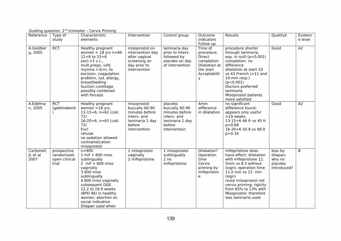
Guiding question: 2nd trimester – Cervix PrimingReference Type of
studyCharacteristic elements
Intervention Control group Outcome indicatorsFollow up
Results Qualityit Evidence level
A.Goldberg, 2005
RCT Healthy pregnant women > 18 yrs n=8412+6 to 15+6excl:>1 s.c., mult.pregn, iufd, myoma >3cm, lis excision, coagulation problem, iud, allergy, breastfeedingSuction curettage possibly combined with forceps
misoprostol on intervention day after vaginal screening on day prior to intervention
laminaria day prior to interv. followed by placebo on day of intervention
Time of procedure Direct completionDilatation at the startAcceptability
procedure shorter through laminaria, esp. in nulli (p<0.001)completion: no differencedilatation at start 33 vs 43 French (=11 and 14 mm resp.)(p<0.001)Doctors preferred laminaria Misoprostol patients more satisfied
Good A2
A.Edelman, 2005
RCT (geblindeerd)
Healthy pregnant women >18 yrs,13-15+6, n=62 (calc 72)16-20+6, n=63 (calc 72)Exclrefusalno sedation allowedcontraindication misoprostol
misoprostol buccally 60-90 minutes before interv. and laminaria 1 day before intervention
placebo buccally 60-90 minutes before interv. and laminaria 1 day before intervention
4mm difference in dilatation
no significant difference found; appears only useful >19 weeks13-15+6 46 fr vs 45 fr p=0.6816-20+6 50.9 vs 48.9 p=0.16
Good A2
Carbonell, JL et al 2007
prospective randomized open clinical trial
n=9001 mif + 600 miso sublingually2 mif + 600 miso vaginally3 600 miso sublingually4 600 miso vaginally subsequent D&E12.2 to 19.9 weeks (BPD 46) in healthy women; abortion on social indicationDilapan used when
1 misoprostol vaginally2 mifepristone
1 misoprostol sublingually2 no mifepristone
Dilatation?Operation timeCervix priming by mifepristone
mifepristone does have effect: dilatation with mifepristone 12. 5mm vs 8.5 without (sign); operation time 11.0 min vs 13 min (sign)route misoprostol notcervix priming: rigicity from 65% to 13% with Misoprostol: therefore less laminaria used
bias by dilapan; why no placebo introduced?
B
139

cervix unripe on day of treatment
Patel et al, 2006
retrospective descriptive
Different regimes in 19 different centresadequacy, (serious) adverse events, side effectsn= 221812 to 23 6/7 weeks
All women second trimester, surgical abortion
rates of (serious) adverse eventsadequatesafepatient satisfactiontreatment over in 1 session
adverse events overall 19.39/100serious adverse events overall 0.45 % (primarily blood loss>500ml)adequate 75 %patient not satisfied 2.3%one session 99.8%
Good C
140

Guiding question: 2nd trimester – D&E versus medicalReference Type of
studyCharacteristic elements Intervention Contr
olOutcome indicatorsFollow up
Results Quality
Evidence level
PA Lohr, 2009
systematic review
Comparing surgical and medical induced abortionRCTs induced abortion > 16 weeks
D & E medical
2 articles 1. D&E better than intra-amniotic Prosta F2alfa2. D&E seems better than mifepristone/ misoprostol, but n=18
Good A2
Grossman et al 2008
Pubmed review
key wordssecond trimester abortionmid-trimester abortiondilation and evacuationmifepristone and misoprotol(only English)RCTs, comp cohort, cohort studies and consecutive case studies > 400
safety surgical versus medical abortion
Uterus perforation or rupturebleeding requiring transfusionincomplete surgical evacuation neededcervix laesioninfectionother complication
1 RCT: more pain and more adverse events in the medical group1 retrospective cohortmedical generally later in pregnancy, but more adverse events in medical group especially incomplete abortions: OR adverse event surgical 0.15 case studies >400
Both methods show low complication rates, D&E is becoming safer
A
141
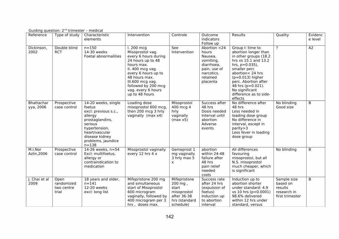
Guiding question: 2nd trimester – medicalReference Type of study Characteristic
elementsIntervention Controle Outcome
indicatorsFollow up
Results Quality Evidence level
Dickinson, 2002
Double blind RCT
n=15014-30 weeksFoetal abnormalities
I. 200 mcg Misoprostol vag. every 6 hours during 24 hours up to 48 hours max.II. 400 mcg vag. every 6 hours up to 48 hours max.III.600 mcg vag. followed by 200 mcg vag. every 6 hours up to 48 hours
See Intervention
Abortion <24 hoursNausea, vomiting, diarrhoea, pain, use of narcotics. retained placenta
Group I: time to abortion longer than in other groups (18.2 hrs vs 15.1 and 13.2 hrs, p=0.035), smaller perc abortion< 24 hrs (p=0.013) higher perc. Abortion after 48 hrs (p=0.021).No significant difference as to side-effects
? A2
Bhattacharyya, 2006
Prospective case control
14-20 weeks, single foetusexcl: previous s.c., allergy prostaglandins, serious hypertension, heart/vascular disease kidney problems, jaundicen=138
Loading dose misoprostol 600 mcg, then 200 mcg 3 hrly vaginally (max x4)
Misoprostol 400 mcg 4 hrlyvaginally (max x5)
Success after 48 hrsDosis neededInterval until abortionAdverse events
No difference after 48 hrsLess needed in loading dose groupNo difference in interval, escept in parity>3Less fever in loading dose group
No blindingGood size
B
M.I.Nor Azlin,2006
Prospective case control
14-26 weeks, n=54Excl: multifoetus, allergy or contraindication to medication
Misoprostol vaginally every 12 hrs 4 x
Gemeprost 1 mg vaginally 3 hrly max 5 x
abortion within 24-48failure after 48 hrspain relief neededcosts
All differences favouring misoprostol, but all N.S. misoprostol much cheaper, which is significant
No blinding B
J. Chai et al 2009
Open randomized two centre trial
18 years and older, n=14112-20 weeksexcl: long list
Mifepristone 200 mg and simultaneous start of Misoprostol 600 microgram vaginally, followed by 400 microgram per 3 hrs , doses max.
Mifepristone 200 mg , start misoprostol after 36-38 hrs (standard schedule)
Success rate after 24 hrs (expulsion of foetus)Induction up to abortion interval
Induction up to abortion shorter under standard: 4.9 vs 10 hrs (p<0.0001)98.6% delivered within 12 hrs under standard, versus
Sample size based on results research in first trimester
B
142

needed misoprotol schedule repeated after 24 hrs
Side effects 73.2% (p<0.0001)24 hrs 100 vs 91.5% p=0.028Side-effects: difference shivering and fever favours standard, otherwise not
Oi Shan Tang et al 2005
Double blind placebo controlled
18 yrs and older N=11812-20 weeks (gestation)excl: chronic medication; IUD, breast feeding, multifoetus, heavy smokers
200 mg mifepristone 36-48 hrs later followed by 400 microgram misprorstol sublingually and 2 tablets placebo per os every 3 hrs 5 doses max. in 24 hrs; possibly to be repeated after 24 hrs
Same except all placebo sublingually and misoprostol per os
Success rate after 24 hrsTime induction-abortionSide effects
Success rate after 24 hrs 91.4% vs 85%: thus relative risk 1.075.Induction to abortion time 5.5 hrs vs 7.5hrs p=0.0009; more fever in sublingual group p<0.0001
N=120 may measure 20% differenceConclusion that sublingual superior is not quite warranted
A?
B. Yilmaz et al 2007
Randomized prospective trial
n=81IUVD or congenital abnormality, healthy women 20-40 years, single foetus 14-24 weeks, no dilatation before and hemodynamically stable Excl: scarred uterus, blood loss, Hb<10 mg/dl, allergy and broken membranes
800 microgram Misoprostol mositened with physiological salt every 6 hrs, max. 3 times per 24 hrs; possibly repeated within 48 hrs
Again, but without physiological salt
Time induction-abortionSuccess rate after 24 hrsSuccess rate after 48 hrsDosage of MisoprostolRetained placentaHb-decreaseSide-effects
No statistically significant differences found
N=76 can measure a difference of 18.6%Vaginosis not excluded, possibly affecting vaginal PH
B
B. Yilmaz et al 2005
Prospective, dobble blind randomized controlled trial
n=66population as above
As above(B)
Again, but moistened with 3 ml acetic acid 5%(A)
Time induction till abortionSuccess within 48 hrsQuantity MisoprostolRetained placentaHb-decrease
Induction-abortionttime group A 8 hrs vs group B 14 hrs (p<0.001)Group A 96.7% within 24 hrs vs 78.1% in group B (p<0.001)Also less misoprostol group A (p<0.05) No
Sample size on basis of pilot, difference 30%Women with vaginitis not in study
B
143

Side-effects other statistically significant differences; all women delivered within 48 hrs
N. Bhattacharjee et al 2008
Randomized comparative study
n=300single foetus 13-20 wkscontra-indications isoprosto: allergy, anemia, irritable colon, cardiovasc diseases, bronchial asthma
400 microgram misoprostol sublingually at intervals of 3 hrs, max 5 doses in 24 hrs(A)
Again, vaginally(B)
Complete abortion rate at 24 and 48 hrsInduction to abortion (fetus)intervalSide effectsWomen’s preference
24 hrs: A 64.03%; B 61.59%; p=0.76748 hrs: A 79.14%; B 82.01; P=651Interval A 14.1; b 14.5 P=0.066 (NS)Side effect NSPreference for sublingual p<0.0001
Good sample size: looking for 20% difference no blinding
B
A. Karsadig et al 2009
Open randomized clinical study
n=4916-28 weeksincl: foetal abnormalitiesexcl: asthma, hemoragghic complaints, allergy,heart disease, scarred uterus, IUVD, hydrops
200 microgram misoprostol sublingually every 6 hrs max. 4 doses in 24 hrsWith Bishop 7 oxytocine i.v. (A)
Again vaginally (B)
Reduced blood supply through umbilical cordVaginal delivery within 24 hrsTime till abortion
No differences in flow Within 24 hrs: A:24, B: 13 p=0.0008Interval A 12.8h, B 22 h; p=0.0001
Sample size looking for 25% difference saturation umbilical cordSlightly cryptic
?
N. Jansen et al 2008
Prospective randomized study
n=16single f. 16-24 weeks,cong res. or genetic res.excl: allergy to medication and scarred uterus
200 milligram mifepristone, after 48 hrs 200 microgram misoprostol vaginally every 3 hrs till expulsion(A): if no delivery after 3 days on to standard)
Dilapan, followed next day by sulproston i.v.(B, standard) after 3 days interval, then repeat
Induction till expulsion time (starting misoprostol vs sulprostone
A 17.8 hrs; B 45.1 hrs NS p=0.3
50% reduction of interval for sample size calc.
Von Hertzen et al 2009
Multiclinic7 countriesRandomized and placebo controlled
n=68013-20 weekshealthy women, single foe. Hb higher than 100mg/dlexcl. various diseases
400 misoprostol sublingually every three hrs up to 5 doses per 24 hrsRepeated after 24 hrs
Again, vag.Repeated after 24 hrs sublingually
Success within 24 hrs complete and incompleteAgain after 48 hrsTime till
After 24 hrs success higher in vaginal groupAfter 48 hrs equalMore fever vaginallyVaginally more success in nulliparae,
Sample size based on less than10 % difference
B
144
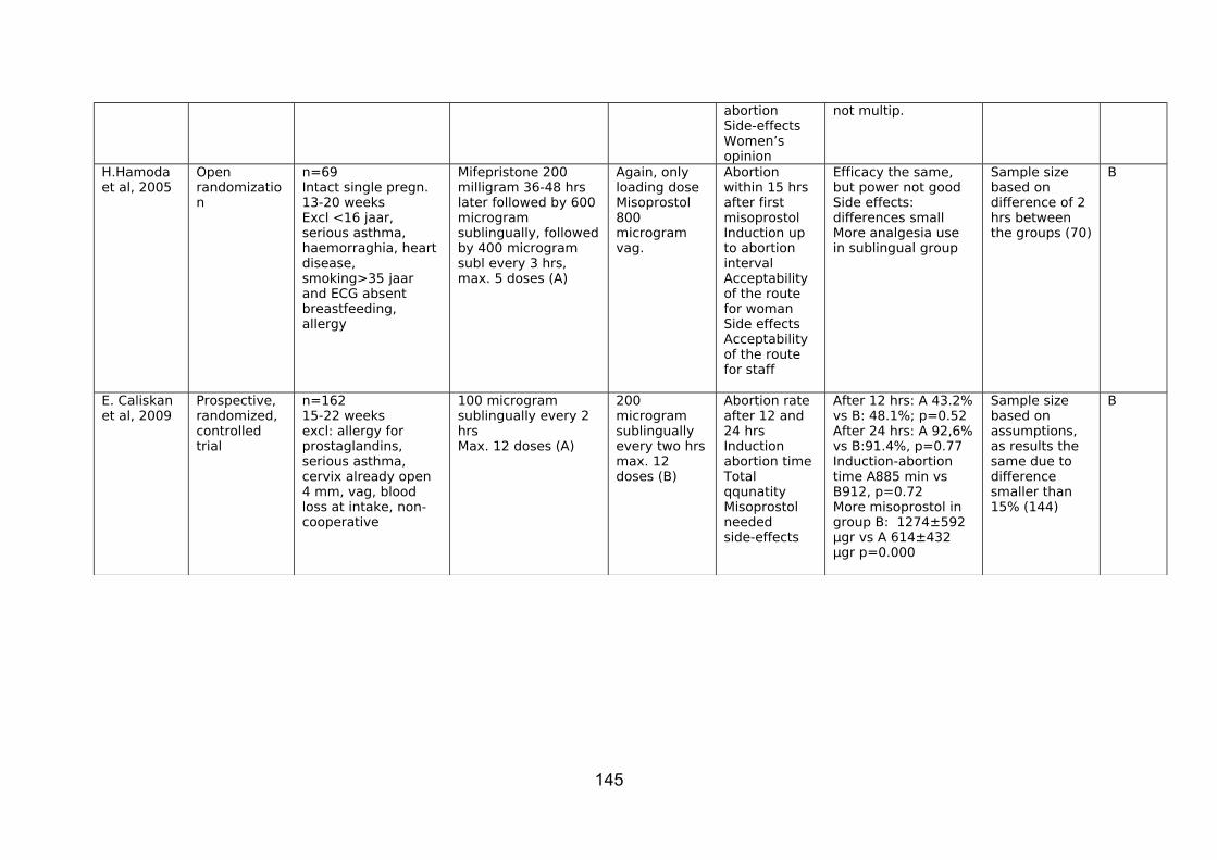
abortionSide-effectsWomen’s opinion
not multip.
H.Hamoda et al, 2005
Open randomization
n=69Intact single pregn. 13-20 weeksExcl <16 jaar, serious asthma, haemorraghia, heart disease, smoking>35 jaar and ECG absent breastfeeding, allergy
Mifepristone 200 milligram 36-48 hrs later followed by 600 microgram sublingually, followed by 400 microgram subl every 3 hrs, max. 5 doses (A)
Again, only loading dose Misoprostol 800 microgram vag.
Abortion within 15 hrs after first misoprostolInduction up to abortion intervalAcceptability of the route for womanSide effectsAcceptability of the route for staff
Efficacy the same, but power not goodSide effects: differences smallMore analgesia use in sublingual group
Sample size based on difference of 2 hrs between the groups (70)
B
E. Caliskan et al, 2009
Prospective, randomized, controlled trial
n=16215-22 weeksexcl: allergy for prostaglandins, serious asthma, cervix already open 4 mm, vag, blood loss at intake, non-cooperative
100 microgram sublingually every 2 hrsMax. 12 doses (A)
200 microgram sublingually every two hrs max. 12 doses (B)
Abortion rate after 12 and 24 hrsInduction abortion timeTotal qqunatity Misoprostol neededside-effects
After 12 hrs: A 43.2% vs B: 48.1%; p=0.52After 24 hrs: A 92,6% vs B:91.4%, p=0.77Induction-abortion time A885 min vs B912, p=0.72More misoprostol in group B: 1274±592 µgr vs A 614±432 µgr p=0.000
Sample size based on assumptions, as results the same due to difference smaller than 15% (144)
B
145
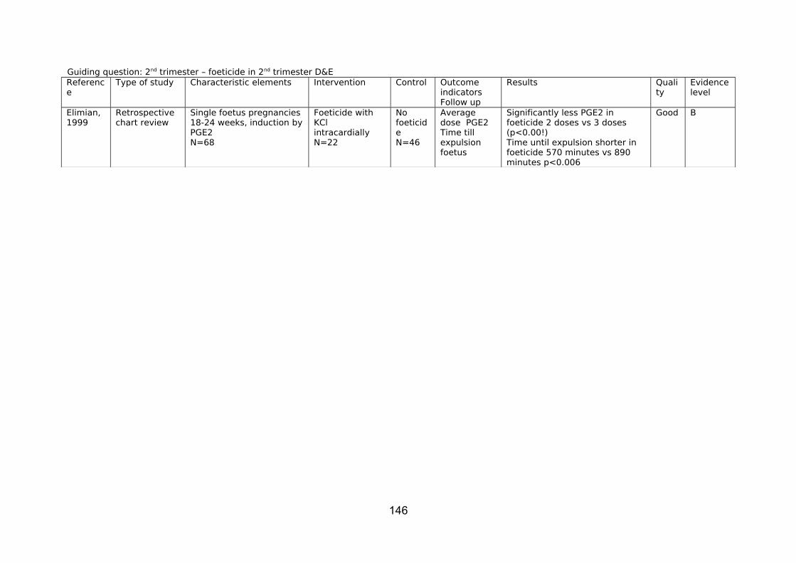
Guiding question: 2nd trimester – foeticide in 2nd trimester D&EReference
Type of study Characteristic elements Intervention Control Outcome indicatorsFollow up
Results Quality
Evidence level
Elimian, 1999
Retrospective chart review
Single foetus pregnancies 18-24 weeks, induction by PGE2N=68
Foeticide with KCl intracardiallyN=22
No foeticideN=46
Average dose PGE2Time till expulsion foetus
Significantly less PGE2 in foeticide 2 doses vs 3 doses (p<0.00!)Time until expulsion shorter in foeticide 570 minutes vs 890 minutes p<0.006
Good B
146

Guiding question: 2nd trimester – scarred uterus Reference Type Characteristic
elementsIntervention Control Outcome
indicatorsFollow up dhrs
Results Quality Evidence level
V. Berghella2009
systematic review
retrospective chartanalysissearch medline, scopus, popline(medical abortion)
complications use of misoprostol in TOP (16-28 weeks)in women with sectio caesarea in anamnesis1 s.c. n=4611 of 2 n=507
None Uterus rupturefrom 1998
1 s.c.:0.4% (CI.008-1.67) uterus rupture
good (in vieew of the possibilities)
C/D
Ido Ben-Ami, 2009
retrospective cohort
TOP in 2nd trimesterSingle pregn.17-24 weeksexcl: multiplesadherent placentano, one or more s.c.(D&E)
sectio in anamnesis1 s.c. 592 or more 36
no s.c.545
Uterus rupture
0 Numbers too small
B
Goyal V., 2009
systematic review
Risk of uterus rupture after use of misoprostol in women with s.c. in anamnesis(medical abortion)16 articles
sectio in anamnesis n=722
no s.c.n=3,556
- uterus rupture- number needed to harm
s.c. + 0,28s.c. – 0,04number needed to harm: 414
Good B?
Dickinson Jan E. 2005
retrospectivecohort
TOP for foetal abnormality 14- 28 weeks, 2 equal time cohort, wit hor without s.c. in anamnesismedical TOP with misoprostol (different regimes)
n=720 of which 101 with a s.c. in anamnesis
101 vs 619 quantity misoprostol used,demographics, time until delivery, rupture or hysterectomy
No difference quantity of misoprostol; in s.c. group older and higher parity and gravidity. Pregn. up to 9 wks, time until delivery not different, no ruptures or hysterectomy in both groups
limited C
Tarim E. et al, 2004
case reports TOP for foetale indications, all
n=29 h/o vaginal delivery
Delieverd before and
Differences with respect
Except age and number of
limited C/D?
147
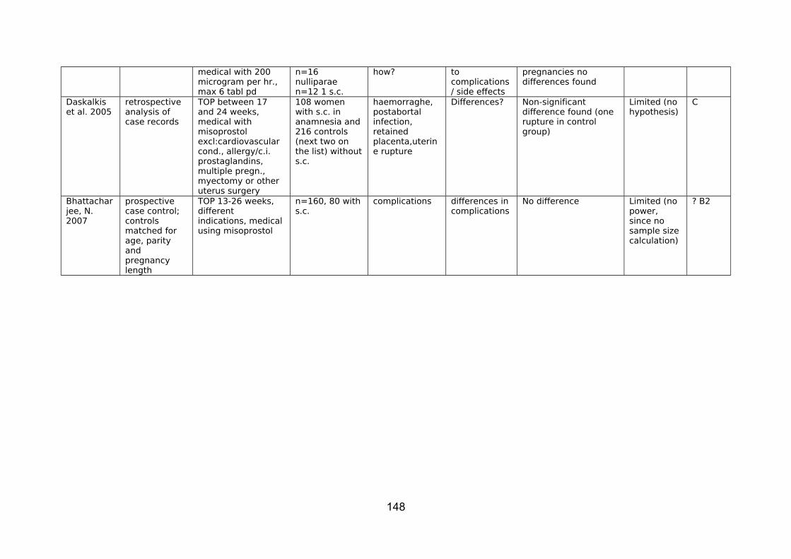
medical with 200 microgram per hr., max 6 tabl pd
n=16 nulliparaen=12 1 s.c.
how? to complications / side effects
pregnancies no differences found
Daskalkis et al. 2005
retrospective analysis of case records
TOP between 17 and 24 weeks, medical with misoprostolexcl:cardiovascular cond., allergy/c.i. prostaglandins, multiple pregn., myectomy or other uterus surgery
108 women with s.c. in anamnesia and 216 controls (next two on the list) without s.c.
haemorraghe, postabortal infection, retained placenta,uterine rupture
Differences? Non-significant difference found (one rupture in control group)
Limited (no hypothesis)
C
Bhattacharjee, N. 2007
prospective case control; controls matched for age, parity and pregnancy length
TOP 13-26 weeks, different indications, medical using misoprostol
n=160, 80 with s.c.
complications differences in complications
No difference Limited (no power, since no sample size calculation)
? B2
148

Guiding question: 2nd trimester – late complicationsReference
Type of study Characteristic elements Intervention Control Outcome indicatorsFollow up
Results Quality Evidence level
Jackson JE et al, 2006
retrospective chart analysis with simultaneous cohort
Wanted pregnancies; first pregnancy after D&E, no multiples or foetal abnormalitiesn=852 next pregnancies timewise of same age (2yrs) without D&E in anamnesisn=170
Past D&E 12-24 weeks
none D&E 12-24 weeks gehad
pregnancy outcome, viz. prematurity, placenta abnormality, multiple complications
Lower birthweight but NSGA at delivery lower (p=0.001)more preterm delivery(<37 weeks) but NS<34 weeks 0-1cerclage 2-1abnormal placenta 4-4 c.s fewer but NShaemorraghe 2-4uterine ruptue 0-1overall 10-12 (NS)
good? C
Winer et al, 2009
Prospective case control
Admission for late spontaneous abortion, premature delivery or prematurely ruptured membranes all before 37 weeks n=245 (1)2 next deliveries at over 37 weeks (n=490) (2)exc: multiple pregnancies, other treatment course to effect induced abortion
induced abortion in anamnesis?
Same odds ratio mere admissions in group 1 than group 2, but after correction for age number of pregnancies OR 1.33 (CI 0.81-2.17)
good B
Turok et al, 2008
Retrospective cohort
major complications1. D&E in academic
hospital n=832. medical abortion in
academic hospitals n=89
3. D&C in private out patient clinic n= 253
Price also considered
abortion 13-24 weeksperiod of 5 years in hospital1 year in private clinic
hypothesis: more complications in hospitals a long list
odds ratio clinic much fewer complications(and cheaper)Apples and oranges in a way
limited C
149

Indications hospital: iuvd, foetal abnormalities or maternal illness indicatiions clinic: 98% social
Nucatola et al 2008
retrospective chart analysis
6,620 elective surgical abortions 12-16 weeks with misoprostol priming (68 months)
complications?
complications 3 perforated uterus (0.45 per 1,000 abortion)1 haemorraghe due to placenta accreta =complication of the pregnancy)Not different from literature
C
Lohr, 2008
Literature review (descriptive)
Modern methods D & E in second trimester
n.a. n.a. SafetyPros and consAcceptabilityComplications
D&E is safe, efficient and cost-effective as a method to effect second trimester termination of pregnancy for women who have access
Non-systematic, expert opinion
D
150
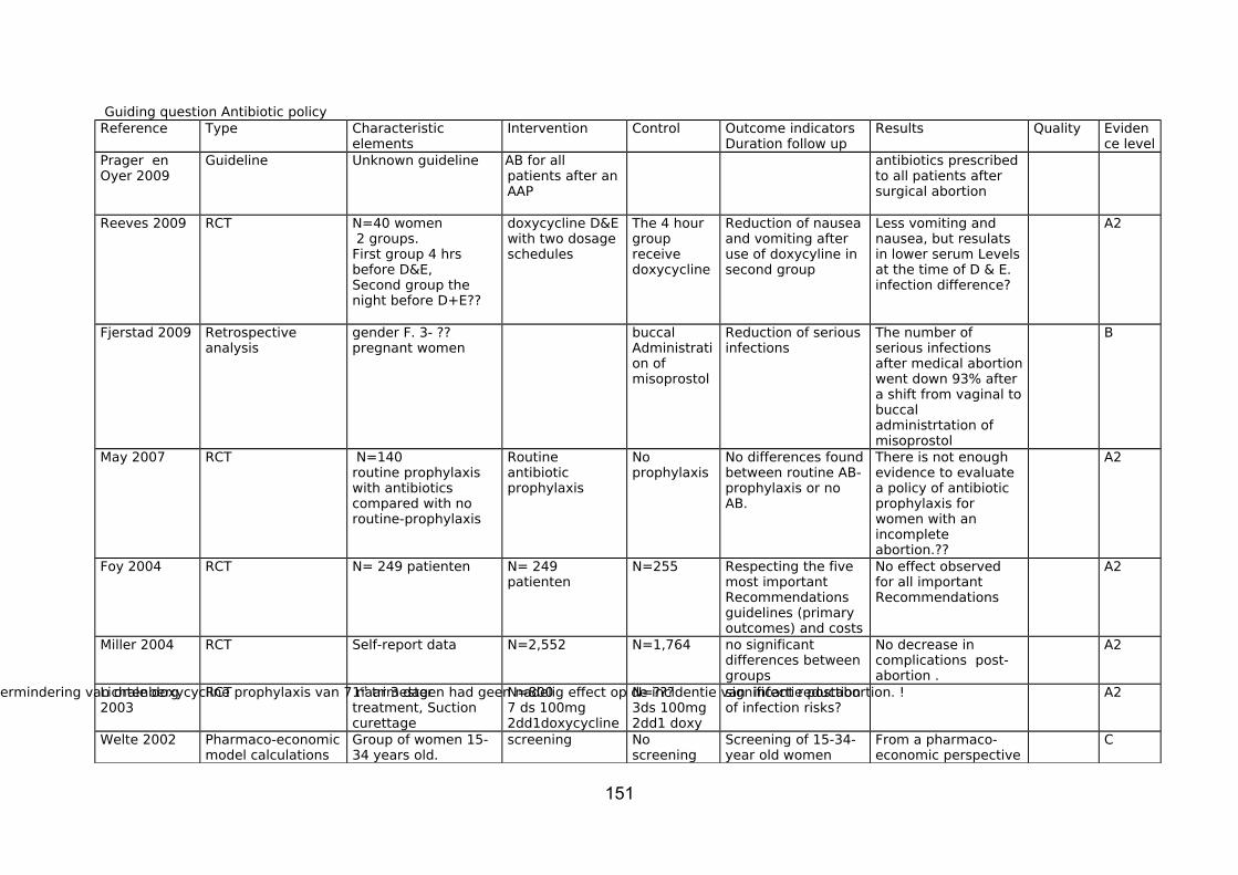
Guiding question Antibiotic policyReference Type Characteristic
elementsIntervention Control Outcome indicators
Duration follow upResults Quality Eviden
ce levelPrager en Oyer 2009
Guideline Unknown guideline AB for all patients after an AAP
antibiotics prescribed to all patients after surgical abortion
Reeves 2009 RCT N=40 women 2 groups.First group 4 hrs before D&E,Second group the night before D+E??
doxycycline D&E with two dosage schedules
The 4 hour group receive doxycycline
Reduction of nausea and vomiting after use of doxycyline in second group
Less vomiting and nausea, but resulats in lower serum Levels at the time of D & E. infection difference?
A2
Fjerstad 2009 Retrospective analysis
gender F. 3- ??pregnant women
buccal Administration of misoprostol
Reduction of serious infections
The number of serious infections after medical abortion went down 93% after a shift from vaginal to buccal administrtation of misoprostol
B
May 2007 RCT N=140 routine prophylaxis with antibiotics compared with no routine-prophylaxis
Routine antibiotic prophylaxis
No prophylaxis
No differences found between routine AB-prophylaxis or no AB.
There is not enough evidence to evaluate a policy of antibiotic prophylaxis for women with an incomplete abortion.??
A2
Foy 2004 RCT N= 249 patienten N= 249 patienten
N=255 Respecting the five most important Recommendations guidelines (primary outcomes) and costs
No effect observed for all important Recommendations
A2
Miller 2004 RCT Self-report data N=2,552 N=1,764 no significant differences between groups
No decrease in complications post-abortion .
A2
Lichtenberg 2003
RCT 1st trimester treatment, Suction curettage
N=800 7 ds 100mg 2dd1doxycycline
N=???3ds 100mg 2dd1 doxy
significant reduction of infection risks?
vermindering van orale doxycycline prophylaxis van 7 naar 3 dagen had geen nadelig effect op de incidentie van infectie postabortion. ! A2
Welte 2002 Pharmaco-economic model calculations
Group of women 15-34 years old.
screening No screening
Screening of 15-34-year old women
From a pharmaco-economic perspective
C
151

would save costs, compared to no screening
a CT screening programme for sexually active women in Amsterdam is advisable.
Crowley 2001 RCT 1st trimester treatment abortion,group1
group 2
N=273 N= There was no difference in risk of transfer to hospital and the frequencies of self-reported symptoms was not different across both groups
Weak indications that metronidazol reduces the risk of infection of upper bronchia and genitals after first trimester AAP??
A2
Grimes 2001 RCT 4 studies (women from Kenia, Nigeria, US and Turkey)Effectiveness of prophylactic antibiotics Administration before implantation of IUD
IUD is safe to use, with or without the use of prophylactic antibiotics.prevalence of infection no difference ?
A2
Larsson 2000 RCT clindamycine 2% cream or placebo
N=1,102 N=placebo
Treatment with clindamycine cream in women with a normal lactobacillus flora showed no difference compared to the non-treated group.
No difference A2
152
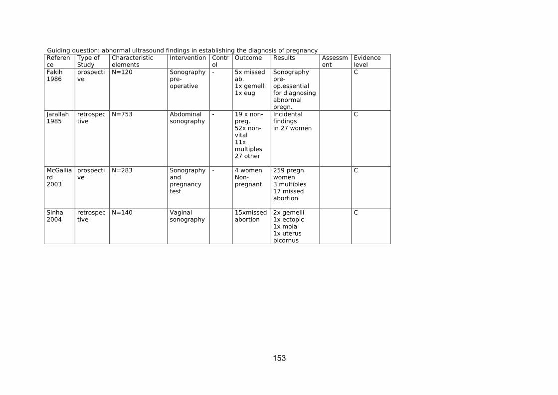
Guiding question: abnormal ultrasound findings in establishing the diagnosis of pregnancyReference
Type ofStudy
Characteristic elements
Intervention Control
Outcome Results Assessment
Evidence level
Fakih1986
prospective
N=120 Sonographypre-operative
- 5x missed ab.1x gemelli1x eug
Sonographypre-op.essentialfor diagnosingabnormal pregn.
C
Jarallah1985
retrospective
N=753 Abdominalsonography
- 19 x non-preg.52x non-vital11x multiples27 other
Incidental findings in 27 women
C
McGalliard2003
prospective
N=283 Sonography and pregnancy test
- 4 womenNon-pregnant
259 pregn. women3 multiples17 missed abortion
C
Sinha2004
retrospective
N=140 Vaginalsonography
15xmissedabortion
2x gemelli1x ectopic1x mola1x uterus bicornus
C
153

Guiding question pregnancy lenth: comparing ultrasound with LMP/VT findingsReference
Type ofStudy
Characteristic elements
Intervention Control
Outcome Results Assessment
Evidence level
Blanchard2007
prospective
N=6738 weeks AM
Comparing reportedLMPWith ultrasound
Estimate of LMP19 days below ultrasound
Medical.abortion pssible without ultrasound
C
Fakih1986
prospective
N=1201st trimester
Comparing reportedLMP and VTWith ultrasound
Between ultrasound andVt, differences:89% <1 week13% 1-2 weeks6 % >2 weeks
No positive correlationBetween ultrasound andLMP; good correlation between ultrasound and LMP plus VT
C
Fielding2002
prospectivemulticenter
N=1,016Pregnancy<63 days’ AM
Medicalabortion
Adequate overallparallel betweenLMP and VT versus uktrasound
VT:87% correct 9 % incorrect
B
Goldstein1988
prospective
N=2501st trimester
Surgicalabortion
4 women with LMP<12 weeksAppeared longer pregnant after Ultrasound
C
Jarallah1985
retrospective
N=753 Abdominal ultrasoundbefore abortion
73% knew LMP 62% equal to ultrasound, 22% Further and 16% less far
C
McGalliard2003
prospective
N=283 1st trimester surgical2nd trimester medical
In 90 out of 262 womenNo difference betweenLMP and ultrasound > 1week
C
Sinha2004
retrospective
N=140 Medical abortion<63 days
Discrepancy between LMP andd sonography in19.3%
C
154
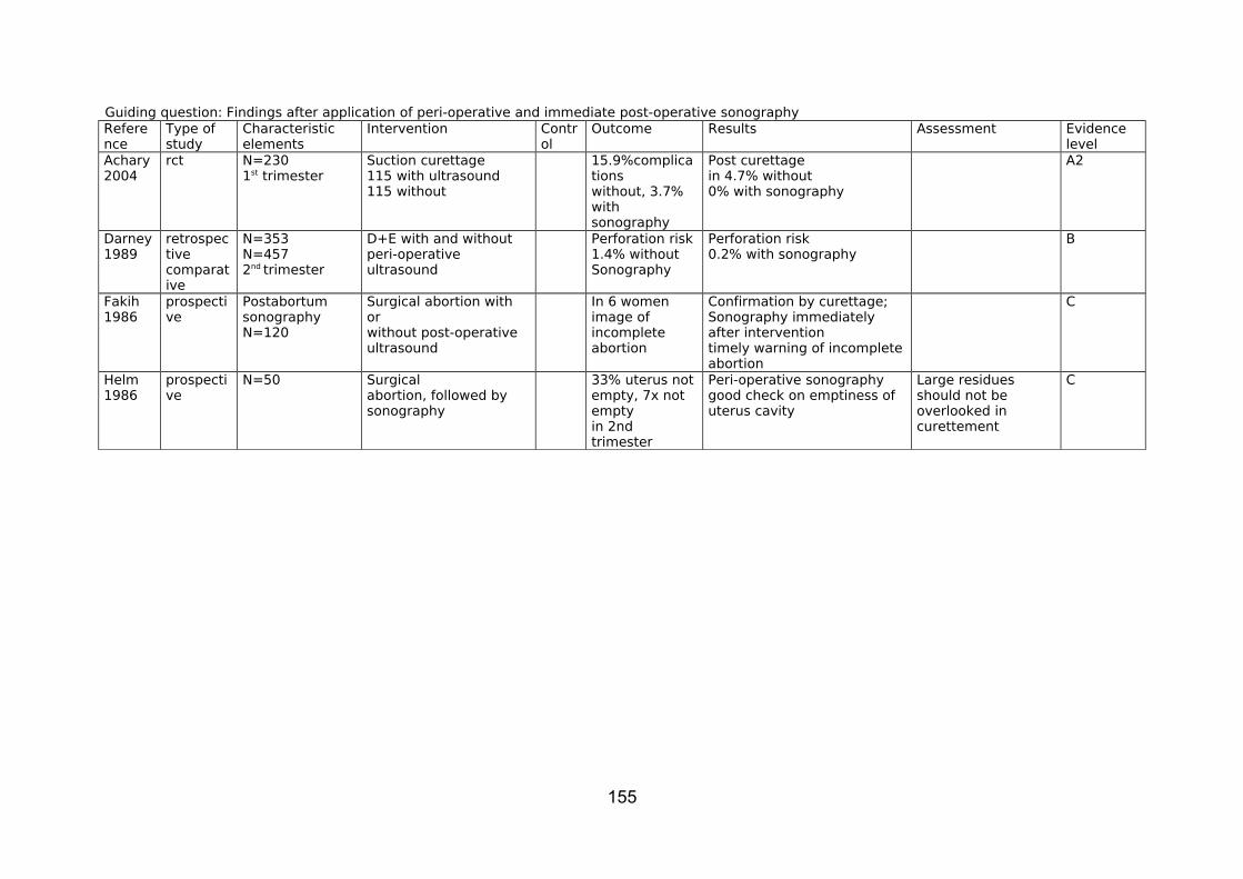
Guiding question: Findings after application of peri-operative and immediate post-operative sonographyReference
Type ofstudy
Characteristic elements
Intervention Control
Outcome Results Assessment Evidence level
Achary2004
rct N=2301st trimester
Suction curettage115 with ultrasound115 without
15.9%complicationswithout, 3.7%with sonography
Post curettagein 4.7% without0% with sonography
A2
Darney1989
retrospectivecomparative
N=353N=4572nd trimester
D+E with and without peri-operative ultrasound
Perforation risk1.4% without Sonography
Perforation risk0.2% with sonography
B
Fakih1986
prospective
PostabortumsonographyN=120
Surgical abortion with or without post-operative ultrasound
In 6 womenimage of incompleteabortion
Confirmation by curettage;Sonography immediately after interventiontimely warning of incomplete abortion
C
Helm1986
prospective
N=50 Surgicalabortion, followed bysonography
33% uterus not empty, 7x not empty in 2nd trimester
Peri-operative sonographygood check on emptiness of uterus cavity
Large residues should not be overlooked in curettement
C
155
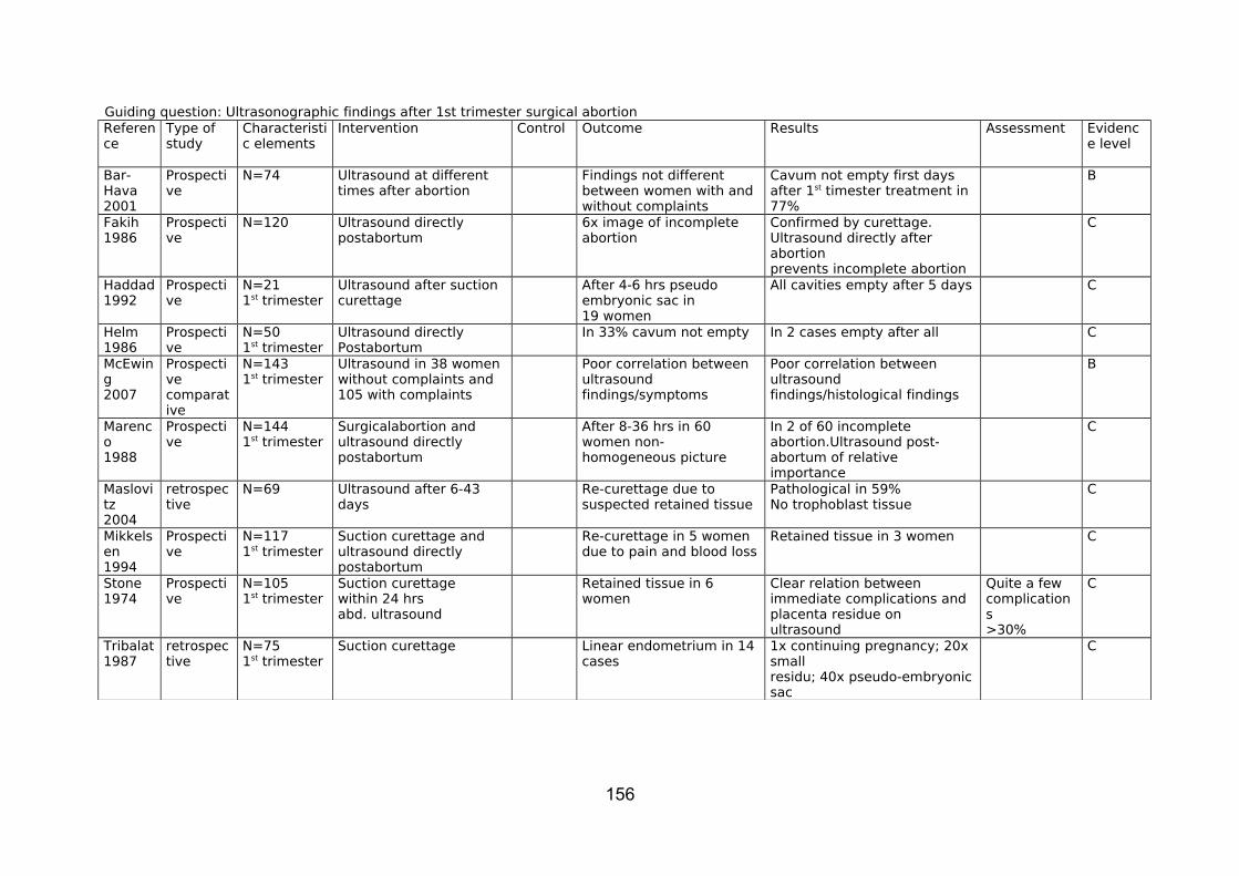
Guiding question: Ultrasonographic findings after 1st trimester surgical abortionReference
Type ofstudy
Characteristic elements
Intervention Control Outcome Results Assessment Evidence level
Bar-Hava2001
Prospective
N=74 Ultrasound at different times after abortion
Findings not different between women with and without complaints
Cavum not empty first days after 1st timester treatment in 77%
B
Fakih1986
Prospective
N=120 Ultrasound directlypostabortum
6x image of incomplete abortion
Confirmed by curettage.Ultrasound directly after abortionprevents incomplete abortion
C
Haddad1992
Prospective
N=211st trimester
Ultrasound after suction curettage
After 4-6 hrs pseudo embryonic sac in19 women
All cavities empty after 5 days C
Helm1986
Prospective
N=501st trimester
Ultrasound directlyPostabortum
In 33% cavum not empty In 2 cases empty after all C
McEwing2007
Prospectivecomparative
N=1431st trimester
Ultrasound in 38 women without complaints and 105 with complaints
Poor correlation between ultrasound findings/symptoms
Poor correlation between ultrasound findings/histological findings
B
Marenco1988
Prospective
N=1441st trimester
Surgicalabortion and ultrasound directly postabortum
After 8-36 hrs in 60 women non- homogeneous picture
In 2 of 60 incomplete abortion.Ultrasound post-abortum of relative importance
C
Maslovitz2004
retrospective
N=69 Ultrasound after 6-43 days
Re-curettage due to suspected retained tissue
Pathological in 59%No trophoblast tissue
C
Mikkelsen1994
Prospective
N=1171st trimester
Suction curettage and ultrasound directly postabortum
Re-curettage in 5 women due to pain and blood loss
Retained tissue in 3 women C
Stone1974
Prospective
N=1051st trimester
Suction curettagewithin 24 hrsabd. ultrasound
Retained tissue in 6 women
Clear relation between immediate complications and placenta residue on ultrasound
Quite a few complications>30%
C
Tribalat1987
retrospective
N=751st trimester
Suction curettage Linear endometrium in 14 cases
1x continuing pregnancy; 20x smallresidu; 40x pseudo-embryonic sac
C
156
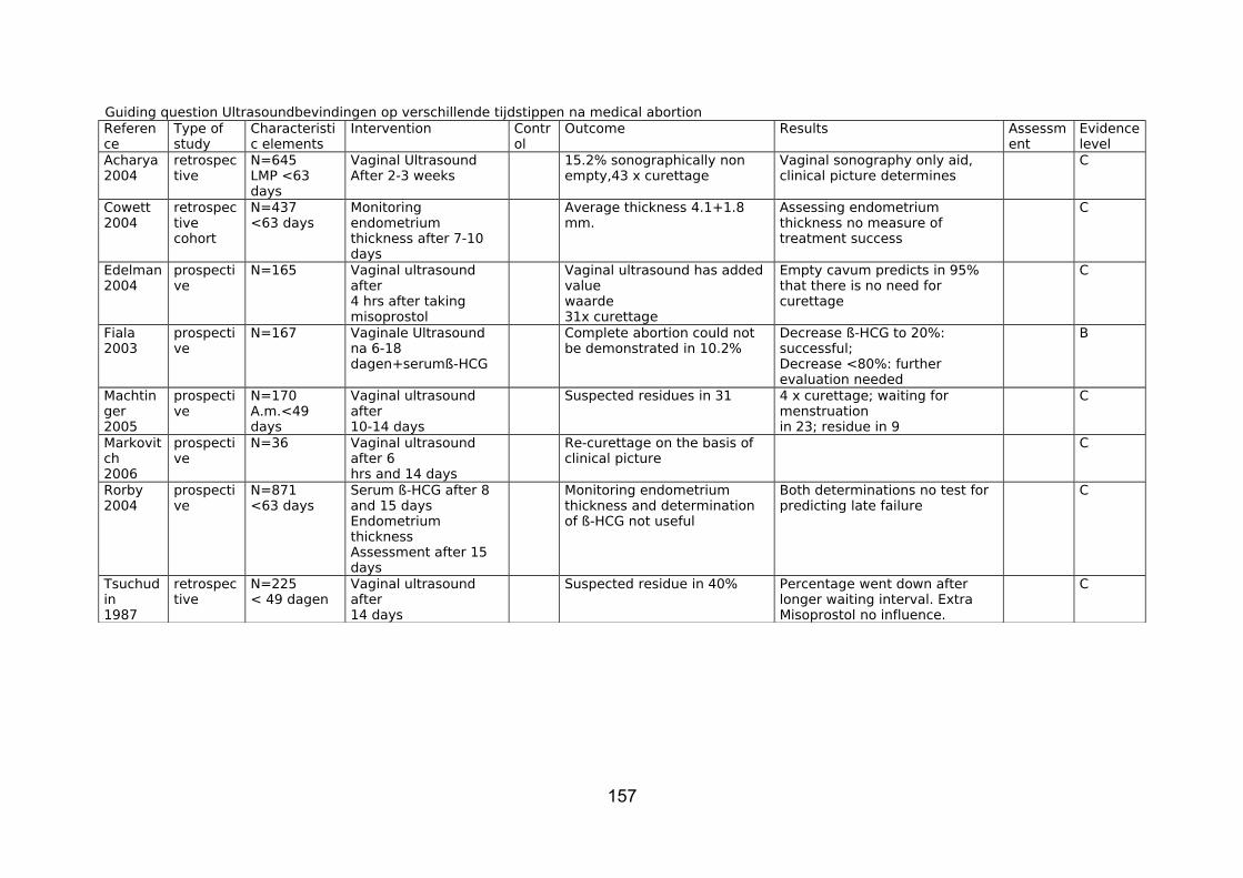
Guiding question Ultrasoundbevindingen op verschillende tijdstippen na medical abortionReference
Type ofstudy
Characteristic elements
Intervention Control
Outcome Results Assessment
Evidence level
Acharya2004
retrospective
N=645LMP <63 days
Vaginal UltrasoundAfter 2-3 weeks
15.2% sonographically non empty,43 x curettage
Vaginal sonography only aid, clinical picture determines
C
Cowett2004
retrospectivecohort
N=437<63 days
Monitoring endometrium thickness after 7-10 days
Average thickness 4.1+1.8 mm.
Assessing endometrium thickness no measure of treatment success
C
Edelman2004
prospective
N=165 Vaginal ultrasound after4 hrs after takingmisoprostol
Vaginal ultrasound has added valuewaarde31x curettage
Empty cavum predicts in 95% that there is no need for curettage
C
Fiala2003
prospective
N=167 Vaginale Ultrasound na 6-18 dagen+serumß-HCG
Complete abortion could not be demonstrated in 10.2%
Decrease ß-HCG to 20%: successful;Decrease <80%: further evaluation needed
B
Machtinger2005
prospective
N=170A.m.<49 days
Vaginal ultrasound after10-14 days
Suspected residues in 31 4 x curettage; waiting for menstruationin 23; residue in 9
C
Markovitch2006
prospective
N=36 Vaginal ultrasound after 6hrs and 14 days
Re-curettage on the basis of clinical picture
C
Rorby2004
prospective
N=871<63 days
Serum ß-HCG after 8and 15 daysEndometrium thicknessAssessment after 15 days
Monitoring endometrium thickness and determination of ß-HCG not useful
Both determinations no test for predicting late failure
C
Tsuchudin1987
retrospective
N=225< 49 dagen
Vaginal ultrasound after14 days
Suspected residue in 40% Percentage went down after longer waiting interval. Extra Misoprostol no influence.
C
157

Guiding question: Rhesus factor, risk of foetomaternal transfusion related to abortion expressed as percentages positive KB testsAuthor Study Number
of women
Pos. KB test pre-op. (> 0.1 ml)
Method Pos. KB test post-op when negKB test preop
Conclusion Remarks Evidence level
Jorgensen1969
Case seriesN=227
4785
1580
00
00
SpontaneousSuction curettageCurettageOtherwise
0 (0%)1 (1.2%)
1 (6.7%)7 (8.6%)
No FMT after spontaneous abortion; after suction curettage in 1.2% and after other methods 8.6%
B
Leong c.s.1979
Case seriesN=75
75 2 Suction curettage
10 (13.7%)
No FMT before 6 weeks a.m.
B
Murray c.s.1970
Case seriesN=483
91243
7376
2139
117
SpontaneousSuction curettageSalineOtherwise
1 (1.4%)0 (0%)
1 (1,6%)0 (0%)
No difference in frequency of FMT between spontaneous and induced abortion
Suction curettage up to 14 weeks’ amenorrhoea
B
Queenan c.s.1971
Case seriesN=606
404
202
0
0
Suction curettageSaline
7.2%
20.2%
Frequency FMT rises with pregnancy length
Suction curettage up to 16 weeks’ amenorrhoea
B
Borst-Eilers1972
Case seriesN=72
1250
82
Not determined000
Suction curettageCurettageSectio parva
Group undefined; Total of 2 (3.3%)
In 2 cases FMT more than 0.1 ml but less than than 0.16 ml
158
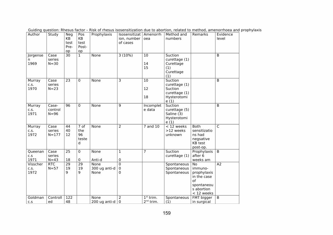
Guiding question: Rhesus factor – Risk of rhesus isosensitization due to abortion, related to method, amenorrhoea and prophylaxisAuthor Study Neg
KB testPre-op
PosKB testPost-op
Prophylaxis Isosensitization, number of cases
Amenorrhoea
Method and numbers
Remarks Evidence level
Jorgensen1969
Case seriesN=30
30 1 None 3 (10%) 10
1415
Suction curettage (1)Curettage (1)Curettage (1)
B
Murray c.s.1970
Case seriesN=23
23 0 None 3 10
12
18
Suction curettage (1)Suction curettage (1)Hysterotomie (1)
B
Murray c.s.1971
Case-controlN=96
96 0 None 9 Incomplete data
Suction curettage (5)Saline (3)Hysterotomie (1)
B
Murray c.s.1972
Case seriesN=177
444012
7 of the 96 tested
None 2 7 and 10 < 12 weeks>12 weeksunknown
Both sensitizations had negsative KB test post-op.
C
Queenan c.s1971
Case seriesN=43
25
18
0
0
None
Anti-d
1
0
7 Suction curettage (1)
Prophylaxis after 6 weeks am
B
Visscher c.s.1972
RTCN=57
29199
29199
None300 ug anti-dNone
000
SpontaneousSpontaneousSpontaneous
No immuno-prophylaxis in the case of spontaneous abortion < 12 weeks
A2
Goldman c.s
Controlled
12248
None200 ug anti-d
20
1st trim.2nd trim.
Spontaneous (1)
FMT bigger in surgical
B
159

1972 clinical trialN=170
1st trim.2st trim.
Spontaneous (1)Surgical(1)Surgical (2)
than spontaneous abortion. Risk increases with gestation length
Keith c.s1977
Controlled clinicalTrialN=315
17298
300 ug anti50 ug anti
00
1st trim.1st trim.
50 ug anti-dis sufficient in 1st trim.
B
Bennebroek Gravenhorst c.s 1986
Case seriesN=103
FMT 1-3.5 mlIn 6-8%
2nd trim. 75 ug anti-dis sufficient in 2nd trim.
C
Hensleigh c.s1997
RCTN=187
FMT in 27%
0 (of N=134 control group)
5-14 weeks
Group with 73 ug anti-d too small for statistical analysis
A2
160