Glycopolymers for Biomedical Applications
114
Glycopolymers for Biomedical Applications by Bo Yuan A dissertation submitted to the Graduate Faculty of Auburn University in partial fulfillment of the requirements for the Degree of Doctor of Philosophy Auburn, Alabama May 04, 2013 Keywords: glycopolymer, biodecomposition, hydrogel, micelle, drug delivery Copyright 2013 by Bo Yuan Approved by Gisela Buschle-Diller, Chair, Professor of Polymer and Fiber Engineering Peter Schwartz, Professor of Polymer and Fiber Engineering Maria Lujan Auad, Associate Professor of Polymer and Fiber Engineering Xinyu Zhang, Assistant Professor of Polymer and Fiber Engineering
Transcript of Glycopolymers for Biomedical Applications

Glycopolymers for Biomedical Applications
by
Bo Yuan
A dissertation submitted to the Graduate Faculty of Auburn University
in partial fulfillment of the requirements for the Degree of
Doctor of Philosophy
Auburn, Alabama May 04, 2013
Keywords: glycopolymer, biodecomposition, hydrogel, micelle, drug delivery
Copyright 2013 by Bo Yuan
Approved by
Gisela Buschle-Diller, Chair, Professor of Polymer and Fiber Engineering Peter Schwartz, Professor of Polymer and Fiber Engineering
Maria Lujan Auad, Associate Professor of Polymer and Fiber Engineering Xinyu Zhang, Assistant Professor of Polymer and Fiber Engineering

ii
Abstract
Glycopolymers are synthetic polymers containing carbohydrate groups. They may play
an important role in a wide range of biomolecular events such as adhesion, inflammation,
cellular recognition, cell growth regulation, and cancer cell metastasis. They may play an
important role in a wide range of biomolecular events such as adhesion, inflammation, cellular
recognition, cell growth regulation, and cancer cell metastasis. In this study, three glycopolymers
were synthesized and their application as biomedical materials was evaluated.
In the first project, a glycomonomer with amide linkage: (maleic acid monoamido)-2-D-
glucopyranose (MAMG) was first synthesized within a one-step reaction in 4 h with relatively
high yield. The product was isolated by precipitation in ethyl acetate. Copolymerization of
MAMG and styrene was conducted in DMSO using AIBN as initiator with different initial
monomer ratios. 1H-NMR was used to characterize the chemical structure of MAMG. The
chemical structure of copolymer PMAMG-ST was confirmed by FTIR and 1H-NMR. Molecular
weight and final monomer ratio on PMAMG-ST was determined by GPC and elemental analysis,
respectively. The biodecomposition and release of the sugar of glycomonomer, glycopolymer
and control sample was evaluated by an oxidative-fermentative test.
In the second project, glucosamine was grafted onto poly(vinyl methyl ether-alt-maleic
acid) to produce a glycopolymer, Glu-PMVE-MAc, with high yield. The product was isolated by
precipitation in ethyl acetate. The chemical structure of the glycopolymer was confirmed by
FTIR and 1H-NMR. Elemental analysis was utilized to determine the amount of grafted

iii
glucosamine groups. Glu-PMVE-MAc was crosslinked by poly(ethylene glycol) (PEG 400 or
PEG 600). Swelling of the Glu-PMVE-MAc hydrogels in aqueous solution at pH of 1.2 to 7.4
was investigated. The mesh size of the hydrogels was calculated from swelling data using
Peppas–Merrill equation. The drug delivery profile of fluorescein isothiocyanate-dextran (FITC-
dextran)-loaded hydrogels in enzyme-free simulated gastric fluid (pH 1.2) and simulated
intestinal fluid (pH 6.8) was studied.
In the third project, a thermo-responsive amphiphilic glycopolymer: poly(2-{[(D-
glucosamine-2N-yl)carbonyl]-oxy}ethylmethacrylate)-b-poly(propylene oxide) (PHEMAGI-
PPO) was synthesized via atom transfer radical polymerization (ATRP). The chemical structure
of glycomonomer (HEMAGI), macroinitiator (PPO-Br), and glycopolymer was confirmed by
1H-NMR or 13C-NMR spectra. Degree of functionalization of PPO-Br was determined to be
more than 99%. Molecular weight of glycopolymers was estimated from integral ratio of specific
peaks on NMR spectra. The critical micelle concentration (CMC) of the glycopolymer was
measured by dye micellization method and the diameter of the formed micelles was determined
by transmission electron microscopy (TEM). The lectin recognition property was evaluated using
Con A as a model lectin.

iv
Acknowledgments
The author would like to express his thanks to his advisor and mentor, Dr. Gisela
Buschle-Diller, for her guidance, encouragement, support and valuable advice. The author is also
grateful to his committee member, Dr. Peter Schwartz, Dr. Maria Auad, and Dr. Xinyu Zhang for
their suggestions and help.
The author is owed a tremendous appreciation to Dr. Zhiwei Xie for his teachings,
discussion, and brilliant advice. Thanks also go to group members: Dr. M. A. Karaasalan, Kai
Wang, and Mei Li. Thanks to Dr. Michael Meadows for being the outside reader and advisor on
the NMR study, and Dr. Michael Miller for his help with microscopic testing methods. Thanks to
the Department of Polymer and Fiber Engineering for providing support and exciting research
environment.
He would like to express his most sincere thanks to his parents, Dixiang Yuan and Chun
Liu; his wife, Rui Liu, for the love and encouragement throughout the years.

v
Table of Contents Abstract ........................................................................................................................................... ii
Acknowledgments.......................................................................................................................... iv
List of Figures .............................................................................................................................. viii
List of Tables ................................................................................................................................ xii
Chapter 1 Synthesis and Biodecomposition of Glycopolymer: Poly((maleic acid monoamido)-2-D-glucopyranose-co-styrene) (PMAMG-ST) ................................................................................ 1
1.1 Introduction ........................................................................................................................... 1
1.1.1 Glycopolymers ............................................................................................................... 1
1.1.2 Synthesis of glycomonomers ......................................................................................... 2
1.1.3 Free radical copolymerization of glycomonomer and styrene ....................................... 9
1.1.4 Approach to a convenient synthesis of a glycopolymer in this project ....................... 18
1.2 Experimental Part................................................................................................................ 18
1.2.1 Materials ...................................................................................................................... 18
1.2.2 Sythesis of glycomonomer and glycopolymer ............................................................. 19
1.2.3 Characterization: .......................................................................................................... 20
1.3 Results and Discussion: ...................................................................................................... 21
1.4 Conclusions ......................................................................................................................... 28
1.5 References ........................................................................................................................... 28
Chapter 2 Synthesis of Glucosamine-Grafted Poly(methyl vinyl ether-alt-maleic acid) (Glu-PMVE-MAc) and pH-Responsive Hydrogel for Drug Delivery Application .............................. 33
2.1 Introduction ......................................................................................................................... 33

vi
2.1.1 Glycopolymers ............................................................................................................. 33
2.1.2 Sugar derivatives grafted onto maleic groups that are connected to polymers ............ 33
2.1.3 pH-responsive glycopolymer hydrogels ...................................................................... 42
2.1.4 Approach ...................................................................................................................... 44
2.2 Experimental Part................................................................................................................ 45
2.2.1 Materials ...................................................................................................................... 45
2.2.2 Synthesis of glucosamine grafted poly(methyl vinyl ether-alt-maleic acid) ............. 45
2.2.3 Synthesis of PEG crosslinked hydrogels ..................................................................... 46
2.2.4 Characterization of Glu-PMVE-MAc and hydrogels .................................................. 48
2.2.5 Determination of network mesh size ........................................................................... 49
2.2.6 Drug release studies ..................................................................................................... 50
2.3 Results and Discussion ....................................................................................................... 52
2.4 Conclusions ......................................................................................................................... 60
2.5 References ........................................................................................................................... 61
Chapter 3 Synthesis of Amphiphilic Glycopolymer (PHEMAGI-PPO) Using Atom Transfer Radical Polymerization (ATRP) for Drug Delivery Application ................................................. 65
3.1 Introduction ......................................................................................................................... 65
3.1.1 Glycopolymers in micelle formation ........................................................................... 65
3.1.2 Synthesis of amphiphilic glycopolymers ..................................................................... 67
3.1.3 Formation of glycopolymer micelles ........................................................................... 69
3.1.4 CMC of amphiphilic glycopolymrs ............................................................................. 72
3.1.5 Hydrodynamic diameter of glycopolymer micelles and interaction with lectins ........ 73
3.1.6 Drug-loaded glycopolymer micelles ............................................................................ 75
3.1.7 Approach ...................................................................................................................... 80

vii
3.2 Experimental part ................................................................................................................ 80
3.2.1 Materials ...................................................................................................................... 80
3.2.2 Synthesis of 2-{[(D-glucosamine-2N-yl)carbonyl] oxy}ethyl methacrylate ............... 81
3.2.3 Synthesis of end-group bromized poly(propylene glycol) (PPO-Br) .......................... 82
3.2.4 Atom transfer radical polymerization of amphiphilic glycopolymer ................... 83
3.2.5 Characterization ........................................................................................................... 84
3.3 Results and Discussion ....................................................................................................... 86
3.4 Conclusions ......................................................................................................................... 98
3.5 References ........................................................................................................................... 99

viii
List of Figures
Figure 1.1 Synthesis of AGA and MAG ......................................................................................... 4
Figure 1.2 Synthesis of Glycomonomers: 3 (4-O-β-D-galactopyranosyl-1-(acrylamido)-1-deoxyglucitol) ................................................................................................................................. 4
Figure 1.3 Synthesis of 2-{[(D-glucosamine-2-N-yl)carbonyl] oxy}ethyl methacrylate ............... 5
Figure 1.4 Synthesis of Glycomonomers: 5a, 5b ............................................................................ 6
Figure 1.5 Synthesis of VLA (N-p-vinylbenzyl-[O-α-D-glucopyranosyl-(1→4)]-D- glucanamide) ................................................................................................................................... 7
Figure 1.6 Synthesis of LAMA and GAMA ................................................................................... 8
Figure 1.7 Synthesis of Glycomonomer: 8 (N-p-vinylbenzyl-D-glucuronamide) .......................... 8
Figure 1.8 CHMA (6-(methacryloyloxy)hexyl β-D-cellobioside) ................................................. 9
Figure 1.9 LIMA (11-(N-p-vinylbenzyl) amidoundecanoyl maltobionamide) ............................ 10
Figure 1.10 Glycomonomers: 11, 12, 13, 14, and 15 .................................................................... 11
Figure 1.11 VLA, VM5A, and VAA ............................................................................................ 12
Figure 1.12 LVO (D-lactose-O-(vinylbenzyl)oxime) ................................................................... 13
Figure 1.13 Glycomonomers: 20, 21, 22, 23, and 24 .................................................................... 14
Figure 1.14 GEMA (Glucosyloxyethyl methacrylate) .................................................................. 15
Figure 1.15 Glycomonomer: 26 (2,4,6,-tri-O-acetyl-3-deoxy-D-erythro-hex-2-enono-1,5- lactone) .......................................................................................................................................... 15
Figure 1.16 Glycomonomers: 27, 28, and 29 ................................................................................ 16
Figure 1.17 Glycomonomers: 30, 31, 32, 33, 34, and 35 .............................................................. 17
Figure 1.18 HEAGI (2-{[(D-glucosamine-2-N-yl) carbonyl]oxy}ethyl acrylate) ....................... 17

ix
Figure 1.19 1H-NMR spectra of (maleic acid monoamido)-2-D-glucopyranose (MAMG) ......... 22
Figure 1.20 FT-IR spectra of poly((maleic acid monoamido)-2-D-glucopyranose-co-styrene) (PMAMG-ST) ............................................................................................................................... 23
Figure 1.21 1H-NMR spectra of poly((maleic acid monoamido)-2-D-glucopyranose-co-styrene) (PMAMG-ST) ............................................................................................................................... 24
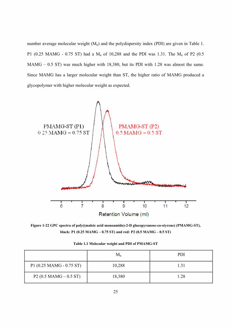
Figure 1.22 GPC spectra of poly((maleic acid monoamido)-2-D glucopyranose-co-styrene) (PMAMG-ST) ............................................................................................................................... 25
Figure 1.23 Biodecomposition evaluation of MAMG, PSMA, and PMAMG-ST by oxidative-fermentative test (OF test) via pH change .................................................................................... 27
Figure 2.1 Grafting D-galactopyranosyl-α-(1→3)-galactopyranosyl-α-p-aminophenol (1) onto poly(styrene-co-maleic acid) (PSMAC) ....................................................................................... 34
Figure 2.2 Grafting (2). (3) and (4) onto poly(styrene-co-maleic anhydride) (PSMAH) ............. 35
Figure 2.3 Grafting (5) – (11) onto poly(styrene-co-maleic anhydride) (PSMAH) ..................... 36
Figure 2.4 Grafting of (12) and (13) onto poly{styrene-co-[(maleic anhydride)-alt-styrene]} (PST-PSMAH) .............................................................................................................................. 37
Figure 2.5 Grafting of N-(4-aminobutyl)-O-β-D-galactopyranosyl-(1→4)-D-gluconamide (14) onto poly(N-vinylpyrrolidone-co-maleic acid) (PNVP-MAC) .................................................... 38
Figure 2.6 Grafting of (15), (16) and (17) onto poly(ethylene-atl-maleic anhydride) (PEMAH) 39
Figure 2.7 Grafting of (18), (19) and (20) onto poly(propylene-alt-maleic anhydride) (PPMAH) thin film ......................................................................................................................................... 40
Figure 2.8 Grafting of glucosamine (21) onto poly(isobutylene-alt-maleic acid) (PIMAC) ........ 41
Figure 2.9 FT-IR spectra of glucosamine grafted poly(methyl vinyl ether-alt-maleic acid) (Glu-PMVE-MAc) ........................................................................................................................ 52
Figure 2.10 1H-NMR spectra of glucosamine grafted poly(methyl vinyl ether-alt-maleic acid) (Glu-PMVE-MAc) ........................................................................................................................ 53
Figure 2.11 Equilibrium swelling ratio of Glu-PMVE-MAc and PMVE-MAc hydrogels in aqueous solution from pH of 1.2 to 7.4 ........................................................................................ 55
Figure 2.12 Mesh size of Glu-PMVE-MAc hydrogels in aqueous solution from pH of 1.2 to 7.4.................................................................................................................................................. 56

x
Figure 2.13 Drug release profile of FITC-dextran-loaded Glu-PMVE-MAc hydrogels in enzyme-free simulated gastric fluid (SGF) (pH=1.2) or simulated intestinal fluid (SIF) (pH=6.8) within 240 min ......................................................................................................................................... 59
Figure 2.14 Drug release profile of FITC-dextran-loaded Glu-PMVE-MAc hydrogels in enzyme-free simulated gastric fluid (SGF) (pH=1.2) or simulated intestinal fluid (SIF) (pH=6.8) within 120 h.............................................................................................................................................. 60
Figure 3.1 Star-shaped poly(ε-caprolactone)-b-poly(D-gluconamidoethyl methacrylate) glycopolymers (SPCL-PGAMA) .................................................................................................. 67
Figure 3.2 Star-shaped poly(ε-caprolactone)-b-poly(D-lactobionamidoethyl methacrylate) glycopolymers (SPCL-PLAMA) .................................................................................................. 68
Figure 3.3 Poly(2-{[(D-glucosamin-2N-yl)carbonyl]- oxy}ethyl methacrylate-b-poly(n-butyl acrylate) (PHEMAGl-PBA) .......................................................................................................... 69
Figure 3.4 Poly(2-(β-D-galactosyloxy)ethyl methacrylate-co-styrene)-b-polystyrene (P(GalEMA-co-S)-b-PS)............................................................................................................... 69
Figure 3.5 Poly(ethylene oxide)-poly(D-gluconamidoethyl methacrylate)-poly(2-(diethylamino)ethyl methacrylate) (PEO-GAMA-DEA) and poly(propylene oxide)-poly(D-gluconamidoethyl methacrylate) (PPO-GAMA) .......................................................................... 70
Figure 3.6 Poly(3-O-methacryloy-α,β-D-glucopyranose)-b-poly(2-(diethylamino)ethyl methacrylate) (PMAGlc-PDEA) ................................................................................................... 71
Figure 3.7 Poly(2’-(4-vinyl-[1,2,3]-triazol-1-yl)ethyl-O-α-D-mannopyranoside)-b-poly(N-isopropyl acrylamide) ................................................................................................................... 71
Figure 3.8 Peracetylated maltoheptaose-b-poly(2-ethhyl-2-oxazoline)-b-peracetylated maltoheptaose (AcMH-b-PEtOz-b-AcMH) .................................................................................. 72
Figure 3.9 Star-shaped poly(γ-benzyl L-glutamate)-b-poly(D-gluconamidoethyl methacrylate) (SPBLG-PGAMA) ........................................................................................................................ 77
Figure 3.10 Star poly(amido amine)-b-poly(ε-caprolactone)-b-poly(D-gluconamidoethyl methacrylate) (PAMAM– PCL–PGAMA) ................................................................................... 78
Figure 3.11 Poly(6-O-(2’-formyl-4’-vinylphenyl)-D-galactopyranose-b-5,6-benzo-2-methylene-1,3-dioxepane) (PVDG-BMDO) ................................................................................................. 79
Figure 3.12 13C-NMR spectra of HEMAN ................................................................................... 86
Figure 3.13 1H-NMR spectra of HEMAGI ................................................................................... 87
Figure 3.14 1H-NMR spectra of PPO-Br ...................................................................................... 88
xi
Figure 3.15 1H-NMR spectra of PHEMAGI-PPO ........................................................................ 89
Figure 3.16 CMC determination of PHEMAGI62-PPO41 (A), PHEMAGI46-PPO41 (B), PHEMAGI31-PPO41 (C), and PHEMAGI15-PPO41 (D) ................................................................. 92
Figure 3-17 Optical transmittance measurement as a function of temperature for PHEMAGI-PPO micelles ......................................................................................................................................... 94
Figure 3-18 TEM images of PHEMAGI62-PPO41 (A), PHEMAGI46-PPO41 (B), PHEMAGI31-PPO41 (C), and PHEMAGI15-PPO41 (D) ...................................................................................... 95
Figure 3.19 Lectin binding ability test of PHEMAGI-PPO using Con A (0.5 mg/mL) as model lectin .............................................................................................................................................. 98

xii
List of Tables
Table 1.1 Molecular weight and PDI of PMAMG-ST ................................................................. 25
Table 1.2 Monomer ratio on PMAMG-ST ................................................................................... 26
Table 2.1 Amount of utilized reagents and yield for synthesis of Glu-PMVE-MAc ................... 46
Table 2.2 Amount of reagents in synthesis of PMVE-MAc or Glu-PMVE-MAc hydrogels ....... 47
Table 2.3 Glucosamine substitution rate on Glu-PMVE-MAc ..................................................... 54
Table 2.4 Actual weight and weight percentage of released Glu-PMVE-MAc of G2-400 hydrogels after immersing in SGF or SIF in 240 minutes ............................................................ 57
Table 3.1 CMC of amphiphilic glycopolymers and common surfactant ...................................... 72
Table 3.2 Dh of glycopolymer aggregates determined by DLS and lectins that can interact with them............................................................................................................................................... 74
Table 3.3 Amount of utilized reagents for synthesis of PHEMAGI-PPO .................................... 84
Table 3.4 Characteristics of PHEMAGI-PPO glycopolymers synthesized by ATRP .................. 90
Table 3.5 CMC and concentration at onset of linear course ......................................................... 93
Table 3.6 Lower critical solution temperature (LCST) of PHEMAGI-PPO…………………… 94
Table 3.7 Size of the micelles and the concentration of the glycopolymer solutions……………96

1
CHAPTER 1 SYNTHESIS AND BIODECOMPOSITION OF GLYCOPOLYMER:
POLY((MALEIC ACID MONOAMIDE)-2-D-GLUCOPYRANOSE-CO-
STYRENE) (PMAMG-ST)
1.1 Introduction
1.1.1 Glycopolymers
Glycopolymers are synthetic polymers with carbohydrate groups; they form a bridge between
purely synthetic polymers and polysaccharides. Other terms for glycopolymers are polyvinyl
saccharides, carbohydrate-containing polymers and synthetically modified sugar-based
polymers. The sugar units can be monomeric, dimeric or oligomeric and built into the polymer
backbone or grafted onto the synthetic polymer.
Applications of glycopolymers in the biomedical area can be found in areas of virus inhibition,
drug delivery, hydrogel and micelle formation, etc. For instance, the infection of the Influenza
virus starts from interaction between hemagglutinins on the surface of the virus with sialosides
on the cell surface. If such an interaction can be prevented, infection might be stopped at this
stage. Sigal and the co-researchers synthesized two glycopolymers containing sialoside pendent
groups by polymerization or grafting (Sigal et al., 1996). Ki value is the minimum concentration
of sialoside groups (not the concentration of glycopolymer) required to prevent hemagglutination
of chicken red blood cells caused by virus. The lower the Ki value, the prevalent is the
interaction. The Ki value decreased with the increasing molecular weight of the glycopolymers.
Compared to single sialoside, the Ki value was lower by a factor of 10-3.

2
Glycopolymer-drug conjugates were synthesized for targeted drug delivery to minimize
undesirable side effects. A glycopolymer containing galactose pendent groups was conjugated
with doxorubicin, which is a common anti-cancer drug that causes significant side effects.
Animal survival tests were performed to evaluate the toxicity (Hopwell et al. 2001). All animals
survived after 12 weeks with glycopolymer-doxorubicin conjugate treatment of 4, 8, and 12
mg/kg. However, animal survival rate was reduced with an increasing dose of free doxorubicin.
Only 4 mg/kg free doxorubicin resulted 100% death after 12 weeks. The system was then
assessed in a Phase Ι clinical trial involving patients with solid hepatic tumors (Seymour et al.
2002). Uptake of the conjugate into hepatic tumors was significantly higher than the control due
to the interaction of galactose groups with corresponding receptors on the tumors.
Glycopolymers were usually synthesized by four different methods: polymerization of
glycomonomers or copolymerization of glycomonomers with vinyl monomers, ring-opening
polymerization of anhydro-sugars, enzyme-mediated polymerization, and grafting of sugars onto
functionalized synthetic polymers (Varma et al. 2004). In this paper, the focus is on the synthesis
of amide linked glycomonomers and free radical copolymerization of glycomonomers with
styrene.
1.1.2 Synthesis of glycomonomers
Sugars have been linked to vinyl molecules to synthesize glycomonomers by amide linkage,
ester linkage, ether linkage, and other linkages. Since the glycomonomer used in this paper
contains amide linked sugar, the main focus is on the introduction of glycomonomers
synthesized by amide linkage. Most sugar precursors that were commonly used in
glycomonomer synthesis are not commercially available. Synthesis of those precursors might

3
occur via one or more reactions. Similarly, most vinyl precursors are commercially unavailable
as well and need to be synthesized. Therefore, the overall synthesis can be tedious and time-
consuming.
Masutda and co-workers developed a glycomonomer: N-acryloyl-D-glucosamine (AGA, 1) (Fig.
1.1) within one reaction with a yield of 38% (Masutda et al., 1996). The glycomonomer was
obtained by reacting D-glucosamine hydrochloride with acryloyl chloride in potassium carbonate
aqueous solution using sodium nitrite as polymerization inhibitor. The total reaction time was 24
h. The glycomonomer was separated by pouring the reaction solution to ethanol and
recrystallized in a mixed solvent. Bernard and co-workers synthesized the same glycomonomer
with minor change (Bernard et al. 2006). They isolated the product by passing the reaction mix
through a silica chromatography column followed by recrystallization. In this case a yield of
only 20% was achieved. Further redesign of synthesis of AGA led to a slightly different
glycomonomer: 2-(methacrylamido)glucopyranose (MAG, 2) (Fig. 1.1) by the same synthesis
and separation method as AGA within 24 h. Methacryloyl chloride was linked with D-
glucosamine hydrochloride to produce such product. A yield of 32% and 58% was reported in
two different papers for this product, respectively (Pearson et al., 2009 and Ting et al., 2010).
Wilson et al. (1998) reported the synthesis of 4-O-β-D-galactopyranosyl-1-(acrylamido)-1-
deoxyglucitol (3) (Fig. 1.2). A sugar precursor containing an amine group was obtained by
treatment of lactose with hydrazine hydrate for 24 h and then with hydrogen gas for another 24 h.
A similar synthetic strategy as the production of AGA was applied to produce 3 from sugar
precursor and acryl chloride. Extraction and precipitation were utilized to obtain the final
product. The total yield, which is defined as the product of yield of each reaction was calculated
to be 58.9%.

4
OHO
HO
OH+ O Cl O
HOHO
OH
NH
AGA, 1
NaNO2 in K2CO3 solution0oC then room temp.
OHO
HO
OH
+ O Cl OHO
HO
OH
NH
MAG, 2
NaNO2 in K2CO3 solution
-10oC then room temp.
NH3+Cl-
OH OH
OHOHNH3
+Cl-
O
O
Figure 1-1 Synthesis of AGA and MAG (Masutda et al., 1996 and Pearson et al., 2009)
Figure 1-2 Synthesis of Glycomonomers: 3 (4-O-β-D-galactopyranosyl-1-(acrylamido)-1-deoxyglucitol;
Wilson et al. 1998)
Another glycomonomer 2-{[(D-glucosamine-2-N-yl)carbonyl] oxy}ethyl methacrylate
(HEMAGI, 4) (Fig. 1.3) was produced by researchers in Garcia’s group (Leon et al., 2010) . The
vinyl precursor was synthesized by reaction between 2-hydroxyethyl methacrylate and p-
nitrophenyl chloroformate for 24 h and isolated by precipitation in cold methanol. D-
glucosamine hydrochloride was then linked to the vinyl precursor and purified by precipitation in
a solvent mixture. The total reaction took 48 h with total yield of 57%.

5
O
OOH
O
OCl
NO2
triethylamine0 C
O
OO
O
O
NO2
OHO
HO
OH
OHtriethylamine
30 C
OHO
HO
OH
HNO
OO
O
HEMAGI, 4
OH
NH3+Cl-
Figure 1-3 Synthesis of 2-{[(D-glucosamine-2-N-yl)carbonyl] oxy}ethyl methacrylate (HEMAGI; Leon et al.,
2010)
Mixture of sialyllactose isomers isolated from bovine milk was aminated and reacted with p-
vinylbenzyl chloride to synthesize two glycomonomers: N-p-vinylbenzoyl-b-sialyllactosylamine
(5a, 5b) (Fig. 1.4) within two reactions (Tsuchida et al., 1998a). Sialyllactosylamine was
obtained from reaction between sialyllactose and ammonium hydrogen carbonate in 72 h. Excess
ammonium hydrogen carbonate was removed by dilution of the residue with water,
concentration of the solution and then filtration. Reaction of sialyllactosylamine with p-
vinylbenzyl chloride took 5 h. The product was purified by chromatography. Total yield was
reported to be 88%. Three enzymatically synthesized oligosaccharides were aminated and linked
with p-vinylbenzyl chloride in the same way by the same research group (Tsuchida et al. 1998b).
Kobayashi et al. (1985) produced N-p-vinylbenzyl-[O-α-D-glucopyranosyl-(1→4)]-D-
glucanamide (VLA, 6) (Fig. 1.5) within four reactions. They first oxidized D-maltose-1-hydrate
to maltono-1,5-lactone. Separation of the sugar precursor required precipitation, recrystallization
and purification by passing through a chromatography column. The vinyl precursor p-
vinylbenzylamine was prepared from p-vinylbenzyl chloride with two steps. To synthesize the

6
glycomonomer, the oxidized six-membered ring on sugar precursor was opened; and an amide
linkage was formed between the sugar precursor and the vinyl precursor. The total reaction time
was estimated to be 72 h; and the total yield was calculated to be 42.4%.
Figure 1-4 Synthesis of Glycomonomers: 5a, 5b (Tsuchida et al., 1998a)

7
A similar synthetic method as for the production of VLA was utilized to prepare 2-
lactobionamidoethyl methacrylate (LAMA, 7a) (Fig. 1.6) with three steps (Narain et al., 2003).
The vinyl precursor 2-aminoethyl methacrylate was obtained by reaction of ethanolamine and
methacryl chloride followed by precipitation and recrystallization. The sugar precursor
lactobionolactone was converted from lactobionic acid by vacuum distillation at 50ºC. The total
yield was reported to be 54.6%. Another glycomonomer, D-gluconamidoethyl methacrylate
(GAMA, 7b) (Fig. 1.6), was synthesized in the same manner.
O
HOO
OH
O
HO
OHI2, KOH
40oCO
HOO
OH
O
HO
OH
+H2C CH
CH2
NH2
OOH
HO
OH
NH
H2C CH
room temp.
VLA, 6
(f rom p-vinylbenzyl chloridewithin two reactions)
HOHO
O
HO
OH
HO
OH
OH
OH
OH O
OH
OH
O
OH
Figure 1-5 Synthesis of VLA (N-p-vinylbenzyl-[O-α-D-glucopyranosyl-(1→4)]-D-glucanamide; Kobayashi et
al. 1985)
N-p-vinylbenzyl-D-glucuronamide (8) (Fig. 1.7) was synthesized by Kim et al. (2000) by a four
step reaction. D-glucuronolatone was first acetalized in acetone using H2SO4 as catalyst. The
ester bond on the sugar precursor was opened when reacting with p-vinylbenzylamine, which
was prepared from p-vinylbenzyl chloride as the glycomonomer. The total reaction time was
estimated to be 48 h and the total yield was calculated to be 34.5%. Glycomonomers having D-
glucaric moieties were obtained by a similar approach (Hashimoto et al., 1999)

8
O
HO
OH
OHOHO
OH
O
OH
O
HO
OH
OOHO
OH
O
50oC
+
NH3+Cl-
O
O
HO
OH
OHO
OH
O
HNO
vacuum distillation
O
triethylamine40oC
OHO
HO
OH
+ NH3+Cl-
O
triethylamine20oC
HOHO
OH
O
HNO
O
LAMA, 7a
GAMA, 7b
(from methacryloyl chloridewithin one reaction)
(f rom methacryloyl chloridewithin one reaction)
HO
OHOH
HO
OHOH
OHOH
HO
OH
OH
OH
OH O
Figure 1-6 Synthesis of LAMA and GAMA (Narain et al., 2003)
Figure 1-7 Synthesis of Glycomonomer: 8 (N-p-vinylbenzyl-D-glucuronamide; Kim et al. 2000)

9
Glycomonomers containing ester linkages were synthesized by several researchers (Ye et al.,
2001, Gotz, et al., 2002, Malinova et al., 2005, Muthukrishnan et al., 2005a,b, Ting et al., 2007,
Cameron et al., 2008, Borges et al., 2009, Ting et al., 2009a, Suriano et al., 2010).
Glycomonomers having ether linkage were reported in various papers as well (Yamada et al.,
2001, D'Agosto et al., 2002, Lu et al., 2005, Guo et al., 2006, Cameron et al., 2008, Broyer et al.,
2008, Ting et al., 2009b, Xu et al., 2009, Otman et al., 2010, Ke et al., 2010, Hu et al., 2010).
1.1.3 Free radical copolymerization of glycomonomer and styrene
A glycopolymer containing styrene was first reported by Charreyre et al. (1993). The
glycomonomer 6-(methacryloyloxy)hexyl β-D-cellobioside (CHMA, 9) (Fig. 1.8) was
copolymerized with styrene at different monomer ratios in DMSO using AIBN as initiator. After
20 h, conversion of CHMA reached about 80%; and conversion of styrene varied from about
60% to about 100% with different initial monomer ratios. Composition of two monomers in
glycopolymers agreed well with theoretical value.
Figure 1-8 CHMA (6-(methacryloyloxy)hexyl β-D-cellobioside; Charreyre et al. 1993)
Copolymerization of glycomonomer 11-(N-p-vinylbenzyl) amidoundecanoyl maltobionamide
(LIMA, 10) (Fig. 1.9) and styrene in DMSO was reported in 1996 (Revilla et al. 1996). AIBN
was used as initiator. Conversion of LIMA was about 80%; and styrene of about 60%.
Copolymerization reactivity ratio was calculated to be rS = 1.23 ± 0.09 and rLIMA = 0.86 ± 0.1.

10
Furthermore, LIMA was used as polymerizable emulsifier for production of sugar functionalized
polystyrene nanoparticles (Revilla et al., 1993). Critical micelle concentration (CMC) of LIMA
was first determined by surface tension measurement as well as fluorescence emission spectra.
Batch emulsion polymerization of styrene was carried out using LIMA as emulsifier, potassium
persulfate as initiator, and dipotassium phosphate as buffer. Various concentrations of LIMA led
to different sizes of nanoparticles ranging from 130 to 330 nm. Seeded emulsion polymerization
of styrene was also conducted.
Figure 1-9 LIMA (11-(N-p-vinylbenzyl) amidoundecanoyl maltobionamide; Revilla et al. 1996))
Wulff and colleagues synthesized a protected glyconomomer: 2,3;4,5-di-O-isopropylidene-1-(4-
vinylphenyl)-D-gluco(D-manno)pentitol (11) (Fig. 1.10) and copolymerized 11 with styrene in
bulk and solution using AIBN as initiator (Wulff et al. 1997). Bulk polymerization achieved a
yield of more than 85%. Mol fraction of 3 in glycopolymers agreed well with feed monomer
ratio in bulk as well as solution polymerization. Films and plates of glycopolymers were
produced by casting and hot pressing, respectively. The water contact angle of deprotected films
decreased with increasing amount of sugar moieties. Later on, the same researchers
copolymerized four glycomonomers: (11), 7,8-didesoxy-1,2;3,4-di-O-isopropylidene-α-D-
galacto-oct-7-en-1,5-pyranose-6-ulose (12), 1,2;5,6-di-O-isopropylidene-3-O-methacryloyl-α-D-
glucofuranose (13), and 2,3;5,6-di-O-isopropylidene-1-(4-vinylphenyl)-keto-D-glucose (14)
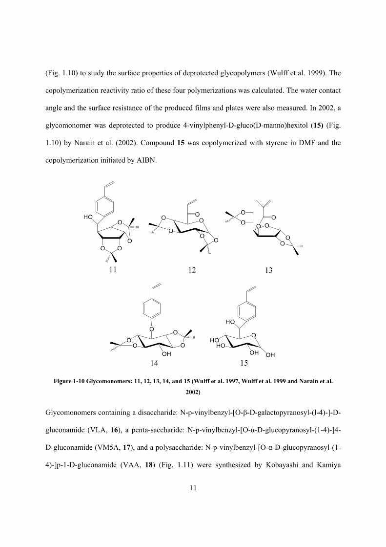
11
(Fig. 1.10) to study the surface properties of deprotected glycopolymers (Wulff et al. 1999). The
copolymerization reactivity ratio of these four polymerizations was calculated. The water contact
angle and the surface resistance of the produced films and plates were also measured. In 2002, a
glycomonomer was deprotected to produce 4-vinylphenyl-D-gluco(D-manno)hexitol (15) (Fig.
1.10) by Narain et al. (2002). Compound 15 was copolymerized with styrene in DMF and the
copolymerization initiated by AIBN.
HO
O O
O
O
11
O
OO
OO
O
12
O
OO
OO
O
O
13
OO
O O
O
14
OHO
HO
HO
15OHOHOH
Figure 1-10 Glycomonomers: 11, 12, 13, 14, and 15 (Wulff et al. 1997, Wulff et al. 1999 and Narain et al.
2002)
Glycomonomers containing a disaccharide: N-p-vinylbenzyl-[O-β-D-galactopyranosyl-(l-4)-]-D-
gluconamide (VLA, 16), a penta-saccharide: N-p-vinylbenzyl-[O-α-D-glucopyranosyl-(1-4)-]4-
D-gluconamide (VM5A, 17), and a polysaccharide: N-p-vinylbenzyl-[O-α-D-glucopyranosyl-(1-
4)-]p-1-D-gluconamide (VAA, 18) (Fig. 1.11) were synthesized by Kobayashi and Kamiya

12
(Kobayashi et al., 1997). The copolymerization of these glycomonomers and styrene was carried
out in DMSO using AIBN as initiator. It was found that with only 0.18 mol % of VAA, the
glycopolymer turned to be insoluble in chloroform whereas pure polystyrene is soluble. Methyl
orange which is insoluble in chloroform was soluble in chloroform containing little amount of
glycopolymers. Hence, methyl orange was solubilized into chloroform with the help of
glycopolymers.
Figure 1-11 VLA, VM5A, and VAA (Kobayashi et al., 1997)
A thorough study of copolymerization of D-lactose-O-(vinylbenzyl)oxime (LVO, 19) (Fig. 1.12)
with styrene was reported by Zhou et al. (1998). The reaction was performed in a mixed solvent
(DMSO/toluene=1:1) and initiated by AIBN. The relationship between conversion and initial
monomer ratio at 65ºC was studied. Conversion of copolymerization at 55ºC, 65ºC, 75ºC was
measured too. TGA characterization showed that pendent disaccharide moieties compromised
the thermal stability of the glycopolymers.

13
Figure 1-12 LVO (D-lactose-O-(vinylbenzyl)oxime; Zhou et al. 1998)
The solubility of glycopolymers containing styrene in various solvents was investigated by two
Japanese research groups. Two protected glycomonomers, N-(p-vinylbenzyl)-1,2-O-
isopropylidene-6-D-glucofuranuronamide (20) and N-(2-methacryloylamino)ethyl-1,2-O-
isopropylidene-6-D-glucofuranuronamide (21) (Fig. 1.13), were copolymerized with styrene
with various initial feed ratios in DMSO and initiated by AIBN (Shimura et al. 2001). The
solubility of the product glycopolymers with different amounts of glycomonomers at the
backbone after deprotection was studied in ten solvents from hexane to water with increasing
solubility parameters. Three glycomonomers with acetylated monosaccharide, disaccharide, and
penta-saccharide pendent groups, 4-vinylbenzyl 2,3,4,6-tetra-O-acetyl-β-D-glucopyranoside
(22), 4-vinylbenzyl O-(2,3,4,6-tetra-O-acetyl-α-D-glucopyranosyl)-(1→4)-O-2,3,6-tri-O-acetyl-
β-D-glucopyranoside (23), and 4-vinylbenzyl O-(2,3,4,6-tetra-O-acetyl-α-D-glucopyranosyl)-
(1→4)-[(O-2,3,6-tri-O-acetyl-α-D-glucopyranosyl)-(1→4)]4-O-2,3,6-tri-O-acetyl-β-D-
glucopyranoside (24) (Fig. 1.13), were copolymerized with styrene in DMF using AIBN as
initiator (Narumi et al. 2001). Varying the initial molar ratios led to various glycopolymers with
different glycomonomer mol fractions. After deacetylation, the solubility of deprotected
glycopolymers in toluene, chloroform, THF, and water was studied. With a Wg value, which is
weight percentage of glucose residue in the glycopolymer, greater than about 50%, the
glycopolymers dissolved in water. However, with Wg value less than about 40%, the

14
glycopolymers precipitated in water.
OHO
OO
CH3
CH3
OHN
H2C CH
20
OHO
OO
CH3
CH3
OHN
21
NHO
OAcO
AcOOAc
OAc
O
22
OOAcO
OAc
OAc
O
OAcO
AcOOAc
OAc
23
OOAcO
OAc
OAc
O
OAcO
AcOOAc
OAc
4
24
OH OH
Figure 1-13 Glycomonomers: 20, 21, 22, 23, and 24 (Shimura et al. 2001 and Narumi et al. 2001)
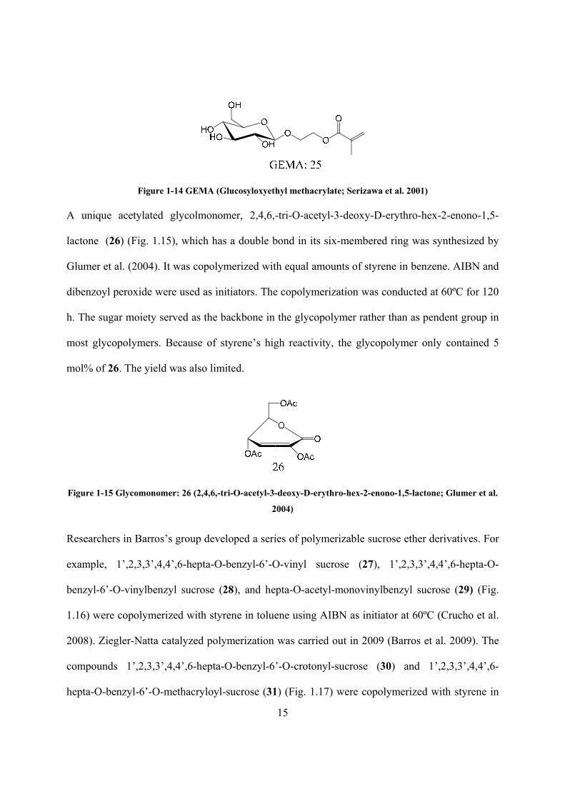
Serizawa et al. (2001) reported an interesting amphiphilic glycopolymer which had a polystyrene
hydrophobic block and a glycopolymer hydrophilic block. The author first initiated the
glycomonomer: glucosyloxyethyl methacrylate (GEMA, 25) (Fig. 1.14) by ammonium
peroxodisulfate in DMF. The resulting oligomer was modified by reacting with 4-vinylbenzoic
acid to produce a macromonomer which further copolymerized with styrene in an ethanol/water
mixture using AIBN as initiator. Micelles were obtained by dialysis against distilled water. From
dynamic light scattering (DLS) and transmission electron microscopy (TEM) images, the mean
diameter of micelles was determined to be 300 to 620 nm depending on the molar ratio of
styrene and the macromonomer. The author also found that the micelles could recognize Con A,
which is a lectin that interacts specifically with glucose.
15
Figure 1-14 GEMA (Glucosyloxyethyl methacrylate; Serizawa et al. 2001) A unique acetylated glycolmonomer, 2,4,6,-tri-O-acetyl-3-deoxy-D-erythro-hex-2-enono-1,5-
lactone (26) (Fig. 1.15), which has a double bond in its six-membered ring was synthesized by
Glumer et al. (2004). It was copolymerized with equal amounts of styrene in benzene. AIBN and
dibenzoyl peroxide were used as initiators. The copolymerization was conducted at 60ºC for 120
h. The sugar moiety served as the backbone in the glycopolymer rather than as pendent group in
most glycopolymers. Because of styrene’s high reactivity, the glycopolymer only contained 5
mol% of 26. The yield was also limited.
Figure 1-15 Glycomonomer: 26 (2,4,6,-tri-O-acetyl-3-deoxy-D-erythro-hex-2-enono-1,5-lactone; Glumer et al.
2004)
Researchers in Barros’s group developed a series of polymerizable sucrose ether derivatives. For
example, 1’,2,3,3’,4,4’,6-hepta-O-benzyl-6’-O-vinyl sucrose (27), 1’,2,3,3’,4,4’,6-hepta-O-
benzyl-6’-O-vinylbenzyl sucrose (28), and hepta-O-acetyl-monovinylbenzyl sucrose (29) (Fig.
1.16) were copolymerized with styrene in toluene using AIBN as initiator at 60ºC (Crucho et al.
2008). Ziegler-Natta catalyzed polymerization was carried out in 2009 (Barros et al. 2009). The
compounds 1’,2,3,3’,4,4’,6-hepta-O-benzyl-6’-O-crotonyl-sucrose (30) and 1’,2,3,3’,4,4’,6-
hepta-O-benzyl-6’-O-methacryloyl-sucrose (31) (Fig. 1.17) were copolymerized with styrene in

16
hexane at 40ºC. TiCl4 and AlEt3 were used as catalysts. Molar rate of Ti/Al was 8:1 and
sugar/styrene was 1, 0.5, 0.1, and 0.05. The relationship of mol fraction of glycomonomers to
glycopolymers and molecular weight of glycopolymers with initial molar ratios of monomers
was studied. In 2010, copolymerization of styrene with 6-O-methacryloyl sucrose (32), 6-O-
crotonoyl sucrose (33), 1’,2,3,3’,4,4’,6’-hepta-O-acetyl-6-O-methacryloyl-sucrose (34), and
1’,2,3,3’,4,4’,6’-hepta-O-acetyl-6-O-crotonoyl-sucrose (35) (Fig. 1.17) were conducted in DMF
for the first two and in toluene for the last two compounds, respectively (Barros et. al 2010). The
relationship of molecular weights of the glycopolymers, degree of monomer conversion, and mol
fraction of glycomonomers to glycopolymers with feed monomer ratios was investigated. A
biodegradation test on the glycopolymers by a fungal (Aspergillus niger) culture method showed
a fungal growth ≥60%, indicating good biodegradability.
Figure 1-16 Glycomonomers: 27, 28, and 29 (Crucho et al. 2008)

17
Figure 1-17 Glycomonomers: 30, 31, 32, 33, 34, and 35 (Barros et al. 2009 and Barros et. al 2010)
A glycopolymer composed of 2-{[(D-glucosamine-2-N-yl) carbonyl]oxy}ethyl acrylate
(HEAGI, 36) (Fig. 1.18) and styrene was made by copolymerization in DMF using AIBN as
initiator (Munoz-Bonilla et al., 2010). A blend of glycopolymer and polystyrene was spin-coated
onto a silicon wafer to fabricate honeycomb-structured films. From AFM study, the diameter of
holes depended on composition of the blend. It was also found from florescence microscope
imaging that FITC-Con A could recognize the sugar moiety on the film.
Figure 1-18 HEAGI (2-{[(D-glucosamine-2-N-yl) carbonyl]oxy}ethyl acrylate; Munoz-Bonilla et al., 2010)

18
1.1.4 Approach to a convenient synthesis of a glycopolymer in this project
In this project, an attempt was made to offer a faster, less cumbersome synthesis route with
reasonable product yield. A glycomonomer with amide linkage: (maleic acid monoamido)-2-D-
glucopyranose (MAMG) was first synthesized within a one-step reaction in 4 h with relatively
high yield. The product was isolated by precipitation in ethyl acetate. Copolymerization of
MAMG and styrene was conducted in DMSO using AIBN as initiator with different initial
monomer ratios. 1H-NMR was used to characterize the chemical structure of MAMG. The
chemical structure of copolymer PMAMG-ST was confirmed by FTIR and 1H-NMR. Molecular
weight and final monomer ratio on PMAMG-ST was determined by GPC and elemental analysis,
respectively. The biodecomposition and release of the sugar of glycomonomer, glycopolymer
and control sample was evaluated by an oxidative-fermentative test.
1.2 Experimental Part
1.2.1 Materials
D-Glucosamine hydrochloride (99.9%, Calbiochem), maleic anhydride (98%, Alfa Aesar),
sodium methoxide (98%, Alfa Aesar), methanol (99.9%, Acros), triethylamine (99%, Alfa
Aesar), ethyl acetate (99.5%, Mallinckrodt Chemicals), anhydrous magnesium sulfate (MgSO4)
(99%, Strem Chemicals) and sodium hydroxide (NaOH) (technical, Spectrum) were used as
received. Styrene (99%, stabilized 10-15 ppm 4-tert-butylcatechol, Alfa Aesar) was purified
from inhibitor by washing with 10% aqueous solution of sodium hydroxide and then with
deionized water; after drying over anhydrous MgSO4, it was filtered and distilled under vacuum.
(2,2’-azobisisobutyronitrile) (AIBN) (98%, Sigma Aldrich) was recrystallized from methanol.

19
Dimethyl sulfoxide (DMSO) (99.9% Malinckrodt) was dried over anhydrous MgSO4 before
reaction.
1.2.2 Sythesis of glycomonomer and glycopolymer
1.2.2.1 Synthesis of (maleic acid monoamido)-2-D glucopyranose (MAMG)
The (maleic acid monoamido)-2-D-glucopyranose was synthesized by modifying the procedure
previously reported (Jin et al., 2009). Maleic anhydride (2.3 g, 23.45 mmol) was added to a 250
ml three neck flask containing D-glucosamine hydrochloride (10.0 g, 46.35 mmol), sodium
methoxide (2.5 g, 46.3 mmol), and methanol (125 ml) while stirring. After stirring at 60ºC for 30
min, maleic anhydride (2.3 g, 23.45 mmol) and triethylamine (9.3 ml, 66.8 mmol) were added.
The reaction was stopped after 4 h. The solution was filtered and the filtrate was slowly added to
250 ml ethyl acetate. The precipitation was isolated by filtration and dried under vacuum at 35 ºC
for 12 h to yield 8.9138 g MAMG (yield: 69.4%)
Scheme 1.1. Synthesis of (maleic acid monoamido)-2-D-glucopyranose (MAMG)
1.2.2.2 Copolymerization of (maleic acid monoamido)-2-D glucopyranose and styrene
MAMG (3.885 g, 15 mmol), styrene (1.56 g, 15 mmol), AIBN (0.136 g, 0.829 mmol) and 15 ml
DMSO were added to a 100 ml Schlenk flask. Air was removed by vacuum and nitrogen was
injected in to create nitrogen atmosphere in the Schlenk flask. After stirring at 60ºC for 15 h,

20
copolymers were isolated by precipitation in methanol, then filtrated and dried under vacuum for
12 h at 35ºC. MAMG and styrene feed ratio was 1:3 or 1:1. P1 (0.25 MAMG – 0.75 ST) and P2
(0.5 MAMG – 0.5 ST) were obtained. Poly(styrene-co-maleic anhydride) was also obtained
using previous described method as control sample.
Scheme 1.2. Synthesis of poly((maleic acid monoamido)-2-D-glucopyranose-co-styrene) (PMAMG-ST)
1.2.3 Characterization:
1.2.3.1 Fourier Transform Infrared Spectroscopy (FT-IR)
The FT-IR of PMAMG-ST was recorded in powder form. The spectra were recorded on a
Thermo Scientific Nicolet 6700 at resolution of 4 cm-1 and the number of scans was 32.
1.2.3.2 Nuclear Magnetic Resonance (NMR) Spectroscopy
1H NMR was used to determine the chemical structures of synthesized products. Spectra were
recorded at room temperature with deuterated dimethyl sulfoxide (DMSO-d6) as a solvent. A
Bruker 400 NMR spectrometer was used. Typical parameters for the proton spectra were a 15 s
pulse delay, a 3 s acquisition time, a 20.68 ppm spectral width, and 16 scans.
1.2.3.3 Gel permeation chromatography (GPC)
21
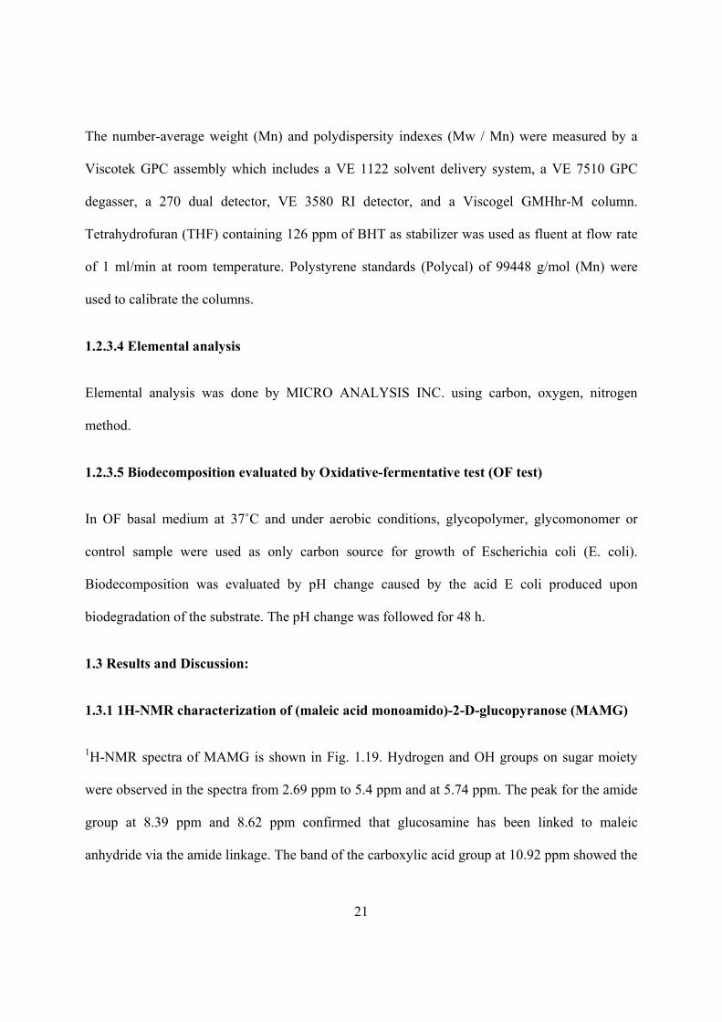
The number-average weight (Mn) and polydispersity indexes (Mw / Mn) were measured by a
Viscotek GPC assembly which includes a VE 1122 solvent delivery system, a VE 7510 GPC
degasser, a 270 dual detector, VE 3580 RI detector, and a Viscogel GMHhr-M column.
Tetrahydrofuran (THF) containing 126 ppm of BHT as stabilizer was used as fluent at flow rate
of 1 ml/min at room temperature. Polystyrene standards (Polycal) of 99448 g/mol (Mn) were
used to calibrate the columns.
1.2.3.4 Elemental analysis
Elemental analysis was done by MICRO ANALYSIS INC. using carbon, oxygen, nitrogen
method.
1.2.3.5 Biodecomposition evaluated by Oxidative-fermentative test (OF test)
In OF basal medium at 37˚C and under aerobic conditions, glycopolymer, glycomonomer or
control sample were used as only carbon source for growth of Escherichia coli (E. coli).
Biodecomposition was evaluated by pH change caused by the acid E coli produced upon
biodegradation of the substrate. The pH change was followed for 48 h.
1.3 Results and Discussion:
1.3.1 1H-NMR characterization of (maleic acid monoamido)-2-D-glucopyranose (MAMG)
1H-NMR spectra of MAMG is shown in Fig. 1.19. Hydrogen and OH groups on sugar moiety
were observed in the spectra from 2.69 ppm to 5.4 ppm and at 5.74 ppm. The peak for the amide
group at 8.39 ppm and 8.62 ppm confirmed that glucosamine has been linked to maleic
anhydride via the amide linkage. The band of the carboxylic acid group at 10.92 ppm showed the

22
anhydride group was opened; only one carboxylic acid group reacted with amine group on
glucosamine during the reaction. H-8 and H-9 proved the double bond on maleic anhydride
remained after the reaction.
Figure 1-19 1H-NMR spectra of (maleic acid monoamido)-2-D-glucopyranose (MAMG); MAMG: (400MHz,
DMSO-d), δ(ppm): 2.69 - 3.8 (H-1 - H-4, H-6), 4.36 – 5.4 (H-a - H-d), 5.74 (H-8), 6.07 (H-9), 8.39, 8.62 (H-7),
10.92 (H-10)
1.3.2 FT-IR characterization of poly((maleic acid monoamido)-2-D-glucopyranose-co-
styrene) (PMAMG-ST)
FT-IR spectra of poly((maleic acid monoamido)-2-D-glucopyranose-co-styrene) (PMAMG-ST)
and poly(styrene-maleic anhydride) (PSMA) are shown in Fig. 1.20. The C-OH groups on sugar
at 1017.0 cm-1 and OH groups between 2339.0 cm-1 and 3685.4 cm-1 confirmed the sugar moiety
DMSO-d6

23
on PMAMG-ST. Peaks for the amide group at 1572.8 cm-1 and 3260.4 cm-1 also proved the
presence of the sugar moiety. The peak at 3027.5 cm-1 which designates the aromatic C-H groups
indicated benzene ring was on PMAMG-ST as well.
Figure 1-20 FT-IR spectra of poly((maleic acid monoamido)-2-D-glucopyranose-co-styrene) (PMAMG-ST)
(P2); PMAMG-ST: wavelength (cm-1): 1017.0 (C-OH on sugar), 1572.8 (C=O on amide group), 2390.0 –
3685.4 (OH on sugar), 3027.5 (aromatic C-H on benzene ring), 3260.4 (N-H on amide group); in comparison
PSMA: wavelength (cm-1): 923.0 (C-O-C on cyclic anhydride), 1221.3 (C-O-C), 1773.0, 1854.9 (C=O on 5-ring
anhydride), 3027.5 (aromatic C-H on benzene ring)
1.3.3 1H-NMR characterization of poly((maleic acid monoamido)-2-D-glucopyranose-co-
styrene) (PMAMG-ST)
Since spectral data confirmed the successful synthesis of the glycomonomer, the
copolymerization with styrene was attempted. The product was confirmed by 1H-NMR analysis.
Fig. 1.21 shows 1H-NMR spectra of poly((maleic acid monoamido)-2-D-glucopyranose-co-
styrene) (PMAMG-ST). H-8 to H-12 designate the backbone of PMAMG-ST. H-1 to H-7 and H-
a to H-d proved the sugar moiety on PMAMG-ST. H-13 to H-17 confirmed the presence of

24
benzene ring on PMAMG-ST. The H-8 peak on the glycomonomer MAMG was the
identification peak of the double bond at 6.07 ppm. It disappeared in the spectra of the
glycopolymer, suggesting that the reaction has occurred.
Figure 1-21 1H-NMR spectra of poly((maleic acid monoamido)-2-D-glucopyranose-co-styrene) (PMAMG-ST)
(P2); PMAMG-ST: (400MHz, DMSO-d), δ(ppm): 0.78 – 2.39 (H-8 - H-12), 2.71 – 4.29 (H-1 - H-4, H-6), 4.45 –
5.24 (H-a – H-d), 5.74 (H-5), 6.59 (H-7), 6.81 – 7.37 (H-13 – H-17)
1.3.4 Molecular weight (Mn) and polydispersity index (PDI) of poly((maleic acid
monoamido)-2-D glucopyranose-co-styrene) (PMAMG-ST)
Copolymerization of MAMG and ST with two different initial monomer ratios was carried out:
P1 (0.25 MAMG – 0.75 ST) and P2 (0.5 MAMG – 0.5 ST). Gel permeation chromatography
(GPC) was used to provide proof for P1 and P2. The GPC spectra are shown in Fig. 1.22. The
DMSO-d6
25
number average molecular weight (Mn) and the polydispersity index (PDI) are given in Table 1.
P1 (0.25 MAMG - 0.75 ST) had a Mn of 10,288 and the PDI was 1.31. The Mn of P2 (0.5
MAMG – 0.5 ST) was much higher with 18,380, but its PDI with 1.28 was almost the same.
Since MAMG has a larger molecular weight than ST, the higher ratio of MAMG produced a
glycopolymer with higher molecular weight as expected.
Figure 1-22 GPC spectra of poly((maleic acid monoamido)-2-D glucopyranose-co-styrene) (PMAMG-ST),
black: P1 (0.25 MAMG – 0.75 ST) and red: P2 (0.5 MAMG – 0.5 ST)
Table 1.1 Molecular weight and PDI of PMAMG-ST
Mn PDI
P1 (0.25 MAMG - 0.75 ST) 10,288 1.31
P2 (0.5 MAMG – 0.5 ST) 18,380 1.28

26
1.3.5 Monomer ratio on poly((maleic acid monoamido)-2-D glucopyranose-co-styrene)
(PMAMG-ST)
Data from elemental analysis are reported in Table 2 as C%, H% and N%. With further
calculation, the monomer ratio of MAMG and ST on PMAMG-ST is given in Table 2 as well.
The MAMG segment on P1 (0.25 MAMG - 0.75 ST) was 20.35%, and on P2 (0.5 MAMG – 0.5
ST) was 47.63%. The monomer ratio on glycopolymer agreed well with the initial monomer
ratio.
Table 1.2 Monomer ratio on PMAMG-ST
C% H% N% MAMG% ST %
P1 (0.25 MAMG - 0.75 ST) 50.6% 5.98% 1.67% 20.35% 79.65%
P2 (0.5 MAMG – 0.5 ST) 60.4% 6.51% 3.72% 47.63% 52.37%
1.3.6 Biodecomposition evaluation by oxidative-fermentative test (OF test)
A biodecomposition test was performed to evaluate the release of glucose from the products. If
sugar can be released, it may act as a sensor for a multitude of applications since glucose is
easily detected by simple means. An oxidative-fermentative test was utilized to investigate this
aspect of the presented research.
PSMA, PMAMG-ST or MAMG were the only carbon sources in the medium for growth of E.
coli. Acid is produced when these materials are consumed by the microorganism. The acid can
be traced back to the original compounds and used as an indicator for their biodegradation.
Therefore, the pH value was used as an indicator for growth of E. coli. The results of OF test are
shown in Fig. 1.23. E. coli produced much higher amounts of acid when consuming PMAMG-

27
ST (P2) than when exposed to PSMA. Moreover, E. coli produced even more acid when
consuming MAMG. Thus, it can be assumed that pendent sugar groups on PMAMG-ST were
removed by E. coli and utilized as carbon source for their growth. In other words, the OF test
indicated that sugar was released from PMAMG-ST. Glucose units could fulfill various
functions in the biomedical field.
Figure 1-23 Biodecomposition evaluation of MAMG, PSMA, and PMAMG-ST by oxidative-fermentative test
(OF test) via pH change

28
1.3.7 Increasing the yield
The synthesis of PMAMG-ST and analysis of the intermediate and final products have been
described in the previous sections and it could be shown that in fact PMAMG-ST was obtained.
However, the yield of the glycomonomer had still been fairly low with less than 20%. Therefore,
the synthesis route had to be modified to lead to a more attractive yield. A better result was
obtained by doubling the amount of catalyst (triethylamine) and changing the method of adding
the reaction solution to ethyl acetate. With these process modifications the yield of the
glycomonomer could be increased to almost 70%.
1.4 Conclusions
With the methods used in this research, it could be shown that (maleic acid monoamido)-2-D
glucopyranose (MAMG) could be synthesized in a one-step reaction that is more efficient with
relatively high yield. The chemical structure of MAMG was confirmed by 1H-NMR. MAMG
could then be used to produce poly((maleic acid monoamido)-2-D glucopyranose-co-styrene)
(PMAMG-ST) successfully. The chemical structure of PMAMG-ST was confirmed by FTIR and
1H-NMR. Molecular weight and PDI of PMAMG-ST was determined by GPC. Elemental
analysis and further calculation showed monomer ratio on glycopolymer agreed well with initial
monomer ratio. Biodecomposition evaluated by oxidative-fermentative test indicated sugar was
released from PMAMG-ST. Glucose units could fulfill various functions in the biomedical field.
1.5 References
Barros MT, Petrova KT, and Raj-Singh TR. European Polymer Journal, 2010, 46, 1151–1157

29
Bernard J, Hao Xj, Davis PT, Barner-Kowollik C, and Stenzel MH. Biomacromolecules, 2006,
7, 232-238
Borges MR, Dos Santos JA, Vieira M, and Balaban R. Materials Science and Engineering C,
2009, 29 519–523
Broyer RM., Quaker GM., and Heather D, and Maynard J. AM. CHEM. SOC., 2008, 130, 1041-
1047
Cameron NR., Spain SG, Kingham JA, Weck S, Albertin L, Barker CA, Battaglia G, Smart T,
and Blanazs A. Faraday Discuss., 2008, 139, 359–368
Charreyre MT, and Boullanger P. Makromol. Chem., 1993, 194, 117-135
Crucho C, Petrova K, Pinto R, and Barros M. Molecules, 2008, 13, 762-770
D'Agosto F, Charreyre MT, Pichot C, and Mandrand B. Macromol. Chem. Phys., 2002, 203,
146-154
Gotz H, Harth E, Schiller SM., Frank CW, Knoll W, and Hawker CJ. Journal of Polymer
Science: Part A: Polymer Chemistry, 2002, 40, 3379–3391
Guo Ty, Liu P, Zhu Jw, Song Md, and Zhang Bh. Biomacromolecules, 2006, 7, 1196-1202
Hashimoto K, Ohsawa R, Imai N, and Okada M. Journal of Polymer Science: Part A: Polymer
Chemistry, 1999, 37, 303–312
Hopwell JW, Duncan R, Wilding D and Chakrabarti K, Hum. Exp. Toxicol., 2001, 20, 461-470
Hu Zg, Fan Xs, and Zhang Gs. Carbohydrate Polymers, 2010, 79, 119–124

30
Ke Bb, Wan Ls, Zhang Wx, and Xu Zk. Polymer, 2010, 51, 2168-2176
Kim SH, Goto M, Cho CS and Akaike T. Biotechnology Letters, 2000, 22, 1049–1057
Kobayashi K, Sumitomo H, and Ina Y. Polymer Journal, 1985, 17, 567-575
Kobayashi K, and Kamiya S. Macromol. Symp., 1997, 120, 139-146
Leon O, Bordege V, Munoz-Bonilla A, Sanchez-Chaves M, and Fernandez-Garcia M. Journal of
Polymer Science: Part A: Polymer Chemistry, 2010, 48, 3623–3631
Lu Fz, Meng Jq, Du Fs, Li Zc, and Zhang By. Macromol. Chem. Phys. 2005, 206, 513–520
Malinova V, and Rieger B. Macromol. Rapid Commun., 2005, 26, 945–949
Matsuda T, and Sugawara T. Macromolecules, 1996, 29, 5375-5383
Muthukrishnan S, Jutz G, Andre X, Mori H, and Muller A HE. Macromolecules, 2005a, 38, 9-18
Muthukrishnan S, Mori H, and Muller A HE. Macromolecules, 2005b, 38, 3108-3119
Narain R, Jhurry D, and Wulff G. European Polymer Journal, 2002, 38, 273-280
Narain R and Armes SP. Biomacromolecules, 2003, 4, 1746-1758
Otman O, Boullanger P, Drockenmuller E, and Hamaide T. Beilstein J. Org. Chem., 2010, 6, No.
58
Pearson S, Allen N, and Stenzel MH. Journal of Polymer Science: Part A: Polymer Chemistry,
2009, 47, 1706–1723

31
Revilla J, Elaissari A, Pichot C, and Gallot B. Polymers for Adv. Tech., 1995, 6, 455-464
Revilla J, Delair T, Gallot B, and Pichot C. Polymer, 1996, 37, 687-698
Serizawa T, Yasunaga S, and Akashi M. Biomacromolecules, 2001, 2, 469-475
Seymour LW., Ferry DR, Anderson D, Hesslewood S, Julyan P J, Poyner R, Doran J, Young
AM, Burtles S, and Kerr DJ, Journal of Clinical Oncology, 2002, 20, 1668-1676
Shimura Y, Hashimoto K, Yamanaka C, and Setojima D. Journal of Polymer Science: Part A:
Polymer Chemistry, 2001, 39, 3893–3901
Sigal GB, Mammen M, Dahmann G, and Whitesides, GM. J. Am. Chem. Soc., 1996, 118, 3789-
3800
Suriano F, Coulembier O, and Dubois P. Reactive & Functional Polymers, 2010, 70, 747-754
Ting SR S, Granville AM, Quemener D, Davis TP, Stenzel MH, and Barner-Kowollik C. Aust. J.
Chem., 2007, 60, 405–409
Ting SR S, Gregory AM., and Stenzel MH. Biomacromolecules, 2009a, 10, 342–352
Ting SR S, Min EH, Escale P, Save M, Billon L, and Stenzel MH. Macromolecules, 2009b, 42,
9422–9434
Ting SR S, Min EH, Zetterlund PB, and Stenzel MH. Macromolecules, 2010, 43, 5211–5221
Tsuchida A, Akimoto S, Usui T, and Kobayashi K. J. Biochem., 1998a, 123, 715-721
32
Tsuchida A, Kobayashi K, Matsubara N, Muramatsu T, Suzuki T, and Suzuki Y. Glycoconjugate
Journal, 1998b, 15, 1047–1054
Varma AJ, Kennedy JF, and Galgali P. Carbohydrate Polymers, 2004, 56, 429-445
Wilson ME, Najdi S, Krochta JM, Hsieh YL, and Kurth MJ. Macromolecules, 1998, 31, 4486-
4492
Wulff G, Zhu Lm, and Schmidt H. Macromolecules, 1997, 30, 4533-4539
Wulff G, Schmidt H, and Zhu Lm. Macromol. Chem. Phys., 1999, 200, 774–782
Xu N, Wang R, Du Fs, and Li Zc. Journal of Polymer Science: Part A: Polymer Chemistry,
2009, 47, 3583–3594
Yamada K, Minoda M, Fukuda T, and Miyamoto T. Journal of Polymer Science: Part A:
Polymer Chemistry, 2001, 39, 459–467
Ye Wj, Wells S, and Desimone JM. Journal of Polymer Science: Part A: Polymer Chemistry,
2001, 39, 3841–3849
Zhou Wj, Naik SS, Kurth MJ, Hsieh Y, and Krochta JM. Journal of Polymer Science: Part A:
Polymer Chemistry, 1998, 36, 2971–2978

33
CHAPTER 2 SYNTHESIS OF GLUCOSAMINE-GRAFTED POLY(METHYL
VINYL ETHER-ALT-MALEIC ACID) (GLU-PMVE-MAC) AND PH-
RESPONSIVE HYDROGEL FOR DRUG DELIVERY APPLICATION
2.1 Introduction
2.1.1 Glycopolymers
Glycopolymers are synthetic polymers that incorporate carbohydrate groups. Generally they can
be synthesized by four different methods: polymerization of glycomonomers or
copolymerization of glycomonomers with vinyl monomers, ring-opening polymerization of
anhydro-sugars, enzyme-mediated polymerization, and grafting of sugars onto functionalized
synthetic polymers (Varma et al. 2004). In the previous chapter, synthesis of an amide linked
glycomonomer and free radical copolymerization of glycomonomer with styrene was reported.
In the present work, glycopolymers were synthesized from grafting sugar onto a maleic acid-
containing synthetic polymer. The properties of the resulting glycopolymers were studied. Very
interesting aspects of this work are the formation and use of these materials as pH-responsive
hydrogel for controlled drug delivery applications.
2.1.2 Sugar derivatives grafted onto maleic groups that are connected to polymers
Vetere et al. (2002) first reported grafting a sugar derivative onto a maleic group- containing
polymer. D-Galactopyranosyl-α-(1→3)-galactopyranosyl-α-p- aminophenol (1) (Fig. 2.1) was
grafted onto poly(styrene-co-maleic acid) (PSMAC) in 2-(N-morpholino)ethanesulfonic acid
(MES) buffer solution with N-(3-dimethylaminopropyl)-N’-ethylcarbodiimide hydrochloride
(EDC) and N-hydroxysuccinimide (NHS) as catalysts. The glycopolymer was aimed at

34
applications for removing natural antibodies that cause hyperacute rejection when implanting an
organ from animals such as a pig or a monkey into the human body. Researchers in the same
group synthesized three amino sugars: 1-amino-1-deoxy-β-D-galactose (2), 1-amino-1-deoxy-β -
D-glucose (3), and 1-amino-1-deoxy-β-D-lactose (4) (Fig. 2.2) and grafted them onto
poly(styrene-co-maleic anhydride) (PSMAH) in sodium bicarbonate buffer solution (Donati, et
al. 2002). The potential application of these glycopolymers as matrix for hepatic cell cultures
was evaluated.
Figure 2-1 Grafting D-galactopyranosyl-α-(1→3)-galactopyranosyl-α-p-aminophenol (1) onto poly(styrene-
co-maleic acid) (PSMAC) (Vetere et al. 2002)

35
Figure 2-2 Grafting (2). (3) and (4) onto poly(styrene-co-maleic anhydride) (PSMAH) (Donati et al. 2002)
Gagali and co-workers grafted glucose (5), lactose (6), and sucrose (7) (Fig. 2.3) directly to
poly(styrene-co-maleic anhydride) (PSMAH) via esterification reaction using 4-
dimethylaminopyridine (DMAP) as catalyst. Fungal degradation tests showed good
biodegradability. FT-IR was utilized to study the structural changes of the glycopolymers after
fungal treatment (Galgali, et al. 2002 and 2004). In 2007, D-glucose (5), 6-O-trityl glucose (8),
methyl glucoside (9), 1,2-5,6-diisopropylidene-D-glucose (10), and 1,2,3,4-tetraacetyl-D-glucose
(11) (Fig. 2.3) grafted poly(styrene-co-maleic anhydride) were synthesized and investigated by
FT-IR (Galgali et al. 2007). Fourier deconvolution was used for the assignment of the different
ester carbonyls formed by the reaction of the different hydroxyl groups on those sugars with the
maleic anhydride compound of PSMAH.

36
Figure 2-3 Grafting (5) – (11) onto poly(styrene-co-maleic anhydride) (PSMAH) (Galgali et al. 2002, 2004,
and 2007) Two sugar derivatives, α-1-O-(2’-aminoethyl)-D-mannopyranoside (12) and β-1-O-(2’-
aminoethyl)-D-galactopyranoside (13) (Fig. 2.4) were grafted onto poly{styrene-co- [(maleic
anhydride)-alt-styrene]} (PST-PSMAH) (Su et al. 2009). Methyl red or rhodamine 6G doped
nanoparticles were formed by sonication. The mannose containing nanoparticle interacted with
concanavalin A which is a lectin that specifically recognizes glucose and mannose.

37
+
HCCH2
CH
CHO
O
O x
room temp.
COONa
O
12
PST-PSMAH
x
OHO
HO
OHOH
ONH2 CH
CH2
y
y
OHO
HO O
OH
NH2
13OH
OHO
HO
OHOH
ONH
Figure 2-4 Grafting of (12) and (13) onto poly{styrene-co-[(maleic anhydride)-alt-styrene]} (PST-PSMAH)
(Su et al. 2009)
Researchers in Auzely-Velty’s group developed a lactose derivative: N-(4-aminobutyl)-O-β-D-
galactopyranosyl-(1→4)-D-gluconamide (14) (Fig. 2.5) and grafted it onto poly(N-
vinylpyrrolidone-co-maleic acid) (PNVP-MAC) with NaOH treatment (Auzely-Velty et al.,
2002). The glycopolymer was reported to interact with a lectin (Ricinus Communis Agglutinin,
RCA120). Amphiphilic copolymers were produced by grafting (14) and dodecylamine onto
PNVP-MAC via the previously described method (Cade et al. 2004). Nanoparticles were
prepared by an emulsification–diffusion procedure. The nanoparticle interacted with RCA120 as
well.

38
Figure 2-5 Grafting of N-(4-aminobutyl)-O-β-D-galactopyranosyl-(1→4)-D-gluconamide (14) onto poly(N-
vinylpyrrolidone-co-maleic acid) (PNVP-MAC) (Auzely-Velty et al. 2002)
Uzawa et al. (2005) grafted three sugar derivatives: 10-aminodecyl O-(α-D-galactopyranosyl)-
(1→4)-β-D-galactopyranoside (15), 4-aminophenyl-O- β-lactoside (16), and 4-aminophenyl-O-
α-D-mannopyranoside (17) (Fig. 2.6) onto poly(ethylene-alt-maleic anhydride) (PEMAH).
Layer-by-layer membranes on gold substrate surface were fabricated by polyanionic
glycopolymer together with polycationic polymer. The derived glycosyl arrays were aimed to
detect Shiga toxin produced by Escherichia coli O-157.

39
Figure 2-6 Grafting of (15), (16) and (17) onto poly(ethylene-atl-maleic anhydride) (PEMAH) (Uzawa et al.
2005)
A glucose derivative, 2-aminoethyloxyl-β-D-glucopyranoside (18) (Fig. 2.7), was grafted onto
spin-coated poly(propylene-alt-maleic anhydride) (PPMAH) thin film (Grombe et al., 2006). A
set of sugar derivatives such as sulfated 2-[2-(2-aminoethyl) -ethyl]-ethyl-β-D- glucopyranoside
(19), sulfated 2-[2-(2- aminoethyl)-ethyl]-ethyl -4-(α-D-glucopyranosyl)-β- D-glucopyranoside
(20) (Fig. 2.7), sulfated cellulose, and heparin was grafted onto PEMAH or PPMAH thin films

40
(Grombe et al. 2007). Antithrombin binding tests and whole blood incubation assays indicated
that those glycopolymers have potential to be used as anticoagulant.
Figure 2-7 Grafting of (18), (19) and (20) onto poly(propylene-alt-maleic anhydride) (PPMAH) thin film
(Grombe et al. 2006, 2007)
Paraskar and colleagues (2010) reported a sugar containing nanoparticle to enhance antitumor
efficacy. Glucosamine (21) (Fig. 2.8) was grafted onto poly(isobutylene -alt-maleic acid)
(PIMAC) using diaza(1,3)bicyclo[5.4.0]undecane (DBU) as catalyst. A chemotherapy drug,

41
cisplatin, was complexed with the glycopolymer via a monocarboxylato and O→platinum (Pt)
coordinate bond. At a certain Pt to glycopolymer ratio, the complex self-assembled into a
nanoparticle that released cisplatin in a pH-dependent manner. In vivo treatment to lung and
breast cancer and biodistribution tests showed that the nanoparticles delayed tumor growth and
decreased biodistribution of platinum to the kidney. Nanoparticles fabricated by oxaliplatin
complex with glucosamine functionalized PIMAC for better antitumor efficacy were also studied
(Paraskar et al. 2012).
Figure 2-8 Grafting of glucosamine (21) onto poly(isobutylene-alt-maleic acid) (PIMAC) (Paraskar et al.
2010)
As a biocompatible material, poly (methyl vinyl ether-co-maleic acid) (PVME-MAc) was mostly
used as denture adhesive. It is the base compound in Super Poligrip®. Because of the strong
reactivity of carboxylic acid groups on the polymer, several natural compounds such as peptides
(Ladaviere et al. 2000) and proteins (Koyama et al. 1992, Allard et al. 2001) have been grafted

42
onto PVME-MAc via amide linkage. Glucosamine, which is a monosaccharide containing an
amine group, is a good candidate to be grafted onto PVME-MAc for specific biological
properties. Meanwhile, PVME-MAc could also be crosslinked by ethylene glycol, 1,4-
butanediol, 1,6-exandiol, 1,8-octanediol, 1,10-decanediol, 1,12-dodecanediol (Luppi et al. 2003),
partially hydrolyzed poly(vinyl acetate) (Guo et al. 2008), cellulosic nanowhisker (Goetz et al.
2009, 2010), and poly(ethylene glycol) (Raj Singh et al. 2009, 2010, Garland et al. 2011).
Applications of those hydrogels were investigated, including topical vehicles for hydrophilic and
hydrophobic drugs (Luppi et al. 2003), tunable denture adhesives to both hydrophobic and
hydrophilic surfaces (Guo et al. 2008), nanocomposites (Goetz et al. 2009, 2010), and controlled
drug delivery devices (Raj Singh et al. 2009, 2010, Garland et al. 2011).
2.1.3 pH-responsive glycopolymer hydrogels
Hydrogels are some of the most common drug carriers. They are three-dimensional, cross-linked
polymers that swell in water or aqueous solvent systems. Because of their high water uptake,
they are generally biocompatible. Drugs are either loaded into the hydrogel during hydrogel
synthesis or diffuse into the hydrogel upon swelling of the hydrogel. Drug delivery is governed
by diffusion of the drug, interaction of drug and hydrogel, and degradation of the matrix.
Therefore, several factors such as pore size and biodegradability of the hydrogel as well as
interaction between drug and matrix influence the speed of drug delivery.
Depending on the pH of the environment, pH-responsive hydrogels naturally display big
differences in swelling properties. They contain pendent acidic (e.g. carboxylic and sulfonic
acids) or basic (e.g. ammonium salt) groups that either accept or release protons in response to
changes in environmental pH. At this event, expulsion of charges on pendent groups increases

43
swelling of hydrogels. pH-responsive hydrogels that stay in collapsed state at low pH and swell
at neutral pH may be used as oral drug delivery devices for pH-sensitive drugs such as proteins.
Researchers have reported that sugar such as lactose, glucose, and sucrose could stabilize
different proteins by a “preferential hydration” mechanism (Arakawa et al. 1982, Lee et al. 1975,
1981), while glucose can prevent the aggregation of insulin (Jeffrey 1974). Thus, pH-responsive
glycopolymer hydrogels as drug delivery devices may better protect proteins in the matrix.
Although hydrogels have been studied insensitively, just a few glycopolymer hydrogels have
been developed. Nakamae and colleagues obtained a glycopolymer hydrogel containing glucose
pendent moieties as drug delivery device for insulin (Miyata et al. 1996). N,N'-methylene
bisacrylamide was used as chemical crosslinking agent and concanavalin A served as physical
crosslinking agent. In glucose solution, physical crosslinks were opened due to prevented
interaction between concanavalin A with pendent glucose groups caused by a surplus of glucose
in the solution. The swelling ratio increased with increasing glucose concentration in the
solution; therefore drugs in the hydrogel may be released upon swelling of the hydrogel. An
insulin-loaded hydrogel can be used as a smart device to control the glucose level in the blood by
itself.
Zhang et al. (2006) synthesized a glycopolymer hydrogel containing blood B group epitope
pendent groups for norovirus treatment. Because of the interaction between norovirus and blood
B group epitope, the hydrogel absorbed most norovirus from the solution. This hydrogel might
be administered to patients with the goal of removing most of the norovirus in the digestive
system, thus reduce the possibility of getting infected.

44
Three pH-responsive glycopolymer hydrogels have been reported by different research groups.
Kim et al. (2002) synthesized a poly(methacrylic acid-co-2-methacyloxyethyl glucoside) based
hydrogel by free-radical photopolymerization. It swelled dramatically above pH 5, which is the
pKa of poly(methacrylic acid) (PMAA). The mesh size of the hydrogel which is the distance
between adjacent crosslinks was calculated by Peppas–Merrill equation (Peppas et al. 1977).
Mahkam (2007) obtained a hydrogel which also contained the PMAA block. It remained in
collapsed state at strongly acidic conditions and swelled in neutral solution. The author loaded a
prodrug named oslalazine into the hydrogel and studied the drug release profile at the pH of 1
and 7.4. As expected, much more drug was released from the hydrogel at pH of 7.4.
A multi-component drug delivery system was developed by Polikarpov et al. (2010). It was
constructed by a thermo- and pH-responsive hydrogel matrix with incorporated maltosylated
hyperbranched poly(ethylene imine) (PEI-Mal). A small, hydrophilic dye (Rose Bengal) was
used as model compound to investigate the drug loading behavior. It was found that PEI-Mal
formed complexes with up to 130 dye molecules per polymer. These complexes were absorbed
by the hydrogel and released quickly at basic and neutral conditions; however, most of them
remained in the matrix at acidic condition.
2.1.4 Approach
In this project, glucosamine was grafted onto poly(vinyl methyl ether-atl-maleic acid) to produce
a glycopolymer, Glu-PMVE-MAc, with high yield. The product was isolated by precipitation in
ethyl acetate. The chemical structure of the glycopolymer was confirmed by FTIR and 1H-NMR.
Elemental analysis was utilized to determine the amount of grafted glucosamine groups. Glu-
PMVE-MAc was crosslinked by poly(ethylene glycol) (PEG 400 or PEG 600). Swelling of the

45
Glu-PMVE-MAc hydrogels in aqueous solution at pH of 1.2 to 7.4 was investigated. The mesh
size of the hydrogels was calculated from swelling data using Peppas–Merrill equation. The drug
delivery profile of fluorescein isothiocyanate-dextran (FITC-dextran)-loaded hydrogels in
enzyme-free simulated gastric fluid (pH 1.2) and simulated intestinal fluid (pH 6.8) was studied.
2.2 Experimental Part
2.2.1 Materials
Poly(methyl vinyl ether-alt-maleic acid) (PMVE-MAc) (Mn 80,000) (Sigma-Aldrich), methanol
(99.8%, BDH), D-Glucosamine hydrochloride (99.9%, Calbiochem), sodium methoxide (98%,
Alfa Aesar), triethylamine (99%, Alfa Aesar), anhydrous magnesium sulfate (MgSO4) (99%,
Strem Chemicals), ethyl acetate (99.5%, Mallinckrodt Chemicals) and poly(ethylene glycol)
(PEG) (400 and 600, Alfa Aesar) were used as received. Fluorescein isothiocyanate–dextran
(FITC–Dextran) (10000, Sigma-Aldrich) was used as model drugs for drug release studies.
2.2.2 Synthesis of glucosamine grafted poly(methyl vinyl ether-alt-maleic acid) (Glu-
PMVE-MAc)
The overall scheme of the grafting reaction is shown in Scheme 2.1; the concentration of the
reagents is listed in Table 2.1. PMVE-MAc in methanol was added dropwise to a mixture of D-
glucosamine hydrochloride, sodium methoxide, and methanol at 60ºC. Subsequently,
triethylamine and anhydrous magnesium sulfate were added. The reaction was terminated after 4
h. The solution was filtered and the filtrate was concentrated using a rotary evaporator. The
product was precipitated by pouring the concentrated filtrate into ethyl acetate. The precipitation
was then collected and dried overnight under vacuum at 35ºC.

46
OO OH
O OH
n + OHO
HO
OH
NH3+Cl-
glucosaminehydrochloride
poly(methyl vinyl ether-alt-maleic acid) (PMVE-MAc)
sodium methoxidetriethylamine 60°C
OO O-Na+
O NH
xO
O O-Na+
O O-Na+
y
OHO
HO
OH
Glu-PMVE-MAc
OH
OH
Scheme 2.1 Synthesis of glucosamine grafted poly(methyl vinyl ether-alt-maleic acid) (Glu-PMVE-MAc)
Table 2.1 Amount of utilized reagents and yield for synthesis of Glu-PMVE-MAc
Glu-PMVE-MAc (1:1) (G1) Glu-PMVE-MAc (1:2) (G2)
glucosamine
hydrochloride
23.2 mmol glucosamine
hydrochloride
11.6 mmol
PMVE-MAc
(base polymer)
23.2 mmol
(repeating units)
PMVE-MAc
(base polymer)
23.2 mmol
(repeating units)
glucosamine:
maleic acid
1 : 1 glucosamine:
maleic acid
1 : 2
Yield 71.4% Yield 86.4%
2.2.3 Synthesis of PEG crosslinked hydrogels
A predetermined amount of Glu-PMVE-MAc or PMVE-MAc, poly(ethylene glycol) (PEG) and
water was mixed to make a brown solution (Table 2.2). A film was prepared by pouring the
solution into a petri dish on a leveled surface and left at room temperature for 72 h. After drying,
the film was cured at 80ºC for 24 h to induce chemical crosslinking (Raj Singh et al., 2009).

47
Table 2.2 Amount of reagents in synthesis of PMVE-MAc or Glu-PMVE-MAc hydrogels
Glu-PMVE-MAc Hydrogel (G1-400) Glu-PMVE-MAc Hydrogel (G1-600)
Glu-PMVE-MAc
(1:1) (G1)
5.894 mmol (repeating
units)
(1.974 g)
Glu-PMVE-MAc
(1:1) (G1)
5.894 mmol (repeating units)
(1.974 g)
PEG 400 2.813 mmol (1.125 g) PEG 600 2.813 mmol (1.688 g)
Water 16.901 g Water 16.338 g
Glu-PMVE-MAc Hydrogel (G2-400) Glu-PMVE-MAc Hydrogel (G2-600)
Glu-PMVE-MAc
(1:2) (G2)
5.894 mmol (repeating
units)
(1.5 g)
Glu-PMVE-MAc
(1:2) (G2)
5.894 mmol (repeating units)
(1.5 g)
PEG 400 2.813 mmol (1.125 g) PEG 600 2.813 mmol (1.688 g)
Water 17.375 g Water 16.813 g
PMVE-MAc Hydrogel (P-400) PMVE-MAc Hydrogel (P-600)
PMVE-MAc 5.894 mmol (repeating
units)
(1.026 g)
PMVE-MAc 5.894 mmol (repeating units)
(1.026 g)
PEG 400 2.813 mmol (1.125 g) PEG 600 2.813 mmol (1.688 g)
Water 17.849 g Water 17.287 g
48
2.2.4 Characterization of Glu-PMVE-MAc and hydrogels
2.2.4.1 Fourier Transform Infrared Spectroscopy (FT-IR)
The FT-IR spectra of Glu-PMVE-MAc was recorded in powder form, using a Thermo Scientific
Nicolet 6700 FT-IR spectrometer at resolution of 4 cm-1; the number of scans was 32.
2.2.4.2 Nuclear Magnetic Resonance Spectroscopy (NMR)
1H NMR was used to determine the structure of the synthesized products. Spectra were recorded
at room temperature from a solution in deuterated dimethyl sulfoxide (DMSO-d6) with a Bruker
400 spectrometer at 400 MHz. Typical parameters for the proton spectra were a 15 s pulse delay,
a 3 s acquisition time, a 20.68 ppm spectral width, and 32 scans.
2.2.4.3 Elemental analysis
Elemental analysis was performed by MICRO ANALYSIS INC. using carbon, oxygen, nitrogen
method.
2.2.4.4 Equilibrium swelling study
Preweighed dry hydrogels were immersed in various buffer solutions at a pH range from 1.2 to
7.4. After the swelling equilibrium was reached, the swollen hydrogels were taken out of the
solution, gently taped with filter paper to remove the surface water, and weighed. All swelling
tests were performed three times on three hydrogels made from three individually synthesized
glycopolymers (Glu-PMVE-MAc) for each kind of hydrogels. The equilibrium swelling ratio
ESR was calculated using the following equation:

49
ESR (%) = (Ws – Wd) / Wd × 100 (1)
where Ws is the mass of the hydrogel at swelling equilibrium and Wd is the initial mass of dry
hydrogel.
Preparation of buffer solutions: NaCl/HCl, pH 1.2-2.0; Na2HPO4/citric acid, pH 2.5-7.4. The
ionic strength was maintained at 0.2 mol/L by adding NaCl to the buffer solutions.
2.2.5 Determination of network mesh size
In order to estimate the mesh size, which is the distance between adjacent crosslinks, of the
swollen hydrogel, the Peppas–Merrill equation (Peppas et al., 1977) was used to calculate the
number average molecular weight between crosslinks. Parameters involved in mesh size
calculation are shown in Table 2.3.
21 2, 2, 1 2,
1/3
2, 2,2,
2, 2,
( / )[ln(1 ) ]1 2
12
s s s
c ns s
sr r
VM M
υ υ υ χυ
υ υυ
υ υ
− + += −
⎡ ⎤⎛ ⎞ ⎛ ⎞⎢ ⎥−⎜ ⎟ ⎜ ⎟⎜ ⎟ ⎜ ⎟⎢ ⎥⎝ ⎠ ⎝ ⎠⎣ ⎦ (2)
Here, n is the number average molecular weight of the uncross-linked polymer, is the
specific volume of the polymer, V1 is the molar volume of the swelling agent, ν2,r is the polymer
volume fraction after crosslinking but before swelling (the relaxed polymer volume fraction), ν2,s
is the polymer volume fraction after equilibrium swelling (swollen polymer volume fraction),
and χ is the Flory polymer-solvent interaction parameter. The values of ν2,r and ν2,s were
determined by an equilibrium swelling experiment.

50
After calculation of c, mesh size ξ of the polymer network was calculated by the equation (3):
1/ 32,
2 cs n
r
MC lM
ξ υ −= (3)
Where Cn is the characteristic ratio of the polymer, Mr is the average molecular weight of the
repeating unit, l is the carbon-carbon bond length.
Table 2.3 Parameters involved in mesh size calculation
Parameters Physical meaning Value
n
number average molecular weight
of the uncross-linked polymer
G1 = 154,000 Da
G2 = 117,000 Da
specific volume of the uncross-linked polymer 0.769 cm3 / g
V1 molar volume of the swelling agent (water) 18 cm3 / mol
χ Flory polymer-solvent interaction parameter 0.5 (χ polyacrylic acid = 0.5a)
Cn
characteristic ratio of the uncross-linked polymer
(glucosamine grafted poly(vinyl methyl ether-alt-
maleic acid))
6.5 (estimated value)
(Cn- poly(acrylic acid) = 6.7a,
Cn-poly(vinyl methyl ether)=6.3b)
Mr
average molecular weight of the repeating
unit of the uncross-linked polymer
G1 = 335 Da
G2 = 254.5 Da
l carbon-carbon bond length 1.54 Å aGudeman et al. 1995, bZeng et al. 2005
2.2.6 Drug release studies
FITC-dextran was used as a model drug. The absorbance of FITC-dextran in solution was
measured by a UV-Vis spectrometer (Thermo Spectronic Genesys 6) at 497 nm. The absorbance
difference of FITC-dextran-loaded hydrogel and FITC-dextran-unloaded hydrogel was recorded

51
to determine the drug release profile. The actual concentration was then determined by standard
calibration curves.
2.2.6.1 FITC-dextran loading
A predetermined amount of FITC-dextran was added to a mixture of Glu-PMVE-MAc, PEG and
water. FITC-dextran-loaded hydrogel was produced from the mixture following the previously
described procedure. FITC-dextran unloaded hydrogel as control sample was prepared using the
same Glu-PMVE-MAc.
2.2.6.2 Preparation of enzyme-free simulated gastric fluid (SGF) (pH=1.2) and simulated
intestinal fluid (SIF) (pH=6.8)
Preparation of SGF: 2 g of sodium chloride and 7 mL of hydrochloric acid were dissolved in 500
mL of water. The solution was then diluted with water to 1 L and adjusted to a pH of 1.2.
Preparation of SIF: 6.8 g of monobasic potassium phosphate was first dissolved in 250 mL of
water. 77 mL of 0.2 N sodium hydroxide and 500 mL of water were then added. The solution
was finally diluted with water to 1 L and the pH was adjusted to be 6.8.
2.2.6.3 In vitro release study
Drug-loaded hydrogels were immersed in 10 mL of SGF (pH=1.2) or SIF (pH=6.8). At a
predetermined time, 3 mL of the solution was analyzed by a UV-Vis spectrophotometer (Thermo
Spectronic Genesys 6). The samples used for testing were then added back to the main solution.
Absorbance of drug-unloaded hydrogels was tested at the same time as control. The absorbance
difference of drug-loaded hydrogels and drug-unloaded hydrogels was the absorbance from

52
FITC-dextran. The cumulative amount of released drug was determined from the standard
calibration curve and the percentage was calculated from division of the amount of released drug
and loaded drug. All release tests were performed in triplicate on three dextran-loaded hydrogels
and three dextran-unloaded hydrogels made from three individually synthesized glycopolymers
(Glu-PMVE-MAc) for each kind of hydrogels.
2.3 Results and Discussion
2.3.1 FT-IR characterization of glucosamine grafted poly(methyl vinyl ether-alt-maleic
acid) (Glu-PMVE-MAc)
Figure 2-9 FT-IR spectra of glucosamine grafted poly(methyl vinyl ether-alt-maleic acid) (Glu-PMVE-MAc)
(G1); Glu-PMVE-MAc: wavelength (cm-1): 1032.7 (C-OH on sugar), 1098.9 (ether group), 1576.2 (amide
group), 1702.9 (C=O on carboxyl group), 2532.5 – 3660.5 (OH on sugar), 3264.1 (NH on amide group); in
comparison PMVE-MAc: wavelength (cm-1): 1098.9 (ether group), 1412.7 (carboxylate anion), 1702.9 (C=O
on carboxyl group)

53
The FT-IR spectra of glucosamine grafted poly(methyl vinyl ether-alt-maleic acid) (Glu-PMVE-
MAc) and poly(methyl vinyl ether-alt-maleic acid) (PMVE-MAc) are shown in Fig. 2.9. The C-
OH groups on sugar at 1032.7 cm-1 and OH groups between 2532.5.0 cm-1 and 3660.5 cm-1
confirmed the sugar moiety on Glu-PMVE-MAc. Peaks for the amide group at 1576.2 cm-1 and
3264.1 cm-1 also proved the presence of the sugar moiety. The intensity was reduced for the
carboxyl group at 1702.8 cm-1 and the fact that the peak of the carboxylate anion at 1412.7 cm-1
on Glu-PMVE-MAc disappeared indicated a lower amount of carboxyl groups on PMVE-MAc
due to the successful grafting reaction.
Figure 2-10 1H-NMR spectra of glucosamine grafted poly(methyl vinyl ether-alt-maleic acid) (Glu-PMVE-
MAc) (G1); Glu-PMVE-MAc: (400MHz, DMSO-d), δ(ppm): 1.13 (H-1), 2.72 (H-5), 2.96 (H-4), 3.03 – 4.13 (H-
2, H-3, H-7 – H-11), 4.36-5.4 ( H-a – H-d), 8.39, 8.62 (H-6)

54
2.3.2 1H-NMR characterization of glucosamine grafted poly(methyl vinyl ether-alt-maleic
acid) (Glu-PMVE-MAc)
The chemical structure of glucosamine grafted poly(methyl vinyl ether-alt-maleic acid) (Glu-
PMVE-MAc) was confirmed by 1H-NMR analysis shown in Fig. 2.10. H-7 to H-11 and H-a to
H-d proved the sugar moiety on Glu-PMVE-MAc. The H-6 which is the N-H on amide group
indicated that the grafting reaction has occurred.
2.3.3 Glucosamine substitution rate on Glu-PMVE-MAc determined by elemental analysis
According to data from elemental analysis and further calculation, about 91.3% of maleic acid
was grafted by one glucosamine in G1, and about 48.1% of maleic acid was grafted by one
glucosamine in G2 (as expressed in the Table 2.3 and the Fig. 2.10 by x and y ratio). The result
agreed well with the theoretically expected ratio based on the feed ratio.
Table 2.4 Glucosamine substitution rate on Glu-PMVE-MAc
C H N x : y (in Sch 2.1 ) x : y (theory)
Glu-PMVE-MAc (1:1) (G1) 46.57% 6.27% 3.98% 0.913 : 0.087 1 : 0
Glu-PMVE-MAc (1:2) (G2) 47.15% 6.09% 2.68% 0.481 : 0.519 1 : 1
2.3.4 Swelling behavior of Glu-PMVE-MAc and PMVE-MAc hydrogels in aqueous solution
from pH of 1.2 to 7.4
Swelling curves were obtained for each of the hydrogels. Their equilibrium swelling ratios (ESR)
in aqueous solution from pH of 1.2 to 7.4 are shown in Fig. 2.11. All Glu-PME-MAc hydrogels
merely swelled in strong acidic solution. However, the ESR increased dramatically above a pH
of 2.5 and reached equilibrium above pH of 3.5. The pH-responsive behavior could be due to the
free carboxyl acid groups in the hydrogels. As introduced before, in acidic condition, most of the

55
carboxyl acid groups did not dissociate. However, at higher pH, negative charges on increasingly
dissociated carboxyl acid groups repelled each other, thus the ESR increased remarkably.
Interestingly, although there were more free carboxyl acid groups in PMVE-MAc hydrogels,
those hydrogels did not show pH-responsive behavior. It might be due to lack of hydrophilic
sugar groups in the matrix that boost water absorption.
Figure 2-11 Equilibrium swelling ratio of Glu-PMVE-MAc and PMVE-MAc hydrogels in aqueous solution from pH of 1.2 to 7.4
The ESR was controlled by both of the glucosamine substitution rate on Glu-PMVE-MAc and
the molecular weight of PEG. Low glucosamine substitution rate and higher molecular weight of
PEG enhanced the ESR. As determined in elemental analysis, 91.3% of maleic acid was grafted
by one glucosamine on G2 and 48.1% of maleic acid was grafted by one glucosamine on G1.
Glucosamine reacted with carboxyl acid groups therefore reduced the amount of carboxyl acid
groups available for the crosslinking reaction. For this reason, the crosslink density, which is

56
defined as moles of crosslinks per unit volume, of G1 hydrogels was higher than G2 hydrogels
when crosslinked by the same type of PEG. From the definition of crosslink density, shorter PEG
resulted in higher crosslink density therefore lower ESR.
2.3.5 Mesh size of Glu-PMVE-MAc hydrogels in aqueous solution from pH of 1.2 to 7.4
Data from the equilibrium swelling study were used to estimate the mesh size of hydrogels by
the Peppas–Merrill equation. Fig. 2.12 shows the mesh size of Glu-PMVE-MAc hydrogels in
aqueous solution from pH of 1.2 to 7.4. Mesh size of Glu-PMVE-MAc hydrogels reduced
dramatically in acidic solution comparing to in higher pH solution.
Figure 2-12 Mesh size of Glu-PMVE-MAc hydrogels in aqueous solution from pH of 1.2 to 7.4
2.3.6 In vitro release study of FITC-dextran-loaded Glu-PMVE-MAc hydrogels in enzyme-
free simulated gastric fluid (SGF) (pH=1.2) and simulated intestinal fluid (SIF) (pH=6.8)

57
Table 2.5 Actual weight and weight percentage of released Glu-PMVE-MAc of G2-400 hydrogels after
immersing in SGF or SIF in 240 minutes
In SGF (pH = 1.2) (10 mL)
Sample
Weight of released
Glu-PMVE-MAc (mg)
Weight of loaded FITC-
dextran (mg)
FITC-dextran-unloaded
hydrogel (control)
4.436 n/a
FITC-dextran-loaded
hydrogel
4.404 1.029
Weight of
hydrogels (mg)
Weight of total
Glu-PMVE-MAc (mg)
Percentage of released
Glu-PMVE-MAc (%)
FITC-dextran-unloaded
hydrogel = 99.7
57.0 7.78
FITC-dextran-loaded
hydrogel = 102.9
58.8 7.49
In SIF (pH = 6.8) (10 mL)
Sample
Weight of released
Glu-PMVE-MAc (mg)
Weight of loaded FITC-
dextran (mg)
FITC-dextran-unloaded
hydrogel
4.224 n/a
FITC-dextran-loaded
hydrogel
3.995 1.145
Weight of
hydrogels (mg)
Weight of total
Glu-PMVE-MAc (mg)
Percentage of released
Glu-PMVE-MAc (%)
FITC-dextran-unloaded
hydrogel = 119.7
68.4 6.17
FITC-dextran-loaded
hydrogel = 114.5
65.4 6.13
Drug release study of FITC-dextran-loaded Glu-PMVE-MAc hydrogels was conducted using a
UV-Vis spectrometer. Fig. 2.13 shows the drug release profile of dextran-loaded G2-600 and

58
G2-400 hydrogels in enzyme-free simulated gastric fluid (SGF) (pH=1.2) and simulated
intestinal fluid (SIF) (pH=6.8). In SIF, dextran was rapidly released from hydrogels at the same
rate within 60 min and reached equilibrium after 120 min. After 240 min, 82.9% of dextran was
released from G2-600 and 71.5% from G2-400. In SGF, none of dextran was released from G2-
400 and G2-600. In fact, a slight negative value was detected due to the simultaneous release of a
small amount of uncrosslinked Glu-PMVE-MAc from the hydrogel, which added to the
absorbance difference of FITC-dextran-loaded hydrogel and FITC-dextran-unloaded hydrogel.
The actual amount of uncrosslinked Glu-PMVE-MAc was determined by standard calibration
curve at 300 nm using a UV-Vis spectrometer. Data in Table 2.4 show actual weight and weight
percentage of released Glu-PMVE-MAc was determined on G2-400 hydrogels. In SGF, 7.8% of
Glu-PMVE-MAc was released from a FITC-dextran-unloaded hydrogel and 7.5% was
discharged from a FITC-dextran-loaded hydrogel. From mesh size calculation, mesh size of G2-
400 at pH of 1.2 was estimated to be about 0.9 nm. Since there was 1.029 mg of FITC-dextran
(Mw=10000 g/mol) in the hydrogel, the loaded FITC-dextran might have blocked the release of
uncrosslinked Glu-PMVE-MAc from the relatively small cavities in the hydrogel. For this
reason, absorbance of FITC-dextran-loaded hydrogel appeared lower than of the control
hydrogel. In SIF, about 6.1% of uncrosslinked Glu-PMVE-MAc was discharged from both
hydrogels. The loaded FITC-dextran could no longer hinder the release of uncrosslinked Glu-
PMVE-MAc since the mesh size had increased to be about 4.7 nm. Interestingly, more
uncrosslinked Glu-PMVE-MAc was released from G2-400 in SGF (pH =1.2) than in SIF (pH =
6.8), possibly because acid might have catalyzed the hydrolysis of ester bonds which served as
crosslinks on the hydrogels. With less than 2%, however, it could safely be assumed that acid did
not significantly damage the hydrogel.
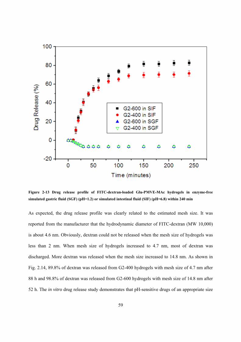
59
Figure 2-13 Drug release profile of FITC-dextran-loaded Glu-PMVE-MAc hydrogels in enzyme-free
simulated gastric fluid (SGF) (pH=1.2) or simulated intestinal fluid (SIF) (pH=6.8) within 240 min
As expected, the drug release profile was clearly related to the estimated mesh size. It was
reported from the manufacturer that the hydrodynamic diameter of FITC-dextran (MW 10,000)
is about 4.6 nm. Obviously, dextran could not be released when the mesh size of hydrogels was
less than 2 nm. When mesh size of hydrogels increased to 4.7 nm, most of dextran was
discharged. More dextran was released when the mesh size increased to 14.8 nm. As shown in
Fig. 2.14, 89.8% of dextran was released from G2-400 hydrogels with mesh size of 4.7 nm after
88 h and 98.8% of dextran was released from G2-600 hydrogels with mesh size of 14.8 nm after
52 h. The in vitro drug release study demonstrates that pH-sensitive drugs of an appropriate size

60
could potentially be retrained in the hydrogel matrix in the stomach and released effectively in
the small intestine.
Figure 2-14 Drug release profile of FITC-dextran-loaded Glu-PMVE-MAc hydrogels in enzyme-free
simulated gastric fluid (SGF) (pH=1.2) or simulated intestinal fluid (SIF) (pH=6.8) within 120 h
2.4 Conclusions
Glucosamine grafted glycopolymer poly(methyl vinyl ether-alt-maleic acid) (Glu-PMVE-MAc)
was successfully synthesized with high yield. The structure of Glu-PMVE-MAc was confirmed
by FT-IR and 1H-NMR. Elemental analysis was utilized to determine the amount of grafted
glucosamine groups. A glycopolymer hydrogel was produced via crosslinking Glu-PMVE-MAc
by PEG 400 or PEG 600. The swelling behavior in aqueous solution from pH of 1.2 to 7.4 was

61
studied. The mesh size was estimated from equilibrium swelling data using Peppas-Merrill
equation. FITC-dextran with a molecular weight of 10,000 was used as a model drug to
investigate the drug delivery profile in enzyme-free simulated gastric fluid (SGF) (pH=1.2) and
enzyme-free simulated intestinal fluid (SIF) (pH=6.8). None of the dextran was released in SGF,
however, most was discharged in SIF. Therefore, these hydrogels could potentially be used to
transport drugs through the stomach and to be released in the intestinal tract.
2.5 References
Allard L, Cheynet V, Oriol G, Veron L, Merlier F, Scremin G, Mandrand B, Delair T, and Mallet
F. Bioconjugate Chemistry, 2001, 12, 972-979
Arakawa T, and Timasheff SN. Biochemistry, 1982, 21, 6536-6544
Auzely-Velty R, Cristea M, and Rinaudo M. Biomacromolecules, 2002, 3, 998-1005
Cade D, Ramus E, Rinaudo M, Auzely-Velty R, Delair T, and Hamaide T. Biomacromolecules,
2004, 5, 922-927
Donati I, Gamini A, Vetere A, Campa C, and Paoletti S. Biomacromolecules, 2002, 3, 805-812
Galgali P, Varma AJ, Puntambekar US, and Gokhale DV. CHEM. COMMUN. , 2002, 2884-
2885
Galgalia P, Puntambekarb US, Gokhaleb DV, and Varma AJ. Carbohydrate Polymers, 2004, 55,
393-399
Galgali P, Agashe M, and Varma AJ. Carbohydrate Polymers, 2007, 67, 576-585

62
Garland MJ, Raj-Singh TR, Woolfson AD, and Donnelly RF. International Journal of
Pharmaceutics, 2011, 406, 91-98
Goetz L, Mathew A, Oksman K, Gatenholm P, and Ragauskas AJ. Carbohydrate Polymers,
2009, 75, 85-89
Goetz L, Foston M, Mathew AP, Oksman K, and Ragauskas AJ. Biomacromolecules, 2010, 11,
2660-2666
Grombe R, Gouzy MF., Nitschke M, Komber H, and Werner C. Colloids and Surfaces A:
Physicochem. Eng. Aspects, 2006, 284–285, 295-300
Grombe R, Gouzy MF, Maitz MF, Freundenberg U, Zschoche S, Simon F, Pompe T, Sperling C,
and Werner C. Macromol. Biosci., 2007, 7, 195-200
Gudeman LF and Peppas NA. J. Appl. Polym. Sci., 1995, 55, 919-928
Guo Xh, Deng F, Li L, and Prudhomme RK. Biomacromolecules, 2008, 9, 1637-1642
Isosaki K, Seno N, and Matsumoto I. Journal of Chromatography, 1992, 597, 123-128
Jeffrey PD. Biochemistry, 1974, 13, 4441-4447
Kim B, and Peppas NA. J. Biomater. Sci. Polymer Edn, 2002, 13, 1271-1281
Ladaviere C, Lorenzo C, Elaıssari A, Mandrand B, and Delair T. Bioconjugate Chemistry, 2000,
11, 146-152
Lee JC, Frigon RP, and Timasheff SN. Ann N.Y. Acad. Sci., 1975, 253, 284-291

63
Lee JC, and Timasheff SN. J. Biol. Chem., 1981, 256, 7 193-7201
Luppi B, Cerchiara T, Bigucci F, Di Pietra AM, Orienti I, and Zecchi V. Drug Delivery, 2003,
10, 239-244
Mahkam M. Drug Delivery, 2007, 14, 147-153
Miyata T, Jijihara A, and Nakamae K. Macromol. Chem. Phys. 1996, 197, 1135-1146
Paraskar AS, Soni S, Chin KT, Chaudhuri P, Muto KW, Berkowitz J, Handlogten MW, Alves
NJ, Bilgicer B, Dinulescu DM, Mashelkar RA, and Sengupta S. PNAS, 2010, 107, 12435–12440
Paraskar A, Soni S, Roy B, Papa AL and Sengupta S. Nanotechnology, 2012, 23, 75-103
Peppas NA, and Merrill EW. J. Appl. Polym. Sci., 1977, 21, 1763-1770
Polikarpov N, Kaufmann A, and Kluge J, abstracts/ journal of controlled release, 2010, 148, 66-
67
Raj-Singh TR, McCarron PA, Woolfson AD, and Donnelly RF. European Polymer Journal,
2009, 45, 1239–1249
Raj-Singh TR, Woolfson AD, and Donnelly RF. Journal of Pharmacy and Pharmacology, 2010,
62, 829–837
Su Rm, Li L, Chen Xp, Han Jh, and Han Sf. Org. Biomol. Chem., 2009, 7, 2040–2045
Uzawa H, Ito H, Izumi M, Tokuhisa H, Taguchi K, and Minoura N. Tetrahedron, 2005, 61,
5895–5905

64
Varma AJ, Kennedy JF, and Galgali P. Carbohydrate Polymers, 2004, 56, 429-445
Vetere A, Donati I, Campa C, Semeraro S, Gamini A, and Paoletti S. Glycobiology, 2002, 12,
283-290
Zeng XG, Chen L, and Yang XZ. Chemical Journal of Chinese Universities, 2005, 26, 1561-
1564
Zhang Yl, Yao Qj, Xia Cf, Jiang X, and Wang PG. ChemMedChem, 2006, 1, 1361-1366

65
CHAPTER 3 SYNTHESIS OF AMPHIPHILIC GLYCOPOLYMER (PHEMAGI-
PPO) USING ATOM TRANSFER RADICAL POLYMERIZATION (ATRP) FOR
DRUG DELIVERY APPLICATION
3.1 Introduction
3.1.1 Glycopolymers in micelle formation
A micelle is an aggregate of amphiphilic molecules, which are molecules consisting of
hydrophilic and hydrophobic blocks, dispersed in form of a liquid colloid. It has a hydrophobic
core and a hydrophilic shell. As a drug delivery device, micelles minimize drug degradation and
loss upon administration, lessen harmful or undesirable side-effects, increase drug
bioavailability, and control drug release to maximize efficiency and minimize toxicity of the
drug. In aqueous solution, the hydrophilic outer layer creates a highly water-bound barrier, which
blocks the adhesion of opsonins. Therefore lower the impact of immune system. (Yamamoto et
al. 2001). Imaging contrast agents and multiple drugs can be loaded into a single micelle,
allowing both diagnosis and therapy (Ferrari 2005). Compared to micelles structured from
smaller molecules, polymeric micelles have much lower critical micelle concentration (CMC),
defined as the lowest concentration of amphiphilic molecules to form a micellar structure. Low
CMC enhances the stability of drugs incorporated in the micelle. Moreover, the larger size of
polymeric micelles retards the rate of body clearance by renal filtration. Therefore they can stay
in the body for a longer time for long-term drug release.
If a sugar-containing block serves as the hydrophilic block, an amphiphilic polymer can be
classified as glycopolymer. Glycopolymers are defined as synthetic polymers that incorporate
carbohydrate groups. They may play an important role in a wide range of biomolecular events
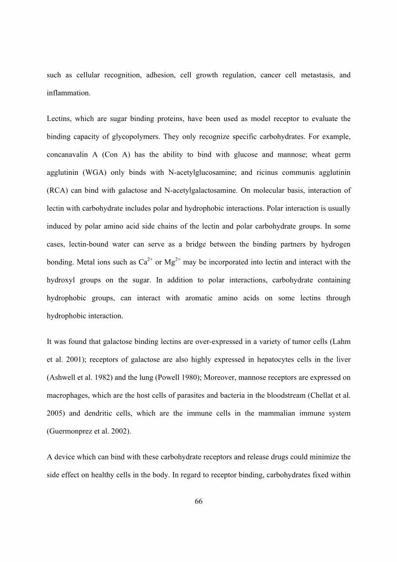
66
such as cellular recognition, adhesion, cell growth regulation, cancer cell metastasis, and
inflammation.
Lectins, which are sugar binding proteins, have been used as model receptor to evaluate the
binding capacity of glycopolymers. They only recognize specific carbohydrates. For example,
concanavalin A (Con A) has the ability to bind with glucose and mannose; wheat germ
agglutinin (WGA) only binds with N-acetylglucosamine; and ricinus communis agglutinin
(RCA) can bind with galactose and N-acetylgalactosamine. On molecular basis, interaction of
lectin with carbohydrate includes polar and hydrophobic interactions. Polar interaction is usually
induced by polar amino acid side chains of the lectin and polar carbohydrate groups. In some
cases, lectin-bound water can serve as a bridge between the binding partners by hydrogen
bonding. Metal ions such as Ca2+ or Mg2+ may be incorporated into lectin and interact with the
hydroxyl groups on the sugar. In addition to polar interactions, carbohydrate containing
hydrophobic groups, can interact with aromatic amino acids on some lectins through
hydrophobic interaction.
It was found that galactose binding lectins are over-expressed in a variety of tumor cells (Lahm
et al. 2001); receptors of galactose are also highly expressed in hepatocytes cells in the liver
(Ashwell et al. 1982) and the lung (Powell 1980); Moreover, mannose receptors are expressed on
macrophages, which are the host cells of parasites and bacteria in the bloodstream (Chellat et al.
2005) and dendritic cells, which are the immune cells in the mammalian immune system
(Guermonprez et al. 2002).
A device which can bind with these carbohydrate receptors and release drugs could minimize the
side effect on healthy cells in the body. In regard to receptor binding, carbohydrates fixed within

67
a glycopolymer structure are more efficient than free carbohydrates due to the multivalent effect
of the clustered saccharide (Lee et al. 1995). Therefore, glycopolymer micelles have the potential
to be used as targeted drug delivery carrier. With the sugar moieties on the hydrophilic block,
glycopolymer micelles may interact with the receptors mentioned above, therefore enhancing the
targeting efficiency of incorporated drugs.
3.1.2 Synthesis of amphiphilic glycopolymers
Amphiphilic glycopolymers were generally synthesized by controlled radical polymerization
which can produce a nearly perfect amphiphilic structure. Controlled polymerization included
atom transfer radical polymerization (ATRP), reversible addition-fragmentation chain transfer
polymerization (RAFT) and nitroxide mediated radical polymerization (NMRP). In the following
paragraphs, glycopolymers obtained by these methods are briefly introduced.
Figure 3-1 Star-shaped poly(ε-caprolactone)-b-poly(D-gluconamidoethyl methacrylate) glycopolymers
(SPCL-PGAMA) (Dai et al. 2008)

68
Figure 3-2 Star-shaped poly(ε-caprolactone)-b-poly(D-lactobionamidoethyl methacrylate) glycopolymers
(SPCL-PLAMA) (Zhou et al. 2008)
Several amphiphilic glycopolymers were produced by ATRP. Dai et al. (2008) synthesized a
biodegradable star-shaped poly(ε-caprolactone)-b-poly(D-gluconamidoethyl methacrylate)
glycopolymer (SPCL-PGAMA) (shown in Fig. 3.1) in 2008. Zhou et al. (2008) further reported a
biodegradable star-shaped poly(ε-caprolactone)-b-poly(D-lactobionamidoethylmethacrylate)
glycopolymer (SPCL-PLAMA) (Fig. 3.2) in the same year. Later on, an amphiphilic
glycopolymer (shown in Fig. 3.3) consisting of poly(2-{[(D-glucosamin-2N-yl)carbonyl]-
oxy}ethyl methacrylate (PHEMAGl)) as hydrophilic block and poly(n-butyl acrylate) (PBA) as
hydrophobic block were prepared by Leon et al. (2010). Amphiphilic glycopolymers synthesized
by ATRP were also prepared by Narain et al. (2003), Qiu et al. (2009), and Dai et al. (2009).
RAFT synthesized amphiphilic glycopolymers were reported by Liu et al. (2010), Hetzer et al.
(2010), and Xiao et al. (2011). In 2009, Ting et al. (2009) obtained an amphiphilic diblock

69
glycopolymer: poly(2-(β-D-galactosyloxy)ethyl methacrylate-co-styrene)-b-polystyrene
(P(GalEMA-co-S)-b-PS) (Fig. 3.4) using NMRP.
Figure 3-3 Poly(2-{[(D-glucosamin-2N-yl)carbonyl]- oxy}ethyl methacrylate-b-poly(n-butyl acrylate)
(PHEMAGl-PBA) (Leon et al. 2010)
Figure 3-4 Poly(2-(β-D-galactosyloxy)ethyl methacrylate-co-styrene)-b-polystyrene (P(GalEMA-co-S)-b-PS)
(Ting et al. 2009)
3.1.3 Formation of glycopolymer micelles
Formation of most glycopolymer micelles was done by adding distilled water slowly into a
glycopolymer organic solution followed by dialysis. In addition to dialysis stimulated micelles,
there are some stimuli-responsive glycopolymers that could form when dissolving in a specific
environment.

70
A poly(ethylene oxide)-b-poly(D-gluconamidoethyl methacrylate)-b-poly(2-(diethylamino)ethyl
methacrylate) (PEO-GAMA-DEA) (Fig. 3.5) was synthesized by Narain et al. (2003). DEA
homopolymer is a weak polybase with a PKb of around 6.7 and is insoluble in neutral or alkaline
aqueous solution. 1H-NMR spectra and surface tension measurements of the triblock copolymer
showed the pH-responsive feature of DEA block. The amphiphilic copolymer formed micelles in
alkaline aqueous solution. A poly(propylene oxide)-b-poly(D-gluconamidoethyl methacrylate)
(PPO-GAMA) (Fig. 3.5) was also reported by Narain et al. in the same paper. The thermo-
responsive feature of PPO block was determined by 1H-NMR spectra and surface tension
measurement of the diblock copolymer. Below 5 ºC, the PPO-GAMA diblock copolymer was
molecularly dissolved. However, micelles were formed at 20 ºC.
Figure 3-5 Poly(ethylene oxide)-poly(D-gluconamidoethyl methacrylate)-poly(2-(diethylamino)ethyl
methacrylate) (PEO-GAMA-DEA) and poly(propylene oxide)-poly(D-gluconamidoethyl methacrylate) (PPO-
GAMA) (Narain et al. 2003)
Liu et al. (2010) prepared a pH-responsive amphiphilic glycopolymer composed of a block of
poly(2-(diethylamino)ethyl methacrylate) (PDEA) and a block of poly(3-O-methacryloy-α,β-D-
glucopyranose) (PMAGlc) (as shown in Fig. 3.6). The pH-responsive feature of DEA block was
also confirmed by 1H-NMR spectra and transmittance measurement. The glycopolymer self-
assembled at alkaline pH.

71
Figure 3-6 Poly(3-O-methacryloy-α,β-D-glucopyranose)-b-poly(2-(diethylamino)ethyl methacrylate)
(PMAGlc-PDEA) (Liu et al. 2010)
Hetzer et al. (2010) synthesized a thermo-responsive amphiphilic glycopolymer: poly(2’-(4-
vinyl-[1,2,3]-triazol-1-yl)ethyl-O-α-D-mannopyranoside)-b-poly(N-isopropyl acrylamide) (Fig.
3.7). Poly(N-isopropyl acrylamide) (NIPAAm) is a common thermo-responsive polymer which
is water-soluble at room temperature. However, it becomes water-insoluble above 32ºC.
Therefore, the amphiphilic glycopolymer formed micelles at the elevated temperature above
40ºC. The formation of micelles was confirmed by measuring the micelle size by dynamic light
scattering (DLS) at different temperatures.
Figure 3-7 Poly(2’-(4-vinyl-[1,2,3]-triazol-1-yl)ethyl-O-α-D-mannopyranoside)-b-poly(N-isopropyl
acrylamide) (Hetzer et al. 2010)
A thermo-responsive amphiphilic glycopolymer (AcMH-b-PEtOz-b-AcMH) (as shown in Fig.
3.8), based on peracetylated maltoheptaose (AcMH) and poly(2-ethyl-2-oxazoline) (PEtOz), was
prepared by Chen et al. (2010). Poly (2-ethyl-2- oxazoline) (PEtOz) is a thermo-sensitive

72
polyelectrolyte with good biocompatibility and biodegradability. The thermo-responsive feature
of the glycopolymer was determined by optical transmittance measurement from 25 ºC to 65 ºC.
Micelles were prepared by direct dissolution in water at room temperature.
Figure 3-8 Peracetylated maltoheptaose-b-poly(2-ethhyl-2-oxazoline)-b-peracetylated maltoheptaose (AcMH-
b-PEtOz-b-AcMH) (Chen et al. 2010)
3.1.4 CMC of amphiphilic glycopolymers
Table 3.1 CMC of amphiphilic glycopolymers and common surfactant
Amphiphilic Glycopolymers
Author Year Composition CMC (μM)
Dai et al.
2008
SPCL15-PGAMA7 1.886
SPCL15-PGAMA18 1.409
Zhou et al.
2008
SPL15-PLAMA7 0.422
SPL15-PLAMA3 0.35
SPL75-PLAMA5 0.0751
Qiu et al.
2009
SPBLG36-PGAMA7 0.411
SPBLG30-PGAMA13 0.62

73
SPBLG36-PGAMA39 0.417
Dai et al.
2009
PAMAM-PCL28-PGAMA3 0.125
PAMAM-PCL28-PGAMA28 0.0423
Chen et al.
2010
AcMH-b-PEtOz50-b-AcMH 0.241
AcMH-b-PEtOz80-b-AcMH 0.376
Common Surfactants
Name Type CMC (μM)
Sodium dodecyl sulfate (SDS) Anionic 8200
Cetyltrimethylammonium bromide (CTAB) Cationic 920
Polysorbate 20 (Tween 20) Nonionic 42
Polysorbate 80 (Tween 80) Nonionic 28
As shown in Table 3.1, the CMC of amphiphilic glycopolymers was determined to be in the
magnitude of 101 to 10-2 μM; however, the CMC of common surfactants was much larger. As
mentioned above, the smaller CMC of the micelles increases the stability of drugs incorporated
in them.
3.1.5 Hydrodynamic diameter (Dh) of glycopolymer micelles and their interaction with
lectins
Data in Table 3.2 show that the Dh of glycopolymer micelles formed from amphiphilic
glycopolymers are much greater than micelles structured by common surfactants. As mentioned
above, the larger size of polymeric micelles makes it possible for these micelles to remain in the
body for a longer time and thus for an extended drug release.
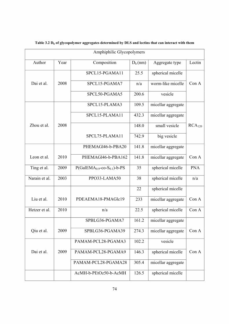
74
Table 3.2 Dh of glycopolymer aggregates determined by DLS and lectins that can interact with them
Amphiphilic Glycopolymers
Author Year Composition Dh (nm) Aggregate type Lectin
Dai et al.
2008
SPCL15-PGAMA11 25.5 spherical micelle
Con A SPCL15-PGAMA7 n/a worm-like micelle
SPCL50-PGAMA5 200.6 vesicle
Zhou et al.
2008
SPCL15-PLAMA3 109.5 micellar aggregate
RCA120
SPCL15-PLAMA11 432.3 micellar aggregate
SPCL75-PLAMA11
148.0 small vesicle
742.9 big vesicle
Leon et al.
2010
PHEMAGl46-b-PBA20 141.8 micellar aggregate
Con A PHEMAGl46-b-PBA162 141.8 micellar aggregate
Ting et al. 2009 P(GalEMA0.9-co-S0.1)-b-PS 35 spherical micelle PNA
Narain et al. 2003 PPO33-LAMA50 38 spherical micelle n/a
Liu et al.
2010
PDEAEMA18-PMAGlc19
22 spherical micelle
Con A 233 micellar aggregate
Hetzer et al. 2010 n/a 22.5 spherical micelle Con A
Qiu et al.
2009
SPBLG36-PGAMA7 161.2 micellar aggregate
Con A SPBLG36-PGAMA39 274.3 micellar aggregate
Dai et al.
2009
PAMAM-PCL28-PGAMA3 102.2 vesicle
Con A PAMAM-PCL28-PGAMA9 146.3 spherical micelle
PAMAM-PCL28-PGAMA28 305.4 micellar aggregate
AcMH-b-PEtOz50-b-AcMH 126.5 spherical micelle
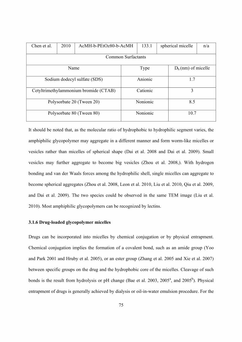
75
Chen et al. 2010 AcMH-b-PEtOz80-b-AcMH 133.1 spherical micelle n/a
Common Surfactants
Name Type Dh (nm) of micelle
Sodium dodecyl sulfate (SDS) Anionic 1.7
Cetyltrimethylammonium bromide (CTAB) Cationic 3
Polysorbate 20 (Tween 20) Nonionic 8.5
Polysorbate 80 (Tween 80) Nonionic 10.7
It should be noted that, as the molecular ratio of hydrophobic to hydrophilic segment varies, the
amphiphilic glycopolymer may aggregate in a different manner and form worm-like micelles or
vesicles rather than micelles of spherical shape (Dai et al. 2008 and Dai et al. 2009). Small
vesicles may further aggregate to become big vesicles (Zhou et al. 2008,). With hydrogen
bonding and van der Waals forces among the hydrophilic shell, single micelles can aggregate to
become spherical aggregates (Zhou et al. 2008, Leon et al. 2010, Liu et al. 2010, Qiu et al. 2009,
and Dai et al. 2009). The two species could be observed in the same TEM image (Liu et al.
2010). Most amphiphilic glycopolymers can be recognized by lectins.
3.1.6 Drug-loaded glycopolymer micelles
Drugs can be incorporated into micelles by chemical conjugation or by physical entrapment.
Chemical conjugation implies the formation of a covalent bond, such as an amide group (Yoo
and Park 2001 and Hruby et al. 2005), or an ester group (Zhang et al. 2005 and Xie et al. 2007)
between specific groups on the drug and the hydrophobic core of the micelles. Cleavage of such
bonds is the result from hydrolysis or pH change (Bae et al. 2003, 2005a, and 2005b). Physical
entrapment of drugs is generally achieved by dialysis or oil-in-water emulsion procedure. For the

76
dialysis method, the drug and amphiphilic copolymer are first dissolved in a solvent in which
they are both soluble. The solution is added to a solvent that is selective only for the hydrophilic
part of the polymer. As the good solvent is replaced by the selective one, the drug is incorporated
while forming micelles. In the oil-in-water emulsion method, an aqueous solution of the
copolymer is added to a solution of the drug in a water-insoluble volatile solvent to form an oil-
in-water emulsion. The drug-loaded micelles are formed as the solvent evaporates. The
physically entrapped drug is released from the micelle by diffusion and upon dissociation of
micelle. Micelle dissociation may result from physical stimuli such as pH (Lee et al. 2003),
temperature (Wei et al. 2006, Nakayama et al. 2006), ultrasound (Husseini et al. 2000 and 2007,
Pruitt et al. 2002, Gao et al. 2005), ultraviolet (Lepage et al. 2007), and near-infrared light
(Goodwin et al. 2005). The incorporated drug may be released at the specific site where those
physical stimuli are applied.
Drug-loaded glycopolymer micelles have been reported by several research groups. A star-
shaped glycopolymer composed of poly(γ-benzyl L-glutamate) and poly(D-gluconamidoethyl
methacrylate) (SPBLG-PGAMA) (shown in Fig. 3.9) was reported by Qiu et al. (2009). An
anticancer drug: doxorubicin was loaded into the micelle aggregates via dialysis method with
loading efficiency of about 20%. Dh of doxorubicin-loaded micelles were found to be more than
twice compared to the blank micelles. Doxorubicin was released in vitro for 1000 h in a triphasic
pattern. About 15% of drug was discharged in 15 h, about 45% of drug was released in 12 days,
and about 20% of drug was delivered within 1 month.

77
Figure 3-9 Star-shaped poly(γ-benzyl L-glutamate)-b-poly(D-gluconamidoethyl methacrylate) (SPBLG-
PGAMA) (Qiu et al. 2009)
A star poly(amido amine)-b-poly(ε-caprolactone)-b-poly(D-gluconamidoethyl methacrylate)
(PAMAM– PCL–PGAMA) (Fig. 3.10) block glycopolymer with a dendrimer core was prepared
by Dai et al. (2009). Nimodipine-loaded micelle aggregates were fabricated by dialysis method.
A high drug loading efficiency of 74.1% was reported. The in vitro drug release underwent a
burst release for about 10 h and a slow release for about 160 h.

78
S
S
OS
S
SS
S
SS
SS
OO
C
O
C
CH3
CH3
CH2 CCH3
OO
Brm
OHHO
HO
OH
NH
OOH
n
OO
O
CO
CCH3
CH3
CH2 C
CH3
OO
Brm
OHHO
HO
OH
NH
OOH
nS =
Figure 3-10 Star poly(amido amine)-b-poly(ε-caprolactone)-b-poly(D-gluconamidoethyl methacrylate)
(PAMAM– PCL–PGAMA) (Dai et al. 2009)
Indomethacin was incorporated into the thermo-responsive amphiphilic glycopolymer (AcMH-b-
PEtOz-b-AcMH) (Fig. 3.8) mentioned above (Chen et al. 2010). The formation of indomethacin-
loaded micelles was achieved by adding distilled water to ethanol solution of glycopolymer and
indomethacin. Unloaded indomethacin was removed by centrifugation and filtration. The
incorporation of indomethacin was confirmed by determining the chemical structure by 1H
NMR, and measuring the size by DLS and TEM. In vitro drug release profiles were obtained at
different temperatures. The indomethacin release profile was apparently different at 25ºC and
37ºC compared to that at 45ºC and 55ºC due to the thermo-responsive property.
The biodegradable and biocompatible aldehyde-functionalized glycopolymer poly(6-O-(2’-
formyl-4’-vinylphenyl)-D-galactopyranose-b-5,6-benzo-2-methylene-1,3-dioxepane) (PVDG-
BMDO) (Fig. 3.11) was synthesized by Xiao et al. (2011). Doxorubicin was conjugated onto the
glycopolymer bearing highly reactive aldehyde groups via an acid-labile Schiff base linkage. The

79
drug efficiency was determined to be as high as 14%. The doxorubicin-loaded micelles exhibited
an average hydrodynamic diameter of about 125 nm determined by DLS. TEM images showed a
slightly smaller diameter. Lower cyctoxicity of doxorubicin-loaded micelles than free
doxorubicin was reported. In vitro release studies indicated that only about 10% of doxorubicin
was released at pH of 7.4 in 120 h. However, about 63% of doxorubicin was discharged at a pH
of 5.0 in the same time period. The pH dependent release profile was attributed to the acid-
cleavable Schiff base linkage. This feature might be useful since tumor sites and inflammatory
tissues have a more acidic environment (Ulbrich et al. 2000, Etrych et al. 2002, Ulbrich et al.
2004, Hruby et al. 2005, and Chytil et al. 2006).
Figure 3-11 Poly(6-O-(2’-formyl-4’-vinylphenyl)-D-galactopyranose-b-5,6-benzo-2-methylene-1,3-dioxepane)
(PVDG-BMDO) (Xiao et al. 2011)
As mentioned above, glycopolymer micelles which are mostly biocompatible, have lower CMC
values and larger sizes than their smaller molecule counterparts (e.g., common surfactant
micelles). Most glycopolymer micelles can be recognized by lectin. Drugs can be incorporated
into the glycopolymer micelles and be released in a controlled manner. In addition, drugs could
be released from stimuli-responsive glycopolymer micelles upon environmental changes. All

80
these features might result in a stable drug-carrying device which could stay in the body for a
longer period of time and release drugs to a targeted site in a regulated manner.
3.1.7 Approach
In this work, a thermo-responsive amphiphilic glycopolymer: poly(2-{[(D-glucosamin-2N-
yl)carbonyl]-oxy}ethylmethacrylate)-b-poly(propylene oxide) (PHEMAGI-PPO) was
synthesized via atom transfer radical polymerization (ATRP). The chemical structure of
glycomonomer (HEMAGI), macroinitiator (PPO-Br), and glycopolymer was confirmed by 1H-
NMR or 13C-NMR spectra. Degree of functionalization of PPO-Br was determined to be more
than 99%. Molecular weight of glycopolymers was estimated from integral ratio of specific
peaks on NMR spectra. The CMC of the glycopolymer was measured by dye micellization
method and the diameter of the formed particles was determined by DLS. The lectin recognition
property was determined using Con A as a model lectin.
3.2 Experimental part
3.2.1 Materials
2-Hydroxyethyl methacrylate (97%), triethylamine (99%), tetrahydrofuran (THF) (99%), 4-
nitrophenyl chloroformate (97%), dimethyl sulfoxide (DMSO) (99%), diethyl ether (99%),
dichloromethane (99.5%) were all from Alfa Aesar and used without further purification.
Anhydrous magnesium sulfate (MgSO4) (99%, Strem Chemicals), D-glucosamine hydrochloride
(99.9%, Calbiochem), dimethylformamide (99.9%, Fisher), methanol (99.8%, EMD),
phosphorus pentaoxide (98%, Spectrum), mono-capped poly(propylene oxide) (Mn=2500,
Sigma-Aldrich), toluene (99.9%, Fisher), 2-bromoisobutyryl bromide (97%, TCI), decolorizing

81
charcoal (Sigma-Aldrich), Eosin Y (0.5% w/v, Sigma-Aldrich), and Concanavalin A (Sigma-
Aldrich) were used as received.
3.2.2 Synthesis of 2-{[(D-glucosamine-2N-yl)carbonyl] oxy}ethyl methacrylate (HEMAGl)
The glycomonomer, HEMAGI was synthesized by modifying the procedure previously reported
(Leon, et al., 2010; shown in Scheme 3.1). Briefly, 2-hydroxyethyl methacrylate (HEMA) (0.021
mol, 2.73 g), triethylamine (0.025 mol, 2.52 g), anhydrous magnesium sulfate (1.5 g), and THF
(10 ml) were placed in a 50 mL flask, which was entirely isolated from the outside to avoid any
contact with humidity. Once the solution was equilibrated at 4ºC, 4-nitrophenyl chloroformate
(0.021 mol, 5.076 g) in pre-dried THF (10 mL) was successively added while stirring. The
reaction was performed at 4ºC for 24 h. The formed triethyl amine chlorohydrate and magnesium
sulfate were filtered off. The product was poured into 100 mL water and separated by separatory
funnel. (Yield: 82.2 %)
Subsequently, the modified monomer with p-nitrophenylcarbonate groups, HEMAN, (0.0085
mol, 2.51 g) was dissolved in 10 mL anhydrous DMSO at 30ºC. Triethylamine (0.01275 mol,
1.288 g), D-glucosamine chloride (0.0085 mol, 1.832 g) and anhydrous magnesium sulfate (1.5
g) were then added while stirring. The reaction was maintained for 24 h at 30ºC. After that,
magnesium sulfate was filtered off. The glycomonomer, HEMAGl, was poured into 100 mL
diethyl ether/dichloromethane mixture (4:1, v/v) and isolated by separatory funnel. The product
was purified by solubilization in DMF/methanol (1:4, v/v) and reprecipitation in diethyl ether/
dichloromethane mixture (4:1, v/v). The resulting product was dried in vacuum at room
temperature in the presence of phosphorus pentoxide until constant weight was reached (Yield:
58.6 %).
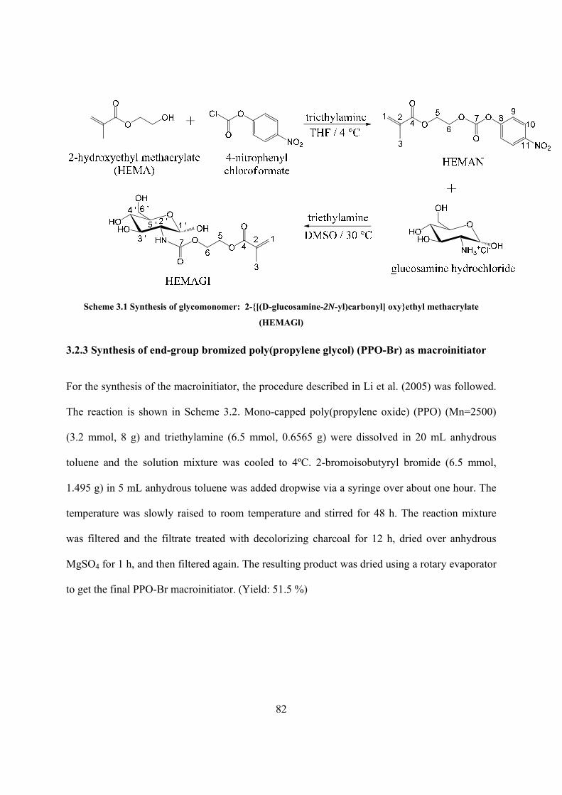
82
Scheme 3.1 Synthesis of glycomonomer: 2-{[(D-glucosamine-2N-yl)carbonyl] oxy}ethyl methacrylate
(HEMAGl)
3.2.3 Synthesis of end-group bromized poly(propylene glycol) (PPO-Br) as macroinitiator
For the synthesis of the macroinitiator, the procedure described in Li et al. (2005) was followed.
The reaction is shown in Scheme 3.2. Mono-capped poly(propylene oxide) (PPO) (Mn=2500)
(3.2 mmol, 8 g) and triethylamine (6.5 mmol, 0.6565 g) were dissolved in 20 mL anhydrous
toluene and the solution mixture was cooled to 4ºC. 2-bromoisobutyryl bromide (6.5 mmol,
1.495 g) in 5 mL anhydrous toluene was added dropwise via a syringe over about one hour. The
temperature was slowly raised to room temperature and stirred for 48 h. The reaction mixture
was filtered and the filtrate treated with decolorizing charcoal for 12 h, dried over anhydrous
MgSO4 for 1 h, and then filtered again. The resulting product was dried using a rotary evaporator
to get the final PPO-Br macroinitiator. (Yield: 51.5 %)

83
RO
OHn
mono-capped poly(propylene oxide)
+ BrBr
O
triethylamineToluene / 4 °Cthen room temp.
2-bromoisobutyryl bromide
RO
b
cO
a
n BrO
d d
PPO-Br
Scheme 3.2 Synthesis of macroinitiator poly(propylene oxide)-Br (PPO-Br)
3.2.4 Atom transfer radical polymerization of amphiphilic glycopolymer PHEMAGI-PPO
HEMAGI was dissolved in a methanol/water (4/1, v/v) mixed solvent (10 mL). Then PPO-Br
macroinitiator was added and the solution was degassed via argon gas purge. After 30 min, the
copper(I) bromide (CuBr) catalyst and 2,2’-bipyridine (bpy) were added to the reaction solution,
which turned dark brown. After reacting at 20 ºC for 24 h, the solution was exposed to air and
diluted with methanol, which led to aerial oxidation of the ATRP catalyst. The final product was
obtained by drying under vacuum using rotary evaporator. Scheme 3.3 illustrates the process.
The amount of utilized reagents is listed in Table 3.3.
Scheme 3.3 Atom transfer radical polymerization of amphiphilic glycopolymer: PHEMAGI-PPO

84
Table 3.3 Amount of utilized reagents for synthesis of PHEMAGI-PPO
HEMAGI PPO-Br CuBr bpy Targeted Composition
1.35 g,
4.03 mmol
0.1625 g,
0.065 mmol
9.5 mg,
0.065 mmol
20 mg,
0.13 mmol
PHEMAGI62-PPO41
1.35g
4.03 mmol
0.219 g
0.088 mmol
12.86 mg
0.088 mmol
27.08 mg
0.176 mmol
PHEMAGI46-PPO41
1.35 g,
4.03 mmol
0.325 g,
0.013 mmol
19 mg,
0.013 mmol
40 mg,
0.026 mmol
PHEMAGI31-PPO41
1.35 g,
4.03 mmol
0.65 g,
0.026 mmol
38 mg,
0.026 mmol
80 mg,
0.052 mmol
PHEMAGI15-PPO41
3.2.5 Characterization
3.2.5.1 Structural characterization by nuclear magnetic resonance (NMR) spectroscopy
1H and 13C NMR were used to determine the structure of the synthesized products. Spectra were
recorded at room temperature on solution in deuterated dimethyl sulfoxide (DMSO-d6) or
deuterated chloroform (CDCl3) with a Bruker 400 spectrometer at 400 MHz. Typical parameters
for the proton spectra were a 15 s pulse delay, a 3 s acquisition time, a 20.68 ppm spectral width,
and 16 scans.
3.2.5.2 Critical micelle concentration (CMC) determination using UV-Vis spectroscopy
The CMC was measured by dye micellization method (Patist et al. 2000) with Eosin Y as
indicator. Absorbance of solutions which contained equal amounts of Eosin Y but increasing
amounts of PHEMAGI-PPO at 542 nm was measured by UV-Vis spectroscopy (Thermo

85
Spectronic Genesys 6). The straight line portion was linear-fitted by Origin 8.0. The interception
of the fitted line and the baseline which was the absorbance without PHEMAGI-PPO in solution
was determined to be the critical micelle concentration.
3.2.5.3 Thermo-responsive behavior of PHEMAGI-PPO micelles
Thermo-responsive behavior of the micelles (2 mg/ml) was measured on a UV-Vis spectroscopy
(Thermo Spectronic Genesys 6) incorporated with a thermocouple. Optical transmittance at
wavelength of 600 nm from 8 ˚C to 22 ˚C was recorded. Temperature at which the transmittance
is 50% of original was defined as the lower critical solution temperature (LCST) (Wang et al.
2003, Weberm et al. 2009).
3.2.5.4 Micellar size measurement in aqueous solution by transmission electron microscopy
(TEM)
The particle size was measured using a Zeiss EM 10CR transmission electron microscopy. One
drop of micellar solution was deposited onto the surface of 300 mesh Formvar-carbon film-
coated copper grids. Excess solution was removed by filter paper. Negative staining using
phosphotungstic acid (0.5 wt %) was carried out to increase the contrast of the specimens. Thirty
micelles each were measured form the images. The average diameter and average deviation was
calculated.
To induce the desired micelle formation, PHEMAGI-PPO was dissolved in water at 4˚C, and the
temperature was slowly increased within 1 h to reach room temperature.
3.2.5.5 Interaction of PHEMAGI-PPO micelles with Con A

86
The lectin recognition property was evaluated by changes in the solution turbidity. Buffer
solution (pH = 7.2) was prepared by adding 0.01 M KH2PO4, 1 mM MnCl2, 1 mM CaCl2, and
0.5 M NaCl in deionized water. Glycopolymer micelles were formed in the buffer solution by
cooling method described above. Con A buffer solution at 0.5 mg/mL was added to buffer
solutions of glycopolymer micelles with various concentration. The solution was stirred
intensively 1 h after adding Con A buffer solution. Then, the turbidity was measured by a UV-
Vis spectrometer (Thermo Spectronic Genesys 6) at 360 nm and room temperature.
3.3 Results and Discussion
3.3.113C-NMR characterization of HEMAN
Figure 3-12 13C-NMR spectra of HEMAN, HEMAN: (400MHz, CDCl3), δ(ppm): 17.7 (C-3), 61.6 (C-5), 66.6 (C-6), 121.5 (C-7), 124.3 (C-8), 125.7 (C-1), 135.5 (C-2), 145.0 (C-11), 152.0 (C-7), 155.1 (C-8), and 166.4 (C-4)

87
The chemical structure of HEMAN was confirmed by 13C-NMR spectra shown in Fig. 3.12. The
signal of C-7 at 121.5 ppm confirmed the success of the reaction. Signals of C-8 to C-11
represent the benzene ring. Signals of C-1 and C-3 indicated that the vinyl group in the structure
was still present, thus the ability of polymerization remained.
3.3.2 1H-NMR characterization of the glycomonomer HEMAGI
Signals of H-4’, H-3’, H-5’, H-6’, H’-1, OH, and NH suggest that glucosamine has been linked
to HEMAN (See Fig. 3.13). Signals of H-1 and H-3 indicated that the vinyl group remained, and
thus the ability for future polymerization was preserved.
Figure 3-13 1H-NMR spectra of HEMAGI, HEMAGI: (400MHz, DMSO-d6), δ(ppm): 1.08 (OH), 1.88 (H-3),
2.99-3.16 (H-4’), 3.38-3.71 (H-3’, H-5’, H-6’), 4.12-4.29 (H-5, H-6), 4.33-4.71 (OH), 4.92 (H-1’), 5.70 (H-1), 6.04
(H-1), and 6.56 (NH), 7.08 (NH)
methanol

88
3.3.3 1H-NMR characterization of macroinitiator: PPO-Br
As shown in Fig. 3.14, the signal of H-d at 1.94 ppm is the evidence that 2-bromosiobutyryl
bromide was linked to PPO via the hydroxyl end group. The degree of bromide functionalization
was estimated to be more than 99% from the integral ratio of H-d and H-a.
Figure 3-14 1H-NMR spectra of PPO-Br, PPO-Br: (400MHz, CDCl3), δ(ppm): 1.14 (H-a), 1.94 (H-d), 3.41 (H-
b), and 3.55 (H-c) 3.3.4 1H-NMR characterization of amphiphilic glycopolymer PHEMAGI-PPO
The signals of H-3 at 1.88 ppm and H-1 at 5.7 ppm and 6.04 ppm of HEMAGI disappeared in
the spectra of PHEMAGI-PPO (Fig. 3.15). It indicated that HEMAGI reacted completely during
the polymerization. The integral ratio of H-d at 1.94 ppm and H-3 from 0.61 to 1.06 ppm could
be used to estimate the molecular weight of the glycopolymer.

89
Figure 3-15 1H-NMR spectra of PHEMAGI-PPO, PHEMAGI-PPO: (400MHz, DMSO-d6), δ(ppm): 0.61-1.06
(H-3), 1.08 (OH), 1.94 (H-d), 2.65-3.16 (H-2’, H-4’), 3.38-3.71 (H-3’, H-5’, H-6’), 4.12-4.29 (H-5, H-6), 4.33-
4.71 (OH), 4.95 (H-1’), and 6.41, 6.89 (NH) 3.3.5 Molecular weight and yield of PHEMAGI-PPO glycopolymer
As shown in Table 3.4, PHEMAGI-PPO glycopolymers were synthesized with high yield. The
molecular weight of the glycopolymers calculated from the integral ratio of specific peaks on 1H-
NMR spectra agreed well with the theoretical molecular weight.

90
Table 3.4 Characteristics of PHEMAGI-PPO glycopolymers synthesized by ATRP
Composition Mn, NMR (g/mol) Mn, Theo (g/mol) Yield (%)
PHEMAGI62-PPO41 21930 23270 92.1
PHEMAGI46-PPO41 16905 17910 87.9
PHEMAGI31-PPO41 11880 12885 90.4
PHEMAGI15-PPO41 7190 7525 89.3
3.3.6 Critical micelle concentration (CMC) determination for PHEMAGI-PPO
CMC is an important parameter for the thermodynamic stability of micelles in aqueous solution
(Du et al. 2006). As determined by dye micellization method, the relationship of the absorbance
intensity as a function of glycopolymer concentration at room temperature is shown in Fig. 3.16.
It can be seen that after reaching a certain concentration, the absorbance intensity increased
linearly. The concentration at the on-set of the linear increase began and the critical micelle
concentration (CMC) of PHEMAGI62-PPO41, PHEMAGI46-PPO41, PHEMAGI31-PPO41, and
PHEMAGI15-PPO41 are recorded in Table 3.5. With the same hydrophobic block, the CMC
reduced from 12.4 μM to 6.4 μM, however, increased to 13.8 μM with the decreasing length of
the hydrophilic segment. Concentration at which linear increase began also followed a similar
trend. As mentioned by Zhou et al. (2008) and Dai et al. (2009), the CMC decreased with the
decreasing length of the hydrophilic segment. A micelle has a hydrophobic core and a
hydrophilic shell. Thus, a higher amount of PHEMAGI15-PPO41 might be necessary to form a
micelle since the short PHEMAGI block on PHEMAGI15-PPO41 may not cover the relatively
large PPO core efficiently to form the core-shell structure.

91

92
Figure 3-16 CMC determination of PHEMAGI62-PPO41 (A), PHEMAGI46-PPO41 (B), PHEMAGI31-PPO41 (C),
and PHEMAGI15-PPO41 (D), (two individually synthesized samples of each glycopolymer were used to
determine the CMC, only one sample is shown in the figure. Average value is listed in table 3.5)

93
Table 3.5 CMC and concentration at onset of linear course
Composition CMC (μM) Concentration at onset of linearity (μM)
PHEMAGI62-PPO41 12.4 ± 0.7 31.9
PHEMAGI46-PPO41 8.1 ± 0.4 29.6
PHEMAGI31-PPO41 6.4 ± 0.3 25.3
PHEMAGI15-PPO41 13.8 ± 0.8 41.7
3.3.7 Thermo-responsive behavior of PHEMAGI-PPO micelles
Fig. 3-17 demonstrates the optical transmittance of PHEMAGI-PPO solutions as a function of
temperature. Initially, the transmittance was higher than 90% at 8 ˚C. However, after reaching a
certain temperature, transmittance reduced dramatically and then equilibrated at room
temperature. It indicated that PHEMAGI-PPO was molecularly dissolved in the solution at 8 ˚C;
however, micelles were formed at room temperature. Such thermo-responsive feature could be
utilized for facile formation of micelles while maintaining stability of the micelles at room
temperature. The LCST of PHEMAGI62-PPO41, PHEMAGI46-PPO41, PHEMAGI31-PPO41, and
PHEMAGI15-PPO41 are recorded in Table 3.6. It was known that PPO exhibited a LCST in the
range of 10 ˚C – 20 ˚C, depending on concentration of polymer (Alexandridis et al. 1995). At 2
mg/ml, LCST of PPO was about 10 ˚C. The LCST of all glycopolymers lied between 11 ˚C to 14
˚C, which showed that the hydrophilic block played a minor role in phase transition. Among all
glycopolymers, PHEMAGI15-PPO41had slightly lower LCST than others. It was due to
inefficient coverage of the short hydrophilic block to hydrophobic block as discussed above.

94
Figure 3-17 Optical transmittance measurement as a function of temperature for PHEMAGI-PPO micelles
(two individually synthesized samples of each glycopolymer were used to determine the LCST, only one
sample is shown in the figure. Average value is listed in table 3.7)
Table 3.6 Lower critical solution temperature (LCST) of PHEMAGI-PPO
Composition LCST (˚C)
PHEMAGI62-PPO41 13.8 ± 0.2
PHEMAGI46-PPO41 13.4 ± 0.1
PHEMAGI31-PPO41 13.2 ± 0.1
PHEMAGI15-PPO41 11.2 ±0.2

95
3.3.8 Size study of PHEMAGI-PPO micelles in aqueous solution
Figure 3-18 TEM images of PHEMAGI62-PPO41 (A), PHEMAGI46-PPO41 (B), PHEMAGI31-PPO41 (C), and
PHEMAGI15-PPO41 (D), scale bar: 100 nm

96
Table 3.7 Size of the micelles and the concentration of the glycopolymer solutions
Composition Size (nm) Concentration (μM)
PHEMAGI62-PPO41 43.8 ± 3.0 13.7
PHEMAGI46-PPO41 32.2 ± 2.2 11.8
PHEMAGI31-PPO41 29.7 ± 1.9 8.4
PHEMAGI15-PPO41 37.8 ± 2.7 13.9
The self-assembly behavior of PHEMAGI-PPO in aqueous solution was studied by transmission
electron microscopy (TEM). The glycopolymer solutions were prepared and the concentration
was slightly higher than the CMC determined above. Morphology of the micellar structure from
PHEMAGI-PPO was investigated by TEM, as shown in Fig. 3.18. The size of the micelles and
the concentration of the glycopolymer solutions are listed in Table 3.7. TEM images
demonstrated the spherical shape of the micelles. As the length of hydrophilic PHEMAGI
segment was reduced, the size of the resulting micelles also decreased. However, the size of
PHEMAGI15-PPO41 (D) which had larger diameter than PHEMAGI46-PPO41 (B) and
PHEMAGI31-PPO41 (C), didn’t follow the trend. As discussed in section on CMC, more
PHEMAGI15-PPO41 may be required to form the core-shell structure because of inefficient
coverage of the short hydrophilic block to hydrophobic block. Therefore, the diameter of the
resulting micelles was larger than expected. Overall, micelles with diameters ranging from 30 to
50 nm may be suitable for drug delivery (Haag, 2004).

97
3.3.9 Interaction of PHEMAGI-PPO micelles with Con A
The lectin binding ability was measured by using Con A as a model lectin. Con A is well-known
for binding specifically to α-manosyl and α-glycosyl residues (Liener et al. 1986). In this test,
Con A interacts with the already formed glycopolymer micelles and is precipitated out of the
solution together with the micelle. After 1 h, the solution was stirred and the turbidity was
measured. Fig. 3.19 presents the turbidity variation with the glycopolymer concentration.
Absorbance indicated the amount of Con A-glycopolymer micelles aggregates. As it can be
observed, the absorbance intensity initially increased with increasing concentration, then reached
a plateau which showed the limitation of binding ability of glycopolymer micelles. By
comparing PHEMAGI31-PPO41 and PHEMAGI62-PPO41, it could be found that PHEMAGI31-
PPO41 with shorter HEMAGI blocks and containing less sugar moieties bound with more Con A
than PHEMAGI62-PPO41 at the same concentration. As the glycopolymers already self-
assembled to micelles in the buffer solution, Con A only interacted with sugar moieties on the
surface of micelles. Therefore, the length of HEMAGI blocks played a minor role for the
interaction of Con A with the micelles. Considering that CMC of PHEMAGI31-PPO41 was about
half of the PHEMAGI62-PPO41’s, more micelles were formed in the PHEMAGI31-PPO41 buffer
solution. Thus, more Con A-micelle aggregates precipitated out of the solution. Interaction of
Con A with PHEMAGI46-PPO41 also depended on its CMC value. The Con A binding ability test
demonstrated the potential application of PHEMAGI-PPO micelles as a targeted drug delivery
device.

98
Figure 3-19 Lectin binding ability test of PHEMAGI-PPO using Con A (0.5 mg/mL) as the model lectin
3.4 Conclusions
Amphiphilic glycopolymers PHEMAGI-PPO with different compositions were successfully
synthesized by ATRP. The chemical structure was confirmed by NMR spectra. Molecular weight
of the glycopolymers, estimated from integral ratio of specific peaks on NMR spectra, agreed
well with the theoretical value. Critical micelle concentration was determined to be low. Large
diameter of self-assembly in micellar solution indicated that single micelles arranged to form
larger aggregates because of hydrogen-bonding ability of the hydrophilic segment. The micelles
were found to bind to Con A which shows that the system could find applications as drug
delivery vehicle.

99
3.5 References
Alexandridis P and Hatton TA. Colloids Surf. A,1995, 96, 1-46
Ashwell G, and Harford J. Annual Review of Biochemistry, 1982, 51, 531–554
Bae Y, Fukushima S, Harada A, and Kataoka K. Angew Chem Int Ed Engl., 2003, 42, 4640–
4643
Bae Y, Nishiyama N, Fukushima S, Koyama H, Matsumura Y, and Kataoka K. Bioconjugate
Chem., 2005a, 16, 122–130
Bae Y, Jang W, Nishiyama N, Fukushima S, and Kataoka K. Mol Biosyst., 2005b, 1, 242–250
Chellat F, Merhi Y, Moreau A, and Yahia LH. Biomaterials, 2005, 26, 7260–7275
Chen Lq, Zhang Lm, Chen Jj, Yang J, and Li Rx. Chinese Sci Bull., 2010, 55, 4187–4196
Chytil P, Etrych T, Konak C, Sirova M, Mrkvan T, Rihova B, and Ulbrich K. J. Controlled
Released, 2006, 115, 26-36
Dai Xh, and Dong Cm. Journal of Polymer Science: Part A: Polymer Chemistry, 2008, 46, 817–
829
Dai Xh, Zhang Hd, and Dong Cm. Polymer, 2009, 50, 4626–4634
Du Jz, Chen Dp, Wang Yc, Xiao Cs, Lu Yj, Wang J, and Zhang GZ. Biomacromolecules, 2006,
7, 1898–1903
Etrych T. Chytil P, Jelinkova M, Rihova B, and Ulbrich K. Macromol. Biosci., 2002, 2, 43-52

100
Ferrari M. Nat Rev Cancer, 2005, 5, 161-171
Gao Z, Fain H, and Rapoport N. J Control Release, 2005, 102, 203–221
Goodwin AP, Mynar JL, Ma Y, Fleming GR, and Frechet JM. J Am Chem Soc., 2005, 127,
9952–9953
Guermonprez P, Valladeau J, Zitvogel L, Thery C, and Amigorena, S. Annual Review of
Immunology, 2002, 20, 621–667
Haag R, Angew Chem Int Ed, 2004, 43, 278-282.
Hetzer M, Chen Gj, Barner-Kowollik C, and Stenzel MH. Macromol. Biosci., 2010, 10, 119–126
Hruby M, Konak C, and Ulbrich K. Journal of Controlled Release, 2005, 103, 137–148
Husseini GA, Myrup GD, Pitt WG, Christensen DA, and Rapoport NY. J Control Release, 2000,
69, 43–52
Husseini GA, Diaz DL, Rosa MA, Gabuji T, Zeng Y, Christensen DA, and Pitt WG. J Nanosci
Nanotechnol., 2007, 7, 1028–1033
Lahm H, Andre S, Hoeflich A, Fischer JR, Sordat B, and Kaltner, H. Journal of Cancer Research
and Clinical Oncology, 2001, 127, 375–386
Lee CY, and Lee TR. Accounts of chemical research, 1995, 28, 321-327
Lee E, Shin H, Na K, and Bae Y. J Control Release, 2003, 90, 363–374
Leon O, Bordege V, Munoz-Bonilla A, Sanchez-Chaves M, and Fernandez-Garcia M. Journal of

101
Polymer Science: Part A: Polymer Chemistry, 2010, 48, 3623–3631
Lepage M, Jiang J, Babin J, Qi B, Tremblay L, and Zhao Y. Phys Med Biol., 2007, 52, 249–255
Li Cm, Buurma NJ, Haq I, Turner C, and Armes SP. Langmuir, 2005, 21, 11026-11033
Liener I, Sharon N, and Goldstein I. The Lectins: Properties, Functions, and Applications in
Biology and Medicine; Academic Press: San Diego, 1986
Liu L, Zhang Jc, Lv Wh, Luo Y, and Wang Xj. Journal of Polymer Science: Part A: Polymer
Chemistry, 2010, 48, 3350–3361
Nakayama M, Okano T, Miyazaki T, Kohori F, Sakai K, and Yokoyama M. J Control Release,
2006, 115, 46–56
Narain R, and Armes SP. Biomacromolecules, 2003, 4, 1746-1758
Patist A, Bhagwat SS, Penfield KW, Aikens P, and Shah DO. J. Surfactants & Detergents., 2000,
3, 53-58
Powell JT. Biochemical Journal, 1980, 187, 123–129
Pruitt JD, and Pitt WG. Drug Deliv., 2002, 9, 253–258
Qiu S, Huang H, Dai Xh, Zhou W, and Dong Cm. Journal of Polymer Science: Part A: Polymer
Chemistry, 2009, 47, 2009–2023
Serizawa T, Yasunaga S, and Akashi M. Biomacromolecules, 2001, 2, 469-475

102
Ting SR S, Min EH, Escale P, Save M, Billon L, and Stenzel MH. Macromolecules, 2009, 42,
9422–9434
Ulbrich K, Subr V, Strohalm J. Plocova D, Jelinkova M, and Rihova B. J. Controlled Release,
2000, 64, 63-79
Ulbrich K, Etrych T, Chytil P, Pechar M, Jelinkova M, and Rihova B. Int. J. Pharm., 2004, 277,
63-72
Wang CH and Hsiue GH. Biomacromolecules, 2003, 4, 1487–1490
Weberm C, Becer CR, and Hoogenboom R. Macromolecules, 2009, 42, 2965–2971
Wei H, Zhang Xz, Zhou Y, Cheng Sx, and Zhuo Rx. Biomaterials, 2006, 27, 2028–2034
Xiao Ny, Liang H, and Lu J. Soft Matter, 2011, 7, 10834-10842
Xie Zg, Lu Tc, and Chen Xs, J. Appy Polym Sci., 2007, 105, 2271-2279
Yamamoto Y, Nagasaki Y, and Kato Y. J Control Release, 2001, 77, 27-338
Yoo HS, and Park TG. Journal of Controlled Release, 2001, 70, 63–70
Zhang Xf, Li Yx, and Chen Xs. Biomaterials, 2005, 26, 2121-2128
Zhou W, Dai Xh, Dong Cm. Macromol. Biosci., 2008, 8, 268–278