gastrovascular cavity This cavity functions in both...
65
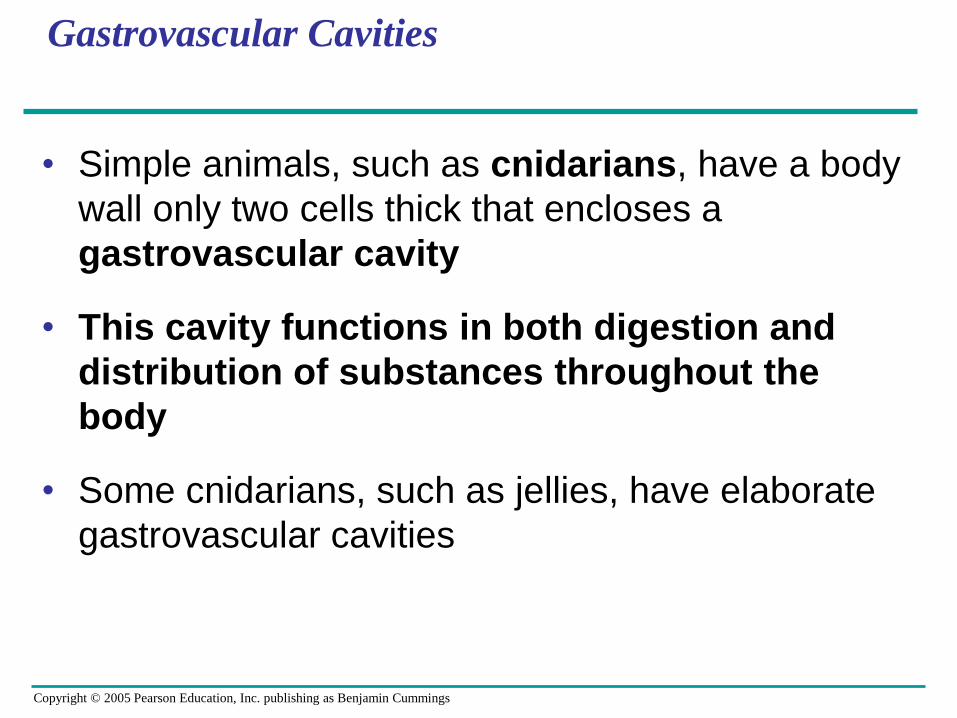
Copyright © 2005 Pearson Education, Inc. publishing as Benjamin Cummings Gastrovascular Cavities • Simple animals, such as cnidarians, have a body wall only two cells thick that encloses a gastrovascular cavity • This cavity functions in both digestion and distribution of substances throughout the body • Some cnidarians, such as jellies, have elaborate gastrovascular cavities
Transcript of gastrovascular cavity This cavity functions in both...
Copyright © 2005 Pearson Education, Inc. publishing as Benjamin Cummings
Gastrovascular Cavities
• Simple animals, such as cnidarians, have a body
wall only two cells thick that encloses a
gastrovascular cavity
• This cavity functions in both digestion and
distribution of substances throughout the
body
• Some cnidarians, such as jellies, have elaborate
gastrovascular cavities
Copyright © 2005 Pearson Education, Inc. publishing as Benjamin Cummings
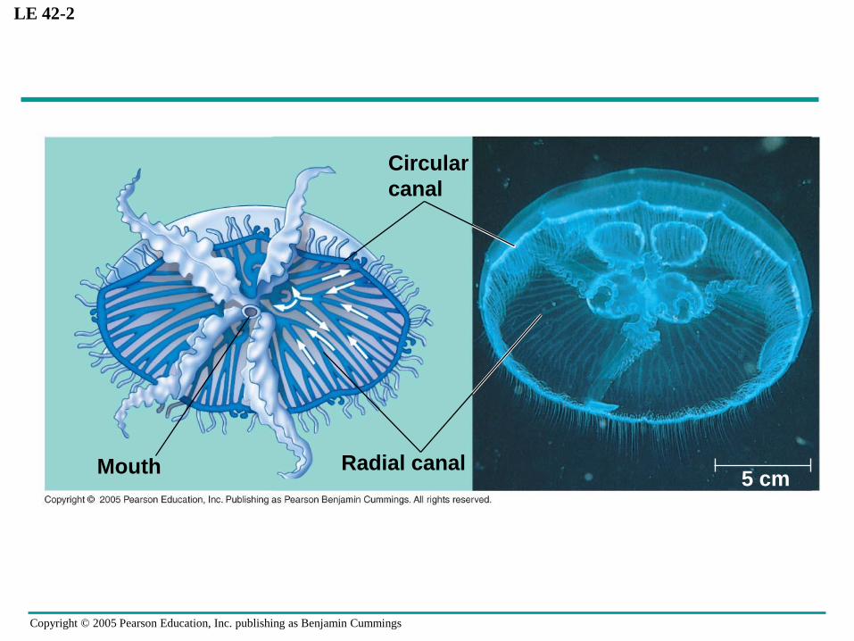
LE 42-2
Mouth Radial canal
Circular
canal
5 cm

Copyright © 2005 Pearson Education, Inc. publishing as Benjamin Cummings
Open and Closed Circulatory Systems
• More complex animals have either open or
closed circulatory systems
• Both systems have three basic components:
– A circulatory fluid (blood or hemolymph)
– A set of tubes (blood vessels)
– A muscular pump (the heart)
Copyright © 2005 Pearson Education, Inc. publishing as Benjamin Cummings
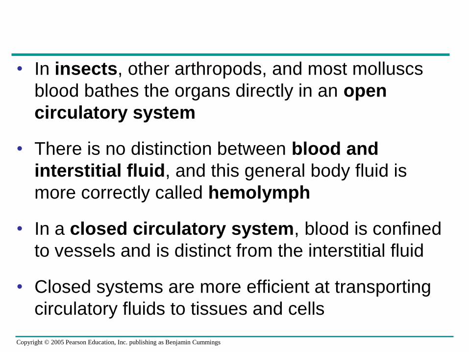
• In insects, other arthropods, and most molluscs
blood bathes the organs directly in an open
circulatory system
• There is no distinction between blood and
interstitial fluid, and this general body fluid is
more correctly called hemolymph
• In a closed circulatory system, blood is confined
to vessels and is distinct from the interstitial fluid
• Closed systems are more efficient at transporting
circulatory fluids to tissues and cells

Copyright © 2005 Pearson Education, Inc. publishing as Benjamin Cummings
LE 42-3
Hemolymph in sinusessurrounding organs
Heart
Anteriorvessel
Ostia
Tubular heart
An open circulatory system.
Lateralvessel
A closed circulatory system.
Auxiliary hearts Ventral vessels
Dorsal vessel(main heart)
Small branch vesselsin each organ
Interstitialfluid
Heart

Copyright © 2005 Pearson Education, Inc. publishing as Benjamin Cummings
Survey of Vertebrate Circulation
• Humans and other vertebrates have a closed
circulatory system, often called the
cardiovascular system
• Blood flows in a closed cardiovascular system,
consisting of blood vessels and a two- to four-
chambered heart
• Arteries carry blood to capillaries, the sites of
chemical exchange between the blood and
interstitial fluid
• Veins return blood from capillaries to the heart

Copyright © 2005 Pearson Education, Inc. publishing as Benjamin Cummings
LE 42-4
FISHES
Gill capillaries
AMPHIBIANS
Lung and skin capillaries
REPTILES (EXCEPT BIRDS)
Lung capillaries
MAMMALS AND BIRDS
Lung capillaries
Gillcirculation
Heart:Ventricle (V)
Atrium (A)
Artery
VeinSystemic
circulation
Systemic capillaries Systemic capillaries
Systemiccircuit
Pulmocutaneouscircuit
Right Left
AA
V
A
V
A
V
Systemic capillaries
Right Left
Pulmonarycircuit
Rightsystemic aorta
V
A
V
Systemic capillaries
Right Left
Pulmonarycircuit
A
Systemiccircuit
Leftsystemic aorta
Systemic circuits include all body tissues except lungs. Note that circulatory systems are depicted
as if the animal is facing you: with the right side of the heart shown at the left and vice-versa.

Copyright © 2005 Pearson Education, Inc. publishing as Benjamin Cummings
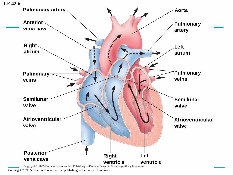
Mammalian Circulation: The Pathway
• Heart valves dictate a one-way flow of blood
through the heart
• Blood begins its flow with the right ventricle
pumping blood to the lungs
• In the lungs, the blood loads O2 and unloads CO2
• Oxygen-rich blood from the lungs enters the heart
at the left atrium and is pumped to the body
tissues by the left ventricle
• Blood returns to the heart through the right atrium

Copyright © 2005 Pearson Education, Inc. publishing as Benjamin Cummings
LE 42-5
Anterior
vena cava
Pulmonary
artery
Capillaries
of right lung
Aorta
Pulmonary
vein
Right atrium
Right ventricle
Posterior
vena cava
Capillaries of
abdominal organs
and hind limbs
Pulmonary
vein
Left ventricle
Left atrium
Aorta
Pulmonary
artery
Capillaries of
head and
forelimbs
Capillaries
of left lung
Copyright © 2005 Pearson Education, Inc. publishing as Benjamin Cummings
LE 42-6
Right
atrium
Posterior
vena cava
Pulmonary
veins
Anterior
vena cava
Pulmonary artery
Pulmonary
veins
Right
ventricle
Aorta
Semilunar
valve
Atrioventricular
valve
Pulmonary
artery
Left
atrium
Semilunar
valve
Atrioventricular
valve
Left
ventricle

Copyright © 2005 Pearson Education, Inc. publishing as Benjamin Cummings
• The heart contracts and relaxes in a rhythmic
cycle called the cardiac cycle
• The contraction, or pumping, phase is called
systole
• The relaxation, or filling, phase is called diastole
• The heart rate, also called the pulse, is the
number of beats per minute
• The cardiac output is the volume of blood
pumped into the systemic circulation per minute

Copyright © 2005 Pearson Education, Inc. publishing as Benjamin Cummings
• The sinoatrial (SA) node, or pacemaker, sets
the rate and timing at which cardiac muscle cells
contract
• Impulses from the SA node travel to the
atrioventricular (AV) node
• At the AV node, the impulses are delayed and
then travel to the Purkinje fibers that make the
ventricles contract
• Impulses that travel during the cardiac cycle can
be recorded as an electrocardiogram (ECG or
EKG)

Copyright © 2005 Pearson Education, Inc. publishing as Benjamin Cummings
LE 42-8
Pacemakergenerates wave ofsignals to contract.
Signals are delayedat AV node.
Signals passto heart apex.
Signals spreadthroughoutventricles.
SA node(pacemaker)
ECG
AV node
Bundlebranches Heart
apex
Purkinjefibers
The pacemaker is influenced by nerves, hormones, body
temperature, and exercise
Copyright © 2005 Pearson Education, Inc. publishing as Benjamin Cummings
LE 42-9
Endothelium
Endothelium
Smooth
muscle
Connective
tissue
Capillary
100 µm
Basement
membrane
Endothelium
Smooth
muscle
Connective
tissue
Valve
Artery
Arteriole Venule
Vein
Artery Vein

Copyright © 2005 Pearson Education, Inc. publishing as Benjamin Cummings
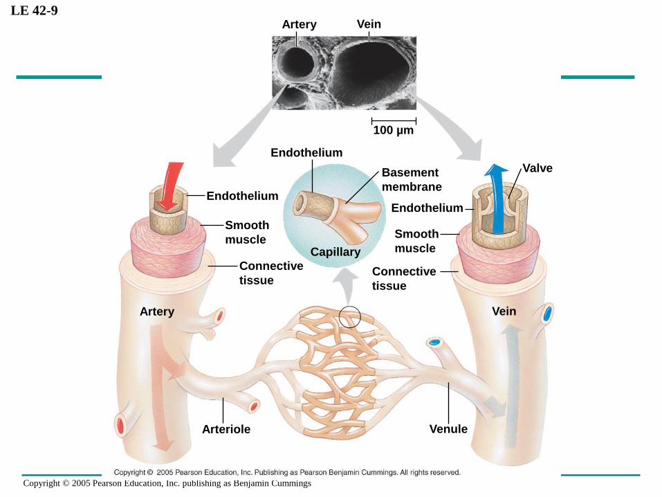
• Structural differences in arteries, veins, and
capillaries correlate with functions
• Arteries have thicker walls that accommodate the
high pressure of blood pumped from the heart
• In the thinner-walled veins, blood flows back to
the heart mainly as a result of muscle action
Blood Vessel Structure and Function

Copyright © 2005 Pearson Education, Inc. publishing as Benjamin Cummings
LE 42-10
Valve (open)
Skeletal muscle
Valve (closed)
Direction of blood flow
in vein (toward heart)

Copyright © 2005 Pearson Education, Inc. publishing as Benjamin Cummings
Blood Flow Velocity
• Physical laws governing movement of fluids
through pipes affect blood flow and blood pressure
• Velocity of blood flow is slowest in the
capillary beds, as a result of the high
resistance and large total cross-sectional area

Copyright © 2005 Pearson Education, Inc. publishing as Benjamin Cummings
LE 42-11
Systolic
pressure
Ve
na
e c
ava
e
Ve
ins
Ve
nu
les
Cap
illa
rie
s
Art
eri
ole
s
Art
eri
es
Ao
rta
Diastolic
pressure
Pre
ss
ure
(m
m H
g)
120100
806040200
Are
a (
cm
2)
5,000
4,000
3,000
2,000
1,000
0
Ve
loc
ity (
cm
/se
c)
50
40
30
20
10
0

Copyright © 2005 Pearson Education, Inc. publishing as Benjamin Cummings
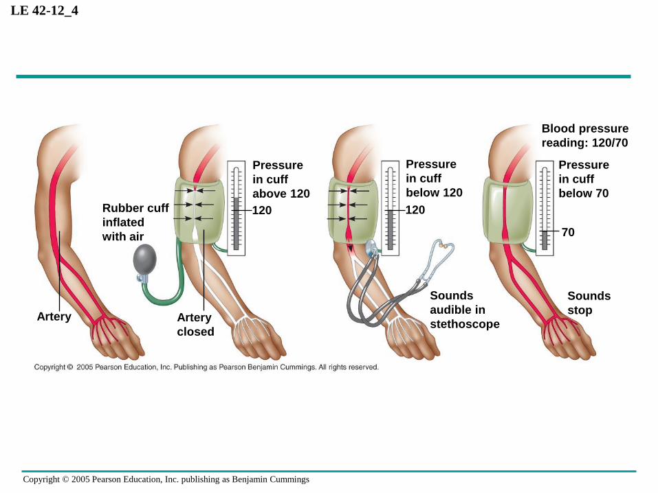
Blood Pressure
• Blood pressure is the hydrostatic pressure that
blood exerts against the wall of a vessel
• Systolic pressure is the pressure in the arteries
during ventricular systole; it is the highest
pressure in the arteries
• Diastolic pressure is the pressure in the arteries
during diastole; it is lower than systolic pressure
• Blood pressure is determined by cardiac output
and peripheral resistance due to constriction of
arterioles
Copyright © 2005 Pearson Education, Inc. publishing as Benjamin Cummings
LE 42-12_4
Artery Artery
closed
Pressure
in cuff
above 120
120Rubber cuff
inflated
with air
Pressure
in cuff
below 120
120
Sounds
audible in
stethoscope
Pressure
in cuff
below 70
70
Blood pressure
reading: 120/70
Sounds
stop
Copyright © 2005 Pearson Education, Inc. publishing as Benjamin Cummings
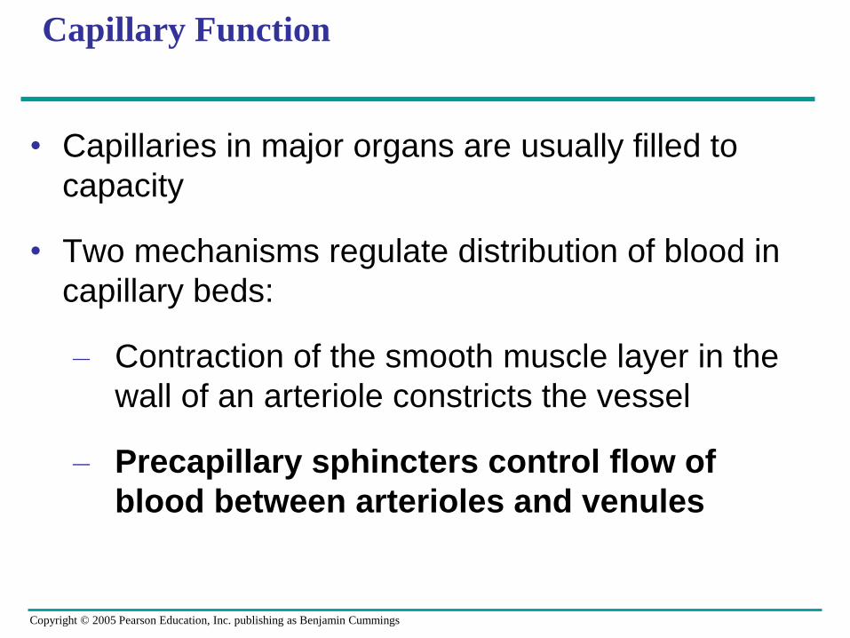
Capillary Function
• Capillaries in major organs are usually filled to
capacity
• Two mechanisms regulate distribution of blood in
capillary beds:
– Contraction of the smooth muscle layer in the
wall of an arteriole constricts the vessel
– Precapillary sphincters control flow of
blood between arterioles and venules

Copyright © 2005 Pearson Education, Inc. publishing as Benjamin Cummings
LE 42-13ab
Precapillary sphincters Thoroughfare
channel
CapillariesVenuleArteriole
Sphincters relaxed
VenuleArteriole
Sphincters contracted

Copyright © 2005 Pearson Education, Inc. publishing as Benjamin Cummings
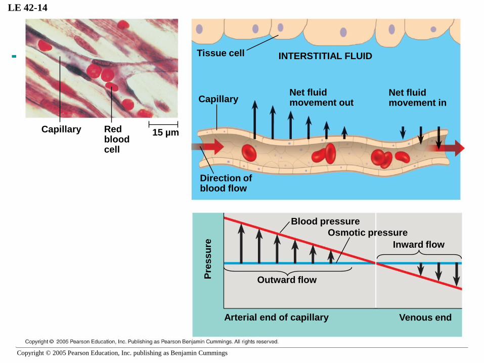
• The critical exchange of substances between the
blood and interstitial fluid takes place across the
thin endothelial walls of the capillaries
• The difference between blood pressure and
osmotic pressure drives fluids out of
capillaries at the arteriole end and into
capillaries at the venule end
Copyright © 2005 Pearson Education, Inc. publishing as Benjamin Cummings
LE 42-14
Capillary Red bloodcell
15 µm
Tissue cell
CapillaryNet fluidmovement out
INTERSTITIAL FLUID
Net fluidmovement in
Blood pressure
Osmotic pressure
Inward flow
Direction ofblood flow
Pre
ss
ure
Outward flow
Venous endArterial end of capillary
Copyright © 2005 Pearson Education, Inc. publishing as Benjamin Cummings
Fluid Return by the Lymphatic System
• The lymphatic system returns fluid to the body
from the capillary beds
• This system aids in body defense
• Fluid reenters the circulation directly at the venous
end of the capillary bed and indirectly through the
lymphatic system

Copyright © 2005 Pearson Education, Inc. publishing as Benjamin Cummings
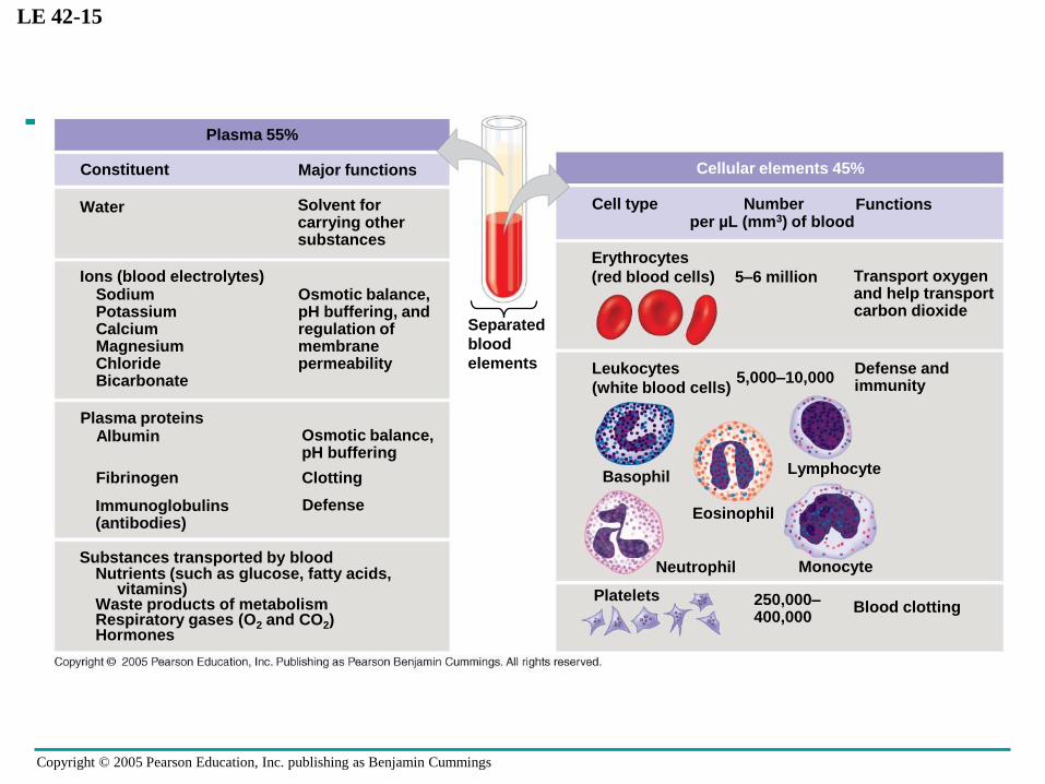
Blood Composition and Function
• Blood consists of several kinds of cells suspended
in a liquid matrix called plasma
• The cellular elements occupy about 45% of the
volume of blood
Copyright © 2005 Pearson Education, Inc. publishing as Benjamin Cummings
Plasma
• Blood plasma is about 90% water
• Among its solutes are inorganic salts in the form of
dissolved ions, sometimes called electrolytes
• Another important class of solutes is the plasma
proteins, which influence blood pH, osmotic
pressure, and viscosity
• Various plasma proteins function in lipid transport,
immunity, and blood clotting
Copyright © 2005 Pearson Education, Inc. publishing as Benjamin Cummings
LE 42-15
SodiumPotassiumCalciumMagnesiumChlorideBicarbonate
Osmotic balance,pH buffering, andregulation ofmembranepermeability
Plasma 55%
Constituent Major functions
Water Solvent forcarrying othersubstances
Ions (blood electrolytes)
Albumin Osmotic balance,pH buffering
Plasma proteins
Fibrinogen
Immunoglobulins(antibodies)
Clotting
Defense
Nutrients (such as glucose, fatty acids, vitamins)
Waste products of metabolismRespiratory gases (O2 and CO2)Hormones
Substances transported by blood
Cellular elements 45%
Cell type Number Functionsper µL (mm3) of blood
5–6 million Transport oxygenand help transportcarbon dioxide
Leukocytes
(white blood cells)5,000–10,000
Defense andimmunity
Monocyte
Basophil
Eosinophil
Lymphocyte
Neutrophil
PlateletsBlood clotting250,000–
400,000
Erythrocytes
(red blood cells)
Separated
blood
elements

Copyright © 2005 Pearson Education, Inc. publishing as Benjamin Cummings
Cellular Elements
• Suspended in blood plasma are two types of cells:
– Red blood cells (erythrocytes) transport
oxygen
– White blood cells (leukocytes) function in
defense
• Platelets, a third cellular element, are
fragments of cells that are involved in clotting

Copyright © 2005 Pearson Education, Inc. publishing as Benjamin Cummings
• Red blood cells, or erythrocytes, are by far the
most numerous blood cells
• There are five major types of white blood cells, or
leukocytes: monocytes, neutrophils, basophils,
eosinophils, and lymphocytes
• They function in defense by phagocytizing
bacteria and debris or by producing antibodies
• Erythrocytes, leukocytes, and platelets all
develop from a common source, pluripotent
stem cells in the red marrow of bones

Copyright © 2005 Pearson Education, Inc. publishing as Benjamin Cummings
LE 42-16
Pluripotent stem cells
(in bone marrow)
Myeloid
stem cellsLymphoid
stem cells
B cells T cells
Lymphocytes
Erythrocytes
Eosinophils
Basophils
Neutrophils
MonocytesPlatelets

Copyright © 2005 Pearson Education, Inc. publishing as Benjamin Cummings
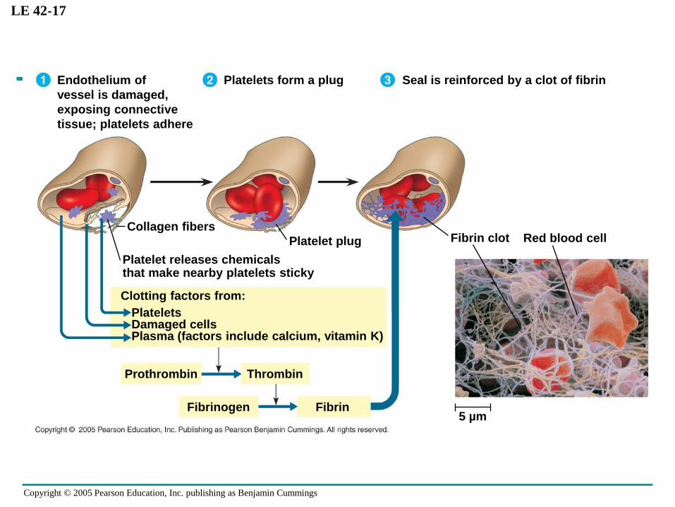
Blood Clotting
• When the endothelium of a blood vessel is
damaged, the clotting mechanism begins
• A cascade of complex reactions converts
fibrinogen to fibrin, forming a clot
Copyright © 2005 Pearson Education, Inc. publishing as Benjamin Cummings
LE 42-17
Endothelium of
vessel is damaged,
exposing connective
tissue; platelets adhere
Platelets form a plug Seal is reinforced by a clot of fibrin
Collagen fibers
Platelet plug
Platelet releases chemicalsthat make nearby platelets sticky
Clotting factors from:
PlateletsDamaged cellsPlasma (factors include calcium, vitamin K)
Prothrombin Thrombin
Fibrinogen Fibrin
Fibrin clot Red blood cell
5 µm
Copyright © 2005 Pearson Education, Inc. publishing as Benjamin Cummings
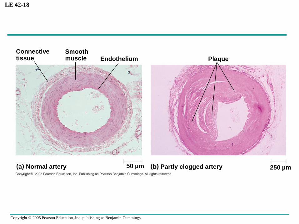
LE 42-18
Connectivetissue
Smoothmuscle Endothelium
50 µmNormal artery Partly clogged artery 250 µm
Plaque

Copyright © 2005 Pearson Education, Inc. publishing as Benjamin Cummings
• Hypertension, or high blood pressure, promotes
atherosclerosis and increases the risk of heart
attack and stroke
• A heart attack is the death of cardiac muscle
tissue resulting from blockage of one or more
coronary arteries
• A stroke is the death of nervous tissue in the brain,
usually resulting from rupture or blockage of
arteries in the head
Cardiovascular Disease
Copyright © 2005 Pearson Education, Inc. publishing as Benjamin Cummings
Gas exchange occurs across specialized respiratory surfaces
• Gas exchange supplies oxygen for cellular
respiration and disposes of carbon dioxide
• Animals require large, moist respiratory
surfaces for adequate diffusion of gases between
their cells and the respiratory medium, either air or
water

Copyright © 2005 Pearson Education, Inc. publishing as Benjamin Cummings
LE 42-19
Respiratory
medium
(air or water)
Organismal
level
Cellular level
Energy-rich
fuel molecules
from food
Respiratory
surface
Circulatory system
Cellular respiration
CO2O2
ATP

Copyright © 2005 Pearson Education, Inc. publishing as Benjamin Cummings
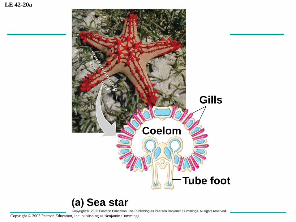
Gills in Aquatic Animals
• Gills are outfoldings of the body surface
specialized for gas exchange
• In some invertebrates, gills have a simple shape
and are distributed over much of the body
• Many segmented worms have flaplike gills that
extend from each segment of their body
• The gills of clams, crayfish, and many other
animals are restricted to a local body region
Copyright © 2005 Pearson Education, Inc. publishing as Benjamin Cummings
LE 42-20a
Gills
Coelom
Tube foot
Sea star

Copyright © 2005 Pearson Education, Inc. publishing as Benjamin Cummings
LE 42-20b
Gill
Parapodia
Marine worm

Copyright © 2005 Pearson Education, Inc. publishing as Benjamin Cummings
LE 42-20c
Gills
Scallop

Copyright © 2005 Pearson Education, Inc. publishing as Benjamin Cummings
LE 42-20d
Gills
Crayfish

Copyright © 2005 Pearson Education, Inc. publishing as Benjamin Cummings
• Effectiveness of gas exchange in some gills,
including those of fishes, is increased by
ventilation and the countercurrent flow of blood
and water

Copyright © 2005 Pearson Education, Inc. publishing as Benjamin Cummings
LE 42-21
Gill
arch
Water
flow Operculum
Gill
arch
Blood
vessel
Oxygen-rich
blood
Water flow
over lamellae
showing % O2
Gill
filaments
O2
Oxygen-poor
blood
Lamella
Blood flow
through capillaries
in lamellae
showing % O2
Countercurrent exchange

Copyright © 2005 Pearson Education, Inc. publishing as Benjamin Cummings
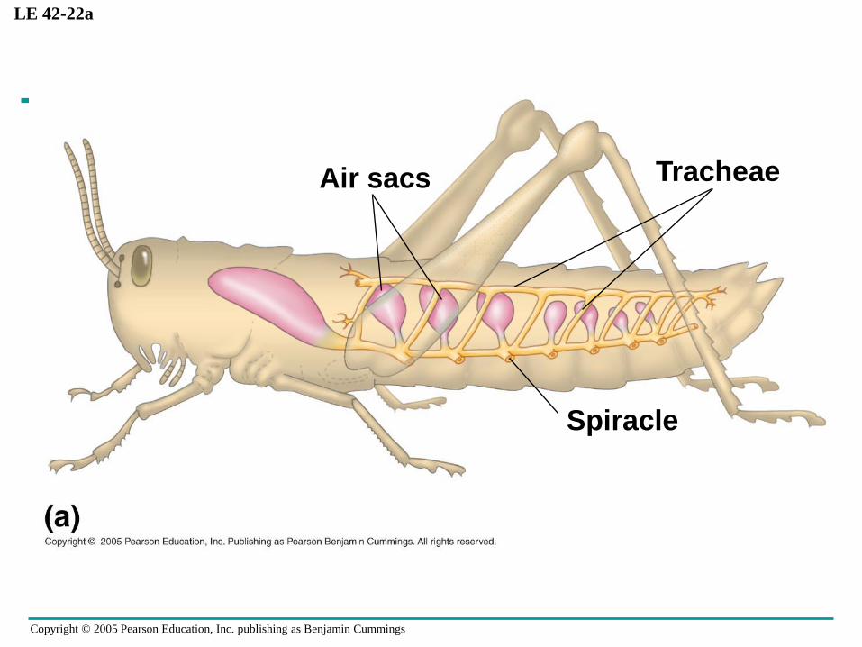
Tracheal Systems in Insects
• The tracheal system of insects consists of tiny
branching tubes that penetrate the body
• The tracheal tubes supply O2 directly to body
cells
Copyright © 2005 Pearson Education, Inc. publishing as Benjamin Cummings
LE 42-22a
Air sacs Tracheae
Spiracle

Copyright © 2005 Pearson Education, Inc. publishing as Benjamin Cummings
LE 42-22b
Bodycell
TracheoleAirsac
Trachea
Air Body wall
MyofibrilsTracheoles Mitochondria
2.5 µm

Copyright © 2005 Pearson Education, Inc. publishing as Benjamin Cummings
Lungs
• Spiders, land snails, and most terrestrial
vertebrates have internal lungs
• An amphibian such as a frog ventilates its lungs by
positive pressure breathing, which forces air down
the trachea
Copyright © 2005 Pearson Education, Inc. publishing as Benjamin Cummings
Mammalian Respiratory Systems: A Closer Look
• A system of branching ducts conveys air to the
lungs
• Air inhaled through the nostrils passes through the
pharynx into the trachea, bronchi, bronchioles,
and dead-end alveoli, where gas exchange occurs

Copyright © 2005 Pearson Education, Inc. publishing as Benjamin Cummings
Branchfrompulmonaryvein(oxygen-richblood)
Terminalbronchiole
Branchfrompulmonaryartery(oxygen-poorblood)
Alveoli
Colorized SEMSEM
Nasalcavity
Leftlung
Heart
Larynx
Pharynx
Esophagus
Trachea
Rightlung
Bronchus
Bronchiole
Diaphragm
Copyright © 2005 Pearson Education, Inc. publishing as Benjamin Cummings
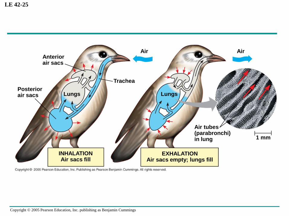
How a Bird Breathes
• Birds have eight or nine air sacs that function as
bellows that keep air flowing through the lungs
• Air passes through the lungs in one direction only
• Every exhalation completely renews the air in the
lungs
Copyright © 2005 Pearson Education, Inc. publishing as Benjamin Cummings
LE 42-25
Anteriorair sacs
LungsPosteriorair sacs
Trachea
Air
Lungs
Air
Air tubes(parabronchi)in lung 1 mm
EXHALATIONAir sacs empty; lungs fill
INHALATIONAir sacs fill

Copyright © 2005 Pearson Education, Inc. publishing as Benjamin Cummings
How a Mammal Breathes
• Mammals ventilate their lungs by negative
pressure breathing, which pulls air into the lungs
• Lung volume increases as the rib muscles and
diaphragm contract

Copyright © 2005 Pearson Education, Inc. publishing as Benjamin Cummings
LE 42-24
Rib cageexpands asrib musclescontract
Airinhaled
Lung
Diaphragm
INHALATIONDiaphragm contracts
(moves down)
Rib cage getssmaller asrib musclesrelax
Airexhaled
EXHALATIONDiaphragm relaxes
(moves up)

Copyright © 2005 Pearson Education, Inc. publishing as Benjamin Cummings
Control of Breathing in Humans
• In humans, the main breathing control centers
are in two regions of the brain, the medulla
oblongata and the pons
• The medulla regulates the rate and depth of
breathing in response to pH changes in the
cerebrospinal fluid
• The medulla adjusts breathing rate and depth to
match metabolic demands

Copyright © 2005 Pearson Education, Inc. publishing as Benjamin Cummings
LE 42-26
Breathingcontrolcenters
Cerebrospinalfluid
Medullaoblongata
Pons
Carotidarteries
Aorta
Diaphragm
Rib muscles

Copyright © 2005 Pearson Education, Inc. publishing as Benjamin Cummings
The Role of Partial Pressure Gradients
• Gases diffuse down pressure gradients in the
lungs and other organs
• Diffusion of a gas depends on differences in a
quantity called partial pressure
• A gas diffuses from a region of higher partial
pressure to a region of lower partial pressure
• In the lungs and tissues, O2 and CO2 diffuse from
where their partial pressures are higher to where
they are lower

Copyright © 2005 Pearson Education, Inc. publishing as Benjamin Cummings
LE 42-27
Inhaled air
Bloodenteringalveolar
capillaries
Alveolarepithelialcells
Alveolar spaces
Alveolarcapillaries
of lung
Exhaled air
Bloodleavingalveolar
capillaries
Pulmonaryveins
Pulmonaryarteries
Tissuecapillaries
HeartSystemicveins
Systemicarteries
Bloodleavingtissue
capillaries
Bloodenteringtissue
capillaries
Tissuecells
CO2O2
CO2O2
O2 CO2
CO2O2
< 40 > 45
40 45
CO2O2
100 40
CO2O2
40 45
CO2O2
104 40
CO2O2
CO2O2
CO2 O2
104 40
120 27160 0.2

Copyright © 2005 Pearson Education, Inc. publishing as Benjamin Cummings
Respiratory Pigments
• Respiratory pigments, proteins that transport
oxygen, greatly increase the amount of oxygen
that blood can carry
• The respiratory pigment of almost all vertebrates
is the protein hemoglobin, contained in
erythrocytes
• Like all respiratory pigments, hemoglobin must
reversibly bind O2, loading O2 in the lungs and
unloading it in other parts of the body

Copyright © 2005 Pearson Education, Inc. publishing as Benjamin Cummings
LE 42-28
Polypeptide chain
O2 unloadedin tissues
O2 loadedin lungs
Iron atomHeme group

Copyright © 2005 Pearson Education, Inc. publishing as Benjamin Cummings
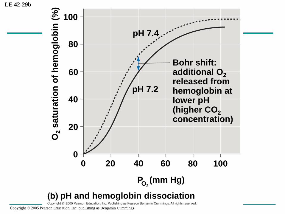
• Loading and unloading of O2 depend on
cooperation between the subunits of the
hemoglobin molecule
• The binding of O2 to one subunit induces the other
subunits to bind O2 with more affinity
• Cooperative O2 binding and release is evident in
the dissociation curve for hemoglobin
• A drop in pH lowers affinity of hemoglobin for O2

Copyright © 2005 Pearson Education, Inc. publishing as Benjamin Cummings
LE 42-29a
O2 unloaded fromhemoglobinduring normalmetabolism
O2 reserve that canbe unloaded fromhemoglobin totissues with highmetabolism
P and hemoglobin dissociation at 37°C and pH 7.4O2
P (mm Hg)O2
Tissues duringexercise
Tissues at rest
Lungs
1008060402000
20
40
60
80
100
O2
satu
rati
on
of
hem
og
lob
in (
%)
Copyright © 2005 Pearson Education, Inc. publishing as Benjamin Cummings
LE 42-29b
Bohr shift:additional O2
released fromhemoglobin atlower pH(higher CO2
concentration)
pH and hemoglobin dissociation
P (mm Hg)O2
1008060402000
20
40
60
80
100
O2
satu
rati
on
of
hem
og
lob
in (
%)
pH 7.2
pH 7.4
Copyright © 2005 Pearson Education, Inc. publishing as Benjamin Cummings
Carbon Dioxide Transport
• Hemoglobin also helps transport CO2 and assists
in buffering
• Carbon from respiring cells diffuses into the blood
plasma and then into erythrocytes and is
ultimately released in the lungs

Copyright © 2005 Pearson Education, Inc. publishing as Benjamin Cummings
LE 42-30
CO2 transportfrom tissuesCO2 produced
Tissue cell
CO2
CO2
CO2
Interstitialfluid
Blood plasmawithin capillary
Capillarywall
Hemoglobinpicks up
CO2 and H+
CO2 transportto lungs
To lungs
H2CO3
Carbonic acid
H2O
Hb
HCO3–
Bicarbonate
Redbloodcell
H++
HCO3–
HCO3–
Hemoglobinreleases
CO2 and H+
H++HCO3–
CO2
H2CO3
H2O
CO2
CO2
CO2
Hb
Alveolar space in lung