Trainees ˇ Symposium - Pathsoc · Preparation prior to MDT meetings ... There is access to all...
88
Trainees’ Symposium: The Pathologist in the multidisciplinary team (MDT); The Surgical, Oncological, Radiological and Pathological Perspective Mr Horgan Dr N Sharma Prof D Dodwell Prof A M Hanby Dr Caroline Young Dr Katie Allen
Transcript of Trainees ˇ Symposium - Pathsoc · Preparation prior to MDT meetings ... There is access to all...
Trainees’ Symposium:
The Pathologist in the multidisciplinary team (MDT); The Surgical, Oncological, Radiological and
Pathological Perspective Mr Horgan
Dr N SharmaProf D DodwellProf A M Hanby
Dr Caroline YoungDr Katie Allen

Todays menu
• 08 55- 10 10• MDT and MDTMs- why we have then.• Attributes of a good MTDM• Views of the core discipline members- what to
expect from the pathologists- i) Dr Sharma, the radiologists perspective ii) Mr Horgan, the surgeons
perspective iii) The oncologists perspective.• Discussion and Q and A

Todays menu
• 10 10- 10 40• Coffee• 10 40 – 10 50• Case discussions


Ruth Picardie Noticed a lump in her breast in 1994
Went to her GP, who referred her to the breast clinic at Guy's.
Lab tests suggested the lump was probably benign, but the report recommended it should be excised in a biopsy, to make sure.
Markandoo, who saw Picardie, told her the lump was harmless and there was no point in surgical removal, which would leave a scar.

Two years later• Picardie had given birth to twins
• The lump was the size of a golf ball.
• Again a GP referred her to the breast clinic.
• This time an aggressive cancer was diagnosed.
• http://www.telegraph.co.uk/culture/3649668/The-Friday-column-breast-cancer.html

Learning points
• Does it clinically seem like cancer, • Does it look like cancer -radiology• Does it look like cancer -pathology
• If the triple assessment is followed this mishaps should be rare.


Core biopsies- ‘B’ grading• B1 - unsatisfactory/normal breast lesion• B2 - benign lesion• B3 - atypical probably benign• B4 - atypical probably malignant• B5 - Malignant (B5a in situ, b invasive).
• Triangulated within the multidisciplinary team meeting.

No man is an island,Entire of itself,Every man is a piece of the continent,A part of the main.If a clod be washed away by the sea,Europe is the less.As well as if a promontory were.As well as if a manor of thy friend'sOr of thine own were:Any man's death diminishes me,Because I am involved in mankind,And therefore never send to know for whom the bell tolls; It tolls for thee.
John Doone 1624
If pathologists be removed from attendance,the MDTM is the less.-
(anonymous lesser poet 2016)

The Characteristics of an Effective Multidisciplinary Team (MDT) National Cancer Action Team
• treatment and care being considered by professionals with specialist knowledge and skills in the relevant aspects of that cancer type;
• patients being offered the opportunity to be entered into high quality and relevant clinical trials;
• patients being assessed and offered the level of information and support they need to cope with their condition;

The Characteristics of an Effective Multidisciplinary Team (MDT)
National Cancer Action Team
• improved equality of outcomes as a result of better understanding and awareness of patients’ characteristics and through reflective practice;
• adherence to national and local clinical guidelines;
• promotion of good working relationships between staff, thereby enhancing their job satisfaction and quality of life;

The Characteristics of an Effective Multidisciplinary Team (MDT)
National Cancer Action Team
• opportunities for education/professional development of team members inc devising and agreeing new protocols and ways of working);
• optimisation of resources – effective MDT working should result in more efficient use of time which should contribute to more efficient use of NHS resources more generally

Categorisation of MDT Characteristics
• 1. The Team • 1.1. Membership • 1.2. Attendance • 1.3. Leadership • 1.4. Team working & culture • 1.5. Personal development & training

Categorisation of MDT Characteristics
• 2. Infrastructure for Meetings • 2.1. Physical environment of meeting venue • 2.2. Technology & equipment (availability & use) • 3. Meeting Organisation & Logistics • 3.1. Scheduling of MDT meetings • 3.2. Preparation prior to MDT meetings • 3.3. Organisation/administration during MDT
meetings • 3.4. Post MDT meeting /co-ordination of service

Categorisation of MDT Characteristics
• 4. Patient-Centred Clinical Decision-Making • 4.1. Who to discuss? • 4.2. Patient-centred care • 4.3. Clinical decision-making process

Categorisation of MDT Characteristics
• 5. Team Governance • 5.1. Organisational Support • 5.2. Data collection, analysis and audit of
outcomes • 5.3. Clinical governance

• 3.3.2. A locally agreed minimum dataset of information is presented on each patient including diagnostic information (pathology and radiology), clinical information (including co-morbidities, psychosocial and specialist palliative care needs) and patient history, views and preferences – the focus is on what the team need to hear to make appropriate recommendations on the patient in question. It may not, for example, be necessary to show/discuss the pathological or radiological findings in all cases.

• 3.3.3. There is access to all relevant information at the meeting including patient notes, test results/images/samples (past and present) and appointment dates (or a proforma /summary record with the necessary information) along with access to PAS, radiology & pathology systems etc – relevant past material should be reviewed prior to the meeting if it is not accessible during the meeting.

• to track patients through the system to ensure that any tests, appointments, treatments are carried out in a timely manner eg. within cancer waits standards where applicable.
• 4.3.9. MDT recommendations are only as good as the information they are based on – if there are concerns that key data is missing this should be documented.
• This means attention to the details of the pathology report
• 5.3.9. Significant discrepancies in pathology, radiology or clinical findings between local and specialist MDTs should be recorded and be subject to audit.
Improving the effectiveness of multidisciplinary team meetings for patients with chronic diseases:
a prospective observational study Rosalind Raine, et al
• Implementation varied by disease category, and was highest in gynaecological cancer (84%) and lowest in mental health (70%).
• High-implementing teams have clear goals, and members shared the view that the main purpose of the MDT meeting was to make treatment recommendations for patients.
• lower-implementing teams, members identified a range of diverse objectives and some stated that meetings lacked clarity of purpose.

The perspectives of the team members/leaders

The radiologists perspectiveDr Nisha Sharma
• Need to work together to obtain the best outcome for the patient
• Radiologists need to provide enough information to ensure that a relevantly detailed pathology report is obtained e.g. If biopsy for calcifications or a mass
• Highlight our level of concern R1-R5• If we biopsy two areas and label them A and B - pathology
report them as A and B and not lesion 1 and 2 as can be confusing
• Tumour size is difficult. If several lesions on imaging we would give an overall extent but in pathology this seldom occurs which means that there is discordance regarding extent

The Surgeons perspectiveMr Kieran Horgan
• The breast surgeon plays a pivotal role to ensure a multidisciplinary team meeting produces any outcomes. A number of colleagues require careful man-management in order to achieve those goals.

The Surgeons perspectiveMr Kieran Horgan
• The radiologist must be persuaded not to describe at length various benign appearances which are only of interest to them and entirely superfluous if the biopsy is benign.

The Surgeons perspectiveMr Kieran Horgan
• The pathologist must be encouraged to reduce their report to the 10% of relevant interest to the meeting, not to embark on endless immunohistochemistry and to retain fascinating discussions on the interface between hyperplasia and in situ neoplasia to coffee breaks with their colleagues.

The Surgeons perspectiveMr Kieran Horgan
• The oncologist must be encouraged to don a visage without agonised perplexion and to reflect that mostly their chemo-therapeutic decisions are best guess and the action will be binary not titrated ; either give the treatment or not.

The Surgeons perspectiveMr Kieran Horgan
• The MDT meeting will not cope well with detailed breast care nurse input as to the details of the patient’s psychosocial background surmised from a clinic visit, valuable though that information is, and a brief “ she wont agree to that treatment “ will suffice.

The Surgeons perspectiveMr Kieran Horgan
• It can be seen therefore that the successful MDT requires all the knowledge , perception , people management skills, modesty and kind collegiate approach typified in the character of the UK breast surgeon.

The Oncologists perspectiveProfessor David Dodwell
• There are two ways of treating breast cancer:
• My way or the wrong way
• And woe betide any MDT decision which challenges this !!

The pathologists perspective- the path report
• Reports are all important.• Relevant data in a logical order.• Most important bits first.• May contain interesting ‘bonus’ material- but
not necessary to regale the a busy MDTM with it all

Pathology report 1
• Breast tissue showing a number of features of fibrocystic change including apocrine metaplasia, adenosis, sclerosing adenosis, usual type ductal hyperplasia and part of a radial scar. Some collagenous spherulosis is also seen, an interesting condition that may resemble some invasive cancer ( 3 references supplied). There is also a grade 3 infiltrating ductal carcinoma with associated DCIS

Pathology report 1
•••• DCIS is present••

Clinical information received
• Mrs Clac•• Age 57 •• Incident screen shows widespread calcification
with a cluster of more concerning calcification-VAB 12 g VAB biopsy undertaken.
•

Pathology report 3
••• High grade solid pattern DCIS with
comedonecrosis and microcalcifications is present

Pathology report 4
••• High grade solid pattern DCIS with associated
DCIS is present in approximately 10% of the sample and contains coarse calcifications. Extensive microcysts with associated micro-calcifications are also present

Ive narrowed the diagnosis down to 16 possibilities


Cases MDT
Case discussion Pathology
Case 1

• 62 year old presented with lump left breast• P4/P5 lump• Mammogram and targeted US left breast and
axilla

Mammogram
Normal right mammogram 51mm mass UOQ left breast R5

US breast and axillaUOQ right breast – 51mm mass U5
Left axilla R4 lymph node 5mm cortical thickening

• US guided core biopsy – ductal grade 3, triple negative (ER/PR/HER 2 negative)
• No DCIS or LVI• Axillary FNA benign – lymphocytes present
• MDT – NACT aiming for conservation rather than mastectomy
• Microbubbles left axilla


SLNB – microbubbles and surgeryCEUS identified SLN which was biopsied 14G x 2 cores and clip inserted
SLNB and LN X-rayed with clip present

Baseline - NACTDynamic, subtracted image – clip in SLN T1W image showing clip in LN

Baseline
55mm mass abutting chest wall T1 W scan

Post SLNB
Post 2 cycles. SLNB cavity T2W – fluid in surgical cavity

Post 2ECPost 2 EC – concentric reduction in tumour 34mm T1 W scan

MDT
• Continue EC chemotherapy• Insert marker clip
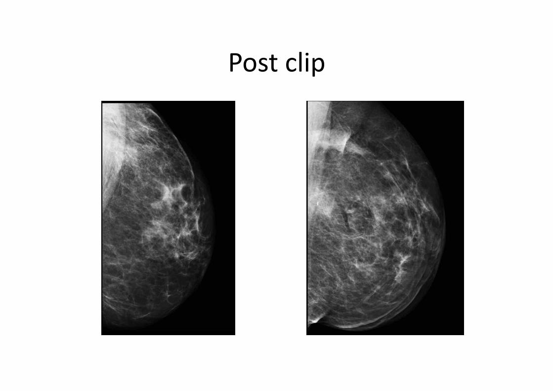
Post clip

NACT post 4EC

NACT post 6EC – end of treatment scan
Residual mass on T1W Blush of contrast

X-Ray wire localisation as not visible on US
Wire localising the clip Specimen X-ray with clip

MDT
• Pathology – clip seen. No residual invasive disease or DCIS
• Radiotherapy• 10 year follow up as grade 3

Case 1 post NACT


Pic of Path pcr

Pic of Path pcr

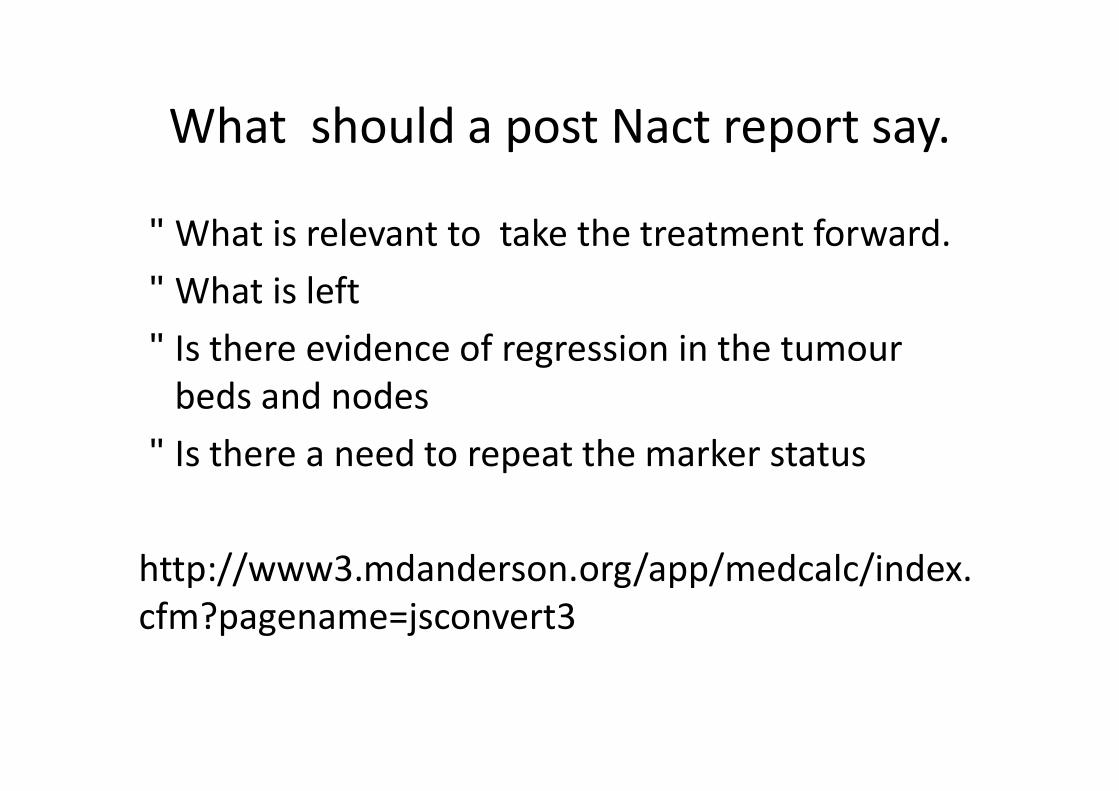
What should a post Nact report say.
• What is relevant to take the treatment forward.• What is left• Is there evidence of regression in the tumour
beds and nodes• Is there a need to repeat the marker status
http://www3.mdanderson.org/app/medcalc/index.cfm?pagename=jsconvert3

Standardization of pathologic evaluation and reporting of postneoadjuvant specimens in clinical trials of breast cancer: recommendations from an international working group. Mod Pathol28, 1185-1201Provenzano, E., Bossuyt, V., Viale, G., Cameron, D., Badve, S., Denkert, C., MacGrogan, G., Penault-Llorca, F., Boughey, J., Curigliano, G., Dixon, J. M., Esserman, L., Fastner, G., Kuehn, T., Peintinger, F., von Minckwitz, G., White, J., Yang, W., Symmans, W. F., and Residual Disease Characterization Working Group of the Breast International Group-North American Breast Cancer Group, C. (2015)

Should we re-test after treatment
Venkatesan, S., and Swanton, C. (2016) TumorEvolutionary Principles: How IntratumorHeterogeneity Influences Cancer Treatment and Outcome. American Society of Clinical Oncology educational book / ASCO. American Society of Clinical Oncology. Meeting 35, e141-149Zardavas, D., Irrthum, A., Swanton, C., and Piccart, M. (2015) Clinical management of breast cancer heterogeneity. Nature reviews. Clinical oncology 12, 381-394

Case 2
• 58 year old • Lump right breast – P5 with skin tethering

Case 2RT MLO – 40mm mass and 8mm ? satellite RT CC

Case 2Known cancer +satellites –32mm mass +8mm+10mm Axilla – 3 abnormal nodes

Case 2Rt MLO mass and clips in 2 satellites Rt CC mass and clips in satellites

Case 2
• US core biopsy – metaplastic, grade 3• Triple negative• 2 satellites with clips C5• Axillary FNA C5


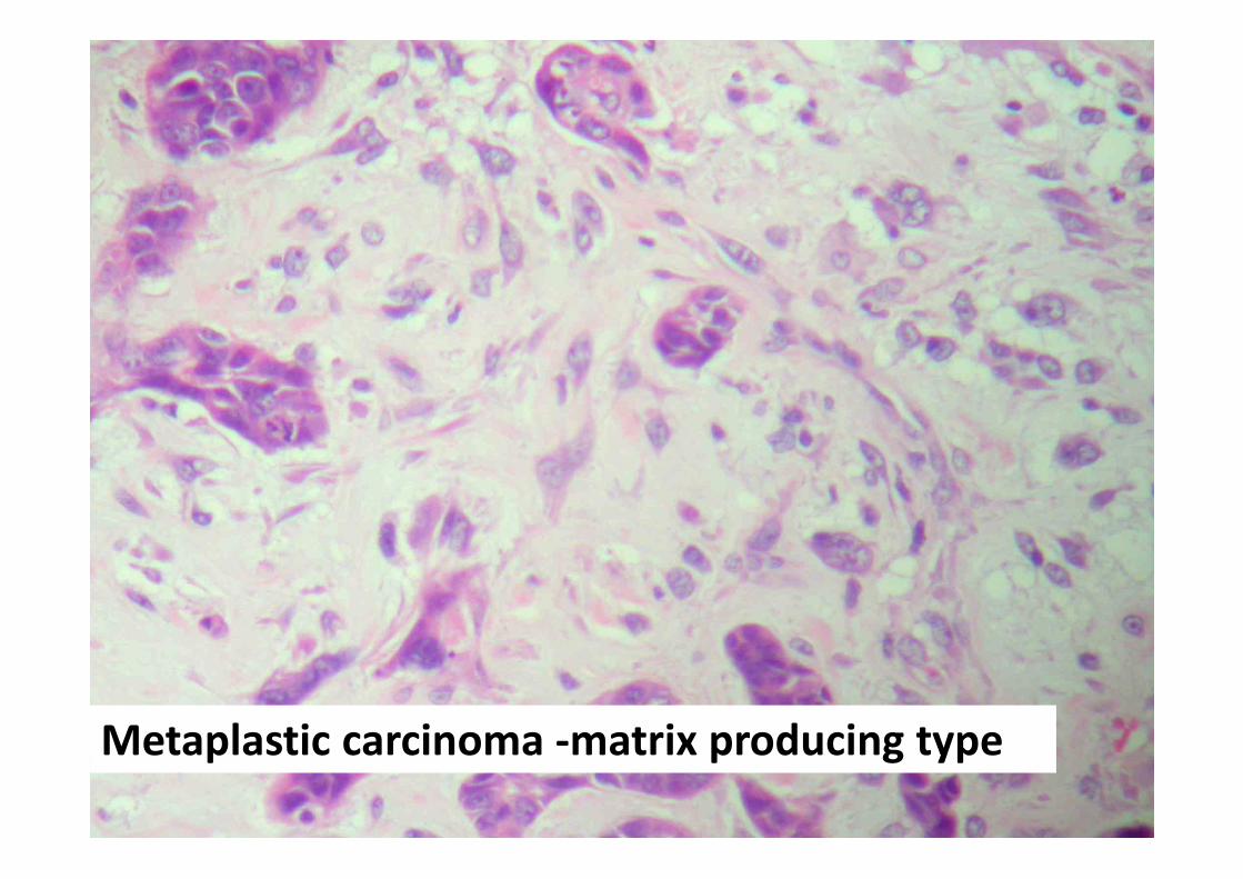
Metaplastic carcinoma -matrix producing type

RIGHT BREAST MASTECTOMY, RIGHT AXILLARY NODE CLEARANCE AND RIGHT APICAL TISSUE
• INVASIVE CARCINOMA• Invasive tumour type Metaplastic carcinoma• Invasive tumour grade 3 (T3, P3, M2)• Invasive tumour size (contiguous) 55mm• Lymphovascular invasion Not seen• Margins >10mm from all margins
• IN-SITU DISEASE• Ductal carcinoma in-situ yes• Grade (NHSBSP): High• Pattern Cribriform,solid• DCIS Necrosis Yes• DCIS Calcification No• DCIS relationship to invasive Admixed and adjacent; plus• at 9 o'clock• measuring 18 mm in maximum dimension.• Margins As above• Paget's disease: Not seen• Lobular neoplasia Yes (ALH and LCIS in background• breast)
• Whole tumour Size 55mm
• LYMPH NODES• Axillary node clearance 0/16 (including one with ITC's)•• Total node count 0/20 (none of the lymph nodes• show any • perinodal scarring/fat necrosis, suggestive of previous aspiration)
• The FNA axilla (LN16-571) has been reviewed and evidence of metastatic carcinoma• to the node is• confirmed.

Amended report following further sampling
• LYMPH NODES• Axillary nodes(A&B) 4/19 (one of the 4 being ITCs)• Apical nodes (C) 0/4• Extracapsular spread One focus• Size of largest metastasis 10mm• Total node count 4/23
• Nottingham Prognostic Index 7.1 (PPG)• Stage (TNM 7th Edition) pT3, pN2, pMx
• Diagnosis
• Right mastectomy - Invasive metaplastic carcinoma.
• Total right axillary lymph nodes - 4/36

Case 2
• Lesson learnt from imagingLow lying axillary lymph nodes – sit within the
breast. Imaging will mention this in the report and clip the node to ensure excision if for breast conservation and ANC

Case 3
• 58 year breast screening referral • Asymptomatic

Mammogram
Left mammogram 2013Left mammogram 2016 – new segmental calcifications
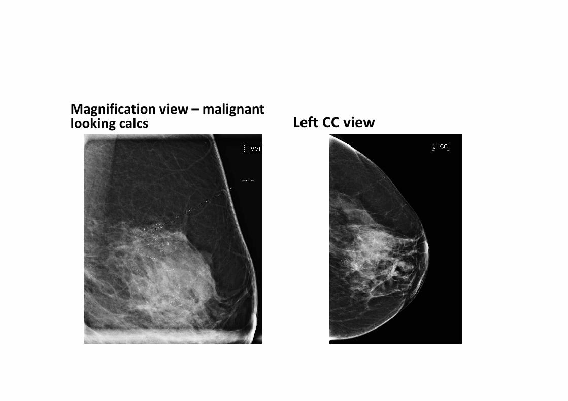
Magnification view – malignant looking calcs Left CC view

• 2 areas biopsied• Area A – HGDCIS• Area B – HGDCIS• Patient declined marker clips

Surgery• LEFT MASTECTOMY AND LEFT SENTINEL LYMPH NODE BIOPSY • INVASIVE CARCINOMA• Invasive tumour type None found currently (though • micrometastasis in lymph node) • IN-SITU DISEASE• Ductal carcinoma in-situ Present• Grade (NHSBSP): High • Pattern Solid• DCIS Necrosis Present• DCIS Calcification Present• DCIS relationship to invasive N/A• Margins 1.5mm posterior, 5mm superior, • rest > 10mm • Paget's disease: Not seen • Cavity biopsies N/A• Whole tumour Size At least 60mm • LYMPH NODES• Sentinel nodes 1/2• Apical node N/A• Extracapsular spread Not seen • Size of largest metastasis 0.5mm• Total node count 1/2 (micrometastasis)• Molecular markers: - N/A • POST NAC PARAMETERS N/A

• DIAGNOSIS:• A: Left breast mastectomy Extensive DCIS, awaiting further levels
• B and C: Left axilla sentinel lymph node biopsy 1/2 (micrometastasis)
• Deeper levels have been performed on all of the original sections which sample• the DCIS and a further 8 blocks have also been taken from the specimen. No• definite areas of invasive carcinoma have been found.
• The micrometastasis seen in the sentinel lymph node was present only on level 1,• cutting out on deeper levels which precluded molecular marker testing. All of• the lymph node blocks have therefore been re-embedded after turning 1800 and• have been examined at three levels. No further carcinoma is seen in any of• these sections and therefore molecular marker testing cannot be performed.
• In summary, despite extensive block sampling and examination at multiple levels,• no foci of invasive carcinoma have been found in the breast though a• micrometastasis is seen within one level of the sentinel lymph node biopsy. As• a final measure, immunohistochemistry for myoepithelial markers will be• performed on the larger DCIS ducts to ensure that these do have an external• myoepithelial cell layer. A supplementary report will be issued with these• results.
• molecular marker testing will be attempted on the sentinel lymph node immunospares on the off chance that these contain tumour (ie. if they were cut before the level 2 H&E which no longer contains tumour). In addition, molecular marker testing will also be performed on an area of DCIS as this may help guide future management decisions.
• There are a few clusters of tumour cells in the capsule of the node, together• measuring 0.5mm, with the largest single cluster memasuring just over 0.2mm.
• The few tumour cells that are present in the sentinel lymph node score 3+ with• Her2 immunohistochemistry and they should therefore be regarded as positive for• Her2 over-expression. The tumour cell nuclei appear to score 0/8 for ER. The• DCIS has also been tested for molecular markers and it scores 3+ with Her2• immunohistochemistry. ER on the DCIS is still awaited.

Issues
• No invasive component found but 1 of 2 nodes micromet
• Molecular markers HER 2+ on node and DCIS HER 2 positive

A case from St Elsewhere’s
• 28 year old woman• Lump left breast and axilla. No FH• Breast biopsy ductal carcinoma• SLNB – Hodgkins disease
‘Please take over care – complex case’!
• What to do?