
Bortezomib sensitizes thyroid cancer to BRAF inhibitor in ...

INTERNATIONAL JOURNAL OF ONCOLOGY 48: 376-384, 2016376
Abstract. Gambogic acid (GA) is a natural compound derived from brownish gamboge resin that shows a range of bioactivity, such as antitumor and antimicrobial properties. Although, GA is already known to induce cell death in a variety of cancer cells, the molecular basis for GA-induced cell death in renal cancer cells is unclear. In this study, a treatment with GA induced cell death in human renal carcinoma Caki cells in a dose-dependent manner. Treatment of Caki cells with GA decreased the levels of antiapoptotic proteins, such as Bcl-2 and XIAP in a dose-dependent manner. In addition, GA decreased the expression of the cFLIPL protein, which was downregulated at the transcriptional level without any change in the levels of cFLIPs expression. z-VAD (pan-caspase inhibitor) partially blocked GA-mediated cell death. GA-induced apoptotic cell death in Caki cells is mediated partly by the AIF translocation from the mitochondria into the nucleus via a caspase-inde-pendent pathway. In contrast, N-acetylcysteine (NAC), a ROS scavenger, had no effect on GA-induced cell death. The restoration of cFLIPL attenuated GA-induced cell death in Caki cells. Furthermore, a sub-toxic dose of GA sensitized TRAIL-mediated apoptosis in Caki cells. Pretreatment with z-VAD completely blocked GA plus TRAIL-mediated apoptosis. On the contrary, pretreatment with NAC partially inhibited GA plus TRAIL-induced apoptosis. Our findings suggested that GA induces apoptosis via the downregulation of cFLIPL and sensitized TRAIL-mediated apoptosis in Caki cells.
Introduction
Gambogic acid (GA) is a constructive component of Garcinia hurburyi, a natural compound derived from brownish
gamboe resin in Southeast Asia countries (1). Gamboge resin is used in conventional Chinese medicine for the treatment of hemostasis, detoxification and as a parasiticide (2). GA was reported to have multiple functions, such as anti-inflammation, anti-angiogenesis and anti-invasion (3-5). GA also has potent anticancer activity in numerous types of human cancers, such as lung cancer, hepatocellular carcinoma, malignant melanoma, breast carcinoma, and chronic myelogenous leukemia by targeting NF-κB, thioredoxin reductase, Bcl-2, Akt/mTOR signaling pathway, and proteasome, respectively (6-10). Furthermore, it exhibits specific cytotoxic activity on rapidly dividing cancerous cells with no side effects on normal cells (11). Despite its anticancer efficacy, the molec-ular mechanism of the GA-induced apoptosis in renal cancer cells is unclear.
TRAIL (tumor necrosis factor (TNF)-related apoptosis-inducing ligand) belongs to the TNF superfamily, which can induce apoptosis in a wide variety of tumor cells, but not normal cells (12). Although TRAIL has beneficial effects in selectively killing tumor cells, many cancer cells are resis-tant to TRAIL (13). The mechanism of TRAIL resistance is unclear but several studies have reported that TRAIL resistance is intimately associated with the overexpression of anti-apoptosis including cellular FADD-like apoptosis regulator (cFLIP), anti-apoptotic Bcl-2 family proteins (e.g., Bcl-2 and Bcl-xL) and inhibitor of apoptosis proteins (IAPs) (13,14). However, a single treatment with TRAIL may not be sufficient for the treatment of malignant tumor cells. Moreover, TRAIL-resistant cancer cells can be sensitized by a TRAIL sensitizer, such as chemotherapeutic drugs and biochemical inhibitors that suppress the expression of anti-apoptosis-associated proteins, indicating that combination of drugs rather than just one drug alone appears to be more effective in cancer therapy. Therefore, the identification of a novel TRAIL sensitizer is important for effective cancer therapy. The aim of this study was to examine the anticancer effects of GA, elucidate the underlying action mechanism of GA, evaluate GA as a sensitizer of TRAIL, and understand the mechanism of the synergy between GA and TRAIL against human renal cancer cells. In the present study, GA was found to induce apoptosis in renal carcinoma (Caki Cells) through the downregulation of cFLIPL. In addition, a GA treatment rendered human renal cancer cells more sensi-tive to TRAIL.
Gambogic acid induces apoptosis and sensitizes TRAIL-mediated apoptosis through downregulation
of cFLIPL in renal carcinoma Caki cellsJI HOON JANG*, JOO-YOUNG KIM*, EON-GI SUNG, EUN-AE KIM and TAE-JIN LEE
Department of Anatomy, College of Medicine, Yeungnam University, Nam-gu, Daegu 705-717, Republic of Korea
Received September 23, 2015; Accepted October 30, 2015
DOI: 10.3892/ijo.2015.3249
Correspondence to: Dr Tae-Jin Lee, Department of Anatomy, College of Medicine, Yeungnam University, 170 Hyeonchung-ro, Nam-gu, Daegu 705-717, Republic of KoreaE-mail: [email protected]
*Contributed equally
Key words: gambogic acid, apoptosis, AIF, cFLIPL, tumor necrosis factor (TNF)-related apoptosis-inducing ligand
JANG et al: GAMBOGIC ACID INDUCES APOPTOSIS AND SENSITIzES TRAIL-MEDIATED APOPTOSIS 377
Materials and methods
Cells and materials. Caki cells were obtained from the American Type Culture Collection (ATCC, Rockville, MD, USA). Dulbecco's modified Eagle's medium, containing 10% fetal bovine serum (FBS), 20 mM HEPES buffer and 100 µg/ml gentamicin was used as the culture medium in these experi-ments. PCR primers were purchased from Bioneer (Daejeon, Korea). The anti-Bcl-2, anti-Mcl-1 and anti-cIAP-2 were acquired from Santa Cruz Biotechnology (Santa Cruz, CA, USA). The anti cFLIPL antibody was obtained from ALEXIS Corp. (San Diego, CA, USA). Anti-PARP, and anti-caspase-3 antibody were purchased from Cell Signaling Technology (Beverly, MA, USA). The anti-XIAP antibody was supplied by R&D systems (Minneapolis, MN, USA). Gambogic acid and the other chemicals were purchased from Sigma (St. Louis, MO, USA). Recombinant human TRAIL was obtained from KOMA Biotech (Seoul, Korea).
Cell count and flow cytometry analysis. The cell counts were performed using a hemocytometer. Approximately 0.5x10⁶ Caki cells were suspended in 100 µl of PBS, and 200 µl of
95% ethanol was added while vortexing. The cells were incubated at 4˚C for 1 h, washed with PBS, and resuspended in 250 µl of 1.12% sodium citrate buffer (pH 8.4) together with 12.5 µg of RNase. Incubation was continued at 37˚C for 30 min. The cellular DNA was then stained by applying 250 µl of propidium iodide (50 µg/ml) for 30 min at room tempera-ture. The stained cells were analyzed by fluorescent activated cell sorting (FACS) on a BD FACSCanto II flow cytometer (BD Biosciences, San Jose, CA, USA) to determine the relative DNA content based on the red fluorescence.
Western blot analysis. The cellular lysates were prepared by suspending 1.2x10⁶ cells in 100 µl of lysis buffer (137 mM NaCl, 15 mM EGTA, 0.1 mM sodium orthovanadate, 15 mM MgCl2, 0.1% Triton X-100, 25 mM MOPS, 100 µM phenyl-methylsulfonyl fluoride, and 20 µM leupeptin, adjusted to pH 7.2). The cells were disrupted by sonication and extracted at 4˚C for 30 min. The proteins were electro-transferred to Immobilon-P membranes (Millipore Corp., Bedford, MA, USA). The detection of specific proteins was carried out using an ECL Western blotting kit according to the manufacturer's instructions.
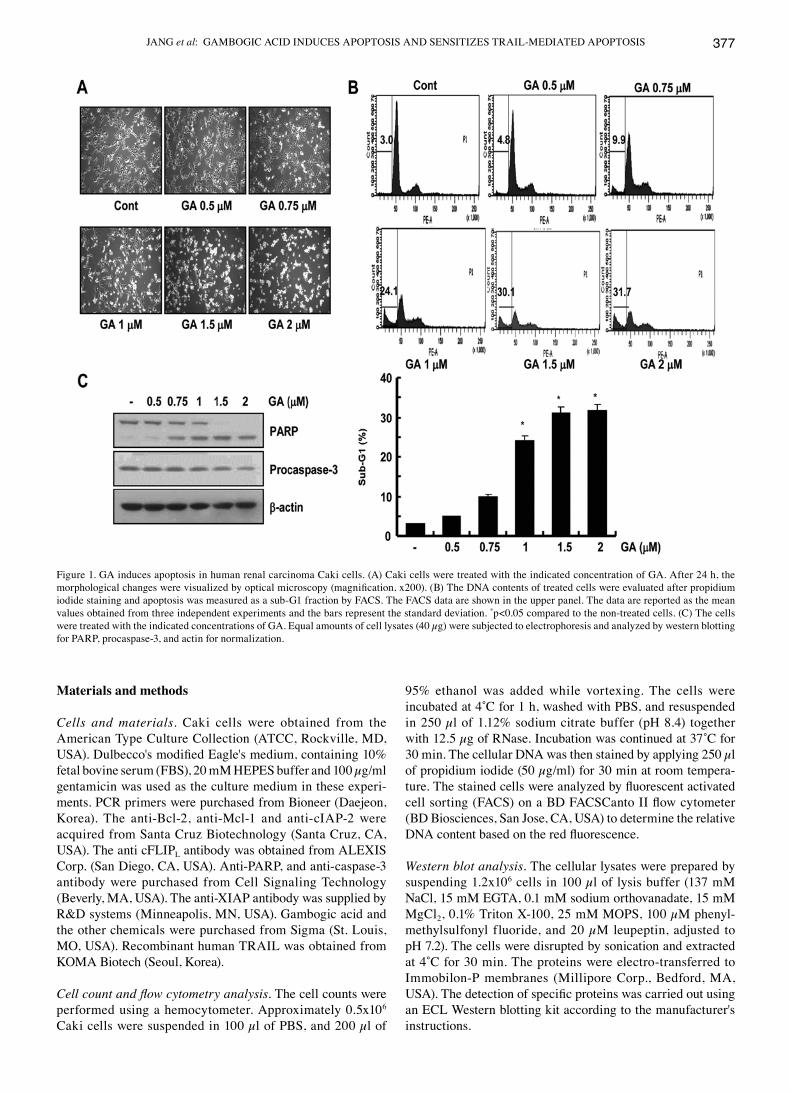
Figure 1. GA induces apoptosis in human renal carcinoma Caki cells. (A) Caki cells were treated with the indicated concentration of GA. After 24 h, the morphological changes were visualized by optical microscopy (magnification, x200). (B) The DNA contents of treated cells were evaluated after propidium iodide staining and apoptosis was measured as a sub-G1 fraction by FACS. The FACS data are shown in the upper panel. The data are reported as the mean values obtained from three independent experiments and the bars represent the standard deviation. *p<0.05 compared to the non-treated cells. (C) The cells were treated with the indicated concentrations of GA. Equal amounts of cell lysates (40 µg) were subjected to electrophoresis and analyzed by western blotting for PARP, procaspase-3, and actin for normalization.
INTERNATIONAL JOURNAL OF ONCOLOGY 48: 376-384, 2016378
Transfection. For transfection, the cells were plated onto 6-well plates at a density of 0.5x10⁶ cells/well and grown overnight. The cells were co-transfected with 2 µg of various plasmid constructs and 1 µg of pCMV-β-galactosidase plasmid for 5 h using the Lipofectamine method. After transfection, the cells were cultured in 10% FBS medium with a vesicle (DMSO) or drug for 24 h.
RNA isolation and reverse transcriptase-PCR. The expres-sion of cFLIPL mRNA was determined by RT-PCR. The total cellular RNA was extracted from the cells using an easyBlue reagent (Life Technologies, Seongnam, Korea), and the cDNA was prepared using M-MLV reverse transcriptase (Gibco-BRL, Gaithersburg, MD, USA), according to the manufacturer's instructions. The cellular RNA sample was reverse-transcribed with a random primer and then amplified by PCR, the GAPDH primer set was used as the internal control. The following primers were used to amplify the cFLIPL and GAPDH. The sequences of the sense and antisense primer for cFLIPL were 5'-CGG ACT ATA GAG TGC TGA TGG-3' and 5'-GAT TAT CAG GCA GAT TCC TAG-3', respectively. The sequences of the sense and antisense primer for GAPDH were 5'-AGG TCG GAG TCA ACG GAT TTG-3' and 5'-GTG ATG GCA TGG ACT GTG GT-3', respectively. The PCR products were analyzed by electrophoresis on a 1.5% agarose gel and detected by UV light.
4',6'-Diamidino-2-phenylindole (DAPI) staining for nuclei condensation and fragmentation. The cells were fixed with 1% paraformaldehyde on a slide glass for 30 min at room temperature. After washing with PBS, 300 nM 4',6'-diamidino-
2-phenylindole (Roche, Mannheim, Germany) was added to the fixed cells for 5 min, and the cells were examined by fluorescence microscopy.
Statistical analysis. The data were analyzed by a one-way ANOVA followed by post-hoc comparisons (Student-Newman-Keuls) using the Statistical Package for Social Sciences 8.0 (SPSS Inc., Chicago, IL, USA).
Results
GA induces apoptosis in renal carcinoma caki cells. To examine the anti-effects of GA in human renal cancer cells, Caki cells were treated with various concentrations of GA. With increasing GA concentration, the GA-treated Caki cells progressively showed the typical features of apoptosis, including cell shrinkage, rounding and detachment of the cell from the plate (Fig. 1A). Cell death was next determined by flow cytometry analysis to detect the hypodiploid cell popu-lations. As shown in Fig. 1B, treatment of Caki cells with GA resulted in a significant increase in the accumulation of sub-G1 phase cells in a dose-dependent manner. In addition, the treatment of Caki cells with GA strongly led to a reduction of the protein levels of 32-kDa precursor together with the concomitant cleavage of PARP, a substrate protein of caspases (Fig. 1C).
GA-induced apoptosis is mediated partly by the AIF trans-location. This study next examined whether the activation of caspase pathway plays a critical role in GA-induced apoptosis. As shown in Fig. 2A, GA-mediated apoptosis was prevented
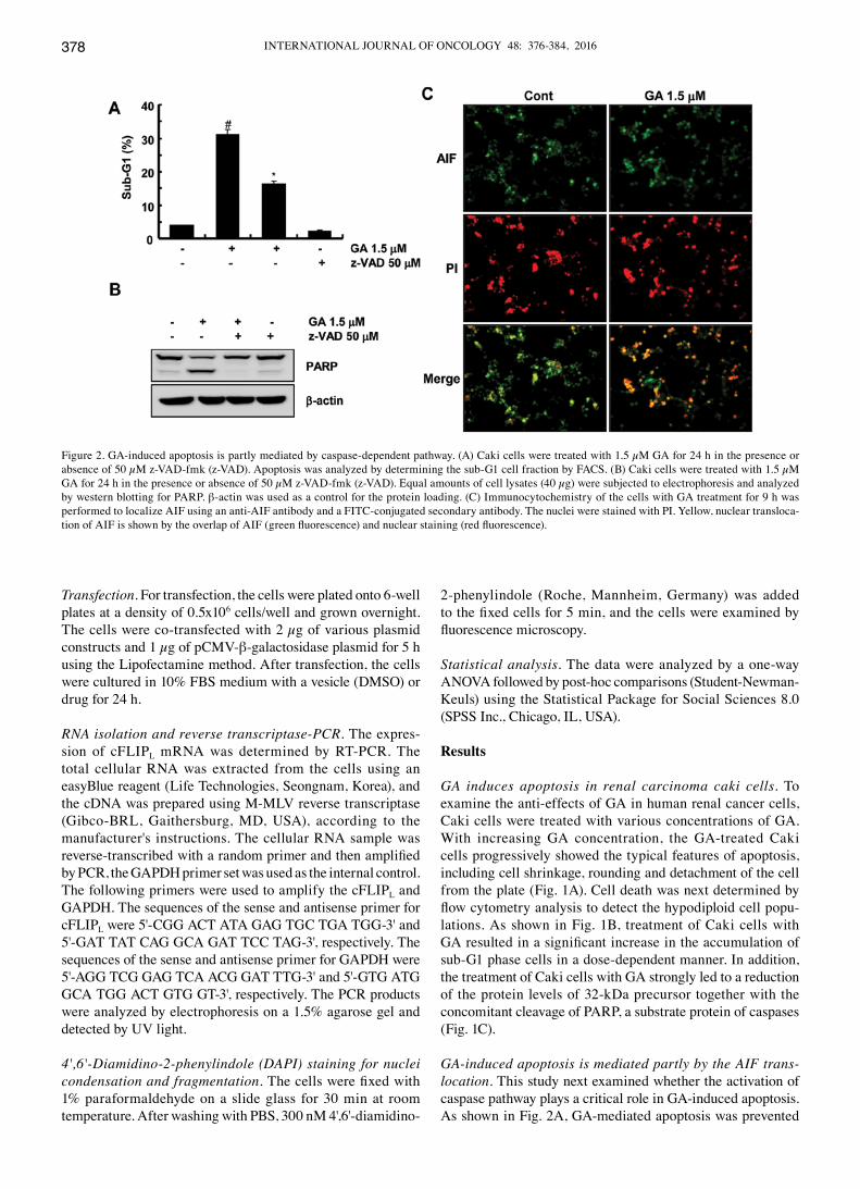
Figure 2. GA-induced apoptosis is partly mediated by caspase-dependent pathway. (A) Caki cells were treated with 1.5 µM GA for 24 h in the presence or absence of 50 µM z-VAD-fmk (z-VAD). Apoptosis was analyzed by determining the sub-G1 cell fraction by FACS. (B) Caki cells were treated with 1.5 µM GA for 24 h in the presence or absence of 50 µM z-VAD-fmk (z-VAD). Equal amounts of cell lysates (40 µg) were subjected to electrophoresis and analyzed by western blotting for PARP. β-actin was used as a control for the protein loading. (C) Immunocytochemistry of the cells with GA treatment for 9 h was performed to localize AIF using an anti-AIF antibody and a FITC-conjugated secondary antibody. The nuclei were stained with PI. Yellow, nuclear transloca-tion of AIF is shown by the overlap of AIF (green fluorescence) and nuclear staining (red fluorescence).
JANG et al: GAMBOGIC ACID INDUCES APOPTOSIS AND SENSITIzES TRAIL-MEDIATED APOPTOSIS 379
partly by a pretreatment with a general and potent inhibitor of caspases, z-VAD-fmk. In contrast, treatment with z-VAD-fmk completely prevented these caspase-related events, such as the cleavage of PARP (Fig. 2B). These results suggest that the GA-induced cell death was mediated partly by the caspase-dependent pathway and caspase-independent cell death in the presence of z-VAD-fmk. Because AIF is involved in the induction of apoptotic cell death through the caspase-independent pathway, this study examined whether AIF plays a role in GA-induced apoptotic cell death. The translocation of AIF was analyzed by the observation of its release from the mitochondria and translocation to the nucleus by fluorescence microscopy. As shown in Fig. 2C, fluorescence microscopy showed that AIF was translocated to the nucleus and caused nuclear condensation after the treatment with GA. This suggests that GA-induced apoptotic cell death in Caki cells is mediated by AIF translocation from the mitochondria into the nucleus via a caspase-independent pathway.
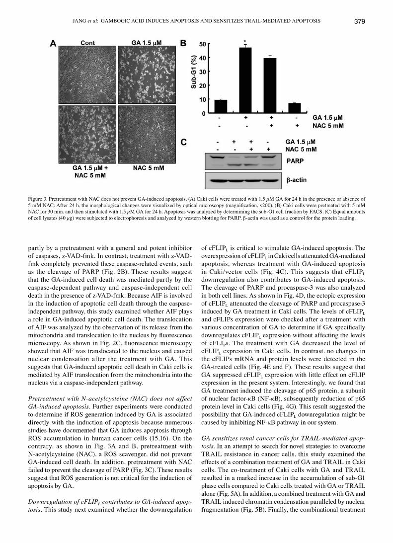
Pretreatment with N-acetylcysteine (NAC) does not affect GA-induced apoptosis. Further experiments were conducted to determine if ROS generation induced by GA is associated directly with the induction of apoptosis because numerous studies have documented that GA induces apoptosis through ROS accumulation in human cancer cells (15,16). On the contrary, as shown in Fig. 3A and B, pretreatment with N-acetylcysteine (NAC), a ROS scavenger, did not prevent GA-induced cell death. In addition, pretreatment with NAC failed to prevent the cleavage of PARP (Fig. 3C). These results suggest that ROS generation is not critical for the induction of apoptosis by GA.
Downregulation of cFLIPL contributes to GA-induced apop-tosis. This study next examined whether the downregulation
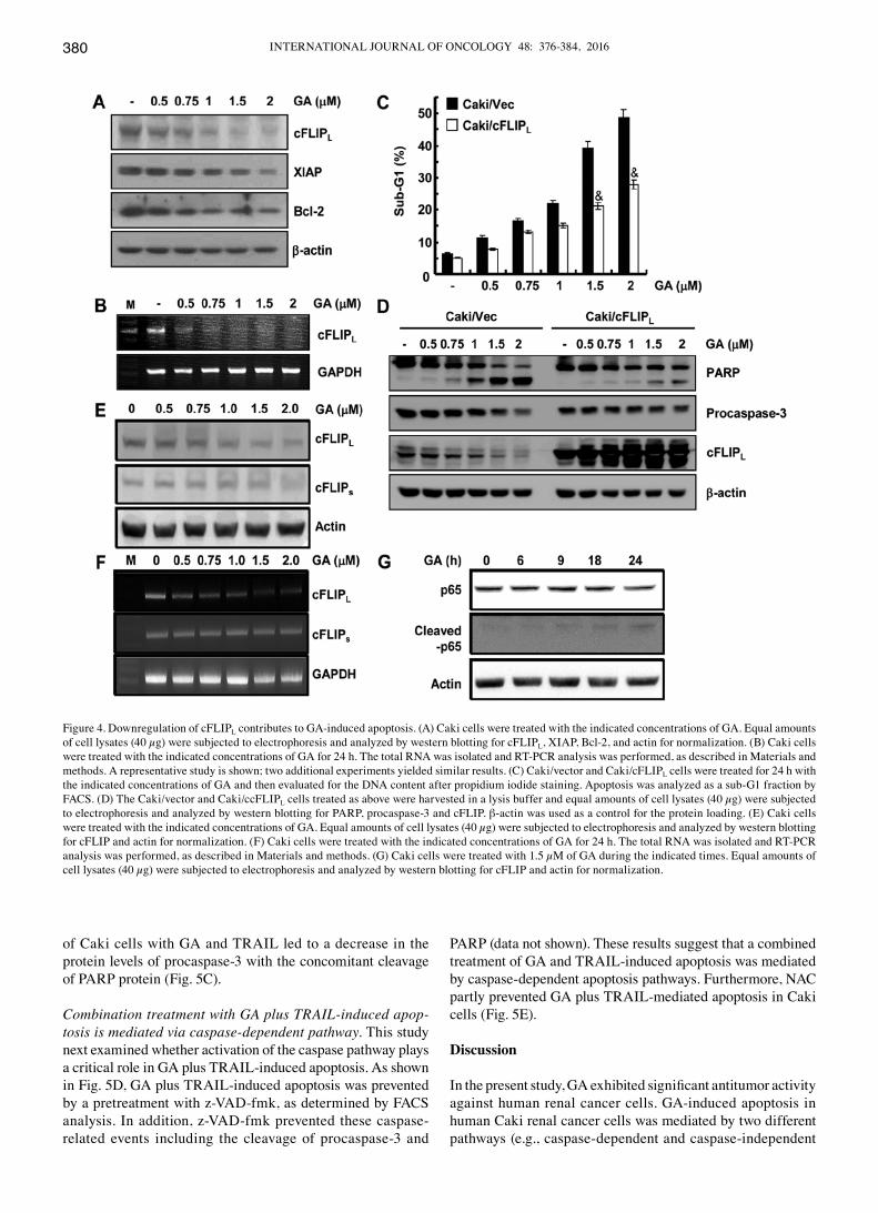
of cFLIPL is critical to stimulate GA-induced apoptosis. The overexpression of cFLIPL in Caki cells attenuated GA-mediated apoptosis, whereas treatment with GA-induced apoptosis in Caki/vector cells (Fig. 4C). This suggests that cFLIPL downregulation also contributes to GA-induced apoptosis. The cleavage of PARP and procaspase-3 was also analyzed in both cell lines. As shown in Fig. 4D, the ectopic expression of cFLIPL attenuated the cleavage of PARP and procaspase-3 induced by GA treatment in Caki cells. The levels of cFLIPL and cFLIPs expression were checked after a treatment with various concentration of GA to determine if GA specifically downregulates cFLIPL expression without affecting the levels of cFLIPs. The treatment with GA decreased the level of cFLIPL expression in Caki cells. In contrast, no changes in the cFLIPs mRNA and protein levels were detected in the GA-treated cells (Fig. 4E and F). These results suggest that GA suppressed cFLIPL expression with little effect on cFLIP expression in the present system. Interestingly, we found that GA treatment induced the cleavage of p65 protein, a subunit of nuclear factor-κB (NF-κB), subsequently reduction of p65 protein level in Caki cells (Fig. 4G). This result suggested the possibility that GA-induced cFLIPL downregulation might be caused by inhibiting NF-κB pathway in our system.
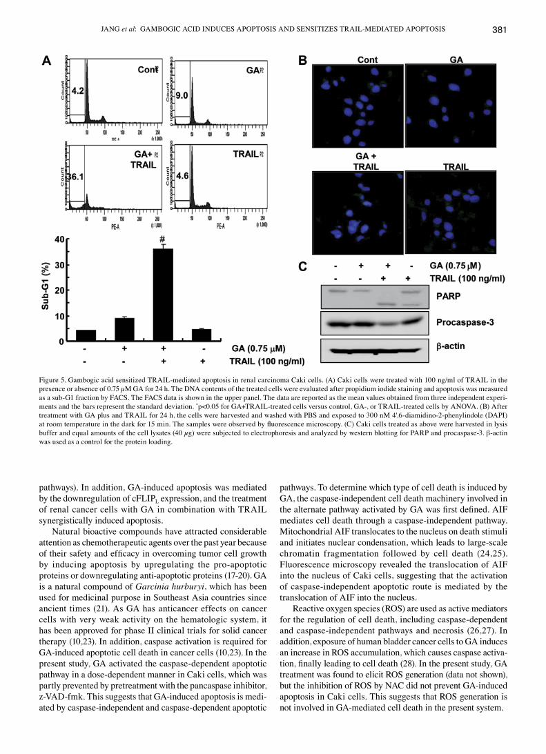
GA sensitizes renal cancer cells for TRAIL-mediated apop-tosis. In an attempt to search for novel strategies to overcome TRAIL resistance in cancer cells, this study examined the effects of a combination treatment of GA and TRAIL in Caki cells. The co-treatment of Caki cells with GA and TRAIL resulted in a marked increase in the accumulation of sub-G1 phase cells compared to Caki cells treated with GA or TRAIL alone (Fig. 5A). In addition, a combined treatment with GA and TRAIL induced chromatin condensation paralleled by nuclear fragmentation (Fig. 5B). Finally, the combinational treatment
Figure 3. Pretreatment with NAC does not prevent GA-induced apoptosis. (A) Caki cells were treated with 1.5 µM GA for 24 h in the presence or absence of 5 mM NAC. After 24 h, the morphological changes were visualized by optical microscopy (magnification, x200). (B) Caki cells were pretreated with 5 mM NAC for 30 min, and then stimulated with 1.5 µM GA for 24 h. Apoptosis was analyzed by determining the sub-G1 cell fraction by FACS. (C) Equal amounts of cell lysates (40 µg) were subjected to electrophoresis and analyzed by western blotting for PARP. β-actin was used as a control for the protein loading.
INTERNATIONAL JOURNAL OF ONCOLOGY 48: 376-384, 2016380
of Caki cells with GA and TRAIL led to a decrease in the protein levels of procaspase-3 with the concomitant cleavage of PARP protein (Fig. 5C).
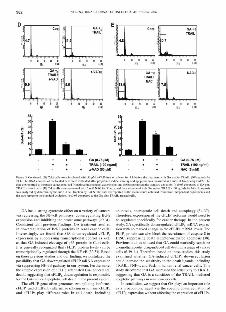
Combination treatment with GA plus TRAIL-induced apop-tosis is mediated via caspase-dependent pathway. This study next examined whether activation of the caspase pathway plays a critical role in GA plus TRAIL-induced apoptosis. As shown in Fig. 5D, GA plus TRAIL-induced apoptosis was prevented by a pretreatment with z-VAD-fmk, as determined by FACS analysis. In addition, z-VAD-fmk prevented these caspase-related events including the cleavage of procaspase-3 and
PARP (data not shown). These results suggest that a combined treatment of GA and TRAIL-induced apoptosis was mediated by caspase-dependent apoptosis pathways. Furthermore, NAC partly prevented GA plus TRAIL-mediated apoptosis in Caki cells (Fig. 5E).
Discussion
In the present study, GA exhibited significant antitumor activity against human renal cancer cells. GA-induced apoptosis in human Caki renal cancer cells was mediated by two different pathways (e.g., caspase-dependent and caspase-independent
Figure 4. Downregulation of cFLIPL contributes to GA-induced apoptosis. (A) Caki cells were treated with the indicated concentrations of GA. Equal amounts of cell lysates (40 µg) were subjected to electrophoresis and analyzed by western blotting for cFLIPL, XIAP, Bcl-2, and actin for normalization. (B) Caki cells were treated with the indicated concentrations of GA for 24 h. The total RNA was isolated and RT-PCR analysis was performed, as described in Materials and methods. A representative study is shown; two additional experiments yielded similar results. (C) Caki/vector and Caki/cFLIPL cells were treated for 24 h with the indicated concentrations of GA and then evaluated for the DNA content after propidium iodide staining. Apoptosis was analyzed as a sub-G1 fraction by FACS. (D) The Caki/vector and Caki/ccFLIPL cells treated as above were harvested in a lysis buffer and equal amounts of cell lysates (40 µg) were subjected to electrophoresis and analyzed by western blotting for PARP, procaspase-3 and cFLIP. β-actin was used as a control for the protein loading. (E) Caki cells were treated with the indicated concentrations of GA. Equal amounts of cell lysates (40 µg) were subjected to electrophoresis and analyzed by western blotting for cFLIP and actin for normalization. (F) Caki cells were treated with the indicated concentrations of GA for 24 h. The total RNA was isolated and RT-PCR analysis was performed, as described in Materials and methods. (G) Caki cells were treated with 1.5 µM of GA during the indicated times. Equal amounts of cell lysates (40 µg) were subjected to electrophoresis and analyzed by western blotting for cFLIP and actin for normalization.
JANG et al: GAMBOGIC ACID INDUCES APOPTOSIS AND SENSITIzES TRAIL-MEDIATED APOPTOSIS 381
pathways). In addition, GA-induced apoptosis was mediated by the downregulation of cFLIPL expression, and the treatment of renal cancer cells with GA in combination with TRAIL synergistically induced apoptosis.
Natural bioactive compounds have attracted considerable attention as chemotherapeutic agents over the past year because of their safety and efficacy in overcoming tumor cell growth by inducing apoptosis by upregulating the pro-apoptotic proteins or downregulating anti-apoptotic proteins (17-20). GA is a natural compound of Garcinia hurburyi, which has been used for medicinal purpose in Southeast Asia countries since ancient times (21). As GA has anticancer effects on cancer cells with very weak activity on the hematologic system, it has been approved for phase II clinical trials for solid cancer therapy (10,23). In addition, caspase activation is required for GA-induced apoptotic cell death in cancer cells (10,23). In the present study, GA activated the caspase-dependent apoptotic pathway in a dose-dependent manner in Caki cells, which was partly prevented by pretreatment with the pancaspase inhibitor, z-VAD-fmk. This suggests that GA-induced apoptosis is medi-ated by caspase-independent and caspase-dependent apoptotic
pathways. To determine which type of cell death is induced by GA, the caspase-independent cell death machinery involved in the alternate pathway activated by GA was first defined. AIF mediates cell death through a caspase-independent pathway. Mitochondrial AIF translocates to the nucleus on death stimuli and initiates nuclear condensation, which leads to large-scale chromatin fragmentation followed by cell death (24,25). Fluorescence microscopy revealed the translocation of AIF into the nucleus of Caki cells, suggesting that the activation of caspase-independent apoptotic route is mediated by the translocation of AIF into the nucleus.
Reactive oxygen species (ROS) are used as active mediators for the regulation of cell death, including caspase-dependent and caspase-independent pathways and necrosis (26,27). In addition, exposure of human bladder cancer cells to GA induces an increase in ROS accumulation, which causes caspase activa-tion, finally leading to cell death (28). In the present study, GA treatment was found to elicit ROS generation (data not shown), but the inhibition of ROS by NAC did not prevent GA-induced apoptosis in Caki cells. This suggests that ROS generation is not involved in GA-mediated cell death in the present system.
Figure 5. Gambogic acid sensitized TRAIL-mediated apoptosis in renal carcinoma Caki cells. (A) Caki cells were treated with 100 ng/ml of TRAIL in the presence or absence of 0.75 µM GA for 24 h. The DNA contents of the treated cells were evaluated after propidium iodide staining and apoptosis was measured as a sub-G1 fraction by FACS. The FACS data is shown in the upper panel. The data are reported as the mean values obtained from three independent experi-ments and the bars represent the standard deviation. *p<0.05 for GA+TRAIL-treated cells versus control, GA-, or TRAIL-treated cells by ANOVA. (B) After treatment with GA plus and TRAIL for 24 h, the cells were harvested and washed with PBS and exposed to 300 nM 4',6-diamidino-2-phenylindole (DAPI) at room temperature in the dark for 15 min. The samples were observed by fluorescence microscopy. (C) Caki cells treated as above were harvested in lysis buffer and equal amounts of the cell lysates (40 µg) were subjected to electrophoresis and analyzed by western blotting for PARP and procaspase-3. β-actin was used as a control for the protein loading.
INTERNATIONAL JOURNAL OF ONCOLOGY 48: 376-384, 2016382
GA has a strong cytotoxic effect on a variety of cancers via repressing the NF-κB pathways, downregulating Bcl-2 expression and inhibiting the proteasome pathways (29-31). Consistent with previous findings, GA treatment resulted in downregulation of Bcl-2 proteins in renal cancer cells. Interestingly, we found that GA downregulated cFLIPL expression by suppressing transcriptional control as well as that GA induced cleavage of p65 protein in Caki cells. It is generally recognized that cFLIPL protein levels can be transcriptionally regulated through the NF-κB (32,33). Based on these previous studies and our finding, we postulated the possibility that GA downregulated cFLIP mRNA expression via suppressing NF-κB pathway in our system. Furthermore, the ectopic expression of cFLIPL attenuated GA-induced cell death, suggesting that cFLIPL downregulation is responsible for the GA-induced apoptotic cell death in the present system.
The cFLIP gene often generates two splicing isoforms, cFLIPL and cFLIPs, by alternative splicing in humans. cFLIPL and cFLIPs play different roles in cell death, including
apoptosis, necroptotic cell death and autophagy (34-37). Therefore, expression of the cFLIP isoforms would need to be regulated specifically for cancer therapy. In the present study, GA specifically downregulated cFLIPL mRNA expres-sion with no marked change in the cFLIPs mRNA levels. The FLIPL protein can also block the recruitment of caspase-8 to DISC, suppressing death receptor-mediated apoptosis (38). Previous studies showed that GA could markedly sensitize chemotherapeutic drug-induced cell death in a range of cancer cells (6,39-41). Therefore, based on these studies, this study examined whether GA-induced cFLIPL downregulation could increase the sensitivity to the death ligands, including TRAIL, TNF-α and FasL in human renal cancer cells. This study discovered that GA increased the sensitivity to TRAIL, suggesting that GA is a sensitizer of the TRAIL-mediated apoptotic pathways in renal cancer cells.
In conclusion, we suggest that GA plays an important role as a proapoptotic agent via the specific downregulation of cFLIPL expression without affecting the expression of cFLIPs.
Figure 5. Continued. (D) Caki cells were incubated with 50 µM z-VAD-fmk or solvent for 1 h before the treatment with GA and/or TRAIL (100 ng/ml) for 24 h. The DNA contents of the treated cells were evaluated after propidium iodide staining and apoptosis was measured as a sub-G1 fraction by FACS. The data are reported as the mean values obtained from three independent experiments and the bars represent the standard deviation. *p<0.05 compared to GA plus TRAIL-treated cells. (E) Caki cells were pretreated with 5 mM NAC for 30 min, and then stimulated with GA and/or TRAIL (100 ng/ml) for 24 h. Apoptosis was analyzed by determining the sub-G1 cell fraction by FACS. The data are reported as the mean values obtained from three independent experiments and the bars represent the standard deviation. *p<0.05 compared to the GA plus TRAIL-treated cells.
JANG et al: GAMBOGIC ACID INDUCES APOPTOSIS AND SENSITIzES TRAIL-MEDIATED APOPTOSIS 383
In addition, GA treatment rendered human renal cancer cells more sensitive to TRAIL. These results suggested that a combined treatment of GA and TRAIL may provide a safe and effective therapeutic strategy against cancers that are resistant to conventional treatments. Furthermore, this study provided novel evidence that the prominent sensitizing effect of GA on TRAIL-induced apoptosis is due to the downregulation of cFLIPL.
Acknowledgements
This study was supported by the Yeungnam University research grants in 2013.
References
1. Guo QL, You QD, Wu zQ, Yuan ST and zhao L: General gambogic acids inhibited growth of human hepatoma SMMC-7721 cells in vitro and in nude mice. Acta Pharmacol Sin 25: 769-774, 2004.
2. Qin Y, Meng L, Hu C, Duan W, zuo z, Lin L, zhang X and Ding J: Gambogic acid inhibits the catalytic activity of human topoisomerase IIalpha by binding to its ATPase domain. Mol Cancer Ther 6: 2429-2440, 2007.
3. Geng J, Xiao S, zheng z, Song S and zhang L: Gambogic acid protects from endotoxin shock by suppressing pro-inflammatory factors in vivo and in vitro. Inflamm Res 62: 165-172, 2013.
4. Lu N, Hui H, Yang H, zhao K, Chen Y, You QD and Guo QL: Gambogic acid inhibits angiogenesis through inhibiting PHD2-VHL-HIF-1α pathway. Eur J Pharm Sci 49: 220-226, 2013.
5. Xin zF, Shen CC, Tao LJ, Yan SG and Wu HB: Gambogic acid inhibits invasion of osteosarcoma via upregulation of TIMP-1. Int J Mol Med 31: 105-112, 2013.
6. Wang LH, Yang JY, Yang SN, Li Y, Ping GF, Hou Y, Cui W, Wang zz, Xiao W and Wu CF: Suppression of NF-κB signaling and P-glycoprotein function by gambogic acid synergistically potentiates adriamycin -induced apoptosis in lung cancer. Curr Cancer Drug Targets 14: 91-103, 2014.
7. Duan D, zhang B, Yao J, Liu Y, Sun J, Ge C, Peng S, Fang J, Xiao W and Wu CF: Gambogic acid induces apoptosis in hepa-tocellular carcinoma SMMC-7721 cells by targeting cytosolic thioredoxin reductase. Free Radic Biol Med 69: 15-25, 2014.
8. Xu X, Liu Y, Wang L, He J, zhang H, Chen X, Li Y, Yang J and Tao J: Gambogic acid induces apoptosis by regulating the expression of Bax and Bcl-2 and enhancing caspase-3 activity in human malignant melanoma A375 cells. Int J Dermatol 48: 186-192, 2009.
9. Li C, Qi Q, Lu N, Dai Q, Li F, Wang X, You Q and Guo Q: Gambogic acid promotes apoptosis and resistance to metastatic potential in MDA-MB-231 human breast carcinoma cells. Biochem Cell Biol 90: 718-730, 2012.
10. Shi X, Chen X, Li X, Lan X, zhao C, Liu S, Huang H, Liu N, Liao S, Song W, et al: Gambogic acid induces apoptosis in imatinib-resistant chronic myeloid leukemia cells via inducing proteasome inhibition and caspase-dependent Bcr-Abl down-regulation. Clin Cancer Res 20: 151-163, 2014.
11. Guo Q, Qi Q, You Q, Gu H, zhao L and Wu z: Toxicological studies of gambogic acid and its potential targets in experimental animals. Basic Clin Pharmacol Toxicol 99: 178-184, 2006.
12. Tan ML, Ooi JP, Ismail N, Moad AI and Muhammad TS: Programmed cell death pathways and current antitumor targets. Pharm Res 26: 1547-1560, 2009.
13. Wu GS: TRAIL as a target in anti-cancer therapy. Cancer Lett 285: 1-5, 2009.
14. Refaat A, Abd-Rabou A and Reda A: TRAIL combinations: The new ‘trail’ for cancer therapy (Review). Oncol Lett 7: 1327-1332, 2014.
15. Yang LJ and Chen Y: New targets for the antitumor activity of gambogic acid in hematologic malignancies. Acta Pharmacol Sin 34: 191-198, 2013.
16. Nie F, zhang X, Qi Q, Yang L, Yang Y, Liu W, Lu N, Wu z, You Q and Guo Q: Reactive oxygen species accumulation contributes to gambogic acid-induced apoptosis in human hepatoma SMMC-7721 cells. Toxicology 260: 60-67, 2009.
17. Li L, Gao Y, zhang L, zeng J, He D and Sun Y: Silibinin inhibits cell growth and induces apoptosis by caspase activation, down-regulating survivin and blocking EGFR-ERK activation in renal cell carcinoma. Cancer Lett 272: 61-69, 2008.
18. Spagnuolo C, Cerella C, Russo M, Chateauvieux S, Diederich M and Russo GL: Quercetin downregulates Mcl-1 by acting on mRNA stability and protein degradation. Br J Cancer 105: 221-230, 2011.
19. Wu YL, Wan Y, Jin XJ, OuYang BQ, Bai T, zhao YQ and Nan JX: 25-OCH3-PPD induces the apoptosis of activated t-HSC/Cl-6 cells via c-FLIP-mediated NF-κB activation. Chem Biol Interact 194: 106-112, 2011.
20. Yu HY, Jin CY, Kim KS, Lee YC, Park SH, Kim GY, Kim WJ, Moon HI, Choi YH and Lee JH: Oleifolioside A mediates caspase-independent human cervical carcinoma HeLa cell apoptosis involving nuclear relocation of mitochondrial apopto-genic factors AIF and EndoG. J Agric Food Chem 60: 5400-5406, 2012.
21. He D, Xu Q, Yan M, zhang P, zhou X, zhang z, Duan W, zhong L, Ye D and Chen W: The NF-kappa B inhibitor, celastrol, could enhance the anti-cancer effect of gambogic acid on oral squamous cell carcinoma. BMC Cancer 9: 343, 2009.
22. Márquez F, Babio N, Bulló M and Salas-Salvadó J: Evaluation of the safety and efficacy of hydroxycitric acid or Garcinia cambogia extracts in humans. Crit Rev Food Sci Nutr 52: 585-594, 2012.
23. Rahman MA, Kim NH and Huh SO: Cytotoxic effect of gambogic acid on SH-SY5Y neuroblastoma cells is mediated by intrinsic caspase-dependent signaling pathway. Mol Cell Biochem 377: 187-196, 2013.
24. Joza N, Susin SA, Daugas E, Stanford WL, Cho SK, Li CY, Sasaki T, Elia AJ, Cheng HY, Ravagnan L, et al: Essential role of the mitochondrial apoptosis-inducing factor in programmed cell death. Nature 410: 549-554, 2001.
25. Kim EA, Jang JH, Lee YH, Sung EG, Song IH, Kim JY, Kim S, Sohn HY and Lee TJ: Dioscin induces caspase-independent apoptosis through activation of apoptosis-inducing factor in breast cancer cells. Apoptosis 19: 1165-1175, 2014.
26. Jacobson MD: Reactive oxygen species and programmed cell death. Trends Biochem Sci 21: 83-86, 1996.
27. Kang YH, Yi MJ, Kim MJ, Park MT, Bae S, Kang CM, Cho CK, Park IC, Park MJ, Rhee CH, et al: Caspase-independent cell death by arsenic trioxide in human cervical cancer cells: Reactive oxygen species-mediated poly(ADP-ribose) polymerase-1 activa-tion signals apoptosis-inducing factor release from mitochondria. Cancer Res 64: 8960-8967, 2004.
28. Ishaq M, Khan MA, Sharma K, Sharma G, Dutta RK and Majumdar S: Gambogic acid induced oxidative stress dependent caspase activation regulates both apoptosis and autophagy by targeting various key molecules (NF-κB, Beclin-1, p62 and NBR1) in human bladder cancer cells. Biochim Biophys Acta 1840: 3374-3384, 2014.
29. Palempalli UD, Gandhi U, Kalantari P, Vunta H, Arner RJ, Narayan V, Ravindran A and Prabhu KS: Gambogic acid covalently modifies IkappaB kinase-beta subunit to mediate suppression of lipopolysaccharide-induced activation of NF-kappaB in macrophages. Biochem J 419: 401-409, 2009.
30. zhao L, Guo QL, You QD, Wu zQ and Gu HY: Gambogic acid induces apoptosis and regulates expressions of Bax and Bcl-2 protein in human gastric carcinoma MGC-803 cells. Biol Pharm Bull 27: 998-1003, 2004.
31. Li X, Liu S, Huang H, Liu N, zhao C, Liao S, Yang C, Liu Y, zhao C, Li S, et al: Gambogic acid is a tissue-specific protea-some inhibitor in vitro and in vivo. Cell Rep 3: 211-222, 2013.
32. Kreuz S, Siegmund D, Scheurich P and Wajant H: NF-kappaB inducers upregulate cFLIP, a cycloheximide-sensitive inhibitor of death receptor signaling. Mol Cell Biol 21: 3964-3973, 2001.
33. Micheau O, Lens S, Gaide O, Alevizopoulos K and Tschopp J: NF-kappaB signals induce the expression of c-FLIP. Mol Cell Biol 21: 5299-5305, 2001.
34. Tenev T, Bianchi K, Darding M, Broemer M, Langlais C, Wallberg F, zachariou A, Lopez J, MacFarlane M, Cain K, et al: The Ripoptosome, a signaling platform that assembles in response to genotoxic stress and loss of IAPs. Mol Cell 43: 432-448, 2011.
35. Feoktistova M, Geserick P, Kellert B, Dimitrova DP, Langlais C, Hupe M, Cain K, MacFarlane M, Häcker G and Leverkus M: cIAPs block Ripoptosome formation, a RIP1/caspase-8 containing intracellular cell death complex differentially regulated by cFLIP isoforms. Mol Cell 43: 449-463, 2011.

INTERNATIONAL JOURNAL OF ONCOLOGY 48: 376-384, 2016384
36. Lee JS, Li Q, Lee JY, Lee SH, Jeong JH, Lee HR, Chang H, zhou FC, Gao SJ, Liang C, et al: FLIP-mediated autophagy regulation in cell death control. Nat Cell Biol 11: 1355-1362, 2009.
37. He MX and He YW: A role for c-FLIP(L) in the regulation of apoptosis, autophagy, and necroptosis in T lymphocytes. Cell Death Differ 20: 188-197, 2013.
38. Safa AR, Day TW and Wu CH: Cellular FLICE-like inhibitory protein (C-FLIP): A novel target for cancer therapy. Curr Cancer Drug Targets 8: 37-46, 2008.
39. Su J, Cheng H, zhang D, Wang M, Xie C, Hu Y, Chang HC and Li Q: Synergistic effects of 5-fluorouracil and gambogenic acid on A549 cells: Activation of cell death caused by apoptotic and necroptotic mechanisms via the ROS-mitochondria pathway. Biol Pharm Bull 37: 1259-1268, 2014.
40. Wang S, Wang L, Chen M and Wang Y: Gambogic acid sensitizes resistant breast cancer cells to doxorubicin through inhibiting P-glycoprotein and suppressing survivin expression. Chem Biol Interact 235: 76-84, 2015.
41. zhao W, You CC, zhuang JP, zu JN, Chi zY, Xu GP and Yan JL: Viability inhibition effect of gambogic acid combined with cisplatin on osteosarcoma cells via mitochondria-independent apoptotic pathway. Mol Cell Biochem 382: 243-252, 2013.


