Bortezomib sensitizes thyroid cancer to BRAF inhibitor in ...
11
https://doi.org/10.1530/ERC-17-0182 http://erc.endocrinology-journals.org © 2018 Society for Endocrinology Printed in Great Britain Published by Bioscientifica Ltd. 25:1 Endocrine-Related Cancer K Tsumagari et al. Bortezomib sensitizes TC to BRAF inhibitors PROOF ONLY 99–109 RESEARCH Bortezomib sensitizes thyroid cancer to BRAF inhibitor in vitro and in vivo Koji Tsumagari 1 , Zakaria Y Abd Elmageed 1 , Andrew B Sholl 2 , Erik A Green 1 , Saboori Sobti 1 , Abdul Razzaq Khan 1 , Abdulrahman Kandil 1 , Fadi Murad 1 , Paul Friedlander 3 , A Hamid Boulares 4,5 and Emad Kandil 1,3 1 Department of Surgery, Tulane University School of Medicine, New Orleans, Louisiana, USA 2 Department of Pathology, Tulane University School of Medicine, New Orleans, Louisiana, USA 3 Department of Otolaryngology, Tulane University School of Medicine, New Orleans, Louisiana, USA 4 The Stanley Scott Cancer Center, Louisiana State University Health Science Center, New Orleans, Louisiana, USA 5 Department of Pharmacology, Louisiana State University Health Science Center, New Orleans, Louisiana, USA Correspondence should be addressed to E Kandil: [email protected] Abstract Although overall survival rate for patients with thyroid cancer (TC) is high, there is an alarming 10-year recurrence rate of up to 30% conferring a ~50% survival among these high-risk patients. The BRAF V600E mutation is estimated to be present in over 50% of papillary thyroid cancer (PTC) cases besides being associated with carcinogenesis and poor prognosis. We assessed the status of NF-κB, Ki-67, cyclin D1 and BRAF V600E in TC tissues and TC cell lines using immunohistochemistry and Western blot analysis. Concurrently, we evaluated the outcomes of combined targeting of the proteasome pathway in addition to selective BRAF inhibitors in cases of PTC. In this study, BRAF V600E -bearing TC cells were treated with BRAF V600E inhibitor, Vemurafenib alone or in combination with the proteasome inhibitor, Bortezomib. The combination of both drugs showed synergistic effects as evidenced by cell growth inhibition (P < 0.05), increased G2-phase cell cycle arrest and induced apoptosis (P < 0.05). In our TC xenograft model, the combination of Vemurafenib and Bortezomib significantly reduced tumor size (P < 0.05) and expression of the markers of cell growth and proliferation, Ki-67 and cyclin D1 (P < 0.001), when compared to monotherapy. Further analysis demonstrated that treatment with Bortezomib sensitized TC cells to Vemurafenib via mitochondrial dysregulation and apoptosis of TC cells, as evidenced by the increase in the expression of p53, Noxa protein, the loss of mitochondrial membrane potential, cytochrome c release and Poly (ADP-ribose) polymerase cleavage. Our results demonstrate a strong clinical potential for the combination of the Bortezomib and the BRAF inhibitor Vemurafenib as an efficient therapeutic approach for the treatment of TC. Introduction Thyroid cancer (TC) is the most common type of endocrine-related cancer (56,870 new cases in 2017) and represents approximately 3.4% of all new cancer cases (Howlader et al. 2017). Although over 90% of differentiated thyroid cancers (DTC) are cured by surgery and have an excellent prognosis, recurrence is possible in patients with DTC, and a small percentage may have a fatal disease course. Anaplastic thyroid cancer (ATC) is Endocrine-Related Cancer (2018) 25, 99–109 Key Words f BRAFV600E f NF-κB f thyroid cancer f Vemurafenib f Bortezomib Downloaded from Bioscientifica.com at 03/18/2022 11:58:04AM via free access
Transcript of Bortezomib sensitizes thyroid cancer to BRAF inhibitor in ...
https://doi.org/10.1530/ERC-17-0182http://erc.endocrinology-journals.org © 2018 Society for Endocrinology
Printed in Great BritainPublished by Bioscientifica Ltd.
25:1Endocrine-Related Cancer
K Tsumagari et al. Bortezomib sensitizes TC to BRAF inhibitors
PROOF ONLY99–109
10.1530/ERC-17-0182
RESEARCH
Bortezomib sensitizes thyroid cancer to BRAF inhibitor in vitro and in vivo
Koji Tsumagari1, Zakaria Y Abd Elmageed1, Andrew B Sholl2, Erik A Green1, Saboori Sobti1, Abdul Razzaq Khan1, Abdulrahman Kandil1, Fadi Murad1, Paul Friedlander3, A Hamid Boulares4,5 and Emad Kandil1,3
1Department of Surgery, Tulane University School of Medicine, New Orleans, Louisiana, USA2Department of Pathology, Tulane University School of Medicine, New Orleans, Louisiana, USA3Department of Otolaryngology, Tulane University School of Medicine, New Orleans, Louisiana, USA4The Stanley Scott Cancer Center, Louisiana State University Health Science Center, New Orleans, Louisiana, USA5Department of Pharmacology, Louisiana State University Health Science Center, New Orleans, Louisiana, USA
Correspondence should be addressed to E Kandil: [email protected]
Abstract
Although overall survival rate for patients with thyroid cancer (TC) is high, there is
an alarming 10-year recurrence rate of up to 30% conferring a ~50% survival among
these high-risk patients. The BRAFV600E mutation is estimated to be present in over 50%
of papillary thyroid cancer (PTC) cases besides being associated with carcinogenesis
and poor prognosis. We assessed the status of NF-κB, Ki-67, cyclin D1 and BRAFV600E
in TC tissues and TC cell lines using immunohistochemistry and Western blot analysis.
Concurrently, we evaluated the outcomes of combined targeting of the proteasome
pathway in addition to selective BRAF inhibitors in cases of PTC. In this study,
BRAFV600E-bearing TC cells were treated with BRAFV600E inhibitor, Vemurafenib alone
or in combination with the proteasome inhibitor, Bortezomib. The combination of
both drugs showed synergistic effects as evidenced by cell growth inhibition (P < 0.05),
increased G2-phase cell cycle arrest and induced apoptosis (P < 0.05). In our TC xenograft
model, the combination of Vemurafenib and Bortezomib significantly reduced tumor
size (P < 0.05) and expression of the markers of cell growth and proliferation, Ki-67 and
cyclin D1 (P < 0.001), when compared to monotherapy. Further analysis demonstrated
that treatment with Bortezomib sensitized TC cells to Vemurafenib via mitochondrial
dysregulation and apoptosis of TC cells, as evidenced by the increase in the expression of
p53, Noxa protein, the loss of mitochondrial membrane potential, cytochrome c release
and Poly (ADP-ribose) polymerase cleavage. Our results demonstrate a strong clinical
potential for the combination of the Bortezomib and the BRAF inhibitor Vemurafenib as
an efficient therapeutic approach for the treatment of TC.
Introduction
Thyroid cancer (TC) is the most common type of endocrine-related cancer (56,870 new cases in 2017) and represents approximately 3.4% of all new cancer cases (Howlader et al. 2017). Although over 90% of
differentiated thyroid cancers (DTC) are cured by surgery and have an excellent prognosis, recurrence is possible in patients with DTC, and a small percentage may have a fatal disease course. Anaplastic thyroid cancer (ATC) is
Endocrine-Related Cancer (2018) 25, 99–109
1
Key Words
f BRAFV600E
f NF-κB
f thyroid cancer
f Vemurafenib
f Bortezomib
25
Downloaded from Bioscientifica.com at 03/18/2022 11:58:04AMvia free access

Printed in Great BritainPublished by Bioscientifica Ltd.https://doi.org/10.1530/ERC-17-0182
http://erc.endocrinology-journals.org © 2018 Society for Endocrinology
100K Tsumagari et al. Bortezomib sensitizes TC to BRAF inhibitors
25:1
PROOF ONLYEndocrine-Related Cancer
one of the most aggressive malignancies that can arise from a transformation of pre-existing DTC (Sanghvi 2013, Howlader et al. 2017). The survival rate of patients with ATC post-diagnosis is usually less than one year. Currently, effective standard therapies for patients with aggressive TC remain limited and need to be developed urgently (Smallridge 2012, Baumunk et al. 2013).
The mitogen-activated protein kinase (MAPK) signaling cascade regulates cell proliferation, differentiation, apoptosis and cell survival (Roberts & Der 2007, Li & Wu 2013). BRAF (v-raf murine sarcoma viral oncogene homolog B), a serine/threonine kinase, is one of the most well-characterized members of the MAPK pathway. The majority of somatic BRAF mutations occur as a substitution of valine with glutamic acid at amino acid residue 600 (V600E). The BRAFV600E mutation constitutively activates its downstream pathway and results in oncogenic transformation of normal cells (Bennedbaek & Hegedüs 2003, Wan et al. 2004). Additionally, it also cooperates with PIK3CA mutations to promote anaplastic thyroid carcinogenesis (Bennedbaek & Hegedüs 1999, Charles et al. 2014). BRAFV600E plays a critical tumorigenic role in several types of cancers, up to 60% of melanomas and 15% of colon cancers (Hegedüs 2009, Davies & Welch 2010, Rustgi et al. 2013, Salama & Flaherty 2013). The BRAFV600E mutation is in up to 50% of papillary thyroid cancers (PTCs) and 25% of ATCs (Jeong et al. 2008, Nucera et al. 2010, Smallridge 2012, Baumunk et al. 2013). Selective BRAFV600E inhibitors have shown promise in treatment of metastatic melanoma (Deandrea et al. 2008, Bollag et al. 2012). However, intrinsic and acquired resistance to selective BRAF inhibitors has proven to be a significant clinical obstacle in the treatment of patients with metastatic melanoma (Deandrea et al. 2008, Davies & Welch 2010, Bollag et al. 2012, Salama & Flaherty 2013).
Despite the fact that Sorafenib (Nexavar), Lenvatinib and multiple kinase inhibitors including BRAF are currently approved for metastatic TC, the median progression-free survival is only approximately 1 year (Spiezia et al. 2009, Brose et al. 2014). The BRAFV600E inhibitors Vemurafenib and Dabrafenib are currently FDA-approved for the treatment of BRAFV600E-harboring melanoma and currently undergoing clinical trials for metastatic TC. Severe toxicities have been observed in these patients, and most patients, even those who responded initially, ultimately developed acquired resistance and rapid disease progression (Schlumberger & Torlantano 2000, Spiezia et al. 2009, Brose et al. 2014, Dadu et al. 2015). Therefore, new alternative therapies are needed for aggressive TCs.
NF-κB has been shown to play a key role in TC by controlling the proliferative and anti-apoptotic signaling pathways of TC cells, including ATCs (Cooper et al. 2006, Hsu et al. 2014, Tsumagari et al. 2015). Paclitaxel induces cell survival through NF-κB activation in ATCs, and the combination of paclitaxel and the NF-κB inhibitor dehydroxymethylepoxyquinomicin has been suggested to compensate for paclitaxel resistance in ATCs (Durante et al. 2006, Polona et al. 2006).
Bortezomib, currently approved for the treatment of advanced multiple myeloma, is a proteasome inhibitor and can effectively control canonical and non-canonical NF-κB signaling (Garrean et al. 2007, Hideshima et al. 2007). Although Bortezomib alone has a modest effect on advanced TC (Bailey et al. 2001, Cooper et al. 2006, Putzer et al. 2013, Tsumagari et al. 2015), a synergetic effect was suggested in combination with other chemotherapeutic agents, portending a promising role for Bortezomib in combination therapy regimens (Smith et al. 2001, Cooper et al. 2006, Mitsiades et al. 2006, Tsumagari et al. 2015). These observations demonstrate the potential for targeting NF-κB for treatment in resistant and aggressive TC.
We therefore hypothesized that the combination of Vemurafenib and Bortezomib would provide synergetic effects in treating aggressive TC. Herein, we investigated the effect of the use of a combination of two FDA-approved drugs in in vitro and in vivo thyroid mouse xenograft models.
Materials and methods
Cell culture
The human anaplastic thyroid carcinoma-derived cell lines, SW1736 and KAT18 cells were provided by Dr Xing (University of Johns Hopkins, Baltimore, MD, USA). DRO and NPA cells were obtained from Dr Guy J F Julliard (University of California Los Angeles School of Medicine, Los Angeles, CA, USA). DRO, NPA and SW1736 cells carry mutant BRAFV600E and KAT18 cells carry BRAFwt. Cells were cultured in RPMI-1640 medium supplemented with 10% fetal bovine serum, 0.1 mM non-essential amino acids, 1 mM sodium pyruvate and 1% penicillin–streptomycin in a 37°C humidified incubator with 5% CO2. Cells were treated with Vemurafenib (PLX4032, SelleckChem, Houston, TX, USA) and Bortezomib (LC Laboratories, Woburn, MA, USA) at various indicated concentrations and time points. The culture medium and drugs were replenished every 24 h during the treatment.
Downloaded from Bioscientifica.com at 03/18/2022 11:58:04AMvia free access

http://erc.endocrinology-journals.org © 2018 Society for Endocrinology
Printed in Great BritainPublished by Bioscientifica Ltd.https://doi.org/10.1530/ERC-17-0182
101K Tsumagari et al. Bortezomib sensitizes TC to BRAF inhibitors
25:1
PROOF ONLYEndocrine-Related Cancer
Human thyroid tissue specimens
Eleven PTC and three goiter tissue specimens were obtained from the Louisiana Cancer Research Center (LCRC) Biospecimen Core following the approval of the Institutional Review Board of Tulane University Health Sciences Center (TUHSC), USA. Real-Time PCR and Sanger sequencing for the detection of BRAFV600E was utilized to detect mutation presence. Clinicopathologic characteristics were retrospectively retrieved from the patient records.
Cell proliferation assay
The cell proliferation assay was performed in triplicate and each experiment was repeated at least thrice. TC cells were seeded into 96-well plates and treated with either drug at the indicated concentrations. After 1, 3 and 5 days of treatment, 10 μL of tetrazolium salt WST-8 (Cell Counting Kit-8, Dojindo Molecular Technologies Inc., MD, USA) was added to cells and incubated for 4 h at 37°C. The plates were read at 450 nm using a microplate reader. For each cell line, the 50% inhibition concentration (IC50) of Vemurafenib and Bortezomib was calculated using GraphPad Prism (version 5.0). Trypan blue (ThermoFisher) exclusion assay was also performed for each cell line. The effectiveness of Vemurafenib and Bortezomib combination therapy was evaluated by a combination index using isobologram analysis which quantifies drug–drug interactions between Vemurafenib and Bortezomib using the described equation (Kanotra et al. 2008, Abd Elmageed et al. 2017).
Colonogenic assay
Cells at a concentration of 500 cells per well were seeded in 6-well plates with 2 mL of media in each well. After overnight incubation (37°C, 5% CO2), each well was treated with Vemurafenib, Bortezomib or a combination of both drugs. After 14 days of treatment, the media were aspirated from each well and a solution of 1% methylene blue and 50% methanol was added. The cells were incubated in this solution for 30 min to fix and stain them. The colony number in each well was counted in duplicate plates.
Cell cycle assay
TC cells were harvested and centrifuged, and the resulting pellets were fixed in ice-cold 70% ethanol. Fixed cells
were recentrifuged, washed and resuspended in PBS containing RNase A (1 mg/mL) and propidium iodide (PI) was added (1.0 mg/mL). PI-stained cells were analyzed by a fluorescence-activated cell sorter (FACSCalibur in the UAMS Flow Cytometry Core Facility, Tulane University, New Orleans, LA, USA), followed by determination of the percentages of cells in G1-, S- and G2/M-phases of the cell cycle.
Apoptosis assay
After treatment with Vemurafenib (1 µM), Bortezomib (20 nM) or both for 48 h, TC cells (2 × 10⁵ cells) were washed with PBS and harvested for the apoptosis assay. The cells were washed and resuspended in cold PBS. PI and Annexin V were added (1.0 mg/mL) following the manufacturer’s instructions (Annexin V-FITC Apoptosis Detection Kit, Sigma). Cells stained by PI and Annexin V were analyzed by fluorescence-activated cell sorting (FACS) as described previously.
Measurement of mitochondrial membrane potential (Δψm) using JC-1
DRO and SW1736 cells were stained with 10 µM JC-1 for 30 min at room temperature in the dark. The intensities of green (520–530 nm) and red fluorescence (>550 nm) of 50,000 individual cells were analyzed by flow cytometry as described previously (Esnault et al. 2010, El Jamal et al. 2016).
Detection of reactive oxygen species (ROS)
The detection of ROS in Bortezomib, Vemurafenib-treated DRO and SW1736 cells was performed by staining with DHR 123 (Sigma) and analyzed by FACS as described previously (Selimovic et al. 2011).
Western blot analysis
Cells treated with inhibitors at the indicated concentrations were lysed in PhosphoSafe Extraction Reagent (EMD Biosciences, Inc., Madison, WI, USA), and protein concentrations were determined using the BCA method (ThermoFisher) as described previously (Cooper et al. 2006, Tsumagari et al. 2015). Briefly, protein samples were boiled in an equal volume of sample loading buffer for 5 min, with equal amounts of protein electrophoresed on a 4–20% Tris–HCL polyacrylamide gel and transferred to a nitrocellulose membrane. After blocking with
Downloaded from Bioscientifica.com at 03/18/2022 11:58:04AMvia free access

Printed in Great BritainPublished by Bioscientifica Ltd.https://doi.org/10.1530/ERC-17-0182
http://erc.endocrinology-journals.org © 2018 Society for Endocrinology
102K Tsumagari et al. Bortezomib sensitizes TC to BRAF inhibitors
25:1
PROOF ONLYEndocrine-Related Cancer
5% skim milk in TBST buffer, membranes were hybridized with the indicated antibodies. The following antibodies were used at the indicated dilution: anti-IκBα (Sc-7182, Santa Cruz, CA, USA), 1:1000; anti-p-IκBα (AF4809, R&D system), 1:1000; anti-PARP (#9542, Cell Signaling Technology Inc.), 1:500); anti-GAPDH (Santa Cruz, CA, USA). Protein expression signals developed by ECL (Pierce, ThermoFisher Scientific) were determined by a gel documentation system (Bio-Rad, Model 700) equipped with Quantity One software.
In vivo studies
Six-week-old inbred homozygous athymic BALB/C nude (nu/nu) male mice (Charles River) were housed in a pathogen-free barrier facility. All animal work was performed at the Tulane University School of Medicine in accordance with federal, local and institutional guidelines and with approved IACUC protocol from Tulane University. Mice were subcutaneously inoculated with 2 × 10⁶ SW1736 cells with Matrigel basement membrane matrix (BD Biosciences, San Jose, CA, USA) in the right flank, and tumor growth was monitored with calipers. After the tumors became palpable, tumor-bearing mice were randomly divided into four groups and administered Vemurafenib (dissolved in 0.5% hydroxypropyl methyl cellulose, 0.1% Polysorbate 80, 50 mg/kg, oral gavage) once per day, Bortezomib (dissolved in 1% DMSO, 0.5 mg/kg, i.p.) twice per week, a combination of Vemurafenib and Bortezomib or vehicle (0.5% hydroxypropyl methyl cellulose, 0.1% Polysorbate 80 once per day and 1% DMSO) once per week. Tumor volume was measured every other day and calculated according to the following formula: π × a2 × b/6, where a is the short axis of the tumor, b is the long axis of the tumor and π ≈ 3.14159. Fractional inhibition of tumor growth was calculated based on the tumor volume. Mice were sacrificed by euthanasia and tumors were harvested on the last day of treatment.
Immunohistochemical (IHC) and immunofluorescence (IF) analyses
Fresh tumors from each group were resected on Day 28 of the efficacy study, fixed in formalin, embedded, cut and mounted. The expressions of BRAFV600E (VE1) (Spring Bioscience, Pleasanton, CA, USA), anti-NF-κB p65 (4-2H22L23, Thermo Scientific), Ki-67 (Thermo Scientific) and cyclin D1 (Novus Biologicals, Littleton, CO, USA) were assessed by IHC and IF according as described previously (Kanotra et al. 2008, Abd Elmageed et al. 2017).
Statistical analysis
All data relating to the study were summarized using descriptive statistics, such as the mean, proportion and standard deviation. The repeated measure analysis of variance method and correlation analysis were applied to find associations and to compare mean differences among different cell lines, tumor samples and concentration levels. The violation of the assumption of sphericity (i.e., the variances of the differences between all groupings of related sets (levels) are equal) for using ANOVAs with repeated measures (within-subject factors) was performed using Mauchly’s sphericity test. Bonferroni’s multiple comparison method was used for a post hoc analysis. A two-sided, 5% significance level was used throughout the analyses. All analyses, summaries and listings were performed using SAS 9.3 software (SAS Institute Inc., Cary, NC, USA).
Results
Analysis of BRAFV600E and NF-κB in human TC tissue and cell lines by IHC and Western blot, respectively
We started first with IHC analysis of BRAFV600E and NF-κB in human TC tissues as well as in goiter specimens. Eleven PTC tissues (Stage III/IV) harboring BRAFV600E (confirmed by real-time PCR, data not shown) and three goiter tissues were stained with BRAFV600E and NF-κB-specific antibodies. All cases of PTC showed overexpression of VE1 and NF-κB as compared to goiter tissues (Fig. 1A and B). The analysis of both BRAFV600E and NF-κB in the TC-derived cell lines NPA, DRO and SW1736 (BRAFV600E), and in KAT18 (BRAFwt) was performed. Data from the Western blot (Fig. 1C) revealed the expression of BRAFV600E in NPA, DRO and SW1736, but not in KAT18, the BRAFwt-expressing cell line. Similarly, the Western blot analysis of the NF-κB pathway of the cytoplasmic and nuclear protein of mutated and wild-type BRAF TC cells (Fig. 1C) demonstrated that the basal activation of NF-κB in BRAFV600E-expressing cells was higher than those noted in BRAFwt-expressing cells, suggesting that acquired resistance could be attributed to activation of NF-κB pathway.
The exposure of TC cells to Vemurafenib or Bortezomib is associated with the inhibition of cell growth
To assess the inhibitory effects of Vemurafenib and Bortezomib on TC cells, we analyzed the viability of TC cell lines using a cell viability assay. First, we estimated the IC50 values of the TC cell lines harboring BRAFV600E
Downloaded from Bioscientifica.com at 03/18/2022 11:58:04AMvia free access

http://erc.endocrinology-journals.org © 2018 Society for Endocrinology
Printed in Great BritainPublished by Bioscientifica Ltd.https://doi.org/10.1530/ERC-17-0182
103K Tsumagari et al. Bortezomib sensitizes TC to BRAF inhibitors
25:1
PROOF ONLYEndocrine-Related Cancer
mutation (NPA, DRO and SW1736) or BRAFwt (KAT18) for the treatment with Vemurafenib (10 μM) and Bortezomib (100 nM) for 120 h and cell viability was determined (Fig. 2A). Then, the cell lines NPA, DRO, SW1736 or KAT18 were exposed to the estimated concentrations of Vemurafenib and Bortezomib. Upon treatment of NPA, DRO and SW1736 cells with Vemurafenib and Bortezomib, we noted significant inhibition of cell viability by 9–22% and 17–29%, respectively, when compared to control cells (Fig. 2B). In contrast, exposure of KAT18, the BRAFwt-expressing cells, to Vemurafenib and Bortezomib showed a cell growth inhibition of 2% and 15%, respectively. Interestingly, the exposure of these cells to both compounds simultaneously for 72 h (Fig. 2B) showed a synergistic effect as evidenced by the level of the growth inhibition of NPA, DRO and SW1736 cells of 44, 58 and 64% (P < 0.01), respectively. Although the effect of Vemurafenib or Bortezomib was not significant in KAT18, the combination significantly inhibited the growth of the BRAFwt KAT18 cells (Fig. 2B). The cell
viability data were further confirmed by the application of a colonogenic assay. Accordingly, the SW1736, BRAFV600E-expressing cells were subjected to a colonogenic assay following the treatment with Vemurafenib, Bortezomib or combination of the two. As expected, a reduction of 25 or 39% of colonies was noted in SW1736 cells in response to the exposure to Vemurafenib or Bortezomib, respectively (Fig. 2C). More interestingly, the combination of Vemurafenib and Bortezomib showed a reduction of 93% in colony formation when compared to control cells (P < 0.01).
Apoptosis induced by the combination of Vemurafenib and Bortezomib in TC cells is associated with mitochondrial dysregulation and accumulation of ROS
Next, we investigated whether Vemurafenib- and Bortezomib-induced growth inhibition of DRO and SW1736 is mediated by an apoptotic mechanism. Both cells were exposed for 48 h to the recommended
Figure 1(A) Immunohistochemistry of PTC-bearing BRAFV600E using NF-κB-, Ki67- and Cyclin-D1-specific antibodies. Advanced PTC and goiter were stained with anti-NF-κB, KI67 or Cyclin D1 as indicated in PTC (right lane) and goiter (left lane). Magnification was 40×. (B) Immunofluorescence staining of PTC with VE1 and/or NF-κB. (C) Western blot analysis of the thyroid cancer cell lines (NPA, DRO, SW1736) expressing mutated BRAFV600E and the thyroid cancer cell line (KAT18) expressing BRAFwt up on the expression of BRAFV600E and activation of NF-κB. GAPDH was used as internal control for loading and transfer.
Downloaded from Bioscientifica.com at 03/18/2022 11:58:04AMvia free access

Printed in Great BritainPublished by Bioscientifica Ltd.https://doi.org/10.1530/ERC-17-0182
http://erc.endocrinology-journals.org © 2018 Society for Endocrinology
104K Tsumagari et al. Bortezomib sensitizes TC to BRAF inhibitors
25:1
PROOF ONLYEndocrine-Related Cancer
concentration of the anticancer agents Vemurafenib and Bortezomib, both individually or in combination. The cells were then subjected to a flow cytometry analysis using Annexin V/PI staining. Data obtained from flow cytometry (Fig. 3A) demonstrated the induction of apoptosis in treated cells. The exposure to Vemurafenib was found to trigger apoptosis up to 15% in both cell lines, whereas the exposure of the same cell lines to Bortezomib demonstrated up to 18% apoptosis rates; however, treatment of cells with a combination of both drugs showed up to a 61% apoptosis rate (P < 0.01) in DRO and SW1736 cells, respectively (Fig. 3A).
We next examined whether there were effects on cell cycle progression after exposure to Vemurafenib and Bortezomib individually and in combination. In SW1736 cells, individual exposure to either compound did not result in any cell cycle changes compared to untreated cells, which were 60–63% in G1-phase, 3–5% in G2-phase and 33–36% in S-phase. Intriguingly, the two drugs in combination resulted in a significant increase in the proportion of cells in the G2-phase, up to 30%, and a decrease of cells entering G1-phase, down to 32% (Fig. 3B). Consequently, exposure to Vemurafenib and Bortezomib simultaneously resulted in a drastic drop in the cellular proliferative responses.
We next analyzed whether the induced apoptosis of TC cells is associated with the inhibition of NF-κB, mitochondrial dysregulation and/or an accumulation of ROS. Apoptosis was confirmed at the molecular level by analysis of the apoptotic marker Poly ADP-ribose polymerase protein (PARP) using Western blot. Although the exposure of TC cells to either Vemurafenib or Bortezomib does not show PARP cleavage, their combination was found to trigger PARP cleavage in both cell lines (Fig. 3C). As a proteasome inhibitor, we speculated that Bortezomib may inhibit the ubiquitination of the inhibitor of NF-κB kinase (IΚBα) and thereby contributes in the modulation of Vemurafenib-induced apoptosis of TC cells. As expected, the Western blot analysis of the IΚBα in control and treated cells (Fig. 3C) revealed the degradation of IΚBα in control and Vemurafenib-treated cells, but not in TC cells treated with either Bortezomib or Vemurafenib combined with Bortezomib (Fig. 3C). Accordingly, the phosphorylation of IΚBα was elevated in control and Vemurafenib-treated cells and inhibited in Bortezomib or in Vemurafenib- and Bortezomib-treated cells (Fig. 3C). This suggests the involvement of Bortezomib-induced inhibition of NF-κB pathway in the modulation of Vemurafenib-induced apoptosis of TC cells.
Figure 2Cell viability assay in TC cells. (A) DRO, SW1736 and KAT18 cells were treated with Vemurafenib (0–10 μM) and Bortezomib (0–100 nM) for 120 h, and cell viability was assayed by WST-8 assay. (B) NPA, DRO, SW1736 or KAT18 cells were exposed to sub-IC50 concentrations of Vemurafenib (0.1, 0.1, 1.0 or 10 µM) and Bortezomib (15, 15, 20 or 35 nM), respectively, for 72 h, and cell viability was tested by Trypan blue nuclear exclusion assay. B; Bortezomib; V; Vemurafenib. (C) SW1736 cells were treated with Vemurafenib (1.0 μM) and Bortezomib (20 nM) for 14 days and assessed by colonogenic assay.
Downloaded from Bioscientifica.com at 03/18/2022 11:58:04AMvia free access

http://erc.endocrinology-journals.org © 2018 Society for Endocrinology
Printed in Great BritainPublished by Bioscientifica Ltd.https://doi.org/10.1530/ERC-17-0182
105K Tsumagari et al. Bortezomib sensitizes TC to BRAF inhibitors
25:1
PROOF ONLYEndocrine-Related Cancer
To show whether the induced apoptosis of TC cells is mediated via mitochondrial dysregulation-dependent mechanisms, the control and treated cells were subjected to flow cytometry analysis following the staining with JC-1. Data from the flow cytometry analysis (Fig. 3D) demonstrated the loss of mitochondrial potential (ΔΨm) only in cells treated with the combination of both Vemurafenib and Bortezomib when compared to control or single drug-treated cells. This represents evidence for the involvement of mitochondrial dysregulation-dependent mechanisms in the modulation of TC cell apoptosis in response to the combination of both Vemurafenib and Bortezomib. We also investigated whether the induced apoptosis of TC cells is associated with the accumulation of ROS. TC control and treated cells were prepared for flow cytometry analysis following the incubation with DHR123 substrates for 1 h under normal tissue culture conditions. Data obtained from flow cytometry (Fig. 3E) showed the accumulation of ROS in Vemurafenib-treated SW1736 (2.11%) and DRO (2.50%) cells, and in Bortezomib-treated SW1736 (3.33%) and DRO (7.52%)
cells, and when compared to corresponding control cells. The exposure SW1736 and DRO cells to the combined therapy (Vemurafenib and Bortezomib) increased the level of ROS accumulation to 17.18% and 28.18%, respectively, when compared to control cells (Fig. 3E). This suggests a possible role for ROS accumulation in the modulation of induced apoptosis in TC cells treated with this drug combination therapy.
The combination of Vemurafenib and Bortezomib synergistically reduced tumor growth in vivo
We assessed the clinical reliability of the Bortezomib and Vemurafenib combination as a potential treatment option for aggressive TC cells bearing BRAFV600E mutation using a mouse xenograft tumor model derived from SW1736 cells. Six-week-old inbred homozygous athymic BALB/C nude (nu/nu) male mice were subcutaneously implanted with 2.0 × 10⁶ SW1736 cells. After the tumor size reached 50–100 mm3, Vemurafenib and/or Bortezomib were administered to the mice for 4 weeks. The effect of
Figure 3Drug combination induces apoptosis and cell cycle arrest in TC cells. (A) DRO and SW1736 cells were treated with Vemurafenib (0.1 or 1.0 μM) and Bortezomib (10 or 20 nM) for 48 h. TC cells were stained with propidium iodide and anti-Annexin V. The percentage of apoptotic cells is indicated (*P < 0.05, **P < 0.01). (B) SW1736 cells were treated with Vemurafenib and/or Bortezomib, and cell cycle progression was assessed by flow cytometric assay. (C) Western blot analysis demonstrates the phosphorylation and the corresponding degradation of IΚBα and cleavage of PARP in control and treated SW1736 and DRO cells. GAPDH is used as an internal control for loading and transfer. Data are representative of three independent experiments. (D) Flow cytometry analysis of treated and control cells following the JC-1-staining shows the loss of mitochondrial membrane potential in response to the exposure of TC cells to the combination therapy. (E) Flow cytometry analysis of the reactive oxygen species (ROS) in control and treated TC cell following the incubation with DHR123 substrates. Data are representative of three independent experiments performed separately. B, Bortezomib; V, Vemurafenib.
Downloaded from Bioscientifica.com at 03/18/2022 11:58:04AMvia free access
Printed in Great BritainPublished by Bioscientifica Ltd.https://doi.org/10.1530/ERC-17-0182
http://erc.endocrinology-journals.org © 2018 Society for Endocrinology
106K Tsumagari et al. Bortezomib sensitizes TC to BRAF inhibitors
25:1
PROOF ONLYEndocrine-Related Cancer
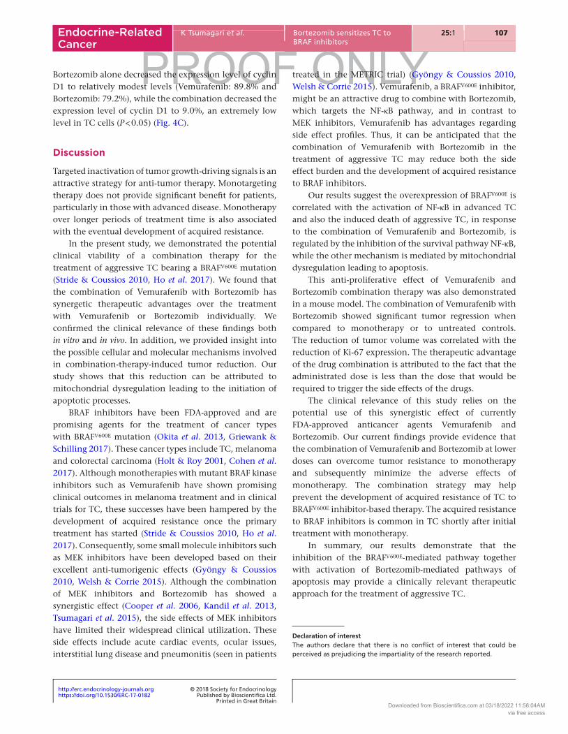
mono- or combination therapy on tumor size was monitored at regulated time intervals until the end of the experiment. Tumors were stained by H&E and apoptotic changes were seen in the group treated with Vemurafenib, Bortezomib or combination. Apoptotic changes were most prominent in the group treated with combination therapy (Fig. 4A) The tumor growth rates of control and treated mice are calculated and depicted in Fig. 4B. Robust tumor growth was seen in the control group without treatment (760% compared to initial time point). In contrast, a significant tumor growth reduction of 35% and 26% was noted in Vemurafenib- and Bortezomib-treated mice, respectively. The combination of the two inhibitors resulted in a significant tumor size reduction of 64% when compared with the control mice (P < 0.05).
To characterize the mechanism of Vemurafenib- and Bortezomib-induced tumor growth inhibition that was observed in the thyroid xenograft model, the mitotic index
of tumor tissues was assessed by the Ki-67 expression using IHC analysis. Active cell proliferation was observed in tumor tissue sections with a 69% relative proliferation rate (Fig. 4B). Monotherapy with Vemurafenib or Bortezomib slightly decreased the percentage of Ki-67-positive proliferating tumor cells, with relative proliferation rates of 74% and 47%, respectively (Fig. 4B). The combination of Vemurafenib and Bortezomib markedly decreased the percentage of Ki-67-positive proliferating tumor cells to 11% (P < 0.01). We next determined whether the combination of Vemurafenib and Bortezomib affected cell cycle progression in vivo by analyzing cyclin D1 expression (Fig. 4C). Cyclin D1, a key regulator for cell cycle progression, is required for maintenance of the G2-phase; the prolongation of G2 and phase arrest is correlated with the expression of cyclin D1 (Stacey 2003, Jensen et al. 2012). The statistical analysis of IHC staining results indicated that the use of Vemurafenib or
Figure 4Effect of drug combination on xenograft thyroid tumors in mice. Mice were engrafted with 2 × 10⁶ SW1736 cells and treated with Vemurafenib and/or Bortezomib. (A) At the end point after treating with the drug Vemurafenib, Bortezomib or combination (V + B), tumors resected and stained with H&E. (B) Treatment of mice with the drug combination (V + B) produced a synergistic effect (64% tumor size reduction compared to initial tumor size), while Vemurafenib (35%) or Bortezomib (26%) alone slightly increased tumor size compared to initial tumor. Data are expressed as the arithmetic mean ± the standard deviation and considered significant at P < 0.05. (C) Statistical analysis of IHC of Ki-67 in control and treated mice with Vemurafenib, Bortezomib or combination for 4 weeks. Nuclear staining was counted from at least five fields and expressed as percentage, * and ** represent significance at P < 0.05 and P < 0.01, respectively, compared with control (no drug). (D) Statistical analysis of IHC of Cyclin D1 in control and treated mice with Vemurafenib, Bortezomib or combination for 4 weeks.
Downloaded from Bioscientifica.com at 03/18/2022 11:58:04AMvia free access
http://erc.endocrinology-journals.org © 2018 Society for Endocrinology
Printed in Great BritainPublished by Bioscientifica Ltd.https://doi.org/10.1530/ERC-17-0182
107K Tsumagari et al. Bortezomib sensitizes TC to BRAF inhibitors
25:1
PROOF ONLYEndocrine-Related Cancer
Bortezomib alone decreased the expression level of cyclin D1 to relatively modest levels (Vemurafenib: 89.8% and Bortezomib: 79.2%), while the combination decreased the expression level of cyclin D1 to 9.0%, an extremely low level in TC cells (P < 0.05) (Fig. 4C).
Discussion
Targeted inactivation of tumor growth-driving signals is an attractive strategy for anti-tumor therapy. Monotargeting therapy does not provide significant benefit for patients, particularly in those with advanced disease. Monotherapy over longer periods of treatment time is also associated with the eventual development of acquired resistance.
In the present study, we demonstrated the potential clinical viability of a combination therapy for the treatment of aggressive TC bearing a BRAFV600E mutation (Stride & Coussios 2010, Ho et al. 2017). We found that the combination of Vemurafenib with Bortezomib has synergetic therapeutic advantages over the treatment with Vemurafenib or Bortezomib individually. We confirmed the clinical relevance of these findings both in vitro and in vivo. In addition, we provided insight into the possible cellular and molecular mechanisms involved in combination-therapy-induced tumor reduction. Our study shows that this reduction can be attributed to mitochondrial dysregulation leading to the initiation of apoptotic processes.
BRAF inhibitors have been FDA-approved and are promising agents for the treatment of cancer types with BRAFV600E mutation (Okita et al. 2013, Griewank & Schilling 2017). These cancer types include TC, melanoma and colorectal carcinoma (Holt & Roy 2001, Cohen et al. 2017). Although monotherapies with mutant BRAF kinase inhibitors such as Vemurafenib have shown promising clinical outcomes in melanoma treatment and in clinical trials for TC, these successes have been hampered by the development of acquired resistance once the primary treatment has started (Stride & Coussios 2010, Ho et al. 2017). Consequently, some small molecule inhibitors such as MEK inhibitors have been developed based on their excellent anti-tumorigenic effects (Gyöngy & Coussios 2010, Welsh & Corrie 2015). Although the combination of MEK inhibitors and Bortezomib has showed a synergistic effect (Cooper et al. 2006, Kandil et al. 2013, Tsumagari et al. 2015), the side effects of MEK inhibitors have limited their widespread clinical utilization. These side effects include acute cardiac events, ocular issues, interstitial lung disease and pneumonitis (seen in patients
treated in the METRIC trial) (Gyöngy & Coussios 2010, Welsh & Corrie 2015). Vemurafenib, a BRAFV600E inhibitor, might be an attractive drug to combine with Bortezomib, which targets the NF-κB pathway, and in contrast to MEK inhibitors, Vemurafenib has advantages regarding side effect profiles. Thus, it can be anticipated that the combination of Vemurafenib with Bortezomib in the treatment of aggressive TC may reduce both the side effect burden and the development of acquired resistance to BRAF inhibitors.
Our results suggest the overexpression of BRAFV600E is correlated with the activation of NF-κB in advanced TC and also the induced death of aggressive TC, in response to the combination of Vemurafenib and Bortezomib, is regulated by the inhibition of the survival pathway NF-κB, while the other mechanism is mediated by mitochondrial dysregulation leading to apoptosis.
This anti-proliferative effect of Vemurafenib and Bortezomib combination therapy was also demonstrated in a mouse model. The combination of Vemurafenib with Bortezomib showed significant tumor regression when compared to monotherapy or to untreated controls. The reduction of tumor volume was correlated with the reduction of Ki-67 expression. The therapeutic advantage of the drug combination is attributed to the fact that the administrated dose is less than the dose that would be required to trigger the side effects of the drugs.
The clinical relevance of this study relies on the potential use of this synergistic effect of currently FDA-approved anticancer agents Vemurafenib and Bortezomib. Our current findings provide evidence that the combination of Vemurafenib and Bortezomib at lower doses can overcome tumor resistance to monotherapy and subsequently minimize the adverse effects of monotherapy. The combination strategy may help prevent the development of acquired resistance of TC to BRAFV600E inhibitor-based therapy. The acquired resistance to BRAF inhibitors is common in TC shortly after initial treatment with monotherapy.
In summary, our results demonstrate that the inhibition of the BRAFV600E-mediated pathway together with activation of Bortezomib-mediated pathways of apoptosis may provide a clinically relevant therapeutic approach for the treatment of aggressive TC.
Declaration of interestThe authors declare that there is no conflict of interest that could be perceived as prejudicing the impartiality of the research reported.
Downloaded from Bioscientifica.com at 03/18/2022 11:58:04AMvia free access

Printed in Great BritainPublished by Bioscientifica Ltd.https://doi.org/10.1530/ERC-17-0182
http://erc.endocrinology-journals.org © 2018 Society for Endocrinology
108K Tsumagari et al. Bortezomib sensitizes TC to BRAF inhibitors
25:1
PROOF ONLYEndocrine-Related Cancer
FundingThis work was supported, in part, by a grant from Tulane School of Medicine Dean to E K and grant HL072889 from the NIH and funds from the Louisiana Cancer Research Center (New Orleans, LA) to A H B.
ReferencesAbd Elmageed ZY, Sholl AB, Tsumagari K, Al-Qurayshi Z, Basolo F,
Moroz K, Boulares AH, Friedlander P, Miccoli P & Kandil E 2017 Immunohistochemistry as an accurate tool for evaluating BRAF-V600E mutation in 130 samples of papillary thyroid cancer. Surgery 161 1122–1128. (https://doi.org/10.1016/j.surg.2016.06.081)
Bailey MR, Couret LN, Sapozhnikov OA, Khokhlova VA, ter Haar G, Vaezy S, Shi X, Martin R & Crum LA 2001 Use of overpressure to assess the role of bubbles in focused ultrasound lesion shape in vitro. Ultrasound in Medicine and Biology 27 695–708. (https://doi.org/10.1016/S0301-5629(01)00342-8)
Baumunk D, Andersen C, Heile U, Ebbing J, Cash H, Porsch M, Liehr UB, Janitzky A, Wendler JJ, Schindele D, et al. 2013 High-intensity focussed ultrasound in low-risk prostate cancer – oncological outcome and postinterventional quality of life of an inexperienced therapy centre in comparison with an experienced therapy centre. Aktuelle Urologie 44 285–292. (https://doi.org/10.1055/s-0033-1348253)
Bennedbaek FN & Hegedüs L 1999 Percutaneous ethanol injection therapy in benign solitary solid cold thyroid nodules: a randomized trial comparing one injection with three injections. Thyroid 9 225–233. (https://doi.org/10.1089/thy.1999.9.225)
Bennedbaek FN & Hegedüs L 2003 Treatment of recurrent thyroid cysts with ethanol: a randomized double-blind controlled trial. Journal of Clinical Endocrinology and Metabolism 88 5773–5777. (https://doi.org/10.1210/jc.2003-031000)
Bollag G, Tsai J, Zhang J, Zhang C, Ibrahim P, Nolop K & Hirth P 2012 Vemurafenib: the first drug approved for BRAF-mutant cancer. Nature Reviews Drug Discovery 11 873–886. (https://doi.org/10.1038/nrd3847)
Brose MS, Nutting CM, Jarzab B, Elisei R, Siena S, Bastholt L, de la Fouchardiere C, Pacini F, Paschke R, Shong YK, et al. 2014 Sorafenib in radioactive iodine-refractory, locally advanced or metastatic differentiated thyroid cancer: a randomised, double-blind, phase 3 trial. Lancet 384 319–328. (https://doi.org/10.1016/S0140-6736(14)60421-9)
Charles RP, Silva J, Iezza G, Phillips WA & McMahon M 2014 Activating BRAF and PIK3CA mutations cooperate to promote anaplastic thyroid carcinogenesis. Molecular Cancer Research 12 979–986. (https://doi.org/10.1158/1541-7786.MCR-14-0158-T)
Cohen R, Cervera P, Svrcek M, Pellat A, Dreyer C, de Gramont A & André T 2017 BRAF-mutated colorectal cancer: what is the optimal strategy for treatment? Current Treatment Options in Oncology 18 9. (https://doi.org/10.1007/s11864-017-0453-5)
Cooper DS, Doherty GM, Haugen BR, Kloos RT, Lee SL, Mandel SJ, Mazzaferri EL, McIver B, Sherman SI, Tuttle RM, et al. 2006 Management guidelines for patients with thyroid nodules and differentiated thyroid cancer. Thyroid 16 109–142. (https://doi.org/10.1089/thy.2006.16.109)
Dadu R, Shah K, Busaidy NL, Waguespack SG, Habra MA, Ying AK, Hu MI, Bassett R, Jimenez C, Sherman SI, et al. 2015 Efficacy and tolerability of vemurafenib in patients with BRAF(V600E) -positive papillary thyroid cancer: M.D. Anderson Cancer Center off label experience. Journal of Clinical Endocrinology and Metabolism 100 E77–81. (https://doi.org/10.1210/jc.2014-2246)
Davies L & Welch HG 2010 Thyroid cancer survival in the United States: observational data from 1973 to 2005. Archives of Otolaryngology: Head and Neck Surgery 136 440–444. (https://doi.org/10.1001/archoto.2010.55)
Deandrea M, Limone P, Basso E, Mormile A, Ragazzoni F, Gamarra E, Spiezia S, Faggiano A, Colao A, Molinari F, et al. 2008 US-guided percutaneous radiofrequency thermal ablation for the treatment of solid benign hyperfunctioning or compressive thyroid nodules. Ultrasound in Medicine and Biology 34 784–791. (https://doi.org/10.1016/j.ultrasmedbio.2007.10.018)
Durante C, Haddy N, Baudin E, Leboulleux S, Hartl D, Travagli JP, Caillou B, Ricard M, Lumbroso JD, De Vathaire F, et al. 2006 Long-term outcome of 444 patients with distant metastases from papillary and follicular thyroid carcinoma: benefits and limits of radioiodine therapy. Journal of Clinical Endocrinology and Metabolism 91 2892–2899. (https://doi.org/10.1210/jc.2005-2838)
El Jamal SM, Taylor EB, Abd Elmageed ZY, Alamodi AA, Selimovic D, Alkhateeb A, Hannig M, Hassan SY, Santourlidis S, Friedlander PL, et al. 2016 Interferon gamma-induced apoptosis of head and neck squamous cell carcinoma is connected to indoleamine-2,3-dioxygenase via mitochondrial and ER stress-associated pathways. Cell Division 11 11. (https://doi.org/10.1186/s13008-016-0023-4)
Esnault O, Rouxel A, Le Nestour E, Gheron G & Leenhardt L 2010 Minimally invasive ablation of a toxic thyroid nodule by high-intensity focused ultrasound. American Journal of Neuroradiology 31 1967–1968.
Garrean S, Hering J, Helton WS & Espat NJ 2007 A primer on transarterial, chemical, and thermal ablative therapies for hepatic tumors. American Journal of Surgery 194 79–88. (https://doi.org/10.1016/j.amjsurg.2006.11.025)
Griewank KG & Schilling B 2017 Next-generation sequencing to guide treatment of advanced melanoma. American Journal of Clinical Dermatology 18 303–310.
Hideshima T, Mitsiades C, Tonon G, Richardson PG & Anderson KC 2007 Understanding multiple myeloma pathogenesis in the bone marrow to identify new therapeutic targets. Nature Reviews Cancer 7 585–598.
Gyöngy M & Coussios CC 2010 Passive spatial mapping of inertial cavitation during HIFU exposure. IEEE Transactions on Biomedical Engineering 57 48–56.
Hegedüs L 2009 Therapy: a new nonsurgical therapy option for benign thyroid nodules? Nature Reviews Endocrinology 5 476–478.
Ho CC, Liao WY, Lin CA, Shih JY, Yu CJ & Chih-Hsin Yang J 2017 Acquired BRAF V600E mutation as resistant mechanism after treatment with osimertinib. Journal of Thoracic Oncology 12 567–572. (https://doi.org/10.1016/j.jtho.2016.11.2231)
Holt RG & Roy RA 2001 Measurements of bubble-enhanced heating from focused, MHz-frequency ultrasound in a tissue-mimicking material. Ultrasound in Medicine and Biology 27 1399–1412. (https://doi.org/10.1016/S0301-5629(01)00438-0)
Howlader N, Noone A, Krapcho M, Miller D, Bishop K, Kosary CL, Yu M, Ruhl J, Tatalovich Z, Mariotto M, et al. 2017 SEER Cancer Statistics Review, 1975–2014. Bethesda, MD: National Cancer Institute. (available at: https://seer.cancer.gov/csr/1975_2014/), based on November 2016 SEER data submission, posted to the SEER web site, April 2017.
Hsu KT, Yu XM, Audhya AW, Jaume JC, Lloyd RV, Miyamoto S, Prolla TA & Chen H 2014 Novel approaches in anaplastic thyroid cancer therapy. Oncologist 19 1148–1155. (https://doi.org/10.1634/theoncologist.2014-0182)
Jensen CR, Ritchie RW, Gyöngy M, Collin JR, Leslie T & Coussios CC 2012 Spatiotemporal monitoring of high-intensity focused ultrasound therapy with passive acoustic mapping. Radiology 262 252–261. (https://doi.org/10.1148/radiol.11110670)
Jeong WK, Baek JH, Rhim H, Kim YS, Kwak MS, Jeong HJ & Lee D 2008 Radiofrequency ablation of benign thyroid nodules: safety and imaging follow-up in 236 patients. European Radiology 18 1244–1250. (https://doi.org/10.1007/s00330-008-0880-6)
Kandil E, Tsumagari K, Ma J, Abd Elmageed ZY, Li X, Slakey D, Mondal D & Abdel-Mageed AB 2013 Synergistic inhibition of
Downloaded from Bioscientifica.com at 03/18/2022 11:58:04AMvia free access
http://erc.endocrinology-journals.org © 2018 Society for Endocrinology
Printed in Great BritainPublished by Bioscientifica Ltd.https://doi.org/10.1530/ERC-17-0182
109K Tsumagari et al. Bortezomib sensitizes TC to BRAF inhibitors
25:1
PROOF ONLYEndocrine-Related Cancer
thyroid cancer by suppressing MAPK/PI3K/AKT pathways. Journal of Surgical Research 184 898–906. (https://doi.org/10.1016/j.jss.2013.03.052)
Kanotra SP, Lateef M & Kirmani O 2008 Non-surgical management of benign thyroid cysts: use of ultrasound-guided ethanol ablation. Postgraduate Medical Journal 84 639–643. (https://doi.org/10.1136/pgmj.2008.072777)
Li S & Wu PH 2013 Magnetic resonance image-guided versus ultrasound-guided high-intensity focused ultrasound in the treatment of breast cancer. Chinese Journal of Cancer 32 441–452. (https://doi.org/10.5732/cjc.012.10104)
Mitsiades CS, McMillin D, Kotoula V, Poulaki V, McMullan C, Negri J, Fanourakis G, Tseleni-Balafouta S, Ain KB & Mitsiades N 2006 Antitumor effects of the proteasome inhibitor bortezomib in medullary and anaplastic thyroid carcinoma cells in vitro. Journal of Clinical Endocrinology and Metabolism 91 4013–4021. (https://doi.org/10.1210/jc.2005-2472)
Nucera C, Lawler J, Hodin R & Parangi S 2010 The BRAFV600E mutation: what is it really orchestrating in thyroid cancer? Oncotarget 1 751–756. (https://doi.org/10.18632/oncotarget.210)
Okita K, Sugiyama K, Takagi S & Matsumto Y 2013 Microbubble behavior in an ultrasound field for high intensity focused ultrasound therapy enhancement. Journal of the Acoustical Society of America 134 1576–1585. (https://doi.org/10.1121/1.4812880)
Palona I, Namba H, Mitsutake N, Starenki D, Podtcheko A, Sedliarou I, Ohtsuru A, Saenko V, Nagayama Y, Umezawa K, et al. 2006 BRAFV600E promotes invasiveness of thyroid cancer cells through nuclear factor kappaB activation. Endocrinology 147 5699–5707. (https://doi.org/10.1210/en.2006-0400)
Putzer D, Kroiss A, Waitz D, Gabriel M, Traub-Weidinger T, Uprimny C, von Guggenberg E, Decristoforo C, Warwitz B, Widmann G, et al. 2013 Somatostatin receptor PET in neuroendocrine tumours: 68Ga-DOTA0,Tyr3-octreotide versus 68Ga-DOTA0-lanreotide. European Journal of Nuclear Medicine and Molecular Imaging 40 364–372. (https://doi.org/10.1007/s00259-012-2286-6)
Roberts PJ & Der CJ 2007 Targeting the Raf-MEK-ERK mitogen-activated protein kinase cascade for the treatment of cancer. Oncogene 26 3291–3310. (https://doi.org/10.1038/sj.onc.1210422)
Rustgi AK 2013 BRAF: a driver of the serrated pathway in colon cancer. Cancer Cell 24 1–2.
Salama AK & Flaherty KT 2013 BRAF in melanoma: current strategies and future directions. Clinical Cancer Research 19 4326–4334. (https://doi.org/10.1158/1078-0432.CCR-13-0779)
Sanghvi NT 2013 High-intensity focused ultrasound treatment of prostate cancer. Journal of the Acoustical Society of America 134 4089. (https://doi.org/10.1121/1.4830936)
Schlumberger MJ & Torlantano M 2000 Papillary and follicular thyroid carcinoma. Best Practice and Research: Clinical Endocrinology and Metabolism 14 601–613. (https://doi.org/10.1053/beem.2000.0105)
Selimovic D, Ahmad M, El-Khattouti A, Hannig M, Haikel Y & Hassan M 2011 Apoptosis-related protein-2 triggers melanoma cell death by a mechanism including both endoplasmic reticulum stress and mitochondrial dysregulation. Carcinogenesis 32 1268–1278. (https://doi.org/10.1093/carcin/bgr112)
Smallridge RC 2012 Approach to the patient with anaplastic thyroid carcinoma. Journal of Clinical Endocrinology and Metabolism 97 2566–2572. (https://doi.org/10.1210/jc.2012-1314)
Smith NB, Temkin JM, Shapiro F & Hynynen K 2001 Thermal effects of focused ultrasound energy on bone tissue. Ultrasound in Medicine and Biology 27 1427–1433. (https://doi.org/10.1016/S0301-5629(01)00454-9)
Spiezia S, Garberoglio R, Milone F, Ramundo V, Caiazzo C, Assanti AP, Deandrea M, Limone PP, Macchia PE, Lombardi G, et al. 2009 Thyroid nodules and related symptoms are stably controlled two years after radiofrequency thermal ablation. Thyroid 19 219–225. (https://doi.org/10.1089/thy.2008.0202)
Stacey DW 2003 Cyclin D1 serves as a cell cycle regulatory switch in actively proliferating cells. Current Opinion in Cell Biology 15 158–163. (https://doi.org/10.1016/S0955-0674(03)00008-5)
Stride EP & Coussios CC 2010 Cavitation and contrast: the use of bubbles in ultrasound imaging and therapy. Proceedings of the Institution of Mechanical Engineers, Part H 224 171–191. (https://doi.org/10.1243/09544119JEIM622)
Tsumagari K, Abd Elmageed ZY, Sholl AB, Friedlander P, Abdraboh M, Xing M, Boulares AH & Kandil E 2015 Simultaneous suppression of the MAP kinase and NF-κB pathways provides a robust therapeutic potential for thyroid cancer. Letters 368 46–53. (https://doi.org/10.1016/j.canlet.2015.07.011)
Wan PT, Garnett MJ, Roe SM, Lee S, Niculescu-Duvaz D, Good VM, Jones CM, Marshall CJ, Springer CJ, Barford D, et al. 2004 Mechanism of activation of the RAF-ERK signaling pathway by oncogenic mutations of B-RAF. Cell 116 855–867. (https://doi.org/10.1016/S0092-8674(04)00215-6)
Welsh SJ & Corrie PG 2015 Management of BRAF and MEK inhibitor toxicities in patients with metastatic melanoma. Therapeutic Advances in Medical Oncology 7 122–136. (https://doi.org/10.1177/1758834014566428)
Received in final form 14 September 2017Accepted 2 October 2017
Downloaded from Bioscientifica.com at 03/18/2022 11:58:04AMvia free access