Flow Cytometry Panel Design Journey
1
Waterfall of Discovery There are hundreds of research applications for flow cytometry. Spectral Mountain Passage Map out the peaks and valleys of the dyes’ spectra to minimize spillover and spread. Lake of Permeabilization For smooth sailing, select the appropriate buffer system for your antibodies. Viability Volcano Beware of hitchhiking dead cells because they can skew your data. Be sure to identify them properly with a viability dye. Flow Cytometry Panel Design Journey We're with you every step of the way Define your experimental hypothesis Step 1: Defining your experimental hypothesis is the first step in panel design. Start with identifying: • The biological information you are trying to achieve • The population(s) of cells you wish to interrogate • Whether targets are found on the cell surface or intracellularly Know your flow cytometer Step 3: Knowing your instrument is essential. Understanding your instrument's configuration will let you know how many markers and which fluorochromes your instrument can detect. Elements to consider include: • Laser wavelength for excitation • Number of detectors off each laser • Filters available to detect the fluorochromes Marker selection Step 2: During the second step of the panel design process, you will need to identify which and how many markers you need to identify the population of interest. Pay attention to: • Marker expression levels • Primary antigen: Expressed at high density, often defining lineages • Secondary antigen: Often expressed over a continuum • Tertiary antigen: Critical markers expressed at low density • Marker coexpression, especially of dim markers • The gating strategy needed to identify the population(s) of cells you wish to interrogate Fluorochrome assignment Step 4: Carefully select fluorochromes to resolve markers at all expression levels and minimize spectral overlap. Consider using tools like a fluorochrome resolution ranking and a spectrum viewer to help assess: • Cross laser excitation • Fluorochrome spillover Remember to pair bright fluorochromes with low expressing antigens and dim fluorochromes with high expressors. Keep in mind that spread only impacts the resolution of coexpressed markers. Review panel Step 5: Review your panel design and begin ordering your reagents. Remember to titrate your mass size reagents and optimize your staining protocol. Include proper controls for compensation, FMO and biological controls to help ensure optimal panel performance. To learn more about flow cytometry panel design resources or for support, please contact your BD Sales Representative. For Research Use Only. Not for use in diagnostic or therapeutic procedures. 23-23036-00 BD Life Sciences, San Jose, CA, 95131, USA bdbiosciences.com BD, the BD Logo, Horizon Brilliant and Horizon Brilliant Violet are trademarks of Becton, Dickinson and Company or its affiliates. © 2020 BD. All rights reserved. Did you know? BD offers over 40 fluorochromes and more than 1,500 specificities to provide researchers flexibility in panel design. Did you know? BD launched the world’s 1 st commercially available flow cytometer in 1974 - kicking off a legacy of research innovation. Did you know? Since the introduction of revolutionary Sirigen polymer dye technology, BD has launched 17 innovative dyes including BD Horizon Brilliant™ UV and BD Horizon Brilliant Violet™ Dyes. Videos Webinars Panel design tools Check out our online flow cytometry panel design educational resources Ready to begin workflow 5 4 2 3 bdbiosciences.com/panel-design 1 TIP: For help choosing fluorochromes, check out our BD Spectrum Viewer: bdbiosciences.com/spectrum-viewer
Transcript of Flow Cytometry Panel Design Journey
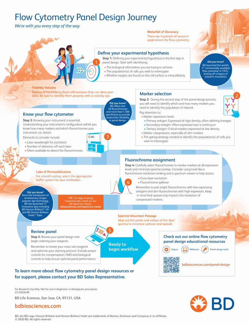
Waterfall of DiscoveryThere are hundreds of research applications for flow cytometry.
Spectral Mountain PassageMap out the peaks and valleys of the dyes’ spectra to minimize spillover and spread.
Lake of PermeabilizationFor smooth sailing, select the appropriate bu�er system for your antibodies.
Viability Volcano Beware of hitchhiking dead cells because they can skew your data. Be sure to identify them properly with a viability dye.
Flow Cytometry Panel Design JourneyWe're with you every step of the way
Define your experimental hypothesisStep 1: Defining your experimental hypothesis is the first step in panel design. Start with identifying:
• The biological information you are trying to achieve• The population(s) of cells you wish to interrogate• Whether targets are found on the cell surface or intracellularly
Know your flow cytometerStep 3: Knowing your instrument is essential. Understanding your instrument's configuration will let you know how many markers and which fluorochromes your instrument can detect. Elements to consider include:
• Laser wavelength for excitation• Number of detectors o� each laser• Filters available to detect the fluorochromes
Marker selectionStep 2: During the second step of the panel design process, you will need to identify which and how many markers you need to identify the population of interest.Pay attention to:
• Marker expression levels• Primary antigen: Expressed at high density, often defining lineages• Secondary antigen: Often expressed over a continuum• Tertiary antigen: Critical markers expressed at low density
• Marker coexpression, especially of dim markers• The gating strategy needed to identify the population(s) of cells you
wish to interrogate
Fluorochrome assignmentStep 4: Carefully select fluorochromes to resolve markers at all expression levels and minimize spectral overlap. Consider using tools like a fluorochrome resolution ranking and a spectrum viewer to help assess:
• Cross laser excitation• Fluorochrome spillover
Remember to pair bright fluorochromes with low expressing antigens and dim fluorochromes with high expressors. Keep in mind that spread only impacts the resolution of coexpressed markers.
Review panel Step 5: Review your panel design and begin ordering your reagents.
Remember to titrate your mass size reagents and optimize your staining protocol. Include proper controls for compensation, FMO and biological controls to help ensure optimal panel performance.
To learn more about flow cytometry panel design resources or for support, please contact your BD Sales Representative.
For Research Use Only. Not for use in diagnostic or therapeutic procedures. 23-23036-00
BD Life Sciences, San Jose, CA, 95131, USA
bdbiosciences.comBD, the BD Logo, Horizon Brilliant and Horizon Brilliant Violet are trademarks of Becton, Dickinson and Company or its affiliates. © 2020 BD. All rights reserved.
Did you know? BD o�ers over
40 fluorochromes and more than 1,500 specificities to provide researchers flexibility
in panel design.
Did you know? BD launched the world’s
1st commercially available flow cytometer in 1974 -
kicking o� a legacy of research innovation.
Did you know? Since the introduction of
revolutionary Sirigen polymer dye technology,
BD has launched 17 innovative dyes including BD Horizon Brilliant™ UV and BD Horizon Brilliant
Violet™ Dyes.
Videos Webinars Panel design tools
Check out our online flow cytometry panel design educational resources
Ready to begin workflow
5
4
2
3
bdbiosciences.com/panel-design
1
TIP: For help choosing fluorochromes, check out our
BD Spectrum Viewer: bdbiosciences.com/spectrum-viewer