Introduction to Flow Cytometry - HemePathReviewhemepathreview.com/Heme-Review/FlowCytometry.pdf ·...
40
Introduction to Flow Cytometry Jesse Manuel Jaso, M.D. 7/28/2015
Transcript of Introduction to Flow Cytometry - HemePathReviewhemepathreview.com/Heme-Review/FlowCytometry.pdf ·...

Introduction to Flow Cytometry
Jesse Manuel Jaso, M.D.
7/28/2015

Flow Cytometry
• Flow cytometry is the measurement of single cells as they pass single file through a beam of light in a fluid stream
• Cells are “flowing” through the instrument (flow cell)
• More control over which cells are being examined (cell sorting)
• Generate data for only the cells you are interested in

Flow Cytometric Immunophenotyping
• Characterization of a cell or group of cells by the presence or absence of certain antigens on their surface or in their cytoplasm
• Pre-described immunophenotypes can be used to aid in the diagnosis of hematopoietic neoplasms
• Pattern recognition + Right context
• Majority of the time, this is what we are talking about when we say “flow cytometry”
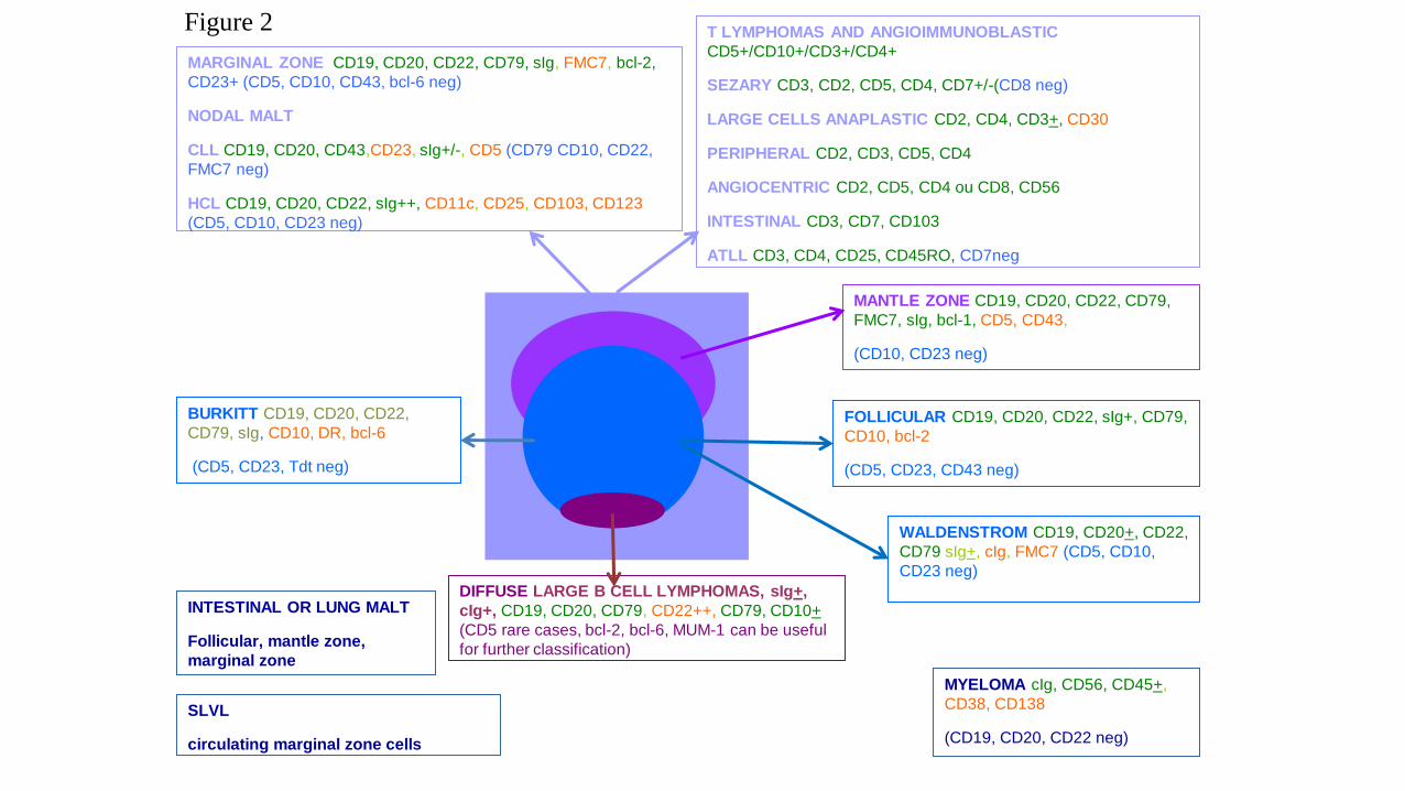
Figure 2
MANTLE ZONE CD19, CD20, CD22, CD79,
FMC7, sIg, bcl-1, CD5, CD43,
(CD10, CD23 neg)
FOLLICULAR CD19, CD20, CD22, sIg+, CD79,
CD10, bcl-2
(CD5, CD23, CD43 neg)
DIFFUSE LARGE B CELL LYMPHOMAS, sIg+,
cIg+, CD19, CD20, CD79, CD22++, CD79, CD10+
(CD5 rare cases, bcl-2, bcl-6, MUM-1 can be useful
for further classification)
BURKITT CD19, CD20, CD22,
CD79, sIg, CD10, DR, bcl-6
(CD5, CD23, Tdt neg)
T LYMPHOMAS AND ANGIOIMMUNOBLASTIC
CD5+/CD10+/CD3+/CD4+
SEZARY CD3, CD2, CD5, CD4, CD7+/-(CD8 neg)
LARGE CELLS ANAPLASTIC CD2, CD4, CD3+, CD30
PERIPHERAL CD2, CD3, CD5, CD4
ANGIOCENTRIC CD2, CD5, CD4 ou CD8, CD56
INTESTINAL CD3, CD7, CD103
ATLL CD3, CD4, CD25, CD45RO, CD7neg
MARGINAL ZONE CD19, CD20, CD22, CD79, sIg, FMC7, bcl-2,
CD23+ (CD5, CD10, CD43, bcl-6 neg)
NODAL MALT
CLL CD19, CD20, CD43,CD23, sIg+/-, CD5 (CD79 CD10, CD22,
FMC7 neg)
HCL CD19, CD20, CD22, sIg++, CD11c, CD25, CD103, CD123
(CD5, CD10, CD23 neg)
INTESTINAL OR LUNG MALT
Follicular, mantle zone,
marginal zone
SLVL
circulating marginal zone cells
WALDENSTROM CD19, CD20+, CD22,
CD79 sIg+, cIg, FMC7 (CD5, CD10,
CD23 neg)
MYELOMA cIg, CD56, CD45+,
CD38, CD138
(CD19, CD20, CD22 neg)


Why?
• For the same reason we look at a sample under a microscope:
• In a heterogeneous collection of cells:
• Determine the presence/absence of cell(s) of interest
• Determine the characteristics of the cell(s) present:
• “Parameters”
• Size, granularity, immunophenotype, proliferation rate, etc.
• MPC: multi-parameter flow cytometry
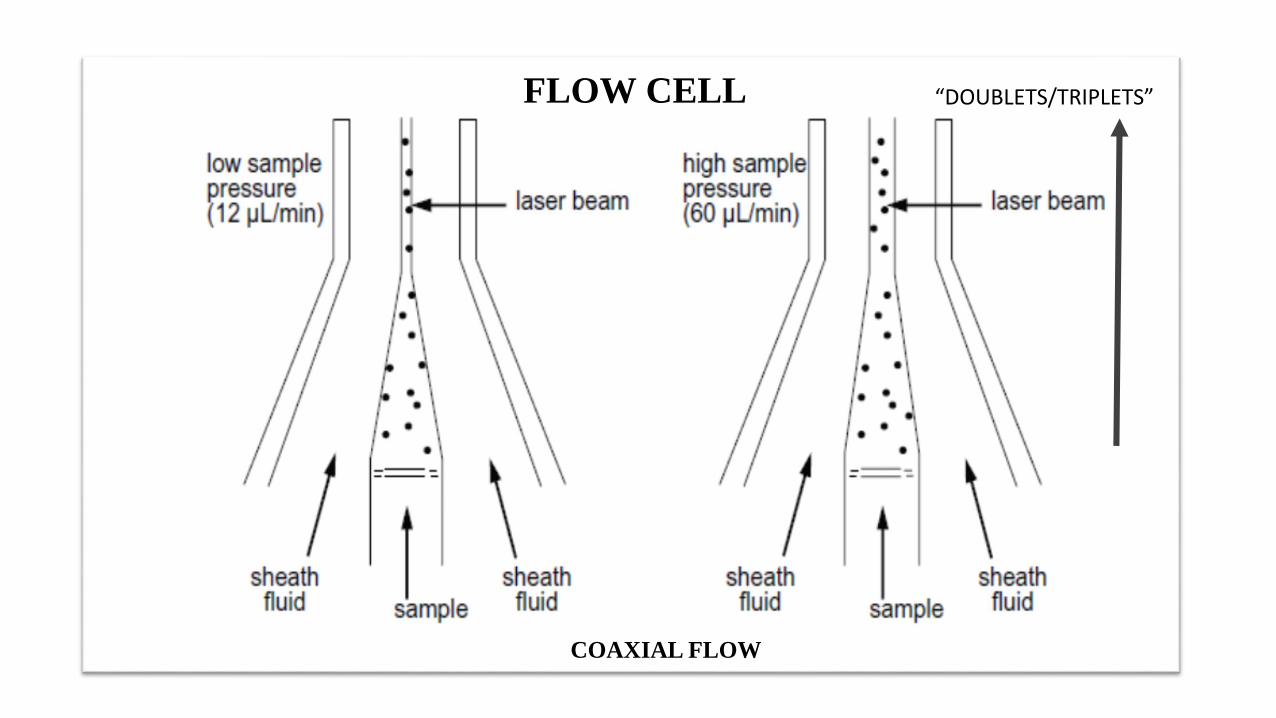
FLOW CELL
COAXIAL FLOW
“DOUBLETS/TRIPLETS”

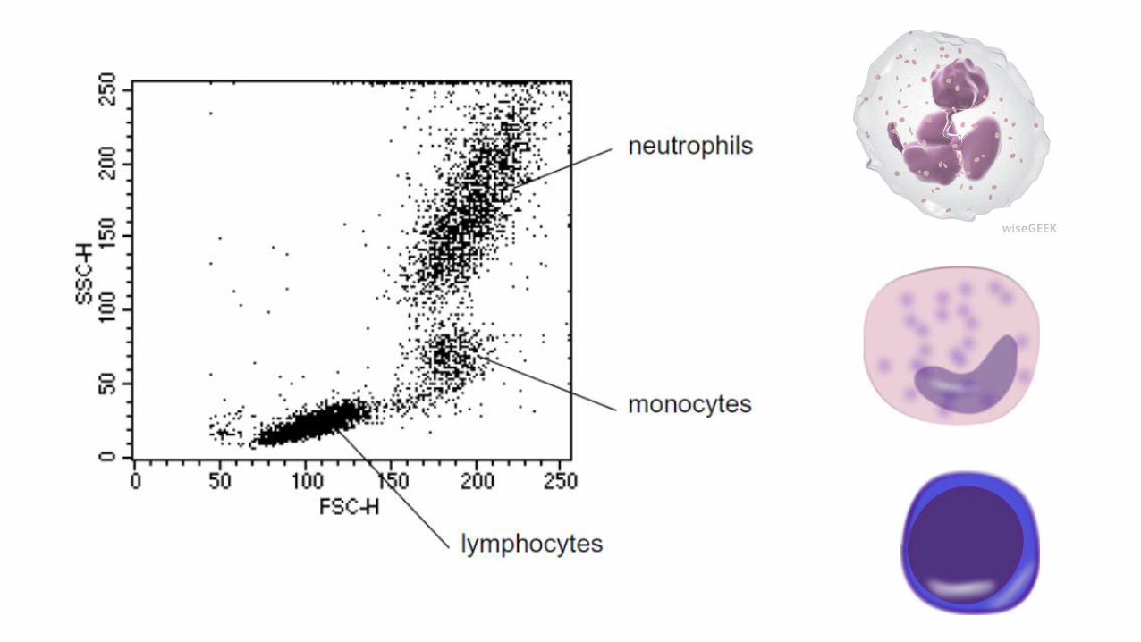
Light Scatter
• Light passing in a “straight line”
• Deflection of light from its straight path is light scatter• Requires some kind of interaction with matter
• Wavelength (energy) of the light
• Characteristics of the matter
• If we control everything else (wavelength, etc.) we can use light scatter to determine characteristics of matter
• Cells in our case

CELL
SIZE
CYTOPLASMIC
GRANULARITY

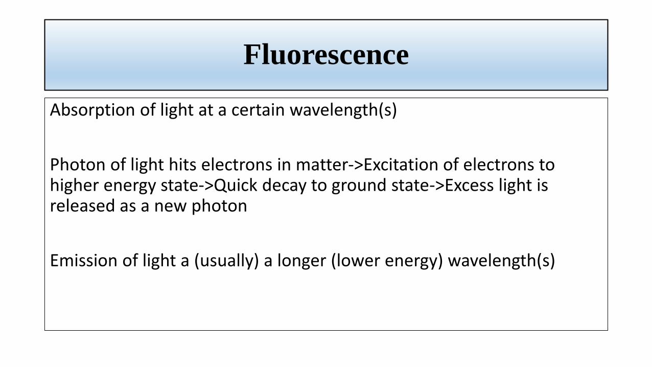
Fluorescence
Absorption of light at a certain wavelength(s)
Photon of light hits electrons in matter->Excitation of electrons to higher energy state->Quick decay to ground state->Excess light is released as a new photon
Emission of light a (usually) a longer (lower energy) wavelength(s)

E = hc/𝝀

Aragonit Crystal
Higher Energy
Wavelength
Absorption
Lower Energy
Wavelength
Emission
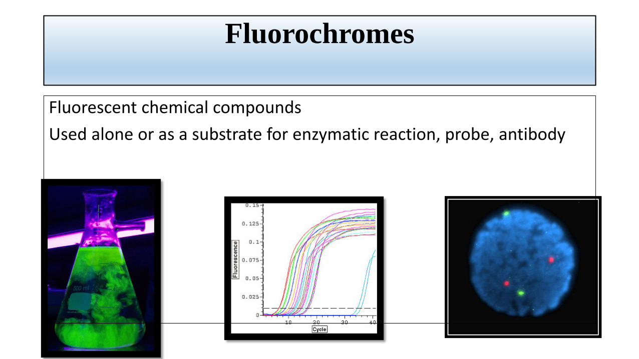
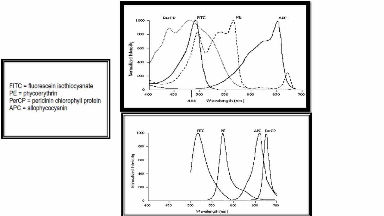
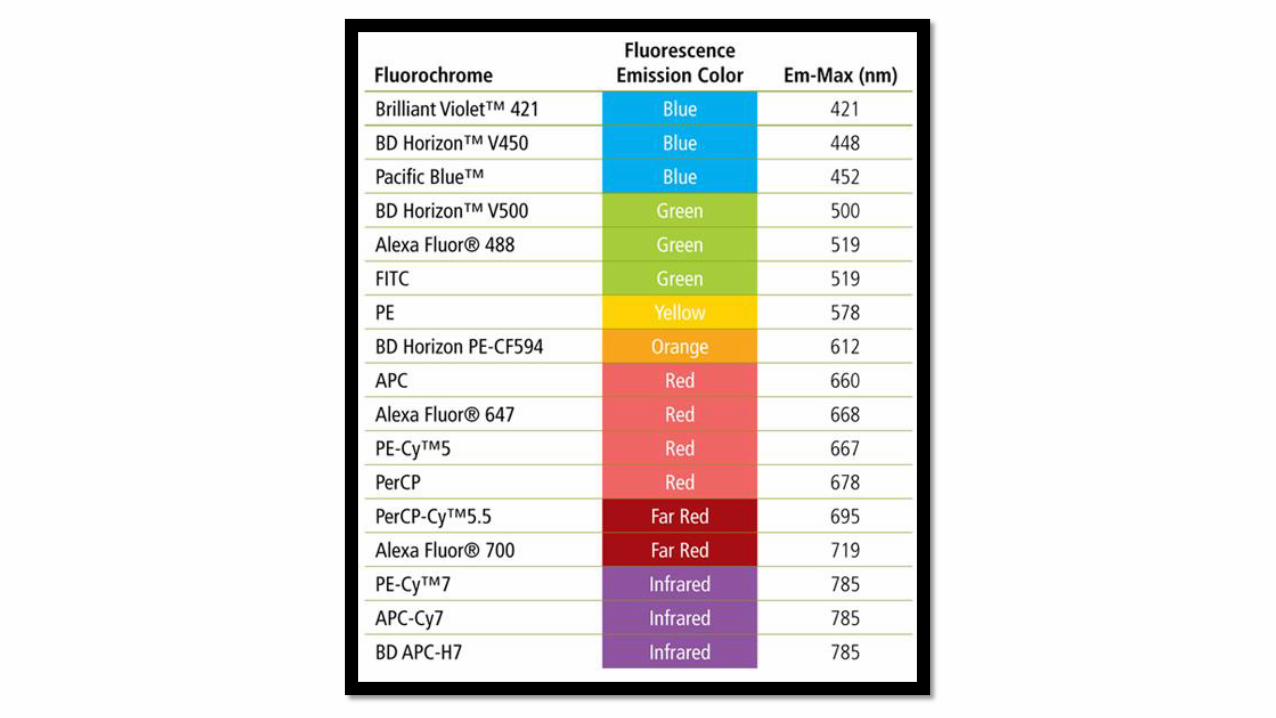
Fluorochromes
Fluorescent chemical compounds
Used alone or as a substrate for enzymatic reaction, probe, antibody
Fluorochromes
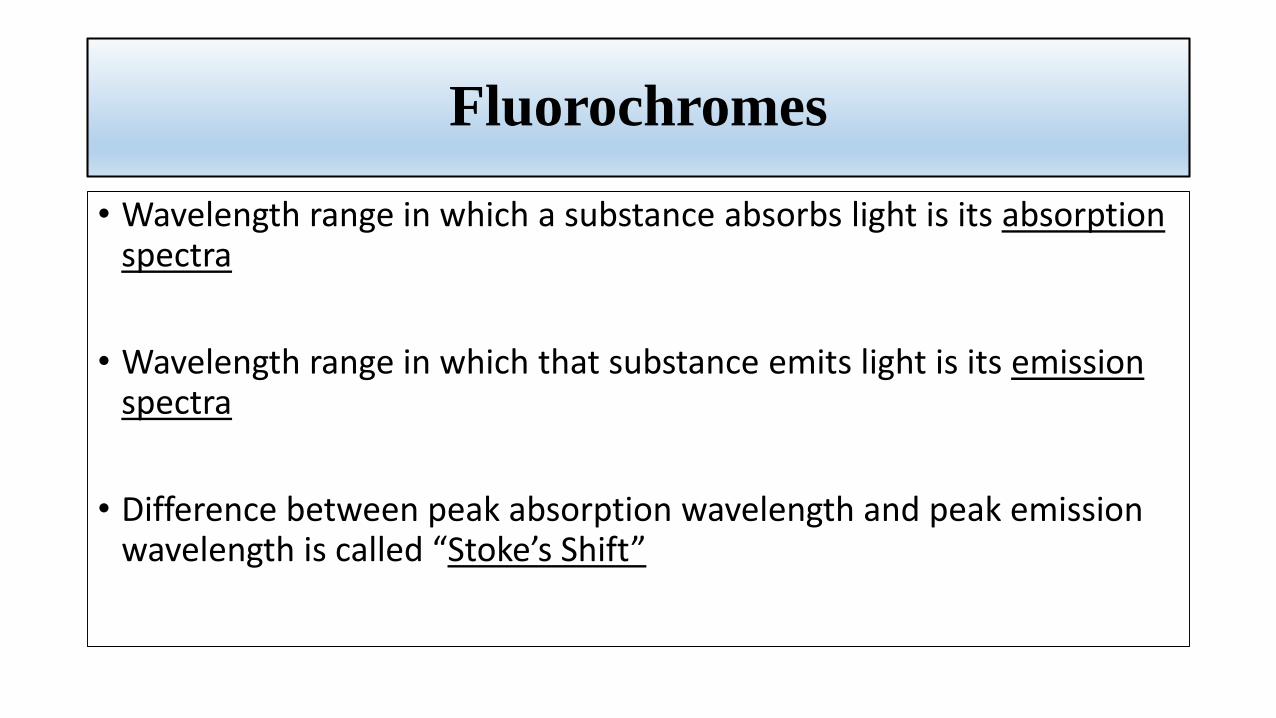
• Wavelength range in which a substance absorbs light is its absorption spectra
• Wavelength range in which that substance emits light is its emission spectra
• Difference between peak absorption wavelength and peak emission wavelength is called “Stoke’s Shift”


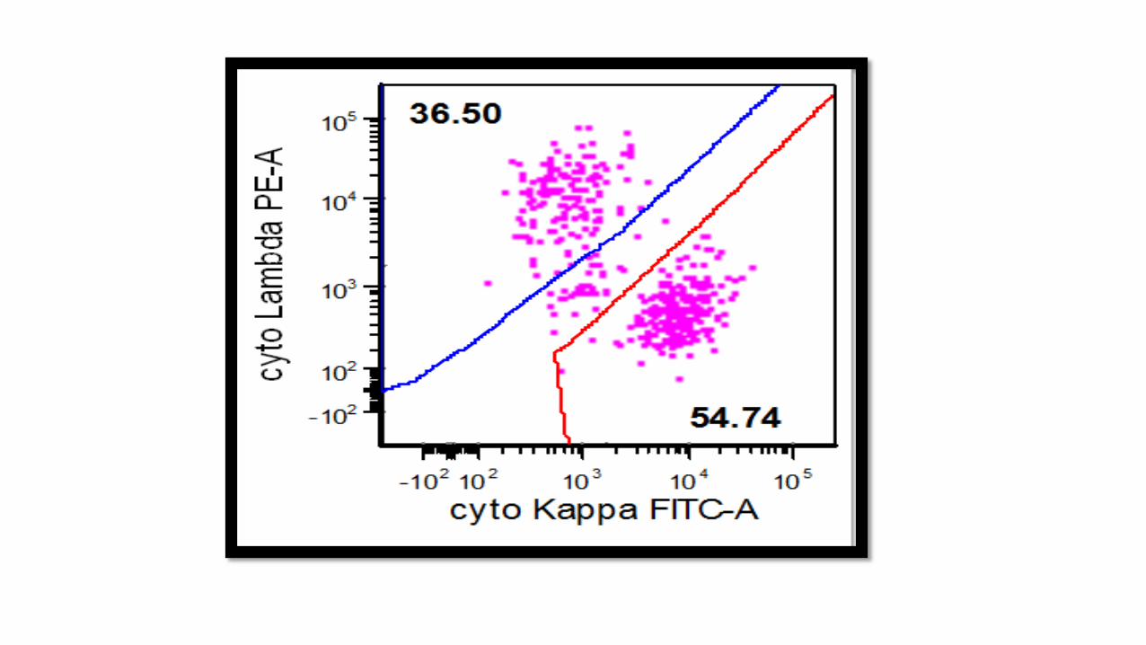
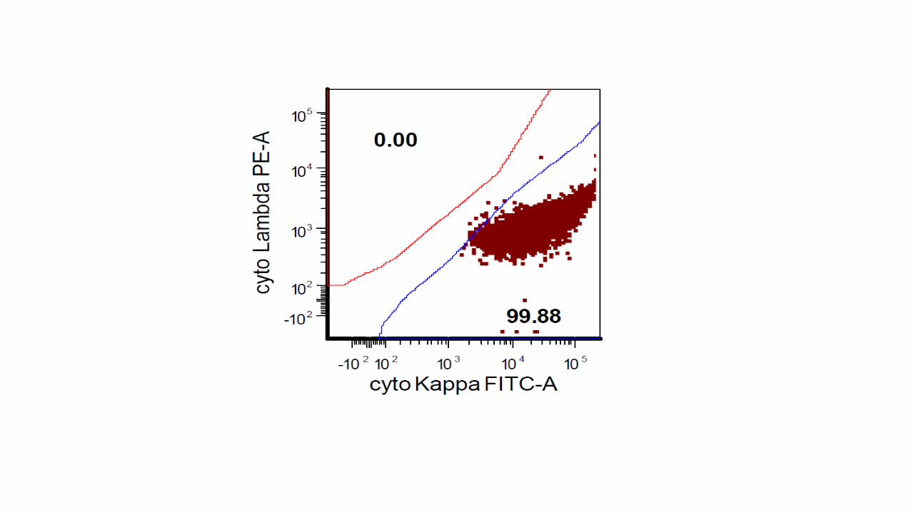
Kappa
Lambda
Permeabilization
Fixation
Kappa
Lambda
sKappa FITC
sLambda PE
cKappa FITC
cLambda PE

104 to 106
per tube

Laser(s)Lenses
&Mirrors
Filters DetectorsDiodes
&PMT
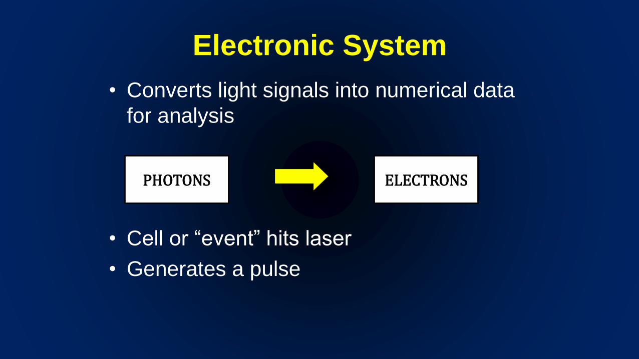
Electronic System
• Converts light signals into numerical data
for analysis
• Cell or “event” hits laser
• Generates a pulse
PHOTONS ELECTRONS
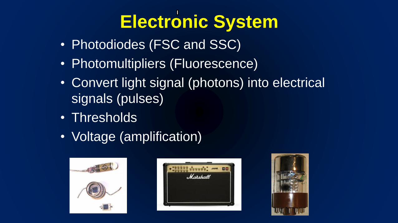
Electronic System• Photodiodes (FSC and SSC)
• Photomultipliers (Fluorescence)
• Convert light signal (photons) into electrical
signals (pulses)
• Thresholds
• Voltage (amplification)
Electronic System
• Each event gets a numeric value ( pulse height, width,
area) and assigned a channel number
• Raw data stored as “list mode data”
• Each channel number is assigned point on a dot plot
• Linear or logarithmic scale


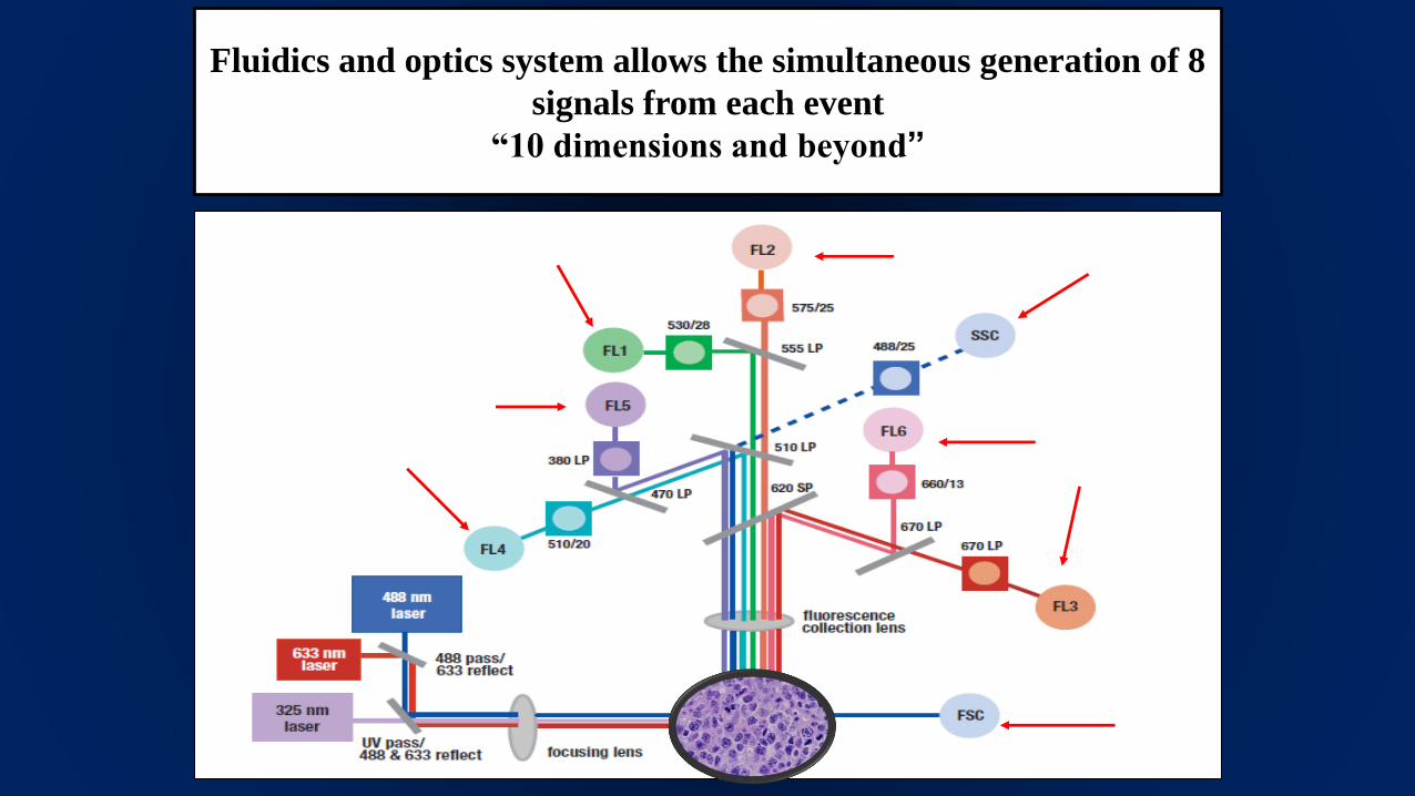
Fluidics and optics system allows the simultaneous generation of 8
signals from each event
“10 dimensions and beyond”

Figure 2
MANTLE ZONE CD19, CD20, CD22, CD79,
FMC7, sIg, bcl-1, CD5, CD43,
(CD10, CD23 neg)
FOLLICULAR CD19, CD20, CD22, sIg+, CD79,
CD10, bcl-2
(CD5, CD23, CD43 neg)
DIFFUSE LARGE B CELL LYMPHOMAS, sIg+,
cIg+, CD19, CD20, CD79, CD22++, CD79, CD10+
(CD5 rare cases, bcl-2, bcl-6, MUM-1 can be useful
for further classification)
BURKITT CD19, CD20, CD22,
CD79, sIg, CD10, DR, bcl-6
(CD5, CD23, Tdt neg)
T LYMPHOMAS AND ANGIOIMMUNOBLASTIC
CD5+/CD10+/CD3+/CD4+
SEZARY CD3, CD2, CD5, CD4, CD7+/-(CD8 neg)
LARGE CELLS ANAPLASTIC CD2, CD4, CD3+, CD30
PERIPHERAL CD2, CD3, CD5, CD4
ANGIOCENTRIC CD2, CD5, CD4 ou CD8, CD56
INTESTINAL CD3, CD7, CD103
ATLL CD3, CD4, CD25, CD45RO, CD7neg
MARGINAL ZONE CD19, CD20, CD22, CD79, sIg, FMC7, bcl-2,
CD23+ (CD5, CD10, CD43, bcl-6 neg)
NODAL MALT
CLL CD19, CD20, CD43,CD23, sIg+/-, CD5 (CD79 CD10, CD22,
FMC7 neg)
HCL CD19, CD20, CD22, sIg++, CD11c, CD25, CD103, CD123
(CD5, CD10, CD23 neg)
INTESTINAL OR LUNG MALT
Follicular, mantle zone,
marginal zone
SLVL
circulating marginal zone cells
WALDENSTROM CD19, CD20+, CD22,
CD79 sIg+, cIg, FMC7 (CD5, CD10,
CD23 neg)
MYELOMA cIg, CD56, CD45+,
CD38, CD138
(CD19, CD20, CD22 neg)


Stop Here
• Review hand outs
• Will disuses use of FCIP for diagnosis of acute leukemia and lymphoma in subsequent lectures (if you invite me back)