FACULDADE DE EDICINA - ULisboarepositorio.ul.pt/bitstream/10451/1150/1/5567_Thesis_Bruno... ·...
189
UNIVERSIDADE DE LISBOA FACULDADE DE MEDICINA IMMUNIZATION WITH MUTANT PLASMODIUM PARASITES: HOW IS THAT ACHIEVED? by Bruno Gonçalo Douradinha Mateus (recipient of a scholarship SFRH/BD/16813/2004 from Fundação para a Ciência e Tecnologia) A dissertation submitted for the degree of Doctor of Philosophy in Biomedical Sciences (specialization in Biopathological Sciences) Supervisor: Prof. Doutora Maria Manuel Mota Faculdade de Medicina da Universidade de Lisboa Instituto de Medicina Molecular 2006
Transcript of FACULDADE DE EDICINA - ULisboarepositorio.ul.pt/bitstream/10451/1150/1/5567_Thesis_Bruno... ·...

UNIVERSIDADE DE LISBOA
FACULDADE DE MEDICINA
IMMUNIZATION WITH MUTANT PLASMODIUM
PARASITES: HOW IS THAT ACHIEVED?
by
Bruno Gonçalo Douradinha Mateus (recipient of a scholarship SFRH/BD/16813/2004
from Fundação para a Ciência e Tecnologia)
A dissertation submitted for the degree of Doctor of Philosophy in Biomedical Sciences
(specialization in Biopathological Sciences)
Supervisor: Prof. Doutora Maria Manuel Mota
Faculdade de Medicina da Universidade de Lisboa Instituto de Medicina Molecular
2006

As opiniões expressas nesta publicação são da exclusiva responsabilidade do seu autor

PREFACE
This thesis presents the results achieved during the research developed at Instituto
de Medicina Molecular, under the supervision of Prof. Doutora Maria Manuel
Mota, from August 2004 to September 2006, aiming to be awarded a degree of
Doctor of Philosophy in Biomedical Sciences by Faculdade de Medicina da
Universidade de Lisboa.
This thesis is structured in 6 chapters, preceded by an abstract (both in English
and Portuguese). The first chapter provides an insight on Malaria Liver Stage
research, with an emphasized focus on Genomics and Vaccines, and the Aims of
this work. The three following chapters (second to fourth) contain the original
data attained regarding this project. The fifth chapter present an overall discussion
of all data obtained. The last chapter concerns the methodologies, materials and
experimental techniques used during the execution of this project. The scientific
publications which outcome from this work are included in Appendixes 1 and 2.
The data presented in this dissertation is purely the result of my
own work and it is clearly acknowledged in the text whenever
data or reagents produced by others were utilized. This work has
not been previously submitted for any degree at this or any other
University.
iii

ACKNOWLEDGMENTS
To my parents, sister and my brother-in-law, for the support, help and patience
during these last years. Love, mum, dad, Célia and Nuno.
To Maria Manuel Mota, for her supervision, availability, ideas, thoughts, patience,
and especially for hers pragmatic view of science, and the opportunity of realizing
this essay in her group, at Instituto de Medicina Molecular.
To Profs. Ana Margarida Vigário and Bruno Silva-Santos, for the advices,
patience and ideas gave in the last years, not only as “professors”, but also as
friends, as well as for the (constant and last minute scheduled…) availability
shown in co-tutoring this thesis.
To Andrew Waters, for having suggested collaboration between our groups,
which allowed the project hereby related.
To Melissa van Dijk and to Kevin Augustijn, for performing the experiments that
gave origin to Plasmodium berghei p36p- and pbcrmp- parasites, respectively,
crucial for this thesis.
To Joanne Thompson, for receiving me in her lab, in Institute of Cell, Animal and
Population Biology, Edinburgh University, United Kingdom, where the
experiments concerning pbcrmp- parasites were done, as well as availability,
patience and assistance shown during my stay in Scotland.
To Olivier Silvie, for a precious tip concerning P. yoelii mosquitoes, crucial for
the cross-species experiments presented in this thesis.
To Richard Culleton and to Dominique Mazier, for supplying us the mosquitoes
infected with P. yoelii 17XL, P. chabaudi chabaudi AS and P. vinckei petteri or
P. yoelii 265 BY, respectively, allowing the experiments regarding cross-species
protection presented in this thesis.
v

ACKNOWLEDGMENTS _
To João Pedro Simas, for his friendship, help and support, especially in the
different routes of immunization used in this project.
To Joana Barros and Russel Foxhall, for critically review some of the texts
presented hereby.
To all my colleagues from Malaria Cell Biology Group at Instituto Gulbenkian de
Ciência: Pat, Sa, Cris, Marta, Sílvia, Sónia, Ana Pamplona, Ricardo Ataíde,
Cristina Afonso, Nuno and Mar, for their help and support with all those little big
things, as well as for their suggestions, and to Carina, Inês, Iana, Ana Roberto and
Miguel for their friendship.
To Dolores Bonaparte and Alina Costa, by their precious assistance concerning
mice experiments, animal care and especially for her sympathetic support and the
needed “last minute” mice.
To my colleagues from neighbor research groups: Elsa, Geni, Bruno, Lénia, Sofia,
Marta M., Marta A. and Filipa for being my friends, supportive and patient.
Kisses and hugs.
To my friends: Antero, Mónica, Jorge, Paulo and Ana Rita for the good moments
shared in these last years of friendship, especially when things were bad. Thanks a
lot, mates.
To all others that, either in IMM or not, who, somehow, helped me during this
work, either with material, support and attention, a big “Thank you all” for your
assistance.
vi

SUMÁRIO
A Malária, causada pelo parasita Apicomplexa da espécie Plasmodium, ceifa a
vida de mais de 1 milhão de crianças anualmente. Infelizmente, não existe
nenhuma vacina contra esta doença está actualmente disponível, sendo crucial
encontrar uma solução para diminuir as devastadoras consequências provocadas
por esta doença, e é de consenso geral no mundo científico que novas
intervenções são realmente necessárias para controlar a Malária. A infecção do
fígado é o primeiro estadio que os parasitas Plasmodium, na forma de
esporozoítos, têm de realizar (em hospedeiros mamíferos) para assegurar o seu
desenvolvimento para o estadio seguinte, os merozoítos. Durante este estadio,
com a duração aproximada de uma semana em humanos, os sintomas associados à
doença não se manifestam, o que o torna ideal para uma terapia de intervenção,
como uma vacina. Actualmente, os parasitas atenuados por radiação são, por
excelência, uma referência nas vacinas contra o estadio hepático da Malária, uma
vez que têm demonstrado uma elevada capacidade de immunizar humanos, outros
primatas e ratinhos. Embora se consiga deste modo conferir uma protecção
duradoura, esta metodologia foi considerada inapta para se conseguir immunizar
pessoas em larga escala, devido a problemas de ordem logística, clínica e técnica.
Este método apresenta ainda outra devantagem, não ser considerado seguro, dado
que é necessário um controlo rígido da dose de radiação aplicada aos
esporozoítos: se esta for demasiado pequena, os parasitas permanecerão
infecciosos e, se for excessiva, o efeito protector induzido pelos esporozoítos
irradiados é completamente anulado.
Neste projecto, estudamos a capacidade de conferir imunidade contra a Malária
por parasitas que foram atenuados através de modificações genéticas, os P.
berghei (um parasita que infecta ratinhos) pb36p-, pbcrmp3- e pbcrmp4-.
Os nossos resultados mostram claramente que os esporozoítos pb36p- são capazes
de conferir protecção, podendo potencialmente ser aplicados como uma vacina
experimental que previna contra a Malária. Estes parasitas são incapazes de
produzir a proteina Pb36p, uma proteína de superfície encontrada nos
micronemas, e actualmente, de função desconhecida. Caracterizámos a protecção
conferida por estes parasitas mutantes e, sempre que possível, comparámo-la com
a induzida por esporozoítos atenuados por radiação. Observámos que a protecção
induzida pelos pb36p- é específica para o estadio hepático, uma vez que os
ratinhos imunizados não estão protegidos contra a inoculação com eritrócitos
vii

SUMÁRIO _
infectados com merozoítos P. berghei. Ratinhos das estirpes BALB/c e C57BL6,
após imunização com pb36p-, ficaram protegidos contra sucessivas re-infecções
até 18 e 12 meses, respectivamente. Adicionalmente, ratinhos BALB/c, após
imunização, ficaram protegidos até 6 meses após a última inoculação de
imunização, sem ser necessário recorrer re-infecções intermédias para
continuarem protegidos. Imunizações com doses menores de esporozoítos pb36p-
e utilizando métodos de administração clinicalmente aprovados (nomeadamente
intramuscular, subcutanea e intradermal) também protegem ratinhos C57BL6
contra a doença, embora não sejam tão eficientes como as imunizações efectuadas
pelo modo intravenoso, no que respeita aos níveis de protecção total. A
capacidade de imunização do pb36p- e dos parasitas atenuados por radiação não
se restringe apenas contra a infecção com a espécie homóloga P. berghei, mas
também contra a espécie heterologa P. yoelii, um outro parasita de ratinho. O
desenvolvimento intrahepático deste último é também fortemente inibida, levando
a uma redução do nível de parasitémia máxima no sangue. Este estudo é o
primeiro, do nosso conhecimento, a abordar este tópico.
Os mecanismos, despoletados pelos parasitas pb36p-, que induzem esta protecção
foram também alvo de estudo. Observámos que os hepatócitos infectados com
esporozoítos pb36p- in vitro apresentam uma elevada taxa de apoptose, bem como
níveis bastante altos da proteina apoptótica Procaspase-3. Os níveis de apoptose in
vivo também são bastante elevados nos fígados de ratinhos imunizados com
parasitas pb36p- quando comparados com os imunizados com parasitas atenuados
por radiação ou infectados com parasitas infecciosos, confirmando os resultados
obtidos in vitro. A taxa de eliminação dos esporozoítos pb36p- é,
consequentemente, bastante rápida. Imunização com parasitas pb36p- induz um
aumento nas células T de memória CD8+ no fígado, que eventualmente se dissipa
nos meses seguintes.
Adicionalmente, foram também caracterizados os parasitas pbcrmp3- e pbcrmp4-.
Estes parasitas não produzem proteinas que se sabe conterem domínios funcionais
relacionados com adesão ou invasão. Estes parasitas não conseguem romper a
membrana dos oocistos e, consequentemente, são incapazes de migrar para as
glândulas salivares do mosquito. Dado este fenótipo aberrante, estes parasitas são
incapazes de serem transmitidos para o hospedeiro através da picada do mosquito
infectado. Nós observámos que os oocistos formados por ambos os parasitas
viii

SUMÁRIO
apresentam uma forma normal e sem defeitos aparentes, tal como os resultantes
de parasitas da estirpe selvagem. Uma vez extraídos os esporozoítos pbcrmp3- e
pbcrmp4- dos oocistos, observámos que eles, tal como os da estirpe selvagem, são
capazes de executar o gliding, de migrar através de vários hepatócitos e de
infectar uma célula final, iniciando assim o seu desenvolvimento para o próximo
estadio. Estes parasitas mutantes infectam um número de células semelhante ao
observado para a estirpe selvagem 24 horas após infecção. Contudo, os vacúolos
parasitóforos dos parasitas pbcrmp3- e pbcrmp4- são menores que os da estirpe
selvagem, e apresentam uma forma aberrante. Mesmo mantendo estes parasitas
em cultura até 56 horas após infecção, não se verifica qualquer aumento no
tamanho dos seus vacúolos parasitóforos.
Os parasitas pbcrmp3- e pbcrmp4-, tal como os parasitas atenuados por radiação e
os pb36p-, são incapazes de se desenvolverem completamente no fígado. Visto
apresentarem um fenótipo semelhante ao observado para os parasitas pb36p- e os
atenuados por radiação, surgiu a hipótese de os esporozoítos pbcrmp3- e
pbcrmp4- também poderem ser utilizados como uma vacina experimental contra
Malária. Infelizmente, esta hipótese revelou não ser válida. Ratinhos BALB/c e
C57BL6 imunizados com parasitas pbcrmp3- ou pbcrmp4- não ficaram protegidos
contra uma infecção posterior com a estirpe selvagem. Pensamos que esta
deficiência em conferir protecção advem do facto destes parasitas mutantes terem
sido extraídos dos oocistos, estando ainda “imaturos”, uma vez que não passaram
pelo estadio de maturação nas glândulas salivares que alguns investigadores
consideram ser fundamental para que os esporozoítos adquiram a sua capacidade
infectiva. Ambos os parasitas pb36p- e atenuados por radiação necessitam de
infectar e iniciar o desenvolvimento intrahepático, de modo a conferir imunidade
contra a Malária. Outra alternativa derivaria do facto destes parasitas atenuados
conseguirem inibir eficientemente a apoptose das células hospedeiras, tal como a
estirpe selvagem. Dada a falta de corpos apoptóticos, os parasitas pbcrmp3- ou
pbcrmp4- não conseguiriam montar uma resposta imune eficiente como os pb36p-
ou os atenuados por radiação.Contudo, ainda estão a ser realizados os estudos que
demonstrarão quais as verdadeiras razões responsáveis pela incapacidade dos
parasitas pbcrmp3- e pbcrmp4- em conferir protecção contra a Malária. Embora
estes parasitas não apresentem qualquer potencial como estratégia de vacinação
contra a Malária, o seu fenótipo durante o estadio hepático é bastante interessante
ix

SUMÁRIO _
e irá fornecer indubitavelmente novas perspectivas ácerca das interacções
parasita-célula hospedeira a este nível.
Os nossos resultados demonstram claramente que os parasitas pb36p- possuem
um forte potencial para serem aplicados como uma vacina experimental contra a
Malária, ao contrário dos parasitas pbcrmp3- e pbcrmp4-. Os parasitas pb36p-
conseguem conferir imunidade a longo termo e protecção contra outra espécie de
parasita de ratinho, o P. yoelii. Imunizações utilizando rotas de administração
utilizadas regularmente em vacinação humana, bem como doses com reduzido
número de esporozoítos pb36p- são também eficazes. Torna-se agora fundamental
compreender os mecanismos que permitem aos parasitas pb36p- conferir
protecção. Os nossos esforços iniciais demonstram que a protecção induzida por
esporozoítos pb36p- está dependente de interferão gama (IFN-γ) e de células T
gama-delta. A apoptose também aparenta ter um papel primordial no efeito
protectivo mediado por estes parasitas atenuados. Estes são apenas os passos
iniciais nesta matéria. Novos esforços com o intuito de compreender e descobrir
os mecanismos que levam à protecção induzida por estes e outros parasitas
geneticamente modificados, o que, sem dúvida, fornecerá dados inovadores e de
importância vital para o desenvolvimento de uma vacina dirigida ao estadio
hepático da Malária. Esses esforços conduzirão a novas estratégias de vacinação
contra esta doença.
Palavras-chave: Malaria, esporozoíto, parasitas geneticamente modificados,
vacina, apoptose, mecanismos do sistema imune
x

ABSTRACT
Malaria claims the lives of more than 1 million children in Sub-Saharan Africa
annually. Yet, no vaccine is currently available against this disease caused by the
Apicomplexa parasites known as Plasmodium. There is no doubt that novel
intervention strategies are required to control malaria. Liver infection is the first
obligatory step, allowing parasites, in the form of sporozoites, to develop into the
next infective stage, merozoites. This stage is clinically silent (no illness-related
symptoms arise) and lasts around a week in human malaria, making it an ideal
target for a vaccine. So far, the golden standard for a vaccine against the malaria
liver stage is immunization with radiation attenuated sporozoites, a whole
organism approach which has proved to be able to confer long lasting protection
to humans, non human primates and mice, but considered unfeasible for mass
immunization, due to logistical, clinical and technical obstacles. This approach
was also considered unsafe, since strict control of radiation dose is vital to achieve
effective protection: if parasites are underirradiated, they remain infectious, and if
overirradiated, the protective effect elicited by them is completely lost.
In this work, we have focused on another whole organism approach
immunization, by exploiting the potential of three P. berghei (a rodent parasite)
genetically-attenuated sporozoites, pb36p-, pbcrmp3- and pbcrmp4-.
pb36p- based immunizations proved that these attenuated parasites may be used
as an experimental vaccine against malaria. These parasites are unable to
synthesize the protein Pb36p, a surface protein found in the sporozoite
micronemes and without a known function. Thus, further studies were undertaken
to characterize pb36p- mediated protection. We observed that the protection
induced by pb36p- is stage specific, being unable to protect mice against
challenge with P. berghei infected erythrocytes. BALB/c and C57BL6 mice are
found to be protected against continuous challenges up to 18 months and 12
months post final immunization respectively (last time point tried). Moreover,
BALB/c mice immunized with three doses of pb36p- sporozoites are protected up
to 6 months after last immunization with no additional challenge. Protection in
C57BL6 mice can be attained even with low doses of pb36p- sporozoites (1,000
sporozoites), a feature not observed in RAS based immunizations. pb36p-
sporozoites still confer protection to C57BL6 mice when administered through
routes commonly used as public health measures, namely intramuscular,
subcutaneous and intradermal, although never attaining complete sterile
xi

ABSTRACT _
protection as seen with intravenous inoculation. pb36p- and RAS mediated
immunity are not species specific, since mice immunized with these attenuated
parasite have a high level of inhibition of intrahepatic parasite development and
reduction in blood stage parasitemia when challenged with P. yoelii sporozoites, a
heterologous species.
Preliminary studies regarding the mechanisms elicited by pb36p- to confer
immunity to mice were also performed. We observed that pb36p- infected
hepatocytes have a much higher rate of apoptosis and Procaspase-3 (the precursor
of active caspase-3 which is necessary for apoptosis) was also detected in vitro.
Apoptosis in vivo was also observed in pb36p- infected mice, and its clearance
rate in livers of immunized mice is greatly increased to that observed under
similar conditions in RAS immunized mice. Moreover, pb36p- immunization lead
to an increase in CD8+ T memory cells in the liver which eventually decreases
with time.
In addition, we characterized pbcrmp3- and pbcrmp4- parasites, which are unable
to express Plasmodium proteins containing motifs implicated in invasion or
adhesion (Plasmodium berghei cysteine repeat modular protein). These parasites
are unable to breach oocysts membrane and, therefore, to migrate to the salivary
glands, abrogating transmission to host via mosquito bite. We observed that
oocysts derived from pbcrmp3- and pbcrmp4- parasites have the normal shape
seen for WT parasites. pbcrmp3- and pbcrmp4- sporozoites from oocysts are able
to glide and migrate through several cells as WT sporozoites do. They also have
the same infection level as WT 24 hours post infection. However, pbcrmp3- and
pbcrmp4- EEFs are small and present aberrant shapes, compared to WT EEFs.
Development in culture up to 56 hours does not promote any further increase in
the size of these attenuated EEFs.
The characterization of pbcrmp3- and pbcrmp4- parasites’ intrahepatic
development led us to think these two attenuated parasites could be possible
candidates for a genetically attenuated sporozoite vaccine. Like RAS and pb36p-,
they arrest during liver development, never forming mature EEFs like WT
sporozoites. Regrettably, this similarity is not reflected in their immunization
potential. No protection is achieved with pbcrmp3- and pbcrmp4- parasites in
either BALB/c or C57BL6 mice. The lack of protective effect demonstrated by
pbcrmp3- and pbcrmp4- sporozoites could derive from their immature stage,
xii

ABSTRACT
resulting in inefficient infection in vivo, an essential step attenuated sporozoites
must perform to confer immunity, or from the ability of efficiently inhibit host
cell apoptosis as WT, therefore not eliciting an immune response against the
parasite as RAS and pb36p- do. However, this remains to be further investigated.
Although these attenuated parasites lack potential as immunization agents, their
phenotype in liver stage development is quite interesting and will hopefully
provide undoubtedly new insights into parasite-host interactions.
Our results clearly confirm the potential of pb36p- as an experimental vaccine
against malaria, whilst pbcrmp3- and pbcrmp4- failed to achieve the same goal.
Furthermore, pb36p- is able to confer long-lasting immunity and cross-species
protection against P. yoelii. This protection can be achieved even with low
immunization doses and using the most common routes of immunization.
Understanding the mechanisms by which pb36p- parasites induce immunity will
help us to design a vaccine against the malaria liver stage. We have already made
an attempt to initiate studies regarding this issue, and observed that pb36p-
mediated immunity is dependent on IFN-γ and γδ T cells. Moreover, apoptosis
seems to play a key role in the protection induced by these attenuated parasites.
These are just the first steps on this subject, and further studies are already
underway to unravel the mechanisms of this and other GAS mediated protection,
which will no doubt provide ground-breaking relevant data for the development of
a vaccine targeting the pre-erythrocytic stage of Malaria.
Keywords: Malaria, sporozoite, genetically attenuated sporozoites, vaccine,
apoptosis, immune mechanisms
xiii


ABBREVIATIONS
APC – Antigen Presenting Cells
CD – Cluster of Differentiation
CelTOS – Cell traversal Protein for Ookinete and sporozoite
CSP – Circumsporozoite Protein
DAPI – Diamidino-phenyl-indole
DC – Dendritic CellS
DMEM – Dulbecco's Modified Eagle Medium
EEF – Exoerythrocytic Form
FACS – Flow Activated Cell Sorting
FCS – Fetal Calf Serum
GAS – Genetically Attenuated Sporozoites
HGF – Hepatocyte Growth Factor
HLA – Human Leucocyte Antigens
HPRT – Hypoxanthine Guanine Phosphoribosyltransferase
HSPG – Heparan Sulfate Proteoglycan
i.d. – intradermal
IFN-γ – interferon γ
Ig – Immunoglobulin
IL – interleukin
i.m. – intramuscular
i.p. – intraperitoneal
ISIF – Infection Susceptibility-Inducing Factor
i.v. – intravenous
MHC – Major Histocompatibility Complex
NK – Natural Killer cells
PCR – Polymerase Chain Reaction
PS – Penicilin-Stretpomycin
PV – Parasitophorous Vacuole
RAS – Radiation Attenuated Sporozoites
RPMI – Roswell Park Memorial Institute (medium)
RT – Room Temperature
RT-PCR – Reverse Transcriptase Polymerase Chain Reaction
RTS,S – Repeat T cell epitopes Surface antigens plus S antigen from hepatitis B
virus
xv

ABBREVIATIONS _
s.c. – subcutaneous
SCS – solution of Sodium Cacodylate 0.1 M plus Sucrose 3.7% pH 7.4 (buffer)
SDS – Sodium Dodecyl Sulfate
SPECT – Sporozoite (micronemes) Protein Essential for Cell Traversal
spp. – species
TBS – Tris Buffered Saline (buffer)
TRAP – Thrombospondin-Related Adhesive Protein
WT – Wild Type
xvi

TABLE OF CONTENTS
PREFACE iii
ACKNOWLEDGMENTS v
SUMÁRIO vii
ABSTRACT xi
ABBREVIATIONS xv
TABLE OF CONTENTS xvii
INDEX OF FIGURES xx
INDEX OF TABLES xxi
1 - INTRODUCTION 1
1.1 – GENERAL OVERVIEW ON MALARIA 3
1.2 – PLASMODIUM LIFE CYCLE 4
1.3 – MALARIA LIVER STAGE 7
1.3.1 – Sporozoite Gliding Motility 7
1.3.2 – Next Stop: the Liver 8
1.3.3 – Migration 10
1.3.4 – Invasion and Development inside Hepatocytes 11
1.3.5 – Leaving the Hepatocyte 14
1.4 – VACCINE AGAINST MALARIA 15
1.4.1 – Aims and History 15
1.4.2 – Vaccine against Plasmodium Liver Stage 17
1.5 – COMPARATIVE GENOMICS IN PLASMODIUM 19
1.5.1 – General Features 19
1.5.2 – Applications 19
1.5.2.1 – Orthology 19
1.5.2.2 – Genetic Tools 20
1.5.2.3 – Genetic Modification elucidates the role of targeted genes in Plasmodium
infection 21
1.6 – AIMS AND STRATEGIES 23
2 - pb36p- AS AN EXPERIMENTAL GAS BASED VACCINE AGAINST MALARIA 25
2.1 – INTRODUCTION 27
2.2 – RESULTS 28
xvii

TABLE OF CONTENTS _
2.2.1 – Immunization with pb36p- sporozoites protects BALB/c and C57BL6 mice against subsequent infection 28
2.2.2 – pb36p- sporozoites induce long-lasting protection against continuous challenge with P. berghei sporozoites 30
2.2.3 – BALB/c mice immunized with pb36p- sporozoites do not require continuous challenge to maintain protection up to 6 months 32
2.2.4 – Lower doses of pb36p- sporozoites are still able to confer protection against subsequent challenge 33
2.2.5 – Different routes of administration of pb36p- and RAS sporozoites partially protect against infection 35
2.2.6 – pb36p- and RAS and confer partial protection against challenge with P. yoelii sporozoites 37
2.3 – CONCLUSIONS 44
3 - MECHANISMS OF PROTECTION INDUCED BY pb36p- 49
3.1 – INTRODUCTION 51
3.2 – RESULTS 53
3.2.1 – pb36p- sporozoites infected Hepatocytes enter Apoptosis more frequently than RAS or WT parasite- infected Hepatocytes 53
3.2.2 – pb36p- sporozoites have a higher Clearance Rate in Livers of infected BALB/c mice 55
3.2.3 – pb36p- induced protection is dependent on IFN-γ and γδ T cells 57
3.2.4 – Memory T and B Cells in pb36p- immunized BALB/c mice 58
3.2.4.1 – Increase in Memory T cells is observed in livers and spleens of pb36p- immunized BALB/c mice 58
3.2.4.2 – Memory B cells do not suffer an increase following pb36p- immunization in BALB/c mice 61
3.3 – CONCLUSIONS 62
xviii

TABLE OF CONTENTS
4 - pbcrmp- AS POTENTIAL GAS BASED VACCINES FOR MALARIA 67
4.1 – INTRODUCTION 69
4.2 – RESULTS 71
4.2.1 – pbcrmp3- and pbcrmp4- sporozoites develop normally inside oocysts 71
4.2.2 – pbcrmp3- and pbcrmp4- sporozoites are able to glide and to migrate through several hepatocytes 73
4.2.3 – pbcrmp3- and pbcrmp4- sporozoites are not able to fully develop into mature EEFs 75
4.2.4 – pbcrmp3- and pbcrmp4- sporozoites are not able to confer protection to mice upon challenge with WT sporozoites 77
4.3 – CONCLUSIONS 79
5 - FINAL CONSIDERATIONS 83
6 - MATERIAL AND METHODS 97
6.1 – MATERIALS 99
6.1.1 – Chemical and General Reagents 99
6.1.2 – Cells 99
6.1.3 – Parasites 99
6.1.4 – Mice 100
6.2 – METHODS 101
6.2.1 – Immunizations 101
6.2.2 – Quantification of Liver Infection 101
6.2.3 – Sporozoite Infection in vitro 102
6.2.4 – Immunofluorescence Assay 103
6.2.5 – In vitro analysis of Apoptosis in RAS and pb36p- parasite-invaded Hepatocytes 103
6.2.6 – In vivo analysis of Apoptosis in RAS and pb36p- parasite-invaded Hepatocytes 104
6.2.7 – Western Blot for Caspase-3 Detection 104
6.2.8 – Memory Cells and FACS 105
6.2.9 – Electron Microscopy 106
xix

TABLE OF CONTENTS _
6.2.10 - Gliding Assays for pbcrmp3- and pbcrmp4- sporozoites 106
6.2.11 - Migration Assays for pbcrmp3- and pbcrmp4- sporozoites 107
REFERENCES 109
APPENDIXES 127
xx

TABLE OF CONTENTS
Index of Figures
Figure 1.1 Endemic areas of Malaria worldwide 3
Figure 1.2 Plasmodium Life Cycle 6
Figure 1.3 Gliding 8
Figure 1.4 Sporozoites migrating in a liver blood vessel 9
Figure 1.5 Plasmodium sporozoites migration in HepG2 cells 10
Figure 1.6 Migration of sporozoites – Quantification 11
Figure 1.7 P. berghei developing inside a hepatocyte 13
Figure 1.8 Merosome leaving a hepatocyte 15
Figure 2.1 Level of inhibition of infection with P. yoelii 265 BY in mice immunized with P. berghei RAS and pb36p- sporozoites 39
Figure 2.2 Maximum blood-stage parasitemias levels achieved in P. berghei p36p- and RAS immunized BALB/c mice subsequently
challenged with P. yoelii 265 BY sporozoites 41
Figure 2.3 Maximum blood-stage parasitemias levels achieved in P. berghei pb36p- and RAS immunized C57BL6 mice subsequently
challenged with P. yoelii 265 BY sporozoites 42
Figure 2.4 Maximum blood-stage parasitemias levels achieved in pb36p- immunized BALB/c mice which were subsequently challenged with P. yoelii 17XL sporozoites 43
Figure 3.1 Apoptosis is increased in pb36p- parasitized liver cells 53
Figure 3.2 High levels of procaspase-3 are detected in HepG2 cells infected with RAS and pb36p- sporozoites 54
Figure 3.3 Hepatic Persistence of RAS and pb36p- sporozoites in BALB/c mice 56
Figure 3.4 Quantification of P. berghei liver load in pb36p- immunized IFNg-/- and TCRδ-/- mice after challenge with infectious
P. berghei sporozoites 58
Figure 3.5 Memory CD8+ and CD4+ in livers of naïve and pb36p- immunized mice 59
Figure 3.6 Memory CD8+ and CD4+ in spleens of naïve and pb36p- immunized mice 60
Figure 3.7 Memory CD8+ and CD4+ in lymph nodes of naïve and pb36p- immunized mice 60
Figure 3.8 Memory B cells in liver, spleen and lymph nodes of naïve and pb36p- immunized mice 62
Figure 4.1 pbcrmp3- and pbcrmp4- mature oocysts are still found in mosquitoes guts 18 days after infective blood meal 73
xxi

TABLE OF CONTENTS _
Figure 4.2 pbcrmp3- and pbcrmp4- mature oocysts are able to migrate through cells as WT sporozoites, although present different migration rates 75
Figure 4.3 pbcrmp3- and pbcrmp4- have the same infection rate in HepG2 cells than WT sporozoites 76
Figure 4.4 pbcrmp3- and pbcrmp4- infection lead to small and aberrant- like EEFs in HepG2 cells 76
Figure 4.5 pbcrmp3- and pbcrmp4- intrahepatic development is already Impaired at 24 hours post infection 77
Figure 4.6 pbcrmp3- and pbcrmp4- immunized C57BL6 mice develop blood-stage parasitemia similarly to naïve control 78
Index of Tables
Table 2.1 Immunization with pb36p- sporozoites protects against a subsequent infection with WT sporozoites 30
Table 2.2 Immunization with pb36p- sporozoites lead to protection against subsequent multiple challenges 31
Table 2.3 Protection elicited by pb36p- sporozoites does not require subsequent multiple challenges to remain fully protective up to 6 months after last immunization in BALB/c mice 33
Table 2.4 Protection elicited by pb36p- sporozoites can be achieved even with low immunization doses in C57BL6 mice 34
Table 2.5 pb36p- sporozoites administered intramuscularly, subcutaneously and intradermally are able to confer protection against intravenous challenge with infectious P. berghei sporozoites in C57BL6 mice 36
Table 2.6 pb36p- sporozoites administered intramuscularly, subcutaneously and intradermally are able to confer protection against WT P. berghei sporozoites infected mosquitoes bite challenge with in C57BL6 mice 37
Table 2.7 Immunization with P. berghei RAS and pb36p- sporozoites lead to a delay on parasitemia onset in BALB/c and C57BL6 mice challenged with the heterologous species P. yoelii, and some even achieve sterile protection 44
Table 4.1 Immunization with pbcrmp3- and pbcrmp4- sporozoites do not protect BALB/c and C57BL6 mice against a subsequent infection with WT sporozoites 79
xxii

Chapter I
Introduction


INTRODUCTION
1. Introduction
1.1. General Overview on Malaria
Malaria exists for millennia. The name of this plague comes from the Italian word
for “bad air”. Nowadays, is still a major global health problem, which affects
around 500 millions people worldwide. It is responsible for the high level of
mortality and morbidity in emerging countries, killing more individuals than any
other cause, including civil wars (Sherman, 1998). In addition, it is a huge
obstacle to the economical development of those regions (Fig. 1.1).
Figure 1.1. Endemic areas of Malaria worldwide according World Health Organization, in January of 2004 (adapted from http://www.who.int/)
Malaria alone is considered the “public health enemy number one” in Africa,
where 90% of all global cases of this disease occur, leading to the loss of 2 billion
of euros annually. Every 30 seconds, an African child dies from malaria
(http://www.who.int/). Several attempts of eradicating the disease were made,
such as the use of drugs and insecticides. However, these strategies were not
successful due to advent of the Plasmodium parasites resistance to drugs widely
3

INTRODUCTION _
used to prevent and fight malarial infection, as chloroquine, and of the Anopheles
mosquitoes to insecticides such as dichlorodiphenyltrichloroethane (DDT) and
others. In fact, in the most recent years, malaria has re-emerged in many parts of
the world where it was assumed to be eradicated (http://www.who.int/). The
global failure to effectively reduce malaria supports the need of finding a cost-
effective vaccine against this disease.
The genus Plasmodium, the causative agent of malaria, is a member of the
Apicomplexa phylum of protozoa that contains more than 5000 named species
(spp.), mostly obligatory intracellular parasites, and all with a complex of
organelles at the apical end of the invasive stages. The phylum includes other
human pathogens like Toxoplasma and Cryptosporidium, which cause
opportunistic infections, or Babesia and Theileria, that cause huge animal stock
losses (Sherman, 1998).
Different Plasmodium spp. can infect different vertebrates, including human,
other primates, rodents, birds, and even reptiles. Four distinct Plasmodium spp.
are known to be pathogenic to man, Plasmodium falciparum, P. malariae, P.
vivax and P. ovale. A significant amount of research has been made in
Plasmodium spp. that infect rodents, due to the many limitations of working with
humans samples and/or subjects, such as lack of clinical history of infections in
patients, legal authorization procedures, amongst others. These are P. berghei, P.
yoelii and P. chabaudi (Sherman, 1998).
1.2. Plasmodium Life Cycle
The Plasmodium life cycle is quite complex and comprises two hosts: (i) the
definitive host, which is the female Anopheles mosquito, and (ii), a vertebrate
host.
In the mosquito, the sporogonic stage is initiated in the Anopheles mosquito after
a blood meal from a vertebrate infected host that contains gametocytes in the
blood. The gametocytes will develop into male and female gametes inside the gut
of the mosquito, a process that is triggered by the shift of temperatures between
vertebrate host (37ºC, in the case of mammal hosts), and invertebrate hosts
(around 20ºC, for the mosquitoes). Fusion of the gametes occurs, forming an
ookinete. This new form traverses the gut wall of the mosquito (part of digestive
4
INTRODUCTION
tract that resembles to stomach), developing into an oocyst which can reach 30 to
40 µm diameter (Sherman, 1998; Vanderberg, 1967). Inside this new form,
sporozoites (around 10 µm length per 1 µm width) will be formed, being released
after 3-4 days of maturation (which corresponds to 10-14 days after the blood
meal). The sporozoites aim towards mosquitoes salivary glands, from where they
will be injected into a new vertebrate host (along with anticoagulants and
vasodilators), when the anopheline mosquito collects its next blood meal
(Sherman, 1998).
Sporozoite migration from gut to salivary glands occurs through the haemolymph
(circulatory fluid of molluscs and insects that have an ‘open’ circulatory system)
and is mediated by a major surface protein of the parasite, the circumsporozoite
protein (CSP). CSP can be found all over Plasmodium sporozoite surface, being
highly conserved between the different species and presenting homology with
other apicomplexan parasite proteins (Myung, 2004). Antibodies against CSP can
block sporozoite infection of mosquito salivary glands (Sidjanski, 1997). Besides,
this, CSP seems also to be essential for sporozoite formation inside oocysts.
Parasites with a targeted disruption for this gene produce a normal number of
oocysts like wild-type (WT), but sporozoite formation is blocked (Ménard, 1997).
In the mammalian host, the schizogonic stage takes place after the injection of
sporozoites. Here, the parasites proliferate in two different sites, first on liver cells
(exoerythrocytic site) and later, in a series of cycles, on red blood cells
(erythrocytic site) (Sherman, 1998). Once inside the host, the sporozoites make
their journey to the liver through blood vessels. Each mosquito bite injects around
15 to 200 sporozoites under the skin of the host, which will remain in that area
approximately 2 minutes (on mice) or 15 up to 30 minutes (on humans). Once in
the liver, Plasmodium schizont development only occurs inside hepatocytes. The
schizont will develop progressively until it gives rise to tens of thousands
merozoites (Sherman, 1998). During the schizont development in the liver, no
symptoms of the disease are observed in infected individuals. After complete
maturation in the hepatocytes (around 2 days on mice and 5.5-7 days in humans),
merozoites leave these cells and enter the bloodstream. The Exoerythrocytic Stage
is now complete, and the parasite development will proceed from now on in the
Erythrocytic Stage. Here, unlike exoerythrocytic stage, the malaria symptoms
arise, which comprise fever, anaemia, nausea and headaches (reviewed in
5

INTRODUCTION _
Stevenson, 2004). Once in the blood stream, merozoites will invade erythrocytes.
This is a cyclic process, progressively amplified by repeated cycles of invasion,
intracellular growth, multiplication and re-invasion, which varies from 24 to 72
hours, depending on the Plasmodium spp, being 24 hours in P. falciparum. Each
mature parasite develops into 8 up to 32 new merozoites (depending on the
Plasmodium spp., being around 20 in P. falciparum and P. vivax) (Bannister,
2003; Sherman, 1998). Some merozoites will not develop into more merozoites,
instead they form gametocytes (male, or microgametocyte, and female, or
macrogametocyte). When a mosquito collects blood for feeding, and ingests
gametocytes, the cycle continues, as mentioned before. The entire life cycle can
be observed in figure 1.2.
Figure 1.2. Plasmodium Life Cycle. An infected Anopheles mosquito feeds on a host, injecting Plasmodium sporozoites into the blood stream. Sporozoites will reach the liver within minutes, infecting a hepatocyte for further development. There, they multiply into the new infective form, merozoites, which will leave the liver and aim to the blood. Once there, they will infect cyclically erythrocytes, developing into more merozoites. However, some of them will develop not in more merozoites, but in gametocytes (male and female). When an Anopheles mosquito take a blood feed on this host, it will collect these gametocytes, which will fecundate and develop into an ookinete, and later in an oocyst. Oocysts will attach to midgut for maturation, giving rise to sporozoites, which will burst the oocyst and migrate to the mosquito salivary glands, ready to infect a new host upon the next mosquito blood feed.
6

INTRODUCTION
1.3. Malaria Liver Stage
The liver infection is the first obligatory step for an efficient malaria infection.
Shortly after injection into mammalian host, sporozoites are rapidly found in the
liver, a crucial step for their further development into merozoites. This step is
clinically silent, since no symptoms of illness are observed. Thus, it is an
attractive target for a vaccine against malaria, since it represents the first step in
infection, presents no symptoms and the development of sporozoites into
merozoites has a time span which would allow an immune response to stop the
infection at an early stage. However, the hepatic stages of infection are poorly
known and understood, and almost none Plasmodium molecules are known to
have an impact on the parasite development inside hepatocytes (Sherman, 1998).
1.3.1. Sporozoite Gliding Motility
Sporozoites, as the majority of the invasive stages of all apicomplexan parasites,
present a form of locomotion, which is not based on cilia or flagella (which they
do not possess). Instead, they move in a substrate-dependent manner, named
gliding motility. This feature seems to be related with secretion of certain proteins
(namely CSP and Thrombospondin-Related Adhesive Protein – TRAP) by the
apical complex of Plasmodium parasites, translocated to its surface by an actin-
myosin dependent process, spreading this way to its posterior end, reaching by
this mean the substrate. As the substrate, in parasite membrane, is immovable, the
posterior translocation of the receptor-ligand complexes results in the forward
movement of the parasite. This process is easily observed in vitro, using
antibodies against CSP (Fig. 1.3). CSP and TRAP are expressed by all
Plasmodium spp. members, and it was observed not only actin-depolymerizing
agents, such as cytochalasin-D or anti-CSP monoclonal antibodies as well as
Plasmodium lines containing null mutations for TRAP, are unable to move and,
consequently, of invading liver cells or mosquitoes salivary glands (Mota, 2002;
Mota and Hafalla, 2002; Sultan, 1999). Thus, it has been shown unequivocally
that gliding motility is required for invasion of both the mosquito and the
hepatocytes.
7

INTRODUCTION _
Figure 1.3. Gliding sporozoite, leaving a trail of CSP behind (Adapted from Mota, 2002)
1.3.2. Next stop: the Liver
Once inside the host, sporozoites enter in circulatory system. Only those which
enter the blood capillaries will reach the liver. The remaining will be drained by
lymphatic vessels and degraded by leukocytes (Amino, 2006). Specific
recognition of hepatocytes by sporozoites is very efficient and TRAP and CSP
also play a pivot role in this process. Both are essential for parasite migration to
mosquito salivary glands, and also for invasion of liver cells (Akhouri, 2004;
Kappe, 2003; Rathore, 2003). These proteins are crucial during this period of the
parasite life cycle, a vital step for further parasite development and the
establishment of infection therefore they represent ideal targets for vaccines
against malaria (Bodescot, 2004; Bruña-Romero, 2001).
In order to leave the blood vessels and reach the liver, the sporozoites have to
traverse either endothelial cells (cells which form those vessels wall) or Kupffer
cells, and the space of Disse, which separates the endothelial layer from liver cells
(Fig. 1.4).
8

INTRODUCTION
K
K
BV
H
SD
SD
EC
K
K
BV
H
SD
SD
EC
Figure 1.4. Schematic representation of sporozoites migrating in a liver blood vessel. Sporozoites (in green), in order to leave the blood vessel (BV) and reach the hepatocytes (H), need to traverse either endothelial cells (EC) or Kupffer cells (K) and the space of Disse (SD).
It has been demonstrated that CSP binds to the glycosaminoglycan chains of
heparan sulfate proteoglycans (HSPGs), present in the basolateral pole of
hepatocytes. Liver endothelial cells possess fenestrations, but not wide enough to
allow sporozoite passage. It has been proposed that HSPGs would be extended
through these fenestrations, allowing the sequestration of sporozoites. Although
this process is necessary for the attachment to the hepatocytes, it is not required
for cell invasion (Kappe, 2003; Mota, 2002). CSP and TRAP recognize distinct
cell type-specific surface proteoglycans, not only on hepatocytes, but also on
Kupffer cells, the resident macrophages in the liver, and stellate cells. Moreover,
because stellate cells synthesize 8× more sulfated proteoglycans than hepatocytes
and incorporate twice the amount of sulfate into HSPGs, stellate cell
proteoglycans protruding through the endothelial fenestrations have been
suggested to mediate the initial arrest of Plasmodium sporozoites in the liver
sinusoid (Pradel, 2004; Pradel, 2002). It has been proposed that sporozoites
migrate to the space of Disse through Kupffer cells. Sporozoites have been found
preferentially inside Kupffer cells shortly after sporozoites infection, but mostly
surrounded by a vacuole. However, it is likely that sporozoites may use more than
one cellular pathway to cross the sinusoidal cell layer, as a proportion of cell
traversal deficient parasites can still infect the liver and propagate in mice (Ishino,
2004; Mota, 2002).
9

INTRODUCTION _
1.3.3. Migration
Plasmodium sporozoites migrate through several cells before infecting a final host
cell. During this migration, sporozoites enter and leave the cells by disrupting cell
membrane. Usually, cells can repair this damage and survive, but, in some cases,
death occurs (Fig. 1.5). This process does not lead to the formation of a
Parasitophorous Vacuole (PV) inside the cell, which is only observed when the
parasite reaches a final hepatocyte for infection.
Figure 1.5. Plasmodium sporozoites migration in HepG2 cells. Plasmodium sporozoites enter and exit a host cell within a minute, as seen above in these time-lapse video images of a P. berghei sporozoite entering and exiting a HepG2 hepatocyte (adapted from Mota, 2001)
Migration was confirmed in experiments in vitro (using human and mouse
hepatoma cell lines) and in vivo (using a mouse model for malarial infection) (Fig.
1.6), and only viable and motile sporozoites were able of migrate through cells. In
fact, heat-inactivated, as well as sporozoites treated with anti-CSP monoclonal
antibody or cytochalasin-D were unable to migrate through cells (Mota, 2002;
Mota and Hafalla, 2002; Mota, 2001; Mota and Rodriguez, 2001).
It has been proposed that sporozoites migration through cells might provide a
biological advantage in establishing a successful infection. It might supply an
unimpeded view of the local host cytoplasmic environment, allowing the
sporozoites to confirm whether or not they reached the liver, and triggers
pathways required for invasion and infection. Migration is commonly observed in
10

INTRODUCTION
other apicomplexan parasites at similar stages of the life cycle. Toxoplasma and
Eimeria bovis (a cattle pathogen) sporozoites are also able to migrate through host
endothelial cells, disrupting the cell membrane and without the formation of a
parasitophorous vacuole (Mota, 2002; Mota and Hafalla, 2002; Mota, 2001; Mota
and Rodriguez, 2001).
Breaching of the cell membranes by the Plasmodium sporozoite is likely to
involve specific lipases, proteases or pore-forming proteins. Four distinct P.
berghei proteins have been shown to play important roles during cell traversal.
They are sporozoite microneme protein essential for cell traversal (SPECT),
SPECT2, cell traversal protein for ookinete and sporozoite (CelTOS) and a
phospholipase (PbPL) (Kariu, 2006; Bhanot, 2005; Ishino, 2005A; Ishino, 2004).
At least two of these, SPECT2 and PbPL, seem to be involved in pore formation
activity while CelTOS has been proposed to be required for movement through
host cell cytosol.
Figure 1.6. Migration of Plasmodium sporozoites in hepatoma cell lines can be quantified using a cell-impermeant fluorescent tracer. Cells incubated with P. yoelii sporozoites (A and B, Hepa 1-6 cell line, C, Balb/c 6 weeks old mice). Dextran-FITC, a marker for wounded cells, was used, so, cells traversed by sporozoites appear with a bright green colour (adapted from Mota, 2001)
1.3.4. Invasion and Development inside Hepatocytes
After migration through several cells, the sporozoite enters the host cell, with the
formation of a PV. Once inside, the sporozoite starts a new stage of infection,
commonly named Exoerythrocytic Form (EEF). Little is known about the process
of invasion and formation of EEFs in Plasmodium. Usually, host cell invasion by
11

INTRODUCTION _
Toxoplasma tachyzoites is used as a model for Plasmodium. Gliding is crucial at
this point. Molecules excreted at the apical end of the parasite bind to a fixed
substrate on cell surface, being transported for the posterior, leading to forward
locomotion. During this process, when the parasite finds and binds to host cell
receptors, invasion takes place. The anterior end forms a tight junction with the
host cell surface. Penetration occurs, and a vacuole is formed around the parasite,
being sealed once invasion process is complete (Silvie, 2004; Mota, 2002).
Another host molecule that seems to interact directly or indirectly with
sporozoites is tetraspanin CD81, a membrane protein that is expressed on the
surface of hepatocytes and a putative receptor for hepatitis C virus (Silvie, 2003).
CD81 seems to play an essential role in the invasion of mouse hepatocytes by P.
yoelii and human hepatocytes by P. falciparum but is not required for cell traverse
by sporozoites. CD81 mechanism is exclusive in hepatic stage of malarial
infection but, most interesting, were not reproducible in another rodent parasite,
P. berghei, a model widely used in malaria research. P. berghei sporozoites seem
to invade hepatocytes by using a CD81-independent pathway. Interestingly, in the
presence of CD81, P. berghei sporozoites can use this molecule to invade
hepatocytes. However, they are still able to invade cells if CD81 is absent
suggesting, again, that P. berghei sporozoites can use alternative invasion
pathways. The same authors have also shown that host cell membrane cholesterol
contributes to CD81-dependent infection by P. yoelii and P. falciparum
sporozoites but not to CD81-independent infection by P. berghei sporozoites
(Silvie, 2006).
Another strategy used by Plasmodium parasites explains the need for a migration
process prior to the final invasion. In order to be ready to infection, hepatocytes
must contact with an Infection Susceptibility-Inducing Factor (ISIF). This was
first observed when cells in culture presented a higher level of infection if treated
with medium where previously other cells have been subjected to infection
instead of fresh medium. Later, this ISIF was identified as been hepatocyte growth
factor (HGF). HGF is released from hepatocytes after wounding, and traversed
cells present a higher level of production of this factor. Cells incubated with
antibodies against HGF show a decrease in sporozoite infection of around 90%
than the matched untreated control. MET is the tyrosine kinase receptor for HGF,
and is activated when HGF is released from neighbour cells. Activation of MET
12

INTRODUCTION
by HGF enhances sporozoite infectivity in hepatocytes. Downmodulation of MET
(e.g., using RNA interference) leads to a reduction in infection. It was seen that
HGF/MET signalling might be required for early parasite establishment and
development inside the host cell. The HGF/MET requirement in infection was
also shown in vivo (Carrolo, 2003). Recently, it has been shown that Plasmodium
parasites exploit HGF to activate anti-apoptotic signalling pathways in the host
cell through HGF/MET signalling (Leirião, 2005). HGF/MET signalling is able to
protect cells from apoptosis by using mainly PI3-kinase/Akt pathway,
contributing to a successful infection by Plasmodium sporozoites. Interestingly, in
the context of infection, apoptosis is being redefined based on a number of studies
demonstrating that apoptotic death of host cells after pathogen infection can
trigger powerful innate and adaptive immune responses. In fact, apoptosis-
induced inflammation is actively being investigated as a way of enhancing
vaccine function improving accessibility of the effector cells of the immune
system to the site of infection (reviewed in Restifo, 2000). The mechanisms and
(parasite) molecules involved in preventing host cell apoptosis by intracellular
parasites including apicomplexans are being investigated (Blaho, 2004; Luder,
2001).
Once inside the final hepatocyte, the sporozoite starts its EEF formation
procedure. Its nucleus increases progressively, giving rise to a spherical form.
Inside an EEF, a single sporozoite develops into a schizont, which contains tens of
thousands uninucleate merozoites (Hoffman, 2000) (Fig. 1.7).
Figure 1.7. P. berghei developing inside a hepatocyte. P. berghei EEF (green) developing inside a HepG2 cell. Inside the EEF, merozoites in formation can be observed (blue). The nucleus of host/neighbour cell (great blue circles) and cells cytoskeleton (red staining for actin) also can be observed (adapted from Mota, 2002)
13

INTRODUCTION _
The process of formation of the Plasmodium parasites intracellular vacuole is
widely studied in the erythrocytic stage, but very little is known how it occurs in
hepatocytes. The two types of host cells are completely different, as are their
surrounding environment (Sherman, 1998). So, very few assumptions that what
occurs in one cell type can be made for the other. Only recently have host
molecules that influence parasite development inside the hepatocytes started to be
identified. Signalling pathways are abundant within cells and represent potential
targets upon which a pathogen can interact with its host as well as likely key
elements of the host’s response to infection. Intracellular signalling mechanisms
rely heavily on phosphorylation, a process that is enzymatically catalyzed by
kinases. A genome wide RNA interference screen of all the human kinases and
kinase associated proteins is currently being performed in our lab to identify
signalling pathways that are involved in the interactions that occur between the
parasite and its host. Our preliminary results indicate that infection is significantly
modulated by kinases involved in cytoskeleton regulation, suggesting a crucial
role for cytoskeletal dynamics in infection. An unbiased microarray approach to
identify genes that are differentially expressed at various time points following
Plasmodium infection of a mouse hepatoma cell line is also being undertaken by
us. Interestingly, it was observed that the number of differentially expressed host
genes decreases as infection proceeds. A host molecule that seems to play an
important role is alipoprotein A1 (ApoA1), which localizes 24 hours post
infection at the parasitophorous vacuole, and seems to interact with the parasite
molecule UIS4 (A. K. Mueller, 2005 Molecular Parasitology Meeting). The
implications of such behaviour remain to be elucidated.
1.3.5. Leaving the Hepatocyte
A final important step in the intracellular life of pathogens is their ability to exit
the host cell after replication, to continue their life cycle. Nevertheless, the
mechanisms which mediated such processes are still poorly understood. The
release of Plasmodium merozoites from hepatocytes is usually referred to as
occurring after hepatocyte rupture. Still, this has never been directly observed and
the signal(s) that trigger their exit remain unknown. Very recently, it has been
14

INTRODUCTION
reported that Plasmodium merozoites are released not by rupture of the hepatocyte
but by the formation of merozoite-filled vesicles (merosomes), which bud off the
infected hepatocyte into the lumen of liver sinusoids (Fig. 1.8). Initially,
merozoites are released from the parasitophorous vacuole membrane and mix
freely with the host cell cytoplasm (Meis, 1985). The plasma membranes of
merosomes are of host cell origin and are thus not recognised by Kupffer cells,
which guide the parasites safely, back to the blood stream. The molecular basis of
the process is not fully understood but it has been shown that proteases mediate
liberation of merozoites from the PV and the formation of merosomes (Sturm,
2006).
Figure 1.8. Schematic representation of a merosome leaving a hepatocyte. Merozoites (Mz), in order to abandon hepatocytes (H) and reach blood vessel (BV), traverse the layer of endothelial cells (EC) and the space of Disse (SD) in a merozoites-filled vesicle, or merosome (M), thus avoiding elimination by Kupffer cells (K).
1.4. Vaccine against Malaria
1.4.1. Aims and History
Vaccination, in principle, is an attempt to mimic certain aspects of an infection for
the purpose of causing an immune response that will protect the individual from
that infection. Vaccine research has three goals: induction of strain-transcending
and durable immune responses, identification of protective antigens for stage
15

INTRODUCTION _
specific immunity and successful combination of candidate immunogens
(Artavanis-Tsakonas, 2003; Kilama, 2003). Promising candidate biomolecules,
with proven potential by research institutions, can be selected by industry for
manufacture, and posterior validation phases, till they reach clinical trials
(Kilama, 2003).
In 1967, mice immunized with X-radiation-attenuated rodent malarial sporozoites
have shown protection against challenge with live and infectious sporozoites
(Nussenzweig, 1967). In 1973, this approach had the same result in humans
(Clyde, 1973). Volunteers infected with irradiated P. falciparum sporozoites were
protected against challenge within nine weeks of immunization. This protection
lasted 23-42 weeks post immunization, in five of the six volunteers. However, this
vaccination consists in the bite of several irradiated mosquitoes that is an
unpractical mean when planning a mass vaccination. Many potential candidate
vaccines against hepatic and also blood stage have been tested in clinical trials in
the last decade, and other warrant preclinical assessment. But, so far, there is not a
single effective vaccine available against malaria (reviewed in Hill, 2005). In the
90s, vaccination with a synthetic peptide, Spf66, against malarial blood stage
parasites, generated much speculation and excitement, but it revealed lack of
efficacy in protection against this disease (Moorthy, 2004; Molano, 1992).
The aim of most vaccines is to induce antibody and T-cell responses to one or a
few antigens, but for effective vaccination, these will need to be of greater
magnitude, duration (and not strain-specific) than in naturally acquired immunity.
At the moment, it is unknown and not understood which responses are needed,
except that is necessary to produce T-cell help for an antibody response. An
alternative approach is to use a cocktail of several antigens, attempting to mimic
natural immunity, but it is a complex and costly process (Moorthy, 2004). Other
problems, which arose from combination with multiple antigens, are interference
and competition. In the last decade a huge effort been allocated to design a
subunit vaccine, which means, to use partial or complete antigens (previously
identified by a proteomics approach from host-pathogen interactions), which
could induce immunity to the whole pathogen in vaccination, e.g., the hepatitis B
vaccine. Nevertheless, these type of vaccine are usually ineffective in induce long-
term immune responses, as effector T cells responses, like CD8+ cytotoxic T
lymphocytes (Moorthy, 2004). Another subunit vaccine type is DNA based. DNA
16

INTRODUCTION
sequences of Plasmodium parasites (namely P. falciparum) have been inserted
into plasmid DNA molecules (DNA vaccine) or various recombinant attenuated
DNA viruses (recombinant vaccines), to generate vaccine candidates. The
delivered DNA molecules are uptaken by host cells, parasites then proteins are
expressed, and T cell epitopes bound to Human Leukocyte Antigens (HLA, the
human Major Histocompatibility Complexes) molecules prime naïve T cells to
form memory T cell population. Recombinant subunit vaccines act similarly, but
are more active in infecting cells and expressing parasite proteins, before infection
process is aborted. DNA based vaccines can induce high levels of effector T cell
immune responses, and permits the combination of multiple antigens, but poor
antibody responses are observed (reviewed in Hill, 2005). The development of an
effective vaccine also requires research into antigenic polymorphism, duration of
efficacy, and means of antigen combination. For a vaccine be licensed for use, the
entire process (from first observations to demonstrations of high level of efficacy)
can take more then a decade (reviewed in Hill, 2005).
1.4.2. Vaccine against Plasmodium Liver Stage
The hepatic stage of the life cycle is an ideal target for vaccine-induced protective
immune responses, because this stage lasts around 5.5-7 days and is not associated
with symptoms. A vaccine that would efficiently target the infected hepatocytes
would prevent both the clinical symptoms of the disease, since they arise only in
the erythrocytic stage, and the transmission of malaria, since formation of
gametocytes occurs during gametogenesis, also in the blood stage (Hoffman,
2000).
An ideal vaccine against parasites at this stage would induce high titres of
functional antibody against sporozoites, to prevent their entry inside hepatocytes,
and stimulate potent cytotoxic T lymphocyte immunogenicity against infected
liver cells, while not harming remaining host tissues. Actually, the leading
candidate vaccine is a recombinant protein vaccine, named RTS,S/A02, developed
by GlaxoSmithKline, in which Hepatitis B surface antigen was fused to DNA
encoding CSP, and mixed in an adjuvant AS02. Although has proven efficacy, it
confers only 30% of protection against parasites of the same strain after infectious
sporozoite challenge. In field trials held in Mozambique, none of the vaccinees
17

INTRODUCTION _
inoculated with RTS,S was protected 6 months post immunization, being a case of
a successful vaccine which misses its target unfortunately (reviewed in Hill,
2005). Although more than 10 vaccines designed to induce protective antibody- or
cell-mediated immune responses against the infected hepatocyte have been
evaluated in humans, only a small number of humans have been protected
(reviewed in Hill, 2005). Other vaccines using viral vectors such as adenoviruses
and modified vaccinia virus Ankara are being developed (Ophorst, 2006; Hill,
2005), by other biotech companies, such as Crucell Holland BV
(http://www.crucell.com/) and Oxxon Therapeutics (http://www.oxti.com/),
respectively.
So far, the golden standard for an exoerythrocytic malaria vaccine is protection
conferred by immunization with radiation attenuated sporozoites (RAS). It has
conferred protection to more than 90% of animal subjects and human volunteer
hosts who were subjected to that immunization. Irradiated sporozoites enter
hepatocytes, but only partially develop within these cells (Surhbier, 1990; Sigler,
1984). As so, the host will never suffer from the symptoms caused by parasites in
blood stage. There is no immunogenic response against blood stage malarial
antigens, because they are not expressed in liver stage. Sexual differentiation,
which also occurs in blood, will not take place as well and consequently, nor will
transmission. Immunization of human volunteers with radiation-attenuated
sporozoites demonstrated that this type of vaccine is feasible in developing
protection against malaria, and provides foundation for further studies aiming to
achieve a subunit pre-erythrocytic stage malaria vaccine. The actual data indicates
that this type of vaccine is not strain-specific, meaning an immunization with
irradiated P. falciparum sporozoites will induce responses not only against that
strain, but that will also confer protection against other strains within the same
spp. (Hoffman, 2002; Doolan, 2000; Hoffman, 2000). RAS mediated protection is
dependent on CD8+ and CD4+ T cells, nitric oxide, cytokines (such as interleukin
12 (IL-12) and γ interferon (IFN-γ)) and natural killer cells (NK cells), although
this varies according with the background of the host (Doolan, 2000).
Immunization strategies using P. falciparum RAS have been considered
impossible due to technical, clinical and logistical hurdles: route of
administration, large quantities of sporozoites needed and regulatory, potency and
safety requirements (Luke, 2003). However, some are pursuing this approach and
18

INTRODUCTION
already have overcome some of those obstacles (S. L. Hoffman, personal
communication; http://www.sanaria.com/).
1.5. Comparative Genomics in Plasmodium
1.5.1. General Features
In October 2002, fruit of seven years of labour resulted in the publication of an
almost complete annotated genome sequence of P. falciparum (PlasmoDB,
http://www.plasmodb.org/; Gardner, 2002). Other simultaneous initiatives have
allowed comparative genomics of this human parasite with P. vivax and the rodent
parasites P. yoelii, P. berghei and P. chabaudi (Thompson, 2001). Comparative
analysis of these genomes can provide substantially more information on parasites
physiology and biology, and expand the ability to assign putative functions to
predicted coding sequences. More than 60% of expressed genes have no known
function. Comparative genomics has given the opportunity to deduce the function
of many of the predicted proteins through the identification of orthologous genes
(genes with high rate of homology, in different species or strains) and motifs in
other organisms, and to analyse genome organization and the phylogenetic
profiles of protein families. It will be also possible to discover orthologues and
paralogues (genes with high rate of homology, within the same specie or strain) of
less well conserved genes, and enhance studies that seek to discover and evaluate
the vaccine and drug target potential of Plasmodium proteins (Hall, 2005; Waters,
2003; Thompson, 2001).
1.5.2. Applications
1.5.2.1. Orthology
Model malaria parasite orthologues of P. falciparum have an important use as
vaccine and drug targets research and development. Some of those orthologues
were previously identified by biochemical, molecular biology and immunological
assays. Accessing to PlasmoDB resources, the identification of orthologues
between P. falciparum and other Plasmodium, as well as paralogues, may be
19
INTRODUCTION _
performed in silico, using BLAST analysis. Once those genes have been
identified, the biology and the function of the encoded proteins can be
investigated in the most appropriate Plasmodium species, using mice as animal
models, to test their potential as vaccines or drug targets. Another rich source of
information will be parasite mutants that fail to undergo specific developmental
phases, such as intrahepatic growth, enabling specific transcription programs to be
elucidated (Waters, 2003; Thompson, 2001).
1.5.2.2. Genetic Tools
In the 90s, several genetic tools for the study of malaria parasite biology were
developed (van Dijk, 1995). Transfection systems for this haploid unicellular
organism are now available, for human and animal Plasmodium spp. Transient
transfection has been used to provide insight into the regulation of gene
expression, while stable transfection has given the opportunity of expressing
transgenes, as well as elucidating the function of proteins by disrupting,
modifying or replacing the genes encoding them. These genetic tools, added to the
information now available of the P. falciparum genome, represent an important
breakthrough for malaria research, contributing significantly to the understanding
of the hugely complex biology of this parasite. Their potential is vast, since more
rational approaches to vaccine and drug design can be achieved, by altering the
parasite genome (Koning-Ward, 2000).
Stable transfections in P. falciparum, P. berghei, P. yoelii and the nonhuman
primate parasites P. knowlesi and P. cynomolgi have proven feasibility, by drug
selection (Mota and Thathy, 2001; Koning-Ward, 2000). Unlike transient
transfections, this system requires that transfected DNA contain a selectable
marker gene encoding a protein that confers a selectable phenotype, such as drug
resistance (pyrimethamine is the commonly used), on the recipient parasite.
Mature schizonts containing fully developed merozoites (in P. berghei, P. yoelii
and P. knowlesi) or intraerythrocytic blood stage parasites (in P. falciparum) are
chosen as target cells for introduction of DNA, because these stages are not
dependent on the erythrocyte for survival, minimizing the problem of host cell
damage during the process. This technology has provided the opportunity to
produce specific gene knockout parasites and to express foreign genes within the
20
INTRODUCTION
parasite. Transient transfections, per turn, provided the opportunity to identify
elements that control gene expression, and to temporarily express or overexpress
genes thought to be lethal to Plasmodium. Although its efficiency is fifty times
higher than in stable transfections, this method is often of little value to
researchers performing transfections experiments with Plasmodium spp., namely
P. falciparum (Skinner-Adams, 2003; Koning-Ward, 2000).
1.5.2.3. Genetic Modification elucidates the role of targeted genes in
Plasmodium infection
Thanks to genetic modifications, a powerful tool was finally accomplished: GFP-
expressing constitutively throughout all life cycle P. berghei parasites were
achieved (Franke-Fayard, 2004). These GFP-expressing lines will, for example,
provide a means to purify by flow sorting cultured, infected hepatocytes or
erythrocytes containing parasites allowing a detailed analysis of the transcriptome
and proteome of both parasite and host cells and as a consequence shed more light
on the parasite strategies used to influence host cell pathways. Luciferase
expressing P. berghei parasites are also available and can be used when GFP
technology fails to provide the insights about host-Plasmodium interactions
(Franke-Fayard, 2005; Koning-Ward, 1999).
Depletion of genes using these tools also led to important findings about
Plasmodium biology. An example is P48/45 gene, member of a gene superfamily
with 10 elements containing six-cysteine (6-cys) structural domain, unique to
Plasmodium. This family appears to be well distributed among Plasmodium spp.
Most of the members are conserved in the genome of P. vivax, P. yoelii, P.
chabaudi and P. berghei. To date, all the members of 6-cys domain superfamily
are conserved. Disruption of p48/45 gene in P. berghei parasites led to the
formation of infertile male gametocytes, making it an excellent candidate for
transmission blocking vaccines (van Dijk, 2001). Other members of this family,
namely P47, the paralogue of P48/45 and P230 are also being studied as
transmission blocking vaccines (Thompson, 2001).
Our previous work with another member of this family, P36p led to a completely
different and unexpected phenotype. Unlike seen for p48/45 gene, disruption of
p36p in P. berghei does not impair the parasite development inside the mosquito.
21

INTRODUCTION _
However, when injected in mice, these attenuated parasites do not give rise to a
patent blood stage infection. We observed that p36p- sporozoites perform all
features necessary for a successful hepatocyte infection (gliding motility,
activation of apical complex and invasion), at the same level WT sporozoites do.
Nevertheless, their intrahepatic development is strongly arrested, never leading to
formation of merozoites and, consequently, to a blood stage infection (van Dijk,
2005). This phenotype is similar to the one observed for RAS (Suhrbier, 1990;
Sigler, 1984) and to other P. berghei genetically-attenuated sporozoites (GAS)
lacking either uis3 (upregulated in sporozoites gene 3) or uis4 genes (Mueller,
2005; Mueller and Camargo, 2005). As mentioned before, RAS can be used as an
immunization strategy against malaria (Nussenzweig, 1967). Interestingly, uis3-
and uis4-, with a similar aborted intrahepatic development also confer protection
to mice against later challenge with WT P. berghei sporozoites (Mueller, 2005;
Mueller and Camargo, 2005). The reasons which lead to this phenotype are still
unclear. So far, we only know that ApoA1 co-localizes with UIS4 protein at the
PV membrane inside the hepatocyte, most probably due to the cholesterol need
for parasite development (A. K. Mueller, 2005 Molecular Parasitology Meeting).
pb36p- sporozoites are able to reduce parasite liver burden in mice upon challenge
with infectious WT sporozoites (van Dijk, 2005). P36p protein localizes in
sporozoites micronemes, and is excreted during gliding (Ishino, 2005B). The
function of P36p is still unknown and is currently being investigated.
Interestingly, another study regarding pb36p- sporozoites demonstrate that they
have a much smaller infectivity in HepG2 cells, leading to a continuous traversal
of cells. Consequently, these attenuated parasites possess a 6× higher rate of
migration through hepatocytes than WT sporozoites (Ishino, 2005B). The
discrepancy between this study and our results is presently unknown.
The examples just described show that the use of these genetic tools in
Plasmodium can lead to groundbreaking discoveries about aspects still
unidentified of this parasite biology and interaction with its host molecules, and
provide insight about novel vaccine and drug targets candidates to fight this
plague.
22

INTRODUCTION
1.6. Aims and Strategies
Our previous results have shown that P. berghei GAS lacking a gene denominated
pb36p were not fully capable of developing inside hepatocytes and demonstrate
potential to be used as a novel vaccine strategy. In this project, we decided to
focus on pb36p- parasites immunization potential, characterizing several features
of this GAS based vaccine, using the mouse strains BALB/c and C57BL6. More
precisely, we proposed (i) to determine the number of doses required to confer
sterile protection, (ii) to determine the minimum dose of attenuated parasites
required to elicit a full protective immune response, (iii) to elucidate the
administration routes by which these parasites (as well as RAS) are still able to
confer protection, (iv) to access the potential of pb36p- and RAS on conferring
protection to a distinct rodent Plasmodium species, P. yoelii, (v) to elucidate the
mechanisms by which pb36p- confers immunity and (vi) to determine whether
two other GAS, pbcrmp3- and pbcrmp4- were also able to protect against
subsequent challenge.
23


Chapter II
pb36p- as an experimental GAS based vaccine against Malaria


pb36p- AS AN EXPERIMENTAL GAS BASED VACCINE AGAINST MALARIA
2.1. Introduction
The current rationale for the characterization of Plasmodium molecules involved
in liver infection lies in the development of a (subunit) vaccine that protects
against liver and subsequent blood stage infection (Tsuji, 2001). However,
vaccination studies with subunit vaccines that contain only (parts of) single
proteins of sporozoites have failed so far to provide any significant, long-lasting
protective immunity (reviewed in Hill, 2005). Contrastingly, immunization
studies using complete radiation-attenuated sporozoites (RAS) showed full
protection against subsequent challenge with infectious sporozoites in both animal
models of malaria (Gwadz, 1979; Nussenzweig, 1967) and in human volunteers
(Clyde, 1975). So far, only P. falciparum RAS immunizations have conferred
long-lasting protection to human volunteers against challenge with infectious
sporozoites (lasting up to 10.5 months) but failed to give protection against cross-
species challenges in humans, not protecting against a challenge with another
human malaria parasite, P. vivax (Hoffman, 2002). The lack of significant
progress with subunit vaccines stimulated recent attempts to produce a vaccine
based on non-replicating, metabolically active RAS (Luke, 2003). Nonetheless,
some major hurdles may impair the development a RAS-based Plasmodium
sporozoites vaccine, namely production of adequate number of sporozoites for
such a vaccine and viability of applying delivery routes commonly used as public
health measures (Luke, 2003). Furthermore, RAS-based approach suffers from
significant drawbacks, not least the question of safety and reproducibility because
the amount of radiation that generates the attenuated state is strictly defined.
Parasites that are underirradiated remain infectious, and those that are
overirradiated do not induce protective immunity (Suhrbier, 1990). Recently, it
has been shown that genetically attenuated sporozoites (GAS) that lack sporozoite
specific conserved genes (uis3- and uis4-) that are apparently important for
sporozoite development in the hepatocyte can induce significant or complete
protective immunity in the P. berghei rodent model of malaria when different
immunization protocols are used (Mueller, 2005; Mueller and Camargo, 2005).
Generation and analysis of biological and immunogenic characteristics of multiple
27

pb36p- AS AN EXPERIMENTAL GAS BASED VACCINE AGAINST MALARIA_
GAS models are essential for further development of GAS as a potential malaria
vaccine.
In this study, we characterized the immunization potential of a recent GAS in P.
berghei, which lacks the protein P36p, a sporozoite-specific microneme protein
that is excreted to surface during gliding (Ishino, 2005B; van Dijk, 2005). P36p is
a member of the 6-cys protein family, which comprises 10 members, some of
them constituting potential vaccine candidates (van Dijk, 2001; Thompson, 2001).
Development of pb36p- parasites aborts within the hepatocyte and immunization
with pb36p- sporozoites reduces significantly or even abolish completely parasites
liver burden after subsequent challenges with wild-type sporozoites, (van Dijk,
2005).
Here, we show that P. berghei p36p- sporozoites are able to induce full protection
in rodent models against Malaria, as seen before for P. berghei RAS, uis3- and
uis4- parasites, and can last up to 18 months. Furthermore, pb36p- sporozoites
require low immunization doses to confer complete immunity, and confer
protection when administered through feasible routes for human vaccination, such
as intramuscular, subcutaneous and intradermal. But, more surprisingly, this
protection is not restricted to challenges with P. berghei homologous parasites.
Both RAS and pb36p- sporozoites also protect mice against challenge with a
heterologous rodent species, P. yoelii, strongly inhibiting this species intrahepatic
development, and some mice even achieve sterile protection. To our knowledge,
our results show for the first time that whole parasite attenuated malaria
sporozoite vaccine is capable of inducing cross-species protection.
2.2. Results
2.2.1. Immunization with pb36p- sporozoites protects BALB/c and
C57BL6 mice against subsequent infection
We have shown that pb36p- development within hepatocytes is arrested, similarly
to which occurs with RAS sporozoites (van Dijk, 2005). Because RAS has been
successfully used in immunization studies for generation of protection against
subsequent challenge with WT sporozoites, we investigated the immunization
28

pb36p- AS AN EXPERIMENTAL GAS BASED VACCINE AGAINST MALARIA
potential of pb36p- sporozoites. Mice were i.v. immunized with pb36p-
sporozoites by using different immunization protocols and subsequently
challenged with WT parasites. Our previous results, using real-time PCR assays to
quantify A-type ribosomal RNA transcripts produced by developing trophozoites
in the liver (van Dijk, 2005), show that mice immunized with RAS and pb36p-
sporozoites had a very strong and comparable reduction (>99%) in liver stage
development of WT parasites compared to unimmunized mice. In several
independent experiments groups of BALB/c and C57BL6 mice were immunized
with a single dose of 50,000 pb36p- sporozoites or multiple times (one to three
immunizations), with a prime of 50,000 and subsequent boosts of 20,000 pb36p-
sporozoites, respectively, and challenged i.v. with 10,000 WT sporozoites on
different days after immunization (Table 2.1). Protection was determined by
monitoring mice daily for blood stage parasitemia in blood smears or using FACS
analysis from day 3 to 3 weeks after challenge. A single immunization dose of
pb36p- sporozoites fully protected BALB/c mice, whereas three subsequent
immunizations with pb36p- sporozoites were required to completely protect
C57BL6 mice, as shown by the absence of subsequent blood stage infection after
challenge with WT sporozoites. C57BL6 mice were partially protected when
immunized only twice (Table 2.1), with either RAS (33%) or pb36p- sporozoites
(25%). The protective immune response induced by pb36p- sporozoites as well as
RAS sporozoites seemed to be parasite stage-specific, because BALB/c mice were
not protected from a challenge with 1×10⁶ parasite-infected erythrocytes and
developed a normal blood stage infection comparable to the control group (Table
2.1).
Occasionally, some mice from both host strains (10% C57BL6, i.e., 5 of 48 mice;
4% BALB/c, i.e., 1 of 26 mice) did develop a 7-day delayed blood stage infection
upon i.v. injection of pb36p- sporozoites. Interestingly, blood stage infections
were never observed when pb36p- sporozoites were transmitted naturally through
mosquito bites (van Dijk, 2005). The resulting blood stage parasites still contained
the knockout genotype, as analyzed by PCR in Leiden University Medical Center,
The Netherlands, by Milly van Dijk (van Dijk, 2005).
29

pb36p- AS AN EXPERIMENTAL GAS BASED VACCINE AGAINST MALARIA_
TABLE 2.1. Immunization with pb36p- sporozoites protects against a subsequent infection with WT sporozoites
No. protected (no. challenged) Mouse
Strain
Immunizationa, RAS/pb36p - ×
1,000
Challengeb, × 1,000
Control RAS pb36p-
BALB/c 50 10 0 (5) ND 5 (5) C57BL6 50 10 0 (3) 0 (3) 0 (1) C57BL6 50/20 10 0 (3) 1 (3) 1 (4) C57BL6 50/20/20 10 0 (5) 5 (5) 4 (4) BALB/c 50 1,000 iRBC 0 (3) 0 (5) 0 (4)
a Groups of mice were immunized i.v. with one dose of RAS or pb36p - sporozoites isolated from different mosquitoes batches. Multiple immunizations with RAS or pb36p- sporozoites were performed with weekly intervals. ND, not determined. b Mice were challenged intravenously (i.v.) with WT P. berghei sporozoites, isolated from different mosquitoes batches, or intraperitoneally (i.p.) with infected red blood cells (iRBC), 10 days after last immunization. The pre-patent period was monitored by FACS analysis. All unimmunized mice become positive on day 3 or 4 after challenge.
2.2.2. pb36p- sporozoites induce long-lasting protection against
continuous challenges with P. berghei sporozoites
BALB/c animals shown protected when challenged 10 days after last
immunization (Table 2.1) were rechallenged, 1, 2, 4 and 6 months after
immunization, respectively, with 10,000 WT sporozoites and did not develop
blood stage infections up to 3 weeks after challenge (Table 2.2, group 1; adapted
from van Dijk, 2005).
In contrast, blood stage parasites could be observed in control mice on day 3.0
after challenge (Table 2.2, group 1). We next sought to test if this protection could
last up to 1 year. The same group of protected BALB/c mice was again challenged
i.v. with 20,000 WT sporozoites nearly 12 months (351 days) after immunization.
A higher dose was used, since although age has no effect on lethality in P. berghei
in mice, older rats are able to completely control the infection (Pierrot, 2003;
Greenberg, 1953; Singer, 1955). Interestingly, mice were fully protected and
failed to develop blood stage parasites up to 3 weeks after the challenge, while
blood stage parasites could be detected on day 3.5 in control animals (Table 2.2;
group 1).
30

pb36p- AS AN EXPERIMENTAL GAS BASED VACCINE AGAINST MALARIA
TABLE 2.2. Immunization with pb36p- sporozoites lead to protection against subsequent multiple challenges
No. protected (no. challenged) Group Mouse
Strain
Immunizationa, RAS/pb36p - ×
1,000
Challengeb, × 1,000
Challenge at ... days post
last immunization Control RAS pb36p-
1 BALB/c 50 10/10/10 30/60/120 0 (5) ND 5 (5) 1 BALB/cc 50 10/20 180/351 0 (5) ND 4 (4) 1 BALB/cd 50 20 531 0 (5) ND 3 (3) 2 C57BL6 50/20/20 10/10/10/20 30/60/90/180 0 (5) 5 (5) 4 (4) 2 C57BL6 50/20/20 20 365 0(5) 5 (5) 2 (4) 3 BALB/c 50/20/20 10/20 60/180 0 (5) 3 (3) 4 (4) 3 BALB/ce 50/20/20 20 365 0 (5) 3 (3) 1 (2) 4 BALB/c 50/20/20 10/20 90/180 0 (5) 3 (3) 3 (3) 4 BALB/cf 50/20/20 10/20 365 0 (5) 1 (1) 2 (2) 5 BALB/c 50/20/20 20 365 0 (5) ND 1 (2) a Groups of mice were immunized i.v. with one dose of RAS or pb36p - sporozoites isolated from different mosquitoes batches. Multiple immunizations with RAS or pb36p- sporozoites were performed with weekly intervals. ND, not determined. b Mice were challenged with WT P. berghei sporozoites, isolated from different mosquitoes batches. The pre-patent period was monitored by FACS analysis. All unimmunized mice become positive on day 3 or 4 after challenge. c,d,e,f Mice were found dead during the time which experiments were performed, but cause of death is not related with malaria
And, most remarkable, these mice were still fully protected when re-challenged
i.v. with 20,000 WT P. berghei sporozoites 18 months after last boost, unlike
matched naïve control mice (all positive on day 3.0 after challenge). The immune
response elicited by pb36p- sporozoites was still able to fully protect BALB/c
mice after 18 months (Table 2.2, group 1).
C57BL6 mice immunized i.v. with three doses of RAS or pb36p- were protected
against subsequent challenge with infectious sporozoites (Table 2.1). Like in
BALB/c mice, we propose to determine the protective immune response
persistence. After the final immunization, these mice received i.v. challenges of
10,000 WT P. berghei parasites 1, 2 and 3 months post final immunization and, of
20,000 WT sporozoites 6 months post final immunization. A complete absence of
blood stage parasites up to 3 weeks post challenge was observed in mice
immunized with either RAS or pb36p- sporozoites regarding all three subsequent
challenges (Table 2.2, group 2; adapted from van Dijk, 2005). Blood stage
parasites were detected in control mice after 3.0 days, for each challenge. pb36p-
sporozoites were able to confer, like RAS-based immunizations, sterile protective
31

pb36p- AS AN EXPERIMENTAL GAS BASED VACCINE AGAINST MALARIA_
immunity that lasts up to 6 months in C57BL6 mice. These mice were re-
challenged, 12 months after last immunization, and 50% of pb36p- immunized
mice were still fully protected (Table 2.2, group 2). Nevertheless, pb36p-
immunized mice which developed parasitemia have a longer pre-patency period
(4.5 days), when compared with naïve matched control (3.0 days).
2.2.3. BALB/c mice immunized with pb36p- sporozoites do not require
continuous challenges to maintain protection up to 6 months
All the previous immunization experiments were performed with subsequent
multiple challenges within the same groups of mice. Therefore, we investigated
whether the long-lasting protection observed was due to the constant
strengthening of the immune response, elicited by the continuous presentation of
antigens during infectious sporozoite challenge. To test whether RAS and pb36p-
based protective immunity could still be evoked when i.v. challenges were
performed after long periods post immunization. Groups of BALB/c mice were
immunized i.v. with three doses (50,000/20,000/20,000 weekly apart) of either
RAS or pb36p- parasites, and only challenged once with 10,000 WT P. berghei
sporozoites 2 or 3 months (60 or 90 days, respectively) or with 20,000 WT P.
berghei sporozoites 6 months (180 days) after final immunization (Table 2.3).
None of these immunized mice became ever positive for blood stage parasites
whereas all control mice for each challenge experiment developed blood-stage
parasitemias as expected. These results show that full and long-lasting protection
does not require continuous challenges to be maintained up to 6 months in
BALB/c mice, when using a prime plus 2 boosts immunization regimen.
Furthermore, immunized mice which have shown to be protected 2 and 3 months
after last immunization were re-challenged with 20,000 infectious parasites 6
months (180 days) after last immunization (Table 2.2, groups 3-4). Not
surprisingly, these mice were still protected and have never developed blood stage
parasites, unlike their matched control. In addition they, as well as those which
shown to be protected 6 months after final immunization were challenged again
12 months subsequent to last immunization (Table 2.2, groups 3-5). After one
year, protection starts waning, since only 50% of mice which suffer challenges at
32

pb36p- AS AN EXPERIMENTAL GAS BASED VACCINE AGAINST MALARIA
2/6/12 months (Table 1, group 3) and at 6/12 months (Table 1, group 5) were still
fully protected. Even so, pre-patency periods for immunized mice which became
infected (Table 2.3, groups 3 and 5) is longer when compared with naïve control
mice (6.0 and 5.0 days, respectively, versus 3.0 days for naïve matched control),
as observed above for pb36p- immunized C57BL6 mice challenged 12 months
after last immunization (Table 2.2, group 2).
TABLE 2.3. Protection elicited by pb36p- sporozoites does not require subsequent multiple challenges to remain fully protective up to 6 months after last immunization in BALB/c mice
No. protected (no. challenged) Immunizationa,
RAS/pb36p - × 1,000
Challengeb, × 1,000
Challenge at ... days post
last immunization Control RAS pb36p-
50/20/20 10 60 0 (5) 3 (3) 4 (4) 50/20/20 10 90 0 (5) 3 (3) 3 (3) 50/20/20 20 180 0 (5) 4 (4) 4 (4)
a Groups of mice were immunized i.v. with one dose of RAS or pb36p- sporozoites isolated from different mosquitoes batches. Multiple immunizations with RAS or pb36p- sporozoites were performed with weekly intervals b Mice were challenged with WT P. berghei sporozoites, isolated from different mosquitoes batches. The pre-patent period was monitored by FACS analysis. All unimmunized mice become positive on day 3 or 4 after challenge.
2.2.4. Lower doses of pb36p- sporozoites are still able to confer
protection against subsequent challenge
It is known that low doses of P. berghei RAS are sufficient to confer protection
against malaria in mice. A single dose of 1,000 RAS is enough to protect BALB/c
mice against a subsequent challenge with 10,000 WT sporozoites, while multiple
doses of 10,000 RAS (but not of 1,000 RAS) are able to protect C57BL6 mice
against a similar concurrent challenge with 10,000 sporozoites (Jaffe, 1990).
Thus, we next sought to determine the minimum dose required of these attenuated
parasites to protect against challenge with infectious WT sporozoites. C57BL6
mice were divided in groups, and each group was immunized i.v. with three equal
doses (with one week interval between them). Doses used were of 10,000, 5,000
or 1,000 sporozoites, either RAS or pb36p-. Control mice were immunized i.v.
33

pb36p- AS AN EXPERIMENTAL GAS BASED VACCINE AGAINST MALARIA_
with a prime of 50,000 and two boosts of 20,000 (weekly apart) of either RAS or
pb36p- sporozoites. Ten days after last immunization, mice were challenged i.v.
with 10,000 WT sporozoites. A group of naïve mice was added at this point. As
expected, mice immunized with three doses of 10,000 RAS sporozoites were fully
protected against challenge with 10,000 WT sporozoites (Table 2.4). Similarly,
mice immunized with three doses of 5,000 RAS sporozoites and challenged also
with 10,000 P. berghei WT sporozoites, are also entirely protected against
disease. Not surprisingly, immunizations using the same doses of pb36p-
sporozoites (10,000 and 5,000) and challenged likewise were also able to prevent
completely the emergence of blood stage parasites (Table 2.4). Matched controls
became blood-stage positive 3.2 days post challenge.
TABLE 2.4. Protection elicited by pb36p- sporozoites can be achieved even with low immunization doses in C57BL6 mice
Pre-patency (days post infection)b
No. protected/no. challenged (%) Immunizationa,
RAS/pb36p- × 1,000
RAS pb36p- RAS pb36p-
50/20/20 - - 5/5 (100%) 4/4 (100%) 10/10/10 - - 5/5 (100%) 5/5 (100%)
5/5/5 - - 5/5 (100%) 5/5 (100%) 1/1/1 6.2 - 0/5 (0%) 5/5 (100%)
None (Control) 3.2 0/5 (0%) a Groups of mice were immunized i.v. with RAS or pb36p - sporozoites isolated from different mosquitoes batches. Multiple immunizations with RAS or pb36p- sporozoites were performed with weekly intervals. Data from three independent experiments are shown. b Mice were challenged with 10,000 WT P. berghei sporozoites, isolated from different mosquitoes batches. Pre-patency is defined as the time until the first appearance of blood-stage parasites, and it was monitored by FACS analysis.
As one could expect, three doses of 1,000 RAS sporozoites were not sufficient to
protect mice against a challenge with 10,000 sporozoites. Nevertheless, a delay in
pre-patency was observed (6.2 days), when compared to naïve control infected
mice (3.2 days), indicating some extent of protection, which is able to reduce
intrahepatic parasite burden (although not enough to provide complete immunity).
But, most amazing, and unlike occur in RAS immunized mice, three doses of
1,000 pb36p- sporozoites confer full protection to mice posterior infected with
10,000 P. berghei sporozoites. Low immunization doses of pb36p- (1,000 pb36p-
34

pb36p- AS AN EXPERIMENTAL GAS BASED VACCINE AGAINST MALARIA
sporozoites per dose) are, therefore, required to fully immunize mice against
subsequent challenge with infectious P. berghei sporozoites.
2.2.5. Different routes of administration of pb36p- and RAS sporozoites
partially protect against infection
All vaccines used as public health measures are administered using intradermal,
subcutaneous or intramuscular routes. Studies are being done to prove that a RAS-
based vaccine can be safely administered using one of the conventional routes
(Luke, 2003). Previous studies in A/J mice (Kramer, 1975) demonstrated that
protection can be achieved administrating RAS sporozoites intramuscularly (but
not subcutaneously), being some mice fully protected. To verify if a pb36p-
mediated vaccine fulfils this requirement, we immunize C57BL6 mice
intramuscularly (i.m.), subcutaneously (s.c.) and intradermally (i.d), using a prime
plus 2 boots regimen (50,000/20,000/20,000 P. berghei RAS or pb36p-
sporozoites, one week interval between them). Control mice were immunized i.v.
using the same regimen. Ten days after last boost, mice were challenged i.v with
10,000 WT P. berghei sporozoites. At this time, a group of naïve mice was added.
All mice immunized i.v with either RAS or pb36p- sporozoites were fully
protected against challenge with infectious P. berghei sporozoites, as expected
(Table 2.5). Some sterile protection was achieved in i.m. immunizations (20% and
13% for RAS and pb36p- immunized mice, respectively) and for RAS i.d.
immunized mice (20%), as can be observed on Table 2.5. The remaining
immunized mice (both RAS and pb36p-) which became positive had a strong
delay in pre-patency period, ranging from 4.5 to 5.3 days, while naïve mice
developed blood-stage parasitemia 3.2 days after challenge (Table 2.5). The delay
in pre-patency of immunized mice which became blood-stage positives strongly
suggests a partial inhibition of intrahepatic development of parasites (as seen
above with mice which were immunized with 1,000 RAS sporozoites), indicating
therefore these mice are partially protected against challenge with P. berghei
infectious parasites.
35

pb36p- AS AN EXPERIMENTAL GAS BASED VACCINE AGAINST MALARIA_
TABLE 2.5. pb36p- sporozoites administered intramuscularly, subcutaneously and intradermally are able to confer protection against intravenous challenge with infectious P. berghei sporozoites in C57BL6 mice
Pre-patency (days post infection)b
No. protected/no. challenged (%) Type of
immunizationa
RAS pb36p- RAS pb36p-
intravenous - - 5/5 (100%) 4/4 (100%) intramuscular 4.5 5.3 1/5 (20%) 1/8 (13%) subcutaneous 4.8 5.0 0/5 (0%) 0/5 (0%) intradermal 4.5 4.8 1/5 (20%) 0/5 (0%)
None (Control) 3.2 0/5 (0%) a Groups of mice were immunized as indicated with RAS or pb36- sporozoites isolated from different mosquitoes batches. Multiple immunizations with RAS or pb36p- sporozoites were performed with weekly intervals. Data from two independent experiments are shown. b Mice were challenged with 10,000 WT P. berghei sporozoites, isolated from different mosquitoes batches. Pre-patency is defined as the time until the first appearance of blood-stage parasites, and it was monitored by FACS analysis.
None of these routes conferred the same level of sterile protection which can be
achieved with intravenous route. However, the challenges with WT parasites were
performed intravenously, which does not correspond to the natural route of
infection, and therefore not providing practical data about the efficiency of these
different routes of immunization with attenuated parasites if applied in the field.
Since malaria is naturally transmitted through infected Anopheles mosquitoes bite,
we decided to repeat pb36p- immunizations applying these different routes
(intramuscular, subcutaneous and intradermal) as before, and challenging the mice
by allowing P. berghei infected mosquitoes to feed on them. Consequently, the
efficacy of these routes of administration using these genetically-attenuated
sporozoites against the natural course of infection versus challenge performed
intravenously could be assessed. C57BL6 mice were immunized i.v., i.m., s.c. and
i.d with pb36p- sporozoites (50,000/20,000/20,000, weekly apart) as above. Ten
days later, P. berghei infected mosquitoes were allowed to feed on these mice, as
well as in a control unimmunized group. Mice were checked daily for blood stage
parasites up to two weeks post blood feed. As expected, mice immunized i.v. were
fully protected, and naïve control mice became all positive. Mice immunized i.m.,
s.c. and i.d. reached much higher levels of sterile protection (83%, 80% and 75%,
36

pb36p- AS AN EXPERIMENTAL GAS BASED VACCINE AGAINST MALARIA
respectively) when compared to mice immunized likewise which suffer i.v.
challenge, as one would expected (Table 2.6).
TABLE 2.6. pb36p- sporozoites administered intramuscularly, subcutaneously and intradermally are able to confer protection against WT P. berghei sporozoites infected mosquitoes bite challenge with in C57BL6 mice
Type of immunizationa
No. protected/no. challengedb (%)
intravenous 4/4 (100%) intramuscular 5/6 (83%) subcutaneous 4/5 (80%) intradermal 3/4 (75%)
None (Control) 0/4 (0%) a Groups of mice were immunized as indicated with pb36p- sporozoites isolated from different mosquitoes batches. Multiple immunizations were performed with weekly intervals. Data from two independent experiments are shown. b Six highly infected mosquitoes (around 50,000-80,000 sporozoites per salivary gland, as confirmed posterior) were allowed to feed on mice. Sterile protection is defined as the complete absence of blood-stage parasites, and it was monitored by FACS analysis.
2.2.6. pb36p- and RAS confer partial protection against challenge with
P. yoelii sporozoites
Until now, our knowledge regarding cross-species protection with attenuated
sporozoites relies on a preliminary study, where one volunteer was immunized
with P. falciparum RAS infected mosquitoes and subsequently infected by
mosquitoes harbouring infectious P. vivax sporozoites (Hoffman, 2002). The
volunteer eventually developed blood-stage parasites, meaning, in this particular
episode, P. falciparum RAS administered through mosquitoes bite were unable to
confer sterile protection against another Plasmodium species, P. vivax.
We tested whether cross-species protection could be conferred by P. berghei
pb36p- sporozoites, by immunizing mice i.v. with these attenuated parasites and
challenging them with another rodent Plasmodium species, P. yoelii. BALB/c
mice were divided in different groups, according to immunization protocol used:
single immunization dose of 50,000 pb36p- sporozoites, prime-boost regimen
(50,000/20,000 pb36p- sporozoites, weekly apart), and full immunization regimen
(50,000/20,000/20,000 pb36p- sporozoites, one week interval between them).
37

pb36p- AS AN EXPERIMENTAL GAS BASED VACCINE AGAINST MALARIA_
Cross-species protection was also assessed for RAS immunizations (prime-boost
and prime-2 boosts regimens). As seen above, a single dose of 50,000 RAS or
pb36p- sporozoites is able to confer sterile protection against a homologous
challenge with 10,000 P. berghei sporozoites in BALB/c mice, so we chose this
dose as a start point for cross-species protection experiments. Additional boosts
(up to two) were also performed, in order to confirm if protection against P. yoelii
requires multiple immunizations. Three weeks after last boost, mice were
challenged with 100, 1,000 or 10,000 P. yoelii 265 BY sporozoites. P. yoelii is
known to be 2,000× more infective in BALB/c mice than P. berghei (Khan,
1991), so we also used lower number of P. yoelii sporozoites for challenge. Naïve
controls were challenged with the same number of P. yoelii sporozoites. Forty
hours after infection, half of the animals of groups which received a single
immunization and prime-boost regimen (n=3) was sacrificed and liver collected
for assess the parasite burden by RT-PCR (Fig. 2.1). Prime-boost immunization
with RAS reduces significantly parasite burden in liver compared with naïve
controls, achieving levels of inhibition of 99.9% (P<0.001) and 94.8% (P=0.042),
against challenges of 100 and 1,000 P. yoelii, in that order (Fig. 2.1A and 2.1B,
respectively). Mice which suffered only a single immunization with pb36p-
sporozoites and challenged with 100 and 1,000 P. yoelii sporozoites, parasite
burden suffered a reduction of 58.5% and 51.1%, correspondingly, (Fig. 2.1A and
2.1B, respectively), although not statistically significant (P>0.05). However, mice
which suffered an extra boost of pb36p- sporozoites, like in RAS immunized
mice, had a significant higher level of inhibition of P. yoelii intrahepatic
development, reaching 94.6% (P=0.001) and 88.6% (P=0.049) versus challenge
with 100 and 1,000 P. yoelii, correspondingly (Fig. 2.1A and 2.1B, respectively).
38

pb36p- AS AN EXPERIMENTAL GAS BASED VACCINE AGAINST MALARIA
RAS I+B pb36p- I pb36p- I+B
0
20
40
60
80
100
51.1%
% li
ver
inhi
bitio
n
B
0
20
40
60
80
100
94.6%
% li
ver
inhi
bitio
n
A
RAS I+B pb36p- I pb36p- I+B
99.9%
88.6%94.8%
58.5%
RAS I+B pb36p- I pb36p- I+B
0
20
40
60
80
100
51.1%
% li
ver
inhi
bitio
n
B
0
20
40
60
80
100
94.6%
% li
ver
inhi
bitio
n
A
RAS I+B pb36p- I pb36p- I+B
99.9%
88.6%94.8%
58.5%
Figure 2.1. Level of inhibition of infection with P. yoelii 265 BY sporozoites in mice immunized with P. berghei RAS and pb36p- sporozoites. BALB/c mice were immunized with RAS sporozoites (prime plus boost) or with pb36p- sporozoites (single immunization and prime plus boost) and challenged 3 weeks after last immunization with 100 (A) or 1,000 (B) P. yoelii sporozoites. Livers were collected 40 hours post challenge and parasite burden was assessed by Real Time PCR. Control mice have no inhibition in infection.
Blood-stage parasitemia were evaluated daily up to 2 weeks post infection, for all
immunization regimens tried in this mouse strain. In mice challenged with 100 P.
yoelii sporozoites, control naïve infected mice had a maximum parasitemia
average of 7.57% ± 0.75 and a pre-patency period of 5.2 days (Fig. 2.2 and Table
2.7, groups 1-3). Mice which suffered a single immunization with pb36p-
sporozoites were all blood-stage positive on day 5.0, reaching a maximum
parasitemia of 5.47% ± 3.67, not presenting a significant decrease when compared
with non-immunized controls (Fig. 2.2A and Table 2.7, group 1). On other hand,
mice which received an extra boost of pb36p- sporozoites only reached a
maximum parasitemia of 1.30% ± 2.08 (Fig. 2.2A), showing an significant
decrease in blood-stage parasitemia (P=0.019). Furthermore, one out of three of
these mice failed to develop blood-stage parasitemia, being fully protected against
a challenge with P. yoelii sporozoites. The remaining mice immunized this way
39

pb36p- AS AN EXPERIMENTAL GAS BASED VACCINE AGAINST MALARIA_
had a delay in pre-patency period (7.0 days), when compared to naïve control
(Table 2.7, group 2). But most remarkable, all mice immunized likewise with
RAS presented sterile protection against P. yoelii challenge (Fig. 2.2B and Table
2.7, group 2). These mice never developed parasitemia. BALB/c mice pb36p-
immunized using a prime-2 boosts regimen did not present a delay on blood stage
parasitemia (Table 2.7, group 3) when compared to naïve counterpart (5.0 days),
although a significant marked inhibition in maximum parasitemia (Fig. 2.2A) is
also observed (2.72% ± 0.87; P=0.047). In RAS immunized mice with a similar
regimen, 75% of sterile protection was attained, and the only mouse which
became positive had a pre-patency period of 6.0 days (Table 2.7, group 3). In
addition, maximum parasitemia reached for this group was 0.88% ± 1.75 (Fig.
2.2B), which is significantly lower (P=0.008), when compared to naïve control
mice.
Concerning challenge with 1,000 P. yoelii sporozoites, blood-stage parasites were
already observed on day 4.0 post infection for both naïve (Table 2.7, groups 4-6)
and pb36p- single-immunized mice (Table 2.7, group 4). In naïve mice,
parasitemia reached a maximum of 12.17% ± 1.87 (Fig. 2). pb36p- single-
immunized mice present an decrease in maximum parasitemia, only reaching
6.00% ± 2.01 (Fig. 2.2A), although not significant (P>0.05). In pb36p- prime-
boost immunized mice, this value reaches 5.60% ± 1.45 (Fig. 2.2A), presenting a
significant decrease (P=0.035), as well as a higher pre-patency period (4.7 days),
when compared with naïve control mice (Table 2.7, group 5). RAS
correspondingly immunized mice had a similar delay (5.5 days) on blood-stage
parasites arise (Table 2.7, group 5). Maximum parasitemia achieved was 2.07% ±
1.79 (Fig. 2.2B), significantly lower than matched unimmunized controls
(P=0.004). Furthermore, one out of the three RAS immunized mice failed to
develop blood-stage parasites, clearly indicating some sterile protection are still
achieved with this immunization method when a 10× higher challenge dose of P.
yoelii is used. Moreover, pb36p-and RAS prime-2 boosts immunizations also led
to higher pre-patency periods, 5.2 and 7.0 days, respectively, when compared to
naïve control (Table 2.7, group 6). Maximum parasitemia value achieved was also
significantly lower for both types of attenuated parasites. pb36p- immunized mice
reached 2.22% ± 1.24 (P=0.02), as can be observed in Fig. 2.2A. Like in RAS
prime-2 boosts immunized which suffered a challenge with 100 P. yoelii, RAS
40

pb36p- AS AN EXPERIMENTAL GAS BASED VACCINE AGAINST MALARIA
likewise immunized mice reached 75% of sterile protection against a challenge of
1,000 P. yoelii, and the maximum parasitemia average was 0.28% ± 0.55
(P=0.003) (Fig. 2.2B).
In mice challenged with 10,000 P. yoelii sporozoites, control mice have a pre-
patency period of 4.0 days (Table 2.7, group 8) and a maximum parasitemia of
14.38% ± 4.60 (Fig. 2.2). Not surprisingly, pb36p- and RAS prime-2 boosts
immunized mice have longer pre-patency periods of 4.6 and 5 days, respectively
(Table 2.7, group 8), as well as significant lower values for maximum
parasitemia, only achieving 2.32% ± 0.52 (P=0.01) and 1.30% ± 0.34 (P=0.008),
respectively.
% P
aras
item
ia
A
B
% P
aras
item
ia
0
3
6
9
12
15
18
21
100 Py 1,000 Py 10,000 Py
controlpb36p- Ipb36p- I+Bpb36p- I+2B
0
3
6
9
12
15
18
21
100 Py 1,000 Py 10,000 Py
controlRAS I+BRAS I+2B
**
*
*
**
*
**
*
% P
aras
item
ia
A
B
% P
aras
item
ia
0
3
6
9
12
15
18
21
100 Py 1,000 Py 10,000 Py
controlpb36p- Ipb36p- I+Bpb36p- I+2B
0
3
6
9
12
15
18
21
100 Py 1,000 Py 10,000 Py
controlRAS I+BRAS I+2B
% P
aras
item
ia
A
B
% P
aras
item
ia
0
3
6
9
12
15
18
21
100 Py 1,000 Py 10,000 Py
controlpb36p- Ipb36p- I+Bpb36p- I+2B
0
3
6
9
12
15
18
21
100 Py 1,000 Py 10,000 Py
controlRAS I+BRAS I+2B
0
3
6
9
12
15
18
21
100 Py 1,000 Py 10,000 Py
controlpb36p- Ipb36p- I+Bpb36p- I+2B
0
3
6
9
12
15
18
21
100 Py 1,000 Py 10,000 Py
controlRAS I+BRAS I+2B
**
*
*
**
*
**
*
Figure 2.2. Maximum blood-stage parasitemias levels achieved in P. berghei p36p- and RAS immunized BALB/c mice which were subsequently challenged with P. yoelii 265 BY sporozoites. BALB/c mice were immunized with pb36p- sporozoites (single immunization, prime plus boost and prime plus 2 boosts) and with RAS sporozoites (prime plus boost and prime plus 2 boosts) and challenged 3 weeks after last immunization with 100, 1,000 or 10,000 P. yoelii sporozoites (only prime plus 2 boosts for the later). pb36p- (A) is able to decrease maximum blood-stage parasitemia after challenge with different doses of P. yoelii sporozoites in BALB/c mice, although at least two immunization doses are required to achieve significant reduction. RAS (B) equally induce diminution on maximum parasitemias observed in the same conditions, even achieving sterile protection in some cases. Parasitemias are expressed as the mean ± the standard deviation of individual animals. Data from two independent experiments are shown. * P<0.05.
41

pb36p- AS AN EXPERIMENTAL GAS BASED VACCINE AGAINST MALARIA_
Partial protection against challenge with P. yoelii is also observed in C57BL6
mice immunized with either P. berghei RAS or pb36p- sporozoites. C57BL6 mice
were immunized according to a regimen of prime plus two boosts with either P.
berghei RAS or pb36p- sporozoites (50,000/20,000/20,000), weekly apart.
Unlikely BALB/c, C57BL6 mice require this regimen, as mentioned previously,
to become fully protected against a challenge with WT P. berghei sporozoites. As
so, we immunize mice according to this routine, to assess its degree of protection
against challenge with the heterologous species P. yoelii. No additional boots
were performed, since that treatment would be too fierce to mice. 3 weeks later,
mice were challenged i.v. with 1,000 P. yoelii sporozoites. Naïve unimmunized
mice were challenged likewise. As in BALB/c mice, blood-stage parasites were
verified daily from day 3 to day 14 post infection. Naïve control mice were all
blood-stage positive already at day 4.0 post infection (Table 2.7, group 7) and
reached a maximum parasitemia of 14.60% ± 3.36 (Fig. 2.3). RAS and pb36p-
immunized mice, as expected, had longer pre-patency periods (6.3 and 5.3 days,
respectively) and reaching significant lower values for maximum parasitemia
observed (5.1% ± 1.16, P=0.038 and 4.5 ± 1.14, P=0.033, correspondingly), when
compared to naïve control mice (Fig. 2.3).
% P
aras
item
ia
0
3
6
9
12
15
18
21ControlRASpb36p-
**
% P
aras
item
ia
0
3
6
9
12
15
18
21ControlRASpb36p-
% P
aras
item
ia
0
3
6
9
12
15
18
21ControlRASpb36p-
**
Figure 2.3. Maximum blood-stage parasitemias levels achieved in P. berghei RAS and pb36p- immunized C57BL6 mice which were subsequently challenged with P. yoelii 265 BY sporozoites. C57BL6 mice were immunized with either RAS or pb36p- sporozoites (prime plus 2 boosts) and challenged 3 weeks after last immunization with 1,000 P. yoelii sporozoites. Both RAS and pb36p- are able to decrease maximum blood-stage parasitemia when challenged with P. yoelii sporozoites, attaining significant reduction. Parasitemias are expressed as the mean ± the standard deviation of individual animals. Data from two independent experiments are shown. * P<0.05
42

pb36p- AS AN EXPERIMENTAL GAS BASED VACCINE AGAINST MALARIA
Our preliminary observations show that pb36p- mediated cross-species protection
is also observed against another (lethal) strain of this parasite, P. yoelii 17XL.
BALB/c mice were immunized with a single dose of 50,000 pb36p- sporozoites.
Three weeks later, these mice were challenged with 10,000 P. yoelii 17XL
sporozoites. Naïve control mice were infected likewise. Blood-stage parasitemia
were assessed from days 3 to 11 days post infection. Control mice have a pre-
patency period of 3.0 days (Table 2.7, group 9) and a maximum parasitemia of
57.11% ± 10.23 (Fig. 2.4). Not surprisingly, pb36p- immunized mice have lower
pre-patency period of 4.6 days (Table 2.7, group 9), and present a decrease in
maximum parasitemia achieved, only reaching 36.50% ± 10.14 (Fig. 2.4).
However, this reduction in parasitemia is not statistically significant.
% P
aras
item
ia
0
20
40
60
80 Controlpb36p-
% P
aras
item
ia
0
20
40
60
80 Controlpb36p-
Figure 2.4. Maximum blood-stage parasitemias levels achieved in pb36p- immunized BALB/c mice which were subsequently challenged with P. yoelii 17XL sporozoites. BALB/c mice were immunized with pb36p- sporozoites (single immunization) and challenged 3 weeks after last immunization with 10,000 P. yoelii 17XL sporozoites. pb36p- are able to decrease maximum blood-stage parasitemia when challenged with a lethal strain of P. yoelii sporozoites, although not significantly. Parasitemias are expressed as the mean ± the standard deviation of individual animals.
Consequently, immunization with these both types of P. berghei live-attenuated
parasites can confer protection against challenge with another rodent Plasmodium
species, P. yoelii. Both delays in pre-patency periods and maximum parasitemia
observed indicate a partial inhibition in P. yoelii liver burden, as clearly confirmed
by RT-PCR (Fig. 2.1), strong enough in some cases to even induce sterile
protection against this heterologous-species challenge.
Additionally, we intended to assess the level of pb36p- cross-species protection
against another rodent heterologous species, P. chabaudi chabaudi AS and P.
43

pb36p- AS AN EXPERIMENTAL GAS BASED VACCINE AGAINST MALARIA_
vinckei petteri, but we did not obtained an adequate amount of sporozoites to
perform this kind of experiment.
TABLE 2.7. Immunization with P. berghei RAS and pb36p- sporozoites lead to a delay on parasitemia onset in BALB/c and C57BL6 mice challenged with the heterologous species P. yoelii, and some even achieve sterile protection.
Pre-patency (days)
No. protected (no. challenged) Groups Mouse
Strain Immunization
× 1,000a
Challenge Dose
(P. yoelii sporozoites)b pb36p- RAS Control pb36p- RAS Control
1 BALB/c 50 100 5.0 ND 5.2 0 (3) ND 0 (3) 2 BALB/c 50/20 100 7.0 - 5.2 1 (3) 3 (3) 0 (3) 3 BALB/c 50/20/20 100 5.0 6.0 5.2 0 (5) 3 (4) 0 (5) 4 BALB/c 50 1,000 4.0 ND 4.0 0 (3) ND 0 (3) 5 BALB/c 50/20 1,000 4.7 5.5 4.0 0 (3) 1 (3) 0 (3) 6 BALB/c 50/20/20 1,000 5.2 7.0 4.0 0 (5) 3 (4) 0 (5) 7 C57BL6 50/20/20 1,000 4.8 5.5 4.0 0 (5) 0 (5) 0 (5) 8 BALB/c 50/20/20 10,000 4.6 5.0 4.0 0 (5) 0 (4) 0 (5) 9 BALB/c 50 10,000 4.6 ND 3.0 0 (6) ND 0 (5)
a Groups of mice were immunized as described before with P. berghei RAS or pb36p- sporozoites. ND, not determined. b Mice were challenged with P. yoelii 265 BY sporozoites, a non lethal strain (groups 1-8). Data from two independent experiments are shown for these groups. Group 9 was challenged with P. yoelii lethal strain 17XL sporozoites. This experiment was only performed once.
For all experiments described above, control mice groups (both BALB/c and
C57BL6, 3-5 animals per group) immunized likewise with either RAS or pb36p-
sporozoites from the same mosquitoes’ batch and challenged with 10,000
homologous P. berghei sporozoites each fail to develop blood stage parasites,
confirming that these sporozoites were fully protective in homologous challenge
in these conditions. All control naïve mice (3-5 animals per group) challenged
equally with WT P. berghei sporozoites developed blood stage parasitemia 3-4
days later, as expected.
2.3. Conclusions
The lack of significant progress with (recombinant) subunit vaccines that contain
only (parts of) single proteins of sporozoites has led to renewed interest in
vaccines based on live, attenuated whole sporozoites (Luke, 2003). Recently, it
has been shown that GAS, like RAS, produced by engineered inactivation of
sporozoite-specific genes can induce protective immunity in the P. berghei rodent
44

pb36p- AS AN EXPERIMENTAL GAS BASED VACCINE AGAINST MALARIA
model of malaria (Mueller, 2005; Mueller and Camargo, 2005; van Dijk, 2005).
Specifically, our studies demonstrate that a GAS produced through the
inactivation of the sporozoite-specific protein P36p induces protective immunity.
P36p is a member of the P48/45 family of surface proteins (Thompson, 2001) that
include a number of promising vaccine candidate antigens, such as P48/45 and
P230 that are expressed on the surface of gametes (Thompson, 2001; van Dijk,
2001). P36p has no function in sporozoite motility and invasion of both salivary
glands and hepatocytes (Ishino, 2005B; van Dijk, 2005), and instead plays an
essential role during development of the liver trophozoite (van Dijk, 2005).
pb36p- parasites apparently fail to maintain a parasitophorous vacuole (PV),
arresting early during intrahepatocytic development (van Dijk, 2005).
Importantly, immunization with pb36p- sporozoites induces a fully protective
immune response against subsequent challenge with WT sporozoites.
Interestingly, as has been shown for RAS (Chatterjee, 2001), only a single
immunizing dose of pb36p- sporozoites is required to elicit a protective immune
response in BALB/c mice. As in the immunization studies performed with uis3-
and uis4- GAS (Mueller, 2005; Mueller and Camargo, 2005) and RAS
(Chatterjee, 2001), three immunizing doses of pb36p- sporozoites are required to
completely protect C57BL6 mice. This protective immune response, as shown
before for RAS, uis3- and uis4- (Mueller, 2005; Mueller and Camargo, 2005;
Clyde, 1975), is parasite stage specific, not preventing infection when mice are
challenged with infected erythrocytes.
Intriguingly, pb36p- sporozoites, like uis4- GAS (Mueller and Camargo, 2005),
albeit at a lower frequency, initiated a delayed blood stage infection in some mice.
The reasons for this are unclear and are currently under investigation (Milly van
Dijk, personal communication).
The protection elicited by pb36p- sporozoites may last up to 12 and 18 months in
C57BL6 and BALB/c, respectively. The length of the protection induced by these
parasites is quite outstanding. Mice live in average 18 months and can reach 3
years-old (Fox, 1984). Thus, experiments were stopped due to the low number of
mice that reached the last time points. On the later time points, protection
conferred by RAS was always stronger than by pb36p- sporozoites. The
prominent differences observed above between BALB/c and C57BL6 regarding
immunization with RAS and pb36p- clearly demonstrate that the choice of the
45

pb36p- AS AN EXPERIMENTAL GAS BASED VACCINE AGAINST MALARIA_
host strain in studying the mechanisms of protective immunity induced by
attenuated sporozoites is critical, as has been shown by earlier studies (Chatterjee,
2001).
Similarly to RAS-induced protection, lower doses than those currently tested of
pb36p- sporozoites are sufficient to confer sterile protection. Immunization of
C57BL6 mice with 3 doses of 10,000 or 5,000 pb36p- sporozoites or RAS fully
protects mice against challenge with 10,000 sporozoites. However, while 3 doses
of 1,000 pb36p- sporozoites are able to confer fully protection to challenge with
10,000 sporozoites, similar RAS immunizations only confer partial protection,
confirming previous reports for RAS (Jaffe, 1990). This suggests that lower doses
of pb36p- sporozoites are required than those required for RAS sporozoites, to
induce the same level of protection. Moreover, RAS immunized mice present a
higher pre-patency period when compared with naïve controls (6.2 days vs. 3.2
days, correspondingly), which lead to think that even low doses as 1,000 RAS
sporozoites are sufficient to induce a partial inhibition on parasites intrahepatic
development (most probably, induced by apoptosis, although that remains to be
elucidated), but unable to enough to confer sterile protection, as clearly
demonstrated.
All vaccines used as public health measures are administered intramuscularly,
subcutaneously or intradermally (Luke, 2003). A vaccine based on live attenuated
sporozoites should also accomplish this requirement. Our results, as well as other
studies for RAS (Kramer, 1975), clearly demonstrated that protection can be
achieved administrating RAS and pb36p- sporozoites intramuscularly, being some
mice fully protected. Protection is also achieved using subcutaneous route in both
types of attenuated parasites, as clearly demonstrated by delay in patency period.
This is not been observed in previous studies (Kramer, 1975), but different mouse
strains and immunization regimens were used, which could explain these
differences. Another GAS, P. berghei uis3-, contrastingly, when administered
subcutaneously, confer full sterile protection against intravenous challenge with
WT P. berghei sporozoites (Mueller, 2005), which leads to think its protection
mechanisms differ greatly from those induced by RAS or pb36p- itself. To our
knowledge, no studies regarding intradermal immunization with live attenuated
Plasmodium sporozoites were described previously. Our results show that both
RAS and pb36p- sporozoite confer some level of protection when administered by
46

pb36p- AS AN EXPERIMENTAL GAS BASED VACCINE AGAINST MALARIA
this route, with only a small proportion of RAS immunized mice being fully
protected.
Mice immunized with pb36p- sporozoites through all these routes followed by
mosquito bite challenge (the natural course of infection) have a higher level of
sterile protection, around 80%, which supports the potential as an experimental
vaccine against malaria of this particular GAS. Unfortunately, none of these
routes confer the same level of sterile protection as immunizations performed
intravenously, which is undoubtedly desirable. Although at the moment these
routes do not lead to complete protection, the results are quite encouraging and
promising, strengthening the potential of a human live attenuated parasites-based
vaccine. Optimization of the conditions used (e.g., higher immunization doses)
may increase (or even lead to full) protection, which ought to be further
investigated.
Our results show for the first time the potential of attenuated malaria sporozoites
to induce cross-species protection. We clearly demonstrated that immunization
with P. berghei RAS and pb36p- sporozoites is able to partially inhibit the
development of P. yoelii in both BALB/c and C57BL6 mice. Immunization with
either RAS or pb36p- sporozoites lead to a significant decrease both in P. yoelii
liver burden and of values of maximum blood-stage parasitemia, achieving, in
some cases, sterile protection. However, significant levels of partial cross-species
protection are only attained with, at least, one extra boost. These results confirm
the potential of live-attenuated parasites as a multi-species vaccine against
Malaria.
Here, we show the immunization potential of P. berghei p36p- as an experimental
vaccine against Malaria, able to induce long-lasting immunity and cross-species
protection against P. yoelii. Furthermore, protection with this GAS can be
achieved even with low immunization doses and using practicable routes of
immunization commonly used as public health measures in humans.
47


Chapter III
Mechanisms of protection induced by pb36p-


MECHANISMS OF PROTECTION INDUCED BY pb36p-
3.1. Introduction
In the previous chapter, we have described the potential of pb36p- sporozoites as
an experimental vaccine against the preerythrocytic stage of Malaria. To make it
more accurate, whenever possible, we compared it with the golden standard for
Malaria vaccine, Radiation-Attenuated Sporozoites (RAS). For decades, RAS
induced immunity has been widely studied and is now known to be mediated by
complex mechanisms involving antibody responses that inhibit sporozoite motility
and host cell invasion and T cell responses directed against intrahepatocytic
stages. Cytotoxic CD8+ T and CD4+ T helper cells lymphocytes recognizing
Major Histocompatibility Complex I and II (MHC I and II), respectively,
presented parasite-derived peptides, as well as cytokines (IL-2, IFN-γ, and IL-12;
TNF- α, IL-1, and IL-6) and nitric oxide, have been shown to be critical effectors
in protection against preerythrocytic malaria (reviewed in Morrot, 2004 and Tsuji,
2003; Doolan, 1999). γδ T cells, although a component of early immunity against
preerythrocytic malaria parasites, are not required for RAS mediated protection
(McKenna, 2000). Moreover, RAS induced long-lasting protection is
characterized by the establishment of a specific subset of memory CD8+ T cells in
the liver (but not of CD4+ T cells), CD44highCD45RBlowCD62LlowCD122low/high
(Berenzon, 2003; Guebre-Xabier, 1999). No increase in splenic memory T cells is
observed in RAS long lasting immunizations in C57BL6 mice (Guebre-Xabier,
1999). B cells are known to be important in the establishment of immune
protective mechanisms against liver stage malaria, since antibodies are paramount
in conferring sterile protection against challenge with sporozoites (Schofield,
1987). However, to the extent of our knowledge, nothing no studies have been
done regarding the role of memory B cells in immunity against malaria liver
stage. Although some authors suggest that memory B cells might be involved in
liver protection using other immunization strategies, such as adenovirus and
modified vaccinia virus (Bruña-Romero, 2001), that remains to be elucidated in
RAS mediated immunity. Another mechanism which is most likely to be involved
in RAS mediated protection is apoptosis of infected host cells. Recently, it has
been shown that both P. berghei sporozoites are able to prevent apoptosis of the
hepatocytes they infect, ensuring so their survival during intrahepatic
development (Leirião, 2005; van de Sand, 2005). Furthermore, P. yoelii RAS,
51

MECHANISMS OF PROTECTION INDUCED BY pb36p- _
which are unable to inhibit apoptosis pathways at the same level than non
irradiated sporozoites, have be identified as a source of antigens for antigen
presenting cells (APC), such as dendritic cells and macrophages (Leirião and
Mota, 2005), initiating a protective immune response.
As stated before, RAS mediated protection presents some concerning
disadvantages. The use of GAS for vaccination might remove the uncertainty
associated with RAS once a more thorough understanding of the mechanisms of
immunity invoked by GAS and their developmental defect(s) are available. In this
chapter, we describe an initial study of potential mechanisms which could lead to
pb36p- induced immunity. Two main features were studied: apoptosis and
immune factors required for mediate/maintain protection. We found evidence that
pb36p- sporozoites have a much higher rate of apoptosis in HepG2 cells, when
compared to RAS and WT sporozoites. pb36p- parasitized cell numbers are also
progressively reduced in vivo and apparently fail to prevent host cell apoptosis,
consequently leading to parasite clearance.
Using deficient mice for either IFN-γ and TCRδ (T cell receptor δ), we assessed
the requirements of these two factors involved in immune response against
pathogens in pb36p- mediated protection. Like in RAS induced immunity, pb36p-
requires IFN-γ to elicit an immune protective response. However, most
surprisingly and, unlike observed in RAS protection (McKenna, 2000), pb36p-
protective immunity is also dependent of γδ T cells. Moreover, pb36p- immunized
BALB/c mice have an increase in intrahepatic memory CD8+ T cells
(CD44highCD62Llow), but not of memory CD4+ T cells, 10 days after
immunization, which eventually fades at 1 month and being scarcely detected 6
months later. Splenic memory CD8+ T cells suffer similar increase, although at a
lower rate. Contrastingly to what happens in RAS long lasting protection, pb36p-
immunized BALB/c mice have a huge increase in splenic memory CD4+ T cells
(CD44highCD62Llow) 1 month after immunization. TO CONTINUED W/ 6 MTHS
DATA…Memory B cells are not increased in liver, spleen or lymph nodes
following pb36p- immunization up to 6 months later.
Our results, although preliminary, clearly demonstrate that pb36p- and RAS
mediated protection have some prominent differences which need to be dissected
in detail, so a vaccine based on this GAS may be accomplished.
52

MECHANISMS OF PROTECTION INDUCED BY pb36p-
3.2. Results
3.2.1. pb36p- sporozoite infected Hepatocytes enter Apoptosis more
frequently than RAS or WT parasite-infected Hepatocytes
Host hepatocyte apoptosis is normally inhibited by WT parasites upon invasion
and establishment of the PV (Leirião, 2005; van de Sand, 2005). Therefore, it was
possible that the observed rapid disappearance of pb36p- sporozoite-infected
HepG2 cells in culture was due to the inability of the parasite to prevent the
hepatocyte from undergoing apoptosis. Thus, the level of apoptosis in pb36p-
sporozoite invaded cells was examined (Fig. 3.1A).
Figure 3.1. Apoptosis is increased in pb36p- parasitized liver cells. (A) Apoptosis rates represent the percentage of parasite-invaded cells that undergo apoptosis 6 hours after infection of HepG2 cells with WT, RAS, or pb36p- sporozoites. Non. Inf. indicates non infected HepG2 cell cultures. Rates are defined as the mean ± the standard deviation of each well. Data from three independent experiments is shown. (B and C) Visualization of pb36p- sporozoite (green, anti-HSP70) infected hepatocytes displaying typical apoptotic signs as detected by DAPI staining (blue) in vitro (B) as well as in vivo (C). Active caspase-3 (red) detection was also performed in B. (Original magnification, × 1,260) (adapted from van Dijk, 2005).
HepG2 cells were incubated with WT, RAS, or pb36p- sporozoites (20,000
sporozoites per well), and apoptosis was determined 6 hours after infection by
detection of active caspase-3 (the factor which will ultimately lead to apoptosis) in
the infected host cell cytoplasm and by analysis of the nuclear morphology of the
53

MECHANISMS OF PROTECTION INDUCED BY pb36p- _
infected cells after DAPI staining by observing chromatin condensation and/or
nuclear fragmentation (Fig. 3.1B). Interestingly, we also detected apoptosis of
pb36p- parasitized hepatocytes in vivo 6 hours after infection (Fig. 3.1C). The
level of apoptosis in pb36p- parasitized cells was significantly higher (P<0.05)
compared to that in RAS-infected cells, which was higher than the level observed
in WT-infected cells (Fig. 3.1A). The different levels of apoptosis are consistent
with the observed longer survival time of RAS in culture (Scheller, 1995).
Our results also show that in HepG2 cells during infection with RAS or pb36p-
parasites, high levels of procaspase-3, the precursor of active caspase-3 are
detected (Fig. 3.2).
Caspase 3
42 kDa
25 kDa
Actin
Non Inf WT RAS pb36p-
0
0.2
0.4
0.6
0.8
1
Lev
el P
roca
spas
e-3
(arb
. uni
ties)
Non Inf WT RAS pb36p-
A
B
Caspase 3
42 kDa
25 kDa
Actin
Non Inf WT RAS pb36p-
Caspase 3
42 kDa
25 kDa
Actin
Non Inf WT RAS pb36p-
0
0.2
0.4
0.6
0.8
1
Lev
el P
roca
spas
e-3
(arb
. uni
ties)
Non Inf WT RAS pb36p-0
0.2
0.4
0.6
0.8
1
Lev
el P
roca
spas
e-3
(arb
. uni
ties)
Non Inf WT RAS pb36p-
A
B
Figure 3.2. High levels of procaspase-3 are detected in HepG2 cells infected with RAS and pb36p- sporozoites. A) High levels of procaspase-3 (MW 32 kDa), the precursor of active caspase-3, which will lead to apoptosis, are detected in HepG2 cells infected with RAS or pb36p- sporozoites, but not in non infected neither in WT-infected HepG2, after 24 hours in culture, using Western Blot. B) Quantification of Procaspase-3 shows that this apoptotic protein is highly expressed in RAS and pb36p- infected HepG2, but not in WT P. berghei infected cells. Non Inf indicates non infected HepG2 cell cultures. To assure samples were equally loaded with the same quantities of protein, detection of actin (MW 42 kDa) was performed. The density of each band was determined using ImageJ (National Institute of Health, Bethesda, Maryland, USA). The values were normalized as the ratio of Procaspase-3 density to the Actin density in the corresponding sample. Results are expressed in arbitrary units. HepG2 cells were infected with either 150,000 WT, RAS or pb36p- sporozoites.
After 24 hours, cells were collected and quantified. 50,000 cells of each type
(including non infected HepG2) were used for detection of caspase-3 by Western
Blot. Procaspase-3 (MW 32 kDa) is detected in cells infected by RAS and pb36p-
54

MECHANISMS OF PROTECTION INDUCED BY pb36p-
sporozoites, but not in uninfected and WT infected hepatocytes (Fig. 3.2),
strengthening the hypothesis that live attenuated parasites are unable to inhibit
apoptosis in host cells, as others have shown (Leirião, 2005; van de Sand, 2005).
3.2.2. pb36p- sporozoites have a higher Clearance Rate in Livers of
infected BALB/c mice
We determined in vivo the persistence of WT, RAS and pb36p- infected
hepatocytes in whole liver extracts of BALB/c mice by using real-time PCR for
liver schizont detection. WT parasites have a continuous growing liver burden up
to 40 hours post infection, as expected (Fig. 3.3A). RAS parasites show an
increasing development up to 18 hours after infection, although at a much slower
rate compared to WT, which begun to decrease from that period onwards (Fig.
3.3B). pb36p- parasites are hardly detected at 6 hours post infection and even
barely at further time points (Fig. 3.3 C). Our results confirm the significantly
more rapid clearance of pb36p- parasitized hepatocytes within 6 hours after
infection compared to RAS-infected hepatocytes as has been observed in vitro
(P=0.002). However, RAS and pb36p- infected hepatocytes are barely detected 1
week after infection (Fig. 3.3B and C).
55

MECHANISMS OF PROTECTION INDUCED BY pb36p- _
0
50000
100000
150000
200000
250000
300000
6 hrs 12 hrs 18 hrs 24 hrs 40 hrs
A
B
C
Nºc
opie
s 18S
cD
NA
P. b
ergh
ei
0200400600800
10001200140016001800
6 hrs 12 hrs 18 hrs 24 hrs 40 hrs 1 week
0200400600800
10001200140016001800
6 hrs 12 hrs 18 hrs 24 hrs 40 hrs 1 week
0
50000
100000
150000
200000
250000
300000
6 hrs 12 hrs 18 hrs 24 hrs 40 hrs
A
B
C
Nºc
opie
s 18S
cD
NA
P. b
ergh
ei
0200400600800
10001200140016001800
6 hrs 12 hrs 18 hrs 24 hrs 40 hrs 1 week
0200400600800
10001200140016001800
6 hrs 12 hrs 18 hrs 24 hrs 40 hrs 1 week
Figure 3.3. Hepatic Persistence of RAS and pb36p- sporozoites in BALB/c mice. Liver burden of P. berghei was quantified in BALB/c mice infected with (A) WT, (B) RAS and (C) pb36p- sporozoites, at several time points. WT parasites continuously grow up to 40 hours. RAS parasites also initiate intrahepatic development up to 18 hours post infection, although decreasing onwards. pb36p- parasites are only hardly detected at 6 hours post infection. After 1 week, very low numbers of copies of both attenuated parasites are detected. Number of copies of P. berghei 18S cDNA are defined as the mean ± the standard deviation of each animal (n=3 per time point). Data from two independent experiments are shown.
56

MECHANISMS OF PROTECTION INDUCED BY pb36p-
3.2.3. pb36p- induced protection is dependent on IFN-γ and γδ T cells
Our previous work has shown that a single immunization with 50,000 pb36p-
sporozoites led to a reduction of parasite liver burden up to 90%, after challenge
with infectious WT P. berghei sporozoites in C57BL6 mice (van Dijk, 2005).
Using deficient mice for either IFN-γ (IFNg-/-) or γδ T cells (TCRδ-/-) in the same
background, we confirmed if these two features were required to establish pb36p-
induced immunity. IFNg-/- and TCRδ-/- C57BL6 mice (as well as a WT mice
group) were immunized once with 50,000 pb36p- sporozoites and challenged with
10,000 WT P. berghei sporozoites 10 days later. Mice were sacrificed 40 hours
after challenge and livers collected and homogenised for quantification of parasite
load by real time PCR. As expected, pb36p- single immunization reduced
significantly liver parasite burden, in 90% (P<0.001), when compared to WT
parasite load (Fig. 3.4). Not unanticipated, protection induced by this GAS in
IFNg-/- is abolished (Fig. 3.4), as in RAS based immunizations (Doolan, 2000).
These mice achieve no reduction in parasite liver burden, implicating a complete
failure in attaining protection when immunized with pb36p- sporozoites.
However, and quite unexpected, pb36p- induced protection requires also the
presence of γδ T cells to became fully effective (Fig. 3.4). Like in IFNg-/- mice, no
reduction in parasite load is observed in TCRδ-/- mice after immunization with
pb36p-, indicating a similar lack of protection. RAS based immunization does not
require this particular subset of T cells to confer full protection in C57BL6 mice
(McKenna, 2000), which suggests that the immune mechanisms elicited by these
two types of attenuated parasites present major differences. However, previous
studies have been done using a different Plasmodium species (P. yoelii), which
may also contribute to the observed differences.
57

MECHANISMS OF PROTECTION INDUCED BY pb36p- _
Nºc
opie
s 18S
cD
NA
0
500000
1000000
1500000
2000000
2500000
3000000
3500000
Inf Cont Imm IFNg-/- TCRδ-/-
Nºc
opie
s 18S
cD
NA
0
500000
1000000
1500000
2000000
2500000
3000000
3500000
Inf Cont Imm IFNg-/- TCRδ-/- Figure 3.4. Quantification of P. berghei liver load in pb36p- immunized IFNg-/- and TCRδ-/- mice after challenge with infectious P. berghei sporozoites. Liver burden of 50,000 pb36p- sporozoites immunized IFNg-/- (n=8) and TCRδ-/- (n=6) C57BL6 mice was quantified 40 hours after a challenge with 10,000 WT P. berghei sporozoites. Inf indicates naive infected mice (n=3) and Cont Imm WT immunized mice (n=5). Lack of either IFN-γ or γδ T cells abrogate pb36p- induced protective effect on C57BL6 mice. Parasite Loads are defined as the mean ± the standard deviation of each animal.
3.2.4. Memory T and B Cells in pb36p- immunized BALB/c mice
3.2.4.1. Increase in Memory T cells is observed in livers and spleens of pb36p-
immunized BALB/c mice
Memory in RAS immunized C57BL6 mice is associated with the establishment of
a specific subset of memory CD8+ T cells (but not of CD4+ T cells) can be found
in the liver, CD44highCD45RBlowCD62LlowCD122low/high (Berenzon, 2003;
Guebre-Xabier, 1999). However, no difference was observed between
unimmunized and immunized mice regarding splenic memory T cells (Guebre-
Xabier, 1999).
Our results show that, concerning pb36p- immunizations, BALB/c mice are
protected longer than C57BL6 mice (previous chapter), suggesting that immune
memory cells might be easily established in that mouse strain. So, we immunize
BALB/c mice with 50,000 pb36p- sporozoites (single dose) and memory CD8+
and CD4+ T and B cells were quantified at later time points in liver, spleen and
lymph nodes of non immunized and immunized mice. Memory T cells were
quantified as CD44highCD62Llow (Berenzon, 2003; Guebre-Xabier, 1999), using
Flow Activated Cell Sorting (FACS).
58

MECHANISMS OF PROTECTION INDUCED BY pb36p-
CD8+ memory cells (CD44highCD62Llow) have an increase of nearly three-fold in
the liver of immunized mice, compared to naïve mice, 10 days after immunization
(Fig. 3.5). However, 1 month after immunization, CD8+CD44highCD62Llow cell
population declines, being only 50% higher than in naïve mice (Fig. 3.5). DATA
FOR 6 MONTHS. Contrastingly, CD4+ liver memory cells present no different
phenotype in both unimmunized and immunized mice up to 6 months (Fig. 3.5).
0
50
100
150
200
250
300
Control 10 days 1 month 6 months
CD8+CD4+
%
0
50
100
150
200
250
300
Control 10 days 1 month 6 months
CD8+CD4+
%
Figure 3.5. Memory CD8+ and CD4+ in livers of naïve and pb36p- immunized mice. Memory T cells in livers of 50,000 pb36p- sporozoites immunized BALB/c mice (n=3 per group) were assessed 10 days, 1 month and 6 months after immunization. CD8+ memory cells have a huge increase 10 days in immunized mice, eventually declining. CD4+ memory cells do not present major differences between non immunized and immunized mice. Cells were quantified by Fluorescence Activated Cell Sorting (FACS). A pool of cells from the three animals within the same group was analysed. Cell population is defined as percentage, being control (cell populations of naïve mice) stipulated as 100%.
In spleens of pb36p- immunized mice, 10 days after immunization, both CD8+
and CD4+ memory cells suffer minor increases (Fig. 3.6), compared to naïve
mice. CD8+CD44highCD62Llow cell population remains stable at further time
points (1 and 6 months) in immunized mice (Fig. 3.6). However,
CD4+CD44highCD62Llow cell population present a huge increase, being 150%
superior to what is observed in naïve mice. DATA FOR 6 MONTHS.
59

MECHANISMS OF PROTECTION INDUCED BY pb36p- _
%
0
50
100
150
200
250
300
Control 10 days 1 month 6 months
CD8+CD4+
%
0
50
100
150
200
250
300
Control 10 days 1 month 6 months
CD8+CD4+
Figure 3.6. Memory CD8+ and CD4+ in spleen of naïve and pb36p- immunized mice. Memory T cells in spleens of 50,000 pb36p- sporozoites immunized BALB/c mice (n=3 per group) were assessed 10 days, 1 month and 6 months after immunization. CD8+ memory cells present a small increase 10 days in immunized mice, remaining stationary at later time points. CD4+ memory cells do not present major differences between non immunized and immunized mice 10 days later, but a huge increase is observed 1 month after immunization. Cells were quantified by FACS. A pool of cells from the three animals within the same group was analysed. Cell population is defined as percentage, being control (cell populations of naïve mice) stipulated as 100%.
The initial site of induction of memory T cells resident in the liver is unclear.
While some defend it is in the liver itself, others claim they are induced on a
draining lymph node, migrating to the liver upon sporozoite challenge (reviewed
in Krzych, 2005). So, we also assess memory T cells in lymph nodes of non
immunized and pb36p- immunized mice, 10 days after immunization. No striking
differences are observed in memory CD8+ and CD4+ T cells for each condition
studied (Fig. 3.7). As so, we decided not to analyse memory populations at later
time points in these organs.
0
50
100
150
200
250
300
Control 10 days
CD8+CD4+
%
0
50
100
150
200
250
300
Control 10 days
CD8+CD4+
%
Figure 3.7. Memory CD8+ and CD4+ in lymph nodes of naïve and pb36p- immunized mice. Memory T cells in lymph mnodes of 50,000 pb36p- sporozoites immunized BALB/c mice (n=3 per group) were assessed 10 days after immunization. No striking differences were observed in both CD8+ and CD4+ memory cells between non immunized and immunized mice. Cells were quantified by FACS. A pool of cells from the three animals within the same group was analysed. Cell population is defined as percentage, being control (cell population of counter parties’ naïve mice) stipulated as 100%.
60

MECHANISMS OF PROTECTION INDUCED BY pb36p-
3.2.4.2. Memory B cells do not suffer an increase following pb36p-
immunization in BALB/c mice
Memory B cells have scarcely been studied in immunization regarding liver stage
of malaria. In RAS immunizations, nothing is known concerning this cell
population behaviour. However, it has been proposed that they play a prominent
role in immunization with adenoviruses and recombinant vaccinia virus
expressing malaria antigen P. yoelii CSP (Bruña-Romero, 2001). To observe if
memory B cells are increased after pb36p- immunization, we quantified them by
FACS, in the same organs (liver, spleen and lymph nodes) and time points (10
days, 1 month and 6 months) as done for memory T cells. Memory B cells were
defined as CD19+CD27high (Fecteau, 2003). After 10 days, no difference in
memory B cell populations is observed in non immunized and pb36p- immunized
mice (Fig. 3.8), in all organs analysed. Memory B cells were also quantified 1 and
6 months after immunization in liver and spleen of both unimmunized and pb36p-
immunized mice. Again, no differences were observed between naïve and
immunized mice (histograms are similar to those on Fig. 3.8, so we decided not to
include them, since they would not provide further essential data). CONFIRM
FOR 6 MONTHS
61

MECHANISMS OF PROTECTION INDUCED BY pb36p- _
M1
M1
M1
A
B
C
M1
M1
M1
M1
M1
M1
M1
M1
M1
A
B
C
Figure 3.8. Memory B cells in liver, spleen and lymph nodes of naïve and pb36p- immunized mice. Memory B cells in (A) liver, (B) spleen and (C) lymph mnodes of 50,000 pb36p- sporozoites from naïve (filled histograms) and immunized (open histograms) BALB/c mice (n=3 per group) were assessed 10 days after immunization. Memory B cells were defined as C19+CD27high (subpopulation defined as M1). No striking differences were observed in B memory cells between non immunized and immunized mice, in all organs analysed. Cells were quantified by FACS. A pool of cells from the three animals within the same group was analysed.
3.3. Conclusions It is now possible to consider a comparative study of the intrahepatocytic
development and the mechanism(s) of immunity invoked by different GAS and
RAS. Like GAS, RAS sporozoites invade liver cells, although trophozoite
development is arrested at a later stage and produces a visible PV compared to
pb36p- sporozoites. Moreover, RAS-infected hepatocytes persist in culture longer
than their GAS-infected counterparts, which, in the case of pb36p- parasites,
results from their failure to prevent the host cell from entering apoptosis.
Plasmodium sporozoites have the ability to prevent host cells from entering
62

MECHANISMS OF PROTECTION INDUCED BY pb36p-
apoptosis during intrahepatic development, to avoid their elimination (Leirião,
2005; van de Sand, 2005). pb36p- sporozoites, in turn, are severely impaired in
protecting hepatocytes from apoptosis, these cells being eliminated from the liver
very early during parasite development (van Dijk, 2005). Host cell apoptosis has
also been shown at early stages during RAS development (Leirião and Mota,
2005). Although RAS populations provoke apoptosis to a greater degree than WT
sporozoites, they do so to a significantly lower degree than pb36p- sporozoites.
Accordingly, high levels of procaspase-3, the precursor of active caspase-3 which
plays a prominent role in promoting apoptosis, are detected in HepG2 cells
infected by both types of attenuated parasites, but not in WT infected cells. We
also determined in vivo the persistence of RAS and pb36p- infected hepatocytes in
whole liver extracts of BALB/c mice by using real-time PCR for liver parasite
load detection/quantification. Our results confirm the significantly more rapid
clearance of pb36p- parasitized hepatocytes within 6 hours after infection
compared to RAS-infected hepatocytes, similarly to what occurs in vitro. One
week after immunization, similar low parasite loads of both attenuated parasites
infected hepatocytes are detected.
The differing biological characteristics of RAS and GAS described here suggest
that the immune responses elicited by RAS and GAS could be different.
Presentation of parasite antigens in the context of host cell apoptosis has recently
been shown as an alternative mechanism involved in the induction of protective
immunity by RAS (Leirião and Mota, 2005). As apoptosis of pb36p- infected
hepatocytes was also detected in vivo, it might well be that host-cell apoptosis is
associated with the ability of pb36p- sporozoites and other GAS to induce
protective immune responses and needs further investigation. Since apoptotic
infected-hepatocytes can provide a huge array of antigens to professional antigen
presenting cells, like dendritic cells, (Leirião and Mota, 2005), one can consider
that the protective effect mediated by pb36p- immunization might be at least
partially initiated by these apoptotic infected hepatocytes.
Immune responses against RAS are complex and involve both cell-mediated and
humoral immunity (reviewed in Morrot, 2004 and Tsuji, 2003). One can only
speculate about possible immune mechanisms that may lead to the protection
induced by pb36p- against both homologous and heterologous species. Unlike
RAS, which immune response in mice is well studied and documented (Doolan,
63

MECHANISMS OF PROTECTION INDUCED BY pb36p- _
2000), the features that lead to pb36p- induced protection are still unknown (van
Dijk, 2005), as well as for other GAS parasites (Mueller, 2005; Mueller and
Camargo, 2005). Whether pb36p- induced immunity is dependent or not on CD8+
T cells, IFN-γ, CD4+ T cells, IL-12, NK cells and nitric oxide as seen for RAS
(Doolan, 2000; Sedegah, 2002) in different genetic backgrounds remains to be
elucidated. Our preliminary data points that pb36p- protection is dependent, like
in RAS, of IFN-γ, but also of γδ T cells, which are known to not exert any effect
on P. yoelii RAS-mediated protection (McKenna, 2000), suggesting therefore that
some striking differences regarding the immune responses evoked by these two
different live-attenuated parasites may occur. However, more detailed
experiments must be undertaken to fully access all immunological features needed
for pb36p- mediated protection and eventual differences when compared to RAS
induced immunity.
As seen for RAS, pb36p- immunizations induce a huge increase of
CD8+CD44highCD62Llow memory T cells in the liver (Guebre-Xabier, 1999) 10
days after immunization, fading at 1 month and barely detectable 6 months later.
This subset of CD8+ T cells most probably is responsible for the long-lasting
protective effect of pb36p- sporozoites described in the previous chapter. On
contrast, memory T cells in the spleen of RAS immunized mice present no
phenotypic differences when compared to counterparts from naïve mice up to 6
months (Guebre-Xabier, 1999). However, our preliminary data shows that
CD4+CD44highCD62Llow memory T cells in the spleen of pb36p- immunized mice
increase at later time points after immunization, reaching values nearly three-fold
higher. One could say that this is in agreement with the previous data, supporting
that pb36p- and RAS immunizations are mediated through different mechanisms.
Nonetheless, this experiment was done in a different mouse strain (BALB/c mice)
than the one used in previous studies (Guebre-Xabier, 1999) and, moreover, it was
only performed once. Memory T cells populations in lymph nodes in pb36p-
immunized mice 10 days later do not suffer any considerable change.
Furthermore, memory B cells in all organs analysed in pb36p- immunized mice
show no differences from naïve mice up to 6 months after immunization.
Nevertheless, only preliminary studies were performed and further analysis
regarding pb36p- immune memory cells using both mouse strains (BALB/c and
64

MECHANISMS OF PROTECTION INDUCED BY pb36p-
C57BL6) must be performed so feasible data and final conclusions may be
accomplished.
65


Chapter IV
pbcrmp- as potential GAS based vaccines for Malaria


pbcrmp- AS POTENTIAL GAS BASED VACCINES FOR MALARIA
4.1. Introduction
Intracellular parasites are able to manipulate the host cell pathways into their own
benefit, making them successful pathogens (reviewed in Nyalwhide, 2003).
Nevertheless, these mechanisms are poorly known and understood for
Plasmodium parasites, especially in the liver stage. In fact, our group was the first
to elucidate one strategy maneuver performed by Plasmodium parasites during
intrahepatocytic development in order to survive, the manipulation of apoptosis of
infected cells (Leirião, 2005). In the last decade, several genetic tools for the study
of malaria parasite biology were developed. Transfection systems for this haploid
unicellular organism are now available, for human and animal Plasmodium spp,
providing insight into the regulation of gene expression and elucidating the
function of proteins by disrupting, modifying or replacing the genes encoding
them. These genetic tools, added to the information now available of the P.
falciparum genome, represent an important breakthrough for malaria research,
contributing significantly to the understanding of the hugely complex biology of
this parasite. Their potential is vast, since more rational approaches to vaccine and
drug design can be achieved, by altering the parasite genome (Koning-Ward,
2000). Intracellular development is essential for Plasmodium in the vertebrate
host and, consequently two features are crucial for this parasite: invasion and
adhesion. Using the recently genetic approaches to search for Plasmodium
proteins containing motifs implicated in invasion or adhesion, a new family of
genes was identified, pbcrmp (Plasmodium berghei cysteine repeat modular
protein), consisting in 4 members, pbcrmp1-4, with homologues in P. falciparum
(Thompson, submitted). pbcrmp1 and pbcrmp2 are transcribed in mature blood-
stage parasites, while pbcrmp3 and pbcrmp4 are only transcribed in gametocytes.
Furthermore, in the mosquito stage, pbcrmp1 and pbcrmp2 are transcribed in
sporulating oocysts. pbcrmp2 is also transcribed in sporozoites. pbcrmp parasites
showed normal growth rates and formed ookinetes both in vitro and in vivo.
Interestingly, sporozoites lacking pbcrmp1 and pbcrmp2 genes (pbcrmp1- and
pbcrmp2-) are not able to efficiently invade mosquito salivary glands, most
remaining in the haemolymph. On the other hand, parasites whose pbcrmp3 or
pbcrmp4 genes were disrupted (pbcrmp3- and pbcrmp4-) are severely impaired in
69

pbcrmp- AS POTENTIAL GAS BASED VACCINES FOR MALARIA _
disrupting the oocyst’s membrane being scarcely found in haemolymph and never
in salivary glands (Thompson, submitted; K. D. Augustijn, personal
communication). Thus, pbcrmp parasites are not able to transmit from the vector
to the host, since they never reach salivary glands. It is anticipated that pbcrmp
family members can mimic the structure of Tumor Necrosis Factor Receptors
(TNFR), which would mean that Plasmodium parasites would be able to bind to
host immune molecules and thereby modify the course of the immune response
(Thompson, submitted). Nevertheless, this remains to be confirmed
experimentally.
We decided to extract pbcrmp3- and pbcrmp4- directly from oocysts and see if
this attenuation presented a particular phenotype in the hepatic stage. We
observed that pbcrmp3- and pbcrmp4- sporozoites develop normally inside
oocysts in mosquitos’ midgut like WT do. Nevertheless, pbcrmp3- and pbcrmp4-
mature oocysts are still observed in midguts on day 18 after infective blood meal,
unlike WT.
To efficiently reach the liver, sporozoites need to be motile. Since they possess no
cilia or flagella, they do so in a substrate-dependent manner, named gliding
motility. This allows them, once in the liver, to migrate through several cells
(hepatocytes and Kupffer cells), a crucial step which will permit an efficient
infection (Mota, 2002; Mota, 2001). Our results show that pbcrmp3- and
pbcrmp4- sporozoites extracted from oocysts are able to glide, migrate through
several hepatocytes and infect a final one for further development. However,
pbcrmp3- and pbcrmp4- intrahepatic development lead to small and aberrant-
shape EEFs, although these are found in similar number compared to WT control,
namely EEFs derived from WT sporozoites from salivary glands.
Recently, it has been shown P. berghei sporozoites which suffered genetic
modifications are as efficient as radiation-attenuated sporozoites in immunization
against malaria (Mueller, 2005; Mueller and Camargo, 2005; van Dijk, 2005).
Importantly, these parasites are severely impaired in intrahepatocytic
development, making them unable to progress to erythrocytic stages and,
consequently, they do not cause disease, making them ideal candidates for a GAS
based vaccine. Since pbcrmp3- and pbcrmp4- sporozoites also present an
impaired development inside hepatocytes, we decided to evaluate their
immunization potential. Both BALB/c and C57BL6 mice were immunized with
70

pbcrmp- AS POTENTIAL GAS BASED VACCINES FOR MALARIA
either pbcrmp3- and pbcrmp4- oocysts-derived sporozoites, according to regimens
proved efficacious using pb36p- immunizations in these mouse strains. Both
pbcrmp3- and pbcrmp4- oocysts-derived sporozoites immunizations failed
completely in conferring protection to all mice tested, confirming therefore that
these attenuated parasites are not suitable candidates for a GAS-based malaria
vaccine.
This chapter provides our results concerning the role of pbcrmp in parasite-host
interactions and development, and is the first study regarding these deficient
parasites in liver stage. Determining the role of this family may not only shed
light on its mechanisms of infection but also provide potential new targets for
therapy.
4.2. Results
4.2.1. pbcrmp3- and pbcrmp4- sporozoites develop normally inside
oocysts
When an anopheline feeds on an infected host, gametes fertilization occurs in the
midgut. The resulting zygote, known as ookinete, will penetrate the midgut
epithelium of the mosquito and continues its sporogonic development as an
encapsulated oocyst attached to the epithelium. As described, a full grown oocyst
is a spherical cell with 30-40 µm in diameter, limited by a plasma membrane and
a thick capsule. It contains numerous dividing nuclei and cytoplasmatic
membranes. Sporozoites will begin to fill oocysts days 10-12 after infective blood
meal (Sherman, 1998). Upon extraction of oocysts, pbcrmp3- and pbcrmp4-
sporozoites could be observed in light microscope, looking similar to WT salivary
glands or haemolymph sporozoites (K. D. Augustijn, personal communication).
So, we hypothesized that this attenuation in P. berghei could lead to deficient
oocysts, presenting morphological differences which could be responsible for the
differences observed between WT, pbcrmp3- and pbcrmp4- parasites. In order to
confirm so, we extracted guts from WT, pbcrmp3- and pbcrmp4- infected
mosquitoes (~5 guts per parasite type) at days 10 and 18 after infective blood
meal and, using Electron Microscopy, checked for any differences between WT
71

pbcrmp- AS POTENTIAL GAS BASED VACCINES FOR MALARIA _
and attenuated parasites oocysts. At day 10 after blood meal, for either WT or
deficient parasites, we observed several oocysts in the external surface of
mosquitoes gut. Like in WT, pbcrmp3- and pbcrmp4- oocysts were still in the
formation process, containing already various nuclei, which would give rise to
sporozoites (Fig. 4.1A-C, respectively). No morphological differences between
WT and attenuated parasites oocysts were detected. P. berghei sporozoites
usually leave oocysts around day 14, and the vast majority of them are found in
salivary glands day 15-16 onwards. At day 18 post infection, we observe almost
none oocyst in WT infected mosquitoes, and those observed had very few nuclei
and, mostly probably, had suffered melanization (Fig. 4.1D). Melanization is a
standard defense mechanism for insects against microbial and parasitic invaders,
leading to their destruction (Sherman, 1998). On the other hand, at the same time
post infection, many pbcrmp3- and pbcrmp4- oocysts were still found, and full of
mature sporozoites (Fig. 4.1E-F, respectively), looking structural-like WT P.
berghei sporozoites found in guts days 12-13 after infective blood meal have been
previously described (Sherman, 1998; Vanderberg, 1967). This leads to conclude
that the attenuation done in pbcrmp3- and pbcrmp4- parasites does not induce any
morphologically changes which render them unable to breach oocysts but, almost
certainly, acts in some molecular pathway, strongly impairing their ability to burst
oocysts and migrate to the salivary glands.
72

pbcrmp- AS POTENTIAL GAS BASED VACCINES FOR MALARIA
spsp
nn
nn
n
n nn
mo
mo
mo mo mo
h
hA
D
B C
E F
spsp
nn
nn
n
n nn
mo
mo
mo mo mo
h
hA
D
B C
E F
spsp
nn
nn
n
n nn
mo
mo
mo mo mo
h
hA
D
B C
E F
Figure 4.1. pbcrmp3- and pbcrmp4- mature oocysts are still found in mosquitoes gut 18 days after infective blood meal. WT (A), pbcrmp3-(B) and pbcrmp4-(C) oocysts are found in mosquitos’ guts 10 days after infective blood meal. Oocysts rely on the external part of the gut, between mosquito epithelium (mo) and haemocoel (h). Several nuclei (n), from which sporozoites will derive, can be observed. No morphological changes are observed between pbcrmp3- and pbcrmp4- oocysts when compared to their WT counterparts At day 18 post infection, very few WT oocysts were observed (D), and those which still remain in the gut, present very few nuclei and most probably are melanized. Per turn, several pbcrmp3- and pbcrmp4- mature oocysts are still found in gut at this time (E and F, respectively), full of sporozoites (sp). These attenuated parasites, unlike WT, are not able to breach oocysts membrane and, therefore, to migrate to salivary glands. (bar 5µm). 4.2.2. pbcrmp3- and pbcrmp4- sporozoites are able to glide and to
migrate through several hepatocytes
WT sporozoites have the ability of gliding, which they use as a motility
mechanism, characterized by spiral movements across the substrate that propel the
parasite forward (Mota, 2002; Mota, 2001). While executing this process, they
leave a trail of CSP behind. This last feature has been routinely used to detect and
quantify Plasmodium sporozoites motility. After incubating pbcrmp3- and
pbcrmp4- sporozoites (extracted from oocysts 21 days after infective blood meal)
in gelatin-coated coverslips (6,000 sporozoites per coverslip), they were stained
for CSP (which became attached to gelatin). pbcrmp3- and pbcrmp4- sporozoites
performed several spiral trails (around 2-6 per sporozoite), as in WT salivary
73

pbcrmp- AS POTENTIAL GAS BASED VACCINES FOR MALARIA _
glands matched control. These results show that pbcrmp3- and pbcrmp4-
sporozoites are motile as WT sporozoites.
As mentioned before, Plasmodium sporozoites migrate through several cells
before the invasion of a last one. This feature is dependent on sporozoite motility
(Mota, 2002; Mota, 2001). Thus, we decided to check whether pbcrmp3- and
pbcrmp4- sporozoites are able to migrate through cells as WT sporozoites. WT
salivary glands and pbcrmp3- and pbcrmp4- oocysts sporozoites (extracted from
mosquitoes 21 days after infective blood meal) were incubated 2 hours with
HepG2cells (seeded 24 hours before in coverslips in 24 wells plate), in the
presence of a cell-impermeant fluorescent tracer, rhodamine-dextran (6,000
sporozoites per coverslip). Sporozoites enter and leave hepatocytes by disrupting
their membrane, which is rapidly repaired, while in infection, sporozoites entry
occurs with formation of a parasitophorous vacuole surrounding the parasite.
Thus, it is possible to determine which cells were traversed, using rhodamine-
dextran, a tracer which enters the cell immediately after membrane disruption, and
becomes trapped inside after its repair (Mota, 2002; Mota, 2001). Rhodamine-
dextran uninfected positive cells, which were those traversed by sporozoites, were
quantified (Fig. 4.2). Surprisingly, these two attenuated parasites lead to distinct
results. Both are able to efficiently perform migration. However, pbcrmp3-
sporozoites present a significant decrease in traversed cells compared to WT
sporozoites (P=0.003), while pbcrmp4- sporozoites significantly migrate through
more cells than their WT counterparts (P=0.03).
74

pbcrmp- AS POTENTIAL GAS BASED VACCINES FOR MALARIA
0
10
20
30
40
WT pbcrmp3- pbcrmp4-
% tr
aver
sed
cells
*
*
WT pbcrmp3- pbcrmp4-0
10
20
30
40
WT pbcrmp3- pbcrmp4-
% tr
aver
sed
cells
*
*
0
10
20
30
40
WT pbcrmp3- pbcrmp4-
% tr
aver
sed
cells
0
10
20
30
40
WT pbcrmp3- pbcrmp4-
% tr
aver
sed
cells
*
*
WT pbcrmp3- pbcrmp4-
Figure 4.2. pbcrmp3- and pbcrmp4- sporozoites are able to migrate through cells as WT sporozoites, although present different migration rates. HepG2 cells were incubated with either WT salivary glands sporozoites, pbcrmp3- or pbcrmp4- oocysts sporozoites. Using a cell-impermeant fluorescent tracer, rhodamine-dextran, the migration rate of these attenuated parasites was quantified. pbcrmp3- sporozoites have a significant decrease in their migration ability compared to WT sporozoites. Contrastingly, pbcrmp4- sporozoites migrate through more cells significantly compared to WT sporozoites. Traversed cells are expressed as the mean ± the standard deviation for each coverslip. * P<0.05.
4.2.3. pbcrmp3- and pbcrmp4- sporozoites are not able to fully develop
into mature EEFs
Our results show that pbcrmp3- and pbcrmp4- sporozoites are able to move and
migrate through host cells. In order to reach the next step, WT sporozoites need
develop into EEFs once inside the hepatocyte, forming, trophozoites, schizonts
and, at a later stage, merozoites, which will reach the bloodstream. Thus, we
decided to determine whether pbcrmp3- and pbcrmp4- sporozoites were able to
develop regular-form EEFs. pbcrmp3- and pbcrmp4- oocysts sporozoites, as well
as a control (WT salivary glands sporozoites) were incubated for 24 hours with
HepG2 cells (as done for migration essays; 6,000 sporozoites per coverslip), and
the number of EEFs was quantified (Fig. 4.3).
75

pbcrmp- AS POTENTIAL GAS BASED VACCINES FOR MALARIA _
0
30
60
90
120
150
WT pbcrmp3- pbcrmp4-
EE
Fs
WT pbcrmp3- pbcrmp4-
0
30
60
90
120
150
WT pbcrmp3- pbcrmp4-
EE
Fs
0
30
60
90
120
150
WT pbcrmp3- pbcrmp4-
EE
Fs
WT pbcrmp3- pbcrmp4-
Figure 4.3. pbcrmp3- and pbcrmp4- sporozoites have the same infection rate in HepG2 cells than WT sporozoites. HepG2 cells were incubated with either WT salivary glands sporozoites, pbcrmp3- or pbcrmp4- oocysts sporozoites. 24 hours later, infection was quantified. pbcrmp3- and pbcrmp4- parasites present no difference in infection rate when compared to WT matched control. Infected cells are expressed as the mean ± the standard deviation for each coverslip.
No major differences were observed in the level of infection regarding WT
salivary glands and these attenuated oocysts sporozoites, being quite similar.
However, attenuated parasites EEFs are quite small and present aberrant forms,
when compared to WT EEFs (Fig. 4.4), similar to what occurs with pb36p-
sporozoites (van Dijk, 2005).
A B CA B CA B CA B CA B CA B C
Figure 4.4. pbcrmp3- and pbcrmp4- infection lead to small and aberrant-like EEFs in HepG2 cells. Although the level of infection of pbcrmp3- and pbcrmp4- in HepG2 cells is quite similar to WT, EEFs derived from both attenuated parasites have a smaller compared to WT and possess abnormal forms. WT EEFs (A) are quite large and present a normal spherical form, contrasting with pbcrmp3- (B) and pbcrmp4- (C) EEFs. Fluorescence pictures of EEF (green), hepatocyte nucleus (blue), DNA labeled with DAPI. (bar 5µm).
However, and unlike what happens with pb36p- infections, the level of infection
of pbcrmp3- and pbcrmp4- in HepG2 cells is quite similar to WT. Thus, the small
size observed of these attenuated parasites could simply mean that they had a
delay in intrahepatic development. As so, we decide to check the intrahepatocytic
stage of these deficient parasites at later time points, to confirm whether or not
76

pbcrmp- AS POTENTIAL GAS BASED VACCINES FOR MALARIA
they could still fully develop. As done previously, HepG2 cells were infected with
WT salivary glands and pbcrmp3- and pbcrmp4- oocysts sporozoites, and
incubated at 24, 48 and 56 hours post infection. WT parasites, as expected,
develop normally, their size increasing through the time (Fig. 4.5A-C). At 56
hours, WT merozoites are easily observed within the Parasitophorous Vacuole
(Fig. 4.5C). Differently, no change in size is observed from 24 to 56 hours in
pbcrmp3- and pbcrmp4- parasites (Fig. 4.5D-F and G-I, respectively). EEFs
originated by these attenuated parasites possess a small size and anomalous form,
which is maintained up to 56 hours post infection.
A B CA B C
E FD
G H I
A B CA B C
E FD
G H I
Figure 4.5. pbcrmp3- and pbcrmp4- intrahepatic development is already impaired at 24 hours post infection. WT parasites show normal development in EEFs at 24 hours post infection (A), increasing successively size at 48 hours (B) and 56 hours (C) post infection. pbcrmp3- (D-F) and pbcrmp4- (G-I), at the same time points, correspondingly, do not show any difference in size throughout all the infection, remaining small and with irregular shapes. Fluorescence pictures of EEF (green), hepatocyte nucleus (blue), DNA labeled with DAPI. (bar 5µm).
4.2.4. pbcrmp3- and pbcrmp4- sporozoites are not able to confer
protection to mice upon challenge with WT sporozoites
Immunization with radiation-attenuated sporozoites (RAS), which are still able to
invade cells and form a parasitophorous vacuole, but do not develop into mature
EEFs, leads to full protection against a consequent infection (Luke, 2003;
Hoffman, 2000). More recently, genetically-attenuated sporozoites (GAS) in P.
77

pbcrmp- AS POTENTIAL GAS BASED VACCINES FOR MALARIA _
berghei, which are also impaired in liver development but are capable of
conferring immunity to mice against challenge with infectious sporozoites have
been described (Mueller, 2005; Mueller and Camargo, 2005; van Dijk, 2005).
Since pbcrmp3- and pbcrmp4- parasites present an in vitro infection phenotype
similar to the one observed with RAS and other GAS, we decided to confirm if
these two attenuated parasites could be used as a vaccine strategy against the pre-
erythrocytic stage of malaria,
Balb/c and C57BL6 mice were immunized intravenously (i.v.) with pbcrmp3- and
pbcrmp4- sporozoites accordingly to regimens proven effective to other GAS
(single dose of 50,000 attenuated sporozoites for BALB/c and
50,000/20,000/20,000 attenuated sporozoites, weekly apart, for C57BL6). Mice
immunized with pbcrmp3- and pbcrmp4- sporozoites were checked before
challenge for blood stage parasites, and all were negative, which confirms the
inability of these attenuated parasites to develop patent blood stage infection.
Mice were challenged i.v. with 10,000 WT P. berghei sporozoites per mouse 10
days after last immunization. Parasitemia was determined by thin smearing tail
blood from the infected mice daily day 4 after infection onwards (Fig. 4.6).
0
3
6
9
12
Day 4 Day 5 Day 6 Day 7
WT pbcrmp3-pbcrmp4-
Para
site
mia
(%)
Day 4 Day 6 Day 7Day 5
WTpbcrmp3-pbcrmp4-
0
3
6
9
12
Day 4 Day 5 Day 6 Day 7
WT pbcrmp3-pbcrmp4-
Para
site
mia
(%)
0
3
6
9
12
Day 4 Day 5 Day 6 Day 7
WT pbcrmp3-pbcrmp4-
Para
site
mia
(%)
Day 4 Day 6 Day 7Day 5
WTpbcrmp3-pbcrmp4-
Figure 4.6. pbcrmp3- and pbcrmp4- immunized C57BL6 mice develop blood-stage parasitemia similarly to naïve control. C57BL6 mice immunized with pbcrmp3- (-■-) and pbcrmp4- (-▲-) are not protected against posterior challenge with WT infectious sporozoites, and develop a patent blood stage infection similar to the one observed in naïve challenged mice (-♦-).
All mice immunized with pbcrmp3- and pbcrmp4- sporozoites developed
parasitemias similar to the ones observed in the control groups used (both Balb/c
78

pbcrmp- AS POTENTIAL GAS BASED VACCINES FOR MALARIA
and C57Bl6). Data regarding development of blood stage establishment
parasitemia in BALB/c immunized mice is similar to the one observed in C57BL6
mice and we consider redundant to show it.
Thus, pbcrmp3- and pbcrmp4- sporozoites are not able to confer protection
against challenge with infectious sporozoites (Table 4.1), and, therefore, do not
present any immunization potential to be used as an experimental GAS-based
malaria vaccine.
TABLE 4.1. Immunization with pbcrmp3- and pbcrmp4- sporozoites do not protect
BALB/c and C57BL6 mice against a subsequent infection with WT sporozoites
No. protected (no. challenged) Mouse Strain
Immunizationa, pbcrmp3-/ pbcrmp4-
× 1,000
Challengeb, × 1,000 Control pbcrmp3- pbcrmp4-
BALB/c 50 10 0 (6) 0 (6) 0 (6) C57BL6 50/20/20 10 0 (6) 0 (6) 0 (6)
a Groups of mice were immunized i.v. with one dose of pbcrmp3- and pbcrmp4- sporozoites isolated from different mosquitoes batches. Multiple immunizations with pbcrmp3- and pbcrmp4- sporozoites were performed with weekly intervals. b Mice were challenged with WT P. berghei sporozoites, isolated from different mosquitoes batches 10 days after last immunization. Parasitemias were monitored by giemsa staining/blood smear. All mice become positive on day 4 after challenge.
4.3. Conclusions
In this work, we characterized two genetically modified P. berghei parasites,
pbcrmp3- and pbcrmp4-, unable to express proteins containing motifs implicated
in invasion or adhesion (P. berghei cysteine repeat modular protein). These
attenuated parasites are unable to breach oocysts membrane, never being found in
mosquitoes salivary glands and, therefore, unable to be transmitted to the
mammalian host (Thompson, submitted). Using Electron Microscopy, we
observed that pbcrmp3- and pbcrmp4- oocysts have a normal structure similarly
to what as been described to WT oocysts (Vanderberg, 1967), which lead us to
think their inability to burst oocysts membrane rely in a molecular pathway(s)
which is(are) strongly impaired due to the attenuation performed in these
parasites.
The hepatic stage of malaria infection in vitro was also studied, to observe if these
deficient parasites present an unusual phenotype compared to WT sporozoites.
79

pbcrmp- AS POTENTIAL GAS BASED VACCINES FOR MALARIA _
Using the hepatoma cell line HepG2, we demonstrate that these mutant
sporozoites were able, as WT sporozoites to perform the known features which
lead to hepatocytes invasion and infection: they are motile and migrate through
several cells before reaching one for further development. pbcrmp3- parasites
present a lower rate of hepatocyte traversion, which is not reflected in their
infectivity (these disruptants parasites rate of EEF formation is similar to matched
control). pbcrmp4- parasites behave differently, presenting a higher rate of
migration through cells. That may be due to the fact of PbCRM 4 protein be a
large protein (687 kDa), and its disruption originate lighter (and, consequently,
swifter) parasites and, as so, able of migrate throughout more cells (J. Thompson,
personal communication). However, the number of EEFs 24 hours post infection
is quite similar to the WT salivary glands.
Nevertheless, both pbcrmp3- and pbcrmp4- are not able to form regular EEFs. It
was noticed impairment in the growth of these attenuated parasites EEFs. Longer
time points post infection were also observed (48 and 56 hours), to determine
whether there was a real impairment in these deficient parasites, or simply a delay
in intrahepatic development. We confirmed that these deficient parasites can
infect HepG2 cells, but impairment in EEF growth was noticed. Even at longer
time-points, these EEFs do not show a significant increase in size. We assume
these parasites lack the ability to mature once inside the hepatocytes, and arrest
development at a very early stage post infection.
It is known that immunization with RAS and GAS can confer protection against
posterior challenge with infectious sporozoites (Mueller, 2005; Mueller and
Camargo, 2005; van Dijk, 2005; Hoffman, 2000). Since pbcrmp3- and pbcrmp4-
parasites are also arrested during intrahepatic development like RAS and GAS, we
considered the hypothesis of they could confer protection as well. Regrettably,
pbcrmp3- and pbcrmp4- parasites failed to confer protection against challenge
with WT P. berghei, These deficient parasites may simply not have the same
potential as other GAS studied (uis3-, uis4- and pb36p-), or maybe sporozoites
from oocysts are just too immature to confer protection. We intended to use γ
irradiated WT oocyst sporozoites (an irradiator was not available in the facilities
where these experiments were performed) in immunization assays, to investigate
its ability or not of inducing protection against malaria in mice. If so, it will be
pointless to carry on these immunizations with pbcrmp3- and pbcrmp4- parasites.
80

pbcrmp- AS POTENTIAL GAS BASED VACCINES FOR MALARIA
Otherwise, if protection will be achieved using WT RAS from oocysts, lack of
maturation of sporozoites is not what is abrogating the protection using pbcrmp3-
and pbcrmp4- parasites and higher doses and/or other regimens of immunization
should be tried.
The crucial question now is if pbcrmp3- and pbcrmp4- sporozoites are or not able
to develop due to simple lack of maturation, since they do not reach the salivary
glands. Some authors considered that this feature may be essential for maturation
and consequently, to confer infectivity to Plasmodium sporozoites (Vanderberg,
1975). The answer to this question will, with no doubt, contribute for a better
understanding of the biology of Plasmodium-host interactions that until now are a
“black hole” in our knowledge. This work is an example how genetic
manipulations in Plasmodium can help to understand the several and complex
mechanisms that underlie this parasite’s genome.
81


Chapter V
Final Considerations

FINAL CONSIDERATIONS
There are no doubts that novel intervention strategies are required to control
malaria. For decades, since its discovery (Nussenzweig, 1967), immunization with
RAS has been considered the golden standard for a malaria preerythrocytic
vaccine. So far, they conferred sterile protection to 93% of vaccinees (human,
non-human primates and mice), making them the most promising vaccine of all
which are being (or have been) developed (Luke, 2003). Due to its immunization
potential, the immune response elicited by these attenuated parasites has been
widely studied and described (Berenzon, 2003; Doolan, 2000; Guebre-Xabier,
1999; Schofield; 1987). However, large scale immunization with RAS was
considered unfeasible and clinically, technically and logistically impracticable
since sporozoites had to be delivered alive through mosquito bite or intravenously
in order to immunize large numbers of individuals (Luke, 2003). Shortly after the
discovery of the immunization potential of RAS, scientists have discovered the
potential of genetic manipulation: the DNA recombinant era was born.
Performing genetic engineering seemed to be the answer for all major hurdles in
medicine, including the synthesis of new vaccines, containing parts of genes
which code for major virulent proteins of certain pathogens. Malaria vaccine was
not excluded. Subunit vaccines against this disease were developed, but all failed
in conferring sterile protection at the same level RAS did years before (reviewed
in Hill, 2005). Even the leading candidate, RTS,S/AS02, containing the B cell
epitope of CSP fused with the hepatitis B virus surface antigen only achieved 30%
of protection, which faded rapidly in the following months (Alonso, 2004). The
constant failures and disappointments in subunit vaccines against the
preerythrocytic stage of malaria led some researchers to focus again on a P.
falciparum RAS based vaccine (Luke, 2003). They are currently developing a
vaccine using cryopreserved RAS, and studying the efficacy of this vaccine, in
addition to overcome the major obstacles presented, meaning routes of
administration, production of adequate sporozoites numbers and physical
requirements (namely safety and potency), to achieve such a vaccine (Luke,
2003). Nevertheless, this approach suffers from significant disadvantages, not
least the question of safety and reproducibility because the amount of radiation
that generates the attenuated state is strictly defined. Parasites that are
underirradiated remain infectious, and those that are overirradiated do not induce
protective immunity (Suhrbier, 1990). Recently, an alternative for RAS has been
85

FINAL CONSIDERATIONS _
found. Genetically attenuated sporozoites (GAS) that lack sporozoite specific
conserved genes (uis3- and uis4-) that are apparently important for sporozoite
development in the hepatocyte can induce significant or complete protective
immunity in the P. berghei rodent model of malaria when different immunization
protocols are used (Mueller, 2005; Mueller and Camargo, 2005). Like RAS, these
attenuated sporozoites are nonreplicating and metabolically active during
intrahepatocytic development. Moreover, since attenuation is done using genetic
engineering, they do not require strict control of radiation dose as RAS do,
making them more feasible for a vaccine based on attenuated sporozoites.
In this study, we characterized three other P. berghei GAS, pb36p-, pbcrmp3- and
pbcrmp4-, to assess their potential as an experimental vaccine against malaria.
Our previous work has shown that pb36p- sporozoites are not able to fully
develop after hepatocytes infection and, moreover, previous inoculations led to a
huge reduction in parasites liver burden upon posterior challenge with infectious
WT sporozoites (van Dijk, 2005). Here, we also observed that pbcrmp3- and
pbcrmp4- parasites are arrested during intrahepatic development. Thus, since
pb36p-, pbcrmp3- and pbcrmp4- parasites are strongly impaired in intrahepatic
development like RAS and other GAS with proven immunization potential, we
ought to confirm if they could also confer protection to mice against challenge
with WT parasites. BALB/c and C57BL6 mice were immunized with pb36p-,
pbcrmp3- or pbcrmp4- sporozoites. We observed that pb36p- sporozoites where
able to protect both BALB/c and C57BL6 mice from later challenge with
infectious WT parasites. BALB/c mice require only one single dose to be fully
protected, while C57BL6 need three immunizations to achieve sterile protection.
On the contrary, all mice immunized with pbcrmp3- or pbcrmp4- sporozoites
develop a patent blood stage infection upon challenge with infectious WT
sporozoites at the same level than naïve challenged mice.
Since pbcrmp3- and pbcrmp4- sporozoites have no immunization potential, no
further studies were done regarding these attenuated parasites. We focused next in
characterizing pb36p- mediated protection and the mechanisms that could lead to
such protection. Whenever possible, we establish comparisons between RAS and
pb36p- induced protections and observed that, although many features are shared
by both attenuated parasites, some differences were also observed. Like RAS,
pb36p- confers full protection to two distinct mouse strains, BALB/c and
86

FINAL CONSIDERATIONS
C57BL6. Moreover, like in RAS based immunizations, different immunizations
regimens are required to confer protection to those different mouse strains
(Chatterjee, 2001). Previous studies have shown that RAS immunity is stage
specific (Clyde, 1975). Mice immunized with RAS sporozoites were not protected
against posterior challenge with Plasmodium infected red blood cells. We
observed the same phenotype using BALB/c mice immunized with either RAS or
pb36p- sporozoites and challenged later with infected erythrocytes. Immunized
mice developed a patent blood stage parasitemia like naïve challenged mice,
showing no degree of protection, leading to conclude pb36p- induced protection is
also stage-specific.
Protection induced by these attenuated parasites is long lasting. BALB/c mice
challenged with infectious sporozoites several times up to 18 months after last
immunization were always protected. Moreover, BALB/c mice immunized with
pb36p- using a prime plus 2 boosts regimen were found protected up to 6 months
without any previous challenge. However, when those mice are re-challenged 12
months after last immunization, some of them are no longer protected. RAS
immunized mice in the same conditions, contrastingly, were always protected.
Thus, BALB/c mice require no additional challenges to become fully protected up
to 6 months. To achieve protection at longer time points, antigen presentation
(through challenges) seems to be required, so the immune response against
Plasmodium sporozoites can be maintained. Most likely, a source of antigens is
necessary to keep the protective effect after 6 months. Berenzon and colleagues
have shown that presence of antigen is needed to maintain the persistence of
intrahepatic memory T cells (Berenzon, 2003), and, therefore, immunity against
the parasite. An antigen depot in the liver would obviously re-stimulate the
protective immune response and it may supplied either by undeveloped
RAS/pb36p- parasites arrested in hepatocytes or through continuous challenges
with WT sporozoites. P. berghei RAS can be found in livers of immunized mice 6
months after immunization (Scheller, 1995), acting as a persistent source of
antigens, at least, during that period. Moreover, when mice are treated with
primaquine to eradicate these RAS persisting liver forms, protection is completely
abrogated (Scheller, 1995), supporting that an antigen depot is in fact crucial for
RAS based immunizations to be completely effective. Most probably, pb36p-
parasites remain long enough in the liver, inducing the protection achieved 6
87

FINAL CONSIDERATIONS _
months after final immunization without the need of performing any additional
challenge. The presence of pb36p- would elicit an immune response, strong
enough to fully protect mice after 6 months without any other type of antigen
presentation, and eventually, starts fading from this period onwards. Moreover, a
higher number of challenges appear to be required to maintain full protection
longer than 6 months in BALB/c mice, almost certainly acting synergistically with
persistent pb36p- in the liver, thus eliciting a strong long lasting immune
response. We have also shown that pb36p- has a higher apoptosis rate than RAS,
being cleared in the liver sooner than RAS parasites. Most likely, these parasites
would not last in the liver as long as RAS do in the same conditions, therefore
exhausting sooner the supply of antigens and, consequently, the length of induced
protection. Our results also show a decline in CD8+CD44highCD62Llow T cells
(memory cells) in the liver from 10 days up to 6 months in pb36p- immunized
BALB/c mice. As mentioned above, it has been shown that in RAS immunized
C57BL6 mice, an antigen depot is necessary for maintenance of these CD8+ T
memory cells (Berenzon, 2003). Although done in a different mouse strain, the
results are quite similar, leading us to think that pb36p- induced immunity is
maintained while these attenuated parasites remain in the liver (up to six months)
and additional antigen presentation derived from challenges is required to achieve
longer periods of sterile protection. Furthermore, intrahepatic memory CD8+ T
cells in pb36p- immunized mice decrease with time, most probably due to
exhaustion of antigen depot of these parasites in the liver. Therefore, as for RAS
induced protection, pb36p- mediated immunity also relies in continuous antigen
presentation, derived either from persisting attenuated parasites in the liver or, at a
later stage, from WT parasites inoculated. Long lasting protection elicited by
pb36p- is also observed in another mouse strain. pb36p- immunized C57BL6 mice
are also protected against several challenges up to 6 months after last
immunization, and 50% of these mice are still fully protected against a re-
challenge performed 12 months later. RAS immunized mice, in contrast, were
always fully protected in these conditions. As in BALB/c mice, continuous
antigen presentation must elicit a protracted immune response, which eventually
declines, and lasting longer in RAS immunized mice most probably also due to
pb36p- higher clearance in the liver. Even so, the length of this protection is quite
88

FINAL CONSIDERATIONS
amazing for both mice strains studied, and for both types of attenuated parasites,
strengthening their potential as a vaccination strategy against malaria.
Nowadays, the development of a P. falciparum RAS based vaccine faces some
hurdles, namely production of adequate quantities of sporozoites and
administration through a route considered clinically practical (Luke, 2003). Thus,
it is fundamental to determine the minimum quantity of attenuated sporozoites
which can confer protection, since that will have serious implications in cost
evaluation. Previous studies have shown that low immunizations doses of RAS
(multiple doses of 10,000) can protect C57BL6 mice against a challenge with
10,000 WT sporozoites (Jaffe, 1990). Our results confirmed these previous results
and, furthermore, showed that protection can be achieved with even lower doses
of either RAS or pb36p-. C57BL6 mice immunized with prime plus two boosts
regimen with equal doses of 10,000 or 5,000 of either attenuated parasite were
fully protected against a challenge of 10,000 WT P. berghei sporozoites. Using
the same immunization protocol, we immunized C57BL6 with an even lower
dose, 1,000 RAS or pb36p- sporozoites, and challenged them likewise with
10,000 WT sporozoites. As some have already described (Jaffe, 1990), this dose is
unable to protect C57BL6 mice against infection. Our results with RAS confirmed
such, since all mice immunized this way developed blood stage infection. A
strong delay (3 days) in the onset of blood stage parasitemia has been observed
compared to naïve challenged mice, indicating some extent of protection,
although obviously not enough to confer sterile protection. However, and most
surprisingly, mice immunized similarly with pb36p- sporozoites achieved fully
protection. Such difference is quite remarkable and clearly shows that pb36p-
sporozoites require lower doses than RAS to attain similar level of protection.
So far, mostly of immunizations using attenuated sporozoites have been
performed either intravenously or through irradiated mosquitoes bite (Luke,
2003). None of these is clinically accepted as a route of administration for a
human vaccine. So far, public health measures state that a vaccine can only be
administrated intramuscularly, subcutaneously or intradermally. Our studies show
that all these routes conferred some extent of protection for either RAS or pb36p-
sporozoites upon intravenous challenge with infectious parasites, as suggested by
the delay on blood stage parasitemia onset in immunized mice. Furthermore, a
proportion of sterile protection is achieved for both attenuated parasites using
89

FINAL CONSIDERATIONS _
intramuscular route and by RAS only in intradermally challenged mice. Previous
studies concerning RAS immunized A/J mice intramuscularly showed similar
results (Kramer, 1975). However, in the same study, mice immunized
subcutaneously presented no protection, developing a patent blood stage infection
as unimmunized challenged mice (Kramer, 1975). We assume that the
discrepancy observed between such and our results derives from the fact of two
distinct mouse strains have been used in these immunizations, leading to the
differences observed. However, that remains to be elucidated. Contrastingly,
another P. berghei GAS, uis3-, is able to confer full sterile protection to C57BL6
when administered subcutaneously, using the same regimen (Mueller, 2005),
suggesting that immune mechanisms elicited by uis3- differ greatly from those
attained by either RAS or pb36p-. So far, to our knowledge, no studies regarding
intradermal immunizations with RAS or other GAS have been done, being this the
first study using this immunization route. However, these results are obtained by
challenging with infectious sporozoites i.v. and not through the natural course of a
malaria infection, the bite of an infected mosquito. Thus, to assess more
realistically the efficacy of these routes, immunizations with pb36p- sporozoites
were repeated, using all these clinically acceptable routes and mice were then
challenged via mosquito bite. Immunized mice were shown to have a higher level
of sterile protection, around 80% for all routes, which supports pb36p- parasites
potential as an experimental vaccine against malaria. Unfortunately, none of these
routes confer the same level of sterile protection as immunizations performed
intravenously, which is undoubtedly desirable. Although at the moment these
routes do not lead to complete protection, the results are quite encouraging and
promising, strengthening the potential of a human live attenuated parasites-based
vaccine.
Our results show for the first time that protection elicited by attenuated
sporozoites is not species specific. P. berghei RAS and pb36p- can confer
protection to BALB/c and C57BL6 mice challenged with a different rodent
Plasmodium species, P. yoelii, even achieving in some cases sterile protection (the
later only observed in BALB/c so far). Cross-protection induced against one
Plasmodium species by other different species has already been observed for the
blood-stages of infection. Primary infection with P. chabaudi chabaudi AS
infected erythrocytes led to protection against mortality and lower parasitemia
90

FINAL CONSIDERATIONS
levels after a challenge with a lethal strain of P. yoelii (Legorreta-Herrera, 2004).
Mice infected with blood stage P. berghei parasites and later re-infected with P.
yoelii, P. vinckei and P. chabaudi parasitized erythrocytes show partial resistance
against mortality (McColm, 1983; Jarra, 1985). In humans, there are similar
evidences for this type of protection. Prior infections with P. malariae and P.
vivax prevent the symptoms associated to subsequent P. falciparum infection,
although not other forms of morbidity (Smith, 2001). Regarding Plasmodium liver
stages of infection, it has been observed that, during treatment of neurosyphilis,
previous infection with P. malariae sporozoites resulted in reduction of
parasitemia levels and fever frequencies following secondary infection with P.
falciparum sporozoites (Collins, 1999). However, there are no reports showing
that immunization with attenuated parasites, including RAS, is able to elicit any
type of cross-protection. So far, to the extent of our knowledge, immunization
with P. falciparum RAS and subsequent challenge with P. vivax sporozoites has
only been performed once, to a single subject, whom did not became protected
(Hoffman, 2002). Our results show that the immune response elicited by both
RAS and GAS P. berghei p36p- sporozoites also acts partially against another
murine parasite, P. yoelii. Considering that BALB/c mice are 2,000 times more
sensitive to P. yoelii than P. berghei sporozoites (Khan, 1991), the partial
protection attained with pb36p- against P. yoelii sporozoites challenge is quite
notable. One can only speculate about the mechanisms which are responsible for
inducing cross-species protection. Most probably, CD8+ T cells are a key feature
in this type of immunity. It has been reported that a cytotoxic T cell clone
obtained from a BALB/c mouse immunized with P. yoelii irradiated sporozoites
recognizes an epitope on both P. yoelii and P. berghei CSP (Weiss, 1992). When
this clone is transferred to naïve mice, it confers protection against posterior
challenge with either P. berghei or P. yoelii sporozoites, leading to believe that
cross-species immunity elicited by RAS (and most likely also by pb36p-
sporozoites) relies on CD8+ T cells.
Apoptosis must also play a prominent role on cross-species protection. It is known
that, in order to survive, Plasmodium sporozoites have the ability to prevent host
cells from entering apoptosis during intrahepatic development (Leiriao, 2005; van
de Sand, 2005). We have shown that pb36p- sporozoites, in turn, are severely
impaired in protecting hepatocytes from apoptosis, these cells being eliminated
91

FINAL CONSIDERATIONS _
from the liver very early during parasite development. Host cell apoptosis has also
been shown at early stages during RAS development (Leiriao and Mota, 2005)
and we observed a similar phenotype, with a higher extent, during pb36p-
intrahepatic growth. Since apoptotic infected-hepatocytes can provide a huge
array of antigens to APC (e.g. dendritic cells) (Leiriao and Mota, 2005), one can
consider that the protective effect mediated by pb36p- immunization against P.
yoelii might be at least partially initiated by these apoptotic infected hepatocytes.
P. berghei and P. yoelii have an average protein identity of 88.2% (Hall, 2005),
which suggests that through apoptosis several conserved antigens common to P.
berghei and P. yoelii would be presented to immune system, therefore eliciting an
protective response strong enough to act against both parasite species. Since P.
berghei and P. chabaudi have also a high average protein identity, 83.2% (Hall,
2005), one might speculate this P. berghei attenuated sporozoite induced immune
response might also exert a protective effect against infection with P. chabaudi
sporozoites. Most likely, optimized immunization protocols, i.e., using other
immunization protocols and/or higher doses of RAS or pb36p- sporozoites per
immunization, might lead to complete sterile protection against P. yoelii and other
murine Plasmodium species, and we are currently investigating those approaches.
Immunization strategies using P. falciparum RAS have been considered
impossible due to technical, clinical and logistical hurdles: route of
administration, large quantities of sporozoites needed and regulatory, potency and
safety requirements (Luke, 2003). However, some are pursuing this approach
(Luke, 2003), which would be quite useful in a possible implementation of P.
falciparum GAS immunization strategy. Orthologues of pb36p- in P. falciparum
and P. vivax have already been described (Thompson, 2001), therefore being
possible to perform this type of genetic modification in human parasites.
Unfortunately, as observed in uis4- parasites (Mueller and Camargo, 2005),
pb36p- sporozoites initiate a delayed blood in some mice, although at a lower
frequency. GAS vaccine efficacy and safety could be improved by developing
more complex GAS strains that lack multiple genes in a single parasite, as some
defend (Matuschewski, 2006; Waters, 2005). There is a close paralogue of pb36p,
pb36 that is also expressed in sporozoites (Thompson, 2001) and located directly
adjacent to pb36p in the parasite genome. Improved levels of protection may be
achieved by creating double mutants, like pb36-/pb36p- or uis3-/pb36p- parasites.
92

FINAL CONSIDERATIONS
Each form of GAS must be evaluated for its immunization potential and the best
single or combination one, taken forward to human trials.
The characterization of pbcrmp3- and pbcrmp4- parasites intrahepatic
development in the liver led us to think these two attenuated parasites could also
be candidates for a GAS experimental vaccine. Like RAS and pb36p-, they arrest
during liver development, never forming mature EEFs like WT sporozoites.
Regrettably, this similarity is not reflected in their immunization potential. No
protection is achieved with pbcrmp3- and pbcrmp4- parasites in either BALB/c or
C57BL6 mice. pbcrmp3- and pbcrmp4- sporozoites were extracted from oocysts
and, consequently never passed through the maturation stage in mosquito salivary
glands, which some authors consider crucial to confer to sporozoites their
infectivity (Vanderberg, 1975). So, the lack of protective effect demonstrated by
pbcrmp3- and pbcrmp4- sporozoites could derive of their immature stage, not
allowing an efficient infection in vivo, an essential step attenuated sporozoites
must perform to confer immunity. However, that remains to be further
investigated. The level of infection observed in pbcrmp3- and pbcrmp4- infections
is quite similar to the one observed in the WT, which mean these attenuated
parasites are able to efficiently inhibit apoptosis pathways as WT parasites do.
The lack of apoptotic infected hepatocytes may also explain the deficiency on
conferring protection of pbcrmp3- and pbcrmp4- sporozoites, but this ought to be
confirmed. Nevertheless, although these attenuated parasites do not present any
immunization potential, their phenotype in liver stage development is quite
interesting and will provide undoubtedly new insights into parasite-host
interactions.
Our results clearly confirm the potential of pb36p- as an experimental vaccine
against malaria, while pbcrmp3- and pbcrmp4- failed in achieve the same goal.
Furthermore, pb36p- is able to confer long-lasting immunity and cross-species
protection against P. yoelii. This protection can be achieved even with low
immunization doses and using the most commonly used routes of immunization.
Understanding the mechanisms by which pb36p- parasites induce immunity will
help us to design a vaccine against malaria liver stage. We have already made an
attempt to initiate studies regarding this issue, and observed that pb36p- mediated
immunity is dependent on IFN-γ and γδ T cells. Moreover, apoptosis seems to be
paramount in these attenuated parasites induced protection. These are just the first
93

FINAL CONSIDERATIONS _
steps on this subject, and further studies are already in course to unravel the
mechanisms of this and other GAS mediated protection, which will no doubt
provide ground-breaking relevant data for the development of a vaccine aimed for
the pre-erythrocytic stage of Malaria.
94


Chapter VI
Material and Methods


MATERIAL AND METHODS
6.1. Materials
6.1.1. Chemical and General Reagents
Analytical or molecular grade chemicals were purchased from Sigma, Merck,
BioRad, Calbiochem, Roche, Riedel-de Haen, Fluka AG, BDH AnalaR, Carlo
Erba Reagenti, Mallincrkodt and TAAB. Molecular biological reagents and
enzymes were purchased from Roche, Applied Biosystems, Promega and New
England Biolabs. Culture media was purchased from Gibco or Sigma. Primers
were acquired from MWG. Antibodies for immunofluorescence were obtained
from Molecular Probes or Jackson Biolabs. Antibodies for Blotting were
purchased from Amersham or Zymed. Antibodies for flow activated cell sorting
(FACS) were purchased from Pharmingen or Serotec.
6.1.2. Cells
For all in vitro experiments, the hepatoma cell line HepG2 (ATCC, HB8065).,
which is efficiently infected by P. berghei parasites and sustains their complete
development (Hollingdale , 1983) was used. Cells were maintained in DMEM
supplemented with 10% Fetal Calf Serum (FCS) and 1% Penicilin-Streptomycin
(P/S) (complete DMEM), in an atmosphere containing 5% CO2. Cells were
periodically tested for mycoplasma infections, as previously described (Uphoff,
2002).
6.1.3. Parasites
Anopheles stephensi mosquitoes infected with P. berghei parasites (clone 15cy1;
ANKA strain) expressing GFP (Franke-Fayard, 2004) and pb36p- P. berghei
parasites (clone 1; ANKA strain) expressing GFP (van Dijk 2005) were either
reared in our own insectary or supplied by Dr. Robert Sauerwein (Department of
Medical Microbiology, University Medical Center St. Radboud, Nijmegen, The
Netherlands.). Anopheles stephensi mosquitoes infected with P. yoelii (265 BY
strain) were supplied by Dr. Dominique Mazier (Inserm U511, Immunobiologie
99

MATERIAL AND METHODS _
Cellulaire et Moléculaire des Infections Parasitaires, Centre Hospitalo-
Universitaire Pitié-Salpêtrière, Université Pierre and Marie Curie, Paris, France).
Anopheles stephensi mosquitoes infected with P. yoelii (17XL strain), P.
chabaudi chabaudi (AS strain) and P. vinckei petteri were supplied by Dr.
Richard Culleton (Institute of Immunology and Infection Research, Ashworth
Laboratories, School of Biological Science, University of Edinburgh, Edinburgh,
United Kingdom). Parasites were maintained by alternating passage of the
parasites in A. stephensi mosquitoes and BALB/c or C57BL6 mice (Gwadz,
1980).
Disruption of pb36p- or pbcrmp3-/pbcrmp4- in P. berghei were performed by Dr.
Milly van Dijk or Dr. Kevin Augustijn (Department of Parasitology, Leiden
University Medical Center, Leiden, The Netherlands), respectively.
Sporozoites were obtained by hand dissection of infected mosquitoes 17-25 days
(P. berghei, P. chabaudi and P. vinckei) or 14-18 days (P. yoelii) after mosquitoes
had taken an infectious blood meal. Salivary glands were collected in an
eppendorf containing RPMI medium, disrupted and centrifuged thrice, 5 minutes
at 800 rpm RT each. The supernatant containing the sporozoites was collected and
placed in other eppendorf. Sporozoites were counted using a haemacytometer
(Ozaki, 1984).
6.1.4. Mice
BALB/c (H-2Kd) and C57BL6 (H-2Kb) were supplied by Instituto Gulbenkian de
Ciência (Oeiras, Portugal) or purchased to Harlan Ibèrica (Barcelona, Spain).
C57BL6 IFN-γ and TCRδ deficient mice (Ifngtm1Ts and TcrdtmMom targeted
mutation, respectively) were purchased from The Jackson Laboratory (Bar
Harbor, Maine, USA) and have been described earlier (Itohara, 1993; Dalton,
1993). For all experiments, 6-8 weeks old mice were used (except for long-lasting
protection, due to obvious reasons). Animal were kept in pathogen-free
environment facilities. All animal care and procedures were in accordance with
European regulations.
100

MATERIAL AND METHODS
6.2. Methods
6.2.1. Immunizations
Balb/c and C57Bl/6 mice were immunized with either RAS or GAS (pb36p-,
pbcrmp3- and pbcrmp4-). RAS were previously submitted to a radiation dose of
16,000 rad in a Co source, using a γ irradiator (Oris Industrie IBL 437C, Gif-sur-
Yvette, France). Immunizations were performed according to a standard protocol
(Orjih, 1982). Briefly, intravenous administration was performed (ocular vein),
with a single dose of 50,000 of attenuated sporozoites for BALB/c and three doses
of 50,000/20,000/20,000 of attenuated sporozoites for C57BL6 (weekly apart).
Other administration routes (intramuscular, subcutaneous and intradermal) were
also performed for RAS and pb36p- immunizations. Immunized mice were
challenged 10 days later with 10,000 WT P. berghei sporozoites (standard
procedure). A naïve control mice group was added at this time for all experiments
performed. As described in chapter 2, different doses and parasites were also used
for challenge of RAS and pb36p- immunized mice.
Parasitemias in blood were detected/measured by FACS analysis (when GFP
expressing parasites were available) or by blood smears and Giemsa staining (van
Dijk, 2005).
6.2.2. Quantification of Liver Infection
Livers were dissected 40 hours post infection, and immediately suspended in 4 ml
volume of Denaturing Solution (4M Guanidium Thiocyanate, 25mM Sodium
Citrate pH 7, and 0.5% N-Laurosyl-sarcosine) plus 0.1M β-mercaptoethanol, and
stored at 4ºC until homogenization. Livers were homogenized with a Tissue
Tearer bladder. Liver homogenates were aliquoted (3×300µl per sacrificed
animal) and stored at -80ºC (Witney, 2001). Total RNA of the samples obtained
was purified using the RNeasy Mini Kit (Qiagen), according to standard protocols
supplied by the manufacturer. RNA was quantified using a Nanodrop
Spectrometer. 1 µg of total RNA (per sample) was reverse transcribed using a
First Strand cDNA Synthesis Kit for RT-PCR, in a 20µl reaction volume,
101
MATERIAL AND METHODS _
according to standard protocols supplied by the manufacturer. cDNA synthesis
was performed at 25ºC for 10 minutes, then 42ºC for 60 minutes, following heat
inactivation at 99ºC for 10 minutes (Witney, 2001), in a BioRad MyCycler
Thermal Cycler. 2 µl of sample cDNA was submitted to real time fluorescent
detection of PCR products, using a Power SyBrGreen PCR Master Mix (12.5 µl),
P. yoelii 18S rRNA specific forward (5’ – GGG GAT TGG TTT TGA CGT TTT
TGC G – 3’, Tm 64.6ºC) and reverse (5’ – AAG CAT TAA ATA AAG CGA
ATA CAT CCT TAT – 3’, Tm 59.2ºC) or P. berghei 18S rRNA specific forward
(5’ – GGA GAT TGG TTT TGA CGT TTA TGT G – 3’, Tm 59.7ºC) and reverse
(5’ – AAG CAT TAA ATA AAG CGA ATA CAT CCT TAC – 3’, Tm 59.9ºC)
primers 10 mg/ml (2 µl of each) and nuclease free water (6.5 µl), in a total 25µl
reaction volume. RT-PCR was performed using a ABI Prism 7000 thermocycler
(Applied Biosystems), using the following thermocycling conditions: 1 cycle of
95ºC for 10 minutes and 45 cycles of 95ºC for 15 seconds (denaturation) and 60ºC
for 1 minute (annealing/extension) (Bruña-Romero, 2001; Witney, 2001). Control
plasmids with standard concentrations of 18S rRNA gene, from 101 to 108 copies
were used in the assay and were synthesized as described elsewhere. A similar
RT-PCR was performed for a house-keeping gene, HPRT, for standardization of
values obtained (Witney, 2001).
6.2.3. Sporozoite Infection in vitro
P. berghei sporozoites were added to cell monolayers (seeded previously and used
when confluence was ~80-90%) in complete DMEM, in 24 well plates, and
centrifuged 10 minutes at 3000 rpm. Plates were then incubated at 37ºC, in an
atmosphere containing 5% CO2. At different time points after infection, such as 2
hours (migration essays), 6 and 24 hours (Apoptosis detection and EEF
development) and 48 and 56 hours (EEF development), cells were fixed with
paraformaldehyde 2% (PFA) and absolute methanol at room temperature (RT),
and stained afterwards.
102

MATERIAL AND METHODS
6.2.4. Immunofluorescence Assay
After fixation, coverslips containing infected cells were incubated with a protein
blocking solution (3% Bovine Serum Albumin, 100mM Glycine, 10% FCS) to
avoid unspecific reaction, containing 0.1% Saponin (Sigma) for permeabilization,
for 30 minutes, rinsed twice shortly in PBS, and incubated 45 minutes RT with a
monoclonal antibody (mAb) against anti HSP70 (also known as 2E6, produced by
a hybridoma cell line; Tsuji, 1994) and, when apoptosis detection was required,
simultaneously with a mAb against active caspase-3. After smooth rinsing of the
coverslips to remove exceeding mAb, they were incubated with mAb Cy2 Alexa
Fluor® 488 for mice IgG (dilution 1:2000) and, when apoptosis detection was
performed, simultaneously with mAb anti Rabbit IgG Texas Red, for 45 minutes
RT, light protected. Following removal of exceeding mAb, the coverslips were
incubated with 20 ng/µl of diamidino-phenyl-indole (DAPI), a DNA stain, up to 2
minutes RT. Then, the coverslips are mounted in glass slides, with a mounting
medium, Mowiole and then observed in a Fluorescence Microscope. Infection rate
was quantified by counting the number of parasite infected cells per coverslip.
Fluorescence pictures were taken using Metamorph software (Molecular Devices,
Sunnyvale, California, USA).
6.2.5. In vitro Analysis of Apoptosis in RAS and pb36p- Parasite-
Invaded Hepatocytes
Two distinct fluorescent methods for apoptosis detection were used: (i) active
caspase-3 detection, and (ii) nuclear morphology by DAPI staining as described
(Leirião, 2005). P. berghei sporozoites (WT, pb36p- or RAS) were added to
HepG2 cells for 6 h before staining with anti-HSP70 mAb (as described in 6.2.3
and 6.2.4) for parasite detection. Apoptotic cells were visualized 6 h after
infection by active caspase-3 detection and DAPI staining. Infection and apoptosis
rates were quantified by counting the number of parasite infected cells per
coverslip and apoptotic parasite-infected cells per coverslip, respectively.
103

MATERIAL AND METHODS _
6.2.6. In vivo Analysis of Apoptosis in pb36p- Parasite-Invaded
Hepatocytes
C57BL6 mice were infected with 500,000 pb36p- or WT P. berghei sporozoites
by i.v. injection. Livers were removed 6 h after infection and snap-frozen in
Tissue Tek (Sakura) in dry ice. Twenty histological sections from each mouse
were obtained with a cryostat (Leica CM 3050 S), and placed in Super Frost Plus
slides (Menzel-Glaser). Afterwards, these slides were fixed and stained for both
parasite detection and nuclear morphology by DAPI staining (as described in
6.2.4 and 6.2.5). All sections were examined for the presence of infected cells
with signs of apoptosis.
6.2.7. Western Blot for Caspase-3 detection
Protein separation was accomplished using a polyacrylamide gel electrophoresis
system composed by a stacking and a resolving gel in tris-glycine running buffer
with 20%. SDS. Protein sample from whole cell lysates (HepG2 uninfected or
incubated 24 hours with 150,000 WT, RAS or pb36p- sporozoites, as described in
6.2.3) in 2x Laemmli buffer were boiled (5 minutes, 100ºC) and loaded into 12 %
SDS-PAGE gels (Burnette, 1981). Pre-stained SDS-PAGE protein standards were
also loaded to determine molecular mass of analyzed proteins. Proteins were
resolved by running the gel using Mini Protean II (BioRad) at 110 V. After
separation by electrophoresis proteins were transferred to a nitrocellulose
membrane through a wet electroblotting (200 mA 90 minutes) using mini protean
II (BioRad) (Burnette, 1981). Membranes were washed in tris-buffered saline
(TBS) and incubated for 1 hour in blocking solution (5% fat-free powder milk in
TBS). Membranes were washed in TBS and probed with primary antibodies in
blocking solution (90 minutes, RT). The primary antibodies used were anti
procaspase-3 and actin (loading control) (both 1:5000), identified by a molecular
weight around 32 and 42 kDa, respectively. After incubation with the primary
antibodies, membranes were washed in TBS (3 times 10 minutes) and all primary
antibodies were detected using horseradish peroxidase conjugated donkey anti–
rabbit (1:4000) or goat anti-mouse (1:4000) secondary antibodies (60 minutes,
104
MATERIAL AND METHODS
RT). After membranes were washed in TBS (3 times 10-15 minutes), HRP
enzymatic activity was visualized using the SuperSignal West Pico
Chemiluminescent substrate (Pierce) according to the manufacturer instructions.
Results were obtained in the form of photo-radiographs (Amersham Hyperfilm) in
a developer (Curix 60, Agfa).
6.2.8. Memory cells and FACS
BALB/c mice immunized as described (6.2.1) were sacrificed (at 10 days, 1 and 6
months after immunization) to collect livers, spleens and lymph nodes for
memory cells quantification.
Livers were perfused via the hepatic portal vein using a Liver Perfusion Medium
(Gibco) and digested using Liver Digestion Medium (Gibco). Livers were then
resected and cells were teased from the tissue using a stainless steel wire mesh.
Cells were centrifuged at 10 minutes and 1500 rpm (4ºC) in complete RPMI
(supplemented with 10% FCS, 1% PS, 1% HEPES, 1% Sodium Pyruvate and
0.1% β-mercaptoethanol). The pellet was ressupended in complete RPMI and, to
eliminate hepatocytes, centrifuged twice over Percoll (60% and 30%,
respectively) 2000 rpm and 10 minutes (20ºC), without brake. Pelleted
lymphocytes were washed once in complete RPMI and treated with ACK
(ammonium, chloride and potassium; 0.15M NH4Cl, 1.0 mM KHCO3, 0.1 mM
Na2 EDTA, pH 7.2), to eliminate erythrocytes (Guebre-Xabier, 1999).
Spleens and lymph nodes suffered similar treatment as just described for liver:
after removal, they were teased in a stainless steel wire mesh. Cells were
centrifuged 1500 rpm for 15 minutes RT, no brake, over Ficoll, for elimination of
red blood cells. Interfase was collected and washed (Guebre-Xabier, 1999).
For FACS analysis, lymphocytes were resuspended PBS 0.2% FCS and incubated
with FACS antibodies for 15 minutes RT. Per group, a pool of cells was analyzed.
Stainings were done with the following conjugated antibodies: CD8+ PerCP
Cy5/CD4+ PE/ CD44 APC/ CD62L FITC (memory T cells) or CD19+ APC/CD27
PE (memory B cells). Cells were washed and resuspended in 200 µl for FACS
acquisition on flow cytometer FACSCalibur (BD Biosciences) and data was
analyzed afterwards using CellQuest software (Becton Dickinson,
Immunocytometry Systems, San Jose, California, USA).
105

MATERIAL AND METHODS _
6.2.9. Electron Microscopy
Guts from mosquitoes infected with WT, pbcrmp3- or pbcrmp4- parasites were
hand dissected, as previously described (Sinden, 1976). Guts were then washed
twice with PBS, and fixed at 4ºC, 30 minutes, with a solution of Sodium
Cacodylate 0.1 M, Sucrose 3.7% and Glutaraldehyde 2%. Next, guts were washed
3 times with a solution of Sodium Cacodylate 0.1 M plus Sucrose 3.7% (buffer
SCS), pH 7.4, and stored overnight in fresh SCS. Then, guts were fixed with
Osmic Acid 1% in buffer SCS, during 10 minutes RT. This process is followed by
3 washes, 10 minutes each, with buffer SCS, and incubation with a solution of
0.2% Tannic Acid in buffer SCS, 30 minutes RT. Guts were washed 3 times
briefly with Sodium Cacodylate 0.1 M pH 7.4, and, next, treated with ethanol 70º
twice, 5 minutes each. In a radioactive handling chemicals facility, guts were
treated with a solution of 0.5% Uranyl Acetate plus 1% Phosphotungstic Acid in
ethanol 70º, during 30 minutes, light protected. Afterwards, guts were dehydrated,
with 2 washes with ethanol 70º, 2 washes with ethanol 95ºC and 3 washes with
absolute ethanol, 5 minutes each, and treated with an pure epoxy resin, EPON,
first once during 10 minutes, and then 4 times 30 minutes each. Then, guts were
incubated at 60ºC overnight, with the remaining EPON. 24 hours later, the
capsules were filled with fresh EPON, and incubated more 24 hours at 60ºC. After
that, the guts were treated with liquid nitrogen, and remove the capsules
containing the cells. The capsules were, afterwards, cut (Leica Reichter Ultracuts),
mounted in Electron Microscopy 200 Mesh copper grids and observed in the
Electron Microscope (TEM Jeol JEM – 100 CX II) (Vanderberg, 1967).
6.2.10. Gliding Assays for pbcrmp3- and pbcrmp4- sporozoites
WT, pbcrmp3- and pbcrmp4- sporozoites were spun for 10 minutes at 3000 rpm
on glass coverslips, previously coated with gelatin 0.02% in water (for 30
minutes, excess removed and left to dry overnight) in sterile conditions and
incubated 2 hours, at 37ºC. After 2 hours incubation in complete DMEM,
coverslips with sporozoites were stained for mAb anti CSP (produced by a
106

MATERIAL AND METHODS
hybridoma cell line; Charoenvit, 1991), as described in 6.2.3 and 6.2.4, except for
the DNA stain with DAPI, which was omitted. Sporozoites motility ability is
considered, when CSP trails originated by parasite gliding trail on the gelatin are
observed (Mota and Hafalla, 2002).
6.2.11. Migration Assays for pbcrmp3- and pbcrmp4- sporozoites
Sporozoite migration through cells was quantified by detection of parasite-
wounded hepatocytes using a cell-impermeant fluorescent tracer macromolecule,
rhodamin-dextran. Sporozoites were incubated for 2 hours at 37ºC (as described
in 6.2.3 and 6.2.4), in complete DMEM containing 1 mg/ml of rhodamine-dextran
(red). After 2 hours, cells were washed twice in PBS, fixed with 2% PFA for 30
minutes (followed by three washes in PBS, 10 minutes each) and in absolute
methanol overnight. Cells were stained afterwards as described in 6.2.10 for CSP
protein. Migration through host cells is quantified as percentage of dextran
positive non infected cells, and the number of sporozoites that reach a final
hepatocyte for infection and further development, as the percentage of parasites
inside dextran negative cells (Mota and Hafalla, 2002).
107


References


REFERENCES
Akhouri, R.R., A. Bhattacharyya, P. Pattnaik, P. Malhotra and A. Sharma. 2004.
Structural and functional dissection of the adhesive domains of Plasmodium
falciparum Thrombospondin Related Anonymous Protein (TRAP). Biochem. J.
379: 815-822.
Alonso, P. L., J. Sacarlal, J. J. Aponte, A. Leach, E. Macete, J. Milman, I.
Mandomando, B. Spiessens, C. Guinovart, M. Espasa, Q. Bassat, P. Aide, O.
Ofori-Anyinam, M. M. Navia, S. Corachan, M. Ceuppens, M. C. Dubois, M. A.
Demoitie, F. Dubovsky, C. Menendez, N. Tornieporth, W. R. Ballou, R.
Thompson and J. Cohen. 2004. Efficacy of the RTS,S/AS02A vaccine against
Plasmodium falciparum infection and disease in young African children:
randomised controlled trial. Lancet. 364: 1411-1420.
Amino, R., S. Thiberge, B. Martin, S. Celli, S. Shorte, F. Frischknecht and R.
Ménard. 2006. Quantitative imaging of Plasmodium transmission from mosquito
to mammal. Nat. Med. 12: 220-224.
Artavanis-Tsakonas, K., J. E. Tongren and E. M. Riley. 2003. The war between
the malaria parasite and the immune system: immunity, immunoregulation and
immunopathology. Clin. Exp. Immunol. 133: 145-152.
Bannister, L. and G. Mitchell. 2003. The ins, outs and roundabouts of malaria.
Trends Parasitol. 19: 209-213.
Berenzon, D., R. J. Schwenk, L. Letellier, M. Guebre-Xabier, J. Williams and U.
Krzych. 2003. Protracted Protection to Plasmodium berghei Malaria is
functionally linked to Functionally and Phenotypically Heterogeneous Liver
Memory CD8+ T Cells. J. Immunol. 171: 2024-2034.
Bhanot, P., K. Schauer, I. Coppens and V. Nussenzweig. 2005. A surface
phospholipase is involved in the migration of plasmodium sporozoites through
cells. J. Biol. Chem. 280: 6752-6760.
111

REFERENCES _
Blaho, J.A. 2004. Virus infection and apoptosis (issue II) an introduction:
cheating death or death as a fact of life? Int. Rev. Immunol. 23: 1-6.
Bodescot , M., O. Silvie, A. Siau, P. Refour, P. Pino, J. F. Franetich, L. Hannoun,
R. Sauerwein and D. Mazier. 2004. Transcription status of vaccine candidate
genes of Plasmodium falciparum during the hepatic phase of its life cycle.
Parasitol. Res. 436: 1061-1069.
Bruña-Romero, O., G. Gonzalez-Aseguinolaza, J. C. R. Hafalla, M. Tsuji and R.
S. Nussenzweig. 2001. Complete, long-lasting protection against malaria of mice
primed and boosted with two distinct viral vectors expressing the same plasmodial
antigen. Proc. Natl. Acad. Sci. USA. 98: 11491-11496.
Burnette, W. N. 1981."Western blotting": electrophoretic transfer of proteins from
sodium dodecyl sulfate--polyacrylamide gels to unmodified nitrocellulose and
radiographic detection with antibody and radioiodinated protein A. Anal
Biochem. 112: 195-203.
Carrolo, M., S. Giordano, L. Cabrita-Santos, S. Corso, A. M. Vigario, S. Silva, P.
Leiriao, D. Carapau, R. Armas-Portela, P. M. Comoglio, A. Rodriguez and M. M.
Mota. 2003. Hepatocyte growth factor and its receptor are required for malaria
infection. Nat Med. 9: 1363-1369.
Charoenvit, Y., S. Mellouk, C. Cole, R. Bechara, M. F. Leef, M. Sedegah, L. F.
Yuan, F. A. Robey, R. L. Beaudoin and S. L. Hoffman. 1991. Monoclonal, but not
polyclonal, antibodies protect against Plasmodium yoelii sporozoites. J. Immunol.
146: 1020-1025.
Chatterjee S., E. Ngonseu, C. Van Overmeir, A. Correwyn, P. Druilhea and M.
Wery. 2001. Rodent malaria in the natural host--irradiated sporozoites of
Plasmodium berghei induce liver-stage specific immune responses in the natural
host Grammomys surdaster and protect immunized Grammomys against P.
berghei sporozoite challenge. Afr J Med Med Sci. 30: 25-33.
112

REFERENCES
Clyde, D. F. 1975. Immunization of man against falciparum and vivax malaria by
use of attenuated sporozoites. Am. J. Trop. Med. Hyg. 24: 397-401.
Collins, W. E. and G. M. Jeffery. 1999. A retrospective examination of
sporozoite- and trophozoite-induced infections with Plasmodium falciparum in
patients previously infected with heterologous species of Plasmodium: effect on
development of parasitologic and clinical immunity. Am. J. Med. Hyg. 61: 36-43.
Dalton, D. K., S. Pitts-Meek, S. Keshav, I. S. Figari, A. Bradley and T. A.
Stewart. 1993. Multiple defects of immune cell function in mice with disrupted
interferon-gamma genes. Science. 259: 1739-1742.
Doolan, D. L. and S. L. Hoffman. 1999. IL-12 and NK cells are required for
antigen-specific adaptive immunity against malaria initiated by CD8+ T cells in
the Plasmodium yoelii model. J. Immunol. 163: 884-892.
Doolan, D. L. and S. L. Hoffman. 2000. The complexity of protective immunity
against liver stage malaria. J. Immunol. 165: 1453-1462.
Fecteau, J. F. and S. Neron. 2003. CD40 stimulation of human peripheral B
lymphocytes: distinct response from naive and memory cells. J. Immunol. 171:
4621-4629.
Fox, J. G., B. J. Cohen and F. M. Loew (Eds). 1984. Laboratory Animal
Medicine. Academic Press, New York, N.Y.
Franke-Fayard, B., H. Trueman, J. Ramesar, J. Mendoza, M. van der Keur, R. van
der Linden, R. E. Sinden, A. P. Waters and C. J. Janse. 2004. A Plasmodium
berghei reference line that constitutively expresses GFP at a high level throughout
the complete life cycle. Mol Biochem Parasitol. 137: 23-33.
Franke-Fayard, B., C. J. Janse, M. Cunha-Rodrigues, J. Ramesar, P. Buscher, I.
Que, C. Lowik, P. J. Voshol, M. A. den Boer, S. G. van Duinen M. Febbraio, M.
M. Mota and A. P. Waters. Murine malaria parasite sequestration: CD36 is the
113

REFERENCES _
major receptor, but cerebral pathology is unlinked to sequestration. Proc. Natl.
Acad. Sci. USA. 102: 11468-11473.
Gardner, M. J., N. Hall, E. Fung, O. White, M. Berriman, R. W. Hyman, J. M.
Carlton et al. 2002. Genome sequence of the human malaria parasite Plasmodium
falciparum. Nature. 419: 498-511.
Greenberg, J., E.M. Nadel and R.G. Coatney. 1953. The influence of strain, sex,
and age of mice on infection with Plasmodium berghei. J. Infect. Dis. 93: 96-100.
Guebre-Xabier, M. R. Schenk and U. Krzych. 1999. Memory phenotype CD8+ T
celss persist in livers of mice protected against malaria by immunization with
attenuated Plasmodium berghei sporozoites. Eur. J. Immunol. 29: 3976-3986.
Gwadz, R. W., A. H. Cochrane, V. Nussenzweig and R. S. Nussenzweig. 1979.
Preliminary studies on vaccination of rhesus monkeys with irradiated sporozoites
of Plasmodium knowlesi and characterization of surface antigens of these
parasites. Bull. World Health Organ. 57, Suppl 1:165-173.
Gwadz, R. W., R. Carter and I. Green. 1980. Gamete vaccines and transmission-
blocking immunity in malaria. Bull. World Health Organ. 57: 175-180.
Hall, N., M. Karras, J. D. Raine, J. M. Carlton, T. W. A. Kooij, M. Berriman, L.
Florens, C. S. Janssen, A. Pain, G. K. Christophides, K. James, K. Rutherford, B.
Harris, D. Harris, C. Churcher, M. A. Quail, D. Ormond, J. Dogget, H. E.
Trueman, J. Mendoza, S. L. Bidwell, M. A. Rajandream, D. J. Carucci, J. R. Yates
III, F. C. Kafatos, C. J. Janse, B. Barrell, C. M. R. Turner, A. P. Waters and R. E.
Sinden. 2005. A Comprehensive Survey of the Plasmodium Life Cycle by
Genomic, Transcriptomic and Proteomic Analyses. Science. 307: 82-86.
Hill, A. V. S. 2005. Pre-erythrocytic malaria vaccines: towards greater efficacy.
Nature. 6: 21-32.
114

REFERENCES
Hoffman, S.L. and D. L. Doolan. 2000. Malaria Vaccines – Targeting Infected
Hepatocytes. Nat. Med. 6: 1218-1219.
Hoffman, S.L., L. M. L. Goh, T. C. Luke, I. Schneider, T. P. Le, D. L. Doolan, J.
Sacci, P. de la Vega, M. Dowler, C. Paul, D. M. Gordon, J. A. Stoute, L. W. P.
Church, M. Sedegah, M., D. G. Heppner, W. R. Ballou and T. L. Richie. 2002.
Protection of Humans against Malaria by Immunization with Radiation-
Attenuated Plasmodium falciparum Sporozoites. J. Infect. Dis. 185: 1155-1164.
Hollingdale M. R., P. Leland and A. L. Schwartz. 1983. In vitro cultivation of the
exoerythrocytic stage of Plasmodium berghei in a hepatoma cell line. Am. J.
Trop. Med. Hyg. 32: 682-684.
Ishino, T., Y. Kazuhiko, Y. Chinzei and M. Yuda. 2004. Cell-Passage Activity is
required for the Malarial Parasite to cross the Liver Sinusoidal Cell-Layer. PLoS
Biol. 2: 77-84.
Ishino, T., Y. Chinzei and M. Yuda. 2005A. A Plasmodium sporozoite protein
with a membrane attack complex domain is required for breaching the liver
sinusoidal cell layer prior to hepatocyte infection. Cell Microbiol. 7: 199-208.
Ishino, T., Y. Chinzei and M. Yuda. 2005B. Two proteins with 6-cys motifs are
required for malarial parasites to commit to infection of the hepatocyte. Mol.
Microbiol. 58: 1264-1275.
Itohara, S., P. Mombaerts, J. Lafaille, J. Iacomini, A. Nelson, A. R. Clarke, M. L.
Hooper, A. Farr and S. Tonegawa. 1993. T cell receptor delta gene mutant mice:
independent generation of alpha beta T cells and programmed rearrangements of
gamma delta TCR genes. Cell. 172: 337-348.
Jaffe, R. I., G. H. Lowell and D. M. Gordon. 1990. Differences in susceptibility
among mouse strains to infection with Plasmodium berghei (ANKA clone)
sporozoites and its relationship to protection by gamma-irradiated sporozoites.
Am. J. Trop. Med. Hyg. 42: 309-313.
115

REFERENCES _
Jarra, W. and K. N. Brown. 1985. Protective immunity to malaria. Studies with
cloned lines of Plasmodium chabaudi and P. berghei in CBA/Ca mice. I. The
effectiveness and inter- and intra-species specificity of immunity induced by
infection. Parasite Immunol. 7: 595-606.
Kappe, S.H.I., K. Kaiser and K. Matuschewski. 2003. The Plasmodium sporozoite
journey: a rite of passage. Trends Parasitol. 19: 135-143.
Kariu, T., T. Ishino, K. Yano, Y. Chinzei and M. Yuda. 2006. CelTOS, a novel
malarial protein that mediates transmission to mosquito and vertebrate hosts. Mol.
Microbiol. 59: 1369-1379.
Khan, Z. M. and J. P. Vanderberg. 1991. Role of Host Cellular Response in
Differential Susceptibility of Nonimmunized BALB/c Mice to Plasmodium
berghei and Plasmodium yoelii Sporozoites. Infect. Immun. 59: 2529-2534.
Kilama, W. L. 2003. Malaria Vaccines in Africa. Acta Trop. 88: 153-159.
Koning-Ward, T. F., M. A. Speranca, A. P. Waters and C. J. Janse. 1999. Analysis
of stage specificity of promoters in Plasmodium berghei using luciferase as a
reporter. Mol. Biochem. Parasitol. 100: 141-146.
Koning-Ward, T. F., C. J. Janse and A. P. Waters. 2000. The Development of
Genetic Tools for Dissecting the Biology of Malaria Parasites. Ann. Rev.
Microbiol. 54: 157-185.
Kramer, L. D. and J. P. Vanderberg. 1975. Intramuscular immunization of mice
with irradiated Plasmodium berghei sporozoites: enhancement of protection with
albumin. Am. J. Trop. Med. Hyg. 24: 913-916.
Krzych, U. and R. J. Schwenk. 2005. The dissection of CD8 T cells during liver-
stage infection. Curr Top Microbiol Immunol. 297:1-24.
116

REFERENCES
Legorreta-Herrera, M., M. L. Ventura-Ayala, R. N. Licona-Chávez, I. Soto-Cruz
and F. F. Hernández-Clemente. 2004. Early treatment during a primary malaria
infection modifies the development of cross immunity. Parasite Immunol. 26: 7-
17.
Leirião, P., S. S. Albuquerque, S. Corso, G. J. Van Gemert, R. W. Sauerwein, A.
Rodriguez, S. Giordano and M. M. Mota. 2005. HGF/MET signalling protects
Plasmodium-infected host cells from apoptosis. Cell. Microbiol. 7: 603-609.
Leiriao, P., M. M. Mota and A. Rodriguez. 2005. Apoptotic Plasmodium-infected
hepatocytes provide antigens to Liver Dendritic Cells. J. Infect. Dis. 191: 1576-
1581.
Luder, C. G., U. Gross and M. F. Lopes. 2001. Intracellular protozoan parasites
and apoptosis: diverse strategies to modulate parasite-host interactions. Trends
Parasitol. 17: 480-486.
Luke, T. C. and S. L. Hoffman. 2003. Rationale and plans for developing a non-
replicating, metabolically active, radiation-attenuated Plasmodium falciparum
sporozoite vaccine. J. Exp. Biol. 206: 3803-3808.
Matuschewski, K. 2006. Vaccine development against Malaria. Curr. Opin.
Immunol. 18: 449-457.
McColm, A. A. and L. Dalton. 1983. Heterologous immunity in rodent malaria:
comparison with the degree of cross-immunity generated by vaccination with that
produced by exposure to live infection. Am. Trop. Med. Parasitol. 77: 355-377.
McKenna, K.C., M. Tsuji, M. Sarzotti, J. B. Sacci Jr., A. A. Whitney and A. F.
Azad. 2000. gammadelta T cells are a component of early immunity against
preerythrocytic malaria parasites. Infect. Imm. 68: 2224-2230.
117

REFERENCES _
Meis, J.F., J. P. Verhave, P. H. Jap and J. H. Meuwissen. 1985. Fine structure of
exoerythrocytic merozoite formation of Plasmodium berghei in rat liver. J.
Protozool. 32: 694-699.
Ménard R., A. A. Sultan, C. Cortes, R. Altszuler, M. R. van Dijk, C. J. Janse, A.
P. Waters, R. S. Nussenzweig and V. Nussenzweig. 1997. Circumsporozoite
protein is required for development of malaria sporozoites in mosquitoes. Nature.
385: 336-340.
Molano, A., C. Segura, F. Guzman, D. Lozada and M. E. Patarroyo. 1992. In
human malaria protective antibodies are directed mainly against the Lys-Glu ion
pair within the Lys-Glu-Lys motif of the synthetic vaccine SPf 66. Parasite
Immunol. 14: 111-124.
Moorthy, V. S., M. F. Good and A. V. S. Hill. 2004. Malaria Vaccine
developments. Lancet. 363: 150-156.
Morrot, A. and F. Zavala. 2004. Effector and memory CD8+ T cells as seen in
immunity to malaria. Immunol. Rev. 201: 291-303.
Mota, M.M., G. Pradel, J. P. Vanderberg, J. C. Hafalla, U. Frevert, R. S.
Nussenzweig, V. Nussenzweig and A. Rodriguez. 2001. Migration of Plasmodium
sporozoites through cells before infection. Science. 291: 140-144.
Mota, M. M. and A. Rodriguez. 2001. Migration through host cells by
apicomplexan parasites. Microbes Infect. 3: 1123-1128.
Mota, M. M., V. Thathy, R. S. Nussenzweig and V. Nussenzweig. 2001. Gene
targeting in the rodent malaria parasite Plasmodium yoelii. Mol. Biochem.
Parasitol. 113: 271-278.
Mota, M. M. and A. Rodriguez. 2002. Invasion of mammalian host cells by
Plasmodium sporozoites. Bioessays. 24:149-156.
118

REFERENCES
Mota, M. M., J. C. R. Hafalla and A. Rodríguez. 2002. Migration through host
cells activates Plasmodium sporozoites for infection. Nat. Med. 8: 1318-1322.
Mueller, A. K., M. Labaied, S. H. I. Kappe and K. Matuschewski. 2005.
Genetically modified Plasmodium parasites as a protective experimental vaccine.
Nature. 433: 164-167.
Mueller, A. K. , N. Camargo, K. Kaiser, C. Andorfer, U. Frevert, K.
Matuschewski and S. H. I. Kappe. 2005. Plasmodium liver stage developmental
arrest by depletion of a protein at the parasite-host interface. Proc. Natl. Acad. Sci.
U.S.A. 102: 3022-3027.
Myung, J.M., P. Marshall and P. Sinnis. 2004. The Plasmodium circumsporozoite
protein is involved in mosquito salivary gland invasion by sporozoites. Mol.
Biochem. Parasitol. 133: 53-59.
Nussenzweig, R.S., J. Vanderberg, H. Most and C. Orton. 1967. Protective
immunity produced by the injection of x-irradiated sporozoites of Plasmodium
berghei. Nature. 216: 160-2.
Nyalwhie, J., U. G. Maier and K. Lingelback. 2003. Intracellular parasitism: cell
biological adaptations of parasitic protozoa to a life inside cells. J. Exp. Med. 197:
143-151.
Ophorst, O.J., K. Radosevic, M. J. Havenga, M. G. Pau, L. Holterman, B.
Berkhout, J. Goudsmit and M. Tsuji. 2006. Immunogenicity and protection of a
recombinant human adenovirus serotype 35-based malaria vaccine against
Plasmodium yoelii in mice. Infect Immun. 74: 313-320.
Orjih, A. U., A. H. Cochrane and R. S. Nussenzweig. 1982. Comparative studies
on the immunogenicity of infective and attenuated sporozoites of Plasmodium
berghei. Trans. R. Soc. Trop. Med. Hyg. 76: 57-61.
119

REFERENCES _
Ozaki, L.S., R. W. Gwadz and G. N. Godson. 1984. Simple centrifugation method
for rapid separation of sporozoites from mosquitoes. J. Parasitol. 70: 831-833.
Pierrot, C., E. Adam, S. Lafitte, C. Godin, D. Dive, M. Capron and J. Khalife.
2003. Age-related susceptibility and resistance to Plasmodium berghei in mice
and rats. Exp. Parasitol. 104: 81-85.
Pradel, G. S. Garapaty and U. Frevert. 2002. Proteoglycans mediate malaria
sporozoite targeting to the liver. Mol. Microbiol. 45: 637-645.
Pradel, G. S. Garapaty and U. Frevert. 2004. Kupffer and stellate cell
proteoglycans mediate malaria sporozoite targeting to the liver. Comp. Hepatol. 3:
Suppl. 1 S:47.
Rathore, D., S. C. L. Hrstka , J. B. Sacci Jr, P. de la Vega, R. J. Linhardt, S.
Kumar and T. F. McCutchan. 2003. Molecular Mechanisms of Host Specifity in
Plasmodium falciparum Infection. J. Biol. Chem. 278: 40905-40910.
Restifo, N. P. 2000. Building better vaccines: how apoptotic cell death can induce
inflammation and activate innate and adaptive immunity. Curr. Opin. Immunol.
12: 597-603.
Scheller, L. F. and A. F. Azad. 1995. Maintenance of protective immunity against
malaria by persistent hepatic parasites derived from irradiated sporozoites. Proc.
Natl. Acad. Sci. USA. 92: 4066-4068.
Schofield, L., J. Villaquiran, A. Ferreira, H. Schellekens, R. Nussenzweig and V.
Nussenzweig. 1987. γ Interferon, CD8+ T cells and antibodies are required for
immunity to malaria sporozoites. Nature. 330: 664-666.
Sedegah, M., G. T. Brice, W. O. Rogers, D. L. Doolan, Y. Charoenvit, T. R.
Jones, V. F. Majam, A. Belmonte, M. Lu, M. Belmonte, D. J. Carucci and S. L.
Hoffman. 2002. Persistence of Protective Immunity to Malaria Induced by DNA
120

REFERENCES
Priming and Poxvirus Boosting: Characterization of Effector and Memory CD8+-
T-Cell Populations. Infect. Immun.70: 3493-3499.
Sherman, I. W. (Ed). 1998. Malaria: Parasite Biology, Pathogenesis and
Protection. American Society for Microbiology Press, Washington DC, USA.
Sidjanski, S. P., J. P. Vanderberg and P. Sinnis. 1997. Anopheles stephensi
salivary glands bear receptors for region I of the circumsporozoite protein of
Plasmodium falciparum. Mol. Biochem. Parasitol. 90: 33-41.
Sigler, C. I., P. Leland and M. R. Hollingdale. 1984. In vitro infectivity of
irradiated Plasmodium berghei sporozoites to cultured hepatoma cells. Am. J.
Trop. Med. Hyg. 33: 544-547.
Silvie, O., E. Rubinstein, J. F. Franetich, M. Prenant, E. Belnoue, L. Renia, L.
Hannoun, W. Eling, S. Levy, C Boucheix and D. Mazier. 2003. Hepatocyte CD81
is required for Plasmodium falciparum and Plasmodium yoelii sporozoite
infectivity. Nat. Med. 9: 93-96.
Silvie, O., J. F. Franetich, S. Charrin, M. S. Mueller, A. Siau, M. Bodescot, E.
Rubinstein, L. Hannoun, Y. Charoenvit, C. H. Kocken, A. H. Thomas, G. J. van
Gemert, R. Sauerwein, M. J. Blackman, R. F. Anders, G. Pluschke and D. Mazier.
2004. A role for apical membrane antigen 1 during invasion of hepatocytes by
Plasmodium falciparum sporozoites. J. Biol. Chem. 279: 9490-9496.
Silvie, O., S. Charrin, M. Billard, J. F. Franetich , K. L. Clark, G. J. van Gemert,
R. W. Sauerwein, F. Dautry, C. Boucheix, D. Mazier and E. Rubinstein. 2006.
Cholesterol contributes to the organization of tetraspanin-enriched microdomains
and to CD81-dependent infection by malaria sporozoites. J. Cell Sci. 119: 1992-
2002.
Sinden, R. E. and M. E. Smalley. 1976. Gametocytes of Plasmodium falciparum:
phagocytosis by leucocytes in vivo and in vitro. Trans. R. Soc. Trop. Med. Hyg.
70: 344-345.
121

REFERENCES _
Singer, I., R. Hadfield and M. Lakonen. 1955. The influence of age on the
intensity of infection with Plasmodium berghei in the rat. J. Infect. Dis. 97: 15-21.
Skinner-Adams, T. S., P. M. Lawrie, P. L. Hawthorne, D. L. and K. R.
Trenholme. 2003. Comparison of Plasmodium falciparum transfection methods.
Malar. J. 2: 19-22.
Smith T., B. Genton, K. Basea, N. Gibson, A. Narara and M. P. Alpers. 2001.
Prospective risk of morbidity in relation to malaria infection in an area of high
endemicity of multiple species of Plasmodium. Am. J. Trop. Med. Hyg. 64: 262-
267.
Stevenson, M. M. and E. M. Riley. 2004. Innate Immunity to Malaria. Nat. Rev.
Immunol. 4: 169-180.
Sturm, A., R. Amino R, C. van de Sand, T. Regen, S. Retzlaff, A. Rennenberg, A.
Krueger, J. M. Pollok, R. Menard and V. T. Heussler. 2006. Manipulation of Host
Hepatocytes by the Malaria Parasite for Delivery into Liver Sinusoids. Science.
(Epub ahead of print).
Suhrbier, A., L. A. Winger, E. Castellano and R. E. Sinden. 1990. Survival and
Antigenic Profile of Irradiated Malarial Sporozoites in Infected Liver Cells. Infec.
Immun. 58: 2834-2839.
Sultan, A.A. 1999. Molecular mechanisms of Malaria sporozoite motility and
invasion of host cells. Int. Microbiol. 2: 155-160.
Thompson J., C. J. Janse and A. P. Waters. 2001. Comparative genomics in
Plasmodium: a tool for the identification of genes and functional analysis. Mol
Biochem Parasitol. 118: 147-154.
Thompson, J., D. Fernandez-Reyes, L. Sharling, S. G. Moore, W. M. Eling, S. A.
Kyes, C. I. Newbold, F. C. Kafatos, C. J. Janse and A. P. Waters. 2006.
122

REFERENCES
Plasmodium CRMP 1-4: multi-domain proteins in host and vector-stages essential
for transmission. (submitted).
Tsuji, M., D. Mattei, R. S. Nussenzweig, D. Eichinger and F. Zavala. 1994.
Demonstration of heat-shock protein 70 in the sporozoite stage of malaria
parasites. Parasitol Res. 80: 16-21.
Tsuji M., E. G. Rodrigues and S. Nussenzweig S. 2001. Progress toward a malaria
vaccine: efficient induction of protective anti-malaria immunity. Biol. Chem. 382:
553-570.
Tsuji, M. and F. Zavala. 2003. T cells as mediators of protective immunity against
liver stages of Plasmodium. Trends Parasitol. 19: 88-93.
Uphoff, C. C. and H. G. Drexler. 2002. Comparative PCR analysis for detection
of mycoplasma infections in continuous cell lines. In Vitro Cell Dev. Biol. Anim.
38: 79-85.
van de Sand, C., S. Horstmann, A. Schmidt, A. Sturm, S. Bolte, A. Krueger, M.
Lutgehetmann, J.M. Pollock, C. Libert and V.T. Heussler. 2005. The liver stage of
Plasmodium berghei inhibits host cell apoptosis. Mol. Microbiol. 58: 731-742.
van Dijk, M. R., A. P. Waters and C. J. Janse. 1995. Stable transfection of malaria
parasite blood stages. Science. 268: 1358-1362
van Dijk, M. R., C. J. Janse, J. Thompson, A. P. Waters, J. A. Braks, H. J.
Dodemont, H. G. Stunnenberg, G. J. Gemert, R. W. Sauerwein and W. Eling.
2001. A central role for P48/45 in malaria parasite male gamete fertility. Cell.
104: 153-164.
van Dijk, M.R., B. Douradinha, B. Franke-Fayard, V. Heussler, M.W. van
Dooren, B. van Schaijk, G.J. van Gemert, R. W. Sauerwein, M. M. Mota, A. P.
Waters and C. J. Janse. 2005. Genetically attenuated, PB36p-deficient malarial
123

REFERENCES _
sporozoites induce protective immunity and apoptosis of infected liver cells. Proc.
Natl. Acad. Sci. U.S.A. 102: 12194-12199.
Vanderberg, J. and J. Rhodin. 1967. Differentiation of nuclear and cytoplasmatic
fine structure during sporogonic development of Plasmodium berghei. J. Cell
Biol. 32: C7-C10.
Vanderberg, J. P. 1975. Development of infectivity by the Plasmodium berghei
sporozoite. J. Parasitol. 61:43-50.
Waters, A. P. 2003. Parasitology. Guilty until proven otherwise. Science. 301:
1487-1488.
Waters, A. P., M. M. Mota, M. R. van Dijk and C. Janse. 2005. Malaria vaccines:
back to the future? Science. 307: 528-530.
Witney A. A., D. L. Doolan, R. M. Anthony, W. R. Weiss, S. L. Hoffman and D.
J. Carucci. 2001. Determining liver stage parasite burden by real time quantitative
PCR as a method for evaluating pre-erythrocytic malaria vaccine efficacy. Mol.
Biochem. Parasitol.118: 233-245.
124


Appendix I


Genetically attenuated, P36p-deficient malarialsporozoites induce protective immunityand apoptosis of infected liver cellsMelissa R. van Dijk*†, Bruno Douradinha†‡, Blandine Franke-Fayard*, Volker Heussler§, Maaike W. van Dooren*,Ben van Schaijk¶, Geert-Jan van Gemert¶, Robert W. Sauerwein¶, Maria M. Mota‡, Andrew P. Waters*�,and Chris J. Janse*
*Department of Parasitology, Leiden University Medical Center, P.O. Box 9600, 2300 RC Leiden, The Netherlands; ‡Instituto Gulbenkian de Ciencia, Rua daQuinta Grande 6, 2780-156 Oeiras, Portugal; §Section of Immunology, Bernhard Nocht Institute for Tropical Medicine, D-20359 Hamburg, Germany; and¶Department of Medical Microbiology, University Medical Centre, P.O. Box 9101, 6500 HB Nijmegen, The Netherlands
Edited by Louis H. Miller, National Institutes of Health, Bethesda, MD, and approved July 2, 2005 (received for review February 3, 2005)
Immunization with Plasmodium sporozoites that have been atten-uated by �-irradiation or specific genetic modification can induceprotective immunity against subsequent malaria infection. Themechanism of protection is only known for radiation-attenuatedsporozoites, involving cell-mediated and humoral immune re-sponses invoked by infected hepatocytes cells that contain long-lived, partially developed parasites. Here we analyzed sporozoitesof Plasmodium berghei that are deficient in P36p (p36p�), amember of the P48�45 family of surface proteins. P36p plays norole in the ability of sporozoites to infect and traverse hepatocytes,but p36p� sporozoites abort during development within the he-patocyte. Immunization with p36p� sporozoites results in a pro-tective immunity against subsequent challenge with infectiouswild-type sporozoites, another example of a specifically geneti-cally attenuated sporozoite (GAS) conferring protective immunity.Comparison of biological characteristics of p36p� sporozoites withradiation-attenuated sporozoites demonstrates that liver cells in-fected with p36p� sporozoites disappear rapidly as a result ofapoptosis of host cells that may potentiate the immune response.Such knowledge of the biological characteristics of GAS and theirevoked immune responses are essential for further investigation ofthe utility of an optimized GAS-based malaria vaccine.
apoptosis � attenuated vaccines � host–pathogen interaction � Plasmodium
Upon introduction into the bloodstream by the femaleAnopheline mosquito during blood feeding, the infectious
sporozoite of Plasmodium invades and multiplies within the hepa-tocyte. Recognition and invasion of a hepatocyte is a complexprocess involving traversal through macrophage-like Kupffer cells(1) and several hepatocytes (2) before forming a parasitophorousvacuole (PV) in the final hepatocyte. Currently, only a few proteinsof sporozoites have been described that play an essential role inestablishing infection of the liver but are thought to be conservedin all species of Plasmodium. These include circumsporozoiteprotein (CS), thrombospondin-related anonymous protein(TRAP), microneme proteins essential for cell traversal (SPECT1,SPECT2 or PPLP1, and CelTOS) and PbIMC1a, which variouslyare involved in motility of sporozoites, recognition of surfacereceptors on host cells, and traversal and invasion of host cells(3–10). Within the PV, the sporozoite transforms and grows(trophozoite stage) and multiplies (schizont stage) for a period ofa few days, resulting in the generation and release of thousands ofmerozoites that invade red blood cells.
The current rationale for the characterization of Plasmodiummolecules involved in liver infection lies in the development of a(subunit) vaccine that protects against liver and subsequent bloodstage infection (11). Immunization studies using complete radia-tion-attenuated sporozoites (RAS) showed full protection againstsubsequent challenge with infectious sporozoites in both animalmodels of malaria and in human volunteers (12). The protective
immunity that is observed after immunization with RAS requiresthat the sporozoites infect hepatocytes and transform into thetrophozoite stage (13). Such immunity is mediated by complexmechanisms involving antibody responses that inhibit sporozoitemotility and host cell invasion and T cell responses directed againstintrahepatocytic stages. CD4� T helper cells and cytotoxic CD8� Tlymphocytes recognizing MHC presented parasite-derived pep-tides, as well as cytokines (IL-2, IFN-�, and IL-12; TNF-�, IL-1, andIL-6) and nitric oxide, have been shown to be critical effectors inprotection against preerythrocytic malaria (13–15).
However, vaccination studies with subunit vaccines that containonly (parts of) single proteins of sporozoites have failed so far toprovide any significant, long-lasting protective immunity (11, 12).The lack of significant progress with subunit vaccines stimulatedrecent attempts to produce a vaccine based on the nonreplicating,metabolically active RAS (12). However, such vaccines suffer fromsignificant drawbacks, not least the question of safety and repro-ducibility because the amount of radiation that generates theattenuated state is strictly defined. Parasites that are underirradi-ated remain infectious, and those that are overirradiated do notinduce protective immunity. Recently, it has been shown thatgenetically attenuated sporozoites (GAS) that lack sporozoite-specific conserved genes (uis3 and uis4) that are apparently impor-tant for sporozoite development in the hepatocyte can inducesignificant or complete protective immunity in the P. berghei rodentmodel of malaria when different immunization protocols are used(16, 17). The use of GAS for vaccination might remove theuncertainty associated with RAS once a more thorough under-standing of the mechanisms of immunity invoked by GAS and theirdevelopmental defect(s) are available.
In this study, we generated a GAS in P. berghei that lacks thesporozoite-specific protein P36p (refs. 18 and 19, and E. Lasonder,personal communication), which is a member of a small family of10 Plasmodium surface proteins, the P48�45 family, that includeother promising vaccine candidate antigens (20–22). Developmentof p36p� parasites aborts within the hepatocyte and immunizationwith p36p� sporozoites induces protective immune responsesagainst subsequent challenges with wild-type sporozoites. P36p-deficient parasitized cell numbers are progressively reduced after24 h in culture and in vivo and apparently fail to prevent host cellapoptosis, consequently leading to parasite clearance. Generationand analysis of biological and immunogenic characteristics of
This paper was submitted directly (Track II) to the PNAS office.
Abbreviations: PV, parasitophorous vacuole; RAS, radiation-attenuated sporozoite; GAS,genetically attenuated sporozoite; SC, selection cassette.
†M.R.v.D. and B.D. contributed equally to this work.
�To whom correspondence should be addressed. E-mail: [email protected].
© 2005 by The National Academy of Sciences of the USA
12194–12199 � PNAS � August 23, 2005 � vol. 102 � no. 34 www.pnas.org�cgi�doi�10.1073�pnas.0500925102

multiple GAS models are essential for further development of GASas a potential malaria vaccine.
Materials and MethodsGeneration of the p36p� Parasite Lines. To disrupt the p36p locus ap36p replacement vector was constructed in vector b3D.DT.�H.�Db containing the pyrimethamine-resistant Toxoplasma gondii(tg) dhfr�ts gene. To introduce gfp into the genome of p36p�
parasites, a vector was constructed with the human (h) dhfr select-able marker and gfp under control of the constitutive pbef-1aapromoter (23–25) and a fragment of 2 kb of the D-type small subunit(dssu) rRNA gene of P. berghei (22). The linearized vector canintegrate in C-type small subunit (cssu) and�or dssu. Integrationinto cssu does not affect the phenotype of the parasites (26). Forfurther details, see Supporting Text, which is published as supportinginformation on the PNAS web site.
P. berghei wild-type (WT) parasites (clone 15cy1; ANKA strain)were used to generate p36p� parasites. Transfection, selection, andcloning of p36p� parasites was performed as described (27). Twoindependent transfection experiments were performed, and twoclones (KO1 and KO2) were selected for further analysis. p36p�
parasites (KO1) were transfected with the gfp vector to create p36p�
mutants expressing gfp constitutively throughout the life cycle.Selection of transformed parasites was performed by treatinginfected animals with WR99210 (16 mg�kg bodyweight) as has beendescribed (25). One parasite clone (KOGFP) in which the gfp wasintegrated into the cssu was selected for further analysis. Correctintegration of constructs into the genome of transformed parasiteswas analyzed by RT-PCR and Southern analysis of restricted DNAor separated chromosomes by field inversion gel electrophoresis(27). PCR on DNA of WT and p36p� parasites was performed byusing primers specific for the WT (L1362 5�-CCGCTCGAGAC-CTTAGGACACTTTGAAATTTG-3� and L1363 5�-CCGCTC-GAGCTACTCATAATAAGAAGAAGAGGTAC-3�; amplify-ing a fragment of 1.2 kb) and disrupted (L1389 5�-ATTTTG-CAACAATTTTATTCTTGG-3� and L313 5�-ACGCAT-TATATGAGTTCATTTTAC-3�; amplifying a fragment of 1.0 kb)locus. PCR on DNA of WT and p36p�:gfp parasites was performedby using primers specific for WT (L270 5�-GTGTAGTAACAT-CAGTTATTGTGTG-3� and L271 5�-CTTAGTGTTTTGTAT-TAATGACGATTTG-3�, amplifying a fragment of 3 kb) anddisrupted cssu (L270 and L635 5�-TTTCCCAGTCAC-GACGTTG-3�, amplifying a fragment of 3 kb). Primers 1389 and313 amplified the expected fragment of 1.0 kb of the disrupted p36plocus in KOGFP parasites. RT-PCR was performed on RNAisolated from WT and p36p� sporozoites as described by Invitro-gen. To amplify cDNA derived from the p36p� or circumsporozoite(CS) gene, primers L1425 (5�-GAAATGAATATGTCGGTAC-TATG-3�) and L1363 (5�-CCGCTCGAGCTACTCATAATAA-GAAGAAGAGGTAC-3�), amplifying a fragment of 0.5 kb, andL1502 (5�-AGTCAACAGATTATTGCCGATG-3�) and L1503(5�-TACAAATCCTAATGAATTGCTTAC-3�), amplifying a0.8-kb fragment, were used, respectively.
Phenotype Analysis of the p36p� Parasite During Blood Stage andMosquito Stage Development. The phenotype of blood stage devel-opment was analyzed in asynchronous infections in Swiss mice andduring standardized synchronized development in vivo and in vitroas described (28). Gamete formation, fertilization and ookineteproduction were studied in vitro as described (20). Oocyst formationand sporozoite development were investigated by using Anophelesstephensi and standard methodologies (29). The number of sporo-zoites per salivary gland was determined by mixing the salivaryglands of 10 infected mosquitoes in 300 �l of PBS and counting thenumbers of sporozoites in duplicate in a cell counter.
Analysis of Characteristics of the Infectivity of p36p� Sporozoites.The capacity of p36p� sporozoites to establish blood stage infec-tions was established by infection of mice (female BALB�c andC57BL6, 15–20 g) through bites of infected mosquitoes or i.v.injection of 5 � 104 purified sporozoites, dissected from infectedmosquito salivary glands (30), per mouse. Twenty to 40 infectedmosquitoes were allowed to feed on each mouse 20 days after theinfectious blood meal. Blood stage infections were monitored inGiemsa-stained bloodsmears or by FACS analysis of tail bloodwhen p36p�:gfp parasites were used (23) on days 4–14 afterinfection. Infection with WT sporozoites results in a 1–10% para-
Fig. 1. Generation of the p36p� parasite lines. (A) Schematic representationof the p36p-locus on chromosome 10 (containing p36 and the paraloguep36p) (20) and the replacement vector MI4. Correct integration of the con-struct results in the disrupted p36p gene as shown. Open box, untranslatedregions; black box, pb36p and pb36 coding regions; gray box, tgdhfr�ts SC. (B)Disruption of p36p was shown by PCR (Right) and by Southern analysis ofseparated chromosomes (Left). PCR on DNA of WT and p36p� clones (KO1 andKO2) results in the amplification of a 1.2-kb WT fragment and a 1.0-kbdisrupted fragment. Chromosomes hybridized to a P. berghei (pb) dhfr�ts 3�UTR region (DT-3�) specific probe detect the endogenous dhfr�ts copy onchromosome 7 and the integrated construct on chromosome 10. (C) Theabsence of p36p transcripts in p36p� parasites as shown by RT-PCR on RNAfrom WT and p36p� sporozoites with (�) or without (�) reverse transcriptase.(D) Schematic representation of the gfp insertion construct pl0019 and thecssu of the rRNA gene unit on chromosome 5 that serves as target locus forintegration of the gfp vector by a single crossover. Black box, cssu; gray box,hdhfr SC; open box, gfp expression cassette. (E) Correct integration of the gfpconstruct in the genome of p36p� parasites (KO1) shown by PCR (Right) andby Southern analysis of separated chromosomes (Left). PCR on DNA of WT andp36p� gfp parasites (KOGFP) results in the amplification of a 3-kb WT frag-ment, a 3-kb fragment of the disrupted cssu, and a 1-kb fragment of thedisrupted p36p locus. Separated chromosomes were hybridized to the DT-3�-probe, detecting pbdhfr�ts on chromosome 7, the gfp construct on chromo-some 5, and the MI4 construct on chromosome 10. (F) GFP-fluorescent oocystsand sporozoites (Left) and blood stages (Right) of the p36p�gfp parasites.
van Dijk et al. PNAS � August 23, 2005 � vol. 102 � no. 34 � 12195
MIC
ROBI
OLO
GY

sitemia at day 4–6 after mosquito feeding and a 0.5–5% parasitemiaon day 4 or 5 after i.v. injection.
Gliding motility of sporozoites was analyzed by counting theaverage number of circles performed by single sporozoites (31).Sporozoites (4 � 104) were spun for 10 min at 1,800 � g onto glasscoverslips previously coated with 0.02% gelatin in water, followedby incubation for 2 h at 37°C and staining with anti-CS 3D11antibody (Ab) for sporozoite and trail visualization. Quantificationwas performed by counting the average number of circles per-formed by 100 sporozoites in three independent coverslips. Hepa-tocyte invasion and traversal were studied in vitro by adding purifiedsporozoites to confluent monolayers of HepG2 cells in supple-mented MEM medium as described (32). Cell traversal was quan-tified by counting parasite-wounded hepatocytes using a cell-impermeant fluorescent tracer macromolecule, rhodamine-dextran(1 mg�ml) (2). Hepatocyte invasion was determined by countingthe percentage of sporozoites inside dextran-negative cells asdescribed (33). Sporozoite development within HepG2 cells in vitrowas determined by staining cells using different Abs: anti-PbEXP-1,detecting a PVM-resident protein (V.H., unpublished results); andanti-HSP90 (V.H., unpublished results) or anti-HSP70 (2), detect-ing the parasite cytoplasmic heat shock protein 90 or 70, respec-tively. Cells were stained with DAPI to visualize the nuclei.Trophozoite development was quantified by counting the numbersof trophozoites 24 h after invasion of sporozoites in a wholecoverslip.
In Vitro Analysis of Apoptosis in RAS and p36p� Parasite-InvadedHepatocytes. Two distinct fluorescent methods for apoptosis detec-tion were used: (i) active caspase-3 detection, and (ii) nuclearmorphology by DAPI staining as described (34). A total of 3 � 104
sporozoites (WT, p36p�, or RAS; gamma source, 16 Krad, ref. 35)were added to monolayers of 2 � 105 HepG2 cells (DMEM�10%FCS�1% penicillin�streptomycin�1 mM glutamine) for 6 h beforestaining with anti-HSP70 Ab (2) for parasite detection. Apoptoticcells were visualized 6 h after infection by using an active caspase-3detection kit (Promega) and DAPI staining. Infection and apopto-sis rates were quantified by counting the number of parasite-infected cells per coverslip and apoptotic parasite-infected cells percoverslip respectively, in triplicate.
In Vivo Analysis of Apoptosis in p36p� Parasite-Invaded Hepatocytes.Two C57BL�6 mice per group were infected with 5 � 105 p36p� orWT sporozoites by i.v. injection. Livers were removed 6 h after
infection and snap-frozen, and 20 histological sections from eachmouse were stained for both parasite detection (anti-HSP70; ref. 2)and nuclear morphology by DAPI staining. All sections wereexamined for the presence of infected cells with signs of apoptosis.
Analysis of the Immunization Potential of p36p� Sporozoites. Groupsof BALB�c and C57BL6 mice were immunized by i.v. injection ofp36p� sporozoites or RAS (35) or PBS and monitored for bloodstage parasitaemia in Giemsa-stained bloodsmears. Mice werechallenged with different doses of WT sporozoites on different timepoints. Animals were either monitored for blood stage parasitemiain bloodsmears every other day starting from day 3 to 3 weeks afterchallenge or killed 40 h after challenge for liver extraction andquantification of infection by real-time PCR quantification ofA-type 18S ribosomal RNA copies (36). For each set of experi-ments, groups of naıve mice were included to verify infectivity of thesporozoite challenge dose.
ResultsGeneration and Characterization of p36p� and p36p�:gfp P. bergheiParasites. To investigate the function of P36p, we created twoindependent, cloned lines of P. berghei parasites that are deficientin P36p (p36p�) by using targeted disruption of the P36p genethrough double crossover homologous recombination (27) (Fig. 1).In addition, to better visualize and count the p36p� parasites, weintroduced gfp under control of a constitutive promoter in thegenome of one of the p36p� lines (p36p�:gfp; Fig. 1). The inabilityof the p36p� parasites to produce transcripts of p36p and thusexpress P36p in sporozoites was shown by RT-PCR (Fig. 1). Allthree p36p� lines show no phenotype that is different from WTparasites during blood stage development (results not shown) anddevelopment in the mosquito. The characteristics of fertilizationand zygote development as well as oocyst and sporozoite produc-tion in the mosquito were not affected in the independent p36p�
lines (Table 1).
P36p Is Essential for Sporozoite Development within the Hepatocyte.The infectivity of p36p� sporozoites to the host (BALB�c andC57BL6 mice) was strongly affected, as was shown by the completeabsence of a blood stage infection after infection through the biteof mosquitoes or after i.v. inoculation of purified salivary glandsporozoites (Table 1). However, occasionally, some mice from bothhost strains (10% C57BL6, i.e., 5 of 48 mice; 4% BALB�c, i.e., 1 of
Table 1. Mosquito development, gliding motility, hepatocyte traversal, and invasion of p36p� sporozoites
Parasite
Ookineteproductionin vitro,* %
Oocyst, no.mean (range)†
Sporozoite no.per salivary
gland†
Mosquitoinfection,no of miceinfected‡
Sporozoiteinjection: no ofmice infected§
Glidingmotility¶
Traversalthroughcells�**
Hepatocyteinvasion**
Livertrophozoitesat 24 h**††
WT 78% (60–85) 165 (8–200) 114,000 4 (4) 5 (5) 4.1 � 1.2 38.3 � 7.6 32.4 � 14.9 248 � 8.1174 (21–230) 72,000 4 (4) 15 (15) 6.4 � 2.1 26.2 � 5.6 38.5 � 12.9 330 � 20.1
KO1 69% (60–75) 210 (120–350) 128,000 0 (5) 0 (3) 4.3 � 1.3 37.3 � 15.4 30.1 � 9.2 1.3 � 1.5183 (112–240) 99,000 0 (3) 0 (15)
KO2 75% (65–80) 120 (35–160) 108,000 0 (4) 0 (3)KOGFP 66% (55–70) 160 (20–260) 98,000 0 (2) 0 (20) 6.1 � 2.3 27.6 � 1.2 39.7 � 16.3 0
*Percentage of female gametocytes that transform into ookinetes in vitro under standard culture conditions as described (19).†Mean number (and range) of oocysts in mosquitoes at day 10 after feeding on infected mice and mean number of sporozoites per salivary gland in glandsdissected from mosquitoes at day 20 after the infectious blood meal.
‡Number of mice that became positive after feeding of 20–40 infected mosquitoes at day 20 after the infectious blood meal. The total number of mice that wereexposed to mosquito infection are shown in parentheses. In experiments using WT parasites, all mice became positive with a parasitemia of 1–10% on day 4or 6 after infection.
§Number of mice that became positive after i.v. injection of 5 � 104 sporozoites. The total number of mice that were injected with sporozoites are shown inparentheses. In experiments using WT sporozoites, all mice became positive with a parasitemia of 0.5–5% on day 4 or 5 after infection.
¶The gliding motility of sporozoites is defined as the average number of circles performed by a single sporozoite.�The capacity of sporozoites to traverse hepatocytes is defined as the percentage of dextran positive hepatocytes in vitro 2 h after adding sporozoites to hepatocytes.**Hepatocyte invasion was determined by counting the number dextran-negative hepatocytes containing sporozoites, 2 h after adding sporozoites to
hepatocytes in vitro.††Parasite development in hepatocytes was determined by counting trophozoites present at 24 h after invasion of sporozoites into hepatocytes in vitro.
12196 � www.pnas.org�cgi�doi�10.1073�pnas.0500925102 van Dijk et al.

26 mice) did develop a 7-day delayed blood stage infection upon i.v.injection of p36p� sporozoites in a parasite dose-independentfashion (data not shown). Interestingly, blood stage infections werenever observed when p36p� sporozoites were transmitted naturallythrough mosquito bites. The resulting blood stage parasites stillcontained the knockout genotype, as analyzed by PCR (data notshown).
Gliding motility is a feature of Plasmodium sporozoites andassociated with invasion of both salivary glands and hepatocytes (4).p36p� sporozoites are unaffected in their ability to glide (Table 1)and, therefore, the loss of infectivity of these sporozoites is not dueto disrupted motility.
We analyzed the ability of p36p� sporozoites to traverse andinfect human hepatocyte cells (HepG2) in culture. HepG2 cellssupport full exoerythrocytic development of P. berghei sporozoitesinto mature schizonts containing erythrocyte-infectious merozoites(37). p36p� sporozoites were able to traverse and infect hepatocytesas well as WT sporozoites (Table 1).
Sporozoite invasion of hepatocytes requires sporozoite apicalsecretion, permitting the subsequent formation of a PV withinwhich the developing parasite resides (38). Induction of apicalsecretion by incubation of sporozoites with HepG2 cell extracts
showed that both WT and p36p� sporozoites present a similar levelof activation of the apical complex (data not shown). Immunoflu-orescent antibody test analysis revealed that WT sporozoites gen-erate a clearly visible PV membrane (PVM) formation containingPbExp1, a PVM-resident protein, in infected HepG2 cells that iscompletely absent in HepG2 cells at 15 and 24 h after infection byp36p�sporozoites (Fig. 2). In sharp contrast to WT sporozoites,which gave rise to mature schizonts after 60–72 h of in vitro culturein HepG2 cells, p36p� sporozoites develop poorly (Fig. 2), and mostparasitised hepatocytes could no longer be detected after 24 h(Table 1). Equal numbers of p36p�, RAS, and WT sporozoites wereobserved in dextran-impermeant hepatocytes; therefore, p36p�
sporozoites might initiate but not maintain a PVM in HepG2 cells.The aborted development of p36p� sporozoites shows several
similar characteristics to the aborted development of RAS, yettrophozoite development of p36p� parasites is aborted at an earlierstage compared to RAS (Fig. 2).
p36p� Sporozoite-Infected Hepatocytes Enter Apoptosis More Fre-quently than RAS or WT Parasite-Infected Hepatocytes. Hosthepatocyte apoptosis is normally inhibited by WT parasitesupon invasion and establishment of the PV (34). Therefore, itwas possible that the observed rapid disappearance of p36p�
sporozoite-infected HepG2 cells in culture was due to the inabilityof the parasite to prevent the hepatocyte from undergoing apopto-sis. Consequently, the level of apoptosis in p36p� sporozoite-invaded cells was examined (Fig. 3A). In three independent exper-iments, HepG2 cells were incubated with WT, RAS, or p36p�
sporozoites, and apoptosis was determined 6 h after infection bydetection of active caspase-3 in the infected host cell cytoplasm andby analysis of the nuclear morphology of the infected cells afterDAPI staining by observing chromatin condensation and�or nu-clear fragmentation (Fig. 3B). Interestingly, we also detected apo-ptosis of p36p� parasitized hepatocytes in vivo 6 h after infection(Fig. 3C). The level of apoptosis in p36p� parasitized cells wassignificantly higher (P � 0.05) compared to that in RAS-infectedcells, which was higher than the level observed in WT-infected cells(Fig. 3A). The different levels of apoptosis are consistent with theobserved longer survival time of RAS in culture (39).
Immunization with p36p� Sporozoites Protects BALB�c and C57BL6Mice Against Subsequent Infection. Because RAS has been success-fully used in immunization studies for generation of protection
Fig. 2. Development of WT, RAS, and p36p� sporozoites in hepatocytes in vitro.Cells were stained by using anti-PbEXP-1 to detect the PVM, anti-HSP90, or HSP70to visualize the parasite cytoplasm and DAPI to stain the nuclei (blue fluores-cence). (A) Visualization of the PVM (green, anti-PbEXP-1) in trophozoites of WTand p36p� parasites (red, anti-HSP90) at 15 and 24 h after infection, respectively.(B) Trophozoite development of WT (a–f ), RAS (g–l), and p36p� (m–r) parasites at6, 12, 15, 18, 21, and 24 h after invasion using anti-HSP70 (green; bar, 10 �m).
Fig. 3. Apoptosis is increased in p36p� parasitized liver cells. (A) Apoptosisrates represent the percentage of parasite-invaded cells that undergo apo-ptosis 6 h after infection of HepG2 cells with WT, RAS, or p36p� sporozoites.Non. Inf. indicates noninfected HepG2 cell cultures. Error bars represent SD. (Band C) Visualization of p36p� sporozoite (green, anti-HSP70) infected hepa-tocytes displaying typical apoptotic signs as detected by DAPI staining (blue)in vitro (B) as well as in vivo (C). Active caspase-3 (red) detection was alsoperformed in B. (Original magnification, �1,260.)
van Dijk et al. PNAS � August 23, 2005 � vol. 102 � no. 34 � 12197
MIC
ROBI
OLO
GY

against subsequent challenge with WT sporozoites, we investigatedthe immunization potential of p36p� sporozoites. Mice were i.v.immunized with p36p� sporozoites by using different immunizationprotocols and subsequently challenged with WT parasites. Protec-tion was determined by using two different detection assays. (i) Inthree independent experiments, groups of four BALB�c mice wereinfected with either 105 p36p� sporozoites or 105 RAS. After 10days, mice were challenged with 5 � 104 WT sporozoites andassessed for liver stage development by using real-time PCR assaysto quantify A-type ribosomal RNA transcripts produced by devel-oping trophozoites (36). Mice immunized with RAS and p36p�
sporozoites showed a very strong and comparable reduction(�99%) in liver stage development of WT parasites compared tononimmunized mice (Fig. 4, which is published as supportinginformation on the PNAS web site). (ii) In several independentexperiments groups of BALB�c and C57BL6 mice were immunizedwith a single dose of 10 to 5 � 104 p36p� sporozoites or multipletimes (one to three immunizations) with 5 to 2 � 104 p36p�
sporozoites, respectively, and challenged with different doses ofWT sporozoites on different days after immunization (Table 2).Protection was determined by monitoring mice intermittently forblood stage parasitemia in bloodsmears or using FACS analysisfrom day 3 to 3 weeks after challenge.
A single immunization dose of p36p� sporozoites fully protectedBALB�c mice (53 of 55) and induced a protective immune responsethat lasted for at least 4 months (120 days), whereas three subse-quent immunizations with p36p� sporozoites were required tocompletely protect C57BL6 mice (9 of 9), resulting in a protectiveimmune response that lasted for at least one month (30 days), asshown by the absence of subsequent blood stage infection afterchallenge with WT sporozoites. Furthermore, in two nonprotectedBALB�c mice (Table 2; experiment 1) liver development wasstrongly inhibited (�103), as shown by a 3-day delay in patentparasitemia. Five protected BALB�c animals (Table 2; experiment1) were rechallenged, 1 and 2 months after immunization, respec-tively, with WT sporozoites and did not develop blood stageinfections up to 2 weeks after challenge. C57BL6 mice werepartially protected when immunized only twice with either RAS(33%) or p36p� sporozoites (25%) (Table 2).
The protective immune response induced by p36p� sporozoitesas well as RAS sporozoites seemed to be parasite stage-specific,because BALB�c mice were not protected from a challenge withparasite-infected red blood cells and developed a normal bloodstage infection comparable to the control group (Table 2).
DiscussionThe lack of significant progress with (recombinant) subunit vac-cines that contain only (parts of) single proteins of sporozoites hasled to renewed interest in vaccines based on live, attenuated wholesporozoites (12). Recently, it has been shown that GAS, like RAS,produced by engineered inactivation of sporozoite-specific genescan induce protective immunity in the P. berghei rodent model ofmalaria (16, 17). Our studies demonstrate another example of GASthat is produced through the inactivation of the sporozoite-specificprotein P36p and induces protective immunity. P36p is a memberof the P48�45 family of surface proteins (21) that include a numberof promising vaccine candidate antigens, such as P48�45 and P230that are expressed on the surface of gametes (20, 22). P36p has nofunction in sporozoite motility and invasion of both salivary glandsand hepatocytes, and instead plays an essential role during devel-opment of the liver trophozoite. P36p� parasites apparently fail tomaintain a PV, arresting early during intrahepatocytic develop-ment; importantly, immunization with p36p� sporozoites induces afully protective immune response against subsequent challenge withWT sporozoites. Interestingly, as has been shown for RAS, only asingle immunizing dose of p36p� sporozoites is required to elicit along-lasting protective immune response in BALB�c mice (40) andinduces a protective immune response that is parasite stage specific(16, 17). As in the immunization studies performed with uis3� anduis4� GAS (16, 17) and RAS (40), three immunizing doses of p36p�
sporozoites are required to completely protect C57BL6 mice forminimally 1 month. Furthermore, our results demonstrate that thechoice of the host strain in studying the mechanisms of protectiveimmunity induced by attenuated sporozoites is critical, as has beenshown by earlier studies (40). Intriguingly, p36p� sporozoites, likeuis4� GAS, albeit at a lower frequency, initiated a delayed bloodstage infection in some mice, but only when p36p� sporozoites areinoculated i.v. The reasons for this are unclear and await furtherinvestigation. There is a close paralogue of p36p, p36 that is alsoexpressed in sporozoites (E. Lasonder, personal communication)and located directly adjacent to p36p in the parasite genome.Improved levels of protection may be achieved by creating a doublep36p��p36p� parasite.
It is now possible to consider a comparative study of theintrahepatocytic development and the mechanism(s) of immunityinvoked by different GAS and RAS. Like GAS, RAS sporozoitesinvade liver cells and transform into the rounded trophozoite stage,but do not enter the process of schizogony. However, trophozoitedevelopment of RAS arrests at a later stage and produces a visiblePV compared to p36p� sporozoites. Moreover, RAS-infected hepa-
Table 2. Immunization with p36p� sporozoites protects against a subsequent infection with WT sporozoites
Experimentno. Mouse strain
Immunization,*RAS�p36p� � 103
Challengedose†, WT � 103
Time ofchallenge, days
after finalimmunization
No. protected(no. challenged)
Control RAS p36p�
1 BALB�c 100 50 10 0 (10) ND 18 (20)2 BALB�c 50 25 10 0 (15) 15 (15) 15 (15)3 BALB�c 50 10 10 0 (5) ND 5 (5)3 BALB�c 50 10 30 0 (5) ND 5 (5)3 BALB�c 50 10 60 0 (5) ND 5 (5)3 BALB�c 50 10 120 0 (5) ND 5 (5)4 BALB�c 50 1,000 iRBC 10 0 (3) 0 (5) 0 (4)1 C57BL6 50 10 10 0 (3) 0 (3) 0 (1)1 C57BL6 50�20 10 10 0 (3) 1 (3) 1 (4)1 C57BL6 50�20�20 10 10 0 (5) 5 (5) 4 (4)1 C57BL6 50�20�20 10 30 0 (5) 5 (5) 5 (5)
*Groups of mice were immunized intravenously with one of PBS (control), RAS, or p36p� sporozoites isolated from different mosquitobatches. Multiple immunizations with RAS or p36p� sporozoites were administered at 7-day intervals.
†Mice were challenged with WT sporozoites or parasite infected red blood cell (iRBC) stages and the prepatent period monitored byeither counting Giemsa-stained bloodsmears or FACS analysis. All control, unimmunized mice became positive on day 4 or 5 afterchallenge. Sporozoites were isolated from different mosquito batches.
12198 � www.pnas.org�cgi�doi�10.1073�pnas.0500925102 van Dijk et al.

tocytes persist in culture longer than their GAS-infected counter-parts, which, in the case of p36p� parasites, results from their failureto prevent the host cell from entering apoptosis. Although RASpopulations provoke apoptosis to a greater degree than WTsporozoites, they do so to a significantly lower degree than p36p�
sporozoites. We also determined in vivo the persistence of RAS-and p36p�-infected hepatocytes in whole liver extracts of BALB�cmice by using real-time PCR for liver schizont detection (36). Ourresults confirm the significantly more rapid clearance of p36p�
parasitized hepatocytes within 6 h after infection compared toRAS-infected hepatocytes as has been observed in vitro (data notshown). However, similar low numbers of RAS- and p36p�-infectedhepatocytes are observed 1 week after infection.
Immune responses against RAS are complex and involve bothcell-mediated and humoral immunity (13–15). However, the dif-fering biological characteristics of RAS and GAS described heresuggest that the immune responses elicited by RAS and GAS couldbe different. For example, cell death by apoptosis was originallydescribed to occur in the absence of inflammation. Interestingly, inthe context of infection, apoptosis is being redefined based on anumber of studies demonstrating that apoptotic death of host cellsafter pathogen infection can trigger powerful innate and adaptiveimmune responses. In fact, apoptosis-induced inflammation isactively being investigated as a way of enhancing vaccine functionimproving accessibility of the effector cells of the immune systemto the site of infection (41, 42). Moreover, presentation of parasiteantigens in the context of host cell apoptosis has recently beenshown as an alternative mechanism involved in the induction ofprotective immunity by RAS (43). As apoptosis of p36p�-infectedhepatocytes was also detected in vivo, it might well be that host-cellapoptosis is associated with the ability of p36p� sporozoites andother GAS to induce protective immune responses and needsfurther investigation.
The mechanisms and (parasite) molecules involved in preventinghost cell apoptosis by intracellular parasites including apicomplex-ans are being investigated (44, 45). Recently, it has been shown thatPlasmodium parasites exploit the host molecule hepatocyte growthfactor (HGF), secreted exclusively by hepatocytes damaged byparasite transit (32), to activate anti-apoptotic signaling pathways inthe host cell through HGF�MET signaling (34). The ability of the
p36p� sporozoites to invoke this pathway is currently unknown;however, such interactions might now be explored in greater detail.In particular, these studies will benefit from the use of a battery ofGFP-expressing parasites available in our laboratory, including(radiation-attenuated) WT parasites expressing GFP constitutively(PbGFPcon) (23) and the p36p�:gfp parasites reported here. TheseGFP-expressing lines will, for example, provide a means to purifyby flow sorting cultured, infected hepatocytes containing abortedparasites allowing a detailed analysis of the transcriptome andproteome of both parasite and host cells and as a consequence shedmore light on the parasite strategies used to influence host cellapoptosis.
Furthermore, our studies show that p36p� sporozoites are able toinvade hepatocytes but seem to be unable to produce a maturePVM, which might be a critical step in the intracellular survival ofthe parasite. It might well be that the presence of a PVM is essentialin escaping from host defense mechanisms (avoiding internalcellular surveillance by the hepatocyte). Indeed, if the parasiteenters the host cell either without a PVM or with an incompletePVM, infection may be readily detected by the host cell, and thismay in turn trigger xenophagy, leading ultimately to an inflamma-tory apoptotic response (46). Therefore, the attenuated parasitemay survive only as long as either the killing response of the hostcell allows or the parasite can avoid or neutralize the host celldefense mechanisms.
P36p may be essential for intracellular survival�developmentthrough being involved in an essential preceding step affectinggene-expression through signaling; therefore, the absence of P36presults in the lack of the necessary parasite mediators that controlparasite survival, formation and�or maintenance of an effective PV,and direct involvement in the regulation of the host cells apoptoticmachinery. Interestingly, P36p is anticipated to be a GPI-anchoredmembrane protein and, therefore, may play a role in the interac-tions between the developing trophozoite and its host cell.
We thank Jai Ramesar for excellent technical help and Dr. Shahid Khanfor helpful discussions. This work was supported by The NetherlandsOrganisation for Health Research and Development (ZonMW) and TheWellcome Trust Functional Genomics Initiative. MMM is supported byFundacao para a Ciencia e Tecnologia and European Molecular BiologyOrganization.
1. Frevert, U. (2004) Trends Parasitol. 9, 417–424.2. Mota, M. M., Hafalla, J. C. & Rodriguez, A. (2001) Science 291, 141–144.3. Sultan, A. A., Thathy, V., Frevert, U., Robson, K. J., Crisanti, A., Nussenzweig, V.,
Nussenzweig, R. S. & Menard, R. (1997) Cell 90, 511–522.4. Menard, R. (2001) Cell Microbiol. 3, 63–73.5. Matuschewski, K., Nunes, A. C., Nussenzweig, V. & Menard, R. (2002) EMBO J. 21, 1597–1606.6. Kappe, S. H., Buscaglia, C. A. & Nussenzweig, V. (2004) Annu. Rev. Cell Dev. Biol. 20, 29–59.7. Ishino, T., Yano, K., Chinzei, Y. & Yuda, M. (2004) PLoS Biol. 2, 77–84.8. Ishino, T., Chinzei, Y. & Yuda, M. (2004) Cell Microbiol. 6, 1119–1125.9. Khater, E. I., Sinden, R. E. & Dessens, J. T. (2004) J. Cell Biol. 167, 425–432.
10. Yuda, M. & Ishino, T. (2004) Cell Microbiol. 6, 1119–1125.11. Tsuji, M., Rodrigues, E. G. & Nussenzweig, S. (2001) Biol. Chem. 382, 553–570.12. Luke, T. C. & Hoffman, S. L. (2003) J. Exp. Biol. 206, 3803–3808.13. Doolan, D. L. & Hoffman, S. L. (1999) J. Immunol. 163, 884–892.14. Tsuji, M. & Zavala, F. (2003) Trends Parasitol. 19, 88–93.15. Morrot, A. & Zavala, F. (2004) Immunol. Rev. 201, 291–303.16. Mueller, A.-K., Labaied, M., Kappe, S. H. I. & Matuschewski, M. (2005) Nature 433, 164–167.17. Mueller, A.-K., Camargo, N., Kaiser, K., Andorfer, C., Frevert, U., Matuschewski, K. &
Kappe, S. H. (2005) Proc. Natl. Acad. Sci. USA 102, 3022–3027.18. Kappe, S. H., Gardner, M. J., Brown, S. M., Ross, J., Matuschewski, K., Ribeiro, J. M.,
Adams, J. H., Quackenbush, J., Cho, J., Carucci, D. J., et al. (2001) Proc. Natl. Acad. Sci.USA 98, 9895–9900.
19. Le Roch, K. G., Zhou, Y., Blair, P. L., Grainger, M., Moch, J. K., Haynes, J. D., De La Vega.,P, Holder, A. A., Batalov, S., Carucci, D. J. & Winzeler, E. A. (2003) Science 301, 1503–1508.
20. van Dijk, M. R. Janse, C. J., Thompson, J., Waters, A. P., Braks, J. A., Dodemont, H. J.,Stunnenberg, H. G., van Gemert, G.-J., Sauerwein, R. W. & Eling, W. (2001) Cell 104, 153–164.
21. Thompson, J., Janse, C. J. & Waters, A. P. (2001) Mol. Biochem. Parasitol. 118, 147–154.22. Stowers, A. & Carter, R. (2001) Exp. Opin. Biol. Ther. 1, 619–628.23. Franke-Fayard, B., Trueman, H., Ramesar, J., Mendoza, J., van der Keur, M., van der Linden,
R., Sinden, R. E., Waters, A. P. & Janse, C. J. (2004) Mol. Biochem. Parasitol. 137, 23–33.24. de Koning-Ward, T. F., Speranca, M. A., Waters, A. P. & Janse, C. J. (1999) Mol. Biochem.
Parasitol. 100, 141–146.25. de Koning-Ward, T. F., Fidock, D. A., Thathy, V., Menard, R., van Spaendonk, R. M.,
Waters, A. P. & Janse, C. J. (2000) Mol. Biochem. Parasitol. 106, 199–212.
26. van Spaendonk, R. M., Ramesar, J., van Wigcheren, A., Eling, W., Beetsma, A. L., vanGemert, G.-J., Hooghof, J., Janse, C. J. & Waters, A. P. (2001) J. Biol. Chem. 276,22638–22647.
27. Menard, R. & Janse, C. J. (1997) Methods 13, 148–157.28. Janse, C. J. & Waters, A. P. (1997) Parasitol. Today 11, 138–143.29. Sinden, R. E. (1997) in Molecular Biology of Insect Diseases Vectors: A Methods Manual, eds.
Crampton, J. M., Beard, C. B. & Louis, C (Chapman and Hall, London), pp. 67–91.30. Ozaki, L. S., Gwadz, R. W. & Godson, G. N. (1984) J. Parasitol. 70, 831–833.31. Stewart, M. J. & Vanderberg, J. P. (1988) J. Protozool. 35, 389–393.32. Carrolo, M., Giordano, S., Cabrita-Santo, S. L., Corso, S., Vigario, A. M., Silva, S.,
Leiriao, P., Carapau, D., Armas-Portela, R., Comoglio, P. M., et al. (2003) Nat. Med. 9,1363–1369.
33. Silvie, O., Rubinstein, E., Franetich, J. F., Prenant, M., Belnoue, E., Renia, L., Hannoun,L., Eling, W., Levy, S., Boucheix, C. & Mazier, D. (2003) Nat. Med. 9, 93–96.
34. Leiriao, P., Albuquerque, S. S., Corso, S., van Gemert, G.-J., Sauerwein, R. W., Rodriguez,A., Giordano, S. & Mota, M. (2005) Cell Microbiol. 7, 603–609.
35. Orjih, A. U., Cochrane, A. H. & Nussenzweig, R. S. (1982) Trans. R. Soc. Trop. Med. Hyg.76, 57–61.
36. Bruna-Romero, O., Hafalla, J. C., Gonzalez-Aseguinolaza, G., Sano, G., Tsuji, M. & Zavala,F. (2001) Int. J. Parasitol. 31, 1499–1502.
37. Suhrbier, A., Janse, C., Mons, B., Fleck, S. L., Nicholas, J., Davies, C. & Sinden, R. E. (1987)Trans. R. Soc. Trop. Med. Hyg. 81, 907–909.
38. Mota, M. M., Hafalla, J. C. & Rodriguez, A. (2002) Nat. Med. 8, 1318–1322.39. Scheller, L. F. & Azad, A. F. (1995) Proc. Natl. Acad. Sci. USA 92, 4066–4068.40. Chatterjee, S., Ngonseu, E., Van Overmeir, C., Correwyn, A., Druilhe, P. & Wery, M. (2001)
Afr. J. Med. Med. Sci. 30, 25–33.41. Restifo, N. P. (2000) Curr. Opin. Immunol. 12, 597–603.42. Leitner, W. W., Hwang, L. N., Bergmann-Leitner, E. S., Finkelstein, S. E., Frank, S. &
Restifo, N. P. (2004) Vaccine 22, 1537–1544.43. Leiriao, P., Mota, M. M. & Rodriguez, A. (2005) J. Infect. Dis. 191, 1576–1581.44. Luder, C. G. K., Gross, U. & Lopes, M. F. (2001) Trends Parasitol. 17, 480–486.45. Blaho, J. A. (2003) Int. Rev. Immunol. 22, 321–326.46. Levine, B. (2005) Cell 120, 159–162.
van Dijk et al. PNAS � August 23, 2005 � vol. 102 � no. 34 � 12199
MIC
ROBI
OLO
GY


Appendix II


Genetically attenuated P36p-deficient Plasmodium berghei sporozoites
confer long-lasting protection and partial cross-species immunity
1
2
3
4
5
6
7
8
9
10
11
12
13
14
15
16
17
18
19
20
21
22
23
24
25
26
27
28
29
30
31
32
33
Running title: GAS whole-organism based experimental malaria vaccine
Bruno Douradinha†1, Melissa R. van Dijk†2, Ricardo Ataide1, Geert-Jan van Gemert3
Joanne Thompson4, Dominique Mazier5, Robert Sauerwein3, Chris J. Janse2, Andrew P.
Waters2, and Maria M. Mota1,*
1Unidade Malaria, Instituto de Medicina Molecular, Universidade de Lisboa, 1649-028,
Lisboa, Portugal 2Department of Parasitology, Leiden University Medical Center, P.O. Box 9600, 2300
RC, Leiden, The Netherlands 3Department of Medical Microbiology, University Medical Centre, P.O. Box 9101,
6500 HB Nijmegen, The Netherlands 4Institute of Immunology and Infection Research, Ashworth Laboratories, School of
Biological Science, University of Edinburgh, The Kings Buildings, West Mains Road,
Edinburgh EH9 3JT, United Kingdom 5Inserm U511, Immunobiologie Cellulaire et Moléculaire des Infections Parasitaires,
Centre Hospitalo-Universitaire Pitié-Salpêtrière, Université Pierre and Marie Curie, F-
75013 Paris, France
† B.D. and M. R. v. D. contributed equally to this work
Submitted to Infection & Immunity (American Society of Microbiology)
* Corresponding author: Mailing address: Unidade Malaria, Instituto de Medicina
Molecular, Faculdade de Medicina de Lisboa, 1649-028, Lisboa, Portugal. Phone: 351
21 799 9509. Fax: 351 21 799 9504. E-mail: [email protected]

Abstract 1
2
3
4
5
6
7
8
9
10
11
12
13
14
15
16
17
18
19
20
21
22
23
24
25
26
27
28
29
30
Immunization with Plasmodium radiation-attenuated sporozoites (RAS) can confer
sterile protection against malaria in humans, monkeys and mice. More recently,
genetically-attenuated sporozoites (GAS) inducing the same protective immunity in
mice were generated by genetic modification of P. berghei parasites.
Our previous work with one of those GAS has demonstrated that sporozoites lacking the
sporozoite specific protein Pb36p (pb36p-), induce a protective immune response in
both C57BL6 and BALB/c mice, that lasted for up to 1 and 4 months, respectively.
Here, we show that protection induced by pb36p- sporozoites can last at least up to 12
and 18 months, in C57BL6 and BALB/c mice, respectively (last time point tried for
either strain). Protection is also achieved when pb36p-parasite numbers were reduced as
low as 1,000 sporozoites per immunization or when different routes of immunization
such as subcutaneous, intradermal or intramuscular were used.
Moreover, immunization with either RAS or pb36p- sporozoites also protects against
subsequent challenge with a heterologous rodent Plasmodium species, P. yoelii. To our
knowledge, this is the first report of attenuated malaria sporozoites inducing cross-
species protection. These results reinforce the potential of using live attenuated
sporozoites as a long-lasting and cross-species experimental vaccine against malaria.

Introduction 1
2
3
4
5
6
7
8
9
10
11
12
13
14
15
16
17
18
19
20
21
22
23
24
25
26
27
28
29
30
31
32
33
Malaria claims the lives of nearly 2 millions African children annually and constitutes a
huge impairment in social and economical development of countries where it is
endemic.
Malaria infection occurs when an infected anopheline mosquito bites a host and
Plasmodium sporozoites reach the bloodstream, heading directly to the liver and invade
the hepatocytes, their first obligatory target, for further development. Such process
involves migration through several hepatocytes (Mota, 2001; Mota and Hafalla, 2002)
and activation of signalling pathways such as HGF/MET (Carrolo, 2003), before
establishing infection in a final hepatocyte with formation of a parasitophorous vacuole.
Subsequently, they develop and mature into trophozoites, schizonts and finally into
merozoites, which will be released to the bloodstream, initiating the symptomatic phase
of infection.
The hepatic stage of a Plasmodium infection constitutes an appealing target for the
development of an intervention strategy, where the aim is to act before the onset of
clinical symptoms. In fact, up to present, the only demonstrable effective vaccine shown
to confer a sterile and lasting protection both in mice (Nussenzweig, 1967) and in
humans (Clyde, 1973) relies upon the inoculation of radiation-attenuated sporozoites
(RAS), that are able to invade but not fully mature inside the hepatocyte. For these
reasons, the development of a vaccine targeting the pre-erythrocytic stage of infection
has drawn much attention in recent years. The leading vaccine candidate,
RTS,S/AS02A, a subunit vaccine, containing parts of the P. falciparum
circumsporozoite protein (CSP) fused to the hepatitis B virus surface antigen, is already
in Phase IIb trials (both in adults and in children). However, to date, the vast majority
has only short-lived protection, achieving low efficacy (reviewed in Hill, 2005). A
vaccine that would induce long-lasting protection against malaria is highly desirable,
since single doses would be able to confer immunity for an extensive period, being ideal
for sub-Saharan, military personnel and travellers. So far, only P. falciparum RAS
immunizations have conferred long-lasting protection to human volunteers against
challenge with infectious sporozoites, lasting up to 10.5 months (Hoffman, 2002).
Additionally, RAS immunizations present some concerning disadvantages, namely
safety and strict control of radiation dose. When high doses are applied to parasites, loss

1
2
3
4
5
6
7
8
9
10
11
12
13
14
15
16
17
18
19
20
21
22
23
24
25
26
27
28
29
30
31
32
33
34
of protective immunity is observed, and low doses do not lead to impairment on
development of parasites in the liver, remaining therefore infectious.
An additional major hurdle for developing a live-attenuated Plasmodium sporozoites
vaccine is the production of adequate number of sporozoites (Luke, 2003). To overcome
this obstacle, one must determine the minimum dose of attenuated parasites which can
confer protection. In humans, it is estimated that the minimum dose of P. falciparum
RAS to confer immunity would be approximately 104-105 sporozoites (Luke, 2003).
To date, public health measures state that all vaccines must be administered by one of
the following routes: subcutaneous, intramuscular or intradermal (Luke, 2003).
Therefore, a vaccine based on live attenuated parasites should also fulfil this
requirement. A small proportion of mice immunized intramuscularly with P. berghei
RAS were fully protected while the remaining had a delay on blood-stage parasitemia.
In similar conditions, no protection was attained using subcutaneous immunization
(Kramer, 1975). Studies in human volunteers need to be performed to validate the
feasibility of these routes of a P. falciparum RAS based vaccine (Luke, 2003).
Recently, genetically-attenuated sporozoites (GAS) in P. berghei, which are also
impaired in liver development but are capable of conferring immunity to mice against
challenge with infectious sporozoites have been described (Mueller, 2005; Mueller and
Camargo, 2005; van Dijk, 2005). One of those GAS suffered a genetic modification
which made it unable to express Pb36p, a sporozoite’s microneme protein that is
excreted to surface during gliding (Ishino, 2005). P36p is a member of the 6-cys protein
family, which comprises 10 members, some of them constituting potential vaccine
candidates (van Dijk, 2001; Thompson, 2001). Here we show that protection induced by
pb36p- sporozoites can last up to 18 months and that a low immunization dose as 1,000
pb36p- sporozoites is sufficient to confer protection. Furthermore, mice immunized
intramuscularly, subcutaneously and intradermally with pb36p- sporozoites are found
protected when challenged with infectious P. berghei sporozoites either intravenously
or by mosquito bite. More surprisingly, this protection is not restricted to challenges
with P. berghei homologous parasites. Both RAS and pb36p- sporozoites also protect
mice against challenge with a heterologous rodent species, P. yoelii, with some mice
even achieving sterile protection. To our best knowledge, this describes for the first time
that experimental attenuated malaria sporozoite vaccines are capable of inducing cross-
species protection. These findings have important implications regarding new strategies
in developing a vaccine against malaria.

1
2
3
4
5
6
7
8
9
10
11
12
13
14
15
16
17
18
19
20
21
22
23
24
25
26
27
28
29
30
31
32
33
Material and Methods
Mice. BALB/c (H-2Kd) and C57BL6 (H-2Kb) were bred in the animal facility of
Instituto Gulbenkian de Ciência (Oeiras, Portugal) or purchased to Harlan Ibèrica
(Barcelona, Spain). For all experiments, 6-8 weeks old mice were use (except for long-
lasting protection experiments, due to obvious reasons). All animal care and procedures
were in accordance with European regulations.
Parasites. P. berghei parasites (clone 15cy1; ANKA strain) expressing GFP
(PbGFPcon; Franke-Fayard, 2004), pb36p- P. berghei parasites (clone 1; ANKA strain)
expressing GFP (p36p-:gfp; van Dijk 2005) and P. yoelii (265 BY strain) were
maintained by alternating passage of the parasites in Anopheles stephensi mosquitoes
and BALB/c mice. Sporozoites for immunizations/challenge studies were obtained by
hand dissection of infected mosquitoes 18-21 days (P. berghei) or 14-17 days (P. yoelii)
after mosquitoes had taken an infectious blood meal, as described elsewhere (Ozaki,
1984).
Immunization and Parasite Challenge Experiments. Mice were immunized with
pb36p-:gfp sporozoites or PbGFPcon RAS (Orjih, 1982). According to the experiment
performed, different immunization doses and administration routes (intravenous,
intramuscular, subcutaneous or intradermal) were used. Immunization protocols are
described further. Mice were challenged intravenously (i.v.) with 10,000 P. berghei
sporozoites or P. yoelii (different doses of sporozoites, according to the experiment
performed), at different time points (as illustrated in next section) or by P. berghei
infected mosquitoes bite (6 highly infected mosquitoes, as confirmed subsequently, per
mouse). Animals were monitored daily for blood stage parasitemia either by
bloodsmears or by FACS analysis (when GFP parasites were available) 2-3 weeks after
challenge, starting day 3. Naive animals were included in all experiments to verify
infectivity of the sporozoite challenge dose.
Real-Time PCR. P. yoelii parasite burden in the liver was measured 40 h after
challenge. Parasite-specific 18S rRNA was evaluated by a quantitative real-time reverse

transcription method, as described somewhere else (Bruña-Romero, 2001). Protection
was defined as the percent of liver infection inhibition, and was estimated according to:
(1 – rRNA
1
2
3
4
5
6
7
8
9
10
11
12
13
14
15
16
17
18
19
20
21
22
23
24
25
26
27
28
29
30
31
32
33
34
immunized / rRNAnaive)×100, were rRNAimmunized and rRNAnaive represent the
number of copies of parasite 18S rRNA in the livers of immunized and naïve challenged
mice, respectively.
Statistical analysis. Student’s t test was performed with the Prism version 4.03
software (GraphPad Software, San Diego, CA). P values of less than 0.05 were
considered statistically significant.
RESULTS
pb36p- sporozoites induce long-lasting protection against continuous challenges
with P. berghei sporozoites
BALB/c mice immunized with pb36p- sporozoites that were protected against challenge
with wild-type (WT) P. berghei up to 4 months (120 days) (van Dijk, 2005) were re-
challenged i.v with 10,000 WT parasites 6 months (180 days) post final immunization,
and are fully protected (none has developed blood-stage parasites up to 3 weeks after
challenge). In contrast, blood stage parasites could be observed in control mice on day
3.0 after challenge (Table 1, group 1). We next sought to test if this protection could last
up to 1 year. The same group of protected BALB/c mice was again challenged i.v. with
20,000 WT sporozoites nearly 12 months (351 days) after immunization. A higher dose
was used, since although age has no effect on lethality in P. berghei in mice, older rats
are able to completely control the infection (Pierrot, 2003; Greenberg, 1953; Singer,
1955). All mice were fully protected and failed to develop blood stage parasites up to 3
weeks after the challenge, while blood stage parasites could be detected on day 3.5 in
control animals (Table 1; group 1). Moreover, these mice were still fully protected when
re-challenged i.v. with 20,000 WT P. berghei sporozoites 18 months after last boost,
unlike matched naïve control mice (all positive on day 3.0 after challenge). The immune
response elicited by pb36p- sporozoites was still able to fully protect BALB/c mice after
18 months (Table 1, group 1).
C57BL6 mice immunized i.v. with three doses of RAS or pb36p- were protected at least
up to 1 month (30 days) after the last immunization (van Dijk, 2005). Like in BALB/c
mice, we propose to determine the protective immune response persistence. After the

final immunization, these mice received i.v. challenges of 10,000 WT P. berghei
parasites 2 and 3 months post final immunization and, of 20,000 WT sporozoites 6
months post final immunization. A complete absence of blood stage parasites up to 3
weeks post challenge was observed in 100% of the mice immunized with either RAS or
pb36p-
1
2
3
4
5
6
7
8
9
10
11
12
13
14
15
16
17
18
19
20
21
22
23
24
25
26
27
28
29
30
31
32
33
34
sporozoites (Table 1, group 2). pb36p- sporozoites were able to confer, like
RAS-based immunizations, sterile protective immunity that lasts up to 6 months in
C57BL6 mice. These mice were re-challenged, 12 months after last immunization, and
50% of pb36p- immunized mice were still fully protected (Table 1, group 2).
Nevertheless, pb36p- immunized mice which developed parasitemia have a longer pre-
patency period (4.5 days), when compared with naïve matched control (3.0 days).
BALB/c mice immunized with pb36p- sporozoites do not require continuous
challenges to maintain protection up to 6 months
Previous immunization experiments were performed with subsequent multiple
challenges within the same groups of mice. We asked whether the long-lasting
protection observed was due to the constant strengthening of the immune response,
elicited by the continuous presentation of antigens during infectious sporozoite
challenge. To test this, RAS and pb36p- immunized mice were challenged i.v. with a
single challenge performed after longer periods post immunization. Groups of BALB/c
mice were immunized i.v. (50,000/20,000/20,000 weekly apart) of either RAS or
pb36p- parasites, and challenged with 10,000 WT P. berghei sporozoites 2 or 3 months
(60 or 90 days, respectively) or with 20,000 WT P. berghei sporozoites 6 months (180
days) after final immunization (Table 2). None of these immunized mice became ever
positive for blood stage parasites whereas all control mice for each challenge
experiment developed blood-stage parasitemias, as expected. These results show that
full and long-lasting protection does not require continuous challenges to be maintained
up to 6 months in BALB/c mice, when using a prime plus 2 boosts immunization
regimen.
Furthermore, immunized mice which have shown to be protected 2 and 3 months after
last immunization were also re-challenged with 20,000 infectious parasites 6 months
(180 days) after last immunization (Table 1, groups 3-4). These mice were still
protected and did not developed blood stage parasites, unlike their matched controls.
These mice, as well as those which shown to be protected 6 months after final
immunization, were challenged again 12 months subsequent to last immunization

1
2
3
4
5
6
7
8
9
10
11
12
13
14
15
16
17
18
19
20
21
22
23
24
25
26
27
28
29
30
31
32
33
34
(Table 1, groups 3-5). The results show that, after one year, protection starts waning as
not all mice are fully protected. Still, pre-patency periods for immunized mice which
became infected (Table 2, groups 3 and 5) is longer when compared with naïve control
mice (6.0 and 5.0 days, respectively, vs. 3.0 days for naïve matched control).
Lower doses of pb36p- sporozoites confer protection against subsequent challenge
In any attenuated whole-organism immunizations the determination of the minimum
number of pathogens required to confer protection is crucial for vaccine design.
C57BL6 mice were immunized i.v. with RAS or pb36p- in three equal doses (with one
week interval between them), of 10,000, 5,000 or 1,000 sporozoites. Control mice were
also immunized i.v. with RAS or pb36p- sporozoites but with the previous regimen
(50,000/20,000/20,000). Ten days after last immunization, all mice were challenged i.v.
with 10,000 WT sporozoites. Mice immunized with three doses of 5,000 or 10,000 of
RAS or pb36p- sporozoites were fully protected (as controls) against challenge with
10,000 WT sporozoites (Table 3). Matched naïve controls became blood-stage positive
3.2 days post challenge.
Three doses of 1,000 RAS sporozoites were not sufficient to protect mice against a
challenge with 10,000 sporozoites. Nevertheless, they were partially protected as a
delay in pre-patency was observed (6.2 days), when compared to naïve control infected
mice (3.2 days). However, and contrary to RAS immunized mice, three doses of 1,000
pb36p- sporozoites confer full protection to mice posterior challenged with 10,000 P.
berghei sporozoites. This difference between RAS and pb36p- immunizations is
reproducible and observed in different independent experiments. The results suggest
that pb36p- can confer protection with lower number of sporozoites than RAS.
Different routes of administration of pb36p- and RAS sporozoites partially protect
against infection
Intravenous or mosquito bite as routes for sporozoite administration are not acceptable
currently for human vaccination. Thus, we sought to determine whether pb36p-
sporozoites would confer protection when administered by suitable routes. C57BL6
mice were immunized intramuscularly (i.m.), subcutaneously (s.c.) and intradermally
(i.d) with P. berghei RAS or pb36p- sporozoites, using a prime plus 2 boots regimen
(50,000/20,000/20,000, one week interval between them). Control mice were
immunized i.v. using the same regimen. Ten days after last boost, mice were challenged

i.v with 10,000 WT P. berghei sporozoites. All mice immunized i.v with either RAS or
pb36p- sporozoites were fully protected against challenge with infectious P. berghei
sporozoites, as expected (Table 4). Some sterile protection was achieved (Table 4), in
both i.m immunizations (20% and 13% for RAS and pb36p- immunized mice,
respectively) and for RAS i.d. immunized mice (20%). The remaining immunized mice
(both RAS and pb36p-), which became positive had a strong delay in pre-patency
period, ranging from 4.5 to 5.3 days, while naïve mice developed blood-stage
parasitemia 3.2 days after challenge (Table 4). Altogether, the results show that
although other routes of administration are not as efficient as i.v., a partial protection is
still achieved.
1
2
3
4
5
6
7
8
9
10
11
12
13
14
15
16
17
18
19
20
21
22
23
24
25
26
27
28
29
30
31
32
33
All challenges until now were performed by i.v. injection with 10,000 sporozoites that
do not mimic the natural infections. Thus, we decide to perform pb36p- immunizations
using the same administrations routes as before but followed by mosquito bite
challenges. C57BL6 mice were immunized i.v., i.m., s.c. and i.d with pb36p-
sporozoites (50,000/20,000/20,000, weekly apart) as above. Ten days later, P. berghei
infected mosquitoes were allowed to feed on these mice, as well as in a control
unimmunized group. Mice were checked daily for blood stage parasites up to two weeks
post blood feed. As expected, mice immunized i.v. were fully protected, and naïve
control mice became all positive. Mice immunized i.m., s.c. and i.d. reached much
higher levels of sterile protection (83%, 80% and 75%, respectively) when compared to
mice immunized likewise which suffer i.v. challenge.
pb36p- and RAS confer partial protection against challenge with P. yoelii
sporozoites
A vaccine that would confer protection to more than one Plasmodium species is highly
desirable. We tested whether pb36p- or RAS could induce such cross-species
protection. BALB/c mice were pb36p- immunized with 3 different regimens: (i) single
immunization dose of sporozoites (50,000), (ii) prime-boost regimen (50,000/20,000,
one week interval between them), and (iii) full immunization regimen
(50,000/20,000/20,000, weekly apart). Cross-species protection was also assessed for
RAS immunizations using the last 2 regimens. Three weeks after last boost, mice were
challenged with 100, 1,000 or 10,000 P. yoelii sporozoites. Low numbers of P. yoelii
sporozoites as 100 or 1,000 were also used, since this Plasmodia species is known to be

2,000× more infective in BALB/c mice than P. berghei (Khan, 1991). Naïve controls
were challenged with the same number of P. yoelii sporozoites.
1
2
3
4
5
6
7
8
9
10
11
12
13
14
15
16
17
18
19
20
21
22
23
24
25
26
27
28
29
30
31
32
33
34
Fourty hours after infection, some mice that received a single immunization or prime-
boost regimen were sacrificed and liver collected for parasite burden assess by
quantitative RT-PCR (qRT-PCR). Prime-boost immunization with RAS reduces
significantly infection in liver compared to naïve controls, achieving levels of inhibition
of 99.9% (P<0.001) and 94.8% (P<0.05), against challenges of 100 and 1,000 P. yoelii,
respectively (Fig. 1A and B). Similar pb36p- sporozoites immunization also show a
significant higher level of P. yoelii infection inhibition when challenged with 100 and
1,000 P. yoelii (94.6% (P<0.001) and 88.6% (P<0.05), correspondingly) (Fig. 1A and
1B, respectively). On the other hand, although a single immunization with pb36p-
sporozoites followed by challenge with 100 and 1,000 P. yoelii sporozoites leads to a
reduction in liver infection (58.5% and 51.1%, respectively) this is not statistically
significant (Fig. 1A and B)
Blood-stage parasitemia were followed daily up to 2 weeks post infection, for all
immunization regimens mentioned. A single immunization with pb36p- sporozoites did
not lead to significant lower maximum blood-stage parasitemias neither to a higher pre-
patency period in mice challenged with either 100 or 1,000 P. yoelii sporozoites, when
compared to naïve infected control (Fig. 2A and Table 5, groups 1 and 4). On other
hand, mice which received an extra boost of pb36p- sporozoites have lower maximum
parasitemias (Fig 2A), showing significant decreases, both regarding challenge with 100
and 1,000 P. yoelii sporozoites (P<0.05 for both) when compared to control infected
mice. Additionally, sterile protection is attained in a proportion of mice that were
challenged with 100 P. yoelii sporozoites (Table 5, group 2). The remaining immunized
mice had a delay in pre-patency period, when compared to naïve control (Table 5,
groups 2 and 5). All mice immunized likewise with RAS achieved sterile protection
when challenged with 100 P. yoelii sporozoites (Fig. 2B and Table 5, group 2), never
developing blood-stage parasitemia. Challenged with 1,000 P. yoelii sporozoites in RAS
immunized mice only leads to partial protection, and mice which became blood stage
positive have both a delay on parasitemia onset and significant lower levels of
maximum parasitemia in the blood (P<0.05), compared to naïve control (Fig. 2B and
Table 5, group 5).
pb36p- immunized mice using a prime plus 2 boosts regimen challenged with 100 P.
yoelii sporozoites did not present a delay on blood stage parasitemia (Table 5, group 3)

1
2
3
4
5
6
7
8
9
10
11
12
13
14
15
16
17
18
19
20
21
22
23
24
25
26
27
28
29
30
31
32
33
34
when compared to naïve counterpart, although a marked inhibition in maximum
parasitemia (Fig. 2A) is also observed (P<0.05). Mice immunized with RAS in an
identical regimen and also challenged with 100 P. yoelii sporozoites, 75% of sterile
protection was attained, and the only mouse which became positive had a higher pre-
patency period (Table 5, group 3) presenting significantly low parasitemia (P<0.01, Fig.
2B) when compared to naïve control mice.
pb36p-and RAS prime plus 2 boosts immunizations also led to higher pre-patency
periods, when compared to naïve control, regarding a challenge with 1,000 P. yoelii
sporozoites (Table 5, group 6). Maximum parasitemia value achieved was also
significantly lower for pb36p- (P<0.05, Fig. 2A). RAS likewise immunized mice
reached 75% of sterile protection against a challenge of 1,000 P. yoelii, and the single
mouse which developed blood stage parasites present a significant decrease in
maximum parasitemia (P<0.01, Fig. 2B).
In mice challenged with 10,000 P. yoelii sporozoites both pb36p- and RAS prime plus 2
boosts immunized mice have lower pre-patency periods (Table 5, group 8), as well as
significant lower values for maximum parasitemia (P<0.01 for both, Fig. 2).
Partial protection against challenge with P. yoelii is also observed in C57BL6 mice
immunized with either P. berghei RAS or pb36p- sporozoites (regimen
50,000/20,000/20,000 weekly apart). As in BALB/c mice, blood-stage parasites were
verified daily and immunized mice showed longer pre-patency periods (Table 5, group
7), reaching significant lower values for maximum parasitemia observed (P <0.05 for
both RAS or pb36p-), when compared to naïve control mice (Fig. 3).
Altogether the results show that both types of P. berghei live-attenuated parasites can
confer protection against challenge with another rodent Plasmodia species, P. yoelii, in
two different mouse strains, BALB/c and C57BL6.

DISCUSSION 1
2
3
4
5
6
7
8
9
10
11
12
13
14
15
16
17
18
19
20
21
22
23
24
25
26
27
28
29
30
31
32
33
34
There are no doubts that novel intervention strategies are required to control malaria.
The results presented in this report, together with our own and others previous work
(Luke, 2003; Mueller, 2005; van Dijk, 2005), illustrates the potential of an attenuated
whole organism vaccine approach.
The results presented in this report clearly show that at least for one of those
genetically-attenuated sporozoites, obtained by deletion of pb36p, protection can be
achieved for as long period of time as for RAS in 2 mouse strains. pb36p- sporozoites
confer protection up to 12 and 18 months in C57BL6 and BALB/c, respectively, the last
time tried to either strain. Mice live in average 18 months (Fox, 1984). Thus,
experiments were stopped due to the low number of mice that reached the last time
points. On the later time points, protection conferred by RAS was always stronger than
by pb36p- sporozoites. Berenzon and colleagues have shown that presence of antigen is
needed to maintain the persistence of intrahepatic memory T cells (Berenzon, 2003),
and, therefore, immunity against the parasite. Since pb36p- sporozoites induce high
rates of infected hepatocyte apoptosis (van Dijk, 2005) and this might mean that
infected hepatocytes disappear faster from the liver. Whether this is responsible for
some loss in memory in the later time points needs to be further investigated.
Similarly to RAS-induced protection, lower doses than those currently tested of pb36p-
sporozoites are sufficient to confer sterile protection. Immunization of C57BL6 mice
with 3 doses of 10,000 or 5,000 pb36p- sporozoites or RAS fully protects mice against
challenge with 10,000 sporozoites. However, while 3 doses of 1,000 pb36p- sporozoites
are able to confer fully protection to challenge with 10,000 sporozoites, similar RAS
immunizations only confer partial protection, confirming previous reports for RAS
(Jaffe, 1990). This suggests that lower doses of pb36p- sporozoites are required than
those required for RAS sporozoites, to induce the same level of protection. We have
shown previously that pb36p- has a higher clearance rate by apoptosis in the liver than
RAS parasites (van Dijk, 2005). In addition, we have also proposed that apoptotic
Plasmodium infected hepatocytes provide a source of parasite antigens for the initiation
of the protective immune response (Leirião and Mota, 2005). Whether this explains the
observed difference in protection remains to be elucidated.
All vaccines used as public health measures are administered intramuscularly,
subcutaneously or intradermally (Luke, 2003). A vaccine based on live attenuated

1
2
3
4
5
6
7
8
9
10
11
12
13
14
15
16
17
18
19
20
21
22
23
24
25
26
27
28
29
30
31
32
33
34
sporozoites should also accomplish this requirement. Our results, as well as other
studies for RAS (Kramer, 1975), clearly demonstrated that protection can be achieved
administrating RAS and pb36p- sporozoites intramuscularly, being some mice fully
protected. Protection is also achieved using subcutaneous route in both types of
attenuated parasites, as clearly demonstrated by delay in patency period. This is not
been observed in previous studies (Kramer, 1975), but different mouse strains and
immunization regimens were used, which could explain these differences. Another
GAS, P. berghei uis3-, contrastingly, when administered subcutaneously, confer full
sterile protection against intravenous challenge with WT P. berghei sporozoites
(Mueller, 2005), which leads to think its protection mechanisms differ greatly from
those induced by RAS or pb36p- itself. To our knowledge, no studies regarding
intradermal immunization with live attenuated Plasmodium sporozoites were described
previously. Our results show that both RAS and pb36p- sporozoite confer some level of
protection when administered by this route, with only a small proportion of RAS
immunized mice being fully protected.
Mice immunized with pb36p- sporozoites through all these routes followed by mosquito
bite challenge (the natural course of infection) have a higher level of sterile protection,
around 80%, which supports the potential as an experimental vaccine against malaria of
this particular GAS. Unfortunately, none of these routes confer the same level of sterile
protection as immunizations performed intravenously, which is undoubtedly desirable.
Although at the moment these routes do not lead to complete protection, the results are
quite encouraging and promising, strengthening the potential of a human live attenuated
parasites-based vaccine. Optimization of the conditions used (e.g., higher immunization
doses) may increase (or even lead to full) protection, which ought to be further
investigated.
Our results show for the first time the potential of attenuated malaria sporozoites to
induce cross-species protection. We clearly demonstrated that immunization with P.
berghei RAS and pb36p- sporozoites is able to partially inhibit the development of P.
yoelii in both BALB/c and C57BL6 mice. Immunization with both RAS and pb36p-
sporozoites lead to a significant decrease both in parasite liver burden and levels of
blood parasitemia. In a proportion of the cases sterile protection was even achieved.
However, significant levels of partial cross-species protection are only attained with, at
least, one extra boost. These results confirm the potential of live-attenuated parasites as
a multi-species vaccine against malaria. Cross-protection induced against one

Plasmodium species by a different one has already been observed for the blood-stages
of infection both in mice (Legorreta-Herrera, 2004; McColm, 1983; Jarra, 1985) and in
humans (Smith, 2001). Regarding Plasmodium liver stages of infection, there are no
reports showing that immunization with attenuated parasites, including RAS, are able to
elicit any type of cross-protection. Immunization with P. falciparum RAS did not confer
protection against subsequent challenge with P. vivax sporozoites as reported only once
in a single human volunteer (Hoffman, 2002). Our results show that the immune
response elicited by both RAS and GAS P. berghei p36p- sporozoites also acts partially
against another murine parasite, P. yoelii. Considering that BALB/c mice are 2,000
times more sensitive to P. yoelii than P. berghei sporozoites (Khan, 1991), the partial
protection (completely sterile in some BALB/c mice) attained with pb36p- against P.
yoelii sporozoites challenge is quite remarkable. P. berghei and P. yoelii have an
average protein identity of 88.2% (Hall, 2005), which suggests that through apoptosis
several conserved antigens common to P. berghei and P. yoelii would be presented to
immune system, therefore eliciting an protective response strong enough to act against
both parasite species. Since P. berghei and P. chabaudi have also a high average protein
identity, 83.2% (Hall, 2005), one might speculate this P. berghei attenuated sporozoite
induced immune response might also exert a protective effect against infection with P.
chabaudi sporozoites, but that remains to be elucidated. Interestingly, a CD8 epitope
present in CSP and known to be very important in RAS protection for P. yoelii is
distinct in P. berghei and P. yoelii (Sano, 2001). Thus, other antigens with possibly
common epitopes much be recognized. The crucial antigens and epitopes responsible
for the cross-protection are an interesting subject of research. Optimized immunization
protocols, i.e., using other immunization protocols and/or higher doses of RAS or
pb36p- sporozoites per immunization, might lead to complete sterile protection against
P. yoelii and other murine Plasmodium species, and require further investigation.
1
2
3
4
5
6
7
8
9
10
11
12
13
14
15
16
17
18
19
20
21
22
23
24
25
26
27
28
29
30
31
32
33
34
One can only speculate about possible immune mechanisms that may lead to the
protection induced by pb36p- against both homologous and heterologous species.
Unlike RAS, which immune response in mice is well studied and documented (Doolan,
2000), the features that lead to pb36p- induced protection are still unknown (van Dijk,
2005), as well as for other GAS parasites (Mueller, 2005; Mueller and Camargo, 2005).
Immunization strategies using P. falciparum RAS have been considered impossible due
to technical, clinical and logistical hurdles: route of administration, large quantities of
sporozoites needed and regulatory, potency and safety requirements (Luke, 2003).

1
2
3
4
5
6
7
8
9
10
11
12
13
14
15
16
17
18
19
20
21
22
23
24
25
26
27
28
29
30
31
32
33
34
However, some are pursuing this approach (Luke, 2003), which would be quite useful
in a possible implementation of P. falciparum GAS immunization strategy. Orthologues
of p36p-, and other GAS, in P. falciparum or P. vivax have already been described
(Thompson, 2001; Kaiser, 2004; Mueller and Camargo, 2005), therefore being possible
to perform this type of genetic modification in human parasites. GAS vaccine efficacy
and safety could be improved by developing more complex GAS strains that lack
multiple genes in a single parasite (Waters, 2005; Matuschewski, 2006). Each form of
GAS must be evaluated for its immunization potential and the best single or
combination one, taken forward to human trials.
Previously, we have shown the immunization potential of p36p- P. berghei (van Dijk,
2005). In this report, we show that potential can be extended to long-lasting immunity
and cross-species protection against P. yoelii. Furthermore, protection can be achieved
even with low immunization doses and using practicable routes of immunization
commonly used as public health measures in humans. Understanding the mechanisms
by which these deficient parasites induce immunity will no doubt provide ground-
breaking relevant data for the development of a vaccine aimed for the pre-erythrocytic
stage of Malaria. Those efforts may pave the way for new vaccination strategies.

ACKNOWLEDGMENTS 1
2
3
4
5
6
7
8
9
10
11
12
13
14
15
16
17
18
19
20
21
22
23
24
25
26
27
28
29
30
31
32
33
34
We thank Dr. João Pedro Simas for his assistance in the different routes of
immunization used in this work, and Sally Moore, for technical support. This work was
supported by The Netherlands Organisation for Health Research and Development
(ZonMW) and The Wellcome Trust Functional Genomics Initiative. B.D. is recipient of
a fellowship from Fundação para a Ciência e Tecnologia (SFRH/16813/BD/2004), and
received support from European Science Foundation (COST STSM 857 00743) and
from European Molecular Biology Organization (EMBO Short Term Fellowship ASTF
242-2005) for this work. M.M.M. is a EMBO YIP, is a recipient of a EURYI Award
from European Science Foundation and is a Howard Hughes Medical Institute
International Scholar.
REFERENCES
Berenzon, D., R. J. Schwenk, L. Letellier, M. Guebre-Xabier, J. Williams and U.
Krzych. 2003. Protracted Protection to Plasmodium berghei Malaria is functionally
linked to Functionally and Phenotypically Heterogeneous Liver Memory CD8+ T Cells.
J. Immunol. 171: 2024-2034.
Bruña-Romero, O., G. Gonzalez-Aseguinolaza, J. C. R. Hafalla, M. Tsuji and R. S.
Nussenzweig. 2001. Complete, long-lasting protection against malaria of mice primed
and boosted with two distinct viral vectors expressing the same plasmodial antigen.
Proc. Natl. Acad. Sci. USA. 98: 11491-11496.
Carrolo, M., S. Giordano, L. Cabrita-Santos, S. Corso, A. M. Vigario, S. Silva, P.
Leiriao, D. Carapau, R. Armas-Portela, P. M. Comoglio, A. Rodriguez and M. M. Mota.
2003. Hepatocyte growth factor and its receptor are required for malaria infection. Nat.
Med. 9: 1363-1369.
Clyde, D.F., H. Most, V.C. McCarthy and J.P. Vanderberg. Immunization of man
against sporozite-induced falciparum malaria. 1973. Am. J. Med. Sci. 266: 169-177.
Doolan, D. L. and S. L. Hoffman. 2000. The complexity of Protective Immunity against
Liver-Stage Malaria. J. Immunol. 165: 1453-1462.

1
2
3
4
5
6
7
8
9
10
11
12
13
14
15
16
17
18
19
20
21
22
23
24
25
26
27
28
29
30
31
32
33
34
Fox, J. G., B. J. Cohen and F. M. Loew (Eds). 1984. Laboratory Animal Medicine.
Academic Press, New York, N.Y.
Franke-Fayard B., H. Trueman, J. Ramesar, J. Mendoza, M. van der Keur, R. van der
Linden, R. E. Sinden, A. P. Waters and C. J. Janse. 2004. A Plasmodium berghei
reference line that constitutively expresses GFP at a high level throughout the complete
life cycle. Mol. Biochem. Parasitol. 137: 23-33.
Greenberg, J., E.M. Nadel and R.G. Coatney. 1953. The influence of strain, sex, and age
of mice on infection with Plasmodium berghei. J. Infect. Dis. 93: 96-100.
Hall, N., M. Karras, J. D. Raine, J. M. Carlton, T. W. A. Kooij, M. Berriman, L.
Florens, C. S. Janssen, A. Pain, G. K. Christophides, K. James, K. Rutherford, B.
Harris, D. Harris, C. Churcher, M. A. Quail, D. Ormond, J. Dogget, H. E. Trueman, J.
Mendoza, S. L. Bidwell, M. A. Rajandream, D. J. Carucci, J. R. Yates III, F. C. Kafatos,
C. J. Janse, B. Barrell, C. M. R. Turner, A. P. Waters and R. E. Sinden. 2005. A
Comprehensive Survey of the Plasmodium Life Cycle by Genomic, Transcriptomic and
Proteomic Analyses. Science. 307: 82-86.
Hill, A. V. S. 2005. Pre-erythrocytic malaria vaccines: towards greater efficacy. Nature.
6: 21-32.
Hoffman, S.L., L. M. L. Goh, T. C. Luke, I. Schneider, T. P. Le, D. L. Doolan, J. Sacci,
P. de la Vega, M. Dowler, C. Paul, D. M. Gordon, J. A. Stoute, L. W. P. Church, M.
Sedegah, M., D. G. Heppner, W. R. Ballou and T. L. Richie. 2002. Protection of
Humans against Malaria by Immunization with Radiation-Attenuated Plasmodium
falciparum Sporozoites. J. Infect. Dis. 185: 1155-1164.
Ishino, T., Y. Chinzei and M. Yuda. 2005. Two proteins with 6-cys motifs are required
for malarial parasites to commit to infection of the hepatocyte. Mol. Microbiol. 58:
1264-1275.
Jaffe, R. I., G. H. Lowell and D. M. Gordon. 1990. Differences in susceptibility among
mouse strains to infection with Plasmodium berghei (ANKA clone) sporozoites and its

relationship to protection by gamma-irradiated sporozoites. Am. J. Trop. Med. Hyg. 42:
309-313.
1
2
3
4
5
6
7
8
9
10
11
12
13
14
15
16
17
18
19
20
21
22
23
24
25
26
27
28
29
30
31
32
33
34
Jarra, W. and K. N. Brown. 1985. Protective immunity to malaria. Studies with cloned
lines of Plasmodium chabaudi and P. berghei in CBA/Ca mice. I. The effectiveness and
inter- and intra-species specificity of immunity induced by infection. Parasite Immunol.
7: 595-606.
Kaiser, K., K. Matuschewski, N. Camargo, J. Ross and S. H. Kappe. 2004. Differential
transcriptome profiling identifies Plasmodium genes encoding pre-erythrocytic stage-
specific proteins. Mol. Microbiol. 51: 1221-1232.
Khan, Z.M. and J. P. Vanderberg. 1991. Role of Host Cellular Response in Differential
Susceptibility of Nonimmunized BALB/c Mice to Plasmodium berghei and
Plasmodium yoelii Sporozoites. Infect. Immun. 59: 2529-2534.
Kramer, L. D. and J. P. Vanderberg. 1975. Intramuscular immunization of mice with
irradiated Plasmodium berghei sporozoites: enhancement of protection with albumin.
Am. J. Trop. Med. Hyg. 24: 913-916.
Legorreta-Herrera, M., M. L. Ventura-Ayala, R. N. Licona-Chávez, I. Soto-Cruz and F.
F. Hernández-Clemente. 2004. Early treatment during a primary malaria infection
modifies the development of cross immunity. Parasite Immunol. 26: 7-17.
Leirião, P., M. M. Mota and A. Rodriguez. 2005. Apoptotic Plasmodium-infected
hepatocytes provide antigens to Liver Dendritic Cells. J. Infect. Dis. 191: 1576-1581.
Luke, T. C. and S. L. Hoffman. 2003. Rationale and plans for developing a non-
replicating, metabolically active, radiation-attenuated Plasmodium falciparum
sporozoite vaccine. J. Exp. Biol. 206: 3803-3808.
Matuschewski, K. 2006. Vaccine development against Malaria. Curr. Opin. Immunol.
18: 449-457.

1
2
3
4
5
6
7
8
9
10
11
12
13
14
15
16
17
18
19
20
21
22
23
24
25
26
27
28
29
30
31
32
33
34
McColm, A. A. and L. Dalton. 1983. Heterologous immunity in rodent malaria:
comparison with the degree of cross-immunity generated by vaccination with that
produced by exposure to live infection. Am. Trop. Med. Parasitol. 77: 355-377.
Mota, M. M., G. Pradel, J. P. Vanderberg, J. C. Hafalla, U. Frevert, R. S. Nussenzweig,
V. Nussenzweig and A. Rodriguez. 2001. Migration of Plasmodium sporozoites through
cells before infection. Science. 291: 141-4.
Mota, M. M., J. C. Hafalla and A. Rodriguez. 2002. Migration through host cells
activates Plasmodium sporozoites for infection. Nat. Med. 8: 1318-1322.
Mueller, A. K., M. Labaied, S. H. I. Kappe and K. Matuschewski. 2005. Genetically
modified Plasmodium parasites as a protective experimental vaccine. Nature. 433: 164-
167.
Mueller, A. K. , N. Camargo, K. Kaiser, C. Andorfer, U. Frevert, K. Matuschewski and
S. H. I. Kappe. 2005. Plasmodium liver stage developmental arrest by depletion of a
protein at the parasite-host interface. Proc. Natl. Acad. Sci. U.S.A. 102: 3022-3027.
Nussenzweig, R., J. Vanderberg, H. Most and C. Orton. 1967. Protective immunity
produced by the injection of x-irradiated sporozoites of plasmodium berghei. Nature.
216: 160-162.
Ozaki, L.S., R. W. Gwadz and G. N. Godson. 1984. Simple centrifugation method for
rapid separation of sporozoites from mosquitoes. J. Parasitol. 70: 831-833.
Orjih, A. U., A. H. Cochrane and R. S. Nussenzweig. 1982. Comparative studies on the
immunogenicity of infective and attenuated sporozoites of Plasmodium berghei. Trans.
R. Soc. Trop. Med. Hyg. 76: 57-61.
Pierrot, C., E. Adam, S. Lafitte, C. Godin, D. Dive, M. Capron and J. Khalife. 2003.
Age-related susceptibility and resistance to Plasmodium berghei in mice and rats. Exp.
Parasitol. 104: 81-85.
1
2
3
4
5
6
7
8
9
10
11
12
13
14
15
16
17
18
19
20
21
22
23
24
25
26
27
28
29
Sano, G., J. C. R. Hafalla, A. Morrot, R. Abe, J. J. Lafaille and F. Zavala. 2001. Swift
Development of Protective Effector Functions in Naive CD8+ T Cells against Malaria
Liver Stages. J. Exp. Med. 194: 173-179.
Singer, I., R. Hadfield and M. Lakonen. 1955. The influence of age on the intensity of
infection with Plasmodium berghei in the rat. J. Infect. Dis. 97: 15-21.
Smith T., B. Genton, K. Basea, N. Gibson, A. Narara and M. P. Alpers. 2001.
Prospective risk of morbidity in relation to malaria infection in an area of high
endemicity of multiple species of Plasmodium. Am. J. Trop. Med. Hyg. 64: 262-267.
Thompson J., C. J. Janse and A. P. Waters. 2001. Comparative genomics in
Plasmodium: a tool for the identification of genes and functional analysis. Mol Biochem
Parasitol. 118: 147-154.
van Dijk, M. R., C. J. Janse, J. Thompson, A. P. Waters, J. A. Braks, H. J. Dodemont,
H. G. Stunnenberg, G. J. Gemert, R. W. Sauerwein and W. Eling. 2001. A central role
for P48/45 in malaria parasite male gamete fertility. Cell. 104: 153-164.
van Dijk, M.R., B. Douradinha, B. Franke-Fayard, V. Heussler, M.W. van Dooren, B.
van Schaijk, G.J. van Gemert, R. W. Sauerwein, M. M. Mota, A. P. Waters and C. J.
Janse. 2005. Genetically attenuated, P36p-deficient malarial sporozoites induce
protective immunity and apoptosis of infected liver cells. Proc. Natl. Acad. Sci. U.S.A.
102: 12194-12199.
Waters, A. P., M. M. Mota, M. R. van Dijk and C. Janse. 2005. Malaria vaccines: back
to the future? Science. 307: 528-530

Figure 1
RAS I+B pb36p- I pb36p- I+B
0
20
40
60
80
100
51.1%
% li
ver
inhi
bitio
n
B
0
20
40
60
80
100
94.6%
% li
ver
inhi
bitio
nA
RAS I+B pb36p- I pb36p- I+B
99.9%
88.6%94.8%
58.5%

Figure 2
% P
aras
item
iaA
B
% P
aras
item
ia
0
3
6
9
12
15
18
21
100 Py 1,000 Py 10,000 Py
controlpb36p- Ipb36p- I+Bpb36p- I+2B
0
3
6
9
12
15
18
21
100 Py 1,000 Py 10,000 Py
controlRAS I+BRAS I+2B
**
*
*
**
*
**
*

Figure 3
% P
aras
item
ia
0
3
6
9
12
15
18
21ControlRASpb36p-
**

FIG 1. Level of inhibition of an infection with P. yoelii 265 BY sporozoites in mice
immunized with P. berghei RAS and pb36p- sporozoites. BALB/c mice were
immunized with RAS sporozoites (prime plus boost) or with pb36p- sporozoites (single
immunization and prime plus boost) and challenged 3 weeks after last immunization
with 100 (A) or 1,000 (B) P. yoelii sporozoites. Livers were collected 40 hours post
challenge and parasite burden was assessed by Real Time PCR. Control mice have no
inhibition in infection.
FIG. 2. Maximum blood-stage parasitemias levels achieved in P. berghei pb36p- and
RAS immunized BALB/c mice which were subsequently challenged with P. yoelii 265
BY sporozoites. BALB/c mice were immunized with pb36p- sporozoites (single
immunization, prime plus boost and prime plus 2 boosts) and with RAS sporozoites
(prime plus boost and prime plus 2 boosts) and challenged 3 weeks after last
immunization with 100, 1,000 or 10,000 P. yoelii sporozoites (only prime plus 2 boosts
for the later). pb36p- (A) is able to decrease maximum blood-stage parasitemia after
challenge with different doses of P. yoelii sporozoites in BALB/c mice, although at least
two immunization doses are required to achieve significant reduction. RAS (B) equally
induce diminution on maximum parasitemias observed in the same conditions, even
achieving sterile protection in some cases. Parasitemias are expressed as the mean ± the
standard deviation of individual animals. Data from two independent experiments are
shown. * P<0.05, ** P<0.1
FIG. 3. Maximum blood-stage parasitemias levels achieved in P. berghei RAS and
pb36p- immunized C57BL6 mice which were subsequently challenged with P. yoelii
265 BY sporozoites. C57BL6 mice were immunized with either RAS or pb36p-
sporozoites (prime plus 2 boosts) and challenged 3 weeks after last immunization with
1,000 P. yoelii sporozoites. Both RAS and pb36p- are able to decrease maximum blood-
stage parasitemia when challenged with P. yoelii sporozoites, attaining significant
reduction. Parasitemias are expressed as the mean ± the standard deviation of individual
animals. Data from two independent experiments are shown. * P<0.05
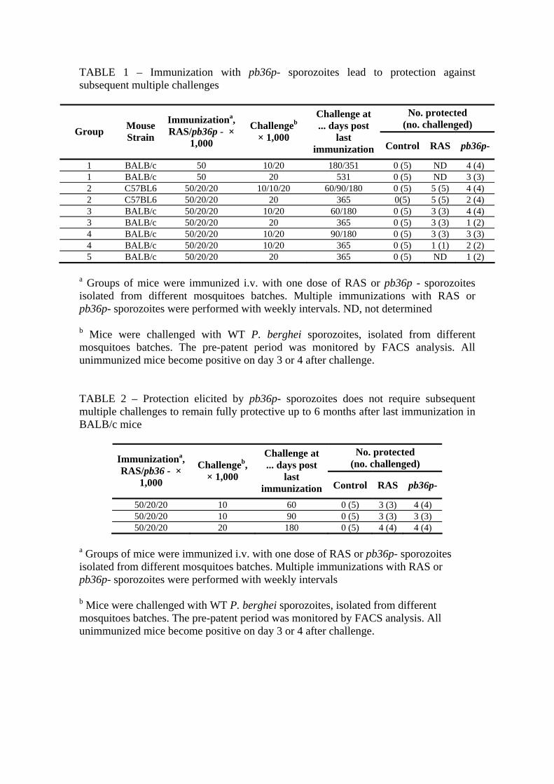
TABLE 1 – Immunization with pb36p- sporozoites lead to protection against subsequent multiple challenges
No. protected (no. challenged)
Group Mouse Strain
Immunizationa, RAS/pb36p - ×
1,000
Challengeb
× 1,000
Challenge at ... days post
last immunization Control RAS pb36p-
1 BALB/c 50 10/20 180/351 0 (5) ND 4 (4) 1 BALB/c 50 20 531 0 (5) ND 3 (3) 2 C57BL6 50/20/20 10/10/20 60/90/180 0 (5) 5 (5) 4 (4) 2 C57BL6 50/20/20 20 365 0(5) 5 (5) 2 (4) 3 BALB/c 50/20/20 10/20 60/180 0 (5) 3 (3) 4 (4) 3 BALB/c 50/20/20 20 365 0 (5) 3 (3) 1 (2) 4 BALB/c 50/20/20 10/20 90/180 0 (5) 3 (3) 3 (3) 4 BALB/c 50/20/20 10/20 365 0 (5) 1 (1) 2 (2) 5 BALB/c 50/20/20 20 365 0 (5) ND 1 (2)
a Groups of mice were immunized i.v. with one dose of RAS or pb36p - sporozoites isolated from different mosquitoes batches. Multiple immunizations with RAS or pb36p- sporozoites were performed with weekly intervals. ND, not determined b Mice were challenged with WT P. berghei sporozoites, isolated from different mosquitoes batches. The pre-patent period was monitored by FACS analysis. All unimmunized mice become positive on day 3 or 4 after challenge. TABLE 2 – Protection elicited by pb36p- sporozoites does not require subsequent multiple challenges to remain fully protective up to 6 months after last immunization in BALB/c mice
No. protected (no. challenged) Immunizationa,
RAS/pb36 - × 1,000
Challengeb, × 1,000
Challenge at ... days post
last immunization Control RAS pb36p-
50/20/20 10 60 0 (5) 3 (3) 4 (4) 50/20/20 10 90 0 (5) 3 (3) 3 (3) 50/20/20 20 180 0 (5) 4 (4) 4 (4)
a Groups of mice were immunized i.v. with one dose of RAS or pb36p- sporozoites isolated from different mosquitoes batches. Multiple immunizations with RAS or pb36p- sporozoites were performed with weekly intervals b Mice were challenged with WT P. berghei sporozoites, isolated from different mosquitoes batches. The pre-patent period was monitored by FACS analysis. All unimmunized mice become positive on day 3 or 4 after challenge.

TABLE 3 – Protection elicited by pb36p- sporozoites can be achieved even with low immunization doses in C57BL6 mice
Pre-patency (days post infection)b
No. protected/no. challenged (%) Immunizationa,
RAS/pb36p- × 1,000
RAS pb36p- RAS pb36p-
50/20/20 - - 5/5 (100%) 8/8 (100%) 10/10/10 - - 5/5 (100%) 5/5 (100%)
5/5/5 - - 5/5 (100%) 8/8 (100%) 1/1/1 6.2 - 0/5 (0%) 10/10 (100%)
None (Control) 3.2 0/8 (0%) a Groups of mice were immunized i.v. with RAS or pb36p - sporozoites isolated from different mosquitoes batches. Multiple immunizations with RAS or pb36p- sporozoites were performed with weekly intervals. Data from three independent experiments are shown. b Mice were challenged with 10,000 WT P. berghei sporozoites, isolated from different mosquitoes batches. Pre-patency is defined as the time until the first appearance of blood-stage parasites, and it was monitored by FACS analysis. TABLE 4 – pb36p- sporozoites administered intramuscularly, subcutaneously and intradermally are able to confer protection against intravenous challenge with infectious P. berghei sporozoites in C57BL6 mice
Pre-patency (days post infection)b
No. protected/no. challenged i.v. (%)
No. protected/no. challenged m.b.c (%) Type of
immunizationa
RAS pb36p- RAS pb36p- pb36p-
intravenous - - 5/5 (100%) 5/5 (100%) 5/5 (100%) intramuscular 4.5 5.3 1/5 (20%) 1/8 (13%) 5/6 (83%) subcutaneous 4.8 5.0 0/5 (0%) 0/10 (0%) 8/10 (80%) intradermal 4.5 4.8 1/5 (20%) 0/9 (0%) 6/8 (75%)
None (Control) 3.2 0/8 (0%) 0/8 (0%) a Groups of mice were immunized as indicated with RAS or pb36 - sporozoites isolated from different mosquitoes batches. Multiple immunizations with RAS or pb36p- sporozoites were performed with weekly intervals. Data from two independent experiments are shown. b Mice were challenged with 10,000 WT P. berghei sporozoites, isolated from different mosquitoes batches. Pre-patency is defined as the time until the first appearance of blood-stage parasites, and it was monitored by FACS analysis. Data concerning pre-patency in this table only concerns i.v. challenge.

c m.b., mosquito bite challenge. Highly infected mosquitoes (50,000-80,000 salivary gland sporozoites per mosquito) were allowed to feed on mice during 10-15 min (5 mosquitoes per mouse) TABLE 5 – Immunization with P. berghei RAS and pb36p- sporozoites lead to a delay on parasitemia onset in BALB/c and C57BL6 mice challenged with the heterologous species P. yoelii, and some even achieve sterile protection.
Pre-patency (days)
No. protected (no. challenged) Groups Mouse
Strain Immunization
× 1,000a
Challenge Doseb
(P. yoelii sporozoites) pb36p- RAS Control pb36p- RAS Control
1 BALB/c 50 100 5.0 ND 5.2 0 (3) ND 0 (3) 2 BALB/c 50/20 100 7.0 - 5.2 1 (3) 3 (3) 0 (3) 3 BALB/c 50/20/20 100 5.0 6.0 5.2 0 (5) 3 (4) 0 (5) 4 BALB/c 50 1,000 4.0 ND 4.0 0 (3) ND 0 (3) 5 BALB/c 50/20 1,000 4.7 5.5 4.0 0 (3) 1 (3) 0 (3) 6 BALB/c 50/20/20 1,000 5.2 7.0 4.0 0 (5) 3 (4) 0 (5) 7 C57BL6 50/20/20 1,000 4.8 5.5 4.0 0 (5) 0 (5) 0 (5) 8 BALB/c 50/20/20 10,000 4.6 5.0 4.0 0 (5) 0 (4) 0 (5)
a Groups of mice were immunized as described before with P. berghei RAS or pb36p- sporozoites. Data from two independent experiments are shown. ND, not determined. b For all experiments described above, matched control mice groups immunized likewise with either RAS or pb36p- sporozoites from the same mosquitoes’ batch and challenged with 10,000 homologous P. berghei sporozoites each. All failed to develop blood stage parasites confirming that these sporozoites were fully protective in homologous challenge in these conditions. All control naïve mice challenged equally with P. berghei sporozoites developed blood stage parasitemia, as expected.