Fabrication of lactobionic-loaded chitosan microcapsules … · Fabrication of lactobionic-loaded...
9
Fabrication of lactobionic-loaded chitosan microcapsules as potential drug carriers targeting the liver Jing Zhang, Cao Li, Zhi-Yuan Xue, Hai-Wei Cheng, Fu-Wei Huang, Ren-Xi Zhuo, Xian-Zheng Zhang ⇑ Key Laboratory of Biomedical Polymers of Ministry of Education, Department of Chemistry, Wuhan University, Wuhan 430072, People’s Republic of China article info Article history: Received 14 August 2010 Received in revised form 5 November 2010 Accepted 30 November 2010 Available online 3 December 2010 Keywords: Layer by layer Multilayered capsules Drug carriers Liver targeting Click chemistry abstract This paper demonstrates a general approach for fabrication of lactobionic chitosan microcapsules using layer-by-layer assembly via click chemistry. Chitosan was selectively modified with either azide (CHI- Az) or alkyne (CHI-Alk) groups. The growth of the CHI-Az/CHI-Alk click multilayer was studied experi- mentally by multilayer assembly on planar supports. Linear buildup of the film was observed. The chito- san click capsules were also analyzed with confocal laser scanning microscopy and transmission electron microscopy. Capsules were found to have regular spherical shapes. In addition, (CHI-Az/CHI-Alk)-coated particles were modified with fluorescein isothiocyanate to ensure that the particles can be easily post- functionalized. Finally, lactobionic acid was conjugated onto the (CHI-Az/CHI-Alk)-coated particles and the lactobionic particles exhibited hepatoma cell (HepG2) targeting behavior. Ó 2010 Acta Materialia Inc. Published by Elsevier Ltd. All rights reserved. 1. Introduction Liver cancer is a common fatal disease [1,2]. However, chemo- therapy for cancer is largely limited by the toxicity of the drugs used to normal tissues [3]. Furthermore, limited circulation in the blood, poor aqueous solubility, limited stability and nonselectivity reduce the therapeutic efficacy and limit the clinical application of antican- cer drugs [3]. To solve these problems, various drug carriers such as micelles, nanogels and microcapsules have been investigated [4–6]. A novel type of hollow polyelectrolyte capsule fabricated via layer- by-layer (LbL) self-assembly on sacrificial template particles, fol- lowed by core removal, has attracted a great deal of research atten- tion. With this technique, the capsules can be engineered with a high level of control of properties such as size and shape, composition, and functionality [7,8]. Due to the toxicity to normal tissues of tradi- tional cancer therapies, the design of drug carriers with active targeting properties is highly important. It was reported that asialo- glycoprotein (ASGP) receptors were specifically abundant on the surface of hepatoma cells [9,10]. Therefore active targeting of these cells can be accomplished via introduction of galactose—which can be recognized by hepatoma cells via ASGP receptors located on their surfaces—into drug carriers for the treatment of liver cancers. Here, we fabricated lactobionic chitosan microcapsules as po- tential drug carriers targeting the liver using LbL assembly via click chemistry. The click chemistry approach for LbL assembly was pro- posed in Refs. [11,12]. The combination of the LbL technique and click chemistry is promising mainly for two reasons. Firstly, various kinds of materials, including those of the same charged or non-charged polymers, can be used for LbL assembly, resulting in single-component polymer, covalently stabilized, mul- tilayer films. For traditional LbL strategies, at least two components are needed for building up the capsule wall. However, the exis- tence of a second polymer component would limit the functional- ities of the specific materials. For example, the biocompatibility and biodegradability of chitosan would be strongly influenced by a second polymer component. Thus, this technique can easily be used to fabricate single-component capsules. Secondly, in our study, the degrees of functionalization of chito- san were kept below 10%, allowing the majority of chitosan to re- tain its functionality. The excess groups of chitosan, which are not modified with click functionalities in the multilayer, can be used to post-functionalize with other materials, including biomacromole- cules, peptides or other functional groups. Herein, we aimed to use this method for the fabrication of biodegradable multilayered capsules with the desired properties. Designing novel materials for biomedical applications generally requires the use of biodegradable materials. Chitosan, a renewable natural polysaccharide, is of great interest due to its good biocom- patibility and biodegradability [13,14], and has been considered for various applications in modern biomedical and pharmaceutical fields [15,16]. Moreover, chitosan is a polycation and carries positive charge at low pH. Thus, it has been widely used for fabricating LbL films [17–21] and capsules [22–25] with other polyelectrolytes. Here, we demonstrate a new way to fabricate chitosan microcap- sules, using click chemistry as the driving force of the LbL assembly. In this study, chitosan was selectively functionalized with either 1742-7061/$ - see front matter Ó 2010 Acta Materialia Inc. Published by Elsevier Ltd. All rights reserved. doi:10.1016/j.actbio.2010.11.042 ⇑ Corresponding author. Tel.: +86 27 6875 4061; fax: +86 27 6875 4509. E-mail address: [email protected] (X.-Z. Zhang). Acta Biomaterialia 7 (2011) 1665–1673 Contents lists available at ScienceDirect Acta Biomaterialia journal homepage: www.elsevier.com/locate/actabiomat
Transcript of Fabrication of lactobionic-loaded chitosan microcapsules … · Fabrication of lactobionic-loaded...

Acta Biomaterialia 7 (2011) 1665–1673
Contents lists available at ScienceDirect
Acta Biomaterialia
journal homepage: www.elsevier .com/locate /actabiomat
Fabrication of lactobionic-loaded chitosan microcapsules as potential drugcarriers targeting the liver
Jing Zhang, Cao Li, Zhi-Yuan Xue, Hai-Wei Cheng, Fu-Wei Huang, Ren-Xi Zhuo, Xian-Zheng Zhang ⇑Key Laboratory of Biomedical Polymers of Ministry of Education, Department of Chemistry, Wuhan University, Wuhan 430072, People’s Republic of China
a r t i c l e i n f o a b s t r a c t
Article history:Received 14 August 2010Received in revised form 5 November 2010Accepted 30 November 2010Available online 3 December 2010
Keywords:Layer by layerMultilayered capsulesDrug carriersLiver targetingClick chemistry
1742-7061/$ - see front matter � 2010 Acta Materialdoi:10.1016/j.actbio.2010.11.042
⇑ Corresponding author. Tel.: +86 27 6875 4061; faE-mail address: [email protected] (X.-Z. Zhan
This paper demonstrates a general approach for fabrication of lactobionic chitosan microcapsules usinglayer-by-layer assembly via click chemistry. Chitosan was selectively modified with either azide (CHI-Az) or alkyne (CHI-Alk) groups. The growth of the CHI-Az/CHI-Alk click multilayer was studied experi-mentally by multilayer assembly on planar supports. Linear buildup of the film was observed. The chito-san click capsules were also analyzed with confocal laser scanning microscopy and transmission electronmicroscopy. Capsules were found to have regular spherical shapes. In addition, (CHI-Az/CHI-Alk)-coatedparticles were modified with fluorescein isothiocyanate to ensure that the particles can be easily post-functionalized. Finally, lactobionic acid was conjugated onto the (CHI-Az/CHI-Alk)-coated particles andthe lactobionic particles exhibited hepatoma cell (HepG2) targeting behavior.
� 2010 Acta Materialia Inc. Published by Elsevier Ltd. All rights reserved.
1. Introduction Firstly, various kinds of materials, including those of the same
Liver cancer is a common fatal disease [1,2]. However, chemo-therapy for cancer is largely limited by the toxicity of the drugs usedto normal tissues [3]. Furthermore, limited circulation in the blood,poor aqueous solubility, limited stability and nonselectivity reducethe therapeutic efficacy and limit the clinical application of antican-cer drugs [3]. To solve these problems, various drug carriers such asmicelles, nanogels and microcapsules have been investigated [4–6].A novel type of hollow polyelectrolyte capsule fabricated via layer-by-layer (LbL) self-assembly on sacrificial template particles, fol-lowed by core removal, has attracted a great deal of research atten-tion. With this technique, the capsules can be engineered with a highlevel of control of properties such as size and shape, composition,and functionality [7,8]. Due to the toxicity to normal tissues of tradi-tional cancer therapies, the design of drug carriers with activetargeting properties is highly important. It was reported that asialo-glycoprotein (ASGP) receptors were specifically abundant on thesurface of hepatoma cells [9,10]. Therefore active targeting of thesecells can be accomplished via introduction of galactose—which canbe recognized by hepatoma cells via ASGP receptors located on theirsurfaces—into drug carriers for the treatment of liver cancers.
Here, we fabricated lactobionic chitosan microcapsules as po-tential drug carriers targeting the liver using LbL assembly via clickchemistry. The click chemistry approach for LbL assembly was pro-posed in Refs. [11,12]. The combination of the LbL technique andclick chemistry is promising mainly for two reasons.
ia Inc. Published by Elsevier Ltd. A
x: +86 27 6875 4509.g).
charged or non-charged polymers, can be used for LbL assembly,resulting in single-component polymer, covalently stabilized, mul-tilayer films. For traditional LbL strategies, at least two componentsare needed for building up the capsule wall. However, the exis-tence of a second polymer component would limit the functional-ities of the specific materials. For example, the biocompatibilityand biodegradability of chitosan would be strongly influenced bya second polymer component. Thus, this technique can easily beused to fabricate single-component capsules.
Secondly, in our study, the degrees of functionalization of chito-san were kept below 10%, allowing the majority of chitosan to re-tain its functionality. The excess groups of chitosan, which are notmodified with click functionalities in the multilayer, can be used topost-functionalize with other materials, including biomacromole-cules, peptides or other functional groups. Herein, we aimed touse this method for the fabrication of biodegradable multilayeredcapsules with the desired properties.
Designing novel materials for biomedical applications generallyrequires the use of biodegradable materials. Chitosan, a renewablenatural polysaccharide, is of great interest due to its good biocom-patibility and biodegradability [13,14], and has been consideredfor various applications in modern biomedical and pharmaceuticalfields [15,16]. Moreover, chitosan is a polycation and carries positivecharge at low pH. Thus, it has been widely used for fabricating LbLfilms [17–21] and capsules [22–25] with other polyelectrolytes.Here, we demonstrate a new way to fabricate chitosan microcap-sules, using click chemistry as the driving force of the LbL assembly.In this study, chitosan was selectively functionalized with either
ll rights reserved.

1666 J. Zhang et al. / Acta Biomaterialia 7 (2011) 1665–1673
azide (CHI-Az) or alkyne (CHI-Alk) groups. The CHI-Alk was thenmodified with rhodamine dye. Chitosan click capsules were ob-tained after removal of the CaCO3 particle template and were shownto have regular spherical shapes. In addition, (CHI-Az/CHI-Alk)-coated particles were modified with fluorescein isothiocyanate(FITC) to ensure that the particles can be easily post-functionalized.Finally, lactobionic acid was conjugated onto the (CHI-Az/CHI-Alk)-coated particles, which enable the particles to bind specifically to theASGP receptors on the surfaces of hepatoma cells (HepG2).
2. Experimental
2.1. Materials
Chitosan (Mw = 50 kDa, deacetylation degree = 85.3%) was pur-chased from Haidebei Marine Bioengineering Co. Ltd. (Jinan, PR Chi-na). Poly(sodium 4-styrene-sulfonate) (PSS) (Mw = 70 kDa) wasobtained from Sigma–Aldrich. 2-Bromo-2-methylpropionic acid(98%), fluoresceinamine isomer (FITC) and lactobionic acid werepurchased from ACROS. Rhodamine B was obtained from Alfa Aesar.1-Ethyl-3-(3-dimethylaminopropyl) carbodiimide hydrochloride(EDC), sodium azide (NaN3), potassium carbonate (K2CO3), calciumchloride (CaCl2), copper(II) sulfate, sodium ascorbate, sodiumhydroxide (NaOH), dimethyl formamide (DMF) and disodium ethyl-enediaminetetraacetate (EDTA) were purchased from Shanghai Re-agent Chemical Co. (PR China). All the reagents were used directlyunless specifically indicated. 4-Oxo-4-(prop-2-ynyloxy) butanoicacid used was synthesized according to our previous report [26]. Fe-tal bovine serum albumin (FBS), Dulbecco’s Modified Eagle’s Med-ium (DMEM), penicillin–streptomycin and phosphate-bufferedsaline (PBS) were purchased from Sigma–Aldrich. All other chemi-cals were obtained from Shanghai Reagent Chemical Co. (PR China)and used as received.
2.2. Synthesis of alkyne-modified chitosan
Chitosan with alkyne functionality (CHI-Alk) was synthesizedvia amide bond formation between the amine groups of chitosanside-chains and carboxyl groups of 4-oxo-4-(prop-2-ynyloxy)butanoic acid. 1.0 g chitosan was dissolved in acetic acid solution(2 wt.%). Then 0.9 g 4-oxo-4-(prop-2-ynyloxy) butanoic acid and2.3 g EDC were added after adjusting pH of the solution to 5–7.The clear solution was stirred at room temperature for 24 h. Thecrude product was dialyzed extensively against de-ionized (DI)water for 3 days and freeze-dried to obtain white polymer in ayield of 0.8 g.
2.3. Synthesis of azide-modified chitosan
Chitosan with azide functionality (CHI-Az) was synthesized viaEDC-mediated coupling of the amine moieties of chitosan with 2-bromo-2-methylpropionic acid. Chitosan (1.0 g) was dissolved in50 ml acetic acid solution (2 wt.%). After adjusting the pH of thesolution to 5–7, 1.0 g 2-bromo-2-methylpropionic acid and 2.3 gEDC were added. The clear solution was then stirred at room tem-perature for 24 h. The crude product was dialyzed and freeze-dried. The product was then dissolved in acetic acid solution(2 wt.%). 0.6 g NaN3 was added after adjusting the solution to neu-tral. The reaction was carried out for 24 h at room temperature.The CHI-Az was obtained by dialyzing and freeze-drying for 3 days,yielding 0.7 g.
2.4. Fluorescent labeling of alkyne-modified chitosan
CHI-Alk was subsequently fluorescently labeled with a dye, rho-damine B. All CHI-Alk was dissolved in distilled water. Then rhoda-
mine B (10 mol.% based on amine groups of the chitosan) and EDC(1.2-fold molar excess compared to rhodamine B) was added. Themixture was stirred for 24 h at room temperature. Rhodamine B-labeled CHI-Alk was obtained by dialyzing against DI water andfreeze-drying.
2.5. Fourier transform infrared spectroscopy
The samples were analyzed by Fourier transform infrared spec-troscopy (FT-IR) using a Perkin–Elmer Spectrum One spectropho-tometer. Before the measurements, the samples were pressedinto potassium bromide (KBr) pellets.
2.6. 1H nuclear magnetic resonance
The 1H nuclear magnetic resonance (NMR) spectra of the poly-mers were recorded on a Mercury VX-300 spectrometer at300 MHz (Varian, USA) using D2O as a solvent and TMS as an inter-nal standard.
2.7. Multilayer assembly on planar supports
Rectangular quartz slides (15 � 8 mm2) were cleaned by H2O2/H2SO4 solution (1:3 v/v) at 80 �C for 90 min and then dipped in amixture solution containing H2O, H2O2 and NH4�OH (5:1:1 v/v/v)at 80 �C for 90 min (Caution! piranha solution is highly causticand great care must be taken when handling.) The slides were fi-nally washed with DI water, and dried in air to obtain a negativelycharged surface. For chitosan click multilayer assembly, the nega-tively charged slides were sequentially immersed in CHI-Az andCHI-Alk solutions containing copper sulfate and sodium ascorbatefor 15 min, accompanied by water rinsing after deposition of eachlayer. The dipping solutions were prepared as follows according tothe literature [27]: (a) CHI-Az (1.0 mg ml�1), (b) CHI-Alk(1.0 mg ml�1), (c) DI water (pH 7.0), (d) copper(II) sulfate(0.4 mg ml�1) and (e) sodium ascorbate (0.8 mg ml�1). The pH ofeach solution was adjusted to 6.1 with 1 M HCl or NaOH solution.Polymer dipping solutions were made up in a constant volumeratio of 3(a or b):1(d):1(e). The washing solutions were made upin a similar ratio except using solution (c) in place of (a) or (b).Dipping solutions and washing solutions were prepared 2 minprior to use.
2.8. Characterization of multilayered films
We used UV–vis spectra obtained on a Lambda Bio40 UV/visspectrometer (Perkin–Elmer) to monitor the LbL assembly process.The morphology of the resulting multilayer was analyzed by atom-ic force microscopy (AFM) using a Shimadzu SPM-9500J3 operatedin tapping mode at 20 �C. The morphology of the quartz slide wasalso analyzed by AFM for comparison. The formation of linkages inthe resulting multilayer was studied by X-ray photoelectron spec-troscopy (XPS). XPS was conducted on a XSAM 800 spectrometer(Kratos, UK) with an Mg KR target. An analysis area of approxi-mately 0.6 � 0.8 mm2 was used to measure three locations persample.
2.9. Fluoresceinamine isomer capture by �4 lm CaCO3 particles
CaCO3 particles with narrow size distribution were preparedaccording to the literature [28,29]. In order to analyze CaCO3 par-ticles by confocal laser scanning microscopy (CLSM), we added flu-oresceinamine isomer in a preparation of CaCO3 to label theparticles with fluorescence. Briefly, 5 ml 0.33 M K2CO3 solutioncontaining 10 mg fluoresceinamine isomer was rapidly poured into5 ml of a 0.33 M solution of CaCl2 containing 10 mg PSS at room

Scheme 1. Synthesis route of azide-modified chitosan (A) and alkyne-modified chitosan (B).
Fig. 1. FTIR spectra of chitosan (A), alkyne-modified chitosan (B), azide-modifiedchitosan (C) and chitosan click capsules (D). The extra curve is an expansion of thealkyne stretch shown in curve B.
J. Zhang et al. / Acta Biomaterialia 7 (2011) 1665–1673 1667
temperature. After intense agitation for 30 s, the reaction mixturewas left still for about 2 min. Then the precipitate was filteredoff, thoroughly washed with DI water and acetone, and dried inair. The whole process was protected from light wherever possible.
2.10. Multilayer assembly on CaCO3 particles
Approximately 60 mg CaCO3 particles and 1.5 ml CHI-Az solu-tion were mixed and the mixture was shaken to form a symmetri-cal suspension. This incubation was allowed to stand for 15 min toestablish a CHI-Az layer. After adsorption, the particles were
centrifuged at 10,000 rpm for 1 min, followed by washing withDI water.
The following dipping solutions were made as per those re-ported in Section 2.7 above. After pre-coating with CHI-Az, 1.5 mlof CHI-Alk solution (containing copper and ascorbate) were addedand allowed to incubate for 15 min. Then the particles were centri-fuged and washed with the washing solutions. This was followedby adsorption of CHI-Az, followed by the same washing protocol.The process was repeated until the desired number of layers wasobtained. The whole process was protected from light whereverpossible.
2.11. Formation of click capsules
To form the click capsules, the multilayer-coated CaCO3 parti-cles were treated with 0.2 M EDTA. Dissolution of the CaCO3 coreoccurred after less than 1 min. The capsules were imaged by CLSMand transmission electron microscopy (TEM).
2.12. Characterization of chitosan click capsules
The core–shell and hollow particles were viewed with CLSM(Nikon C1-si, BD Laser at 488 nm). For TEM, a drop of a concentratedcapsule solution was placed on a clean TEM grid and allowed to dryin air. TEM analysis was carried out with a JEM-100CX II instrumentoperating at an acceleration voltage of 100 kV.
2.13. Post-functionalization of (CHI-Az/CHI-Alk)-coated particles byFITC
The particles were incubated in anhydrous DMF containing FITC(10 mg ml�1) for 24 h. The functionalized particles were obtainedafter several washing steps with anhydrous DMF and at least sevenwashing steps with DI water. A control sample was incubated inDMF without FITC for 24 h. The conjugation of FITC was then

1668 J. Zhang et al. / Acta Biomaterialia 7 (2011) 1665–1673
analyzed by monitoring the fluorescence intensity of the resultingparticles by flow cytometry.
2.14. Post-functionalization of (CHI-Az/CHI-Alk)-coated particles bylactobionic acid
Since �4 lm CaCO3 particles are too big to be internalized byHepG2 cells, we used CaCO3 particles with a small size in thecell-targeting assay in order to assess the cell internalization ofthe capsules into tumor cells. The preparation of small CaCO3 par-ticles is as follows: 5 ml 0.33 M K2CO3 solution was rapidly pouredinto 5 ml 0.33 M CaCl2 solution at room temperature. The subse-quent steps are similar to those used for the preparation of�4 lm (CHI-Az/CHI-Alk)-coated CaCO3 particles. (CHI-Az/CHI-Alk)-coated CaCO3 particles were then incubated in lactobionicacid solution (10 mg ml�1, pH 7.0) containing EDC (2-fold molar ra-tio based on lactobionic acid) for 24 h. The functionalized particleswere obtained after five washing steps with DI water. A controlsample was incubated in solution without lactobionic acid for24 h. The lactobionic adsorption was then analyzed using a HepG2cell targeting experiment.
After HepG2 cells were incubated in DMEM containing 10% FBSand 1% antibiotics (penicillin–streptomycin, 10,000 U ml�1) at37 �C in a humidified atmosphere containing 5% CO2 for 1 day inan incubator, the culture was replaced by medium containingpost-functionalized (CHI-Az/CHI-Alk)-coated particles (0.5 mgml�1). The mixture was then further incubated for 4 h. Cellularadhesion or internalization was studied by CLSM after the plates
Fig. 2. 1H NMR spectra of chitosan (A), Br-modified chitosan (B), a
had been washed six times with PBS. A control experiment wascarried out, using non-functionalized (CHI-Az/CHI-Alk)-coated par-ticles instead of post-functionalized ones.
2.15. Statistical analysis
The results are representative of replicate experiments and theresults are represented by the mean and SD. The statistical analy-ses were performed using Student’s t-test. Probability values lessthan 0.05 (P < 0.05) were considered to be indicative of statisticalsignificance.
3. Results and discussion
3.1. Synthesis of chitosan with pendant groups
Alkyne and azide pendant groups were introduced onto chito-san to investigate the possibility of LbL assembly by click chemis-try. The introduction of an alkyne functional moiety onto chitosanis achieved via the coupling reaction between carboxyl and aminegroups in the presence of the coupling reagent EDC (Scheme 1). Asfor the introduction of the azide functional group, the substitutionreaction between Br and NaN3 at room temperature is widely used[30,31]. As shown in Scheme 1, the precursor Br-modified chitosanwas synthesized. The azide-modified chitosan was then obtainedvia substitution reactions. Since LbL assembly of CHI-Az and CHI-Alk only requires a few functionalized groups, while the low degreeof functionalization would retain the original biocompatibility and
zide-modified chitosan (C) and alkyne-modified chitosan (D).

Fig. 3. Plots of absorbance at 241 nm as a function of multilayered films assembled on quartz with increasing bilayer number by UV–vis spectroscopy (A). XPS spectra ofchitosan click multilayer assembly on planar supports (B). Inset: AFM image of clean quartz slide (a) and (CHI-Az/CHI-Alk)8 multilayered film assembled on quartz (b)(area = 2 � 2 lm2).
Scheme 2. Assembly of click chitosan multilayer (CHI-Az/CHI-Alk) on CaCO3 particle and chitosan click capsule formation.
J. Zhang et al. / Acta Biomaterialia 7 (2011) 1665–1673 1669

Fig. 4. CLSM images of chitosan multilayer coating on the CaCO3 particles (A), CaCO3 particles filled with fluoresceinamine isomer (B) and chitosan-coated CaCO3 particles (C).The profile in (D) corresponds to the line in (C).
1670 J. Zhang et al. / Acta Biomaterialia 7 (2011) 1665–1673
biodegradability of chitosan, the degrees of functionalization ofchitosan were kept below 10%, and all of the experiments were car-ried out in water. The CHI-Alk was then modified with rhodamineB dye to enable the capsules to be studied by CLSM.
Fig. 1 shows that the IR spectra of chitosan with functional groups(Fig. 1B and C) are similar to those of chitosan itself (Fig. 1A). For CHI-Alk (Fig. 1B), the typical absorption of alkyne appeared at 2123 cm�1.In addition, the stretching variation absorption of C@O in alkynependant units existed at 1745 cm�1. Based on the 1H NMR spectraof alkyne-modified chitosan, the chemical shift at 4.6 ppm wasattributed to the methylene protons of AOCH2C„CH. And the chem-ical shifts at 2.2, 2.25, 2.4 and 4.5 ppm were mainly associated withthe protons of the alkyne pendant groups AC@OCH2CH2C@OOCH2C„CH, AC@OCH2CH2C@OOCH2C„CH, AC@OCH2CH2C@OOCH2C„CH and AC@OCH2CH2C@OOCH2C„CH, respectively. Otherresonances are assigned to the backbone chain of chitosan. In thecase of the azide-modified chitosan, to ensure the complete conver-sion from Br to azide pendant groups, a large excess of NaN3 com-pared to the Br in the polymeric chain was added. In this study, the
Fig. 5. CLSM (A) and TEM (B, C) images
product was dialyzed against DI water for several days and the dia-lyzing water was changed many times to ensure the excess NaN3 wasremoved completely. As shown in Fig. 1C, the peak at 2037 cm�1
indicated the existence of azide pendant groups. For 1H NMR spectra,after the substitution reaction between Br and NaN3, the originalchemical shift of methyl protons (AC(CH3)2– Br) at 1.75 ppm disap-peared and a new signal appeared at 1.2 ppm in the 1H NMR spectraof the azide-modified chitosan, which was associated with the corre-sponding methyl protons of AC(CH3)2AN3. This demonstrated theconversion from Br to azide pendant groups after the substitutionreaction. All the other signals are related to chitosan backbone chain.
Herein, the real degree of substitution (DS) of CHI-Alk and CHI-Azwere determined from their 1H NMR spectra (Fig. 2B and C). On thebasis of the 1H NMR spectra, the DS of alkyne and azide functionalgroups in the polymeric chain was calculated by comparing the inte-gration ratio of the methylene protons (2H) (AC@OCH2CH2C@OOCH2C„CH) of alkyne pendant units at 2.4 ppm and the methylprotons (6H) (AC@OC(CH3)2-N3) at 1.2 ppm to that of the protons(5H) which positioned on C-3, C-4, C-5 and C-6 appearing at around
of (CHI-Az/CHI-Alk)8 click capsules.

J. Zhang et al. / Acta Biomaterialia 7 (2011) 1665–1673 1671
3.6 ppm respectively. 1H NMR analysis confirmed that �10% of thechitosan side chains were functionalized with alkyne moieties and�6% were modified with azide moieties.
3.2. Multilayer assembly on planar supports
The growth of the CHI-Az/CHI-Alk click multilayer was studiedvia an experiment involving multilayer assembly on planar sup-ports. The LbL assembly of CHI-Az/CHI-Alk multilayer was moni-tored by UV–vis spectroscopy [27,32,33] (Fig. 3A). Linear growthof the film was observed by monitoring the peak at 241 nm men-tioned in a previous study [27]. A control of chitosan without clickgroups (control, }) showed a plateau in absorbance after only fourlayers, indicating that the click functionalities are essential for thedeposition of consecutive chitosan layers and the formation ofchitosan multilayer. If no copper(II) sulfate and sodium ascorbatewas added (control, D), the absorbance remained constant low,suggesting that film did not build up and indirectly confirming thatcopper(I) acts as a catalyst for LbL assembly of click-modified poly-mers. Moreover, the absorbance of CHI-Az/CHI-Alk without copperis even lower than that of chitosan without click groups, which wasattributed to the fact that the intermolecular interaction of chito-san was weakened after chitosan had been functionalized withclick groups. For XPS study, a peak at 400 eV, which has been com-monly assigned to triazole functionality [34], confirmed the forma-tion of triazole linkages in the chitosan multilayer (Fig. 3B). Themorphology of clean quartz slide and air-dried chitosan multilay-ered film was examined by AFM. The AFM images are shown inFig. 3B as an insert picture. The z scale of chitosan multilayeredfilm is �130 nm (Fig. 3Bb) compared to �5 nm of clean quartz slide(Fig. 3Ba). The rough surface of multilayered film proved the suc-cessful deposition of chitosan onto the quartz slide.
Fig. 6. Relative fluorescence intensity of (CHI-Az/CHI-Alk)8-coated particles beforeand after post-functionalization as monitored by flow cytometry. Inset: schematicillustration of the post-functionalization process. Data are shown as mean ± SD(n = 3). (⁄P < 0.05 as compared with other samples in the same group).
3.3. Multilayer assembly on CaCO3 particles
LbL assembly on colloids was performed by sequentially expos-ing �4 lm CaCO3 particles captured with fluoresceinamine isomerto CHI-Az and CHI-Alk solutions (1.0 mg ml�1) containing coppersulfate (0.4 mg ml�1) and sodium ascorbate (0.8 mg ml�1) at pH6.1. The particles were incubated for 15 min in each chitosan solu-tion, and were then centrifuged and washed with DI water(Scheme 2). The chitosan-coated CaCO3 was studied by CLSM.Fig. 4 shows CLSM images of chitosan-coated CaCO3 particles.Fig. 4A and B show the localization of labeled chitosan and theCaCO3 core captured with fluoresceinamine isomer, respectively.Fluorescence microscopy confirmed that the chitosan multilayercoating on the CaCO3 particles was uniform. From Fig. 4B, we cansee that fluoresceinamine isomer, which was added during theCaCO3 particle preparation process, was successfully captured byCaCO3 particles and distributed evenly. Fig. 4C represents thoseCaCO3 particles coated with rhodamine-labeled CHI-Alk/CHI-Azshell layers. It can be seen that LbL multilayer-coated CaCO3 parti-cles have a red corona layer surrounding a green CaCO3 inner core.Moreover, the diameters of the red rings are larger than those ofthe green ones, proving that chitosan was almost adsorbed onthe surface of CaCO3 particles to form a capsule wall instead ofbeing captured into the porous CaCO3. Moreover, the profile inFig. 4D represents the fluorescence intensity of a single chitosan-coated CaCO3 particle. As shown in Fig. 4D, the fluorescence inten-sity of labeled chitosan is higher on the edges of the particles thanthat in the interior, which also confirms the core–shell structure ofthe chitosan-coated CaCO3 particles. The red fluorescence in theinterior shown in Fig. 4D may due to the porous structure of CaCO3
particles, leading to a small portion of labeled chitosan being cap-tured in the interior.
3.4. Characterization of chitosan click capsules
The chitosan click multilayered core–shell particles containCaCO3 cores. Fig. 5A shows a fluorescent image of chitosan clickcapsules. A uniform coating with red fluorescently labeled chitosanand a regular spherical ring of the click capsules were observed.After EDTA treatment at pH 7, only a red shell layer can be seen,confirming the complete removal of the CaCO3 core. At the sametime, the small molecule fluoresceinamine isomer permeated outof the capsules because of the semi-permeable property of poly-electrolyte shells, which are generally known to be permeable tomolecules with molecular weights below 5 kDa [35]. Moreover, itcan be seen that chitosan click capsules swelled to 6.5 ± 0.5 lmcompared with the 4 lm CaCO3 templates. The slightly enlargedsize of the hollow capsules after the CaCO3 core removal maydue to increased osmotic pressure in the interior void spaces filledwith ionic species [36]. From the confocal images, it can be con-cluded that the triazole linkages between chitosan chains are read-ily formed under these mild conditions. To investigate the reactionof alkyne and azide functional groups in the capsule wall, thefreeze-dried capsules were analyzed by FT-IR. From the IR spectraof the chitosan click capsule (Fig. 1D), we that see that the typicalpeaks of alkyne and azide have disappeared, suggesting that mostof functional groups have been reacted. The TEM image shows thatthe click capsules collapsed and folded but remained intact afterdrying (Fig. 5B).
3.5. Post-functionalization of (CHI-Az/CHI-Alk)-coated particles byFITC
The modification of chitosan with click groups used only a min-or fraction of the amine moieties on chitosan. Therefore, theremaining free amine groups in the multilayer on the surface ofthe particles could be used for post-functionalization with variousmaterials, including small molecules such as drugs as well as largemolecules such as peptides or biomacromolecules. The capabilityof the particles to be post-functionalized was demonstrated byreacting FITC with (CHI-Az/CHI-Alk)8 multilayer. Chitosan multi-layered click films were assembled as mentioned above with eightbilayers of (CHI-Az/CHI-Alk).
Both FITC-functionalized and non-functionalized particles wereanalyzed by flow cytometry. As shown in Fig. 6, non-functionalizedparticles exhibit �6% fluorescence intensity, due to the fluorescein-amine isomer being captured in the CaCO3 core. However, the
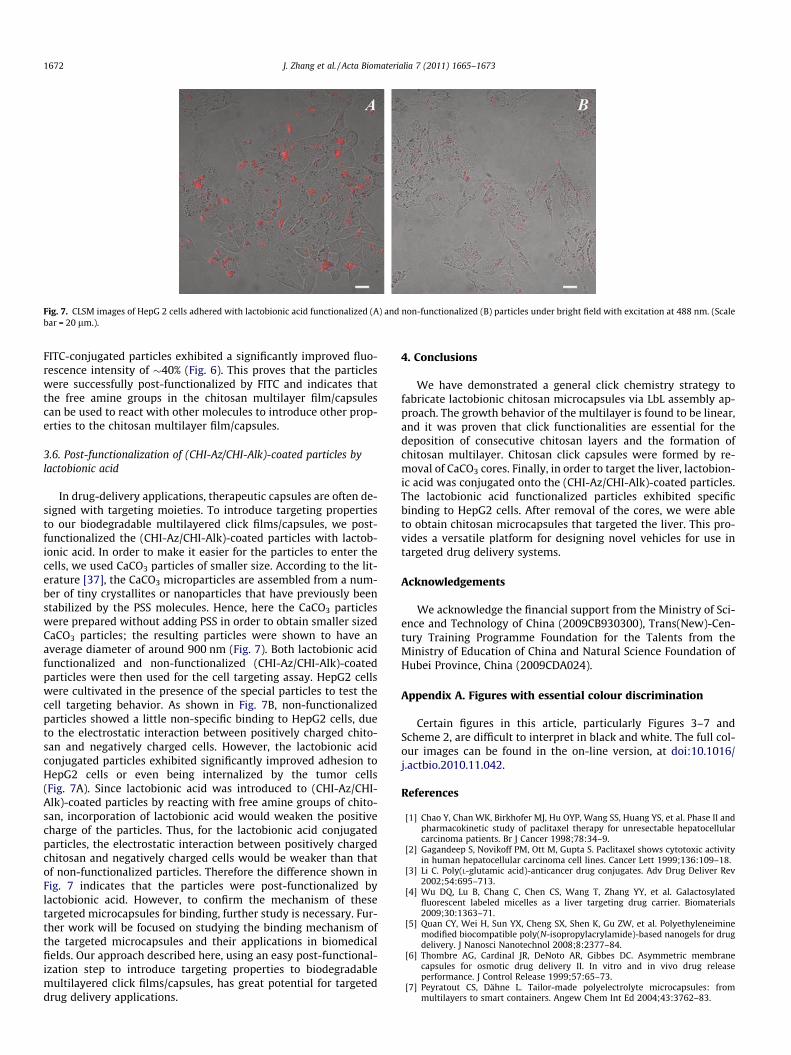
Fig. 7. CLSM images of HepG 2 cells adhered with lactobionic acid functionalized (A) and non-functionalized (B) particles under bright field with excitation at 488 nm. (Scalebar = 20 lm.).
1672 J. Zhang et al. / Acta Biomaterialia 7 (2011) 1665–1673
FITC-conjugated particles exhibited a significantly improved fluo-rescence intensity of �40% (Fig. 6). This proves that the particleswere successfully post-functionalized by FITC and indicates thatthe free amine groups in the chitosan multilayer film/capsulescan be used to react with other molecules to introduce other prop-erties to the chitosan multilayer film/capsules.
3.6. Post-functionalization of (CHI-Az/CHI-Alk)-coated particles bylactobionic acid
In drug-delivery applications, therapeutic capsules are often de-signed with targeting moieties. To introduce targeting propertiesto our biodegradable multilayered click films/capsules, we post-functionalized the (CHI-Az/CHI-Alk)-coated particles with lactob-ionic acid. In order to make it easier for the particles to enter thecells, we used CaCO3 particles of smaller size. According to the lit-erature [37], the CaCO3 microparticles are assembled from a num-ber of tiny crystallites or nanoparticles that have previously beenstabilized by the PSS molecules. Hence, here the CaCO3 particleswere prepared without adding PSS in order to obtain smaller sizedCaCO3 particles; the resulting particles were shown to have anaverage diameter of around 900 nm (Fig. 7). Both lactobionic acidfunctionalized and non-functionalized (CHI-Az/CHI-Alk)-coatedparticles were then used for the cell targeting assay. HepG2 cellswere cultivated in the presence of the special particles to test thecell targeting behavior. As shown in Fig. 7B, non-functionalizedparticles showed a little non-specific binding to HepG2 cells, dueto the electrostatic interaction between positively charged chito-san and negatively charged cells. However, the lactobionic acidconjugated particles exhibited significantly improved adhesion toHepG2 cells or even being internalized by the tumor cells(Fig. 7A). Since lactobionic acid was introduced to (CHI-Az/CHI-Alk)-coated particles by reacting with free amine groups of chito-san, incorporation of lactobionic acid would weaken the positivecharge of the particles. Thus, for the lactobionic acid conjugatedparticles, the electrostatic interaction between positively chargedchitosan and negatively charged cells would be weaker than thatof non-functionalized particles. Therefore the difference shown inFig. 7 indicates that the particles were post-functionalized bylactobionic acid. However, to confirm the mechanism of thesetargeted microcapsules for binding, further study is necessary. Fur-ther work will be focused on studying the binding mechanism ofthe targeted microcapsules and their applications in biomedicalfields. Our approach described here, using an easy post-functional-ization step to introduce targeting properties to biodegradablemultilayered click films/capsules, has great potential for targeteddrug delivery applications.
4. Conclusions
We have demonstrated a general click chemistry strategy tofabricate lactobionic chitosan microcapsules via LbL assembly ap-proach. The growth behavior of the multilayer is found to be linear,and it was proven that click functionalities are essential for thedeposition of consecutive chitosan layers and the formation ofchitosan multilayer. Chitosan click capsules were formed by re-moval of CaCO3 cores. Finally, in order to target the liver, lactobion-ic acid was conjugated onto the (CHI-Az/CHI-Alk)-coated particles.The lactobionic acid functionalized particles exhibited specificbinding to HepG2 cells. After removal of the cores, we were ableto obtain chitosan microcapsules that targeted the liver. This pro-vides a versatile platform for designing novel vehicles for use intargeted drug delivery systems.
Acknowledgements
We acknowledge the financial support from the Ministry of Sci-ence and Technology of China (2009CB930300), Trans(New)-Cen-tury Training Programme Foundation for the Talents from theMinistry of Education of China and Natural Science Foundation ofHubei Province, China (2009CDA024).
Appendix A. Figures with essential colour discrimination
Certain figures in this article, particularly Figures 3–7 andScheme 2, are difficult to interpret in black and white. The full col-our images can be found in the on-line version, at doi:10.1016/j.actbio.2010.11.042.
References
[1] Chao Y, Chan WK, Birkhofer MJ, Hu OYP, Wang SS, Huang YS, et al. Phase II andpharmacokinetic study of paclitaxel therapy for unresectable hepatocellularcarcinoma patients. Br J Cancer 1998;78:34–9.
[2] Gagandeep S, Novikoff PM, Ott M, Gupta S. Paclitaxel shows cytotoxic activityin human hepatocellular carcinoma cell lines. Cancer Lett 1999;136:109–18.
[3] Li C. Poly(L-glutamic acid)-anticancer drug conjugates. Adv Drug Deliver Rev2002;54:695–713.
[4] Wu DQ, Lu B, Chang C, Chen CS, Wang T, Zhang YY, et al. Galactosylatedfluorescent labeled micelles as a liver targeting drug carrier. Biomaterials2009;30:1363–71.
[5] Quan CY, Wei H, Sun YX, Cheng SX, Shen K, Gu ZW, et al. Polyethyleneiminemodified biocompatible poly(N-isopropylacrylamide)-based nanogels for drugdelivery. J Nanosci Nanotechnol 2008;8:2377–84.
[6] Thombre AG, Cardinal JR, DeNoto AR, Gibbes DC. Asymmetric membranecapsules for osmotic drug delivery II. In vitro and in vivo drug releaseperformance. J Control Release 1999;57:65–73.
[7] Peyratout CS, Dähne L. Tailor-made polyelectrolyte microcapsules: frommultilayers to smart containers. Angew Chem Int Ed 2004;43:3762–83.

J. Zhang et al. / Acta Biomaterialia 7 (2011) 1665–1673 1673
[8] Donath E, Sukhorukov GB, Caruso F, Davis SA, Möhwald H. Novel hollowpolymer shells by colloid-templated assembly of polyelectrolytes. AngewChem Int Ed 1998;37:2201–5.
[9] Fallon RJ, Schwartz AL. Receptor-mediated delivery of drugs to hepatocytes.Adv Drug Deliv Rev 1989;4:49–63.
[10] Donati I, Gamini A, Vetere A, Campa C, Paoletti S. Synthesis, characterization,and preliminary biological study of glycoconjugates of poly(styrene-co-maleicacid). Biomacromolecules 2002;3:805–12.
[11] Such GK, Tjipto E, Postma A, Johnston APR, Caruso F. Ultrathin, responsivepolymer click capsules. Nano Lett 2007;7:1706–10.
[12] De Geest BG, Van Camp W, Du Prez FE, De Smedt SC, Demeester J, Hennink WE.Degradable multilayer films and hollow capsules via a ‘Click’ strategy.Macromol Rapid Commun 2008;29:1111–8.
[13] Azab AK, Doviner V, Orkin B, Kleinstern J, Srebnik M, Nissan A, et al.Biocompatibility evaluation of crosslinked chitosan hydrogels aftersubcutaneous and intraperitoneal implantation in the rat. J Biomed MaterRes 2007;83A:414–22.
[14] Peluso G, Petillo O, Ranieri M, Santin M, Ambrosio L, Calabro D, et al. Chitosan-mediated stimulation of macrophage function. Biomaterials 1994;15:1215–20.
[15] Ravi Kumar MNV. A review of chitin and chitosan applications. React FunctPolym 2000;46:1–27.
[16] Agnihotri SA, Mallikarjuna NN, Aminabhavi TM. Recent advances on chitosan-based micro- and nanoparticles in drug delivery. J Control Release2004;100:5–28.
[17] Richert L, Lavalle P, Payan E, Shu XZ, Prestwich GD, Stoltz JF, et al. Layer bylayer buildup of polysaccharide films: physical chemistry and cellularadhesion aspects. Langmuir 2004;20:448–58.
[18] Constantine CA, Gattas-Asfura KM, Mello SV, Crespo G, Rastogi V, Cheng TC,et al. Layer-by-layer biosensor assembly incorporating functionalizedquantum dots. Langmuir 2003;19:9863–7.
[19] Constantine CA, Gattas-Asfura KM, Mello SV, Crespo G, Rastogi V, Cheng TC,et al. Layer-by-layer films of chitosan, organophosphorus hydrolase andthioglycolic acid-capped CdSe quantum dots for the detection of paraoxon. JPhys Chem B 2003;107:13762–4.
[20] dos Santos Jr DS, Bassi A, Rodrigues Jr JJ, Misoguti L, Oliveira Jr ON, MendonçaCR. Light-induced storage in layer-by-layer films of chitosan and an azo dye.Biomacromolecules 2003;4:1502–5.
[21] Camilo CS, dos Santos Jr DS, Rodrigues Jr JJ, Vega ML, Campana Filho SP,Oliveira Jr ON, et al. Surface-relief gratings and photoinduced birefringence inlayer-by-layer films of chitosan and an azopolymer. Biomacromolecules2003;4:1583–8.
[22] Berth G, Voigt A, Dautzenberg H, Donath E, Mohwald H. Polyelectrolytecomplexes and layer-by-layer capsules from chitosan/chitosan sulfate.Biomacromolecules 2002;3:579–90.
[23] Georgieva R, Moya S, Leporatti S, Neu B, Bäumler H, Reichle C, et al.Conductance and capacitance of polyelectrolyte and lipid-polyelectrolytecomposite capsules as measured by electrorotation. Langmuir 2000;16:7075–81.
[24] Qiu X, Leporatti S, Donath E, Mohwald H. Studies on the drug releaseproperties of polysaccharide multilayers encapsulated ibuprofenmicroparticles. Langmuir 2001;17:5375–80.
[25] Gaponik N, Radtchenko IL, Gerstenberger MR, Fedutik YA, Sukhorukov GB,Rogach AL. Labeling of biocompatible polymer microcapsules with near-infrared emitting nanocrystals. Nano Lett 2003;3:369–72.
[26] Zhang J, Xu XD, Wu DQ, Zhang XZ, Zhuo RX. Synthesis of thermosensitiveP(NIPAAm-co-HEMA)/cellulose hydrogels via ‘‘click’’ chemistry. CarbohydratePolymers 2009;77:583–9.
[27] Such GK, Quinn JF, Quinn A, Tjipto E, Caruso F. Assembly of ultrathin polymermultilayer films by click chemistry. J Am Chem Soc 2006;128:9318–9.
[28] Volodkin DV, Petrov AI, Prevot M, Sukhorukov GB. Matrix polyelectrolytemicrocapsules: new system for macromolecule encapsulation. Langmuir2004;20:3398–406.
[29] Sukhorukov GB, Volodkin DV, Günther AM, Petrov AI, Shenoy DV, Möhwald H.Porous calcium carbonate microparticles as templates for encapsulation ofbioactive compounds. J Mater Chem 2004;14:2073–81.
[30] Opsteen JA, Van Hest JM. Modular synthesis of ABC type block copolymers by‘‘click’’ chemistry. J Polym Sci Part A Polym Chem 2007;45:2913–24.
[31] Liu QC, Zhao P, Chen YM. Divergent synthesis of dendrimer-likemacromolecules through a combination of atom transfer radicalpolymerization and click reaction. J Polym Sci A Polym Chem 2007;45:3330–41.
[32] Chen J, Huang SW, Lin WH, Zhuo RX. Tunable film degradation and sustainedrelease of plasmid DNA from cleavable polycation/plasmid DNA multilayersunder reductive conditions. Small 2007;3:636–43.
[33] Chen J, Xia XM, Huang SW, Zhuo RX. A cleavable-polycation template methodfor the fabrication of noncrosslinked, porous polyelectrolyte multilayeredfilms. Adv Mater 2007;19:979–83.
[34] Collman JP, Deraraj NK, Eberspacher TPA, Chidsey CEO. Mixed azide-terminated monolayers: a platform for modifying electrode surfaces.Langmuir 2006;22:2457–64.
[35] Sukhorukov GB, Brumen M, Donath E, Mohwald H. Hollow polyelectrolyteshells: exclusion of polymers and donnan equilibrium. J Phys Chem B1999;103:6434–40.
[36] Gao CY, Moya S, Lichtenfeld H, Casoli A, Fiedler H, Donath E, et al. Thedecomposition process of melamine formaldehyde cores: the key step in thefabrication of ultrathin polyelectrolyte multilayer capsules. Macromol MaterEng 2001;286:355–61.
[37] Tong WJ, Dong WF, Gao CY, Mohwald H. Charge-controlled permeability ofpolyelectrolyte microcapsules. J Phys Chem B 2005;109:13159–65.